Embed Size (px)
Citation preview
Role of eNOS and Nrf2 in
the vascular effects of
(-‐)-‐epicatechin
[Die Rolle von eNOS und Nrf2 bei den vaskulären Effekten von
(-‐)-‐Epicatechin]
Inaugural-‐Dissertation
zur Erlangung des Doktorgrades
der Mathematisch-‐Naturwissenschaftlichen Fakultät
der Heinrich-‐Heine-‐Universität Düsseldorf
vorgelegt von
Tomasz Krenz
aus Lukow, Polen
Düsseldorf, 2014
I
Aus der Klinik für Kardiologie, Pneumologie und Angiologie des
Universitätsklinikums der Heinrich-‐Heine-‐Universität, Düsseldorf
Gedruckt mit der Genehmigung der
Mathematisch-‐Naturwissenschaftlichen Fakultät der
Heinrich-‐Heine-‐Universität Düsseldorf
Referent: Univ.-‐Prof. Dr. med. Malte Kelm
Koreferent: Univ.-‐Prof. Dr. rer. nat. Eckhard Lammert
Tag der mündlichen Prüfung 03.02.2014
II
Hiermit erkläre ich ehrenwörtlich, dass ich die vorliegende Dissertation
selbständig angefertigt habe und keine anderen als die angegebenen
Quellen und Hilfsmittel benutzt habe.
Diese Dissertation wurde weder in gleicher noch in ähnlicher Form in
einem anderen Prüfungsverfahren vorgelegt. Außerdem erkläre ich, dass
ich bisher noch keine weiteren akademischen Grade erworben oder zu
erwerben versucht habe.
Düsseldorf, den 06.01.2014
Tomasz Krenz
Abstract
III
Abstract
The mechanisms by which dietary flavanols mediate their vascular effects are not well
understood. Accumulating data support the evidence that flavanol-‐rich foods like
cocoa are able to improve endothelium-‐dependent vasorelaxation in healthy subjects,
as well as in pathological conditions. Proposed mechanisms include an increase in
nitric oxide (NO)-‐bioavailability by direct activation of the endothelial NO synthase
(eNOS) or by affecting the redox state of vascular cells. (-‐)-‐Epicatechin is the most
abundant flavanol monomer in cocoa and seems to be the active component
responsible for those effects. However a direct link between the eNOS/NO signaling
pathway and vascular response to (-‐)-‐epicatechin in vivo has never been demonstrated
so far. The transcription factor nuclear factor (erythroid-‐derived 2)-‐like 2 (Nrf2) is a key
master switch controlling the expression of enzymes involved in the regulation of the
cellular redox state, is activated by electrophilic compounds, and it is a potential in
vivo target of (-‐)-‐epicatechin or its circulating metabolites.
This work aimed at investigating the role of eNOS and Nrf2 in the vascular response to
(-‐)-‐epicatechin in living mice. This study provides novel evidence that both eNOS and
Nrf2 are required to improve vascular function by (-‐)-‐epicatechin in vivo and indeed
are targets of circulating (-‐)-‐epicatechin metabolites in vascular tissue. The underlying
mechanisms include an activation of eNOS by phosphorylation and increased NO-‐
bioavailability as assessed by increases in the secondary messenger cyclic guanosine
monophosphate (cGMP), and activation of cGMP-‐dependent kinase (PKG)-‐dependent
vasodilator-‐stimulated phosphoprotein (VASP) phopsphorylation, along with
regulation of glutathione (GSH) levels and Nrf2-‐dependent expression of antioxidant
response genes and redox state in the heart and in the lung of the mice. These effects
are absent by administration of NOS inhibitors, and in mice lacking eNOS (eNOS-‐/-‐) or
Nrf2 (Nrf-‐/-‐). In addition, novel potential pathways targeted by (-‐)-‐epicatechin were
identified in vascular tissue by global transcriptional profiling, including regulation of
the xenobiotic defense pathway controlled by the aryl hydrocarbon receptor (AhR) as
well as calcium signaling/handling in the smooth muscle cell. In conclusion both,
increased NO production by eNOS and reduced NO consumption by Nrf2 mediated
antioxidant defense can increase NO-‐bioactivity in the vascular wall.
Zusammenfassung
IV
Zusammenfassung
Die Mechanismen für die vaskulären Effekte von Nahrungsflavanolen sind weitgehend
unbekannt. Immer mehr Daten deuten darauf hin, dass favanolreiche Nahrungsmittel,
wie beispielsweise Kakao in der Lage sind die endothelabhängige Gefäßfunktion im
Menschen unter physiologischen und pathophysiologischen Bedingungen zu
verbessern. Es wird angenommen, dass eine Erhöhung der Stickstoffmonoxid (NO)
Verfügbarkeit durch Aktivierung der endothelialen NO Synthase (eNOS), und eine
Regulation des Redoxsystems vaskulärer Zellen zu den zugrundeliegenden
Mechanismen gehören. (-‐)-‐Epicatechin ist die häufigste monomere Flavanol Substanz
im Kakao und scheint als solche aktiv für die Effekte von Flavanolen verantwortlich zu
sein. Allerdings wurde der direkte Zusammenhang zwischen der eNOS/NO
Signalkaskade und der (-‐)-‐Epicatechin vermittelten vaskulären Antwort noch nicht
gezeigt. Der Transkriptiosfaktor nuclear factor (erythroid-‐derived 2)-‐like 2 (Nrf2) ist ein
Master-‐Regulator für die Kontrolle des zellulären Redoxstatus durch Regulation der
Expression beteiligter Enzyme. Nrf2 kann durch elektrophile Substanzen aktiviert
werden, potentiell auch durch (-‐)-‐Epicatechin oder entsprechende zirkulierende
Metabolite.
Das Ziel dieser Arbeit war es daher die Rolle von eNOS und Nrf2 in der Gefäßantwort
auf (-‐)-‐Epicatechin in vivo zu untersuchen. Diese Arbeit zeigt, dass sowohl eNOS als
auch Nrf2 notwendig sind, um die Gefäßfunktion in vivo zu erhöhen, und dass diese
Faktoren an den Mechanismen im vaskulären Gewebe beteiligt sind. Zu den
zugrundeliegenden Mechanismen gehören: Eine Aktivierung der eNOS durch Erhöhung
der Phosphorylierung; Eine Erhöhung der NO Verfügbarkeit gemessen als Erhöhung
des sekundären Botenstoffes zyklisches Guanosinmonophosphat (cGMP) und
Aktivierung der cGMP-‐abhängigen Proteinkinase (PKG), gezeigt als Erhöhung der
vasodilatator-‐stimulierten Phosphoprotein (VASP) phosphorylierung. Darüber hinaus
wurde eine Regulation des Redoxsystems im kardiovaskulären Gewebe wie dem
Herzen und der Lunge als Erhöhung der Glutathion (GSH) Level nachgewiesen. Wichtig
ist, dass diese Effekte in eNOS-‐/-‐ und Nrf-‐/-‐ Mäusen, und mit NOS-‐Inhibition nicht länger
nachweisbar waren. Des weitern was es durch Microarray Analysen in dieser Arbeit
möglich neue potentielle Signalwege für die vaskulären Effekte von (-‐)-‐Epicatechin zu

Zusammenfassung
V
identifizieren, darunter Signalkaskaden zum Abbau xenobiotischer Substanzen unter
Kontrolle des Aryl-‐Hydrocarbon-‐Rezeptors (AhR), sowie die Regulation der Kalzium-‐
Homöostase in vaskulären, glatten Muskelzellen. Zusammenfassend sind eine
Erhöhung der NO Produktion durch eNOS und ein erniedrigter NO Abbau durch die
Nrf2 vermittelte antioxidative Antwort maßgeblich für Erhöhung der NO Verfügbarkeit
in der Gefäßwand.
Table of Contents
VI
Table of Contents
ABSTRACT III
ZUSAMMENFASSUNG IV
TABLE OF CONTENTS VI
FIGURE INDEX IX
TABLE INDEX X
ABBREVIATIONS XI
INTRODUCTION 1
Flavanols and cardiovascular health 1 Cardiovascular disease and endothelial dysfunction 1
Flavanols and control of endothelial function: a role of eNOS ? 3 The vascular NO pool 5 Flavanols and vascular function 6
Regulation of vascular antioxidant state by dietary flavanols 7
In vivo assessment of endothelial function 10
AIM OF THE STUDY 13
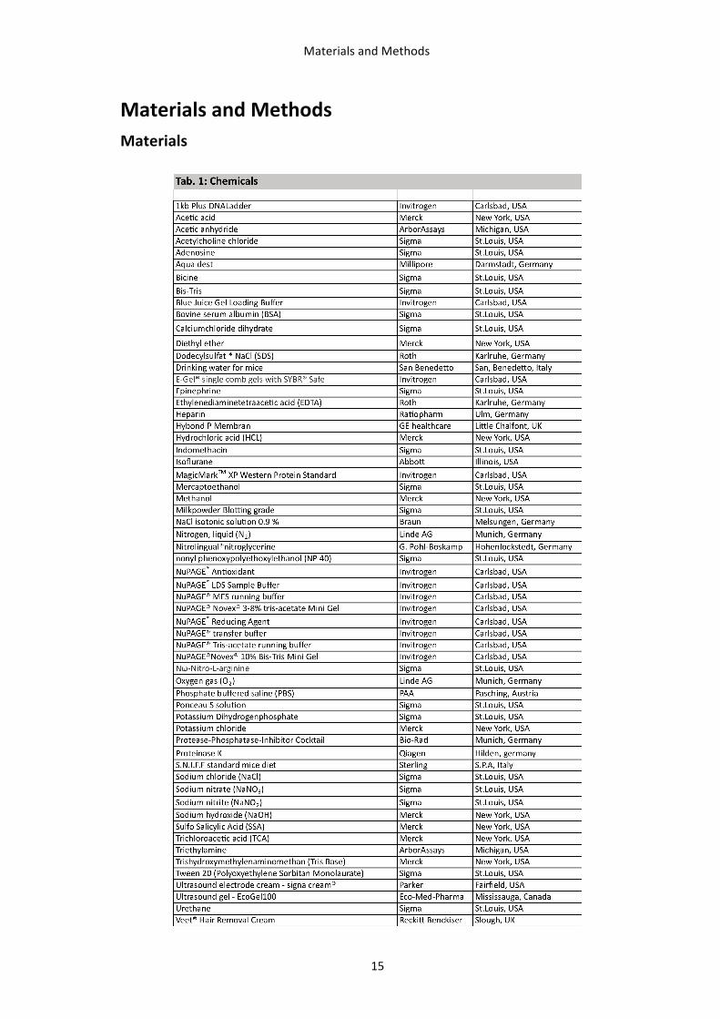
MATERIALS AND METHODS 15
Materials 15
Mice 17 Genotyping 18 Collection of mice blood and organs 18
Experimental setup 19 Oral administration of (-‐)-‐epicatechin to mice 19 Inhibition of nitric oxide synthase (NOS)-‐ and cyclooxygenase (COX) 19 Treatment with vasoactive drugs 20
In vivo measurements in mice 21 LDPI Measurements 21 Ultrasound Measurements 22 Assessment of PORH 24
Table of Contents
VII
Surgical excision of the femoral artery 25 Assessment of blood pressure 26
Biochemical analysis 26 Western Blot 26 cGMP 27 GSH 28
Molecular biological analysis 28 Real Time PCR 28 Microarray analysis 29
Analytical analysis 30 Measurement of nitrate and nitrite 30 Measurement of (-‐)-‐epicatechin metabolites 30
Statistical Analyses 31
RESULTS 32
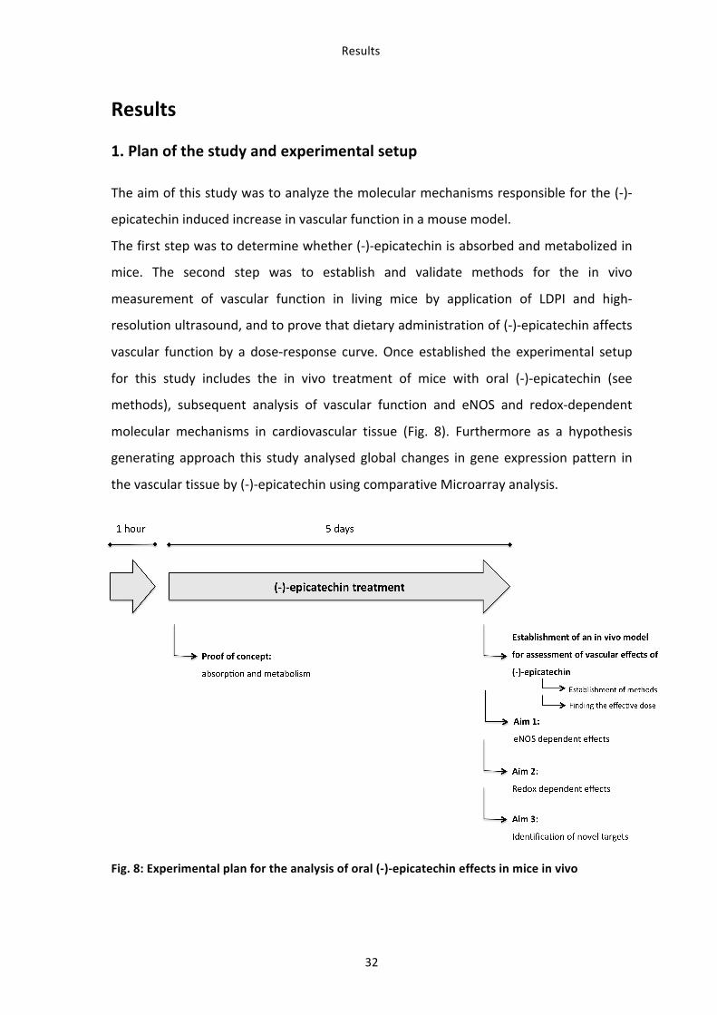
1. Plan of the study and experimental setup 32
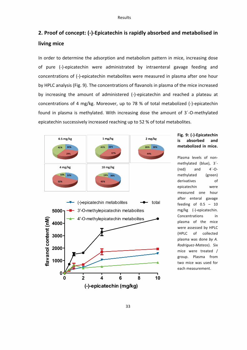
2. Proof of concept: (-)-Epicatechin is rapidly absorbed and metabolised in living mice 33
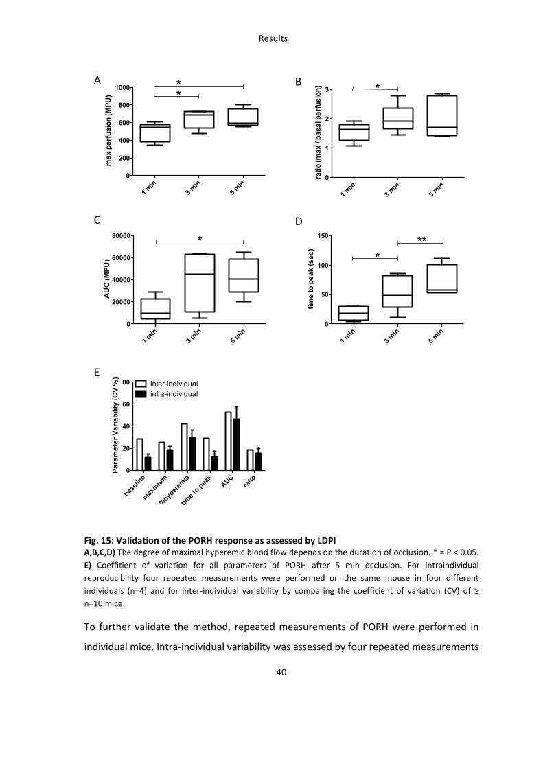
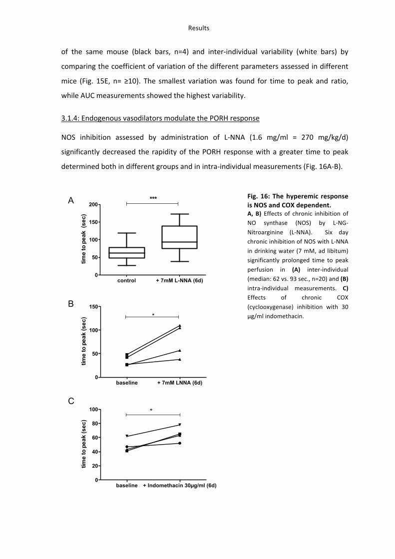
3. (-)-Epicatechin increases vascular reactivity in mice in vivo 34 3.1: Establishment of in vivo methods for assessment of vascular function: ultrasound and LDPI 34 3.1.1: Vasoactive molecules modulate the vascular diameter and mean perfusion in the hindlimb. 34 3.1.2: Post-‐occlusive reactive hyperemia (PORH) in the hindlimb of the mouse 37 3.1.3: Validation of PORH in mouse hindlimb assessed by LDPI 39 3.1.4: Endogenous vasodilators modulate the PORH response 41 3.1.5: PORH response decreases with age in mice. 42
3.2: Oral (-‐)-‐epicatechin increases vascular function in living mice 44 3.2.1: (-‐)-‐Epicatechin increases vascular function in an eNOS-‐dependent fashion 44 3.2.2: (-‐)-‐Epicatechin increases vascular function in a Nrf2-‐dependent fashion 47
4: Molecular mechanisms (I) – (-)-Epicatechin activates eNOS mediated NO-signaling 50 4.1: (-‐)-‐Epicatechin activates eNOS in the vasculature in vivo by increasing phosphorylation 50 4.2: Increased eNOS activity leads to an increase in vascular NO-‐bioactivity 52 4.2.1: The increase in vascular NO-‐bioactivity is eNOS dependent 52 4.2.2: The increase in vascular NO-‐bioactivity in Nrf2 dependent 54
4.3: Effect of (-‐)-‐epicatechin on systemic NO-‐bioavailability. 55
5. Molecular mechanisms (II) – effects of (-)-epicatechin on tissue redox state (GSH) 56 5.1: (-‐)-‐Epicatechin increases redox state in tissues of the cardiovascular system 56 5.2: The increase in redox state in tissues of the cardiovascular system is Nrf2 dependent 58 5.3: (-‐)-‐Epicatechin regulates the expression of antioxidant genes in cardiovascular tissues 59
6. Identification of novel targets of (-)-epicatechin in vascular tissue by microarray analysis 61 6.1: Transcriptional profiling of genes in an (-‐)-‐epicatechin dose-‐response model 61 6.2: Functional analysis of genes constitutively induced or suppressed by (-‐)-‐epicatechin 64
Table of Contents
VIII
6.3: Analysis of pathways constitutively regulated by low and high dose (-‐)-‐epicatechin 67 6.4: Novel potential targets of (-‐)-‐epicatechin – AhR and Xenobiotic metabolism 69 6.5: Novel potential targets of (-‐)-‐epicatechin – Ca++-‐signaling in vascular smooth muscle cells 71
7. Summary of main findings 74
DISCUSSION 75
Why assessing the effects of (-)-epicatechin in vivo in mice ? 75
Is eNOS required for the beneficial vascular effects of (-)-epicatechin in vivo? 76
Is maintenance of NO-bioavailability and redox state essential for the effects of (-)-epicatechin ? 78
Is Nrf2 required for the beneficial vascular effects of (-)-epicatechin in vivo? 81
What other mechanisms are involved in the vascular effects of (-)-epicatechin ? 86 Transcriptional profiling of genes in an (-‐)-‐epicatechin dose-‐response model 86 Novel (-‐)-‐epicatechin targets: vascular smooth muscle signalling and protection from toxic and oxidative stress 87
Establishing a mouse model for assessment of vascular function 92 What is the origin of the LDPI signal in mouse hindlimb? 92 Measurements of PORH: reproducibility and dependency on physiological stimuli. 93
Summary and Conclusion 95
Why is this study relevant for translational flavanol research ? 97
REFERENCES 99
ACKNOWLEDGMENT 111
CURRICULUM VITAE 113
Figure index
IX
Figure index Fig. 1: Groups, structures, and examples of flavonoid substances and their food distribution. 2 Fig. 2: eNOS derived NO from the endothelium mediates vasodilation by regulation of sGC/cGMP
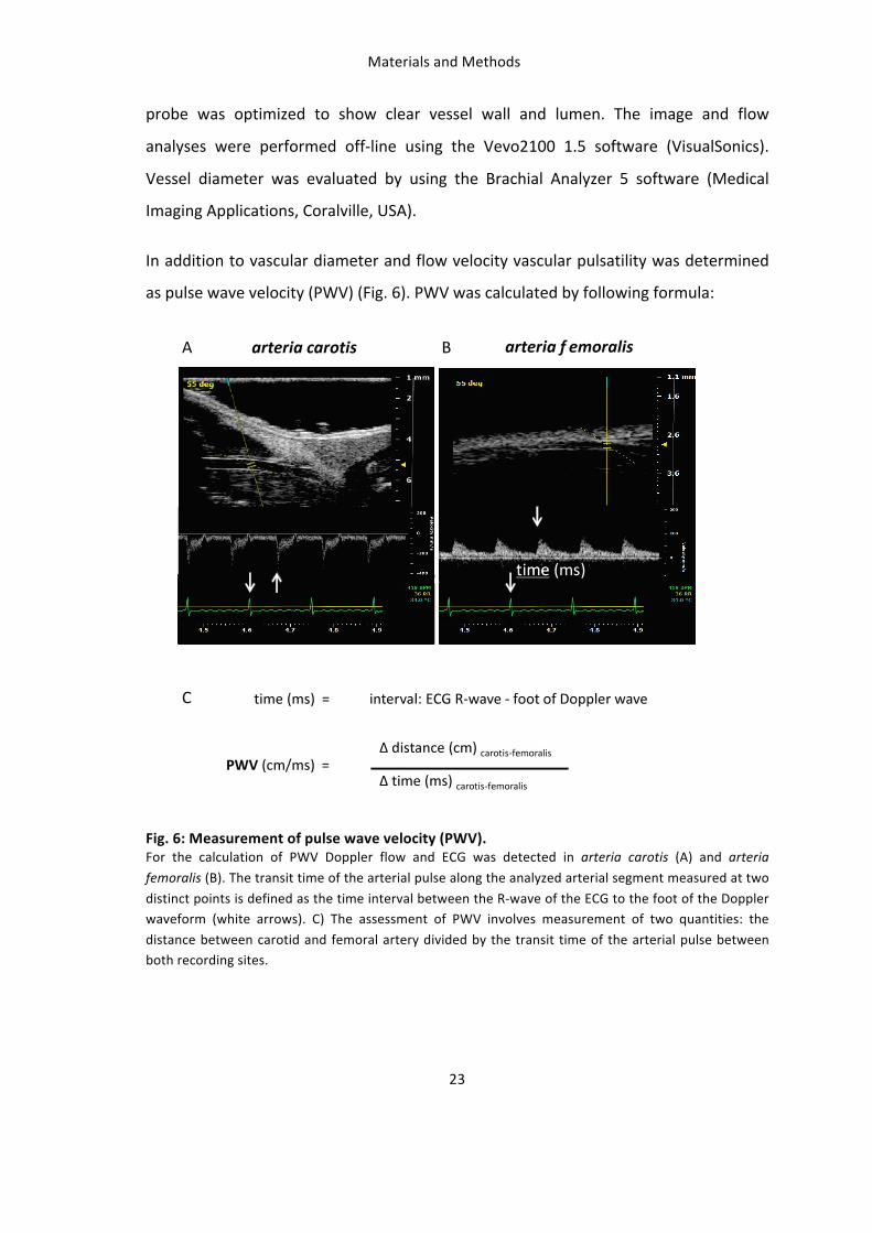
signaling. 4 Fig. 3: eNOS and NO in the circulation. 5 Fig. 4: Potential effects of flavanols on NO-‐bioactivity. 14 Fig. 5: Set up and measurement principles of Laser Doppler perfusion imaging (LDPI). 22 Fig. 6: Measurement of pulse wave velocity (PWV). 23 Fig. 7: Induction of reactive hyperemic blood flow in mouse hindlimb. 25 Fig. 8: Experimental plan for the analysis of oral (-‐)-‐epicatechin effects in mice in vivo 32 Fig. 9: (-‐)-‐Epicatechin is absorbed and metabolized in mice. 33 Fig. 10: (-‐)-‐Epicatechin has a dose-‐dependent effect on vascular function 34 Fig. 11: Pharmacological modulation of vascular diameter and hindlimb perfusion. 36 Fig. 12: Physiological modulation of diameter change in the femoral artery by PORH. 37 Fig. 13: Physiological modulation of hindlimb perfusion by PORH. 38 Fig. 14: Course and slope of PORH assessed as FMD and changes in MPU. 39 Fig. 15: Validation of the PORH response as assessed by LDPI 40 Fig. 16: The hyperemic response is NOS and COX dependent. 41 Fig. 17: The degree of PORH decreases with age. 43 Fig. 18: (-‐)-‐Epicatechin increases FMD and accelerates PORH in vivo in an eNOS dependent fashion. 45 Fig. 19: (-‐)-‐Epicatechin increases vascular pulsatility in mice. 46 Fig. 20: (-‐)-‐Epicatechin does not improve FMD in Nrf2-‐/-‐ mice in vivo. 48 Fig. 21: (-‐)-‐Epicatechin does not improve PORH in Nrf2-‐/-‐ mice in vivo 49 Fig. 22: (-‐)-‐Epicatechin increases eNOS phosphorylation in the aorta. 51 Fig. 23: (-‐)-‐Epicatechin increases eNOS phosphorylation in the lung. 52 Fig. 24: (-‐)-‐Epicatechin activates the NO-‐stimulated cGMP – PKG pathway in aorta in a NOS dependent
fashion. 53 Fig. 25: Effect of (-‐)-‐epicatechin on NO-‐bioavailability in Nrf2-‐/-‐ mice. 54 Fig. 26: Effect of (-‐)-‐epicatechin on systemic NO-‐bioavailability. 55 Fig. 27: (-‐)-‐Epicatechin increases redox state in cardiovascular tissues. 57 Fig. 28: Effect of (-‐) epicatechin on redox state in Nrf2-‐/-‐ mice. 58 Fig. 29: Protein expression of antioxidative genes in aorta after (-‐)-‐epicatechin treatment. 59 Fig. 30: Protein expression of genes involved in GSH metabolism in heart, lung and liver after (-‐)-‐
epicatechin treatment. 60 Fig. 31: Strategy of comparative micro-‐array analysis to identify gene clusters regulated by (-‐)-‐
epicatechin in vivo. 62 Fig. 32: Transcriptional profiling of targets dose-‐dependently regulated by (-‐)-‐epicatechin in mice in vivo.
63 Fig. 33: (-‐)-‐Epicatechin regulated pathways contributing to maintenance of vascular health by regulation
of stress response and vascular functionality. 68 Fig. 34: mRNA expression of AhR in the aorta after 2 mg/kg and 10 mg/kg (-‐)-‐epicatechin treatment. 69 Fig. 35: Regulation of MLCK and MYPT in the aorta. 73 Fig. 36: Summary of the main results of this study. 74 Fig. 37: Network of (-‐)-‐epicatechin regulated genes involved in the NO-‐ and Ca++-‐mediated process of
vascular muscle relaxation / contraction. 91 Fig. 38: (-‐)-‐Epicatechin increases vascular reactivity by regulation of eNOS/sGC/cGMP signaling and its
effectors in smooth muscle cells. 96
Table index
X
Table index Tab. 1: Chemicals 15
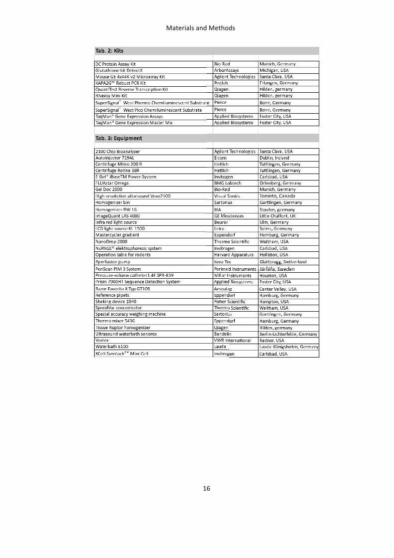
Tab. 2: Kits 16
Tab. 3: Equipment 16
Tab. 4: Instruments 17
Tab. 5: Software 17
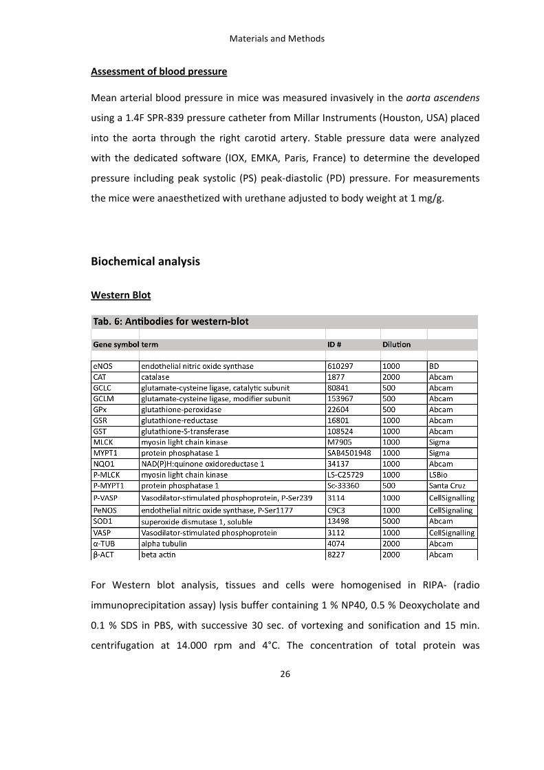
Tab. 6: Antibodies for western-‐blot 26
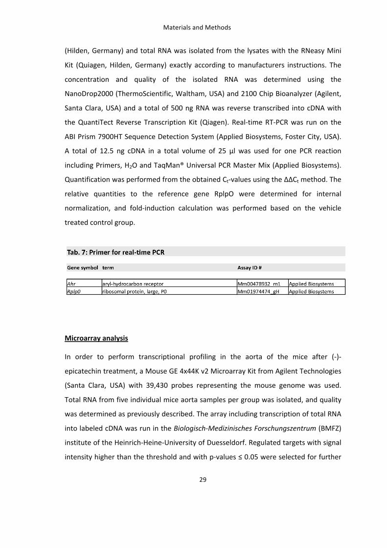
Tab. 7: Primer for real time PCR 29
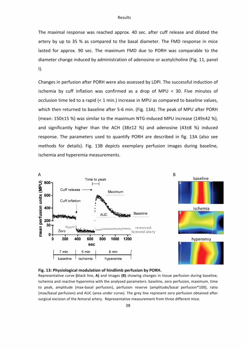
Tab. 8: Physiological parameter during administration of vasoactive drugs 37
Tab. 9: NOS-‐dependence of PORH by LDPI 42
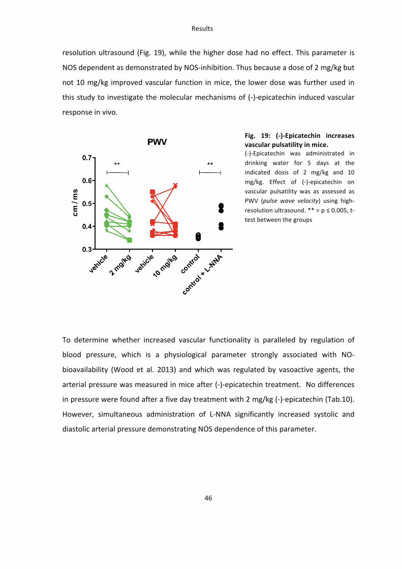
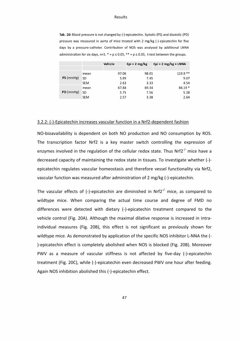
Tab. 10: Blood pressure is not changed by (-‐)-‐epicatechin 47
Tab. 11: Level of nitrite and nitrate in plasma of Nrf2 knockout and wildtype mice after 2 mg/kg (-‐)-‐epicatechin treatment 56
Tab. 12: Top 20 most regulated genes in the aorta by 2 mg/kg and 10 mg/kg (-‐)-‐epicatechin determined by microarray analysis. 64
Tab. 13: Annotation Clustering of Genes constitutively induced or suppressed by 2 mg/kg (-‐)-‐epicatechin using DAVID Analysis 65
Tab. 14: Annotation Clustering of Genes constitutively induced or suppressed by 10 mg/kg (-‐)-‐
epicatechin using DAVID Analysis 66
Tab. 15: Functional Clustering of Genes Constitutively regulated by 2 mg/kg and 10 mg/kg (-‐)-‐epicatechin using DAVID Analysis and KEGG pathway Analysis 67
Tab. 16: Summary of genes involved in Glutathione , Xenobiotic and Drug metabolism that are up-‐ or down-‐regulated by 2 mg/kg and 10 mg/kg (-‐)-‐epicatechin in the aorta 70
Tab. 17: Summary of genes involved in Ca++-‐signaling and vascular smooth muscle contraction that are up-‐ or down-‐regulated by 2 mg/kg and 10 mg/kg (-‐)-‐epicatechin in the aorta 72
Introduction
1
Introduction Flavanols and cardiovascular health
Cardiovascular disease and endothelial dysfunction
Cardiovascular disease is a leading cause of death worldwide. Risk factors of modern
life-‐style for experiencing an initial or recurrent cardiovascular event are obesity, lack
of physical activity, smoking and alcohol consumption (Wang et al. 2009). Heart failure,
coronary artery disease, hypertension and others are associated with endothelial
dysfunction (Widlansky et al. 2003). A dysfunctional endothelium is characterized by a
loss of endothelial control over vascular tone, impairment of endothelium-‐dependent
vasorelaxation, alteration of the anticoagulant and anti-‐ inflammatory properties
leading to thrombosis, and vessel wall remodeling (impaired regulation of vascular
growth) (Widlansky et al. 2003, Seals et al. 2011).
A growing number of therapeutic interventions known to decrease cardiovascular risk,
including exercise, lipid lowering, smoking cessation, weight reduction, medication
with angiotensin-‐converting enzyme inhibitors and statins and also dietary
interventions, have been shown to improve endothelial function (Widlansky et al.
2003). The daily diet can play a major role in prevention of vascular disease initiation
and progression (Eyre et al. 2004). Besides regulation of fat consumption, blood
pressure and blood glucose levels current dietary recommendations include the
consumption of fruits and vegetables (Lichtenstein et al. 2006) proven by
epidemiological studies demonstrating a lower risk for cardiovascular disease (He et al.
2007) (Bhupathiraju et al. 2013). Despite the fact that they are low in calories and fat
and have a favorable mineral ratio the mechanisms by which these diets mediate their
beneficial effects on health still are not well understood (Dauchet et al. 2009).
There is epidemiological evidence that flavonoids mediate beneficial cardiovascular
effects and lower the risk for vascular diseases and mortality (Mulvihill et al. 2010,
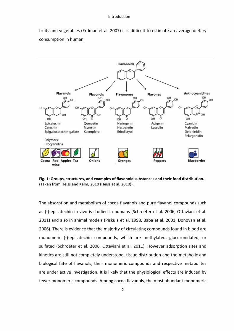
McCullough et al. 2012, Toh et al. 2013). The group of flavanoids includes a number of
various substances with different chemical properties including flavanols, flavonols,
flavones, isoflavones, flavanones, and anthocyanins (Fig. 1). This can result in severe
differences in their absorption, metabolism and bioactivities (Erdman et al. 2007).
Moreover since the distribution of these flavonoids varies considerably in different
Introduction
2
fruits and vegetables (Erdman et al. 2007) it is difficult to estimate an average dietary
consumption in human.
subgroup of the flavonoid family (Figure 1), are a focus of attentionas epidemiological investigations have shown an independentinverse correlation between the dietary intake of flavanol-richfoods and CAD mortality.10 While the epidemiological findingsare provocative, no randomized controlled trials with hard clinicalendpoints have been published that corroborate a cause and effectrelationship between the intake of flavanols and vascular health.
However, several controlled human dietary intervention studieswith flavanol-rich foods and beverages have demonstrated positiveeffects, including the recovery of endothelial function,11–15
improvements in insulin sensitivity, decreased blood pressure,16,17
and reductions in platelet aggregation.18–20 The pharmacologicalmechanisms of action of flavanols have yet to be identified, butthey likely include an enhancement in nitric oxide (NO) bioactivity,modulation of the immune system, and enhanced endothelialhomeostatic vascular repair.21 While it has been speculated thatthe intrinsic antioxidant capacity of flavanols, and flavonoids ingeneral, underlies their positive vascular effects (refs 22,23 andreferences therein), this is as unlikely, although select flavonoidsmay influence the overall level of oxidative stress through second-ary mechanisms.24,25
Quantitatively, flavanols represent a major group of flavonoids inthe western diet.26 Major sources include chocolate and cocoa(up to 920–1220 mg/100 g), apples (up to 120 mg/200 g), and
tea (up to 300 mg/infusion), however, it must be noted thatthe profile of flavanols (e.g. (2)-epicatechin, (+)-epicatechin,(2)-catechin, (+)-catechin) in these foods can vary considerably,and it can be changed as a consequence of food processing.27
Importantly, the methodologies that are typically used tomeasure flavanols in foods, as well as in biological fluids, do notprovide information on the profile of flavanol stereoisomers. Theaverage daily flavanol intake of an adult has been approximatedto be in the range of 50–100 mg.7 However, there is considerableconfusion in the literature, as many authors when reportingdietary intakes do not make a distinction between flavanolsper se (which are by definition monomers) and procyanidins,which are oligomers of flavanols.7 Generally, when viewed as acomposite, the flavanol monomers ((2)- and (+)-epicatechin,(2)- and (+)-catechin) are approximately 10%26,28 of the com-bined monomer and oligomer total. We suggest that the poolingof monomers and procyanidins when presenting dietary intakedata is inappropriate, given that while monomers and dimers areabsorbed in the small intestine,29,30 the longer oligomers arenot absorbed. Thus, the longer oligomers are unlikely to havedirect effects on the vascular endothelium, although we notethey may have biological effects within the intestinal track (e.g.they might influence the microbiota, or act as immunemodulators). Depending on many factors, peak monomer and
Figure 1 (A) Basic structures and examples of the main subclasses of dietary flavonoids. (B) Whereas the majority of flavanols are present asoligomers in food (i.e. cocoa), metabolized flavanol monomers are the dominant flavanols in blood and may be partly responsible for theobserved vascular effects.
C. Heiss et al.2584
Fig. 1: Groups, structures, and examples of flavonoid substances and their food distribution. (Taken from Heiss and Kelm, 2010 (Heiss et al. 2010)).
The absorption and metabolism of cocoa flavanols and pure flavanol compounds such
as (-‐)-‐epicatechin in vivo is studied in humans (Schroeter et al. 2006, Ottaviani et al.
2011) and also in animal models (Piskula et al. 1998, Baba et al. 2001, Donovan et al.
2006). There is evidence that the majority of circulating compounds found in blood are
monomeric (-‐)-‐epicatechin compounds, which are methylated, glucuronidated, or
sulfated (Schroeter et al. 2006, Ottaviani et al. 2011). However adsorption sites and
kinetics are still not completely understood, tissue distribution and the metabolic and
biological fate of flavanols, their monomeric compounds and respective metabolites
are under active investigation. It is likely that the physiological effects are induced by
fewer monomeric compounds. Among cocoa flavanols, the most abundant monomeric
Introduction
3
compound is (-‐)-‐epicatechin (Gu et al. 2004, Heiss et al. 2010). It has been shown that
administration of pure (-‐)-‐epicatechin, accounts for the beneficial effects of flavanols
on endothelial function and the increase of circulating NO metabolites in humans
(Schroeter et al. 2006) and is the single stereoisomer capable of mediating such
significant vasodilatory responses (Ottaviani et al. 2011).
Flavanols and control of endothelial function: a role of eNOS ?
The health and functionality of the vascular system is strongly correlated to the
functionality of the endothelium, which lines the luminal side of the vessels. The
endothelial cell layer controls many biological events occurring either on the
endoluminal or the interstitial side of the vasculature. Many processes including
haemostasis, haematopoiesis, inflammatory reactions and immune response require
close interactions between circulating cells or cytokines and the vascular endothelium
(Mantovani et al. 1997). One of the most important signal transduction cascades is the
release of a vasodilating factor by endothelial cells in response to blood flow mediated
shear stress (Furchgott et al. 1980, Smiesko et al. 1993). The identification of this
“endothelium-‐derived relaxing factor” as nitric oxide (NO) over 20 years ago is a
milestone of cardiovascular research and was awarded with the nobel price in
medicine (Furchgott et al. 1987, Ignarro et al. 1987, Palmer et al. 1987). One year later
the source of this endothelial NO production was found to be the type 3 isoform of
nitric oxide synthase or endothelial nitric oxide synthase (eNOS) (Palmer et al. 1988).
Meanwhile the mechanism and signal transduction cascade leading to vasodilation by
endothelial NO release is well investigated (Nausch et al. 2008, Morgado et al. 2012).
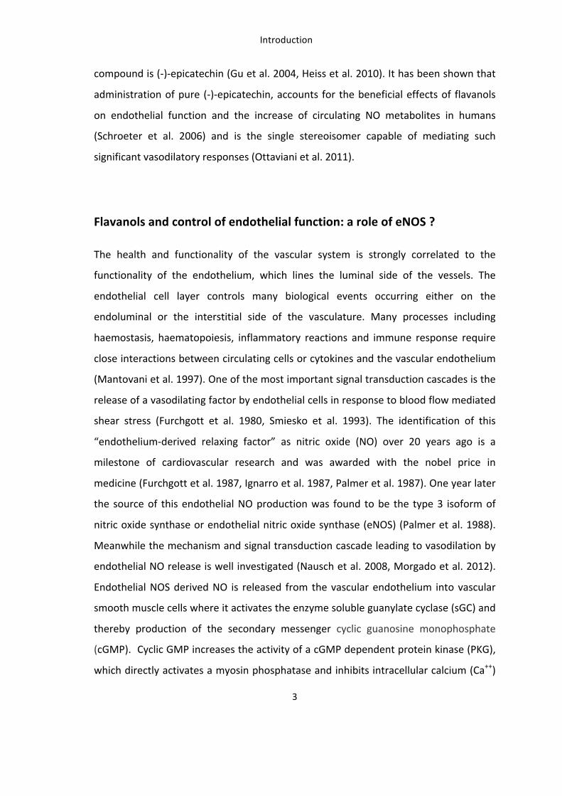
Endothelial NOS derived NO is released from the vascular endothelium into vascular
smooth muscle cells where it activates the enzyme soluble guanylate cyclase (sGC) and
thereby production of the secondary messenger cyclic guanosine monophosphate
(cGMP). Cyclic GMP increases the activity of a cGMP dependent protein kinase (PKG),
which directly activates a myosin phosphatase and inhibits intracellular calcium (Ca++)
PNAS, 2001, Nat med 2003,Circ Res 2002, Blood 2006
Introduction
6
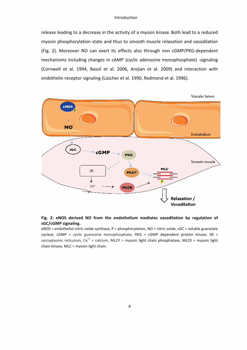
The balanced production and release of vasodilators, most importantly eNOS derived
NO maintains vascular homeostasis and regulates vascular tone. In addition to its
vasodilating feature, endothelial NO has antiatherosclerotic properties, such as
inhibition of platelet aggregation, leukocyte adhesion, smooth muscle cell
proliferation, and expression of genes involved in atherogenesis (Li et al. 2000). Thus a
decrease in NO-‐bioavailability (Kleinbongard et al. 2003, Heiss et al. 2006, Rassaf et al.
2006) and alterations in eNOS activity and expression are strongly associated with
endothelial dysfunction and cardiovascular disease (Widlansky et al. 2003, Seals et al.
2011).
Flavanols and vascular function
The oral intake of cocoa flavanols and pure (-‐)-‐epicatechin has beneficial effects in
particular on improvement of endothelium-‐dependent vascular function as has been
shown by work conducted by our and other groups (Heiss et al. 2003, Schroeter et al.
2006, Heiss et al. 2007). Further an improvement of insulin sensitivity, a decrease in
blood pressure (Hooper et al. 2012) and a reduced platelet aggregation (Ostertag et al.
2010) are reported for dietary cocoa flavanols. The pharmacology and mechanistic
action of flavanols in vivo are not well understood, but they likely include a modulation
of the immune response as shown for the cocoa compound procyanidin (Kenny et al.
2007), an activation of angiogenic cells by flavanol-‐rich cocoa drinks (Heiss et al. 2010).
Most importantly cocoa flavanols and (-‐)-‐epicatechin enhance NO-‐bioavailability in
health (Schroeter et al. 2006, Heiss et al. 2007, Loke et al. 2008) and state of increased
cardiovascular risk including hypertension (Grassi et al. 2005), age (Holt et al. 2012)
and smoking (Heiss et al. 2005, Heiss et al. 2007).
Importantly the oral intake of a pure (-‐)-‐epicatechin drink improved vascular function
assessed as flow-‐mediated dilation (FMD) of the brachial artery in healthy male
subjects with a maximum response after two hours (Schroeter et al. 2006). Also a
longer intake of a high-‐flavanol drink containing (-‐)-‐epicatechin gradually increased
FMD after one, three, five and seven days in healthy smokers (Heiss et al. 2007) and in
healthy adults (Engler et al. 2004). Long-‐term improvement of FMD by cocoa flavanols
Introduction
7
has also been shown in pathological state for patients with coronary artery disease
(Heiss et al. 2010), diabetes (Balzer et al. 2008), obesity (Njike et al. 2011) and
hypertension (Grassi et al. 2005).
The underlying mechanisms, which mediate the beneficial action of cocoa flavanols on
vascular health are not well understood and are very poorly investigated in vivo.
Proposed mechanisms include an increase in NO-‐bioavailability by direct activation of
eNOS or by affecting the redox state of vascular cells. Endothelial NOS activity can be
regulated by post-‐translational modifications, most importantly phosphorylation, but
the enzyme can also be regulated on the transcriptional level (Forstermann et al.
1998). Studies mostly in cultured vascular endothelial cells suggest that red wine
polyphenols (RWPs) and (-‐)-‐epicatechin are able to activate the eNOS enzyme (Leikert
et al. 2002, Wallerath et al. 2003, Ndiaye et al. 2005, Ramirez-‐Sanchez et al. 2010,
Ramirez-‐Sanchez et al. 2011). However, in vitro findings cannot be directly transferred
to the in vivo situation because of the distinct limitations such as 1) lack of
metabolism, 2) lack of systemic action and 3) lack of the direct interaction between
vascular endothelium and vascular smooth muscle which is mandatory for vessel
function. In human it is not possible to directly measure eNOS and NO-‐signaling to the
vessels. The in vivo effect in rodents is poorly investigated and limited to studies in rats
with RWPs (Andriambeloson et al. 1997, Benito et al. 2002, Agouni et al. 2009) and (-‐)-‐
epicatechin (Gomez-‐Guzman et al. 2012) demonstrating elevated level of NO and
cGMP in the aorta.
Regulation of vascular antioxidant state by dietary flavanols
The occurrence of endothelial dysfunction in pathophysiological state is not only
correlated to reduced NO production but also to increased oxidative stress within the
tissue. For instance, the decline in NO-‐bioavailability observed in a dysfunctional
endothelium seems to be determined by an accelerated degradation of NO by reactive
oxygen species (ROS) during oxidative stress, as well as by decreased activity or
Introduction
8
expression of eNOS (Weseler et al. 2010, Seals et al. 2011). In the vasculature, oxidant
molecules can originate from extracellular or intracellular sources and can be
produced by enzymatic or non-‐enzymatic reactions. The blood is an excellent external
carrier of oxidants and ROS as it is in permanent contact with vascular endothelial
cells. Within the vascular wall intracellular sources are the mitochondrial respiratory
chain and enzymes catalyzing oxidative reactions such as: Nicotinamide adenine
dinucleotide phosphate oxidase (NAPDH), xanthine oxidase, lipooxygenase,
myeloperoxidase and also uncoupled NOS (Sugamura et al. 2011).
However, in particular vascular cells are not defenseless and are able to protect the
tissue against oxidative damage e.g. by deactivation of radical species (Sies 1993). One
of the most abundant and important cellular antioxidants is the tripeptide glutathione
(GSH) which synthesis is mainly controlled by gene regulation (Janssen-‐Heininger et al.
2013) , while the most important cellular sensor for pro-‐oxidative molecules is the
transcription factor (erythroid-‐derived 2)-‐like 2 or Nrf21 (Zhu et al. 2008).
GSH exerts its antioxidant activity by directly scavenging free radicals and other ROS or
as a cofactor in enzymatic reactions in which GSH is oxidized to form GSSG (GSH
disulfide) (Sies 1999). The GSH-‐mediated antioxidant defense mechanism of
endothelial cells includes several enzymes participating in the synthesis and
metabolism of GSH: glutamate-‐cysteine ligase (GCL) and GSH synthetase mediate de novo synthesis of GSH while glutathione peroxidase (GPX), glutathione reductase (GSR)
and glutathione-‐S-‐transferase (GST) metabolise GSH using H2O2 and NADPH as
reducing agents (Sies 1993, Sies 1999).
In addition an adaptive response is activated to prevent further damage in eukaryotic
cells. The redox-‐sensitive transcription factor Nrf2 activates protective proteins, which
are able to prevent, deactivate and repair oxidative stress and oxidative damage. Most
prominent targets of Nrf2 are phase II detoxifying enzymes including NAD(P)H quinone
1 Gene ID: 4780 (homo sapiens), 18024 (mus musculus); Official name: nuclear factor, erythroid 2-‐like 2; Official symbol: NFE2L2 alias Nrf2 (homo sapiens), Nfe2I2 alias Nrf2 (mus musculus). According to the scientific consense this work uses Nrf2 when speaking of the functional protein.
Introduction
9
oxidoreductase 1 (NQO1), heme oxygenase-‐1 (HO-‐1), GST and GCL, but also other
antioxidants, proteasomes and drug metabolizing proteins (Kaspar et al. 2009, Baird et
al. 2011). The transcriptional activity of Nrf2 can be induced by molecules, chemicals
and compounds with electrophilic properties (Baird et al. 2011). Many of plant
phytochemicals, which are abundantly contained in human diet possess such
electrophilic chemical structures, including flavanols.
Besides the supposed effects on the NO-‐pathway a controversial discussion describes
the antioxidant properties of flavanoids and their impact on cellular and tissue redox
state. However investigations at non-‐physiological conditions ex vivo (Rice-‐Evans et al.
1996, Galleano et al. 2010) and the use of cell culture systems (Romeo et al. 2009,
Martin et al. 2010, Rodriguez-‐Ramiro et al. 2011, Martin et al. 2013, Ruijters et al.
2013) including the reaction with medium additives (Long et al. 2010) contribute to
strongly limit the impact of these results for interpretation of the physiological
mechanisms.
Even though there is no direct evidence for antioxidant effects of cocoa and green tea
polyphenols from controlled human interventional studies (Scheid et al. 2010, Ellinger
et al. 2011), some studies report an increase in plasma antioxidant capacity and a
decrease in plasma oxidation products associated with elevated plasma (-‐)-‐epicatechin
concentrations (Rein et al. 2000, Wang et al. 2000). Furthermore an impact of (-‐)-‐
epicatechin has been demonstrated on the redox-‐state in hepatic and neuronal cells in
vitro (Martin et al. 2010, Martin et al. 2013) and in tissue of the rat in vivo (Litterio et
al. 2012, Seymour et al. 2013). Whether or not these potential indirect antioxidant
properties regulate the actual vascular functionality in vivo is not clear. Also the
molecular mechanisms underling these effects are not well understood.
As reported the transcription factor Nrf2 is a redox-‐sensitive sensor of oxidative stress
and can be activated by numerous oxidative and electrophilic species, including
reactive oxygen and nitrogen species, aldehydes and metals (Zhu et al. 2008, Baird et
al. 2011). In this paradigm flavanol compounds may directly interact with Nrf2 due to
their chemical structure and electrophilic properties. In vitro (-‐)-‐epicatechin is able to
Introduction
10
activate Nrf2 in cultured neuronal cells (Shah et al. 2010). In vivo studies in mice and
rats further support the contribution of Nrf2 to prevention of stroke damage and heart
failure by (-‐)-‐epicatechin (Shah et al. 2010, Leonardo et al. 2013). However, the role of
Nrf2 in the vascular effects of dietary (-‐)-‐epicatechin has never been investigated in
vivo so far.
In vivo assessment of endothelial function
In human endothelial function is assessed as FMD of the brachial artery in the forearm
(Pyke et al. 2005). The measurement of vascular function in living mice is challenging
and not applied so far.
A wide range of animal models of cardiovascular disease is available, including
hyperlipidemic mice and rats, spontaneously hypertensive rats, genetically modified
mice lacking/overexpressing specific apolipoproteins, hypertensive mice lacking eNOS
and surgical models of vessel lesion (Getz et al. 2012). However, there are only a
limited number of methods available for measuring vascular reactivity and
functionality in vivo in living animals. The molecular mechanisms responsible for
modification of vascular function are classically studied in aortic rings isolated from
animals, maintained in an organ bath, and pre-‐constricted with phenylephrine.
Vasodilation is determined as a decrease in tension or increase in diameter during the
addition of vasoactive substances (Furchgott et al. 1980) (Hutchison et al. 1999) (Zhu
et al. 2003) (Woodman et al. 2000). This methodology was also applied to other
vessels including femoral (Stoen et al. 2001), iliac (Cooke et al. 1991), mesenteric
(Akata et al. 1995), and renal arteries (Ruiz-‐Nuno et al. 2004) from various species. The
results obtained for these conduit arteries were similar and showed predominantly
endothelium-‐ and NOS-‐dependent vasodilation in response to acetylcholine, as well as
similar dose-‐response curves for endothelium-‐independent vasodilators, e.g.
nitroglycerine (NTG) (Ruiz-‐Nuno et al. 2004). The other frequently used method to
determine vascular reactivity is the assessment of changes in perfusion pressure or
flow in the coronary circulation of isolated hearts as a response to vasoactive
Introduction
11
substances (Kanatsuka et al. 1992, Godecke et al. 1998). A decrease in perfusion
pressure or increase in flow predominantly reflects dilation of the resistance arteries.
These ex vivo experiments have enabled very powerful reductionist approaches to the
study of vascular reactivity although isolated vessel systems are greatly limited due to
the absence of blood and their removal from the context of the whole organism (e.g.,
endocrine factors, hemodynamics, or nervous system). Moreover, they do neither
allow assessment of experimental intra-‐individual effects nor longitudinal studies.
Similar to the assessment of perfusion flow in isolated systems in order to determine
vascular reactivity, detection of tissue perfusion can also be used in vivo as a reliable
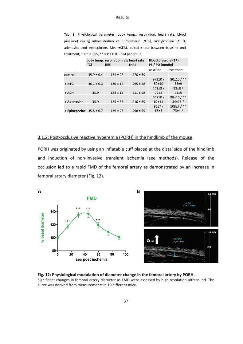
readout for vascular function. Post occlusive reactive hyperemia (PORH) is the
physiological increase of blood flow to the tissue following ischemia after transient
arterial occlusion. PORH has also been applied for testing endothelial function of
conduit vessels in humans, e.g. by measurement of FMD (Frick M 2002, Pyke et al.
2007), of venous occlusion plethysmography, or of skin microcirculation by laser
Doppler flowmetry or imaging (Kubli et al. 2000, Keymel et al. 2010). Scanning laser
doppler perfusion imaging (LDPI) has been applied in clinical settings to assess
functional responses of the cutaneous microcirculation to vasoactive molecules or
PORH in patients with endothelial dysfunction in hypertension (Farkas et al. 2004),
peripheral arterial disease (Kluz et al. 2013), coronary artery disease (Keymel et al.
2010), diabetes (Jarnert et al. 2012), smoking (Petschke et al. 2006) (Fujii et al. 2013)
or age (Algotsson et al. 1995). In swine and dog animal models LDPI was applied for
measuring revascularization in wound healing (Mauskar et al. 2013) (Karayannopoulou
et al. 2010), mucosal microcirculation (Kaner et al. 2013) or retinal capillary blood flow
(Gelatt-‐Nicholson et al. 1999). In rodents and especially in mice LDPI is classically used
to study angiogenesis and neovascularization after hindlimb ischemia (Long et al.
2013) (Sachdev et al. 2013) (Rivard et al. 1999, Limbourg et al. 2009), but it has never
been applied for measuring reactive hyperemic responses in mice. Scanning LDPI is
based on the physical phenomenon defined as the doppler effect, which is a change in
the frequency of a wave occurring when the source and observer are in motion
relative to each other. When laser light penetrates a tissue, it is backscattered and
Introduction
12
returns to its source without change of frequency. But if the laser light is backscattered
by moving objects (i.e. blood cells) it undergoes a shift in frequency directly
proportional to their concentration and velocity (Wardell et al. 1993). The measuring
depth of the laser depends on the energy and wave length of the laser, on the
measuring distance and, most importantly, on characteristics and vascularization of
the tissue influencing its backscattering properties, including pigmentation, density,
biophysical composition, presence of hairs, density and diameter of the vessels
(macrocirculation, microcirculation, resistance vessels), amount of blood cells etc.
(Jakobsson et al. 1993).
A direct measurement of FMD in animal models has been previously demonstrated in
rats (Heiss et al. 2007) but was never applied in living mice. Similar PORH has never
been assessed in mice in vivo.
Aim of the study
13
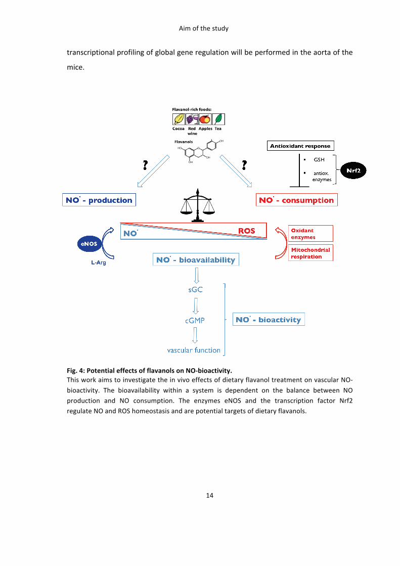
Aim of the study
The mechanisms by which dietary flavanols mediate their vascular effects are not well
understood. Accumulating data support the evidence that flavanol-‐rich foods like
cocoa are able to improve endothelium-‐dependent vasorelaxation in healthy subjects,
as well as in pathological conditions. Proposed mechanisms include an increase in
nitric oxide (NO)-‐bioavailability by direct activation of the endothelial NO synthase or
by affecting the redox state of vascular cells. (-‐) Epicatechin is the most abundant
flavanol monomer in cocoa and seems to be the active component responsible for
those effects. However a direct link between the eNOS/NO/cGMP signaling pathway
and vascular response to (-‐)-‐epicatechin in vivo has never been demonstrated so far.
The transcription factor nuclear factor (erythroid-‐derived 2)-‐like 2 (Nrf2) is a key
master switch controlling the expression of enzymes involved in the regulation of the
cellular redox state, and is activated by electrophilic compounds, and potentially (-‐)-‐
epicatechin or its circulating metabolites.
This work aims at investigating the vascular effects of dietary (-‐)-‐epicatechin in vivo by
raising following questions: 1) Is eNOS required for the beneficial vascular effects of (-‐)-‐
epicatechin in vivo ? 2) are maintenance of NO-‐bioavailability and redox state essential
for the effects of (-‐)-‐epicatechin ? 3) Is Nrf2 required for the beneficial vascular effects
of (-‐)-‐epicatechin in vivo ? 4) What other potential targets/pathways in vascular tissue
are activated and regulated by administration of (-‐)-‐epicatechin ?.
As a proof of concept adsorbtion and metabolism of (-‐)-‐epicatechin in mice will be
assessed by analyzing plasma concentrations of (-‐)-‐epicatechin and its metabolites. To
determine whether eNOS or Nrf2 are required for the beneficial vascular effects of (-‐)-‐
epicatechin in vivo, vascular function will be assessed in mice treated with a NOS
inhibitor, and in mice lacking eNOS (eNOS-‐/-‐) or Nrf2 (Nrf2-‐/-‐) by scanning LDPI and
high-‐resolution ultrasound. To test whether eNOS dependent NO-‐bioactivity is
regulated in vascular tissue eNOS expression, eNOS phosphorylation and cGMP level
will be assessed in the aorta of the mice. To determine whether (-‐)-‐epicatechin
regulates the redox state in vivo GSH-‐level and expression of antioxidant enzymes will
be assessed in tissue of the cardiovascular system from wildtype and Nrf2-‐/-‐ mice.
Finally to identify novel potential targets of (-‐)-‐epicatechin in the vasculature
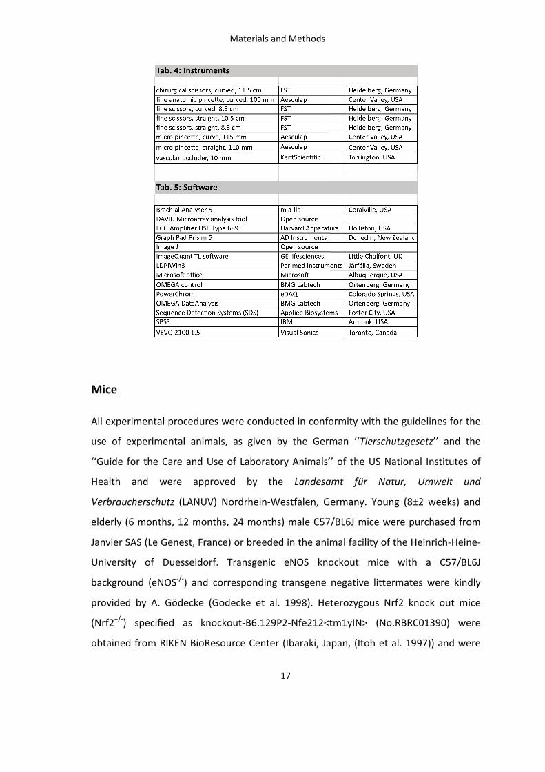
Materials and Methods
18
bred in the animal facility to obtain a population with Nrf2+/+, Nrf2+/-‐ and Nrf2-‐/-‐ genetic
background. Mice were kept in groups, at 19-‐21 °C in 50-‐60 % humidified atmosphere
in a 12 h day/night rhythm. All mice were fed a standard diet ad libitum (SNIFF, S.P.A,
Italy), with low content in NO3-‐ an NO2
-‐. For all physiological in vivo measurements
mice were anesthetized with isoflurane (3 % induction and 1.8-‐2 % maintenance). For
final anesthesia 1 mg/g body weight urethane was administrated intraperitoneal (i.p.).
Genotyping
The genotype of transgenic animals was determined and confirmed by RT-‐PCR using
custom designed primers. For genotyping of the mice fresh or freshly frozen biopsies
were used from the tail or ear. To isolate genomic DNA the tissue was lysed in 300 µl
50 mM NaOH for 40 minutes at 95 °C. The lysates were neutralized with 25 µl 1M Tris-‐
HCl pH 4.5 and centrifuged at 14.000 rpm at 4°C for 10 minutes. The supernatant
containing genomic DNA was directly used for PCR or stored at -‐20 °C. For the
detection of the wildtype or disrupted Nrf2 allele HPLC-‐grade oligonucleotides
purchased from Eurofinsgenomics (Luxemburg) were used: Nrf2: 5’-‐
TGGACGGGACTATTGAAGGCTG-‐3’, Nrf2: 5’-‐GCCGCCTTTTCAGTAGATGGAGG-‐3’, lacZ: 5’-‐
GCGGATTGACCGTAATGGGATAGG-‐3’. The DNA was amplified using the KAPA2G™
Robust PCR Kit, a ready master mix containing Hot Start polymerase and dNTPs
(peqlab, Erlangen, Germany), following the manusfacturer´s instructions. Briefly, for
one PCR reaction 5.5 µl H2O, 12.5 µl TaqMix and 2 µl of each primer were added to 1 µl
of template. A standard PCR program (step 1: 95°C, 15 sec.; step 2: 60°C, 15 sec.; step
3: 72°C, 15 sec.) was run with the primer mix, water and the KAPA2G™ Robust PCR Kit.
Respective bands were identified on a commercial prestained 2 % agarose E-‐Gel®
system from Invitrogen (Carsbad, USA).
Collection of mice blood and organs
For final collection of blood samples up to 1.5 ml blood was drawn from anesthetized,
heparinized mice (40.000 IU/kg heparin i.p.) via heart puncture with closed chest using
a 1 ml nozzle with Ø 0.2 mm cannula and collected into heparin (50 µL) tubes. To
Materials and Methods
19
separate plasma blood was centrifuged at 3000 g for 2 min. at 4 °C and the
supernatant was directly shock frozen in liquid nitrogen before storage at -‐80 °C. For
explanations of the organs the abdominal and thoracic cavities were opened by ventral
incision, the vena cava was cut and the organs were perfused via heart injection with
ice cold PBS until the lung and liver was cleared of blood. After harvesting heart, lung
and liver the complete aorta including ascending, descending and abdominal part was
carefully removed in one peace into ice cold PBS, carefully stripped of adventicia or
blood coagulate and directly frozen in liquid nitrogen prior to storage at -‐80 °C. In case
of transgenic animals a tail biopsy was harvested for a re-‐genotyping procedure.
Experimental setup
Oral administration of (-‐)-‐epicatechin to mice
A dose of 2 mg/kg and 10 mg/kg (-‐)-‐epicatechin (Sigma-‐Aldrich, St.Louis, USA) was
administrated by enteral gavage feeding or ad libitum in drinking water. For acute
treatment mice were fed a low flavanol diet for one week and fastened overnight. A
single dose of 150 µl (-‐)-‐epicatechin was administrated. The solution was prepared by
dissolving (-‐)-‐epicatechin in ethanol (stock: 20 µg/µl) and dilution to a final
concentration of 0.4 µg/µl in drinking water. Mice were measured and sacrificed after
one hour. For longer-‐term administration a daily drinking volume of 5 ml per mouse
per day was defined and the required amount of a 100 mM (29 mg/ml) (-‐)-‐epicatechin
stock solution was calculated and added in 100 ml drinking water. The water was
changed daily for five days. Respective vehicle preparations with ethanol and water
were used in control groups. The dietary treatment of mice with (-‐)-‐epicatechin had no
effect on the body weight or daily drinking volume of the mice.
Inhibition of nitric oxide synthase (NOS)-‐ and cyclooxygenase (COX)
NOS-‐inhibition was accomplished by administration of 1.6 mg/ml (=270 mg/kg/d) Nω-‐
nitro-‐L-‐arginine (L-‐NNA) in 100 ml drinking water for six days (Stock: 160 mg/ml in 1 M
Materials and Methods
20
HCL). When (-‐)-‐epicatechin effects were investigated L-‐NNA was added simultaneously
to the drinking water. Indomethacine was used for chronic inhibition of COX for six
days in a concentration of 30 µg/ml (=5 mg/kg/d) dissolved in 100 ml drinking water.
Corresponding vehicle controls were used in separate animal groups. All chemicals
were purchased by Sigma-‐Aldrich.
Treatment with vasoactive drugs
All stock solutions were freshly prepared on the experimental day with warm (37 °C)
saline as a vehicle. The effects of injection of the vehicle control were tested in the
same groups of animals before injecting the vasoactive substances. Measurements
were performed in n = 4 animals, as indicated.
Nitroglycerin (NTG). NO-‐dependent and endothelium-‐independent vasodilation was
induced by bolus intraperitonal (i.p.) administration of 12 mg/kg NTG in 100 µl vehicle
(0.9% NaCl) (stock: 1 mg/ml, G.Pohl-‐Boskamp, Hohenlockstedt, Germany). Readings
were taken immediately after injection as described.
Adenosine. To pharmacologically mimic the blood flow increase during reactive
hyperemia, a 0.1 mg/kg bolus dose of adenosine was administered in 100 µl vehicle
(stock 10-‐4 mol/l; Sigma-‐Aldrich) into the tail vein.
Epinephrine. To induce vasoconstriction, a local subcutaneous injection of 0.7 mg/kg
epinephrine was used in 100 µl vehicle (stock 10-‐3 mol/l; Sigma-‐Aldrich) into the upper
thigh.
Acetylcholine (ACH). To induce endothelium-‐dependent vasodilation, 0.07 mg/kg
acetylcholine in 100 µl vehicle (stock 10-‐4 mol/L; Sigma-‐Aldrich) was injected into the
tail vein.
Materials and Methods
21
In vivo measurements in mice
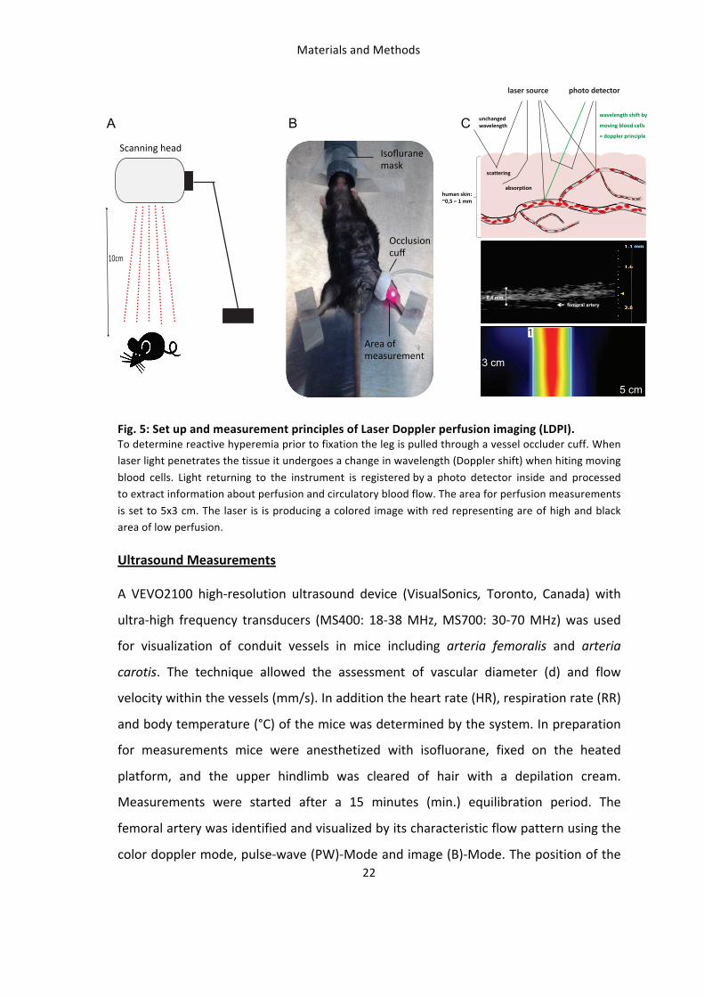
LDPI Measurements
In order to determine perfusion in mouse hindlimb scanning laser doppler perfusion
imaging (LDPI) measurements were performed by using a PeriScan PIM 3 System
(Perimed-‐Instruments, Järfälla, Sweden) provided with a monochromatic 670-‐690 nm
wavelength laser with standard fiber separation (0.25 mm). Mice were anesthetized
with isoflurane (3 % induction and 2 % maintenance) and were positioned and fixed on
a heated platform (set to 39°C, body temperature approx. 36-‐37 °C) lying on the back
(Fig. 5). The hindlimb was prepared for measurements by clearing the inner thigh of
hair using depilation cream. The scanning head is adjusted to a measuring distance of
10 cm (as assessed by the laser). A 5x3 cm scanning area is rapidly scanned (1
image/sec) and the signal is quantified as arbitrary perfusion units (MPU = Mean
Perfusion Units). Measurements were started after a 15 min. equilibration period and
after achieving stable baseline values. Image analysis was performed off-‐line using the
LDPIwin software (Perimed-‐Instruments). As depicted in Fig. 5C, the scanning LDPI
signal is mainly dependent on the penetration depth of the laser and on the blood flow
in the vessels encountered by the laser within the tissue. The femoral artery was found
at 0.4 cm penetration depth, as visualized by high-‐resolution ultrasound (Fig. 5C,
middle). In its proximity the perfusion map obtained by scanning the area showed a
discrete region with very high signal (Fig. 5C, bottom), which was quantified as MPU.
~ 0,4 mmfemoral artery
5 cm
3 cm
A B C
Materials and Methods
27
determined with the DC Protein Assay Kit from Bio-‐Rad (Munich, Germany). Briefly, 5
µl of protein lysate was mixed with 25µl of provided reagent A and 200 µl of reagent B,
incubated for 15 min. at room temperature and the optical density was determined at
740 nM. A total of 10-‐15 µg protein of tissue or 30 µg of cell lysate was
electrophoretically separated on 3-‐8 % or 10 % polyacrylamide gels at 160 V for one
hour. The proteins were transferred to a nitrocellulose membrane at 30 V for one hour
and probed with respective primary antibodies over night at 4°C and with secondary
antibodies for one hour at room temperature. Gels and membranes were purchased
from invitrogen, life technologies (Carlsbad, USA). Immunoreactive bands were
detected using the Super Signal West Chemiluminescent System (Pierce, Bonn,
Germany) following the manufactures protocol and the ImageQuant device and
software. Used primary antibodies with respective dilutions prepared in 0.1 % Tween
TBS buffer containing 5 % BSA are listed in tab. 6. As a positive control for western-‐blot
analysis lysates from cultured human umbilical vein endothelial cells (HUVEC) and
human hepatocellular carcinoma cells (HepG2) were used, both purchased from
PromoCell (Heidelberg, Germany).
cGMP
To determine cGMP concentration the frozen tissue was homogenized in 1 ml ice-‐cold
5 % trichloroacetic acid (TCA) per 100 mg and centrifuged at 2000 g for 15 min. at 4°C.
TCA in the supernatant fraction was extracted by addition of 3 ml diethyl ether per 1
ml TCA used and the samples were then lyophilized by 3-‐5 h vacuum centrifugation at
60 °C. The samples were reconstituted in 1 ml sample diluent per 1 ml TCA. All plasma
and tissue samples were acetylated by addition of 15 µl of the provided
triethylamine/acetic anhydride (2:1) acetylation reagent to 300 µl sample prior to
measurement. cGMP was measured using an enzyme immunoassay kit (Arbor Assays,
Michigan, USA) according to manufacturers descriptions. Briefly a cGMP-‐ peroxidase
conjugate is added to the standard and samples in the wells coated with an anti-‐IgG
antibody. The binding reaction is initiated by the addition of a monoclonal antibody to
cGMP. After an overnight incubation, the plate is washed and the chemiluminescent
Materials and Methods
28
substrate is added. The substrate reacts with the bound cGMP-‐peroxidase conjugate to
produce light detected as luminescence. Concentrations in the samples are calculated
from the standard curve.
GSH
To determine GSH concentrations the tissue was homogenized in 250 μL/10 mg ice
cold 0,01 M HCl, the sample was mixed thoroughly, sonificated for 30 sec. and
centrifuged at 14000 rpm and 4°C for 10 min. The supernatant was mixed with an
equal volume of ice cold 5% sulfo salicylic acid to yield a final concentration of 2.5 %,
incubated on ice for five min. and centrifuged at 14000 rpm and 4°C for 10 min. to
remove precipitated protein. The clear supernatant was used to measure GSH with the
DetectX® Fluorescent Detection kit (Arbor Assays, Michigan, USA) according to
manufactures descriptions. The kit utilizes a proprietary non-‐fluorescent molecule that
covalently binds to the free thiol group on GSH to yield a highly fluorescent product.
After mixing the sample and incubating at room temperature for 15 minutes, the GSH-‐
generated signal is read at 510 nm in a fluorescent plate reader with excitation at 390
nm. Addition of a reaction mixture that converts all the GSSG into free GSH which then
reacts during a second 15 min. incubation period yields the signal related to total GSH
content.
Molecular biological analysis
Real Time PCR
A quantitative real-‐time reverse transcription (RT)–PCR technique was used to quantify
messenger RNA (mRNA) expression of the Aryl hydrogen receptor (AhR) in the aorta.
Commercial available TaqMan oligonucleotides labelled with a fluorescent dye
purchased from Applied Biosystems (Foster City, USA) were used as primers to
quantify mRNA expression. Tissue lysates were made by homogenisation in RLT-‐buffer
(guanidine thiocyanate buffer) using the Tissue Ruptor homogenizer from Qiagen
Materials and Methods
30
analysis. The results were sorted and interpreted by affiliation and functional
classification of the targets using the Database for Annotation, Visualization and
Integrated Discovery (DAVID) (open source, http://david.abcc.ncifcrf.gov/) and the
Kyoto Encyclopedia of Genes and Genomes (KEGG) (open source,
http://www.genome.jp/kegg/).
Analytical analysis
Measurement of nitrate and nitrite
Plasma and tissue nitrate and nitrite levels were measured with high-‐performance
liquid chromatography (ENO20, Eicom, Dublin, Ireland). This method employs an
autoinjection system (Eicom), ion chromatography with a chloride buffer carrier
solution (Eicom) and on-‐line reduction of nitrate to nitrite and subsequent postcolumn
derivatization with the Griess reagent (Hendgen-‐Cotta et al. 2008). Calculations of
concentrations were performed with prepared standards from NaNO2 and NaNO3
using dedicated software (PowerChrom, eDAQ, Colorado Springs, USA). The detection
limit for nitrite and nitrate was 10 nM (0.1 pmol) for either anion at an injection
volume of 100 µl. The measurements were performed by Sivatharsini Thasian-‐
Sivarajah, samples were prepared by myself as described. In preparation for the
measurement the shock-‐frozen plasma samples were thawed on ice and ice-‐cold
methanol was added in equal volume, the sample was mixed thoroughly and
centrifuged for 10 min. at 14000 rpm and 4°C to precipitate proteins. After
centrifugation 100 µl of the supernatant was subjected to analysis.
Measurement of (-‐)-‐epicatechin metabolites
The assessment of (-‐)-‐epicatechin metabolites in blood plasma of the mice was done
by kindly help of Dr. A. Rodriguez-‐Mateos from the Molecular Nutrition Group, School
of Chemistry, Food and Pharmacy at the University of Reading, England. To determine
the concentrations the plasma samples were processed and measured by HPLC-‐
Materials and Methods
31
analysis using a system from Agilent Technologies (1100 series) as previously described
by the group (Rodriguez-‐Mateos et al. 2013). Briefly, plasma (0.5 ml) samples were
prepared by using enzymatic hydrolysis with β-‐glucuronidase and sulfatase (10.000 IU
β-‐glucuronidase, 300 IU sulfatase; 40 min. at 37°C) to produce nonglucuronidated and
nonsulfated metabolites for analysis. Samples were mixed with 1 ml 0.5 % acetic acid
in water (vol:vol), and centrifuged at 17.000 g for 15 min. at 4 °C. The supernatant (10
μl) was injected onto a column with 0.1 % (vol:vol) formic acid in HPLC water and 0.1 %
(vol:vol) formic acid in acetonitrile as mobile phase. The detection and quantification
of phenolic acids and their metabolites was performed by using authentic standards.
Statistical Analyses
When not stated otherwise values reported are means ± SEM or medians with
quartiles and whiskers in Tukey box plots. Comparisons between groups were made
using either 2-‐sided student’s t-‐test or ANOVA, followed by Bonferroni or Games-‐
Howell posthoc test for multiple comparisons. Differences were deemed significant
when p < 0.05. Statistical analyses were performed using GraphPad Prism 5.00 (AD
instruments, El Paso, USA) or SPSS 18.0 statistics (IBM, Amonk, USA).
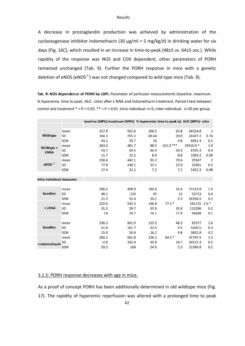
Results
34
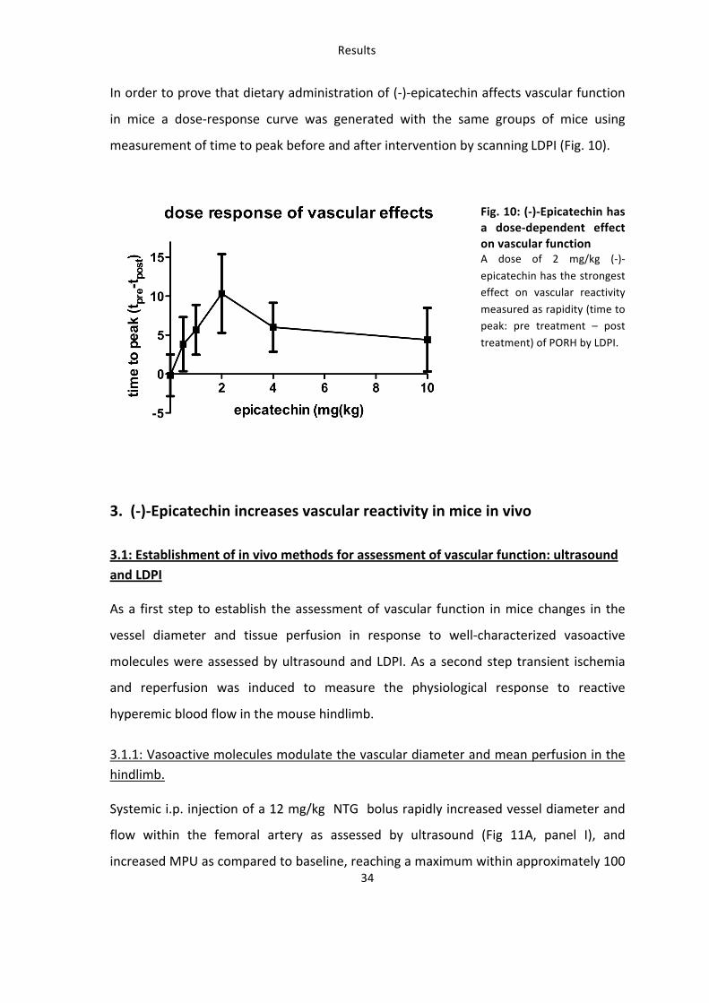
In order to prove that dietary administration of (-‐)-‐epicatechin affects vascular function
in mice a dose-‐response curve was generated with the same groups of mice using
measurement of time to peak before and after intervention by scanning LDPI (Fig. 10).
Fig. 10: (-‐)-‐Epicatechin has a dose-‐dependent effect on vascular function A dose of 2 mg/kg (-‐)-‐
epicatechin has the strongest effect on vascular reactivity
measured as rapidity (time to peak: pre treatment – post
treatment) of PORH by LDPI.
3. (-‐)-‐Epicatechin increases vascular reactivity in mice in vivo
3.1: Establishment of in vivo methods for assessment of vascular function: ultrasound and LDPI
As a first step to establish the assessment of vascular function in mice changes in the
vessel diameter and tissue perfusion in response to well-‐characterized vasoactive
molecules were assessed by ultrasound and LDPI. As a second step transient ischemia
and reperfusion was induced to measure the physiological response to reactive
hyperemic blood flow in the mouse hindlimb.
3.1.1: Vasoactive molecules modulate the vascular diameter and mean perfusion in the hindlimb.
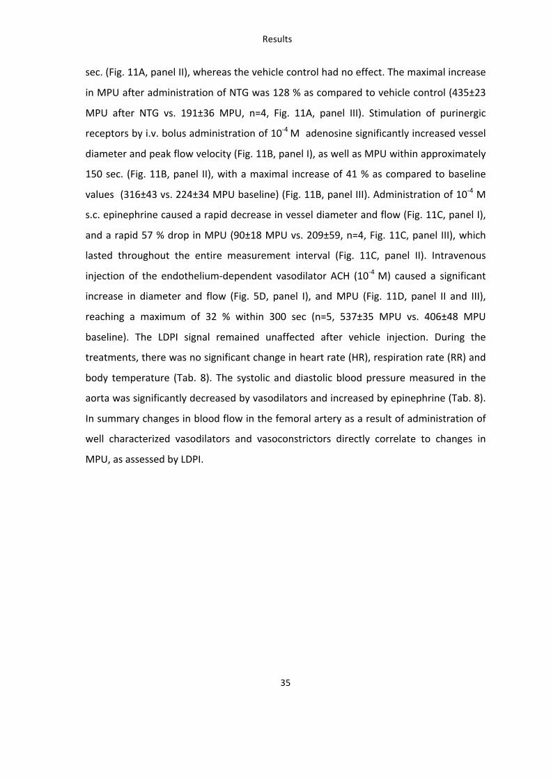
Systemic i.p. injection of a 12 mg/kg NTG bolus rapidly increased vessel diameter and
flow within the femoral artery as assessed by ultrasound (Fig 11A, panel I), and
increased MPU as compared to baseline, reaching a maximum within approximately 100
Results
35
sec. (Fig. 11A, panel II), whereas the vehicle control had no effect. The maximal increase
in MPU after administration of NTG was 128 % as compared to vehicle control (435±23
MPU after NTG vs. 191±36 MPU, n=4, Fig. 11A, panel III). Stimulation of purinergic
receptors by i.v. bolus administration of 10-‐4 M adenosine significantly increased vessel
diameter and peak flow velocity (Fig. 11B, panel I), as well as MPU within approximately
150 sec. (Fig. 11B, panel II), with a maximal increase of 41 % as compared to baseline
values (316±43 vs. 224±34 MPU baseline) (Fig. 11B, panel III). Administration of 10-‐4 M
s.c. epinephrine caused a rapid decrease in vessel diameter and flow (Fig. 11C, panel I),
and a rapid 57 % drop in MPU (90±18 MPU vs. 209±59, n=4, Fig. 11C, panel III), which
lasted throughout the entire measurement interval (Fig. 11C, panel II). Intravenous
injection of the endothelium-‐dependent vasodilator ACH (10-‐4 M) caused a significant
increase in diameter and flow (Fig. 5D, panel I), and MPU (Fig. 11D, panel II and III),
reaching a maximum of 32 % within 300 sec (n=5, 537±35 MPU vs. 406±48 MPU
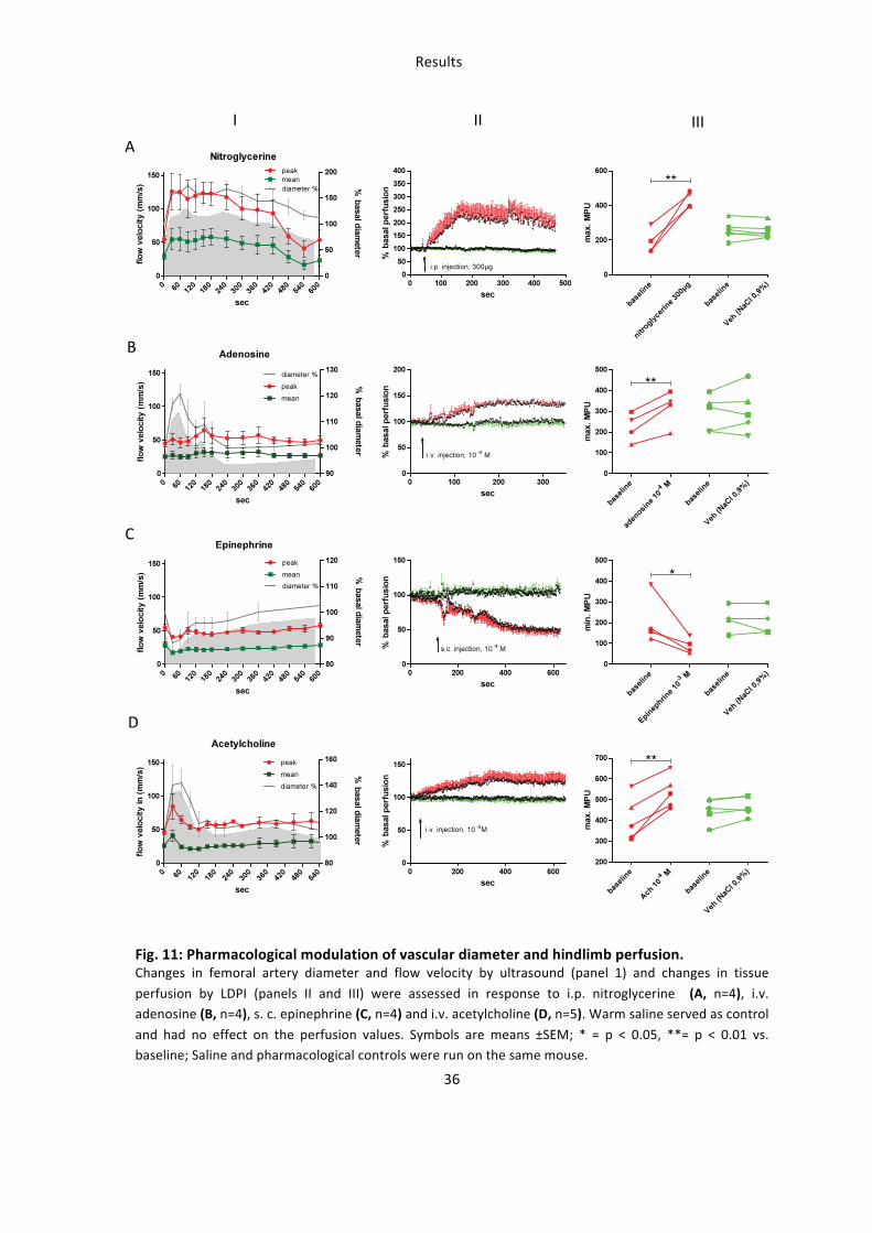
baseline). The LDPI signal remained unaffected after vehicle injection. During the
treatments, there was no significant change in heart rate (HR), respiration rate (RR) and
body temperature (Tab. 8). The systolic and diastolic blood pressure measured in the
aorta was significantly decreased by vasodilators and increased by epinephrine (Tab. 8).
In summary changes in blood flow in the femoral artery as a result of administration of
well characterized vasodilators and vasoconstrictors directly correlate to changes in
MPU, as assessed by LDPI.
1 min
3 min
5 min
0
200
400
600
800
1000**
max
per
fusi
on (M
PU)
1 min
3 min
5 min
0
1
2
3 *
ratio
(max
/ ba
sal p
erfu
sion
)
1 min
3 min
5 min
0
20000
40000
60000
80000 *
AU
C (M
PU)
1 min
3 min
5 min
0
50
100
150 ***
time
to p
eak
(sec
)
baseli
ne
maxim
um
%hypere
mia
time t
o peak
AUCrat
io0
20
40
60
80 inter-individual intra-individual
Para
met
er V
aria
bilit
y (C
V %
)
control + 7mM L-NNA (6d)0
50
100
150
200***
time
to p
eak
(sec
)
baseline + 7mM LNNA (6d)0
50
100
150
*
time
to p
eak
(sec
)
baseline + Indomethacin 30µg/ml (6d)0
20
40
60
80
100 *
time
to p
eak
(sec
)
A
B
C
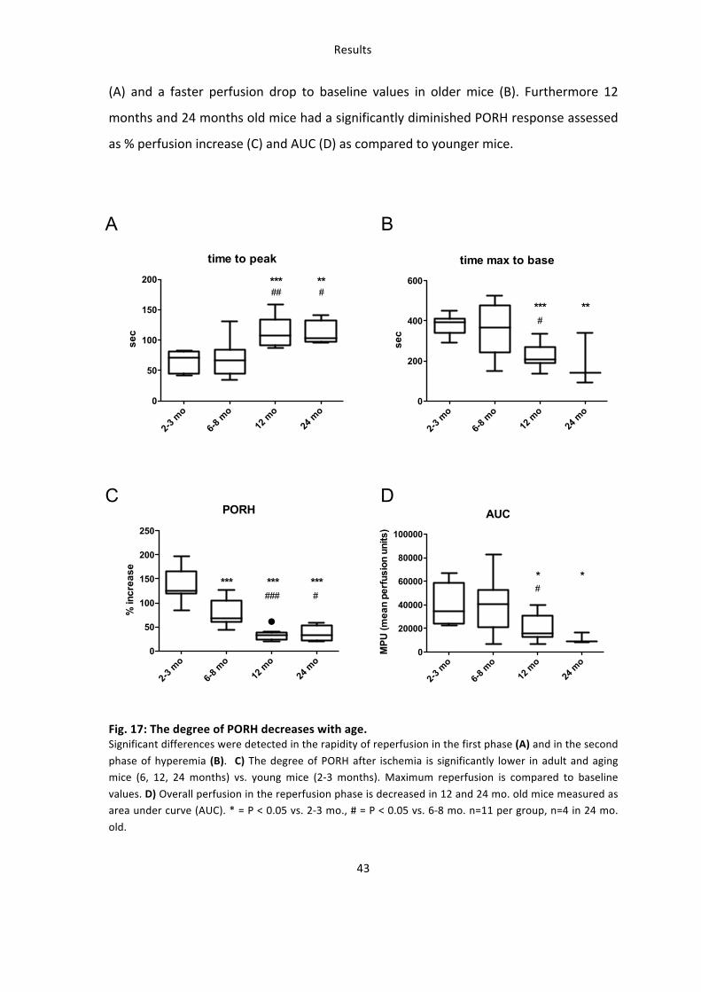
Results
43
(A) and a faster perfusion drop to baseline values in older mice (B). Furthermore 12
months and 24 months old mice had a significantly diminished PORH response assessed
as % perfusion increase (C) and AUC (D) as compared to younger mice.
PORH
2-3 m
o
6-8 m
o12
mo
24 m
o0
50
100
150
200
250
****** ***####
% in
crea
se
AUC
2-3 m
o
6-8 m
o12
mo
24 m
o0
20000
40000
60000
80000
100000
*#
*
MPU
(mea
n pe
rfus
ion
units
)
time to peak
2-3 m
o
6-8 m
o12
mo
24 m
o0
50
100
150
200 ***##
**#
sec
time max to base
2-3 m
o
6-8 m
o12
mo
24 m
o0
200
400
600
***#
**
sec
A B
C D
Fig. 17: The degree of PORH decreases with age. Significant differences were detected in the rapidity of reperfusion in the first phase (A) and in the second
phase of hyperemia (B). C) The degree of PORH after ischemia is significantly lower in adult and aging mice (6, 12, 24 months) vs. young mice (2-‐3 months). Maximum reperfusion is compared to baseline
values. D) Overall perfusion in the reperfusion phase is decreased in 12 and 24 mo. old mice measured as area under curve (AUC). * = P < 0.05 vs. 2-‐3 mo., # = P < 0.05 vs. 6-‐8 mo. n=11 per group, n=4 in 24 mo.
old.
Results
44
Taken together these data show that the measurement of perfusion in the mouse
hindlimb by LDPI is sensitive to pharmacological and flow-‐dependent changes in vessel
diameter and is valid for evaluating intra-‐individual changes for significant in vivo
investigations with limited numbers of animals. Because of small variation, dependence
on NOS/COX and physiological relevance the time to peak parameter was used for
analysis of vascular (-‐)-‐epicatechin effects.
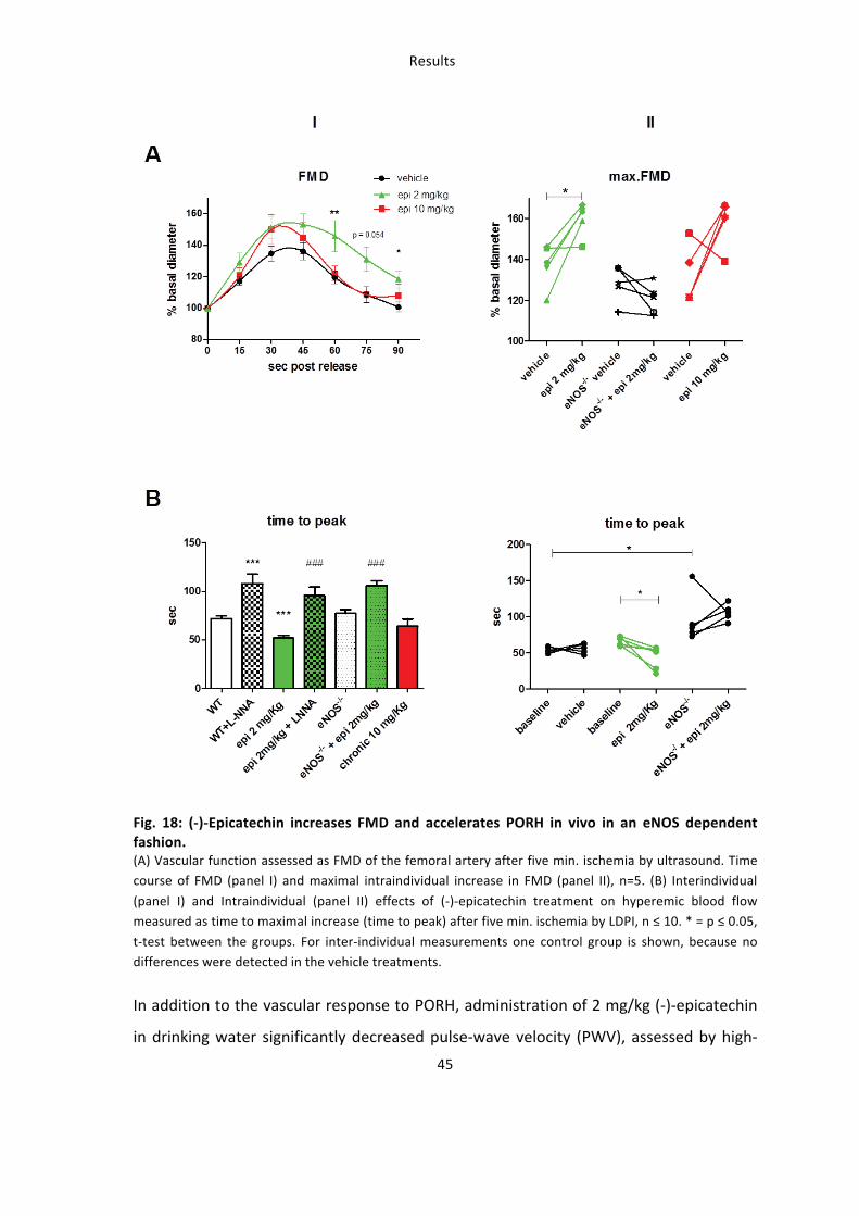
3.2: Oral (-‐)-‐epicatechin increases vascular function in living mice
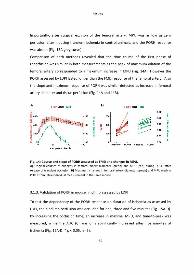
Measurements of FMD by high-‐resolution ultrasound and PORH by LDPI were applied in
order to determine whether oral (-‐)-‐epicatechin treatment improved vascular response
in living mice.
3.2.1: (-‐)-‐Epicatechin increases vascular function in an eNOS-‐dependent fashion
Dietary treatment of mice with 2 mg/kg (-‐)-‐epicatechin corresponded to a significant 1.5
fold increase in FMD of the femoral artery compared to control treatment as assessed
by high resolution ultrasound (Fig. 18A). Maximal dilation of the femoral artery is
reached 45 seconds after releasing the occluding cuff as shown in panel I (53.3±6.7 % 2
mg/kg (-‐)-‐epicatechin (green) vs. 36.2±5.3 % vehicle (black)) and panel II (59.8±3.6 % vs.
37.2±4.8 %). These effects corresponded to a significant increase in the rapidity of
PORH, as determined by measuring the time-‐to-‐maximum response (Fig. 18B).
Importantly these beneficial effects of (-‐)-‐epicatechin on vascular function were fully
blunted by administration of the specific NOS inhibitor L-‐NNA as well as in eNOS-‐/-‐ mice
(Fig 18). Increasing the dose to 10 mg/kg fully blunted the response indicating a very
small window of effective dose.
Results
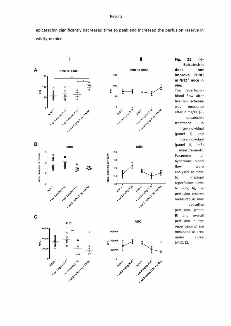
49
epicatechin significantly decreased time to peak and increased the perfusion reserve in
wildtype mice.
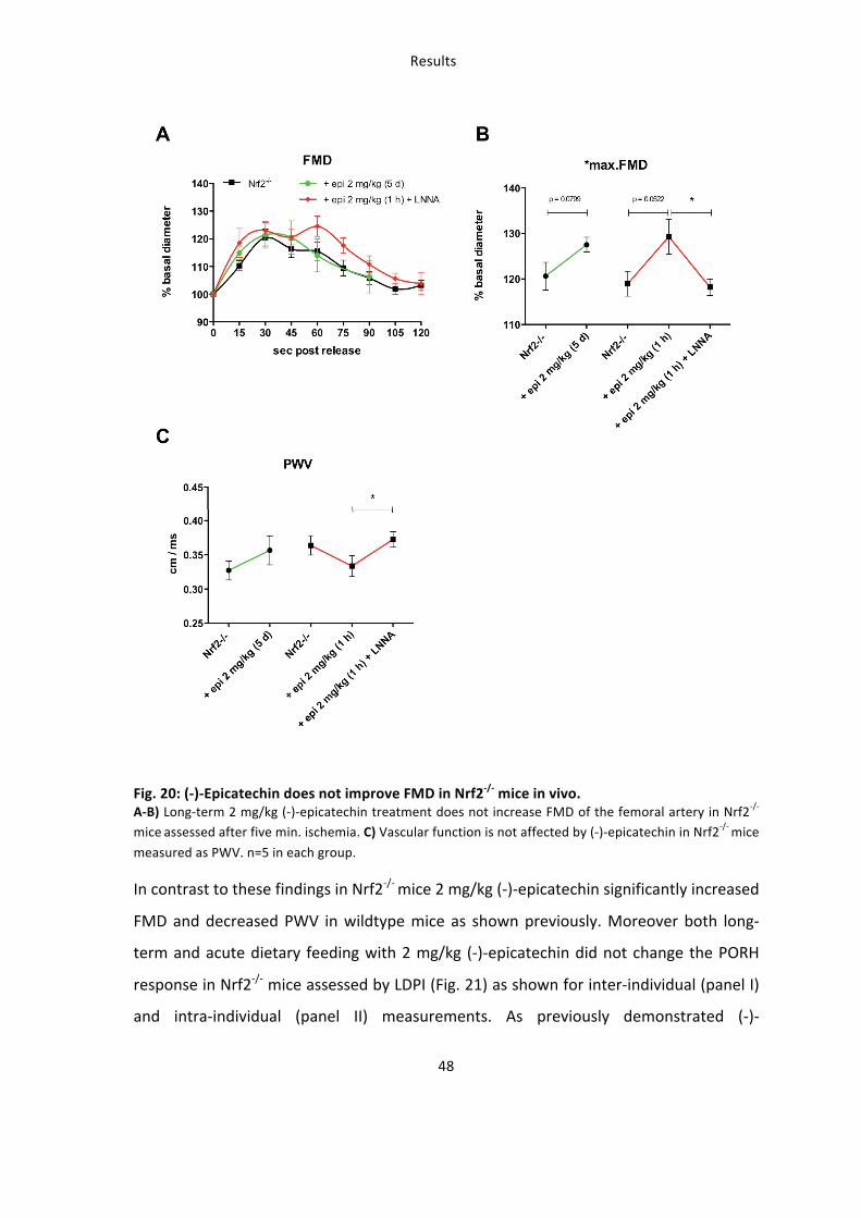
Fig. 21: (-‐)-‐Epicatechin
does not improve PORH in Nrf2-‐/-‐ mice in vivo The reperfusion
blood flow after five min. ischemia
was measured after 2 mg/kg (-‐)-‐
epicatechin treatment in
inter-‐individual (panel I) and
intra-‐individual (panel II, n=5)
measurements. Parameter of
hyperemic blood flow were
analysed as time to maximal
reperfusion (time to peak; A), the
perfusion reserve measured as max
/baseline perfusion (ratio;
B) and overall perfusion in the
reperfusion phase measured as area
under curve (AUC; C).
time to peak
Nrf2-/-
+ epi 2
mg/kg
(5 d
)
+ epi 2
mg/kg
(1 h
)
+ epi 2
mg/kg
(1 h
) + L
NNA0
50
100
150 ****
sec
ratio
Nrf2-/-
+ epi 2
mg/kg
(5 d
)
+ epi 2
mg/kg
(1 h
)
+ epi 2
mg/kg
(1 h
) + L
NNA0
1
2
3
max
/ ba
selin
e pe
rfus
ion
AUC
Nrf2-/-
+ epi 2
mg/kg
(5 d
)
+ epi 2
mg/kg
(1 h
)
+ epi 2
mg/kg
(1 h
) + L
NNA0
20000
40000
60000***
*
MP
U
time to peak
Nrf2-/-
+ epi 2
mg/kg
(5 d
)
Nrf2-/-
+ epi 2
mg/kg
(1 h
)
+ epi 2
mg/kg
(1 h
) + L
NNA0
50
100
150
sec
ratio
Nrf2-/-
+ epi 2
mg/kg
(5 d
)
Nrf2-/-
+ epi 2
mg/kg
(1 h
)
+ epi 2
mg/kg
(1 h
) + L
NNA1.2
1.4
1.6
1.8
2.0
max
/ ba
selin
e pe
rfus
ion
AUC
Nrf2-/-
+ epi 2
mg/kg
(5 d
)
Nrf2-/-
+ epi 2
mg/kg
(1 h
)
+ epi 2
mg/kg
(1 h
) + L
NNA0
20000
40000
60000
**MP
U
A
B
C
I II
Results
50
Taken together these findings demonstrate that (-‐)-‐epicatechin treatment for five days
improves vascular function in wildtype mice but not in eNOS-‐/-‐, after NOS-‐inhibition or in
Nrf2-‐/-‐ mice. However (-‐)-‐epicatechin still improves vascular function when response is
measured one hour after enteral feeding in Nrf2-‐/-‐ mice.
These studies demonstrate that 1) (-‐)-‐epicatechin increases vascular function in a NOS-‐
dependent fashion as show by simultaneous NOS inhibition and measurements in eNOS-‐
/-‐ mice 2) the effects of (-‐)-‐epicatechin administration in water for five days are blunted
in Nrf2-‐/-‐ mice.
4: Molecular mechanisms (I) – (-‐)-‐Epicatechin activates eNOS mediated
NO-‐signaling
4.1: (-‐)-‐Epicatechin activates eNOS in the vasculature in vivo by increasing phosphorylation
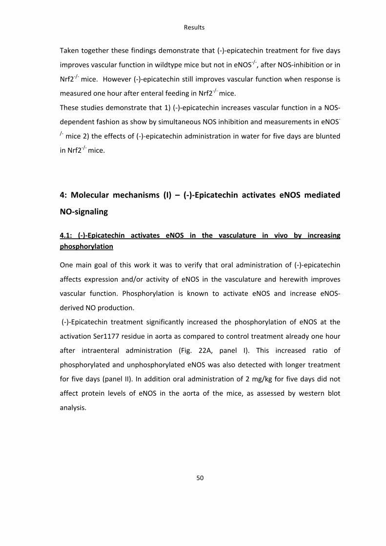
One main goal of this work it was to verify that oral administration of (-‐)-‐epicatechin
affects expression and/or activity of eNOS in the vasculature and herewith improves
vascular function. Phosphorylation is known to activate eNOS and increase eNOS-‐
derived NO production.
(-‐)-‐Epicatechin treatment significantly increased the phosphorylation of eNOS at the
activation Ser1177 residue in aorta as compared to control treatment already one hour
after intraenteral administration (Fig. 22A, panel I). This increased ratio of
phosphorylated and unphosphorylated eNOS was also detected with longer treatment
for five days (panel II). In addition oral administration of 2 mg/kg for five days did not
affect protein levels of eNOS in the aorta of the mice, as assessed by western blot
analysis.
Results
54
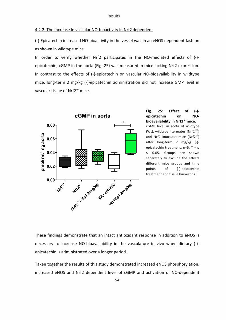
4.2.2: The increase in vascular NO-‐bioactivity in Nrf2 dependent
(-‐)-‐Epicatechin increased NO-‐bioactivity in the vessel wall in an eNOS dependent fashion
as shown in wildtype mice.
In order to verify whether Nrf2 participates in the NO-‐mediated effects of (-‐)-‐
epicatechin, cGMP in the aorta (Fig. 25) was measured in mice lacking Nrf2 expression.
In contrast to the effects of (-‐)-‐epicatechin on vascular NO-‐bioavailability in wildtype
mice, long-‐term 2 mg/kg (-‐)-‐epicatechin administration did not increase GMP level in
vascular tissue of Nrf2-‐/-‐ mice.
Fig. 25: Effect of (-‐)-‐epicatechin on NO-‐bioavailability in Nrf2-‐/-‐ mice. cGMP level in aorta of wildtype (Wt), wildtype litermates (Nrf2+/+)
and Nrf2 knockout mice (Nrf2-‐/-‐) after long-‐term 2 mg/kg (-‐)-‐
epicatechin treatment, n=5. * = p ≤ 0.05. Groups are shown separately to exclude the effects
different mice groups and time points of (-‐)-‐epicatechin
treatment and tissue harvesting.
These findings demonstrate that an intact antioxidant response in addition to eNOS is
necessary to increase NO-‐bioavailability in the vasculature in vivo when dietary (-‐)-‐
epicatechin is administrated over a longer period.
Taken together the results of this study demonstrated increased eNOS phosphorylation,
increased eNOS and Nrf2 dependent level of cGMP and activation of NO-‐dependent
Results
55
downstream signaling in the vasculature of mice by dietary (-‐)-‐epicatechin feeding. This
was accompanied by increased eNOS and Nrf2 dependent vascular function.
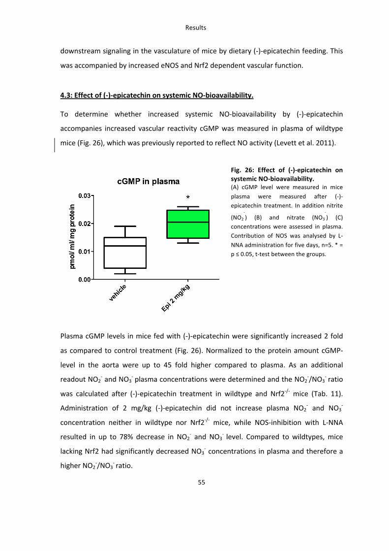
4.3: Effect of (-‐)-‐epicatechin on systemic NO-‐bioavailability.
To determine whether increased systemic NO-‐bioavailability by (-‐)-‐epicatechin
accompanies increased vascular reactivity cGMP was measured in plasma of wildtype
mice (Fig. 26), which was previously reported to reflect NO activity (Levett et al. 2011).
Fig. 26: Effect of (-‐)-‐epicatechin on systemic NO-‐bioavailability. (A) cGMP level were measured in mice
plasma were measured after (-‐)-‐epicatechin treatment. In addition nitrite
(NO2
-‐) (B) and nitrate (NO3
-‐) (C)
concentrations were assessed in plasma. Contribution of NOS was analysed by L-‐
NNA administration for five days, n=5. * = p ≤ 0.05, t-‐test between the groups.
Plasma cGMP levels in mice fed with (-‐)-‐epicatechin were significantly increased 2 fold
as compared to control treatment (Fig. 26). Normalized to the protein amount cGMP-‐
level in the aorta were up to 45 fold higher compared to plasma. As an additional
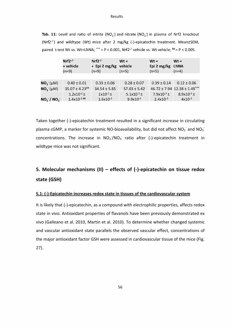
readout NO2-‐ and NO3
-‐ plasma concentrations were determined and the NO2-‐/NO3
-‐ ratio
was calculated after (-‐)-‐epicatechin treatment in wildtype and Nrf2-‐/-‐ mice (Tab. 11).
Administration of 2 mg/kg (-‐)-‐epicatechin did not increase plasma NO2-‐ and NO3
-‐
concentration neither in wildtype nor Nrf2-‐/-‐ mice, while NOS-‐inhibition with L-‐NNA
resulted in up to 78% decrease in NO2-‐ and NO3
-‐ level. Compared to wildtypes, mice
lacking Nrf2 had significantly decreased NO3-‐ concentrations in plasma and therefore a
higher NO2-‐/NO3
-‐ ratio.
Results
57
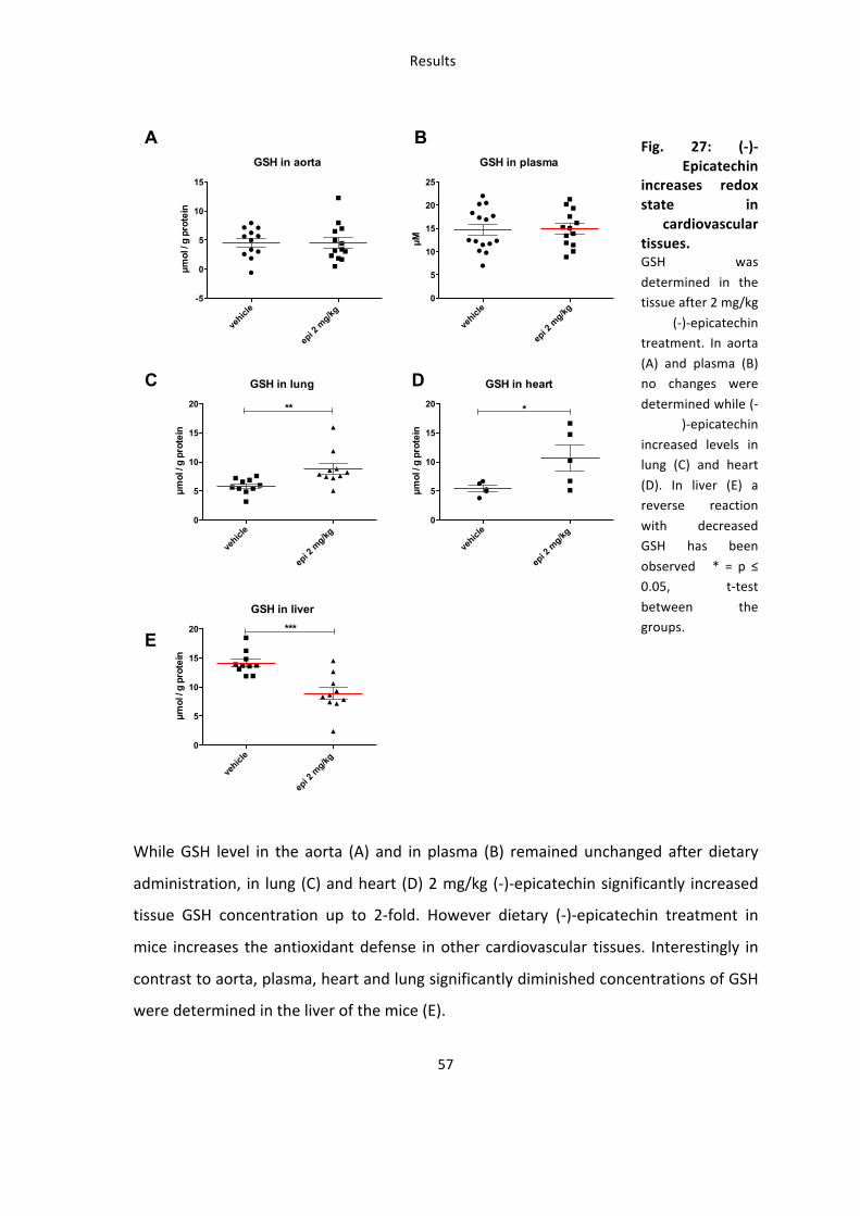
Fig. 27: (-‐)-‐Epicatechin
increases redox state in
cardiovascular tissues. GSH was
determined in the tissue after 2 mg/kg
(-‐)-‐epicatechin treatment. In aorta
(A) and plasma (B) no changes were
determined while (-‐)-‐epicatechin
increased levels in lung (C) and heart
(D). In liver (E) a reverse reaction
with decreased GSH has been
observed * = p ≤ 0.05, t-‐test
between the groups.
While GSH level in the aorta (A) and in plasma (B) remained unchanged after dietary
administration, in lung (C) and heart (D) 2 mg/kg (-‐)-‐epicatechin significantly increased
tissue GSH concentration up to 2-‐fold. However dietary (-‐)-‐epicatechin treatment in
mice increases the antioxidant defense in other cardiovascular tissues. Interestingly in
contrast to aorta, plasma, heart and lung significantly diminished concentrations of GSH
were determined in the liver of the mice (E).
GSH in aorta
vehicl
e
epi 2
mg/kg
-5
0
5
10
15!m
ol /
g pr
otei
n
GSH in heart
vehicl
e
epi 2
mg/kg
0
5
10
15
20 *!m
ol /
g pr
otei
n
A B
C
GSH in plasma
vehicl
e
epi 2
mg/kg
0
5
10
15
20
25
!M
D
E
GSH in lung
vehicl
e
epi 2
mg/kg
0
5
10
15
20 **
!mol
/ g
prot
ein
GSH in liver
vehicl
e
epi 2
mg/kg
0
5
10
15
20 ***
!mol
/ g
prot
ein
Results
58
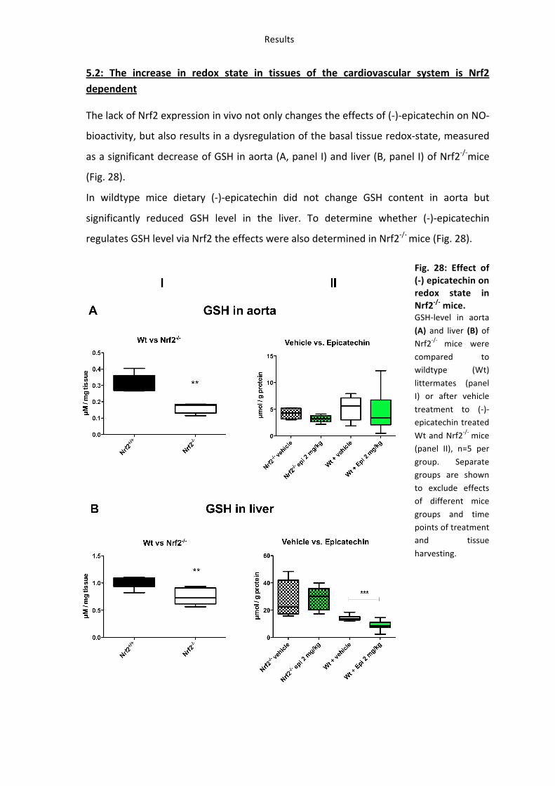
5.2: The increase in redox state in tissues of the cardiovascular system is Nrf2 dependent
The lack of Nrf2 expression in vivo not only changes the effects of (-‐)-‐epicatechin on NO-‐
bioactivity, but also results in a dysregulation of the basal tissue redox-‐state, measured
as a significant decrease of GSH in aorta (A, panel I) and liver (B, panel I) of Nrf2-‐/-‐mice
(Fig. 28).
In wildtype mice dietary (-‐)-‐epicatechin did not change GSH content in aorta but
significantly reduced GSH level in the liver. To determine whether (-‐)-‐epicatechin
regulates GSH level via Nrf2 the effects were also determined in Nrf2-‐/-‐ mice (Fig. 28).
Fig. 28: Effect of (-‐) epicatechin on redox state in Nrf2-‐/-‐ mice. GSH-‐level in aorta
(A) and liver (B) of Nrf2-‐/-‐ mice were
compared to wildtype (Wt)
littermates (panel I) or after vehicle
treatment to (-‐)-‐epicatechin treated
Wt and Nrf2-‐/-‐ mice (panel II), n=5 per
group. Separate groups are shown
to exclude effects of different mice
groups and time points of treatment and tissue
harvesting.
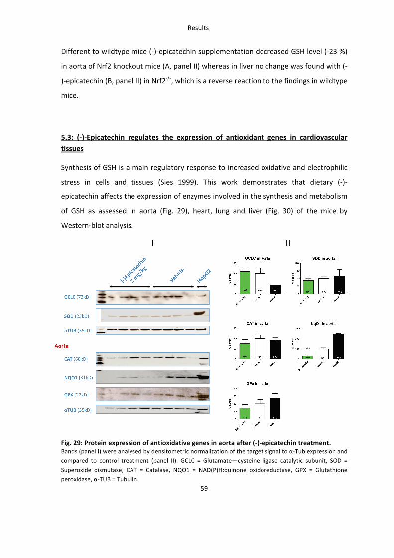
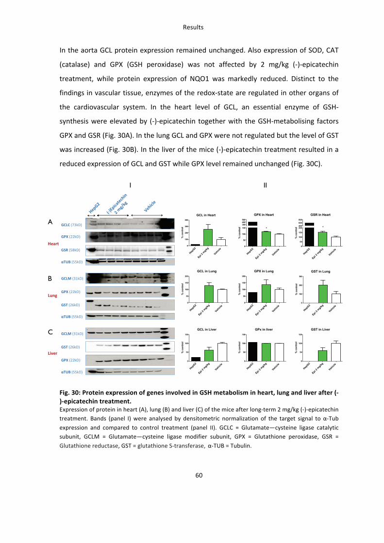
A
B
C
GCL in Heart
HepG2
Epi 2 m
g/kg
Vehicl
e 0
100
200
300
400
% c
ontro
l
GPX in Heart
HepG2
Epi 2 m
g/kg
Vehicl
e 0
50
100
150300400500
*
% c
ontro
l
GSR in Heart
HepG2
Epi 2 m
g/kg
Vehicl
e 0
50
100
150
200400600800
*
% c
ontro
l
GCL in Lung
HepG2
Epi 2 m
g/kg
Vehicl
e 0
50
100
150
200
% c
ontro
l
GPX in Lung
HepG2
Epi 2 m
g/kg
Vehicl
e 0
50
100
150
200
% c
ontro
l
GST in Lung
HepG2
Epi 2 m
g/kg
Vehicl
e 0
100
200
300
% c
ontro
l
GCL in Liver
HepG2
Epi 2 m
g/kg
Vehicl
e 0
50
100
150
% c
ontro
l
GPx in liver
HepG2
Epi 2 m
g/kg
Vehicl
e 0
50
100
150
% c
ontro
l
GST in Liver
HepG2
Epi 2 m
g/kg
Vehicl
e 0
50
100
150
% c
ontro
l
I II
Results
61
Taken together dietary (-‐)-‐epicatechin affected the redox-‐state of tissues of the
cardiovascular system (lung and heart) in mice in vivo demonstrated by increased level
of GSH and regulation of GSH metabolizing enzymes. These regulatory responses are
Nrf2 dependent, because the GSH regulation pattern is altered in Nrf2-‐/-‐ mice.
6. Identification of novel targets of (-‐)-‐epicatechin in vascular tissue by
microarray analysis
6.1: Transcriptional profiling of genes in an (-‐)-‐epicatechin dose-‐response model
To investigate the effect of long-‐term in vivo (-‐)-‐epicatechin treatment on gene
transcription profiles in aorta of mice, genomic mRNA expression was characterized by
comparative microarray analysis. The transcriptional profile of (-‐)-‐epicatechin treated
mice was generated using the Agilent Murine Genome GE 4x44K v2 Microarray Kit and
was logically analysed for set of genes regulated. Three categories of comparisons were
performed: 2 mg/kg vs. vehicle, 10 mg/kg vs. vehicle and 10 mg/kg vs. 2 mg/kg (Fig. 31).
With approximately 39,430 probes representing the mouse genome, targets were
detected with signal intensity higher than the threshold and were selected for further
analysis. Up-‐regulated and down-‐regulated genes were analysed with p-‐values ≤ 0.05.
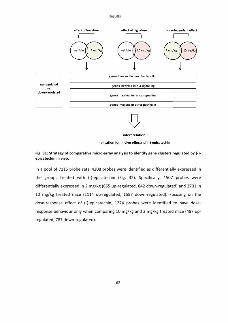
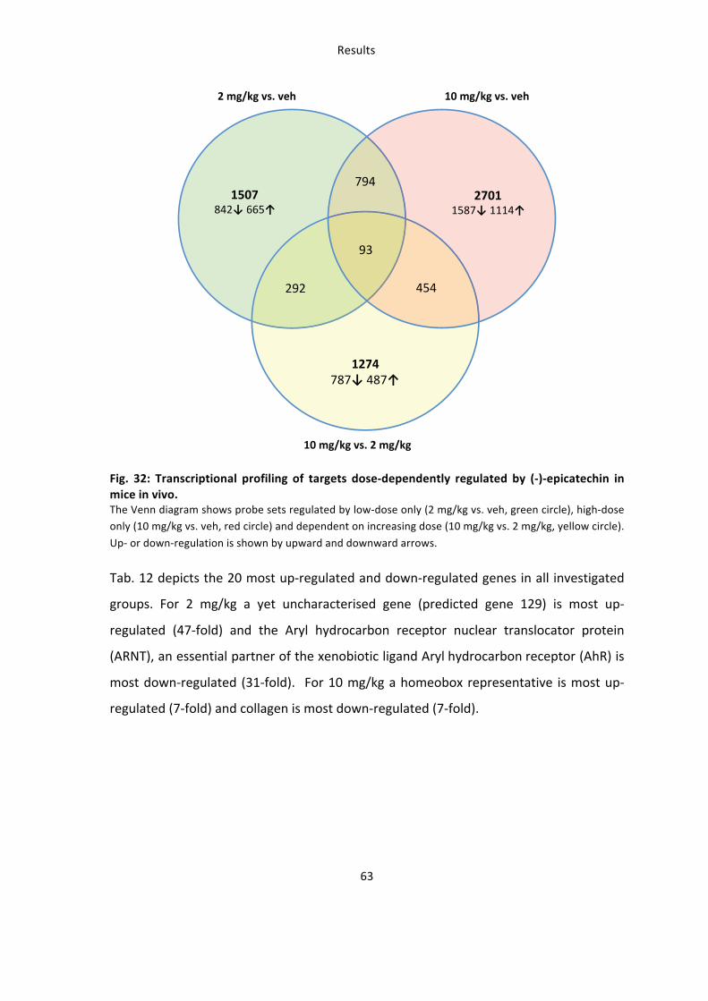
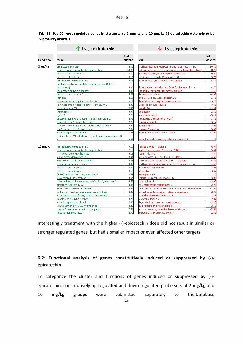
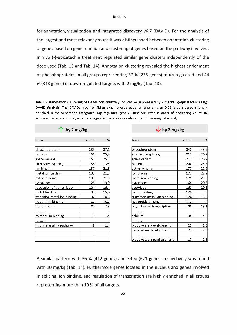
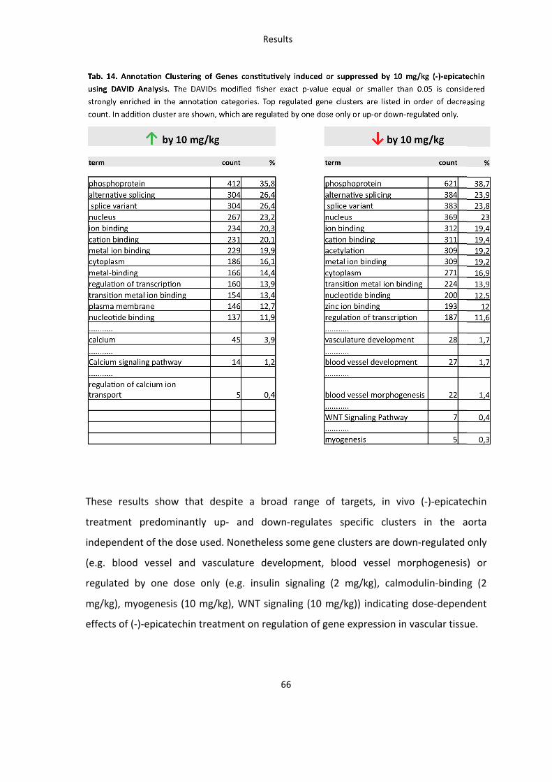
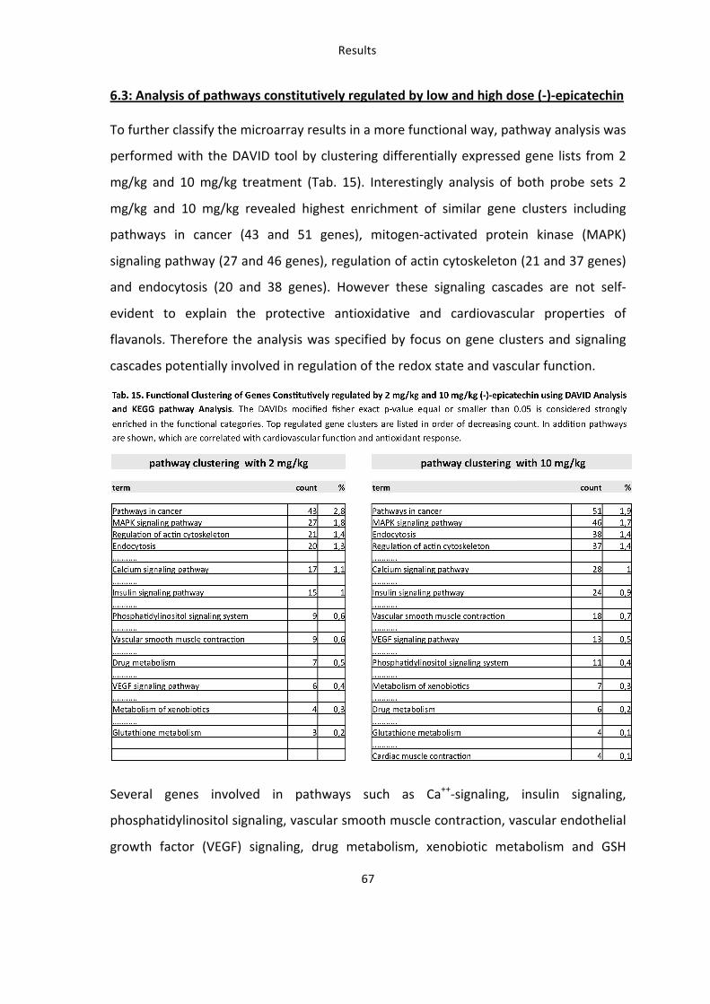
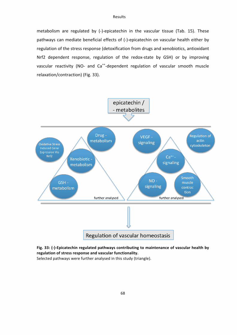
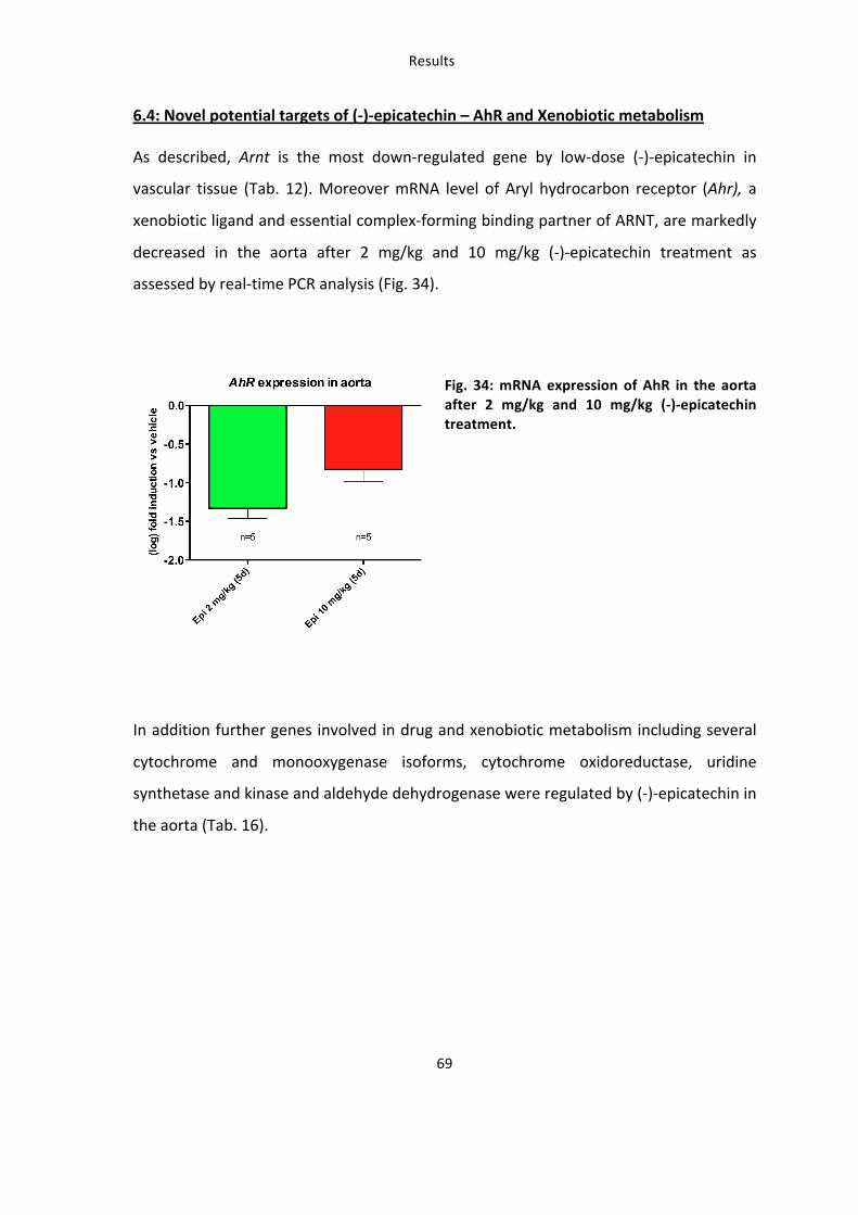
Results
69
6.4: Novel potential targets of (-‐)-‐epicatechin – AhR and Xenobiotic metabolism
As described, Arnt is the most down-‐regulated gene by low-‐dose (-‐)-‐epicatechin in
vascular tissue (Tab. 12). Moreover mRNA level of Aryl hydrocarbon receptor (Ahr), a
xenobiotic ligand and essential complex-‐forming binding partner of ARNT, are markedly
decreased in the aorta after 2 mg/kg and 10 mg/kg (-‐)-‐epicatechin treatment as
assessed by real-‐time PCR analysis (Fig. 34).
Fig. 34: mRNA expression of AhR in the aorta after 2 mg/kg and 10 mg/kg (-‐)-‐epicatechin treatment.
In addition further genes involved in drug and xenobiotic metabolism including several
cytochrome and monooxygenase isoforms, cytochrome oxidoreductase, uridine
synthetase and kinase and aldehyde dehydrogenase were regulated by (-‐)-‐epicatechin in
the aorta (Tab. 16).
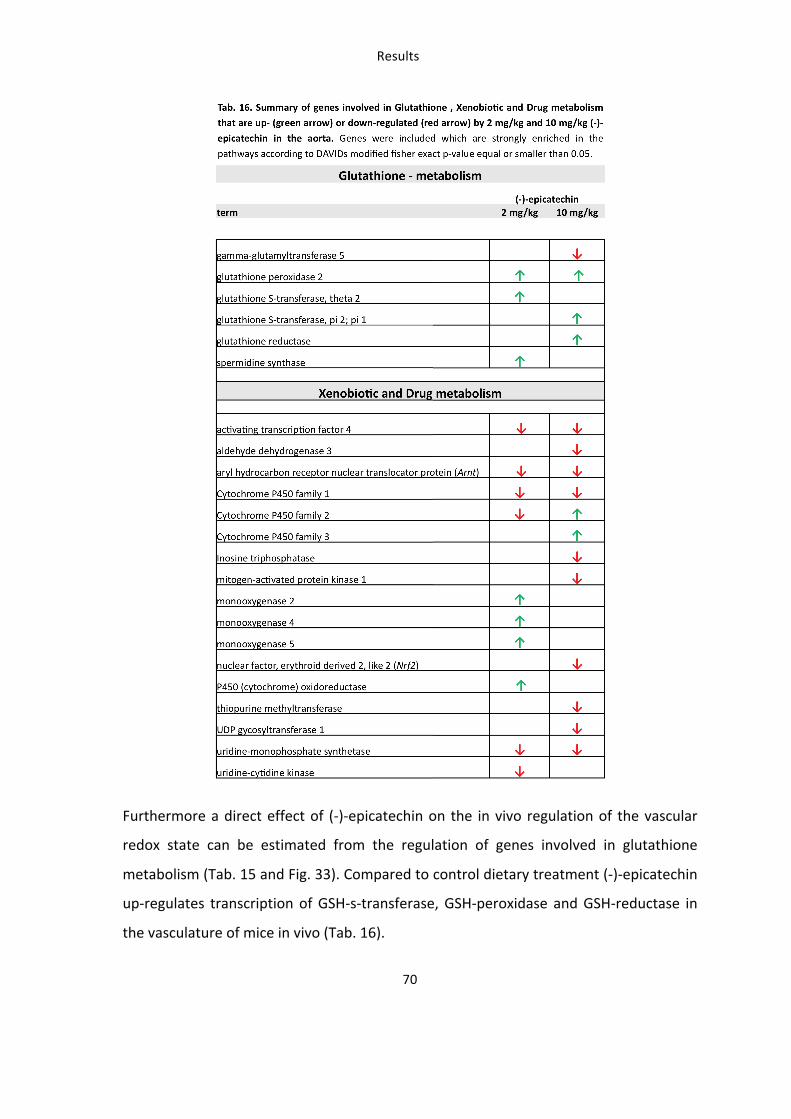
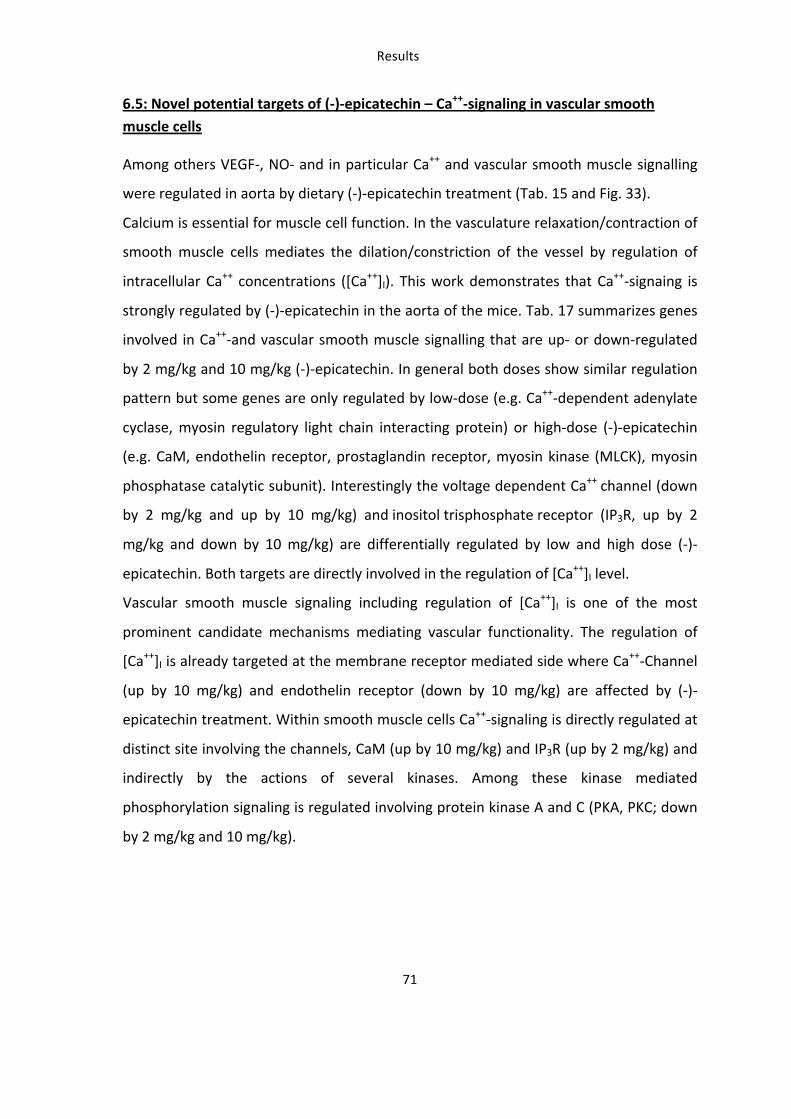
Results
71
6.5: Novel potential targets of (-‐)-‐epicatechin – Ca++-‐signaling in vascular smooth muscle cells
Among others VEGF-‐, NO-‐ and in particular Ca++ and vascular smooth muscle signalling
were regulated in aorta by dietary (-‐)-‐epicatechin treatment (Tab. 15 and Fig. 33).
Calcium is essential for muscle cell function. In the vasculature relaxation/contraction of
smooth muscle cells mediates the dilation/constriction of the vessel by regulation of
intracellular Ca++ concentrations ([Ca++]I). This work demonstrates that Ca++-‐signaing is
strongly regulated by (-‐)-‐epicatechin in the aorta of the mice. Tab. 17 summarizes genes
involved in Ca++-‐and vascular smooth muscle signalling that are up-‐ or down-‐regulated
by 2 mg/kg and 10 mg/kg (-‐)-‐epicatechin. In general both doses show similar regulation
pattern but some genes are only regulated by low-‐dose (e.g. Ca++-‐dependent adenylate
cyclase, myosin regulatory light chain interacting protein) or high-‐dose (-‐)-‐epicatechin
(e.g. CaM, endothelin receptor, prostaglandin receptor, myosin kinase (MLCK), myosin
phosphatase catalytic subunit). Interestingly the voltage dependent Ca++ channel (down
by 2 mg/kg and up by 10 mg/kg) and inositol trisphosphate receptor (IP3R, up by 2
mg/kg and down by 10 mg/kg) are differentially regulated by low and high dose (-‐)-‐
epicatechin. Both targets are directly involved in the regulation of [Ca++]I level.
Vascular smooth muscle signaling including regulation of [Ca++]I is one of the most
prominent candidate mechanisms mediating vascular functionality. The regulation of
[Ca++]I is already targeted at the membrane receptor mediated side where Ca++-‐Channel
(up by 10 mg/kg) and endothelin receptor (down by 10 mg/kg) are affected by (-‐)-‐
epicatechin treatment. Within smooth muscle cells Ca++-‐signaling is directly regulated at
distinct site involving the channels, CaM (up by 10 mg/kg) and IP3R (up by 2 mg/kg) and
indirectly by the actions of several kinases. Among these kinase mediated
phosphorylation signaling is regulated involving protein kinase A and C (PKA, PKC; down
by 2 mg/kg and 10 mg/kg).
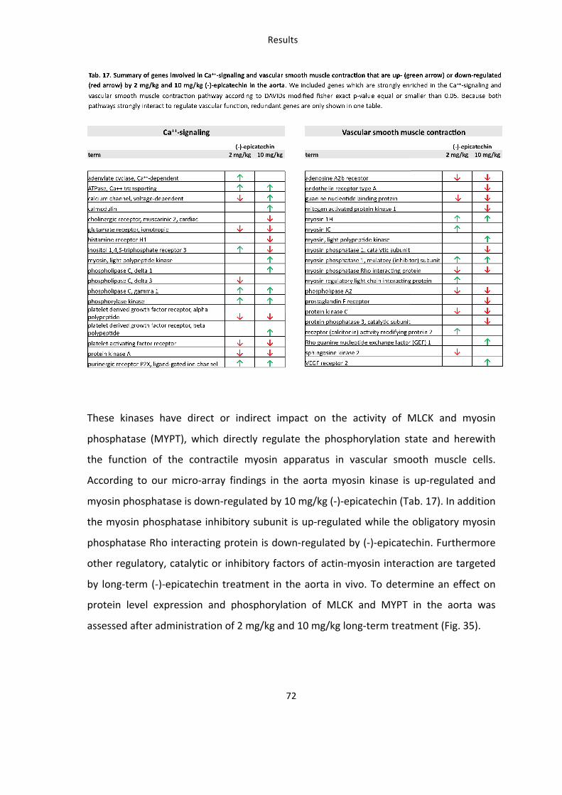
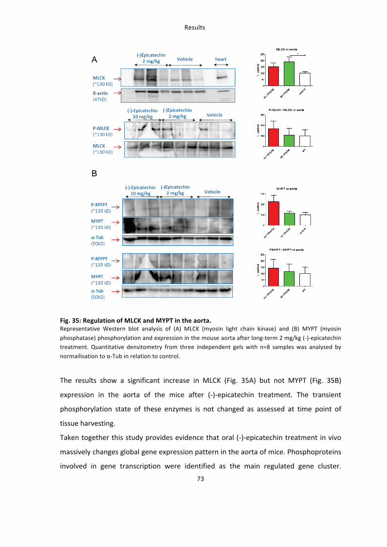
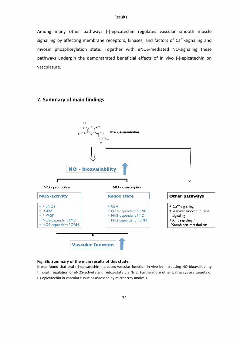
Discussion
75
Discussion The present work investigated the mechanisms underlying the beneficial effects of
dietary (-‐)-‐epicatechin in the cardiovascular system in vivo. It was driven by the
hypothesis that flavanols increase vascular function through increase of NO-‐
bioavailability by increasing both – NO synthesis and antioxidant defense. The main
findings of this study are:
1) (-‐)-‐Epicatechin increases vascular function in an eNOS and Nrf2 dependent fashion
2) (-‐)-‐Epicatechin activates the NO-‐ cGMP pathway and downstream signaling in the
vessel wall in dependence of eNOS and Nrf2
3) (-‐)-‐Epicatechin regulates the redox state of tissues belonging to the cardiovascular
system
4) (-‐)-‐Epicatechin modulates expression of > 4000 targets in vascular tissue thus
contributing to long term changes in the vasculature in vivo. Novel pathways
significant for vascular action of (-‐)-‐epicatechin are: Ca++-‐signaling, vascular smooth
muscle signaling, AhR-‐signaling and Xenobiotic metabolism
Why assessing the effects of (-‐)-‐epicatechin in vivo in mice ?
The analysis of molecular mechanisms underpinning vasodilatory effects in tissue is an
extraordinary advantage of animal in vivo studies, and a main limitation of human
studies. In this study mice models were chosen to analyze the molecular mechanisms
underlying the effects of (-‐)-‐epicatechin on vascular function. In human studies it was
also possible to verify a correlation between administration of cocoa flavanols or pure
(-‐)-‐epicatechin and increased FMD, and increase in nitroso-‐species in plasma. While
these parameters are in some conditions dependent on eNOS activity (i.e. can be
inhibited by administration of NOS inhibitors) a biochemical link between
administration of cocoa flavanols and/or (-‐)-‐ epicatechin and NO-‐bioavailability was
not demonstrated so far. In addition the mechanisms of (-‐)-‐epicatechin cannot be
studied in vitro in cultured cells, because the concentrations of (-‐)-‐epicatechin in the
plasma compartment are very low as compared to the concentration of its metabolites
Discussion
76
as also shown here. A further advantage of the use of mice was the possibility to test
the role of specific targets (i.e. eNOS and Nrf2) by applying both pharmacological
inhibition and genetic depletion in parallel, to overcome eventual unspecific or
compensatory effects, which are typical for the first or the second approach. Pure (-‐)-‐
epicatechin was administrated at a dose previously demonstrated to increase
endothelium-‐dependent vascular function in human in acute (Schroeter et al. 2006)
and long-‐term administration (Heiss et al. 2007) (Engler et al. 2004). This approach
allowed to analyse the effects of vascular function and at the same time the underlying
mechanisms in cardiovascular tissue.
Is eNOS required for the beneficial vascular effects of (-‐)-‐epicatechin in
vivo?
Data presented in this work clearly demonstrate that dietary administration of 2 mg/kg
(-‐)-‐epicatechin improves vascular function in mice, as assessed by increased
FMD/PORH and decreased PWV (Fig. 18 and 19). Importantly eNOS expression and
activity are required for these effects because both pharmacological inhibition and
genetic deletion of eNOS in eNOS-‐/-‐ mice completely block the (-‐)-‐epicatechin effects.
Previously it was shown that exposure of pre-‐constricted rat aortic and femoral rings
from hypertensive rats to (-‐)-‐epicatechin resulted in an increased endothelium-‐
dependent relaxation (Gomez-‐Guzman et al. 2012, Galleano et al. 2013). Importantly,
when NOS was chronically inhibited rats developed hypertension and (-‐)-‐epicatechin
was not longer able to restore defects in endothelium-‐dependent ex vivo aortic ring
vasorelaxation in those animals (Gomez-‐Guzman et al. 2011). The only study
demonstrating the in vivo effects of (-‐)-‐epicatechin describes an effective dilation of
the femoral artery in living rats after direct arterial injection of (-‐)-‐epicatechin into
hindlimb circulation (Ottaviani et al. 2011). However the effects of absorption and
metabolism were not taken into consideration.
Discussion
77
The vascular effects of (-‐)-‐epicatechin very likely include a regulation of NO-‐dependent
vasodilation. Compared to wildtype mice, eNOS-‐/-‐ mice have a decreased, but not
significantly blunted vascular function. Similar results were observed before by
analyzing coronary circulation in the same mouse strain: it was shown that eNOS-‐/-‐
mice compensate for the deficiency for instance by endothelium derived release of
other vasoactive factors e.g. prostaglandins (Godecke et al. 1998).
In addition to an increase in acute vascular response to occlusion this work for the first
time shows that (-‐)-‐epicatechin treatment affects vascular stiffness in mice measured
as reduced PWV. An increased PWV is widely accepted as a measure for endothelial
dysfunction and also for structural changes of the vascular bed for instance in chronic
cardiovascular pathological disorders (Mattace-‐Raso F 2010). Moreover also acute
changes in PWV have been reported within few hours as shown for exercise and
exposure to smoke (Mc Clean et al. 2007, Doonan et al. 2011, Figueroa et al. 2011,
Unosson et al. 2013) and with acute inhibition of NOS (Davies et al. 2003). In the
present study inhibition of NOS also increased PWV in mice.
In conclusion this is the first work demonstrating an improved vascular reactivity and a
direct contribution of eNOS to the (-‐)-‐epicatechin effects in an animal in vivo model.
Other investigations in rodents are limited to ex vivo aortic ring explantation, a
method uncoupling the flavanol effects from systemic response of other organ and cell
systems, metabolism and distribution by blood. It is important to notice, that young,
healthy BL/6 mice were used in this study, which amongst others are known to have a
pronounced ability for vascular regeneration (Helisch et al. 2006). Therefore the
beneficial effect of nutritional flavanol supplementation on endothelium-‐dependent
vascular function might be even stronger in aged mice, mice models of cardiovascular
disease or other strains, when a functional eNOS is presupposed and should be subject
of further investigation.
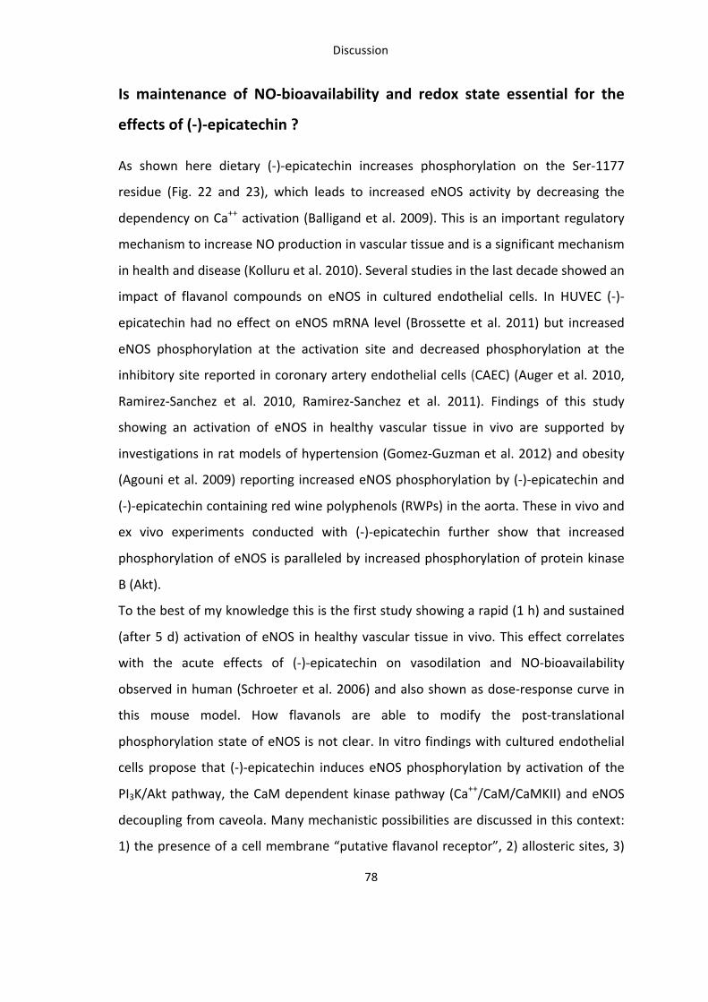
Discussion
78
Is maintenance of NO-‐bioavailability and redox state essential for the
effects of (-‐)-‐epicatechin ?
As shown here dietary (-‐)-‐epicatechin increases phosphorylation on the Ser-‐1177
residue (Fig. 22 and 23), which leads to increased eNOS activity by decreasing the
dependency on Ca++ activation (Balligand et al. 2009). This is an important regulatory
mechanism to increase NO production in vascular tissue and is a significant mechanism
in health and disease (Kolluru et al. 2010). Several studies in the last decade showed an
impact of flavanol compounds on eNOS in cultured endothelial cells. In HUVEC (-‐)-‐
epicatechin had no effect on eNOS mRNA level (Brossette et al. 2011) but increased
eNOS phosphorylation at the activation site and decreased phosphorylation at the
inhibitory site reported in coronary artery endothelial cells (CAEC) (Auger et al. 2010,
Ramirez-‐Sanchez et al. 2010, Ramirez-‐Sanchez et al. 2011). Findings of this study
showing an activation of eNOS in healthy vascular tissue in vivo are supported by
investigations in rat models of hypertension (Gomez-‐Guzman et al. 2012) and obesity
(Agouni et al. 2009) reporting increased eNOS phosphorylation by (-‐)-‐epicatechin and
(-‐)-‐epicatechin containing red wine polyphenols (RWPs) in the aorta. These in vivo and
ex vivo experiments conducted with (-‐)-‐epicatechin further show that increased
phosphorylation of eNOS is paralleled by increased phosphorylation of protein kinase
B (Akt).
To the best of my knowledge this is the first study showing a rapid (1 h) and sustained
(after 5 d) activation of eNOS in healthy vascular tissue in vivo. This effect correlates
with the acute effects of (-‐)-‐epicatechin on vasodilation and NO-‐bioavailability
observed in human (Schroeter et al. 2006) and also shown as dose-‐response curve in
this mouse model. How flavanols are able to modify the post-‐translational
phosphorylation state of eNOS is not clear. In vitro findings with cultured endothelial
cells propose that (-‐)-‐epicatechin induces eNOS phosphorylation by activation of the
PI3K/Akt pathway, the CaM dependent kinase pathway (Ca++/CaM/CaMKII) and eNOS
decoupling from caveola. Many mechanistic possibilities are discussed in this context:
1) the presence of a cell membrane “putative flavanol receptor”, 2) allosteric sites, 3)
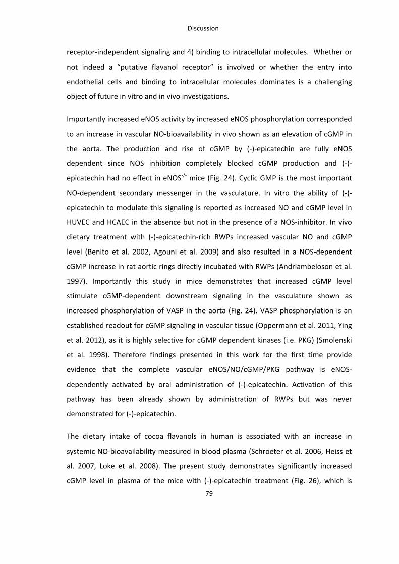
Discussion
79
receptor-‐independent signaling and 4) binding to intracellular molecules. Whether or
not indeed a “putative flavanol receptor” is involved or whether the entry into
endothelial cells and binding to intracellular molecules dominates is a challenging
object of future in vitro and in vivo investigations.
Importantly increased eNOS activity by increased eNOS phosphorylation corresponded
to an increase in vascular NO-‐bioavailability in vivo shown as an elevation of cGMP in
the aorta. The production and rise of cGMP by (-‐)-‐epicatechin are fully eNOS
dependent since NOS inhibition completely blocked cGMP production and (-‐)-‐
epicatechin had no effect in eNOS-‐/-‐ mice (Fig. 24). Cyclic GMP is the most important
NO-‐dependent secondary messenger in the vasculature. In vitro the ability of (-‐)-‐
epicatechin to modulate this signaling is reported as increased NO and cGMP level in
HUVEC and HCAEC in the absence but not in the presence of a NOS-‐inhibitor. In vivo
dietary treatment with (-‐)-‐epicatechin-‐rich RWPs increased vascular NO and cGMP
level (Benito et al. 2002, Agouni et al. 2009) and also resulted in a NOS-‐dependent
cGMP increase in rat aortic rings directly incubated with RWPs (Andriambeloson et al.
1997). Importantly this study in mice demonstrates that increased cGMP level
stimulate cGMP-‐dependent downstream signaling in the vasculature shown as
increased phosphorylation of VASP in the aorta (Fig. 24). VASP phosphorylation is an
established readout for cGMP signaling in vascular tissue (Oppermann et al. 2011, Ying
et al. 2012), as it is highly selective for cGMP dependent kinases (i.e. PKG) (Smolenski
et al. 1998). Therefore findings presented in this work for the first time provide
evidence that the complete vascular eNOS/NO/cGMP/PKG pathway is eNOS-‐
dependently activated by oral administration of (-‐)-‐epicatechin. Activation of this
pathway has been already shown by administration of RWPs but was never
demonstrated for (-‐)-‐epicatechin.
The dietary intake of cocoa flavanols in human is associated with an increase in
systemic NO-‐bioavailability measured in blood plasma (Schroeter et al. 2006, Heiss et
al. 2007, Loke et al. 2008). The present study demonstrates significantly increased
cGMP level in plasma of the mice with (-‐)-‐epicatechin treatment (Fig. 26), which is
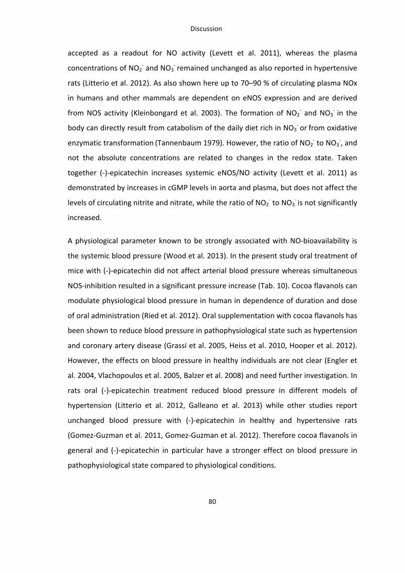
Discussion
80
accepted as a readout for NO activity (Levett et al. 2011), whereas the plasma
concentrations of NO2-‐ and NO3
-‐ remained unchanged as also reported in hypertensive
rats (Litterio et al. 2012). As also shown here up to 70–90 % of circulating plasma NOx
in humans and other mammals are dependent on eNOS expression and are derived
from NOS activity (Kleinbongard et al. 2003). The formation of NO2-‐ and NO3
-‐ in the
body can directly result from catabolism of the daily diet rich in NO3-‐ or from oxidative
enzymatic transformation (Tannenbaum 1979). However, the ratio of NO2-‐ to NO3
-‐, and
not the absolute concentrations are related to changes in the redox state. Taken
together (-‐)-‐epicatechin increases systemic eNOS/NO activity (Levett et al. 2011) as
demonstrated by increases in cGMP levels in aorta and plasma, but does not affect the
levels of circulating nitrite and nitrate, while the ratio of NO2-‐ to NO3
-‐ is not significantly
increased.
A physiological parameter known to be strongly associated with NO-‐bioavailability is
the systemic blood pressure (Wood et al. 2013). In the present study oral treatment of
mice with (-‐)-‐epicatechin did not affect arterial blood pressure whereas simultaneous
NOS-‐inhibition resulted in a significant pressure increase (Tab. 10). Cocoa flavanols can
modulate physiological blood pressure in human in dependence of duration and dose
of oral administration (Ried et al. 2012). Oral supplementation with cocoa flavanols has
been shown to reduce blood pressure in pathophysiological state such as hypertension
and coronary artery disease (Grassi et al. 2005, Heiss et al. 2010, Hooper et al. 2012).
However, the effects on blood pressure in healthy individuals are not clear (Engler et
al. 2004, Vlachopoulos et al. 2005, Balzer et al. 2008) and need further investigation. In
rats oral (-‐)-‐epicatechin treatment reduced blood pressure in different models of
hypertension (Litterio et al. 2012, Galleano et al. 2013) while other studies report
unchanged blood pressure with (-‐)-‐epicatechin in healthy and hypertensive rats
(Gomez-‐Guzman et al. 2011, Gomez-‐Guzman et al. 2012). Therefore cocoa flavanols in
general and (-‐)-‐epicatechin in particular have a stronger effect on blood pressure in
pathophysiological state compared to physiological conditions.
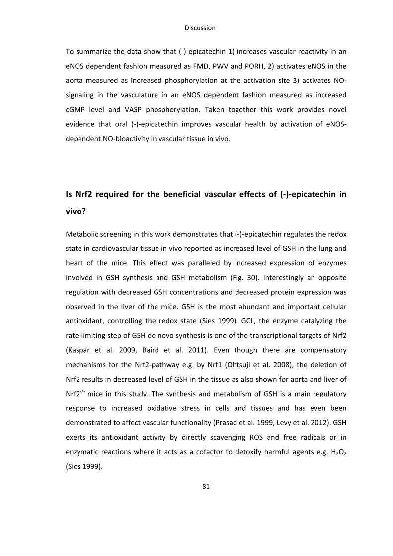
Discussion
81
To summarize the data show that (-‐)-‐epicatechin 1) increases vascular reactivity in an
eNOS dependent fashion measured as FMD, PWV and PORH, 2) activates eNOS in the
aorta measured as increased phosphorylation at the activation site 3) activates NO-‐
signaling in the vasculature in an eNOS dependent fashion measured as increased
cGMP level and VASP phosphorylation. Taken together this work provides novel
evidence that oral (-‐)-‐epicatechin improves vascular health by activation of eNOS-‐
dependent NO-‐bioactivity in vascular tissue in vivo.
Is Nrf2 required for the beneficial vascular effects of (-‐)-‐epicatechin in
vivo?
Metabolic screening in this work demonstrates that (-‐)-‐epicatechin regulates the redox
state in cardiovascular tissue in vivo reported as increased level of GSH in the lung and
heart of the mice. This effect was paralleled by increased expression of enzymes
involved in GSH synthesis and GSH metabolism (Fig. 30). Interestingly an opposite
regulation with decreased GSH concentrations and decreased protein expression was
observed in the liver of the mice. GSH is the most abundant and important cellular
antioxidant, controlling the redox state (Sies 1999). GCL, the enzyme catalyzing the
rate-‐limiting step of GSH de novo synthesis is one of the transcriptional targets of Nrf2
(Kaspar et al. 2009, Baird et al. 2011). Even though there are compensatory
mechanisms for the Nrf2-‐pathway e.g. by Nrf1 (Ohtsuji et al. 2008), the deletion of
Nrf2 results in decreased level of GSH in the tissue as also shown for aorta and liver of
Nrf2-‐/-‐ mice in this study. The synthesis and metabolism of GSH is a main regulatory
response to increased oxidative stress in cells and tissues and has even been
demonstrated to affect vascular functionality (Prasad et al. 1999, Levy et al. 2012). GSH
exerts its antioxidant activity by directly scavenging ROS and free radicals or in
enzymatic reactions where it acts as a cofactor to detoxify harmful agents e.g. H2O2
(Sies 1999).
Discussion
82
Since investigations on the beneficial effects of cocoa flavanols concentrate on
endothelial and vascular function and therefore NO-‐bioactivity, still not much is known
about the in vivo antioxidant effects of these compounds. Because of their chemical
structure flavanols have the potential to exert redox active properties as
predominantly demonstrated through thermodynamic and kinetic analysis (Galleano
et al. 2010) and in solution (Rice-‐Evans et al. 1996). Some authors (Ruijters et al. 2013)
attribute to flavanols in general and (-‐)-‐epicatechin in particular direct antioxidant
effects. However the concentrations needed for such effects are 100-‐1000 fold higher
as compared to circulating (-‐)-‐epicatechin concentrations. Activation of the GSH
metabolism depends on the cell type: while (-‐)-‐epicatechin increased GSH level and
activated GSH metabolizing enzymes in hepatic and neuronal cells (Martin et al. 2010,
Martin et al. 2013), unchanged GSH-‐level and enzymatic activities were reported in a
colon-‐derived cell line (Rodriguez-‐Ramiro et al. 2011).
This study reports an in vivo regulation of the redox state in tissues of the
cardiovascular system by oral (-‐)-‐epicatechin. These findings are supported by similar
approaches in rats showing increased GSH level and enzyme activity in the heart after
administration of flavanol-‐rich grape extract (Seymour et al. 2013) but unchanged
regulation of plasma GSH after administration of (-‐)-‐epicatechin (Litterio et al. 2012).
However, the GSH pool, and herewith the NO pool, along the vascular tree is also
determined by the blood and blood cells permanently streaming through the lumen.
Regulation of GSH level in red blood cells (Lee et al. 2004) and also white cells (Zhu et
al. 2008) is dependent on the presence of Nrf2 as demonstrated in Nrf2-‐/-‐ mice.
Moreover, in cardiovascular and metabolic disorders with vascular dysfunction GSH
level are decreased in red blood cells as shown for hypertension (Kumar et al. 2010)
and type2 diabetes (Rizvi et al. 2001) in human and for bacterial infection with
increased oxidative stress in mice (Guleria et al. 2002). Importantly, ex vivo incubation
with (-‐)-‐epicatechin increased GSH-‐level in red blood cells from healthy people and
patients in a dose-‐dependent bell-‐shaped fashion. In vivo oral flavanol-‐rich green tea
extract increased red blood cell GSH in infected mice and oral administration of
Discussion
83
flavanols from grapes increased the GSH/GSSG ratio in human red blood cells (Weseler
et al. 2011). The physiological relevance of GSH in blood cells is further supported by
positive correlation of decreased GSH in mononuclear cells with decreased FMD
(Fratta Pasini et al. 2012) and in red blood cells with the pathogenesis of slow coronary
flow in human (Enli et al. 2008). As demonstrated in this study, (-‐)-‐epicatechin
increased GSH level in the heart of mice in vivo. Previously it has been shown that GSH
is able to increase coronary vasodilation and flow in isolated rat heart by a NO and sGC
dependent mechanism (Cheung et al. 1997, Levy et al. 2012) and that cardiac
dysfunction in hypertension is associated with decreased GSH in the rat heart
(Seymour et al. 2013). Thus this work provides further evidence for the accumulating
data of flavanol-‐mediated regulation of the physiological relevant tissue redox state in
vivo. It is obvious that the type of tissue and even the cellular type has strong impact
on the described effects. On the one hand this is certainly due to different flavanol
distribution, metabolism and action under different physiological and biological
conditions, on the other hand the redox state itself, its regulation and sources of
oxidative stress vary when comparing different cellular types and tissues (Sies 1999,
Sugamura et al. 2011). Although it is often communicated that cocoa flavanol-‐
mediated antioxidant effects are also an underlying principle in human (Keen et al.
2005, Corti et al. 2009) and may increase plasma antioxidant capacity and decrease
plasma oxidation products (Rein et al. 2000, Wang et al. 2000), convenient evidence
from controlled interventional trails remains elusive (Heiss et al. 2007, Heiss et al.
2010, Scheid et al. 2010, Ellinger et al. 2011, Hollman et al. 2011).
This work provides strong evidence that the (-‐)-‐epicatechin effects on vascular function
are absent if GSH levels are decreased and antioxidant regulation is less effective, as
shown in Nrf2-‐/-‐ mice. In contrast to wildtype mice oral administration of (-‐)-‐
epicatechin to Nrf2-‐/-‐ mice did not improve vascular function (Fig. 20 and Fig. 21), did
not increase NO-‐bioactivity in the vasculature (Fig. 25) and did not regulate tissue
redox state (Fig. 28). These are unreported novel findings, which describe the
contribution of Nrf2 to the vascular (-‐)-‐epicatechin effects in vivo. In other context Nrf2
Discussion
84
is essential for (-‐)-‐epicatechin mediated prevention of stroke damage in mice (Shah et
al. 2010, Leonardo et al. 2013) and is associated with a reduction of cardiac
dysfunction by flavanol-‐rich grape extract in hypertensive rats (Seymour et al. 2013). In
vitro findings confirm that (-‐)-‐epicatechin is able to activate Nrf2 signaling as
demonstrated in cultured neuronal cells (Shah et al. 2010, Leonardo et al. 2013), while
this effect is lacking (Ruijters et al. 2013) or not reported in endothelial cells. Thus
although flavanols process an electrophilic chemical structure and although there is
evidence for an activation of Nrf2 in non vascular cells this is the first work to
demonstrate that Nrf2 is required to increase vascular reactivity and plays an
important role in the in vivo effects of oral administration of (-‐)-‐epicatechin in vascular
tissue. Since cGMP levels are not increased when Nrf2 is lacking, the activation of
eNOS by (-‐)-‐epicatechin is obviously not sufficient to restore the defects in Nrf2-‐/-‐ mice.
Because regulation of the redox state is dependent on Nrf2, the involved mechanism
may include an increased NO degradation due to increased oxidative stress. These
effects have been demonstrated for longer term treatment, while (-‐)-‐epicatechin was
still able to increase vascular function after one hour. This indicates, that the rapid
activation of eNOS by (-‐)-‐epicatechin in the vessel wall acutely increases vascular
function, whereas longer term treatment additionally includes (-‐)-‐epicatechin effects
via regulation of the redox state by Nrf2.
In summary, so far there was no evidence for regulation of the antioxidant state and a
role of Nrf2 in the cardiovascular system in vivo. This work for the first time
demonstrates that maintenance of the redox state and GSH level is required for the
beneficial effects of (-‐)-‐epicatechin on vascular function in the living animal. Orally
administrated (-‐)-‐epicatechin regulates the NO-‐bioactivity, redox state and antioxidant
gene expression in wildtype mice, while these effects are absent when Nrf2 is deleted.
As this regulation is different in aorta, heart/lung and the liver, it is likely that an
altered distribution and metabolism or activation of different signaling cascades in
different tissues underlies the antioxidant properties of (-‐)-‐epicatechin in vivo.
However, the hypothesis generating approach of metabolic screening for the tissue
Discussion
85
redox-‐state can not picture the complete signaling-‐cascades and herewith the
(patho)physiological mechanisms of the (-‐)-‐epicatechin effect. Still, the improvement
of vascular function by increased NO-‐bioactivity might not only be determined by
increased eNOS dependent NO production but also by stabilization of the NO-‐pool.
Thus regulation of GSH and enzymes belonging to antioxidant defense (=Nrf2 targets)
may be necessary for the (-‐)-‐epicatechin induced effects.
Discussion
86
What other mechanisms are involved in the vascular effects of (-‐)-‐
epicatechin ?
Endothelial NOS-‐dependent NO-‐signaling and Nrf2-‐dependent antioxidant response
are mechanisms by which oral (-‐)-‐epicatechin mediates its beneficial cardiovascular
effects. However, a modulation of daily diet, especially under longer-‐term conditions,
also results in global changes, e.g. in the regulation of gene transcription (Liang et al.
2001, Stephanie J. Muga 2004, Swain et al. 2007). Surprisingly, this has never been
investigated for the effects of dietary flavanols in the vasculature in vivo, although the
identification of novel targets and signaling cascades would permit a better and more
comprehensive understanding of the in vivo effects of flavanols on cardiovascular
health. Transcriptional profiling in this work demonstrates that oral (-‐)-‐epicatechin
treatment has a dose-‐dependent, global impact on the transcriptional profile in
vascular tissue in vivo. Interventional studies with flavanols predominantly focus on
one or few potential targets only and no other study performed comparative
transcriptome analysis in vascular tissue, nor in other organs, after in vivo treatment.
Examples of global gene expression analysis involving dietary/nutritional intervention
in mice are numerous and include effects of iron in intestinal tissue (Swain et al. 2007),
linoleic acid in colon (Stephanie J. Muga 2004) or leptin in liver (Liang et al. 2001). An
indication for similar potential of flavanols is provided from the effects of flavanol-‐rich
green tea on lung cancer gene profile in mice (Lu et al. 2006) and epigallocatechin-‐3-‐
gallate (EGCG) on global gene expression profile in human prostate cancer cells (Wang
et al. 2002) and neuronal cells (Weinreb et al. 2003).
Transcriptional profiling of genes in an (-‐)-‐epicatechin dose-‐response model
Oral administration of (-‐)-‐epicatechin regulates 4208 targets from which 1507 are
affected by 2 mg/kg and 2701 by 10 mg/kg. First, these findings demonstrate that oral
feeding with (-‐)-‐epicatechin in vivo results in strong changes of the vascular gene
expression profile. Second, a dose-‐dependent response strongly determines the
number and identity of genes regulated. Therefore results of this study suggest
important implications for nutritional studies with (-‐)-‐epicatechin in vivo. Several
mechanisms such as NO-‐bioactivity, antioxidant properties, and others are currently in
the focus to explain the beneficial action of cocoa flavanols on cardiovascular health
Discussion
87
(Heiss et al. 2010) and it is reasonable that possible targets and signaling cascades are
analyzed separately to explain these effects. However results of this work provide
evidence that other targets out of the main focus are regulated and may play an
essential role. In addition the dose used in in vivo nutritional studies is important
because transcriptional profiling in vascular tissues identified many targets, which are
regulated by low or high dose (-‐)-‐epicatechin only.
To further categorize the targets it was distinguished between annotation clustering of
genes based on gene affiliation and functional clustering of genes based on the
pathway involved. The strongest enrichment of (-‐)-‐epicatechin regulated targets was
found in the phosphoprotein cluster, followed by genes located in the nucleus,
involved in regulation of transcription, splicing and ion binding (Tab. 13 and 14).
Sublist-‐analysis revealed that most of these phosphoproteins are located in the
nucleus and are involved in regulation of gene transcription. Phosphorylation plays a
major role in cellular signaling and is a key reversible modification that can regulate
enzymatic activity, subcellular localization, complex formation and degradation of
proteins (Delom et al. 2006). Therefore global activation/suppression of genes
regulating transcription and other essential cellular processes accounts for the
mechanisms discussed for cocoa flavanols on vasculature. However, affiliation
clustering of genes limits the possibility of a functional and more stringent
interpretation of the results. To connect the novel findings with known vascular in vivo
effects pathway clustering of (-‐)-‐epicatechin regulated genes was done with focus on
signaling cascades potentially involved in regulation of the redox state and vascular
function.
Novel (-‐)-‐epicatechin targets: vascular smooth muscle signalling and protection from toxic and oxidative stress
Besides others (-‐)-‐epicatechin regulates gene clusters of cellular xenobiotic and
antioxidant defense in the aorta in vivo (Tab. 15). Among all targets Arnt is the most
down-‐regulated gene by low-‐dose (-‐)-‐epicatechin (Tab. 12). The ARNT protein forms a
complex with AhR, which is also down-‐regulated by (-‐)-‐epicatechin at mRNA level (Fig.
Discussion
88
34). Regulation of these factors by (-‐)-‐epicatechin is a significant mechanism because
the ARNT/AhR complex plays a major role in the adaptive response of biological
systems to xenobiotic (e.g. drugs, dioxins) stress by binding eligible ligands. However,
the detoxification process itself produces reactive and harmful intermediate
compounds, which have to be further processed and detoxified by the Nrf2-‐dependent
pathway (Wakabayashi et al. 2010). Importantly cocoa flavonoids can also act as
exogenous ligands for the ARNT/AhR complex as demonstrated in vitro as a
competitive antagonist role and an inhibition of ARNT/AhR-‐mediated signal
transduction (Mukai et al. 2010, Han et al. 2012), and in vivo in liver of mice and rats as
disrupted formation of the ARNT/AhR complex by inhibited binding between AhR and
its toxic ligands (Fukuda et al. 2007, Mukai et al. 2008). Therefore there is
accumulating evidence that the intake of flavanoids suppresses the toxicological
effects of xenobiotic agents in the body by inhibition of ARNT/AhR signaling. Even
though a transcriptional down-‐regulation of Arnt and Ahr in aorta by (-‐)-‐epicatechin as
demonstrated in this study disagrees with recent findings with grape extract in the
hearts of rats (Seymour et al. 2013), this mechanism would permit a suppression of
ligand mediated toxicological effects in the vascular system which is similar to
ARNT/AhR inhibition. Importantly the AhR-‐signaling includes a negative feedback
regulation in which an inactive ARNT/AhR complex inhibits the transcription of Ahr
(Denison et al. 2011). Therefore a disrupted formation of the ARNT/AhR complex by (-‐
)-‐epicatechin, as previously demonstrated for other class of flavonoids, can explain a
down-‐regulation of these factors at mRNA level. These novel findings in vascular tissue
in vivo have significant implications for pathophysiological conditions because toxic
AhR ligands mediate cardiovascular disease development and progression in mammals
and vertebrates which is correlated with an activation of ARNT-‐AhR signaling (Korashy
et al. 2006).
The AhR system strongly interacts with the antioxidant Nrf2 signaling pathway
(Wakabayashi et al. 2010), which also can be activated by flavanol compounds (Shah et
al. 2010, Scapagnini et al. 2011, Seymour et al. 2013). As a main protective antioxidant
response to regulate cellular and tissue redox state, enzymes involved in GSH-‐
Discussion
89
metabolism are transcriptionally activated (Janssen-‐Heininger et al. 2013).
Transcriptional profiling in mouse aorta demonstrates that genes clustered in the
phaseII GSH-‐metabolism pathway (GSH-‐s-‐transferase, GSH-‐peroxidase, GSH-‐reductase)
are up-‐regulated by (-‐)-‐epicatechin treatment in vascular tissue in vivo (Tab. 16). While
this pattern could not be proven in aorta on protein level (Fig. 29) an increase in GSH
metabolizing enzymes was also detected in the heart and highly vascularized lung of
the mice (Fig. 30). As previously discussed an activation of GSH metabolizing enzymes
and an elevation of GSH by (-‐)-‐epicatechin has been predominately reported in
cultured cells of non-‐vascular origin in vitro (Martin et al. 2010, Martin et al. 2013)
whereas these effects are not proven for vascular tissue in vivo.
A focus of the functional clustering approach was to identify candidate pathways that
explain the increase of vascular function by (-‐)-‐epicatechin. Among others this work
determined gene clusters of calcium-‐signaling and vascular smooth muscle signaling to
be regulated by (-‐)-‐epicatechin in vascular tissue in vivo. Both pathways essentially
contribute to the vasoactive properties of the vascular wall by regulation of [Ca++]I
through NO-‐cGMP signaling (Nausch et al. 2008, Morgado et al. 2012). Since (-‐)-‐
epicatechin eNOS-‐dependently increases cGMP level and PKG activity in vascular tissue
in vivo (Fig. 24) a regulation of [Ca++]I and myosin phosphorylation is a self-‐evident
mechanism for the effect on vascular function. In fact pathway analysis revealed that
essential factors controlling vascular smooth muscle signaling and [Ca++]I are regulated
by (-‐)-‐epicatechin in the aorta of mice in vivo (Tab. 17) including the Ca++-‐channel, CaM
and IP3R. In addition the enzymes phospholipase C, PKA and PKG are regulated by (-‐)-‐
epicatechin, all which can regulate IP3R mediated Ca++-‐release from intracellular
storage in smooth muscle (Murthy et al. 2003, Narayanan et al. 2012). On the other
hand [Ca++]I can be regulated by entry through voltage–dependent Ca++-‐channel, and
action of the endothelin receptor, both are targets also regulated by (-‐)-‐epicatechin. As
downstream effectors microarray analysis together with western-‐blot analysis
revealed a regulation of MLCK and MYPT.
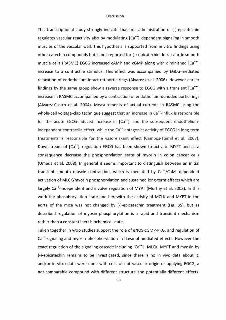
Discussion
90
This transcriptional study strongly indicate that oral administration of (-‐)-‐epicatechin
regulates vascular reactivity also by modulating [Ca++]I dependent signaling in smooth
muscles of the vascular wall. This hypothesis is supported from in vitro findings using
other catechin compounds but is not reported for (-‐)-‐epicatechin. In rat aortic smooth
muscle cells (RASMC) EGCG increased cAMP and cGMP along with diminished [Ca++]I
increase to a contractile stimulus. This effect was accompanied by EGCG-‐mediated
relaxation of endothelium-‐intact rat aortic rings (Alvarez et al. 2006). However earlier
findings by the same group show a reverse response to EGCG with a transient [Ca++]I
increase in RASMC accompanied by a contraction of endothelium-‐denuded aortic rings
(Alvarez-‐Castro et al. 2004). Measurements of actual currents in RASMC using the
whole-‐cell voltage-‐clap technique suggest that an increase in Ca++-‐influx is responsible
for the acute EGCG-‐induced increase in [Ca++]I and the subsequent endothelium-‐
independent contractile effect, while the Ca++-‐antagonist activity of EGCG in long-‐term
treatments is responsible for the vasorelaxant effect (Campos-‐Toimil et al. 2007).
Downstream of [Ca++]I regulation EGCG has been shown to activate MYPT and as a
consequence decrease the phosphorylation state of myosin in colon cancer cells
(Umeda et al. 2008). In general it seems important to distinguish between an initial
transient smooth muscle contraction, which is mediated by Ca++/CaM -‐dependent
activation of MLCK/myosin phosphorylation and sustained long-‐term effects which are
largely Ca++-‐independent and involve regulation of MYPT (Murthy et al. 2003). In this
work the phosphorylation state and herewith the activity of MCLK and MYPT in the
aorta of the mice was not changed by (-‐)-‐epicatechin treatment (Fig. 35), but as
described regulation of myosin phosphorylation is a rapid and transient mechanism
rather than a constant inert biochemical state.
Taken together in vitro studies support the role of eNOS-‐cGMP-‐PKG, and regulation of
Ca++-‐signaling and myosin phosphorylation in flavanol mediated effects. However the
exact regulation of the signaling cascade including [Ca++]I, MLCK, MYPT and myosin by
(-‐)-‐epicatechin remains to be investigated, since there is no in vivo data about it,
and/or in vitro data were done with cells of not vascular origin or applying EGCG, a
not-‐comparable compound with different structure and potentially different effects.
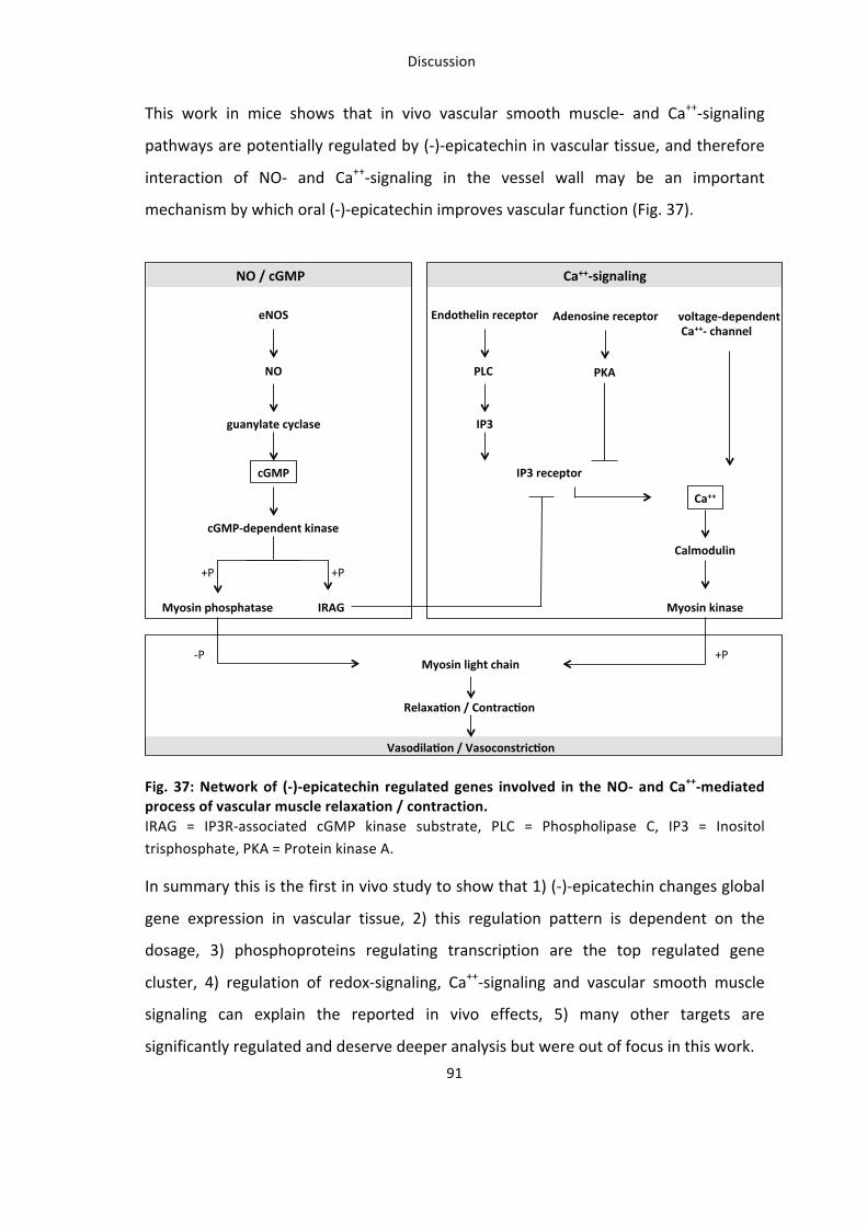
Discussion
92
Limitations of global gene expression analysis in this study is the lack of target
verification by real-‐time PCR for all targets and lack of functional analysis but this are
steps, which are currently under investigation. Since aorta lysates were used, the
discrimination between gene regulation in endothelial cells and smooth muscle cells is
not possible. However, it is important to consider that most of the template origins
from smooth muscles because they outnumber endothelial cells in the aorta. Whether
or not decreased [Ca++]I in smooth muscle parallels increased vasorelaxation by (-‐)-‐
epicatechin in vivo should be subject of future investigations.
Establishing a mouse model for assessment of vascular function
The establishment of methods for the non-‐invasive measurement of vascular function
in living mice was an essential part of this work. Scanning LDPI together with high-‐
resolution ultrasound has been introduced for the first time as a robust non-‐invasive
method for the assessment of vascular function in mice in vivo. Determination of
PORH in the hindlimb allowed the reproducible intra-‐ and inter-‐individual
measurements, which may be applied to evaluate vascular function in mice models of
cardiovascular disease. In particular this study shows that, 1) changes in diameter and
flow in the femoral artery in response to vasoactive drugs correlate with changes in
MPU, 2) dilation of the femoral artery in response to reperfusion after transient
ischemia (FMD) correlate with increased tissue reperfusion (PORH), 3) measurements
of PORH by scanning LDPI are reproducible, depend on availability of endogenous
vasodilators and decrease with increasing animal age.
What is the origin of the LDPI signal in mouse hindlimb?
The perfusion map obtained by scanning a defined area of the hindlimb shows a
discrete region of very high signal intensity, which corresponds to the area of tissue
with and around the femoral artery (see methods). According to the manufacturer’s
information the measurement depth of the system used in this study has been
calculated to be around 1 mm in human skin. In mice the femoral artery can be
Discussion
93
visualized at 0.4 cm penetration depth with high-‐resolution ultrasound. Thus, a major
part of the measured “mean perfusion units” is dependent on blood flow in the
femoral artery (macrocirculation) and less a measure of microcirculatory tissue
perfusion. Therefore, the use of scanning LDPI in the mouse hindlimb is not
comparable to its use in human skin, where – considering the penetration depth of the
laser – it is applied to assess microcirculatory function.
Measurements of PORH: reproducibility and dependency on physiological stimuli.
Measurement of PORH with different methods is routinely applied in clinical settings
to evaluate the endothelial functionality of different vessel beds, and it is known to be
dependent on occlusion time and on endogenous vasodilators.
As a first step of scanning LDPI validation for assessment of hyperemic responses, this
study shows that the LDPI signal is directly correlated to changes in diameter and
blood flow in the femoral artery assessed by ultrasound (Fig. 11). Thus, administration
of endothelium independent and endothelium dependent pharmacological
vasodilators or a vasoconstrictor caused an increase or decrease in vessel diameter,
and blood flow, which corresponded to a comparable response in MPU.
As a second step, it has been verified that PORH-‐induced changes in peak MPU
corresponded to peak FMD of the femoral artery, which is a widely accepted
parameter for determining endothelial function. The elevation of MPU lasted longer
than the FMD response (Fig. 14), indicating a microvascular component of the LDPI
signal. Interestingly, maximal MPU after PORH was comparable with the maximum
NTG-‐induced MPU increase, and significantly higher than the ACh or adenosine
induced response. Importantly, basal perfusion was blunted and PORH was absent in
chronic hindlimb ischemia induced by surgical excision of the femoral artery (Fig. 13).
In addition PORH-‐induced changes in MPU were dependent on the duration of the
occlusion (Fig. 15) and increased with longer occlusion times, further validating this
measurement approach.
As a third step, the reproducibility and variability of PORH assessed by LDPI has been
analysed by repeated intra-‐individual and inter-‐individual measurements (Fig. 15). The
Discussion
94
most robust parameter was the time to peak, which is an indicator of the rapidity of
the response, and the ratio of maximum and baseline perfusion, which is an indicator
of the magnitude of the response. Interestingly, the rapidity of the hyperemic
response was dependent on synthesis of both NO and prostaglandins, as the NOS
inhibitor L-‐NNA as well as the COX inhibitor indomethacin increased time to peak (Fig.
16).
As a fourth step the physiological significance of PORH has been proven. Vascular aging
is known to induce endothelial dysfunction in humans (Seals et al. 2011) and in rats
(Heiss et al. 2007) and is associated with impaired angiogenesis (Rivard et al. 1999) and
aortic ring relaxation (Gendron et al. 2012) in mice. Here, it was found that 12 and 24
months old mice have a slower vascular responsiveness (time to peak), significantly
decreased magnitude of response (perfusion reserve and AUC) and a less sustained
hyperemia as compared to younger mice (Fig. 17).
Although the use of in vivo models of cardiovascular disease is widely spread, methods
for evaluating endothelial function in rodents are mainly restricted to ex vivo
techniques, or high resolution ultrasound in rats (Heiss et al. 2007). Even if a direct
correlation between the changes in arterial diameter, arterial blood flow and changes
in perfusion has been demonstrated in this study, the LDPI technique cannot fully
substitute ultrasound for determination of FMD in vivo. As compared to ultrasound,
scanning LDPI is less specific for endothelial function (i.e. does not measure diameter
and flow velocity in the vessel) and cannot distinguish macrocirculation and
microcirculation. However, it allows to measure tissue perfusion in a defined region of
interest, is easier to use, requires less training time to achieve reproducibility, and it is
more robust as additionally demonstrated for basal perfusion measurements in mouse
hindlimb (Greco et al. 2013). Moreover it is less sensible than ultrasound to tiny
changes in hindlimb position during inflation and deflation of the cuff, temperature
and small changes in cardiac and circulatory parameters, and it is more affordable.
To conclude, in order to perform reliable measurements for assessing vascular
function in mice in vivo both methods were applied – high-‐resolution ultrasound and
Discussion
95
scanning LDPI. Both methods combined allow to test the effects of pathophysiological
changes of vascular function in correlation to genetic or pharmacological manipulation,
or physiological processes like aging both in a cross-‐sectional or longitudinal
experimental settings in the living animal. Thus they permit an easier translation of the
findings to the clinical application in humans.
Summary and Conclusion
The present work analysed the mechanisms underlying the vascular effects of oral (-‐)-‐
epicatechin in vivo. The beneficial action of flavanols on cardiovascular health is widely
reported but the mechanisms are not well understood and mechanistic studies are
mainly limited to in vitro findings with (-‐)-‐epicatechin. This study demonstrates that in
vivo effects of (-‐)-‐epicatechin are dependent on both eNOS-‐dependent regulation of
NO production and Nrf2-‐dependent regulation of the redox state (Fig. 38).
The underlying mechanisms include an activation of the eNOS/cGMP/PKG pathway,
downstream signaling in the vessel wall, regulation of the antioxidant defense and
regulation of redox state in tissue of the cardiovascular system. Moreover
transcriptional profiling in vascular tissue identified novel (-‐)-‐epicatechin targets in vivo
including vascular smooth muscle and Ca++ signaling in interaction of NO-‐signaling.
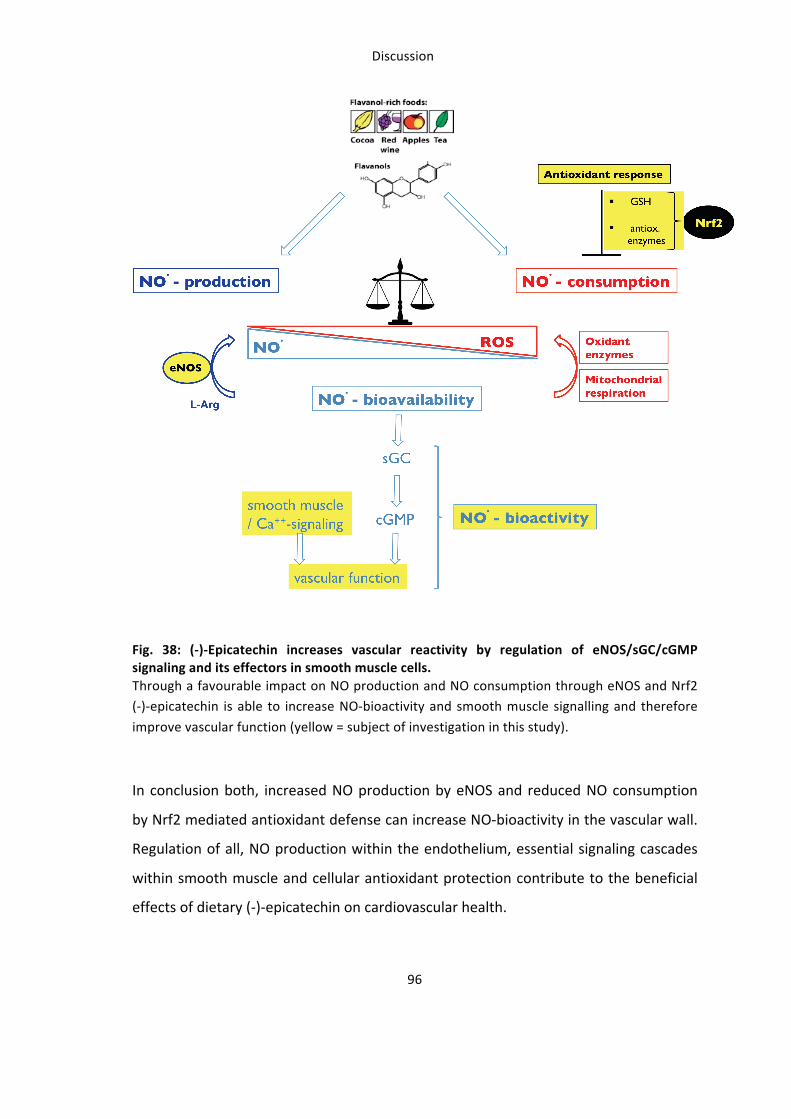
Discussion
97
Why is this study relevant for translational flavanol research ?
This work provides significant contribution to translational flavanol research because it
identified relevant underlying mechanisms, which are able to explain how flavanols
improve vascular function in vivo. Analysis of biochemical and biological mechanisms
involved in effects of a oral intervention is neither possible in tissue in humans, nor is
reported in other in vivo models in this extent so far. There is cumulative evidence that
flavanols can reverse or diminish the initiation and progression of cardiovascular
disease. The identification of potential targets and regulated signaling cascades can
help to develop and specify their application in dietary supplementation or medical
treatment. In this study mice models were chosen to analyze the mechanisms in
cardiovascular tissue because genetic models with a deletion in potential target genes
were available. Importantly a dose of 2 mg/kg (-‐)-‐epicatechin was used in this study
which improved vascular function in mice after five days, but was also demonstrated
to improve FMD of the brachial artery in human in acute (2h) (Schroeter et al. 2006)
and longer term conditions (1-‐7d) (Heiss et al. 2007) (Engler et al. 2004).
This study demonstrates that (-‐)-‐epicatechin is rapidly absorbed and metabolised in
mice. HPLC-‐analysis of plasma samples revealed a dose-‐dependent absorption and
metabolism of (-‐)-‐epicatechin in mice within one hour (Fig. 9). The saturation is
reached at 4-‐10 mg/kg and up to 78 % of total (-‐)-‐epicatechin found in plasma is
metabolized as methylated 3’-‐ and 4’-‐O-‐methylepicatechin. In human the highest
amount of circulating flavanols was found two hours after oral administration
(Schroeter et al. 2006) and also the most abundant metabolite was methylated
epicatechin while unmethylated (-‐)-‐epicatechin represented the minor part. Moreover
it was shown that (-‐)-‐epicatechin is best absorbed, metabolised, and bioactive in
human compared to other flavanol stereoisomers such as (+)-‐epicatechin, (−)-‐catechin,
and (+)-‐catechin (Ottaviani et al. 2011). Interestingly compared to human total plasma
flavanol concentration is threefold higher in mice (1500 nM vs. 600 nM) after
administration of (-‐)-‐epicatechin. This underlines the importance of in vivo
experiments to investigate flavanol mediated effects, as in vitro studies only rarely use
Discussion
98
metabolites including the methylated derivatives of (-‐)-‐epicatechin for treatment of
cells (Steffen et al. 2007).
Another phenomenon found in this study which is relevant for translational flavanol
research, is a bell-‐shaped dose-‐dependence of (-‐)-‐epicatechin in vivo reported as dose-‐
response curve measured as PORH and increased vascular reactivity and pulsatility
with 2 mg/kg but not 10 mg/kg (-‐)-‐epicatechin measured as FMD, PWV and PORH.
Similar to these findings in mice, treatment of healthy rats with 10 mg/kg (-‐)-‐
epicatechin did not result in an increased aortic ring relaxation (Gomez-‐Guzman et al.
2012). The effects of different dose of dietary flavanols are reported but not
satisfactory discussed in studies from animal models and also in human. Both dose of
(-‐)-‐epicatechin, 2 mg/kg and 10 mg/kg, restored endothelial defects but only the
higher dose reduced blood pressure and endothelin level in hypertensive rats (Gomez-‐
Guzman et al. 2012). Moreover, this study provides further novel evidence for a bell-‐
shaped action of (-‐)-‐epicatechin in vivo supporting the reverse effects: 1) The
absorption and metabolism of (-‐)-‐epicatechin in mice reaches a plateau at 4 mg/kg
(Fig. 9), this indicates a saturation effect where (-‐)-‐epicatechin can no longer be
absorbed and metabolized. Importantly this resulting excess of unprocessed
polyphenolic compounds in the organism may even have toxicl effects for the
organism (Takami et al. 2008, Eagle 2012); 2) Treatment with the higher (-‐)-‐epicatechin
dose massively changes global gene regulation in vascular tissue compared to the
lower dose and control conditions. This is can be seen as a proof for the importance of
the dose of flavanols used in in vivo studies, in particular when vascular effects are the
matter of interest. Which mechanisms underlie the adverse effects on vascular
function of higher dose of (-‐)-‐epicatechin in vivo, is current topic of research in our lab.
In summary this study provides new insides for mechanistic action, but also from
metabolism and dose-‐dependence and thus helps to understand how (-‐)-‐epicatechin
may mediate its beneficial cardiovascular effects in human and may therefore have
relevant impact on future translational research in this field.
References
99
References Agouni, A., A. H. Lagrue-‐Lak-‐Hal, H. A. Mostefai, A. Tesse, P. Mulder, P. Rouet, F. Desmoulin, C. Heymes, M. C. Martinez and R. Andriantsitohaina (2009). "Red wine polyphenols prevent metabolic and cardiovascular alterations associated with obesity in Zucker fatty rats (Fa/Fa)." PLoS One 4(5): e5557. Akata, T., M. Nakashima, K. Kodama, W. A. Boyle, 3rd and S. Takahashi (1995). "Effects of volatile anesthetics on acetylcholine-‐induced relaxation in the rabbit mesenteric resistance artery." Anesthesiology 82(1): 188-‐204. Algotsson, A., A. Nordberg and B. Winblad (1995). "Influence of age and gender on skin vessel reactivity to endothelium-‐dependent and endothelium-‐independent vasodilators tested with iontophoresis and a laser Doppler perfusion imager." J Gerontol A Biol Sci Med Sci 50(2): M121-‐127. Alvarez, E., M. Campos-‐Toimil, H. Justiniano-‐Basaran, C. Lugnier and F. Orallo (2006). "Study of the mechanisms involved in the vasorelaxation induced by (-‐)-‐epigallocatechin-‐3-‐gallate in rat aorta." British journal of pharmacology 147(3): 269–280. Alvarez-‐Castro, E., M. Campos-‐Toimil and F. Orallo (2004). "(-‐)-‐Epigallocatechin-‐3-‐gallate induces contraction of the rat aorta by a calcium influx-‐dependent mechanism." Naunyn-‐Schmiedeberg's archives of pharmacology 369(5): 496–506. Andriambeloson, E., A. L. Kleschyov, B. Muller, A. Beretz, J. C. Stoclet and R. Andriantsitohaina (1997). "Nitric oxide production and endothelium-‐dependent vasorelaxation induced by wine polyphenols in rat aorta." British journal of pharmacology 120(6): 1053–1058. Arejian, M., Y. Li and M. B. Anand-‐Srivastava (2009). "Nitric oxide attenuates the expression of natriuretic peptide receptor C and associated adenylyl cyclase signaling in aortic vascular smooth muscle cells: role of MAPK." Am J Physiol Heart Circ Physiol 296(6): H1859-‐1867. Auger, C., J.-‐H. Kim, P. Chabert, M. Chaabi, E. Anselm, X. Lanciaux, A. Lobstein and V. B. Schini-‐Kerth (2010). "The EGCg-‐induced redox-‐sensitive activation of endothelial nitric oxide synthase and relaxation are critically dependent on hydroxyl moieties." Biochemical and biophysical research communications 393(1): 162–167. Baba, S., N. Osakabe, M. Natsume, Y. Muto, T. Takizawa and J. Terao (2001). "Absorption and urinary excretion of (-‐)-‐epicatechin after administration of different levels of cocoa powder or (-‐)-‐epicatechin in rats." Journal of agricultural and food chemistry 49(12): 6050–6056. Baird, L. and A. T. Dinkova-‐Kostova (2011). "The cytoprotective role of the Keap1-‐Nrf2 pathway." Arch Toxicol 85(4): 241-‐272. Balligand, J. L., O. Feron and C. Dessy (2009). "eNOS activation by physical forces: from short-‐term regulation of contraction to chronic remodeling of cardiovascular tissues." Physiol Rev 89(2): 481-‐534. Balzer, J., T. Rassaf, C. Heiss, P. Kleinbongard, T. Lauer, M. Merx, N. Heussen, H. B. Gross, C. L. Keen, H. Schroeter and M. Kelm (2008). "Sustained benefits in vascular function through flavanol-‐containing cocoa in medicated diabetic patients a double-‐masked, randomized, controlled trial." Journal of the American College of Cardiology 51(22): 2141–2149. Bassil, M. and M. B. Anand-‐Srivastava (2006). "Nitric oxide modulates Gi-‐protein expression and adenylyl cyclase signaling in vascular smooth muscle cells." Free Radic Biol Med 41(7): 1162-‐1173. Benito, S., D. Lopez, M. P. Saiz, S. Buxaderas, J. Sanchez, P. Puig-‐Parellada and M. T. Mitjavila (2002). "A flavonoid-‐rich diet increases nitric oxide production in rat aorta." Br J Pharmacol 135(4): 910-‐916. Bhupathiraju, S. N., N. M. Wedick, A. Pan, J. E. Manson, K. M. Rexrode, W. C. Willett, E. B. Rimm and F. B. Hu (2013). "Quantity and variety in fruit and vegetable intake and risk of coronary heart disease." The American journal of clinical nutrition.
References
100
Brossette, T., C. Hundsdorfer, K. D. Kroncke, H. Sies and W. Stahl (2011). "Direct evidence that (-‐)-‐epicatechin increases nitric oxide levels in human endothelial cells." Eur J Nutr 50(7): 595-‐599. Campos-‐Toimil, M. and F. Orallo (2007). "Effects of (-‐)-‐epigallocatechin-‐3-‐gallate in Ca2+ -‐permeable non-‐selective cation channels and voltage-‐operated Ca2+ channels in vascular smooth muscle cells." Life sciences 80(23): 2147–2153. Cheung, P. Y. and R. Schulz (1997). "Glutathione causes coronary vasodilation via a nitric oxide-‐ and soluble guanylate cyclase-‐dependent mechanism." Am J Physiol 273(3 Pt 2): H1231-‐1238. Cooke, J. P., E. Rossitch, Jr., N. A. Andon, J. Loscalzo and V. J. Dzau (1991). "Flow activates an endothelial potassium channel to release an endogenous nitrovasodilator." J Clin Invest 88(5): 1663-‐1671. Cornwell, T. L., E. Arnold, N. J. Boerth and T. M. Lincoln (1994). "Inhibition of smooth muscle cell growth by nitric oxide and activation of cAMP-‐dependent protein kinase by cGMP." Am J Physiol 267(5 Pt 1): C1405-‐1413. Cortese-‐Krott, M. M., A. Rodriguez-‐Mateos, R. Sansone, G. G. Kuhnle, S. Thasian-‐Sivarajah, T. Krenz, P. Horn, C. Krisp, D. Wolters, C. Heiss, K. D. Kroncke, N. Hogg, M. Feelisch and M. Kelm (2012). "Human red blood cells at work: identification and visualization of erythrocytic eNOS activity in health and disease." Blood 120(20): 4229-‐4237. Corti, R., A. J. Flammer, N. K. Hollenberg and T. F. Luscher (2009). "Cocoa and cardiovascular health." Circulation 119(10): 1433-‐1441. Dauchet, L., P. Amouyel and J. Dallongeville (2009). "Fruits, vegetables and coronary heart disease." Nature reviews. Cardiology 6(9): 599–608. Davies, J. I. and A. D. Struthers (2003). "Pulse wave analysis and pulse wave velocity: a critical review of their strengths and weaknesses." J Hypertens 21(3): 463-‐472. Delom, F. and E. Chevet (2006). "Phosphoprotein analysis: from proteins to proteomes." Proteome Sci 4: 15. Denison, M. S., A. A. Soshilov, G. He, D. E. DeGroot and B. Zhao (2011). "Exactly the same but different: promiscuity and diversity in the molecular mechanisms of action of the aryl hydrocarbon (dioxin) receptor." Toxicol Sci 124(1): 1-‐22. Donovan, J. L., V. Crespy, M. Oliveira, K. A. Cooper, B. B. Gibson and G. Williamson (2006). "(+)-‐Catechin is more bioavailable than (-‐)-‐catechin: relevance to the bioavailability of catechin from cocoa." Free Radic Res 40(10): 1029-‐1034. Doonan, R. J., P. Scheffler, A. Yu, G. Egiziano, A. Mutter, S. Bacon, F. Carli, M. E. Daskalopoulos and S. S. Daskalopoulou (2011). "Altered arterial stiffness and subendocardial viability ratio in young healthy light smokers after acute exercise." PLoS One 6(10): e26151. Eagle, K. (2012). "Toxicological effects of red wine, orange juice, and other dietary SULT1A inhibitors via excess catecholamines." Food Chem Toxicol 50(6): 2243-‐2249. Ellinger, S., N. Muller, P. Stehle and G. Ulrich-‐Merzenich (2011). "Consumption of green tea or green tea products: is there an evidence for antioxidant effects from controlled interventional studies?" Phytomedicine 18(11): 903-‐915. Engler, M. B., M. M. Engler, C. Y. Chen, M. J. Malloy, A. Browne, E. Y. Chiu, H.-‐K. Kwak, P. Milbury, S. M. Paul, J. Blumberg and M. L. Mietus-‐Snyder (2004). "Flavonoid-‐rich dark chocolate improves endothelial function and increases plasma epicatechin concentrations in healthy adults." Journal of the American College of Nutrition 23(3): 197–204. Enli, Y., M. Turk, R. Akbay, H. Evrengul, H. Tanriverdi, O. Kuru, D. Seleci, A. Kaftan, O. Ozer and H. Enli (2008). "Oxidative stress parameters in patients with slow coronary flow." Adv Ther 25(1): 37-‐44. Erdman, J. W., Jr., D. Balentine, L. Arab, G. Beecher, J. T. Dwyer, J. Folts, J. Harnly, P. Hollman, C. L. Keen, G. Mazza, M. Messina, A. Scalbert, J. Vita, G. Williamson and J. Burrowes (2007).
References
101
"Flavonoids and heart health: proceedings of the ILSI North America Flavonoids Workshop, May 31-‐June 1, 2005, Washington, DC." J Nutr 137(3 Suppl 1): 718S-‐737S. Eyre, H., R. Kahn and R. M. Robertson (2004). "Preventing cancer, cardiovascular disease, and diabetes: a common agenda for theAmerican Cancer Society, the American Diabetes Association, and the American Heart Association." CA: a cancer journal for clinicians 54(4): 190–207. Farkas, K., E. Kolossváry, Z. Járai, J. Nemcsik and C. Farsang (2004). Non-‐invasive assessment of microvascular endothelial function by laser Doppler flowmetry in patients with essential hypertension. Atherosclerosis. 173: 97-‐102. Figueroa, A., F. Vicil and M. A. Sanchez-‐Gonzalez (2011). "Acute exercise with whole-‐body vibration decreases wave reflection and leg arterial stiffness." American journal of cardiovascular disease 1(1): 60–67. Forstermann, U., J. P. Boissel and H. Kleinert (1998). "Expressional control of the 'constitutive' isoforms of nitric oxide synthase (NOS I and NOS III)." FASEB J 12(10): 773-‐790. Fratta Pasini, A., A. Albiero, C. Stranieri, M. Cominacini, A. Pasini, C. Mozzini, P. Vallerio, L. Cominacini and U. Garbin (2012). "Serum oxidative stress-‐induced repression of Nrf2 and GSH depletion: a mechanism potentially involved in endothelial dysfunction of young smokers." PLoS One 7(1): e30291. Frick M, N. T. S. W. T. (2002). Flußvermittelte Vasodilatation (FMD) der Arteria brachialis: Methodik und klinischer Stellenwert. Journal für Kardiologie: 1-‐7. Fujii, N., M. C. Reinke, V. E. Brunt and C. T. Minson (2013). Impaired acetylcholine-‐induced cutaneous vasodilation in young smokers: roles of nitric oxide and prostanoids. AJP: Heart and Circulatory Physiology. 304: H667-‐673. Fukuda, I., R. Mukai, M. Kawase, K. Yoshida and H. Ashida (2007). "Interaction between the aryl hydrocarbon receptor and its antagonists, flavonoids." Biochem Biophys Res Commun 359(3): 822-‐827. Furchgott, R. F., M. H. Carvalho, M. T. Khan and K. Matsunaga (1987). "Evidence for endothelium-‐dependent vasodilation of resistance vessels by acetylcholine." Blood Vessels 24(3): 145-‐149. Furchgott, R. F. and J. V. Zawadzki (1980). "The obligatory role of endothelial cells in the relaxation of arterial smooth muscle by acetylcholine." Nature 288(5789): 373-‐376. Galleano, M., I. Bernatova, A. Puzserova, P. Balis, N. Sestakova, O. Pechanova and C. G. Fraga (2013). "(-‐)-‐Epicatechin reduces blood pressure and improves vasorelaxation in spontaneously hypertensive rats by NO-‐mediated mechanism." IUBMB Life. Galleano, M., S. V. Verstraeten, P. I. Oteiza and C. G. Fraga (2010). "Antioxidant actions of flavonoids: thermodynamic and kinetic analysis." Arch Biochem Biophys 501(1): 23-‐30. Gelatt-‐Nicholson, K. J., K. N. Gelatt, E. O. MacKay, D. E. Brooks and S. M. Newell (1999). Comparative Doppler imaging of the ophthalmic vasculature in normal Beagles and Beagles with inherited primary open-‐angle glaucoma. Vet Ophthalmol. 2: 97-‐105. Gendron, M. E., N. Thorin-‐Trescases, A. M. Mamarbachi, L. Villeneuve, J. F. Theoret, Y. Mehri and E. Thorin (2012). "Time-‐dependent beneficial effect of chronic polyphenol treatment with catechin on endothelial dysfunction in aging mice." Dose Response 10(1): 108-‐119. Getz, G. S. and C. A. Reardon (2012). "Animal models of atherosclerosis." Arterioscler Thromb Vasc Biol 32(5): 1104-‐1115. Godecke, A., U. K. Decking, Z. Ding, J. Hirchenhain, H. J. Bidmon, S. Godecke and J. Schrader (1998). "Coronary hemodynamics in endothelial NO synthase knockout mice." Circ Res 82(2): 186-‐194. Gomez-‐Guzman, M., R. Jimenez, M. Sanchez, M. Romero, F. O'Valle, R. Lopez-‐Sepulveda, A. M. Quintela, P. Galindo, M. J. Zarzuelo, E. Bailon, E. Delpon, F. Perez-‐Vizcaino and J. Duarte (2011).
References
102
"Chronic (-‐)-‐epicatechin improves vascular oxidative and inflammatory status but not hypertension in chronic nitric oxide-‐deficient rats." Br J Nutr 106(9): 1337-‐1348. Gomez-‐Guzman, M., R. Jimenez, M. Sanchez, M. J. Zarzuelo, P. Galindo, A. M. Quintela, R. Lopez-‐Sepulveda, M. Romero, J. Tamargo, F. Vargas, F. Perez-‐Vizcaino and J. Duarte (2012). "Epicatechin lowers blood pressure, restores endothelial function, and decreases oxidative stress and endothelin-‐1 and NADPH oxidase activity in DOCA-‐salt hypertension." Free Radic Biol Med 52(1): 70-‐79. Grassi, D., S. Necozione, C. Lippi, G. Croce, L. Valeri, P. Pasqualetti, G. Desideri, J. B. Blumberg and C. Ferri (2005). "Cocoa reduces blood pressure and insulin resistance and improves endothelium-‐dependent vasodilation in hypertensives." Hypertension 46(2): 398–405. Greco, A., M. Ragucci, R. Liuzzi, S. Gargiulo, M. Gramanzini, A. R. D. Coda, S. Albanese, M. Mancini, M. Salvatore and A. Brunetti (2013). Repeatability, reproducibility and standardisation of a laser Doppler imaging technique for the evaluation of normal mouse hindlimb perfusion. Sensors (Basel). 13: 500-‐515. Gu, L., M. A. Kelm, J. F. Hammerstone, G. Beecher, J. Holden, D. Haytowitz, S. Gebhardt and R. L. Prior (2004). "Concentrations of proanthocyanidins in common foods and estimations of normal consumption." J Nutr 134(3): 613-‐617. Guleria, R. S., A. Jain, V. Tiwari and M. K. Misra (2002). "Protective effect of green tea extract against the erythrocytic oxidative stress injury during mycobacterium tuberculosis infection in mice." Mol Cell Biochem 236(1-‐2): 173-‐181. Han, S. G., S. S. Han, M. Toborek and B. Hennig (2012). "EGCG protects endothelial cells against PCB 126-‐induced inflammation through inhibition of AhR and induction of Nrf2-‐regulated genes." Toxicol Appl Pharmacol 261(2): 181-‐188. He, F. J., C. A. Nowson, M. Lucas and G. A. MacGregor (2007). "Increased consumption of fruit and vegetables is related to a reduced risk of coronary heart disease: meta-‐analysis of cohort studies." Journal of human hypertension 21(9): 717–728. Heiss, C., A. Dejam, P. Kleinbongard, T. Schewe, H. Sies and M. Kelm (2003). "Vascular effects of cocoa rich in flavan-‐3-‐ols." JAMA 290(8): 1030-‐1031. Heiss, C., D. Finis, P. Kleinbongard, A. Hoffmann, T. Rassaf, M. Kelm and H. Sies (2007). "Sustained increase in flow-‐mediated dilation after daily intake of high-‐flavanol cocoa drink over 1 week." J Cardiovasc Pharmacol 49(2): 74-‐80. Heiss, C., S. Jahn, M. Taylor, W. M. Real, F. S. Angeli, M. L. Wong, N. Amabile, M. Prasad, T. Rassaf, J. I. Ottaviani, S. Mihardja, C. L. Keen, M. L. Springer, A. Boyle, W. Grossman, S. A. Glantz, H. Schroeter and Y. Yeghiazarians (2010). "Improvement of endothelial function with dietary flavanols is associated with mobilization of circulating angiogenic cells in patients with coronary artery disease." Journal of the American College of Cardiology 56(3): 218–224. Heiss, C., C. L. Keen and M. Kelm (2010). "Flavanols and cardiovascular disease prevention." Eur Heart J 31(21): 2583-‐2592. Heiss, C., P. Kleinbongard, A. Dejam, S. Perré, H. Schroeter, H. Sies and M. Kelm (2005). "Acute consumption of flavanol-‐rich cocoa and the reversal of endothelial dysfunction in smokers." Journal of the American College of Cardiology 46(7): 1276–1283. Heiss, C., T. Lauer, A. Dejam, P. Kleinbongard, S. Hamada, T. Rassaf, S. Matern, M. Feelisch and M. Kelm (2006). "Plasma nitroso compounds are decreased in patients with endothelial dysfunction." J Am Coll Cardiol 47(3): 573-‐579. Heiss, C., R. E. Sievers, N. Amabile, T. Y. Momma, Q. Chen, S. Natarajan, Y. Yeghiazarians and M. L. Springer (2007). In vivo measurement of flow-‐mediated vasodilation in living rats using high-‐resolution ultrasound. AJP: Heart and Circulatory Physiology. 294: H1086-‐H1093.
References
103
Helisch, A., S. Wagner, N. Khan, M. Drinane, S. Wolfram, M. Heil, T. Ziegelhoeffer, U. Brandt, J. D. Pearlman, H. M. Swartz and W. Schaper (2006). "Impact of mouse strain differences in innate hindlimb collateral vasculature." Arterioscler Thromb Vasc Biol 26(3): 520-‐526. Hendgen-‐Cotta, U., M. Grau, T. Rassaf, P. Gharini, M. Kelm and P. Kleinbongard (2008). "Reductive gas-‐phase chemiluminescence and flow injection analysis for measurement of the nitric oxide pool in biological matrices." Methods Enzymol 441: 295-‐315. Hollman, P. C., A. Cassidy, B. Comte, M. Heinonen, M. Richelle, E. Richling, M. Serafini, A. Scalbert, H. Sies and S. Vidry (2011). "The biological relevance of direct antioxidant effects of polyphenols for cardiovascular health in humans is not established." J Nutr 141(5): 989S-‐1009S. Holt, R. R., C. Heiss, M. Kelm and C. L. Keen (2012). "The potential of flavanol and procyanidin intake to influence age-‐related vascular disease." J Nutr Gerontol Geriatr 31(3): 290-‐323. Hooper, L., C. Kay, A. Abdelhamid, P. A. Kroon, J. S. Cohn, E. B. Rimm and A. Cassidy (2012). "Effects of chocolate, cocoa, and flavan-‐3-‐ols on cardiovascular health: a systematic review and meta-‐analysis of randomized trials." Am J Clin Nutr 95(3): 740-‐751. Hutchison, S. J., K. Sudhir, R. E. Sievers, B. Q. Zhu, Y. P. Sun, T. M. Chou, K. Chatterjee, P. C. Deedwania, J. P. Cooke, S. A. Glantz and W. W. Parmley (1999). "Effects of L-‐arginine on atherogenesis and endothelial dysfunction due to secondhand smoke." Hypertension 34(1): 44-‐50. Ignarro, L. J., G. M. Buga, K. S. Wood, R. E. Byrns and G. Chaudhuri (1987). "Endothelium-‐derived relaxing factor produced and released from artery and vein is nitric oxide." Proc Natl Acad Sci U S A 84(24): 9265-‐9269. Itoh, K., T. Chiba, S. Takahashi, T. Ishii, K. Igarashi, Y. Katoh, T. Oyake, N. Hayashi, K. Satoh, I. Hatayama, M. Yamamoto and Y. Nabeshima (1997). "An Nrf2/small Maf heterodimer mediates the induction of phase II detoxifying enzyme genes through antioxidant response elements." Biochem Biophys Res Commun 236(2): 313-‐322. Jakobsson, A. and G. E. Nilsson (1993). Prediction of sampling depth and photon pathlength in laser Doppler flowmetry. Med Biol Eng Comput. 31: 301-‐307. Janssen-‐Heininger, Y. M., J. D. Nolin, S. M. Hoffman, J. L. van der Velden, J. E. Tully, K. G. Lahue, S. T. Abdalla, D. G. Chapman, N. L. Reynaert, A. van der Vliet and V. Anathy (2013). "Emerging mechanisms of glutathione-‐dependent chemistry in biology and disease." J Cell Biochem 114(9): 1962-‐1968. Jarnert, C., M. Kalani, L. Rydén and F. Böhm (2012). Strict glycaemic control improves skin microcirculation in patients with type 2 diabetes: a report from the Diabetes mellitus And Diastolic Dysfunction (DADD) study. Diab Vasc Dis Res. 9: 287-‐295. Kanatsuka, H., N. Sekiguchi, K. Sato, K. Akai, Y. Wang, T. Komaru, K. Ashikawa and T. Takishima (1992). Microvascular sites and mechanisms responsible for reactive hyperemia in the coronary circulation of the beating canine heart. Circ Res. 71: 912-‐922. Kaner, D., H. Zhao, H. Terheyden and A. Friedmann (2013). Submucosal implantation of soft tissue expanders does not affect microcirculation. Clin. Oral Impl. Res.: n/a-‐n/a. Kapakos, G., A. Bouallegue, G. B. Daou and A. K. Srivastava (2010). "Modulatory Role of Nitric Oxide/cGMP System in Endothelin-‐1-‐Induced Signaling Responses in Vascular Smooth Muscle Cells." Current cardiology reviews 6(4): 247–254. Karayannopoulou, M., L. P, P. LG, V. Tsioli, A. TL, A. N, C. TC, A. AN, K. E and P. VP (2010). Naturally occurring isohexenylnaphthazarins and wound healing: experimental study in dogs. J Cutan Med Surg. 14: 62-‐70. Kaspar, J. W., S. K. Niture and A. K. Jaiswal (2009). "Nrf2:INrf2 (Keap1) signaling in oxidative stress." Free Radic Biol Med 47(9): 1304-‐1309.
References
104
Keen, C. L., R. R. Holt, P. I. Oteiza, C. G. Fraga and H. H. Schmitz (2005). "Cocoa antioxidants and cardiovascular health." Am J Clin Nutr 81(1 Suppl): 298S-‐303S. Kenny, T. P., C. L. Keen, H. H. Schmitz and M. E. Gershwin (2007). "Immune effects of cocoa procyanidin oligomers on peripheral blood mononuclear cells." Exp Biol Med (Maywood) 232(2): 293-‐300. Keymel, S., J. Sichwardt, J. Balzer, E. Stegemann, T. Rassaf, P. Kleinbongard, M. Kelm, C. Heiss and T. Lauer (2010). Characterization of the Non-‐Invasive Assessment of the Cutaneous Microcirculation by Laser Doppler Perfusion Scanner. Microcirculation: no-‐no. Kleinbongard, P., A. Dejam, T. Lauer, T. Rassaf, A. Schindler, O. Picker, T. Scheeren, A. Gödecke, J. Schrader, R. Schulz, G. Heusch, G. A. Schaub, N. S. Bryan, M. Feelisch and M. Kelm (2003). "Plasma nitrite reflects constitutive nitric oxide synthase activity in mammals." Free radical biology & medicine 35(7): 790–796. Kluz, J., R. Małecki and R. Adamiec (2013). Practical importance and modern methods of the evaluation of skin microcirculation during chronic lower limb ischemia in patients with peripheral arterial occlusive disease and/or diabetes. Int Angiol. 32: 42-‐51. Kolluru, G. K., J. H. Siamwala and S. Chatterjee (2010). "eNOS phosphorylation in health and disease." Biochimie 92(9): 1186–1198. Korashy, H. M. and A. O. El-‐Kadi (2006). "The role of aryl hydrocarbon receptor in the pathogenesis of cardiovascular diseases." Drug Metab Rev 38(3): 411-‐450. Kubli, S., B. Waeber, A. Dalle-‐Ave and F. Feihl (2000). Reproducibility of laser Doppler imaging of skin blood flow as a tool to assess endothelial function. J. Cardiovasc. Pharmacol. 36: 640-‐648. Kumar, N., R. Kant and P. K. Maurya (2010). "Concentration-‐dependent effect of (-‐) epicatechin in hypertensive patients." Phytother Res 24(10): 1433-‐1436. Lee, J. M., K. Chan, Y. W. Kan and J. A. Johnson (2004). "Targeted disruption of Nrf2 causes regenerative immune-‐mediated hemolytic anemia." Proc Natl Acad Sci U S A 101(26): 9751-‐9756. Leikert, J. F., T. R. Rathel, P. Wohlfart, V. Cheynier, A. M. Vollmar and V. M. Dirsch (2002). "Red wine polyphenols enhance endothelial nitric oxide synthase expression and subsequent nitric oxide release from endothelial cells." Circulation 106(13): 1614-‐1617. Leonardo, C. C., M. Agrawal, N. Singh, J. R. Moore, S. Biswal and S. Dore (2013). "Oral administration of the flavanol (-‐)-‐epicatechin bolsters endogenous protection against focal ischemia through the Nrf2 cytoprotective pathway." Eur J Neurosci. Levett, D. Z., B. O. Fernandez, H. L. Riley, D. S. Martin, K. Mitchell, C. A. Leckstrom, C. Ince, B. J. Whipp, M. G. Mythen, H. E. Montgomery, M. P. Grocott, M. Feelisch and G. Caudwell Extreme Everest Research (2011). "The role of nitrogen oxides in human adaptation to hypoxia." Sci Rep 1: 109. Levy, A. S., C. Vigna and J. W. Rush (2012). "Glutathione enhances endothelium-‐mediated control of coronary vascular resistance via a ROS-‐ and NO intermediate-‐dependent mechanism." J Appl Physiol (1985) 113(2): 246-‐254. Li, H. and U. Forstermann (2000). "Nitric oxide in the pathogenesis of vascular disease." J Pathol 190(3): 244-‐254. Liang, C. P. and A. R. Tall (2001). "Transcriptional profiling reveals global defects in energy metabolism, lipoprotein, and bile acid synthesis and transport with reversal by leptin treatment in ob/ob mouse liver." J Biol Chem 276(52): 49066-‐49076. Lichtenstein, A. H., L. J. Appel, M. Brands, M. Carnethon, S. Daniels, H. A. Franch, B. Franklin, P. Kris-‐Etherton, W. S. Harris, B. Howard, N. Karanja, M. Lefevre, L. Rudel, F. Sacks, L. van Horn, M. Winston and J. Wylie-‐Rosett (2006). "Diet and lifestyle recommendations revision 2006: a
References
105
scientific statement from the American Heart Association Nutrition Committee." Circulation 114(1): 82–96. Limbourg, A., T. Korff, L. C. Napp, W. Schaper, H. Drexler and F. P. Limbourg (2009). Evaluation of postnatal arteriogenesis and angiogenesis in a mouse model of hind-‐limb ischemia. Nat Protoc. 4: 1737-‐1746. Litterio, M. C., G. Jaggers, G. Sagdicoglu Celep, A. M. Adamo, M. A. Costa, P. I. Oteiza, C. G. Fraga and M. Galleano (2012). "Blood pressure-‐lowering effect of dietary (-‐)-‐epicatechin administration in L-‐NAME-‐treated rats is associated with restored nitric oxide levels." Free Radic Biol Med 53(10): 1894-‐1902. Loke, W. M., J. M. Hodgson, J. M. Proudfoot, A. J. McKinley, I. B. Puddey and K. D. Croft (2008). "Pure dietary flavonoids quercetin and (-‐)-‐epicatechin augment nitric oxide products and reduce endothelin-‐1 acutely in healthy men." Am J Clin Nutr 88(4): 1018-‐1025. Long, J., S. Wang, Y. Zhang, X. Liu, H. Zhang and S. Wang (2013). "The therapeutic effect of vascular endothelial growth factor gene-‐ or heme oxygenase-‐1 gene-‐modified endothelial progenitor cells on neovascularization of rat hindlimb ischemia model." J Vasc Surg 58(3): 756-‐765 e752. Long, L. H., A. Hoi and B. Halliwell (2010). "Instability of, and generation of hydrogen peroxide by, phenolic compounds in cell culture media." Arch Biochem Biophys 501(1): 162-‐169. Lu, Y., R. Yao, Y. Yan, Y. Wang, Y. Hara, R. A. Lubet and M. You (2006). "A gene expression signature that can predict green tea exposure and chemopreventive efficacy of lung cancer in mice." Cancer Res 66(4): 1956-‐1963. Lüscher, T. F., Z. Yang, M. Tschudi, L. v. Segesser, P. Stulz, C. Boulanger, R. Siebenmann, M. Turina and F. R. Bühler (1990). "Interaction between endothelin-‐1 and endothelium-‐derived relaxing factor in human arteries and veins." Circulation research 66(4): 1088–1094. Mantovani, A., F. Bussolino and M. Introna (1997). "Cytokine regulation of endothelial cell function: from molecular level to the bedside." Immunol Today 18(5): 231-‐240. Martin, M. A., S. Ramos, R. Mateos, M. Izquierdo-‐Pulido, L. Bravo and L. Goya (2010). "Protection of human HepG2 cells against oxidative stress by the flavonoid epicatechin." Phytother Res 24(4): 503-‐509. Martin, S., E. Gonzalez-‐Burgos, M. E. Carretero and M. P. Gomez-‐Serranillos (2013). "Protective effects of Merlot red wine extract and its major polyphenols in PC12 cells under oxidative stress conditions." J Food Sci 78(1): H112-‐118. Mattace-‐Raso F, H. A., Verwoert GC, Wittemana JC, Wilkinson I, Cockcroft J, McEniery C, Yasmin, Laurent S, Boutouyrie P, Bozec E, Hansen TW, Torp-‐Pedersen C, Ibsen H, Jeppesen J, Vermeersch SJ, Rietzschel E, De Buyzere M, Gillebert TC, Van Bortel L, Segers P, Vlachopoulos C, Aznaouridis C, Stefanadis C, Benetos A, Labat C, Lacolley P, Stehouwer C, Nijpels G, Dekker JM, Stehouwer C, Ferreira I, Twisk JW, Czernichow S, Galan P, Hercberg S, Pannier B, Guérin A, London G, Cruickshank JK, Anderson SG, Paini A, Agabiti Rosei E, Muiesan ML, Salvetti M, Filipovsky J, Seidlerova J, Dolejsova M. (2010). "Determinants of pulse wave velocity in healthy people and in the presence of cardiovascular risk factors: 'establishing normal and reference values'." European heart journal 31(19): 2338–2350. Mauskar, N. A., S. Sood, T. E. Travis, S. E. Matt, M. J. Mino, M.-‐S. Burnett, L. T. Moffatt, P. Fidler, S. E. Epstein, M. H. Jordan and J. W. Shupp (2013). Donor Site Healing Dynamics: Molecular, Histological, and Noninvasive Imaging Assessment in a Porcine Model. J Burn Care Res. Mc Clean, C. M., J. Mc Laughlin, G. Burke, M. H. Murphy, T. Trinick, E. Duly and G. W. Davison (2007). "The effect of acute aerobic exercise on pulse wave velocity and oxidative stress following postprandial hypertriglyceridemia in healthy men." European journal of applied physiology 100(2): 225–234.
References
106
McCullough, M. L., J. J. Peterson, R. Patel, P. F. Jacques, R. Shah and J. T. Dwyer (2012). "Flavonoid intake and cardiovascular disease mortality in a prospective cohort of US adults." Am J Clin Nutr 95(2): 454-‐464. Morgado, M., E. Cairrão, A. J. Santos-‐Silva and I. Verde (2012). "Cyclic nucleotide-‐dependent relaxation pathways in vascular smooth muscle." Cellular and molecular life sciences : CMLS 69(2): 247–266. Mukai, R., I. Fukuda, S. Nishiumi, M. Natsume, N. Osakabe, K. Yoshida and H. Ashida (2008). "Cacao polyphenol extract suppresses transformation of an aryl hydrocarbon receptor in C57BL/6 mice." J Agric Food Chem 56(21): 10399-‐10405. Mukai, R., Y. Shirai, N. Saito, I. Fukuda, S. Nishiumi, K. Yoshida and H. Ashida (2010). "Suppression mechanisms of flavonoids on aryl hydrocarbon receptor-‐mediated signal transduction." Arch Biochem Biophys 501(1): 134-‐141. Mulvihill, E. E. and M. W. Huff (2010). "Antiatherogenic properties of flavonoids: implications for cardiovascular health." Can J Cardiol 26 Suppl A: 17A-‐21A. Murthy, K. S. and H. Zhou (2003). "Selective phosphorylation of the IP3R-‐I in vivo by cGMP-‐dependent protein kinase in smooth muscle." American journal of physiology. Gastrointestinal and liver physiology 284(2): G221-‐230. Narayanan, D., A. Adebiyi and J. H. Jaggar (2012). "Inositol trisphosphate receptors in smooth muscle cells." American journal of physiology. Heart and circulatory physiology 302(11): H2190-‐2210. Nausch, L. W. M., J. Ledoux, A. D. Bonev, M. T. Nelson and W. R. Dostmann (2008). "Differential patterning of cGMP in vascular smooth muscle cells revealed by single GFP-‐linked biosensors." Proceedings of the National Academy of Sciences of the United States of America 105(1): 365–370. Ndiaye, M., M. Chataigneau, I. Lobysheva, T. Chataigneau and V. B. Schini-‐Kerth (2005). "Red wine polyphenol-‐induced, endothelium-‐dependent NO-‐mediated relaxation is due to the redox-‐sensitive PI3-‐kinase/Akt-‐dependent phosphorylation of endothelial NO-‐synthase in the isolated porcine coronary artery." FASEB J 19(3): 455-‐457. Njike, V. Y., Z. Faridi, K. Shuval, S. Dutta, C. D. Kay, S. G. West, P. M. Kris-‐Etherton and D. L. Katz (2011). "Effects of sugar-‐sweetened and sugar-‐free cocoa on endothelial function in overweight adults." International journal of cardiology 149(1): 83–88. Ohtsuji, M., F. Katsuoka, A. Kobayashi, H. Aburatani, J. D. Hayes and M. Yamamoto (2008). "Nrf1 and Nrf2 play distinct roles in activation of antioxidant response element-‐dependent genes." J Biol Chem 283(48): 33554-‐33562. Oppermann, M., T. Suvorava, T. Freudenberger, V. T. Dao, J. W. Fischer, M. Weber and G. Kojda (2011). "Regulation of vascular guanylyl cyclase by endothelial nitric oxide-‐dependent posttranslational modification." Basic Res Cardiol 106(4): 539-‐549. Ostertag, L. M., N. O'Kennedy, P. A. Kroon, G. G. Duthie and B. de Roos (2010). "Impact of dietary polyphenols on human platelet function-‐-‐a critical review of controlled dietary intervention studies." Mol Nutr Food Res 54(1): 60-‐81. Ottaviani, J. I., T. Y. Momma, C. Heiss, C. Kwik-‐Uribe, H. Schroeter and C. L. Keen (2011). "The stereochemical configuration of flavanols influences the level and metabolism of flavanols in humans and their biological activity in vivo." Free Radic Biol Med 50(2): 237-‐244. Palmer, R. M., D. S. Ashton and S. Moncada (1988). "Vascular endothelial cells synthesize nitric oxide from L-‐arginine." Nature 333(6174): 664-‐666. Palmer, R. M., A. G. Ferrige and S. Moncada (1987). "Nitric oxide release accounts for the biological activity of endothelium-‐derived relaxing factor." Nature 327(6122): 524-‐526. Petschke, F. T., T. O. Engelhardt, H. Ulmer and H. Piza-‐Katzer (2006). [Effect of cigarette smoking on skin perfusion of the hand]. Chirurg. 77: 1022-‐1026.
References
107
Piskula, M. K. and J. Terao (1998). "Accumulation of (-‐)-‐epicatechin metabolites in rat plasma after oral administration and distribution of conjugation enzymes in rat tissues." The Journal of nutrition 128(7): 1172–1178. Prasad, A., N. P. Andrews, F. A. Padder, M. Husain and A. A. Quyyumi (1999). "Glutathione reverses endothelial dysfunction and improves nitric oxide bioavailability." J Am Coll Cardiol 34(2): 507-‐514. Pyke, K. E. and M. E. Tschakovsky (2005). The relationship between shear stress and flow-‐mediated dilatation: implications for the assessment of endothelial function. The Journal of Physiology. 568: 357-‐369. Pyke, K. E. and M. E. Tschakovsky (2007). "Peak vs. total reactive hyperemia: which determines the magnitude of flow-‐mediated dilation?" J Appl Physiol (1985) 102(4): 1510-‐1519. Ramirez-‐Sanchez, I., L. Maya, G. Ceballos and F. Villarreal (2010). "(-‐)-‐epicatechin activation of endothelial cell endothelial nitric oxide synthase, nitric oxide, and related signaling pathways." Hypertension 55(6): 1398-‐1405. Ramirez-‐Sanchez, I., L. Maya, G. Ceballos and F. Villarreal (2011). "(-‐)-‐Epicatechin induces calcium and translocation independent eNOS activation in arterial endothelial cells." Am J Physiol Cell Physiol 300(4): C880-‐887. Rassaf, T., C. Heiss, U. Hendgen-‐Cotta, J. Balzer, S. Matern, P. Kleinbongard, A. Lee, T. Lauer and M. Kelm (2006). "Plasma nitrite reserve and endothelial function in the human forearm circulation." Free radical biology & medicine 41(2): 295–301. Redmond, E. M., P. A. Cahill, R. Hodges, S. Zhang and J. V. Sitzmann (1996). "Regulation of endothelin receptors by nitric oxide in cultured rat vascular smooth muscle cells." Journal of cellular physiology 166(3): 469–479. Rein, D., S. Lotito, R. R. Holt, C. L. Keen, H. H. Schmitz and C. G. Fraga (2000). "Epicatechin in human plasma: in vivo determination and effect of chocolate consumption on plasma oxidation status." J Nutr 130(8S Suppl): 2109S-‐2114S. Rice-‐Evans, C. A., N. J. Miller and G. Paganga (1996). "Structure-‐antioxidant activity relationships of flavonoids and phenolic acids." Free Radic Biol Med 20(7): 933-‐956. Ried, K., T. R. Sullivan, P. Fakler, O. R. Frank and N. P. Stocks (2012). "Effect of cocoa on blood pressure." Cochrane Database Syst Rev 8: CD008893. Rivard, A., J. E. Fabre, M. Silver, D. Chen, T. Murohara, M. Kearney, M. Magner, T. Asahara and J. M. Isner (1999). "Age-‐dependent impairment of angiogenesis." Circulation 99(1): 111-‐120. Rizvi, S. I. and M. A. Zaid (2001). "Intracellular reduced glutathione content in normal and type 2 diabetic erythrocytes: effect of insulin and (-‐)epicatechin." J Physiol Pharmacol 52(3): 483-‐488. Rodriguez-‐Mateos, A., C. Rendeiro, T. Bergillos-‐Meca, S. Tabatabaee, T. W. George, C. Heiss and J. P. Spencer (2013). "Intake and time dependence of blueberry flavonoid-‐induced improvements in vascular function: a randomized, controlled, double-‐blind, crossover intervention study with mechanistic insights into biological activity." Am J Clin Nutr 98(5): 1179-‐1191. Rodriguez-‐Ramiro, I., M. A. Martin, S. Ramos, L. Bravo and L. Goya (2011). "Comparative effects of dietary flavanols on antioxidant defences and their response to oxidant-‐induced stress on Caco2 cells." Eur J Nutr 50(5): 313-‐322. Romeo, L., M. Intrieri, V. D'Agata, N. G. Mangano, G. Oriani, M. L. Ontario and G. Scapagnini (2009). "The major green tea polyphenol, (-‐)-‐epigallocatechin-‐3-‐gallate, induces heme oxygenase in rat neurons and acts as an effective neuroprotective agent against oxidative stress." J Am Coll Nutr 28 Suppl: 492S-‐499S.
References
108
Ruijters, E. J., A. R. Weseler, C. Kicken, G. R. Haenen and A. Bast (2013). "The flavanol (-‐)-‐epicatechin and its metabolites protect against oxidative stress in primary endothelial cells via a direct antioxidant effect." Eur J Pharmacol 715(1-‐3): 147-‐153. Ruiz-‐Nuno, A., A. Rosado, A. G. Garcia, M. G. Lopez and M. Villarroya (2004). "Differences in the vascular selectivity and tolerance between the NO donor/beta-‐blocker PF9404C and nitroglycerin." Eur J Pharmacol 498(1-‐3): 203-‐210. Sachdev, U., X. Cui and E. Tzeng (2013). HMGB1 and TLR4 mediate skeletal muscle recovery in a murine model of hindlimb ischemia. J. Vasc. Surg. 58: 460-‐469. Scapagnini, G., S. Vasto, N. G. Abraham, C. Caruso, D. Zella and G. Fabio (2011). "Modulation of Nrf2/ARE pathway by food polyphenols: a nutritional neuroprotective strategy for cognitive and neurodegenerative disorders." Mol Neurobiol 44(2): 192-‐201. Scheid, L., A. Reusch, P. Stehle and S. Ellinger (2010). "Antioxidant effects of cocoa and cocoa products ex vivo and in vivo: is there evidence from controlled intervention studies?" Curr Opin Clin Nutr Metab Care 13(6): 737-‐742. Schroeter, H., C. Heiss, J. Balzer, P. Kleinbongard, C. L. Keen, N. K. Hollenberg, H. Sies, C. Kwik-‐Uribe, H. H. Schmitz and M. Kelm (2006). "(-‐)-‐Epicatechin mediates beneficial effects of flavanol-‐rich cocoa on vascular function in humans." Proc Natl Acad Sci U S A 103(4): 1024-‐1029. Seals, D. R., K. L. Jablonski and A. J. Donato (2011). "Aging and vascular endothelial function in humans." Clin Sci (Lond) 120(9): 357-‐375. Seymour, E. M., M. R. Bennink and S. F. Bolling (2013). "Diet-‐relevant phytochemical intake affects the cardiac AhR and nrf2 transcriptome and reduces heart failure in hypertensive rats." J Nutr Biochem 24(9): 1580-‐1586. Shah, Z. A., R. C. Li, A. S. Ahmad, T. W. Kensler, M. Yamamoto, S. Biswal and S. Dore (2010). "The flavanol (-‐)-‐epicatechin prevents stroke damage through the Nrf2/HO1 pathway." J Cereb Blood Flow Metab 30(12): 1951-‐1961. Sies, H. (1993). "Strategies of antioxidant defense." Eur J Biochem 215(2): 213-‐219. Sies, H. (1999). "Glutathione and its role in cellular functions." Free Radic Biol Med 27(9-‐10): 916-‐921. Smiesko, V. and P. C. Johnson (1993). The arterial lumen is controlled by flow-‐related shear stress. Physiology, Am Physiological Soc. 8: 34-‐38. Smolenski, A., C. Bachmann, K. Reinhard, P. Honig-‐Liedl, T. Jarchau, H. Hoschuetzky and U. Walter (1998). "Analysis and regulation of vasodilator-‐stimulated phosphoprotein serine 239 phosphorylation in vitro and in intact cells using a phosphospecific monoclonal antibody." J Biol Chem 273(32): 20029-‐20035. Steffen, Y., T. Schewe and H. Sies (2007). "(-‐)-‐Epicatechin elevates nitric oxide in endothelial cells via inhibition of NADPH oxidase." Biochem Biophys Res Commun 359(3): 828-‐833. Stephanie J. Muga, P. T., Qixia Zhang, Paula Martin, R. Scott Hudson, Lee Mangiante, and Elisabeth McCauley (2004). "Effect of dietary conjugated linoleic acid on apoptosis and gene expression in mouse colon and human colon cancer cell lines." AACR Meeting Abstracts. Stoen, R., K. Lossius, A. A. Persson and J. O. Karlsson (2001). "Relative significance of the nitric oxide (NO)/cGMP pathway and K+ channel activation in endothelium-‐dependent vasodilation in the femoral artery of developing piglets." Acta Physiol Scand 171(1): 29-‐35. Sugamura, K. and J. F. Keaney, Jr. (2011). "Reactive oxygen species in cardiovascular disease." Free Radic Biol Med 51(5): 978-‐992. Swain, J. H., A. M. Kresak and T. P. Pretiow (2007). "Tumor number and comparative micro-‐array analysis of tumor and adjacent non-‐tumor intestinal tissue after iron supplementation in ApcMin/+ mice." Faseb Journal 21(5): A384-‐A384.
References
109
Takami, S., T. Imai, M. Hasumura, Y. M. Cho, J. Onose and M. Hirose (2008). "Evaluation of toxicity of green tea catechins with 90-‐day dietary administration to F344 rats." Food Chem Toxicol 46(6): 2224-‐2229. Tannenbaum, S. R. (1979). "Nitrate and nitrite: origin in humans." Science 205(4413): 1332, 1334-‐1337. Toh, J. Y., V. M. Tan, P. C. Lim, S. T. Lim and M. F. Chong (2013). "Flavonoids from fruit and vegetables: a focus on cardiovascular risk factors." Curr Atheroscler Rep 15(12): 368. Umeda, D., S. Yano, K. Yamada and H. Tachibana (2008). "Involvement of 67-‐kDa laminin receptor-‐mediated myosin phosphatase activation in antiproliferative effect of epigallocatechin-‐3-‐O-‐gallate at a physiological concentration on Caco-‐2 colon cancer cells." Biochemical and biophysical research communications 371(1): 172–176. Unosson, J., A. Blomberg, T. Sandström, A. Muala, C. Boman, R. Nyström, R. Westerholm, N. L. Mills, D. E. Newby, J. P. Langrish and J. A. Bosson (2013). "Exposure to wood smoke increases arterial stiffness and decreases heart rate variability in humans." Particle and fibre toxicology 10(1): 20. Vlachopoulos, C., K. Aznaouridis, N. Alexopoulos, E. Economou, I. Andreadou and C. Stefanadis (2005). "Effect of dark chocolate on arterial function in healthy individuals." American journal of hypertension 18(6): 785–791. Wakabayashi, N., S. L. Slocum, J. J. Skoko, S. Shin and T. W. Kensler (2010). "When NRF2 talks, who's listening?" Antioxid Redox Signal 13(11): 1649-‐1663. Wallerath, T., D. Poleo, H. Li and U. Forstermann (2003). "Red wine increases the expression of human endothelial nitric oxide synthase: a mechanism that may contribute to its beneficial cardiovascular effects." J Am Coll Cardiol 41(3): 471-‐478. Wang, J. and M. E. Widlansky (2009). "Lifestyle choices and endothelial function: risk and relevance." Current vascular pharmacology 7(2): 209–224. Wang, J. F., D. D. Schramm, R. R. Holt, J. L. Ensunsa, C. G. Fraga, H. H. Schmitz and C. L. Keen (2000). "A dose-‐response effect from chocolate consumption on plasma epicatechin and oxidative damage." J Nutr 130(8S Suppl): 2115S-‐2119S. Wang, S. I. and H. Mukhtar (2002). "Gene expression profile in human prostate LNCaP cancer cells by (-‐-‐) epigallocatechin-‐3-‐gallate." Cancer Lett 182(1): 43-‐51. Wardell, K., A. Jakobsson and G. E. Nilsson (1993). Laser Doppler perfusion imaging by dynamic light scattering. Biomedical Engineering, IEEE Transactions on, IEEE. 40: 309-‐316. Weinreb, O., S. Mandel and M. B. Youdim (2003). "cDNA gene expression profile homology of antioxidants and their antiapoptotic and proapoptotic activities in human neuroblastoma cells." Faseb Journal 17(8): 935-‐937. Weseler, A. R. and A. Bast (2010). "Oxidative stress and vascular function: implications for pharmacologic treatments." Curr Hypertens Rep 12(3): 154-‐161. Weseler, A. R., E. J. Ruijters, M. J. Drittij-‐Reijnders, K. D. Reesink, G. R. Haenen and A. Bast (2011). "Pleiotropic benefit of monomeric and oligomeric flavanols on vascular health-‐-‐a randomized controlled clinical pilot study." PLoS One 6(12): e28460. Widlansky, M. E., N. Gokce, J. F. Keaney, Jr. and J. A. Vita (2003). "The clinical implications of endothelial dysfunction." J Am Coll Cardiol 42(7): 1149-‐1160. Wood, K. C., M. M. Cortese-‐Krott, J. C. Kovacic, A. Noguchi, V. B. Liu, X. Wang, N. Raghavachari, M. Boehm, G. J. Kato, M. Kelm and M. T. Gladwin (2013). "Circulating blood endothelial nitric oxide synthase contributes to the regulation of systemic blood pressure and nitrite homeostasis." Arteriosclerosis, thrombosis, and vascular biology 33(8): 1861–1871. Woodman, O. L., O. Wongsawatkul and C. G. Sobey (2000). "Contribution of nitric oxide, cyclic GMP and K+ channels to acetylcholine-‐induced dilatation of rat conduit and resistance arteries." Clin Exp Pharmacol Physiol 27(1-‐2): 34-‐40.

References
110
Ying, L., X. Xu, J. Liu, D. Dou, X. Yu, L. Ye, Q. He and Y. Gao (2012). "Heterogeneity in relaxation of different sized porcine coronary arteries to nitrovasodilators: role of PKG and MYPT1." Pflügers Archiv : European journal of physiology 463(2): 257–268. Zhu, B. Q., R. E. Sievers, A. E. Browne, R. J. Lee, K. Chatterjee, W. Grossman, J. S. Karliner and W. W. Parmley (2003). "Comparative effects of aspirin with ACE inhibitor or angiotensin receptor blocker on myocardial infarction and vascular function." J Renin Angiotensin Aldosterone Syst 4(1): 31-‐37. Zhu, H., Z. Jia, L. Zhang, M. Yamamoto, H. P. Misra, M. A. Trush and Y. Li (2008). "Antioxidants and phase 2 enzymes in macrophages: regulation by Nrf2 signaling and protection against oxidative and electrophilic stress." Exp Biol Med (Maywood) 233(4): 463-‐474.
Acknowledgment
111
Acknowledgment
This work was supported by the VIVID (in vivo investigations in metabolic
pathomechanisms and diseases in Düsseldorf) graduate program funded by the
Heinrich-‐Heine University of Düsseldorf, by the Deutsche Forschungsgemeinschaft
(DFG KE405/5-‐1), as well as by the Susanne-‐Bunnenberg-‐ Stiftung at Düsseldorf Heart
Center to Prof. Dr. med. Malte Kelm.
As a result of four years of hard scientific work this dissertation would not be possible
without the direct involvement of the persons I am going to thank. Nevertheless I
would like to excuse in advance that not all persons that deserve an acknowledgment
will be mentioned in this place.
I would like to thank:
Prof. Dr. med. Malte Kelm, director of the Department of Cardiology, Pneumology and
Angiology, for giving me the opportunity to work in these exiting conditions of
translational research. Further I appreciate his outstanding expertise and support to
my work and I thank him for reviewing and examine this dissertation.
Prof. Dr. rer. nat. Eckhard Lammert, director of the Institute of Metabolic Physiology
and spokesperson of VIVID, for being my supervisor, for the essential
recommendations he gave me during progression and completion of this project and
for review of the final examination.
A special thank is dedicated to Dr. rer. nat. Miriam M. Cortese-‐Krott for the supervision
during the entire process of being a PhD candidate. She taught me how to deal with
success and failure in daily laboratory work, how to manage the pressure of a young
scientist and helped me to be independent and to develop my own ideas in interesting
discussions. Also her practical experimental help was unalienable for this work. Finally I
thank her for reviewing this dissertation and for the help in preparation of the final
examination.
Acknowledgment
112
Dr. med. Christian Heiss, for giving me an insight into the field of flavanol research. He
helped me to better understand the physiological complexity of the human
cardiovascular system and supported the establishment of methods for assessment of
vascular function in mice. I thank him for all the ideas, tips and discussions.
I sincerely thank all members of the cardiology lab. In particular I would like to thank
Sivatharsini Thasian-‐Sivarajah for the experimental help in the establishment of the
staining conditions for Western-‐blot analysis, assessment of NO metabolites in plasma
and many others. Leena, you know that without you I would have never come this far.
I am thanking you not only for the work you have done for me in the lab but in first
place for your understanding and for being who you are. Please, never change your
character, its unique.
I further thank Kim Weber for performing Western-‐Blot and real-‐time PCR analysis of
targets in the mice tissues, Matthias Weidenbach for the help in establishment of in
vivo methods and performing the measurement of blood pressure in mice and Dr. Ana
Rodriguez-‐Mateos for measurement of metabolites in plasma of the mice.
Further special acknowledgement is dedicated to the stuff of our “mice-‐lab”, in
particular Steffi, Dominik and Rabia for the patient, detailed and comprehensive
introduction into the difficult methodology of animal handling and experiments.
I additionally thank meanwhile called “Dr. rer. nat.” Simone Zander, Rainer Windler
and Martina Kropp, but also Dr. rer. nat. Margarete Lukosz and Sonja Ecken who
helped me with all kind of questions, gave me the motivation and guided me the way
on this difficult road.
I do not have to especially thank my family, my mother and my brother, they know
that my life and all that I do would be worthless without them. The same is true for
the other half of me and for my friends.
Curriculum Vitae
113
Curriculum Vitae Personal Details First Name: Tomasz Family Name: Krenz Date of Birth: September, 3th, 1983 Place of Birth: Lukow (Poland) Current Address: Eichenkreuzstr. 19, 40589 Düsseldorf, Germany Contact: [email protected] Family Status: single Nationality: German Academic Studies 03/02/2014 Final exam. Title of PhD thesis: “Role of eNOS
and Nrf2 in the vascular effects of (-)- epicatechin“ 06/2012 – 02/2012 Membership of the VIVID graduate school (in vivo
models for human metabolic diseases Düsseldorf). Membership of iGRAD (Interdisciplinary Graduate
and Research Academy Düsseldorf) 12/2009 – PhD Candidate Heinrich-Heine-Universität Düsseldorf
Division of Cardiology, Pulmonology and Angiology
WS 2007/2008 – SS 2009: Master Biology Heinrich-Heine-Universität Düsseldorf
Course specialization: Cell and Molecular Biology, Biochemistry
Master thesis: „Generation and characterization of clonal Unrestricted Somatic Stem Cells (USSC) from cord blood and analysis of the mesodermal differentiation potential“
Institute for Transplantation Diagnostics and Cell Therapeutics (ITZ)
Qualification: Master of Science (“excellent")
WS 2004/2005 – SS 2007: Bachelor Biology Heinrich-Heine-Universität Düsseldorf
Bachelor thesis: „A comparison of methods for the qualitative and quantitative determination of RNA molecules in living cells “
Institute: Institute for Biophysics
Curriculum Vitae
114
Qualification: Bachelor of Science (Final grade: ”very good”)
Civilien Service 09/2003 – 06/2004: Central Laboratory of the Community Hospital in Solingen Education 06/1994 – 06/2003: Secondary School August-Dicke in Solingen Qualification: Abitur (Final grade: 2,8)
09/1991 – 05/1994: Primary School Meigen in Solingen Work Experience 07/2007 – 09/2009: José Carreras Stem Cell Bank, Düsseldorf 02/2007 –03/2007: During Bachelor Study at a laboratory for
hematology of the St.Lukas hospital in Solingen 10/2001 – 11/2001: DuPont® Performance Coatings
Publications / Abstracts T. Krenz*, M. Weidenbach*, M. Cortese-Krott, and M. Kelm. Endothelial dysfunction in mice lacking nuclear factor (erythroid-derived 2)-like 2. Submitted for presentation in DGK´s 80th Annual Meeting (2014)
T. Krenz, M. Cortese-Krott, M. Weidenbach, M. Totzeck, T. Rassaf, M. Kelm, C. Heiss. - Laser Doppler perfusion imaging for non-invasive assessment of vascular responses in living mice. Submitted for presentation in DGK´s 79th Annual Meeting. Clin Res Cardiol 102, Suppl 1, (2013)
M. Cortese-Krott*, T. Krenz*, A. Rodriguez-Mateos, F. Oberle, K. Weber, D. Pullman, M. Haeberlein, S. Thasian-Sivarajah, J. Spencer, M. Kelm, M. W. Merx, C. Heiss. - Analysis of acute and chronic effects of (-)-epicatechin on NO bioavailability and organ redox state in the cardiovascular system in vivo. Submitted for presentation in DGK´s 79th Annual Meeting. Clin Res Cardiol 102, Suppl 1, (2013)
Miriam M. Cortese-Krott, Ana Rodriguez-Mateos, Roberto Sansone, Gunter G. C. Kuhnle, Sivatharsini Thasian-Sivarajah, Thomas Krenz, Patrick Horn, Christoph Krisp, Dirk Wolters, Christian Heiß, Klaus-Dietrich Kröncke, Neil Hogg, Martin Feelisch and Malte Kelm. Human red blood cells at work: identification and visualization of erythrocytic eNOS activity in health and disease. Blood (2012).
Thomas Krenz, Miriam M Cortese-Krott, Malte Kelm, and Christian Heiss. Non-Invasive Assessment of Vascular Responses in Living Mice Using Laser Doppler Perfusion Imaging. Submitted for presentation in SFRBM´s 19th Annual Meeting. Free Radical Biology and Medicine, Volume 53, Supplement 2, Page S45, (2012).
M.M. Cortese-Krott*, T. Krenz*, A. Rodriguez-Mateos, F. Oberle, S. Sivarajah, M. Kelm. (-)-Epicatechin increases systemic Nrf2-dependent response and vascular function in mice. Submitted for
Curriculum Vitae
115
presentation in SFRBM´s 19th Annual Meeting. Free Radical Biology and Medicine, Volume 53, Supplement 1, Page S167, (2012).
Miriam M Cortese-Krott*, Thomas Krenz*, Ana Rodriguez-Mateos, Friederike Oberle, Kim Weber, Sivatharsini Thasian-Sivarajah, Malte Kelm, and Christian Heiss. (-)-Epicatechin Increases NO Bioavailability and Nrf2- Dependent Response in the Vessel Wall in Vivo. Submitted for presentation in SFRBM´s 19th Annual Meeting. Free Radical Biology and Medicine, Volume 53, Supplement 2, Page S179, (2012).
Cortese-Krott, MM.*, Krenz, T.*, Rodriguez-Mateos, A., Oberle, F., Sivarajah, S., Heiss, C. and Kelm, M. Molecular mechanisms involved in cocoa-induced vascular protection: a role of Nrf2, submitted for presentation in DGK´s 78th Annual Meeting. Clin Res Cardiol 101, Suppl 1. (2012). Cortese-Krott, MM., Sansone, R., Sivarajah, S., Rodriguez-Mateos, A., Kuhnle, G., Krenz, T., Krisp, C., Horn, P., Wolters, D., Heiss, C. and Kelm, M. Decreased expression and activity of red cell eNOS correlate with endothelial dysfunction in humans, submitted for presentation in SFRBM´s 18th Annual Meeting. Free Radical Biology and Medicine, Volume 51, Supplement 1, Page S157, (2011). Cortese-Krott, MM., Horn, P., Krenz, T., Krisp, C., Sivarajah, S., Lysaja, K., Strigl, F., Kröncke, K-D., Heiss, C. and Kelm, M. Isolation, Characterisation, and Activity of an Endothelial Nitric Oxide Synthase in Human Red Blood Cells, submitted for presentation in SFRBM´s 17th Annual Meeting. Free Radical Biology and Medicine, Volume 49, Supplement, (2010) Kluth, S., Buchheiser, A., Houben, A., Geyh, S., Krenz, T., Radke, TF., Wiek, C., Hanenberg, H., Reinecke, P., Wernet, P., Kögler, G. DLK-1 as a marker to distinguish unrestricted somatic stem cells and mesenchymal stromal cells in cord blood, Stem Cells Dev. 2010 Krenz, T., Geyh, S., Kluth, S., Wernet, P., Wiek, C. Hanenberg, H., Buchheiser, A. and Kögler, G. Generation of clonal derived Unrestricted Somatic Stem Cells (USSC) from cord blood and analysis of their expression and differentiation potential, submitted for poster presentation in Stem Cell Network North Rhine-Westphalia (2009).
Additional Language skills: English (written and spoken) Polish (native language) Spanish (school knowledge) French (school knowledge) Achievements during studies: VIVID scholarship funded by the Heinrich-Heine-
Universität Düsseldorf Grant from the Dr. Jost Henkel Stiftung
Poster-Award (5th International Meeting, Stem Cell Network North Rhine-Westphalia)
Additional “Time Management-Get more done with less Effort” Courses and “Writing workshop – research paper”

Curriculum Vitae
116
Workshops “Presenting science 1 – comprehensive, competent and convincing” “Good Scientific Practice” “Felasa - Federation for Laboratory Animal Science Associations “English – Grammar I“ “English – Grammar II“ “Adobe Photoshop – workshop“
Interests: sportive activities in privacy as well as at college
(Soccer, Swimming, Running, Bicycling) Music, Cinema, Movies


