Embed Size (px)
Citation preview
Role of FoxP2 during functional recruitment of post-hatch-generated medium
spiny neurons in a brain region relevant for vocal learning in male zebra finches
Inaugural-Dissertation
to obtain the academic degree
Doctor rerum naturalium (Dr. rer. nat.)
submitted to the Department of Biology, Chemistry, Pharmacy
of Freie Universität Berlin
by
Jennifer Kosubek-Langer
2020


Die vorliegende Arbeit wurde von Juni 2013 bis April 2020 am Institut für Biologie in der
Arbeitsgruppe Verhaltensbiologie unter Leitung von Prof. Constance Scharff, Ph.D.
angefertigt.
1. Gutachterin: Prof. Constance Scharff, Ph.D
2. Gutachterin: PD Dr. Mirjam Knörnschild
Disputation am: 28.08.2020
Table of contents
3
Table of contents
Table of contents ....................................................................................................................... 3
Abstract ..................................................................................................................................... 5
General Introduction ............................................................................................................... 7
A brief history of adult neurogenesis ..................................................................................... 7
Songbirds as a model to study adult neurogenesis ................................................................. 7
Vocal learning and song production ....................................................................................... 8
The song system ..................................................................................................................... 9
Adult neurogenesis in nuclei of the song system ................................................................. 10
FOXP2 – a transcription factor implicated in speech and language ..................................... 15
Aberrant Foxp2 protein levels impair learning and striatal signaling in mammals and
songbirds ............................................................................................................................... 16
Foxp2 expression promotes developmental neurogenesis in mice ....................................... 17
Foxp2 enhances neuronal outgrowth .................................................................................... 18
Thesis outline ........................................................................................................................ 18
Publication A .......................................................................................................................... 20
Maturation, Behavioral Activation, and Connectivity of Adult-Born Medium Spiny
Neurons in a Striatal Song Nucleus ...................................................................................... 20
Publication B ........................................................................................................................... 34
Dynamic FoxP2 levels in male zebra finches are linked to morphology of adult-born
Area X medium spiny neurons ............................................................................................. 34
General Discussion ................................................................................................................. 47
Songbirds offer a model for studying functional adult striatal neurogenesis and its role for
the maintenance of a learned behavior ................................................................................. 47
FoxP2 function is extended by an implication in adult neurogenesis within a pallial-basal
ganglia-thalamo-pallial circuit .............................................................................................. 52
References ............................................................................................................................... 57
List of Publications ................................................................................................................. 78
Zusammenfassung .................................................................................................................. 80
Danksagung ............................................................................................................................. 83
Eidesstattliche Erklärung ...................................................................................................... 85

4
Abstract
5
Abstract
Adult neurogenesis is a process in which new neurons are generated in neurogenic niches and
become recruited into distinct regions of the mature brain. In the adult songbird brain, new
neurons are incorporated into areas that facilitate learning, production and maintenance of song.
The striatal song nucleus Area X constantly receives new medium spiny neurons (MSNs)
throughout adulthood, but it was not known if they are functionally integrated into the
preexisting circuitry. To address this question, I applied Bromodeoxyuridine (BrdU) and
lentiviral vector-mediated labelling of progenitor cells and examined the maturation,
connectivity and singing elicited activation and of their progeny in Area X after different
survival periods. Six weeks after their birth, the majority of new neurons expressed a marker
for mature MSNs, show pre- and postsynaptic connections and expressed dopamine receptors,
indicative of dopaminergic innervation. The expression of the immediate early gene EGR-1
(early growth response protein 1) was used to assess if and at what age new neurons were
activated by singing. Already three weeks after their labelling, a small fraction of new MSNs
expressed EGR-1 after singing and this fraction increased with progressing maturation.
Measuring MSN densities in zebra finches up to seven years of age provided insights into the
dynamics of striatal adult neurogenesis and revealed that it is a process of constant new neuron
addition.
New MSNs that are recruited into Area X express the forkhead box protein P2 (FoxP2).
This transcription factor has important functions in mammalian brain development and
mutations in FOXP2 cause speech and language impairments in humans. In zebra finches,
correct FoxP2 expression levels in Area X are crucial for successful song learning and for song
modulation between different social contexts. FoxP2 levels in Area X are high during the phase
of song learning but generally low in adults and are downregulated by singing. MSNs in Area X
exhibit different FoxP2 expression levels. Since FoxP2 downregulation after singing only
occurs in MSNs with low FoxP2 levels (FoxP2low) and not in MSNs with high FoxP2 levels
(FoxP2high), I postulated that the latter were recently recruited and need to become FoxP2low
MSNs before they would be activated by singing. This hypothesis was tested by measuring
FoxP2 protein levels and EGR-1 expression in individual new MSNs of singing and non-
singing birds at different time points after BrdU birth dating. Interestingly, FoxP2high and
FoxP2low MSNs were equally activated during singing, indicating that this is a process
independent of FoxP2 levels. Further, I identified that one third of new MSNs expressed FoxP2
at high levels during early stages of their maturation. However, the majority of matured MSNs
expressed FoxP2 at low levels, indicating an age-related decrease of FoxP2 levels in a subset
Abstract
6
of newly recruited MSNs. Because Foxp2 was shown to enhance neuronal outgrowth and
differentiation, I analyzed the dendrite morphology and the density of dendritic spines of
FoxP2high and FoxP2low new MSNs that were virally labelled and expressed the green
fluorescent protein. FoxP2high new MSNs had more complex dendrites and a higher density of
the mature mushroom spines than FoxP2low new MSNs and thus probably received more pallial
inputs during a narrow timeframe of their maturation. Comparing my results to what is known
about MSNs of the direct and indirect pathway of the basal ganglia of rodents, I hypothesize
that early differences in FoxP2 levels and concomitant diverging new MSNs morphology might
indicate the existence of distinct MSN subtypes in Area X of zebra finches.
Altogether, the presented data illustrate that new MSNs recruited into Area X of adult
zebra finches are functional and might play a role for the maintenance of song. Within the first
six weeks after their birth new MSNs exhibited dynamic FoxP2 expression levels which are
liked to their dendritic arborization and spine density, thus broadening FoxP2 function by an
implication in striatal adult neurogenesis.
General Introduction
7
General Introduction
A brief history of adult neurogenesis
As many fundamental scientific discoveries, the first detection of newly generated neurons in
adult brains was a coincidence. In the early sixties, Joseph Altman studied glia proliferation
after injury and observed many newly generated cells far away from the lesion site (Altman,
1962b). He suspected that new neurons were generated in the adult brain (Altman, 1962a) and
systematically injected rats and cats with tritiated thymidine at different ages (Altman, 1963).
In follow-up studies he described postnatal neurogenesis in different species and brain regions,
including the hippocampus and the olfactory bulb (Altman and Das, 1965a; b; 1967; Altman,
1969). Despite publishing his reports on postnatal neurogenesis in respected journals (Altman
and Das, 1965b; 1967; Altman, 1969) strong criticism by prominent scientists of the time who
maintained that neurogenesis was limited to pre-natal development (Rakic, 1974) caused these
findings to be largely forgotten for two decades (Altman, 2011). The claims that “adult centers,
the nerve paths are something fixed, ended, and immutable” and “everything may die, nothing
may be regenerated” postulated by Ramón y Cajal in 1913 (Ramón y Cajal, 1913) kept being
the dogma for another 20 years.
In 1983, Fernando Nottebohm was wondering about the seasonal volume changes in
nuclei of the canary brain. Might fluctuations of neurons account for the volume differences?
Using similar techniques as Altman had used, Nottebohm and his PhD student Steve Goldman
found unequivocal evidence that neurons were born in the adult songbird brain (Goldman and
Nottebohm, 1983). Tour-de-force follow up paper demonstrated that these new neurons were
incorporated into functioning neural networks by showing that they responded physiologically
to sound and made synapses with neighboring neurons (Paton and Nottebohm, 1984; Burd and
Nottebohm, 1985). This “rediscovery” of neurogenesis in the adult telencephalon opened doors
for many further investigations of adult neurogenesis in songbirds as well as in mammals
(Doetsch and Scharff, 2001; Gould, 2007; Barnea and Pravosudov, 2011; Kempermann et al.,
2015).
Songbirds as a model to study adult neurogenesis
Neural plasticity, the brains ability to adapt constantly to changing conditions, can be observed
on many levels ranging from single molecules, strengthening or weakening of synapses to new
connections within neuronal circuits (Citri and Malenka, 2008; Ho et al., 2011; Frisen, 2016).
The addition of new neurons into preexisting functional circuits is another intriguing way to
General Introduction
8
generate plasticity, because neurons as single units can be variably tuned to current demands
(Toda and Gage, 2018). Songbirds offer an excellent model to study neurogenesis in the adult
brain because many new neurons are recruited into brain areas that are exclusively associated
with one particular behavior: singing. By studying adult neurogenesis in birds, it is possible to
relate new neuron addition to a measurable behavioral output and thus interpret its role for vocal
learning and song production.
Vocal learning and song production
Vocal communication is a common trait in the animal kingdom, but vocal production learning
(i.e. the imitation of an acoustically perceived sound) has been demonstrated only in eight
animal groups: three groups of birds (hummingbirds, parrots and songbirds) and five groups of
mammals (bats, cetaceans, elephants, humans and pinnipeds) (Janik and Slater, 1997; 2000). In
the majority of the more than 4500 songbird species both males and females sing, which also
seems to have been the ancestral state (Odom et al., 2014). However, in other species, including
the Australian zebra finch, and also in most of those living in the temperate zone, only the males
sing. The time and duration when songbirds learn to produce the acoustic elements of their song
also varies considerably. Open-ended learners like the European starling incorporate new song
elements throughout their lives, whereas closed learners like the zebra finch only sing the
elements that they learned during a sensitive period early in their lives (Brainard and Doupe,
2002; London, 2017). During a sensorimotor phase, juvenile zebra finches hear and store the
tutor’s song while they simultaneously produce a subsong. At around 90 days post hatch the
song has crystallized and usually resembles the tutor’s song to a large extent. In comparison to
many other songbirds, adult zebra finch song is quite stereotyped, with little variation between
song renditions. It consists of multiple elements that form a motif. Introductory notes followed
by multiple motifs form a song bout. Zebra finches sing in two different social contexts; they
either direct their song towards a conspecific, in most cases during courtship (directed song),
or they sing without directing their song towards a conspecific (undirected song, Sossinka and
Böhner, 1980). Directed song is more stereotyped and faster than undirected song and often
accompanied by a courtship dance (Sossinka and Böhner, 1980; Cooper and Goller, 2006;
Ullrich et al., 2016). Despite subtle differences in song features, directed and undirected song
elicit different neuronal activity, gene expression and neurotransmitter release (Jarvis et al.,
1998; Leblois et al., 2010; Woolley et al., 2014).
General Introduction
9
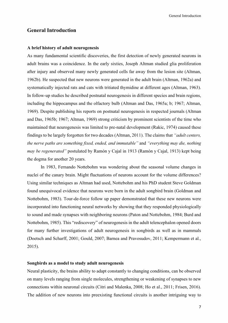
The song system
The neural substrate underlying hearing, producing and learning song consists of several song
nuclei in three interconnected pathways (Fig. 1). The motor pathway controls song production
and connects the pallial (“cortical” like) nucleus HVC (proper name) to the robust nucleus of
the archistriatum (RA). RA in turn connects to motor neurons in the tracheosyringeal portion
of the hypoglossal nucleus (nXIIts), which innervates the vocal organ, called syrinx (Nottebohm
et al, 1976). The auditory pathway processes auditory information entering the brain via the
ears, ascending through a vertebrate-canonical series of nuclei and regions and also connects
indirectly towards HVC and other pallial nuclei (Vates et al., 1996; Mandelblat-Cerf et al.,
2014; Murphy et al., 2017). The anterior forebrain pathway (AFP) enables song learning, song
maintenance and social context dependent modulation of song (Bottjer et al., 1984; Sohrabji et
al., 1990; Scharff and Nottebohm, 1991; Murugan et al., 2013; Kubikova et al., 2014; Woolley
and Kao, 2015; Kojima et al., 2018; Xiao et al., 2020). It forms a pallial-basal ganglia-thalamo-
pallial feedback loop and connects HVC and lateral magnocellular nucleus of the anterior
nidopallium (LMAN) to RA via striatal Area X and the medial dorsolateral nucleus of the
anterior thalamus (DLM). Area X receives dopaminergic innervation from the ventral tegmental
area (VTA) and the substantia nigra pars compacta (SNc, Lewis et al., 1981; Bottjer, 1993; Gale
et al., 2008).
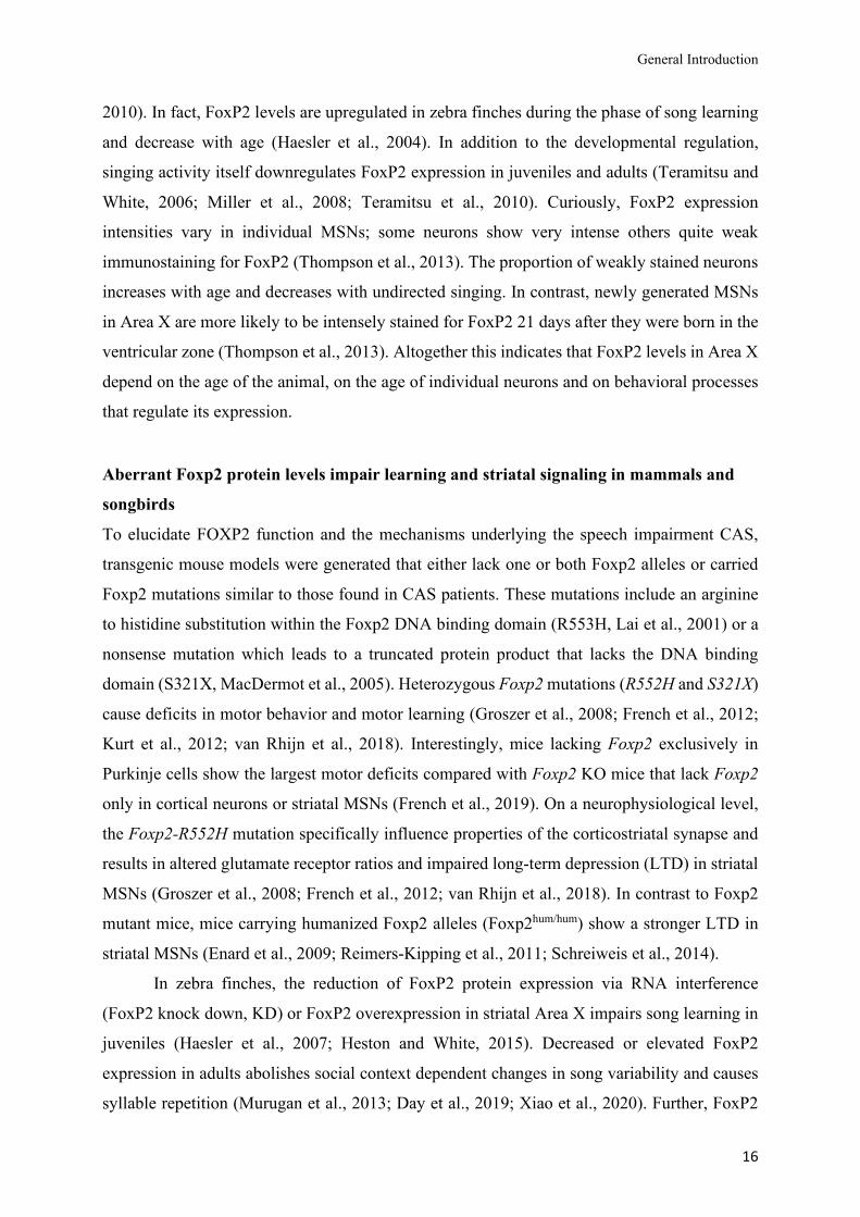
Figure 1. The song system
The song motor pathway (shown in black) controls the vocal organ (syrinx) via HVC >RA >nXIIts. The anterior
forebrain pathway (AFP, shown in blue) forms a pallial-basal ganglia-thalamic-pallial loop, connecting HVC
and RA via Area X>DLM>LMAN. Area X receives dopaminergic innervation from VTA and SNc (shown in
green).
General Introduction
10
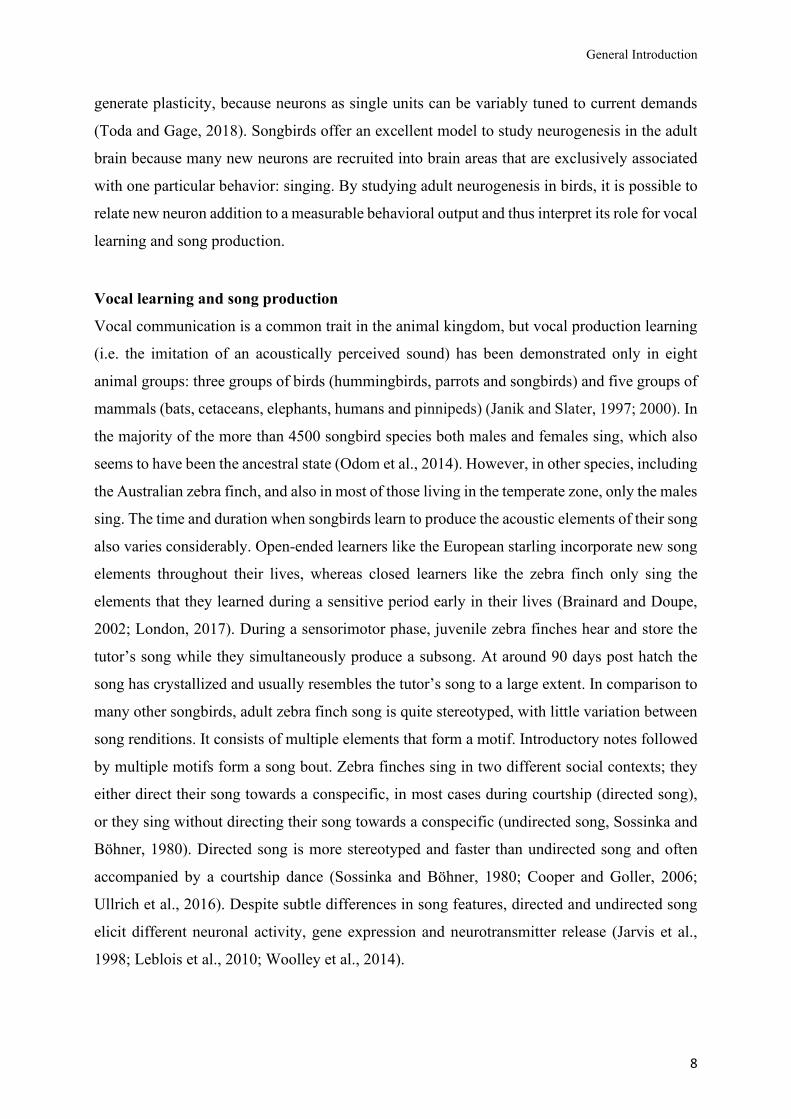
Adult neurogenesis in nuclei of the song system
In the adult songbird brain, new neurons are generated, migrate into the telencephalon and
incorporate into existing circuits (Alvarez-Buylla and Nottebohm, 1988). Proliferation hot spots
at the wall of the lateral ventricle give rise to new neurons that migrate along radial glia cells
into the parenchyma (Alvarez-Buylla and Nottebohm, 1988; Alvarez-Buylla et al., 1988b;
1990). In the song system, pallial HVC, the caudomedial nidopallium (NCM, part of the
auditory pathway) and striatal Area X receive new neurons (Alvarez-Buylla and Nottebohm,
1988; Nordeen and Nordeen, 1988a, Fig. 2). First, I will expand on adult neurogenesis in HVC,
then on adult neurogenesis Area X. HVC contains two types of projection neurons: HVCRA
neurons are part motor pathway and project to RA whereas HVCX neurons send their axons to
Area X (Nottebohm et al., 1976). Only HVCRA neurons are generated postnatally and in
adulthood, in contrast to HVCX neurons, that are mainly generated in ovo (Alvarez-Buylla et
al., 1988a; Nordeen and Nordeen, 1988b; Scotto-Lomassese et al., 2007). The specific ablation
of either HVCRA or HVCX neurons leads to an increased recruitment of only HVCRA neurons,
indicating that cell death of HVCX neurons does not induce their recruitment (Scharff et al.,
2000). New HVC neurons can be detected as early as one week after their birth and their
connection to RA is robustly established between 22 and 31 days after their birth (Burek et al.,
1994; Kirn et al., 1999; Tokarev et al., 2015). They respond to auditory stimuli (Paton and
Nottebohm, 1984) and robustly express immediate early genes after singing as early as three
weeks after they were born, indicating that they are firing during singing (Tokarev et al., 2015).
New neuron recruitment in songbirds can either occur as a process of addition or replacement.
In the seasonally breeding canary, new neurons in HVC replace older ones that have died (Kirn
and Nottebohm, 1993). In the zebra finch, new HVC neurons are added to the existing circuitry,
resulting in a doubling of neuron density over time (Walton et al., 2012).
Age, experience during early development, social environment and behavior impact on
new neuron recruitment and/or survival in HVC. As zebra finches age, the rate of new neuron
addition declines in HVC but not in other parts of the song system (Wang et al., 2002; Pytte et
al., 2007). Since singing enhances new neuron survival in HVC of canaries (Li et al., 2000;
Alvarez-Borda and Nottebohm, 2002), it might be possible that decreased singing rates in aged
zebra finches cause a decline of adult neurogenesis in HVC. However, in Pytte et al. (2007),
there was no difference in motifs per song bout between young and old zebra finches but a
detailed analysis of singing rates across different ages might reveal a relationship between age-
dependent singing rates and the recruitment of new neurons into HVC.
General Introduction
11
Besides age, social environment influences adult neurogenesis in HVC. New neuron number in
HVC co-varies among related and even unrelated adult male zebra finches when they shared
the same nest (Hurley et al., 2008) and individuals that were held in a large mixed sex group
had more new neurons in HVC and Area X compared to zebra fiches that were held in pairs or
in isolation (Lipkind et al., 2002). On a cellular level, postsynaptic neural activity in RA was
shown to be crucial for the survival of new HVC neurons (Larson et al., 2013). Further, singing
induced brain-derived neurotrophic factor (BDNF) expression enhances the survival of new
neurons in HVC of canaries (Li et al., 2000) and its interaction with testosterone influences
proliferation and new neuron survival especially in seasonally breeding songbirds (Brenowitz,
2014). HVC receives input from the auditory pathway (Vates et al., 1996) and new neurons in
HVC respond to sound early during their maturation (Paton and Nottebohm, 1984). Auditory
deprivation caused by bilateral deafening of adult zebra finches decreases the total number of
new neurons in HVC indicating that auditory input is necessary for their survival (Wang et al.,
1999). Other studies found a positive or no effect of deafening on HVC neurogenesis in adults
(Hurley et al., 2008; Pytte et al., 2012), whereby social context and different composition of
nest mates may account for the differences between the studies. The quality of song structure
and rates of adult neurogenesis in HVC are connected; the magnitude of song deterioration after
paralysis of syringeal muscles and the rate of song recovery correlate positively with the number
of new neurons incorporated into HVC (Pytte et al., 2011). If song quality affects rates of new
neurons in HVC or vice versa awaits further investigation.
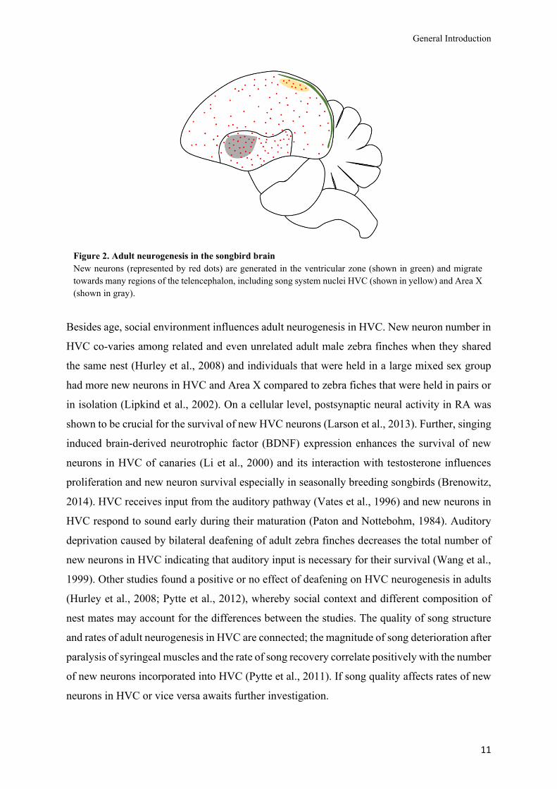
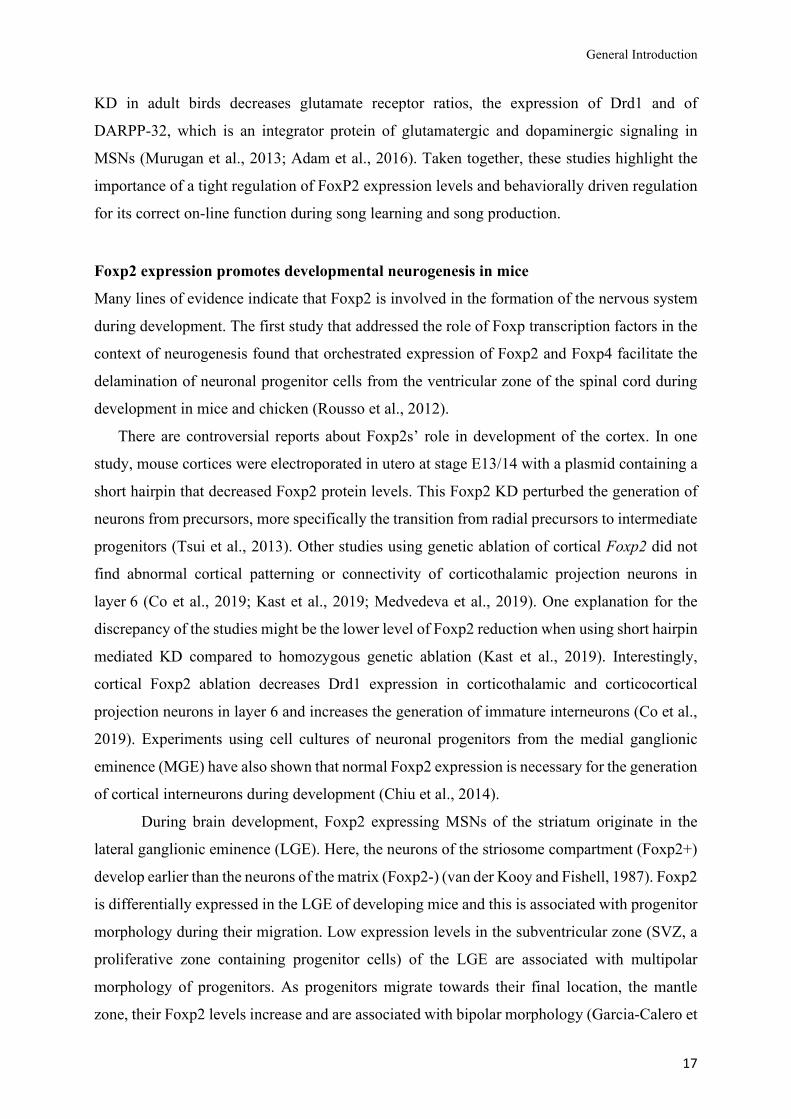
Figure 2. Adult neurogenesis in the songbird brain
New neurons (represented by red dots) are generated in the ventricular zone (shown in green) and migrate
towards many regions of the telencephalon, including song system nuclei HVC (shown in yellow) and Area X
(shown in gray).
General Introduction
12
The following paragraph will summarize the knowledge on adult neurogenesis in Area X, of
which much less is known compared to adult neurogenesis in HVC. Area X is formed from the
Islet1+ ventral striatal domain between 8 and 10 days after hatching and increases in volume
until 40 days after hatching (Nixdorf-Bergweiler, 1996; Garcia-Calero and Scharff, 2013).
Islet1 is a marker for the lateral ganglionic eminence where medium spiny neurons originate
during brain development in mammals (Stenman et al., 2003). Newly generated neurons in
Area X are also MSNs, which is the most abundant neuron type in the striatum of mammals
and birds (Freund et al., 1984; Farries and Perkel, 2000; Rochefort et al., 2007; Scott and Lois,
2007). New MSNs that migrate into Area X originate at the ventricular zone (VZ) at the wall
of the lateral ventricle adjacent to the striatum (Alvarez-Buylla et al., 1990; Scott and Lois,
2007). MSNs are characterized by a medium sized cell soma, spiny dendrites and distinct
electrophysiological and transcriptional profiles (Surmeier et al., 2007; Cepeda et al., 2008;
Gokce et al., 2016; Stanley et al., 2019).
MSNs in Area X receive glutamatergic (excitatory) innervation from HVC and LMAN
and dopaminergic innervation from VTA/SNc. Both glutamatergic and dopaminergic
projections converge onto dendritic spines of the same MSN (Bouyer et al., 1984; Kornfeld et
al., 2020). MSNs are GABAergic (inhibitory), they show sparse firing during singing and their
function is feed-forward inhibition of pallial signaling (Goldberg and Fee, 2010) . They do not
project out of Area X but innervate pallidal-like neurons (PNs) that project to thalamic DLM
(Farries and Perkel, 2002; Kornfeld et al., 2020). Two types PNs can be distinguished in
Area X; direct PNs that innervate DLM and indirect PNs that only innervate direct PNs (Farries
et al., 2005; Goldberg et al., 2010; Xiao et al., 2020, Figure 3). Based on electrophysiological
recordings, PNs have been proposed to resemble the mammalian external and internal segments
of the globus pallidus (Goldberg et al., 2010), that are innervated by different populations of
MSNs (Calabresi et al., 2014, Fig. 3). Gene expression profiles of single PNs in Area X,
however, contradict this hypothesis and show that PNs appear more similar to arkypallidal cells
of the external globus pallidus (Xiao et al., 2020) that do not project forward to neurons of the
subthalamic nucleus but project back to the striatum (Mallet et al., 2012; Abdi et al., 2015). The
existence of different MSN subtypes that exclusively innervate direct or indirect PNs in
songbirds has been proposed but was not yet proven by electrophysiological recordings (Gale
and Perkel, 2010; Pidoux et al., 2015). A recent study that analyzed the Area X transcriptome
distinguishes even five different MSNs clusters (Xiao et al., 2020). Differential gene expression
analysis delineated MSNs clusters that expressed classical markers of MSNs
General Introduction
13
that innervate the direct and indirect pathway of the mammalian basal ganglia (Xiao et al., 2020,
Fig. 3). In rodents, direct pathway MSNs express the dopamine receptor type 1 (Drd1) and the
forkhead box protein P2 (Foxp21), a transcription factor implicated in striatal function (Enard,
2011, and see next section). MSNs of the indirect pathway express the dopamine receptor type
2 (Drd2) but no or only little Foxp2 (Vernes et al., 2011; van Rhijn et al., 2018; Stanley et al.,
2019). In zebra finch Area X, FoxP2 expression is less segregated between MSN cluster; about
60% and 20% of the MSNs that correspond to the direct or indirect pathway express FoxP2,
respectively (Xiao et al., 2020).
New MSNs that migrate into Area X also express FoxP2 (Rochefort et al., 2007). FoxP2
downregulation in the SVZ decreased new MSN spine density in Area X and led to a small but
statistically not significant effect on the rate of recruited new MSNs during song development
in juveniles (Schulz et al., 2010). In mammals, Foxp2 is implicated in embryonic development
of the cortex and the striatum (Tsui et al., 2013; Chiu et al., 2014; Kast et al., 2019), but which
role FoxP2 plays for the process of adult neurogenesis in songbirds is not known.
1 FOXP2 refers to the human gene, Foxp2 refers to the mouse gene and FoxP2 refers to all other
species. FOXP2, Foxp2 and FoxP2 correspond to the protein product (Kaestner et al., 2000).
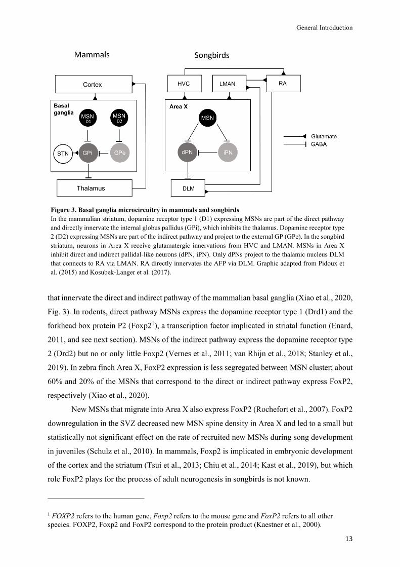
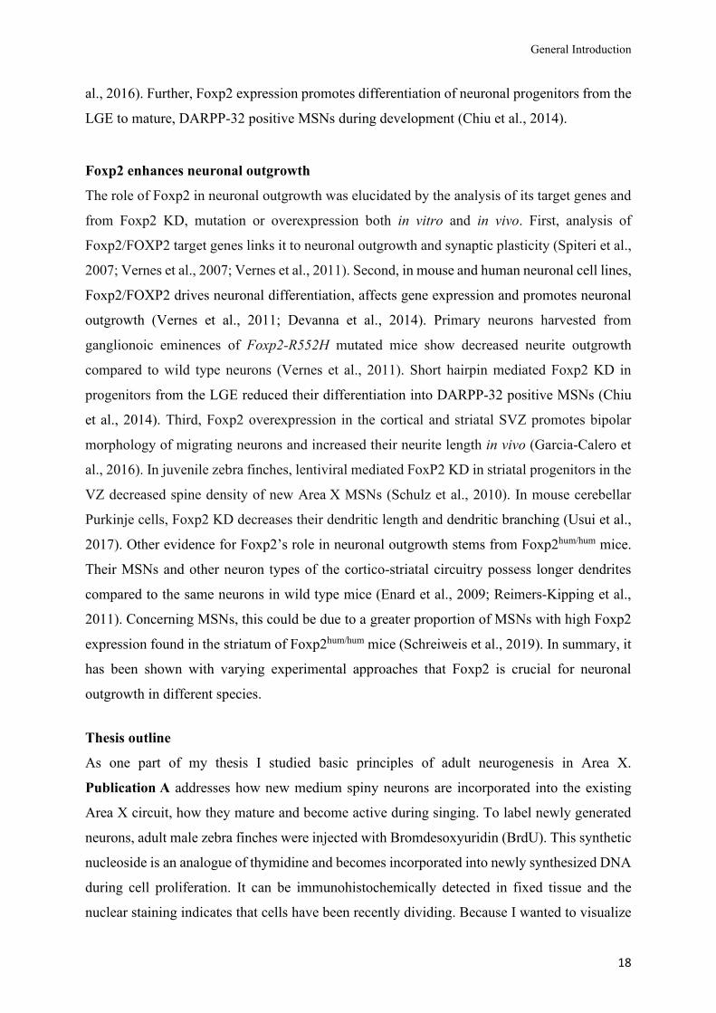
Figure 3. Basal ganglia microcircuitry in mammals and songbirds
In the mammalian striatum, dopamine receptor type 1 (D1) expressing MSNs are part of the direct pathway
and directly innervate the internal globus pallidus (GPi), which inhibits the thalamus. Dopamine receptor type
2 (D2) expressing MSNs are part of the indirect pathway and project to the external GP (GPe). In the songbird
striatum, neurons in Area X receive glutamatergic innervations from HVC and LMAN. MSNs in Area X
inhibit direct and indirect pallidal-like neurons (dPN, iPN). Only dPNs project to the thalamic nucleus DLM
that connects to RA via LMAN. RA directly innervates the AFP via DLM. Graphic adapted from Pidoux et
al. (2015) and Kosubek-Langer et al. (2017).

General Introduction
14
One striking difference between the process of adult neurogenesis in HVC and Area X is that
in the latter the rate of recruited neurons does not decrease with age (Pytte et al., 2007), which
is why I suspected that is has an ongoing function for song maintenance. To find out whether
new neurons in Area X are functionally integrated into the circuitry and behaviorally relevant,
in Publication A of this dissertation, I addressed the maturation course of new MSNs, their
participation in singing related activity and the question if their integration into Area X is a
process of replacement or addition.
General Introduction
15
FOXP2 – a transcription factor implicated in speech and language
Mutations of the transcription factor FOXP2 cause a severe speech and language impairment
in humans, called childhood apraxia of speech (CAS, Lai et al., 2001; MacDermot et al., 2005;
Morgan et al., 2016). Besides a mildly impaired language perception, affected individuals
mostly have difficulties performing fine orofacial movements that underlie speech production
(Vargha-Khadem et al., 1998). Functional imaging revealed that the brains of affected
individuals show structural and functional differences in cortical, cerebellar and basal ganglia
regions (Vargha-Khadem et al., 1998; Watkins et al., 2002; Liégeois et al., 2003; Liégeois et
al., 2016). Additional to its role in speech and language impairments, FOXP2 variants are
associated with attention deficit/ hyperactivity disorder and FOXP2 is a risk gene in autism
spectrum disorders (Demontis et al., 2019; Satterstrom et al., 2020). Considering the underlying
mechanism, both brain development and later brain function may be involved (Ehninger et al.,
2008). Consistent with the former, many of FOXP2’s target genes are linked to
neurodevelopmental disorders (Mukamel et al., 2011). Deciphering Foxp2 expression pattern
during brain development and adulthood is crucial to understand which cell types and neuronal
circuits are affected by Foxp2 variants or mutations. Foxp2 expression patterns in cortex,
cerebellum, thalamus and striatum are strongly conserved across reptiles, birds and mammals
(Ferland et al., 2003; Takahashi et al., 2003; Haesler et al., 2004; Takahashi et al., 2008;
Campbell et al., 2009; Rodenas-Cuadrado et al., 2018). In the cortices of mice, Foxp2 is
enriched in corticothalamic projection neurons but not in corticocortical projection neurons of
layer 6 (Kast et al., 2019). In cerebellar cortex, Foxp2 is expressed in Purkinje cells, which send
the main motor coordination output signals to deep cerebellar nuclei. In the striatum, Foxp2 is
expressed in MSNs of the striosome compartment but not in the ones of the matrix (Takahashi
et al., 2003; Takahashi et al., 2008; Chen et al., 2016). In the striosome, Foxp2 is enriched in
dopamine receptor 1 (Drd1) expressing MSNs that innervate the direct pathway of the cortico-
striatal-thalamic motor circuit (Vernes et al., 2011; van Rhijn et al., 2018, Fig. 3). Only a small
fraction of dopamine receptor 2 expressing (Drd2) MSNs of the indirect pathway express Foxp2
(van Rhijn et al., 2018; Stanley et al., 2019).
Given that the learned song in songbirds and speech in humans exhibit molecular, neural
and behavioral similarities (Fee and Scharff, 2010; Brainard and Doupe, 2013), it is interesting
that FoxP2 expression is low in pallial (“cortical”) regions of songbirds and high in striatum
and thalamus (Haesler et al., 2004). In Area X of the avian striatum, FoxP2 expression levels
are developmentally and behaviorally regulated. This is consistent with an important conserved
role for FOXP2/FoxP2 function in the basal ganglia of humans and songbirds (Bolhuis et al.,
General Introduction
16
2010). In fact, FoxP2 levels are upregulated in zebra finches during the phase of song learning
and decrease with age (Haesler et al., 2004). In addition to the developmental regulation,
singing activity itself downregulates FoxP2 expression in juveniles and adults (Teramitsu and
White, 2006; Miller et al., 2008; Teramitsu et al., 2010). Curiously, FoxP2 expression
intensities vary in individual MSNs; some neurons show very intense others quite weak
immunostaining for FoxP2 (Thompson et al., 2013). The proportion of weakly stained neurons
increases with age and decreases with undirected singing. In contrast, newly generated MSNs
in Area X are more likely to be intensely stained for FoxP2 21 days after they were born in the
ventricular zone (Thompson et al., 2013). Altogether this indicates that FoxP2 levels in Area X
depend on the age of the animal, on the age of individual neurons and on behavioral processes
that regulate its expression.
Aberrant Foxp2 protein levels impair learning and striatal signaling in mammals and
songbirds
To elucidate FOXP2 function and the mechanisms underlying the speech impairment CAS,
transgenic mouse models were generated that either lack one or both Foxp2 alleles or carried
Foxp2 mutations similar to those found in CAS patients. These mutations include an arginine
to histidine substitution within the Foxp2 DNA binding domain (R553H, Lai et al., 2001) or a
nonsense mutation which leads to a truncated protein product that lacks the DNA binding
domain (S321X, MacDermot et al., 2005). Heterozygous Foxp2 mutations (R552H and S321X)
cause deficits in motor behavior and motor learning (Groszer et al., 2008; French et al., 2012;
Kurt et al., 2012; van Rhijn et al., 2018). Interestingly, mice lacking Foxp2 exclusively in
Purkinje cells show the largest motor deficits compared with Foxp2 KO mice that lack Foxp2
only in cortical neurons or striatal MSNs (French et al., 2019). On a neurophysiological level,
the Foxp2-R552H mutation specifically influence properties of the corticostriatal synapse and
results in altered glutamate receptor ratios and impaired long-term depression (LTD) in striatal
MSNs (Groszer et al., 2008; French et al., 2012; van Rhijn et al., 2018). In contrast to Foxp2
mutant mice, mice carrying humanized Foxp2 alleles (Foxp2hum/hum) show a stronger LTD in
striatal MSNs (Enard et al., 2009; Reimers-Kipping et al., 2011; Schreiweis et al., 2014).
In zebra finches, the reduction of FoxP2 protein expression via RNA interference
(FoxP2 knock down, KD) or FoxP2 overexpression in striatal Area X impairs song learning in
juveniles (Haesler et al., 2007; Heston and White, 2015). Decreased or elevated FoxP2
expression in adults abolishes social context dependent changes in song variability and causes
syllable repetition (Murugan et al., 2013; Day et al., 2019; Xiao et al., 2020). Further, FoxP2
General Introduction
17
KD in adult birds decreases glutamate receptor ratios, the expression of Drd1 and of
DARPP-32, which is an integrator protein of glutamatergic and dopaminergic signaling in
MSNs (Murugan et al., 2013; Adam et al., 2016). Taken together, these studies highlight the
importance of a tight regulation of FoxP2 expression levels and behaviorally driven regulation
for its correct on-line function during song learning and song production.
Foxp2 expression promotes developmental neurogenesis in mice
Many lines of evidence indicate that Foxp2 is involved in the formation of the nervous system
during development. The first study that addressed the role of Foxp transcription factors in the
context of neurogenesis found that orchestrated expression of Foxp2 and Foxp4 facilitate the
delamination of neuronal progenitor cells from the ventricular zone of the spinal cord during
development in mice and chicken (Rousso et al., 2012).
There are controversial reports about Foxp2s’ role in development of the cortex. In one
study, mouse cortices were electroporated in utero at stage E13/14 with a plasmid containing a
short hairpin that decreased Foxp2 protein levels. This Foxp2 KD perturbed the generation of
neurons from precursors, more specifically the transition from radial precursors to intermediate
progenitors (Tsui et al., 2013). Other studies using genetic ablation of cortical Foxp2 did not
find abnormal cortical patterning or connectivity of corticothalamic projection neurons in
layer 6 (Co et al., 2019; Kast et al., 2019; Medvedeva et al., 2019). One explanation for the
discrepancy of the studies might be the lower level of Foxp2 reduction when using short hairpin
mediated KD compared to homozygous genetic ablation (Kast et al., 2019). Interestingly,
cortical Foxp2 ablation decreases Drd1 expression in corticothalamic and corticocortical
projection neurons in layer 6 and increases the generation of immature interneurons (Co et al.,
2019). Experiments using cell cultures of neuronal progenitors from the medial ganglionic
eminence (MGE) have also shown that normal Foxp2 expression is necessary for the generation
of cortical interneurons during development (Chiu et al., 2014).
During brain development, Foxp2 expressing MSNs of the striatum originate in the
lateral ganglionic eminence (LGE). Here, the neurons of the striosome compartment (Foxp2+)
develop earlier than the neurons of the matrix (Foxp2-) (van der Kooy and Fishell, 1987). Foxp2
is differentially expressed in the LGE of developing mice and this is associated with progenitor
morphology during their migration. Low expression levels in the subventricular zone (SVZ, a
proliferative zone containing progenitor cells) of the LGE are associated with multipolar
morphology of progenitors. As progenitors migrate towards their final location, the mantle
zone, their Foxp2 levels increase and are associated with bipolar morphology (Garcia-Calero et
General Introduction
18
al., 2016). Further, Foxp2 expression promotes differentiation of neuronal progenitors from the
LGE to mature, DARPP-32 positive MSNs during development (Chiu et al., 2014).
Foxp2 enhances neuronal outgrowth
The role of Foxp2 in neuronal outgrowth was elucidated by the analysis of its target genes and
from Foxp2 KD, mutation or overexpression both in vitro and in vivo. First, analysis of
Foxp2/FOXP2 target genes links it to neuronal outgrowth and synaptic plasticity (Spiteri et al.,
2007; Vernes et al., 2007; Vernes et al., 2011). Second, in mouse and human neuronal cell lines,
Foxp2/FOXP2 drives neuronal differentiation, affects gene expression and promotes neuronal
outgrowth (Vernes et al., 2011; Devanna et al., 2014). Primary neurons harvested from
ganglionoic eminences of Foxp2-R552H mutated mice show decreased neurite outgrowth
compared to wild type neurons (Vernes et al., 2011). Short hairpin mediated Foxp2 KD in
progenitors from the LGE reduced their differentiation into DARPP-32 positive MSNs (Chiu
et al., 2014). Third, Foxp2 overexpression in the cortical and striatal SVZ promotes bipolar
morphology of migrating neurons and increased their neurite length in vivo (Garcia-Calero et
al., 2016). In juvenile zebra finches, lentiviral mediated FoxP2 KD in striatal progenitors in the
VZ decreased spine density of new Area X MSNs (Schulz et al., 2010). In mouse cerebellar
Purkinje cells, Foxp2 KD decreases their dendritic length and dendritic branching (Usui et al.,
2017). Other evidence for Foxp2’s role in neuronal outgrowth stems from Foxp2hum/hum mice.
Their MSNs and other neuron types of the cortico-striatal circuitry possess longer dendrites
compared to the same neurons in wild type mice (Enard et al., 2009; Reimers-Kipping et al.,
2011). Concerning MSNs, this could be due to a greater proportion of MSNs with high Foxp2
expression found in the striatum of Foxp2hum/hum mice (Schreiweis et al., 2019). In summary, it
has been shown with varying experimental approaches that Foxp2 is crucial for neuronal
outgrowth in different species.
Thesis outline
As one part of my thesis I studied basic principles of adult neurogenesis in Area X.
Publication A addresses how new medium spiny neurons are incorporated into the existing
Area X circuit, how they mature and become active during singing. To label newly generated
neurons, adult male zebra finches were injected with Bromdesoxyuridin (BrdU). This synthetic
nucleoside is an analogue of thymidine and becomes incorporated into newly synthesized DNA
during cell proliferation. It can be immunohistochemically detected in fixed tissue and the
nuclear staining indicates that cells have been recently dividing. Because I wanted to visualize
General Introduction
19
not only the nucleus but entire new neurons, I applied a second approach and virally labelled
cells in the ventricular zone (VZ). Therefore, a lentiviral vector carrying the green fluorescent
protein reporter gene (GFP) was injected into the VZ adjacent to the lateral ventricle, where
new neurons destinated for Area X emerge (Alvarez-Buylla et al., 1990; Scott and Lois, 2007).
After sufficient survival time, GFP expressing new MSNs were analyzed in Area X. BrdU and
virally mediated labelling techniques were applied in the experiments underlying the datasets
presented in both publications. The results of Publication A show that new medium spiny
neurons in Area X show the same properties as older, resident MSNs. Once incorporated, they
are robustly active during singing behavior. Further, new neurons are constantly added to Area
X, leading to a more than doubling of the neuronal density in aged zebra finches.
The second part of this thesis especially addresses FoxP2’s role in the process of adult
neurogenesis in Area X. Previous studies indicated that Foxp2 is implicated in both
neurogenesis and neural outgrowth. It was known that new neurons in Area X expressed FoxP2
but why this might be so was unknown. In Publication B, I virally labelled progenitor cells and
later analyzed the decedents of these cells once they differentiated into new MSNs in Area X.
The results of Publication B show that new MSNs in Area X express dynamic FoxP2 levels in
an age dependent manner, which influence their morphology at specific timepoints during their
maturation.
Publication A
20
Publication A
Maturation, Behavioral Activation, and Connectivity of Adult-Born Medium Spiny
Neurons in a Striatal Song Nucleus
ORIGINAL RESEARCHpublished: 07 June 2017
doi: 10.3389/fnins.2017.00323
Frontiers in Neuroscience | www.frontiersin.org 21 June 2017 | Volume 11 | Article 323
Edited by:
Irmgard Amrein,
University of Zurich, Switzerland
Reviewed by:
Tom V. Smulders,
Newcastle University, United Kingdom
Antonia Marin-Burgin,
IBioBA-CONICET-Max Planck Society
Partner, Argentina
*Correspondence:
Jennifer Kosubek-Langer
Specialty section:
This article was submitted to
Neurogenesis,
a section of the journal
Frontiers in Neuroscience
Received: 24 March 2017
Accepted: 23 May 2017
Published: 07 June 2017
Citation:
Kosubek-Langer J, Schulze L and
Scharff C (2017) Maturation,
Behavioral Activation, and
Connectivity of Adult-Born Medium
Spiny Neurons in a Striatal Song
Nucleus. Front. Neurosci. 11:323.
doi: 10.3389/fnins.2017.00323
Maturation, Behavioral Activation,and Connectivity of Adult-BornMedium Spiny Neurons in a StriatalSong NucleusJennifer Kosubek-Langer *, Lydia Schulze and Constance Scharff
Animal Behavior, Freie Universität Berlin, Berlin, Germany
Neurogenesis continues in the adult songbird brain. Many telencephalic song control
regions incorporate new neurons into their existing circuits in adulthood. One song
nucleus that receives many new neurons is Area X. Because this striatal region is crucial
for song learning and song maintenance the recruitment of new neurons into Area
X could influence these processes. As an entry point into addressing this possibility,
we investigated the maturation and connectivity within the song circuit and behavioral
activation of newly generated Area X neurons. Using BrdU birth dating and virally
mediated GFP expression we followed adult-generated neurons from their place of
birth in the ventricle to their place of incorporation into Area X. We show that newborn
neurons receive glutamatergic input from pallial/cortical song nuclei. Additionally, backfills
revealed that the new neurons connect to pallidal-like projection neurons that innervate
the thalamus. Using in situ hybridization, we found that new neurons express the
mRNA for D1- and D2-type dopamine receptors. Employing DARPP-32 (dopamine and
cAMP-regulated phosphoprotein of 32 kDa) and EGR-1 (early growth response protein
1) as markers for neural maturation and activation, we established that at 42 days after
labeling approximately 80% of new neurons were mature medium spiny neurons (MSNs)
and could be activated by singing behavior. Finally, we compared theMSN density in Area
X of birds up to seven years of age and found a significant increase with age, indicating
that new neurons are constantly added to the nucleus. In summary, we provide evidence
that newborn MSNs in Area X constantly functionally integrate into the circuit and are
thus likely to play a role in the maintenance and regulation of adult song.
Keywords: adult neurogenesis, songbird, basal ganglia, Area X, EGR-1, DARPP-32, dopamine
INTRODUCTION
Adult neurogenesis is an enigmatic trait. Only some neurons continue to be generated in adulthoodwhereas the majority are born during development and persist throughout the animal’s life.Why these differences exist is still not known but much progress has been made elucidatingthe mechanism and function of adult neurogenesis during the past decades (Song et al., 2016).Neurons born in adulthood originate in regions adjacent to the ventricles that also give rise toneurons during development. From these neurogenic niches, neural precursors delaminate andthen migrate through the dense parenchyma, incorporate into functional circuits and influencebehavior (Paredes et al., 2016).
Kosubek-Langer et al. Striatal Neurogenesis in Songbirds
Considerable differences exist with respect to the extent ofadult neurogenesis in different species. As a rule of thumb,adult-born new neurons are recruited to many brain regions invertebrates like teleost fish, amphibians, and reptiles, whereasin birds the extent is still widespread but more restricted tothe forebrain (Kaslin et al., 2008). In mammals, there are evenfewer regions that continue to recruit new neurons in adulthood,principally the dentate gyrus (DG) of the hippocampal formation(Kempermann et al., 2015) and the olfactory bulb (Lim andAlvarez-Buylla, 2016). Interestingly, in rats, rabbits, monkeys andhumans but not in mice, adult-generated neurons have also beenobserved in the striatum (Bedard et al., 2002; Dayer et al., 2005;Tonchev et al., 2005; Luzzati et al., 2006; Ernst et al., 2014). Inthese cases, the newly generated neurons belong primarily to theclass of GABAergic interneurons, which constitute less than 5%of the striatal neurons (Tepper et al., 2010). The most abundantstriatal cell type are medium spiny projection neurons (MSNs)(Gerfen andWilson, 1996). In adult rodents, generation of MSNshas only been reported in response to experimentally inducedstroke, ischemia, or lesions (Arvidsson et al., 2002; Tattersfieldet al., 2004; Hou et al., 2008). In contrast, in songbirds adultMSNskeep immigrating in substantial numbers into the striatum undernatural conditions (Alvarez-Buylla et al., 1990). Striatal newbornneurons originate from the progenitor containing subpallialregion in the lateral ventricle that expresses the transcriptionfactors ISL-1/2, NKX2.1, and DLX but not TBR1 (Scott and Lois,2007). Of particular interest is the recruitment of MSNs into AreaX (Nordeen and Nordeen, 1988; Rochefort et al., 2007; Scottand Lois, 2007) a region unique to songbirds relevant for songplasticity in juveniles and adults (Sohrabji et al., 1990; Scharffand Nottebohm, 1991; Jarvis et al., 1998; Hessler and Doupe,1999; Woolley et al., 2014). In songbirds, new neurons destinedfor Area X migrate between 1,000 and 2,000µm to their finaldestination.
The dynamics of neural recruitment are best understood inthe DG and the olfactory bulb. In the former, new neuronsare added, whereas in the latter, they replace older neuronsthat undergo apoptosis (Crespo et al., 1986; Imayoshi et al.,2008). In both cases, the time it takes for new neurons toincorporate into preexisting circuits is similar (Deshpande et al.,2013). In songbirds, the dynamics of neural recruitment haveonly been studied in the pallial/cortical song control regionHVC (proper name, Figure 1A), where glutamatergic projectionneurons undergo neurogenesis (Kirn et al., 1999; Scott and Lois,2007; Tokarev et al., 2016).
To gain insight into the integration of GABAergic MSNs intoexisting circuits, we studied their differentiation, connectivityand activation by singing in Area X. To do so we tracednew neurons by injections of green fluorescent protein (GFP)-expressing lentivirus into the lateral wall of the lateral ventricleand with systemic injections of the cell birth marker 5-bromo-2′-deoxyuridine (BrdU). We also injected retrograde tracer into oneof the target regions of Area X, and used immuno- and in situ-histochemistry to characterize the new neurons. We report thatadult born MSNs receive glutamatergic and dopaminergic input,connect to pallidal-like projection neurons and are activatedduring singing like older, resident MSNs.
Because new HVC neurons seem to replace older ones incanaries (Kirn and Nottebohm, 1993), whereas in zebra finchesconstant neuronal addition was observed (Walton et al., 2012)we also addressed the issue of replacement vs. addition. Wequantified neuron numbers in adult zebra finches of varyingage and found that the density of MSNs in Area X increasedwith age, supporting the idea of neuron addition rather thanreplacement. Overall, our results suggest that Area X receives aconstant addition of functional new GABAergic MSNs.
MATERIALS AND METHODS
AnimalsAdult male zebra finches (Taeniopygia guttata) were bredand housed at the Department of Animal Behavior at FreieUniversität Berlin. The colony was kept under a 12:12 hlight:dark-cycle and food and water were available ad libitum.All procedures were reviewed and approved by the veterinarydepartment of the Freie Universität Berlin and by the ethicscommittee of the Regional Office for Health and Social AffairsBerlin (LAGeSo). The permit numbers are G0116/13 andG0296/15. In total, we used 53 adult male zebra finches. Forthe expression analysis of the early growth response protein1 (EGR-1) and the dopamine- and cAMP-regulated neuronalphosphoprotein (DARPP-32) in newborn cells we used 29 birds(age 462± 158 days, mean± standard deviation, SD). Dopamine(DA) receptor expression was studied in 5 birds (age 172 days ±13 days, mean± SD). Five birds received lentiviral injections (age367 days ± 109 days, mean ± SD). Density measures in Area Xwere performed in 14 birds (age ranging from 372 to 2,526 days).
BrdU InjectionsBirds for EGR-1 and DA receptor analysis received BrdU(50µg/g) via intramuscular injections in the mornings for 5consecutive days. Birds were assigned to three groups withdifferent survival times after BrdU injection (21, 31, and 42 days).We choose the first survival time to be 21 days, because BrdU+neurons in Area X were previously shown to express immediateearly genes after singing at that time (Tokarev et al., 2016).
Song MonitoringFor subsequent EGR-1 analysis, birds were kept in soundattenuated chambers for three nights and were perfused inthe morning of the 4th day 1.5 h after the lights went on.Vocalizations were continuously monitored via Sound AnalysisPro (Tchernichovski et al., 2000). During those 1.5 h birds had tosing at least 150 motifs to be included in the subsequent analysisof EGR-1 expression.
Birds that received lentiviral injections and retrograde tracerwere isolated in sound attenuated chambers for one night beforesacrifice. Birds were kept from singing by the experimenter sittingnearby for 1.5 h after lights went on in the morning and thenkilled. This was necessary because we used some of the brainsections in another experiment to be reported elsewhere.
Birds used for DA receptor analysis were decapitated withoutprevious songmonitoring and their brains were quickly dissected
Frontiers in Neuroscience | www.frontiersin.org 22 June 2017 | Volume 11 | Article 323
Kosubek-Langer et al. Striatal Neurogenesis in Songbirds
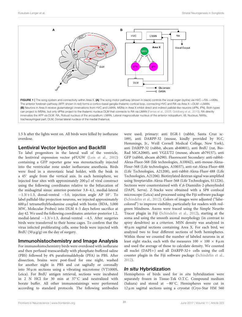
FIGURE 1 | The song system and connectivity within Area X. (A) The song motor pathway (shown in black) controls the vocal organ (syrinx) via HVC->RA->nXIIts.
The anterior forebrain pathway (AFP, shown in red) forms a cortico-basal ganglia-thalamic-cortical loop, connecting HVC and RA via Area X->DLM->LMAN.
(B) Neurons in Area X receive glutamatergic innervations from HVC and LMAN. MSNs in Area X inhibit direct and indirect pallidal-like neurons (dPN, iPN). Both types
can project to MSNs, but only dPNs project to the thalamic nucleus DLM that connects to RA via LMAN (Farries et al., 2005; Goldberg et al., 2010). RA directly
innervates the AFP via DLM. RA, Robust nucleus of the arcopallium; LMAN, Lateral magnocellular nucleus of the anterior nidopallium; XII, Nucleus; NXIIts,
tracheosyringeal part; DLM, Dorsal lateral nucleus of the medial thalamus.
1.5 h after the lights went on. All birds were killed by isofluraneoverdose.
Lentiviral Vector Injection and BackfillTo label progenitors in the lateral wall of the ventricle,the lentiviral expression vector pFUGW (Lois et al., 2002)containing a GFP reporter gene was stereotactically injectedinto the ventricular zone under isofluorane anesthesia. Birdswere fixed in a stereotaxic head holder, with the beak ina 45◦ angle from the vertical axis. In each hemisphere, weinjected four sites with approximately 200µl of viral constructusing the following coordinates relative to the bifurcation ofthe midsagittal sinus: anterior-posterior 3.8–4.1, medial-lateral−1.3/+1.3, dorsal-ventral −5.0, injection angle AP 10◦. Tolabel pallidal-like projection neurons, we injected approximately600µl tetramethylrhodamine coupled with biotin (BDA, 3,000MW, Molecular Probes) into DLM 4–5 days before sacrifice atday 42.We used the following coordinates: anterior-posterior 1.2,medial-lateral −1.3/+1.3, dorsal-ventral −4.5. After surgeriesbirds were transferred to their home cages. To confirm that thevirus infected proliferating cells, some birds were injected withBrdU (50µg/g) on the day of surgery.
Immunohistochemistry and Image AnalysisFor immunohistochemistry birds were overdosed with isofluraneand then perfused transcardially with phosphate-buffered saline(PBS) followed by 4% paraformaldehyde (PFA) in PBS. Afterdissection, brains were post-fixed for one night, washedfor another night in PBS and cut sagitally or coronallyinto 50µm sections using a vibrating microtome (VT1000S,Leica). For BrdU antigen retrieval, sections were incubatedin 2 N HCl for 30 min at 37◦C and neutralized withborate buffer. All other immunostainings were performedaccording to standard protocols. The following antibodies
were used; primary: anti EGR-1 (rabbit, Santa Cruz sc-189), anti DARPP-32 (mouse, kindly provided by H.C.Hemmings, Jr., Weill Cornell Medical College, New York),anti DARPP-32 (rabbit, abcam ab40801), anti BrdU (rat, Bio-Rad MCA2060), anti VGLUT2 (mouse, abcam ab79157), antiGFP (rabbit, abcam ab290). Fluorescent Secondary: anti-rabbit-Alexa-Fluor-568 (life technologies, A10042), anti-mouse-Alexa-Flour-568 (Life technologies, A10037), anti-rat-Alexa-Fluor-488(Life Technologies, A21208), anti-rabbit-Alexa-Fluor-488 (LifeTechnologies, A21206). Biotinylated dextran signal was amplifiedusing Streptavidin-Alexa-Fluor-568 (Life Technologies, S11226).Sections were counterstained with 4′,6-Diamidin-2-phenylindol(DAPI, Serva). Z-Stacks were obtained with a SP8 confocalmicroscope (Leica) and processed using the Fiji software package(Schindelin et al., 2012). Colors of images were adjusted (“false-colored”) to improve visibility, particularly for readers with red-green blindness. Axons were traced using the Simple NeuriteTracer plugin in Fiji (Schindelin et al., 2012), starting at thesoma and using the smooth axonal morphology (in contrast tospiny dendrites) as a criterion. MSN density was analyzed in40µm sagittal sections containing Area X. For each bird, weanalyzed two to four different sections of both hemispheres.Within those we counted the number of labeled neurons in atleast eight stacks, each with the measures 100 × 100 × 8µmand used the average of those to calculate density. We countedall nuclei (DAPI+) and all DARPP-32+ cells using the cellcounter plugin in the Fiji software package (Schindelin et al.,2012).
In situ HybridizationHemispheres of birds used for in situ hybridization wereseparately frozen in Tissue-Tek O.T.C. Compound medium(Sakura) and stored at −80◦C. Hemispheres were cut in12µm sagittal sections using a cryostat (Cryo-Star HM 560
Frontiers in Neuroscience | www.frontiersin.org 23 June 2017 | Volume 11 | Article 323
Kosubek-Langer et al. Striatal Neurogenesis in Songbirds
Cryostat, MICROM). Sections were fixed with 4% PFA for10 min and then acetylated with 0.25% acetic anhydride intriethanolamine for 10 min. Sections were rinsed in 2x insaline sodium citrate (SSC) buffer, dehydrated (75% EtOH,95% EtOH, and 100% EtOH, each for 2 min) and airdried. Sections were prehybridized for 1 h at 60◦C in ahybridization mix consisting of 50% deionized formamide, 5xSSC (pH 4.5), 2% blocking reagent (Roche, 11096176001) in1x maleic acid buffer, 2% sodium dodecyl sulfate, yeast tRNA(Invitrogen, 0.25 mg/ml), and heparin (Polysciences, 0.1 mg/ml).Sections were hybridized overnight with 1% digoxigenin orfluorescin labeled RNA probe in hybridization mix at 60◦C ina mineral oil bath. The next day, slides were rinsed twice withchloroform followed by 2x SSC and 1x SSC. A series of post-hybridization washes followed: 30 min in 1x SSC containing50% formamide at hybridization temperature (60◦C). Then,sections were washed once in 2x SSC and twice in 0.2x SSC20 min each at hybridization temperature. After the post-hybridization washing steps, sections were washed twice in 1xMABT (pH 7.5), consisting of 100 mM maleic acid, 150 mMNaCl and 0.1% Tween-20. Afterwards, sections were incubatedin 1x Roti-ImmunoBlock (Carl Roth) in 1x MABT for 30min, then with either alkaline phosphatase (AP)-conjugatedsheep anti-DIG antibody (Roche) or AP-conjugated sheep anti-fluorescein antibody (Roche), that were diluted 1:200 in 1x Roti-ImmunoBlock in 1x MABT. Slices were incubated overnightat 4◦C in a humidity chamber. After antibody incubation,slides were washed with 1x MABT 4 times for 5 min andequilibrated in alkaline phosphatase buffer NTMT, consistingof 100 mM NaCl, 100 mM Tris hydrochloride pH 9.5, 50 mMMgCl2 and 0.1% Tween-20 for 10 min. AP-labeled probes weredetected colorimetrically via the nitro blue tetrazolium/5-Bromo-4-chloro-3-indolyl phosphate substrate system (NBT/BCIP;Roche). NBT (final concentration: 337.5µg/ml) and BCIP (finalconcentration: 175µg/ml) were diluted in NTMT and sliceswere covered with this solution. Slices were incubated for 6–8 h, then fresh NBT/BCIP solution was added and sectionswere incubated overnight. The reaction was stopped by 10 minof incubation in a stop solution consisting of 10 mM Trishydrochloride pH 8.0 and 1 mM EDTA. Afterwards, slideswere washed three times with 1x PBS for 5 min. Sectionswere further used for immunohistochemical BrdU detection (seeImmunohistochemistry) and examined with a Zeiss Axiovert 200fluorescent microscope.
Analysis and StatisticsData were analyzed with the data analysis software R (RDevelopment Core Team, 2013) and GraphPad Prismversion 5.00 (GraphPad Software, San Diego CaliforniaUSA). Data for EGR-1, DARPP-32 and DA receptorexpression passed the D’Agostino’s K2 test for normaldistribution and were then evaluated with an analysis ofvariance (ANOVA) followed by a post hoc Tukey’s HonestlySignificant Difference test (HSD). To test the correlationbetween DARPP-32 density and age, we performed a linearregression analysis. Significance level was p < 0.05 for alltests.
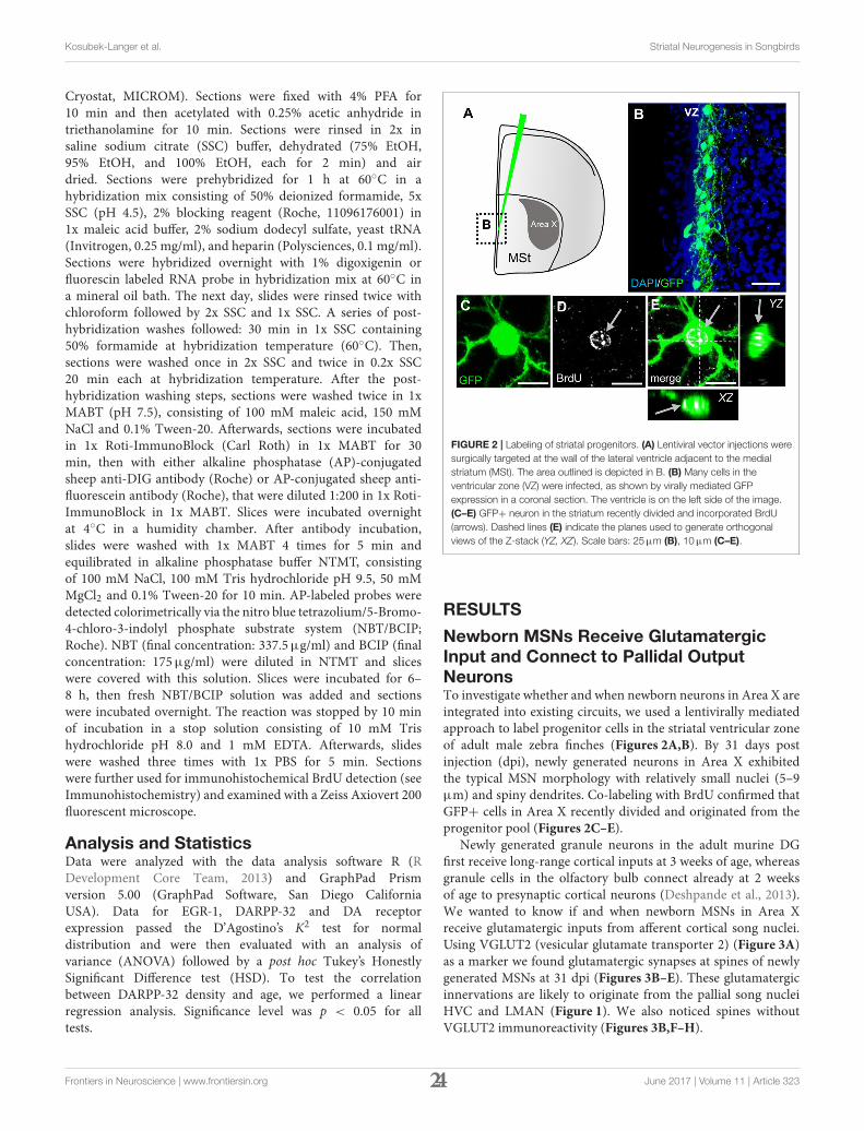
FIGURE 2 | Labeling of striatal progenitors. (A) Lentiviral vector injections were
surgically targeted at the wall of the lateral ventricle adjacent to the medial
striatum (MSt). The area outlined is depicted in B. (B) Many cells in the
ventricular zone (VZ) were infected, as shown by virally mediated GFP
expression in a coronal section. The ventricle is on the left side of the image.
(C–E) GFP+ neuron in the striatum recently divided and incorporated BrdU
(arrows). Dashed lines (E) indicate the planes used to generate orthogonal
views of the Z-stack (YZ, XZ). Scale bars: 25µm (B), 10µm (C–E).
RESULTS
Newborn MSNs Receive GlutamatergicInput and Connect to Pallidal OutputNeuronsTo investigate whether and when newborn neurons in Area X areintegrated into existing circuits, we used a lentivirally mediatedapproach to label progenitor cells in the striatal ventricular zoneof adult male zebra finches (Figures 2A,B). By 31 days postinjection (dpi), newly generated neurons in Area X exhibitedthe typical MSN morphology with relatively small nuclei (5–9µm) and spiny dendrites. Co-labeling with BrdU confirmed thatGFP+ cells in Area X recently divided and originated from theprogenitor pool (Figures 2C–E).
Newly generated granule neurons in the adult murine DGfirst receive long-range cortical inputs at 3 weeks of age, whereasgranule cells in the olfactory bulb connect already at 2 weeksof age to presynaptic cortical neurons (Deshpande et al., 2013).We wanted to know if and when newborn MSNs in Area Xreceive glutamatergic inputs from afferent cortical song nuclei.Using VGLUT2 (vesicular glutamate transporter 2) (Figure 3A)as a marker we found glutamatergic synapses at spines of newlygenerated MSNs at 31 dpi (Figures 3B–E). These glutamatergicinnervations are likely to originate from the pallial song nucleiHVC and LMAN (Figure 1). We also noticed spines withoutVGLUT2 immunoreactivity (Figures 3B,F–H).
Frontiers in Neuroscience | www.frontiersin.org 24 June 2017 | Volume 11 | Article 323
Kosubek-Langer et al. Striatal Neurogenesis in Songbirds
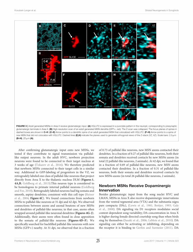
FIGURE 3 | Adult generated MSNs in Area X receive glutamatergic input. (A) VGLUT2 is expressed in a punctate pattern in the neuropil, corresponding to presynaptic
glutamatergic terminals in Area X. (B) High-resolution scan of an adult generated MSN dendrite (GFP+, red). The Z-scan was collapsed. The focus planes of spines in
dashed boxes are shown in C–H. (C–E) Arrow points to a dendritic spine of an adult generated MSN that colocalized with VGLUT2. (F–H) Arrow points to a spine of
new MSN that did not colocalize with VGLUT2. Dashed lines (E,H) indicate the planes used to generate orthogonal views of the Z-stack (YZ, XZ). Scale bars: 2.5µm
(B–H), 25µm (A).
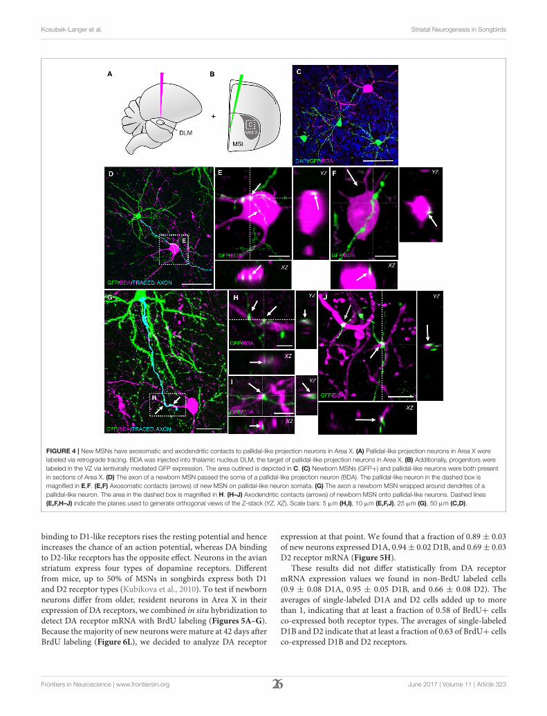
After confirming glutamatergic input onto new MSNs, wetested if they contribute to signal transmission via pallidal-like output neurons. In the adult HVC, newborn projectionneurons were found to be connected to their target nucleus at3 weeks of age (Tokarev et al., 2016). We therefore predictedthat newborn MSNs connected to their target cells in a similarway. Additional to GFP-labeling of progenitors in the VZ, weretrogradely labeled one class of pallidal-like neurons that projectdirectly from Area X to the thalamic nucleus DLM (Figures 1,4A,B; Goldberg et al., 2013).This neuron type is considered tobe homologous to primate internal pallidal neurons (Goldbergand Fee, 2010). Retrogradely labeled neurons had big somata andsmooth, aspiny dendrites; consistent with this cell type (Reineret al., 2004; Figure 4C). We found connections from newbornMSNs to pallidal-like neurons at 31 dpi and 42 dpi. We observedconnections between axons and axonal boutons of new MSNsand dendrites of pallidal-like neurons; in that case, axons oftenwrapped around pallidal-like neuronal dendrites (Figures 4G–J).Additionally, their axons were often found in close appositionto the somata of pallidal-like neurons (Figures 4D–F). Wespecifically searched for backfilled pallidal-like neurons with newMSNs (GFP+) nearby. At 31 dpi, we observed that in a fraction
of 0.73 of pallidal-like neurons, new MSN axons contacted theirdendrites. In a fraction of 0.27 of pallidal-like neurons, both theirsomata and dendrites received contacts by new MSNs axons (intotal 22 pallidal-like neurons, 2 animals). At 42 dpi, we found thatin a fraction of 0.69 of pallidal-like neurons, new MSN axonscontacted their dendrites. In a fraction of 0.31 of pallidal-likeneurons, both their somata and dendrites received contacts bynew MSNs axons (in total 26 pallidal-like neurons, 2 animals).
Newborn MSNs Receive DopaminergicInnervationBesides glutamatergic input from the song nuclei HVC andLMAN, MSNs in Area X also receive dopaminergic innervationsfrom the ventral tegmental area (VTA) and the substantia nigrapars compacta (SNc), (Lewis et al., 1981; Bottjer, 1993; Galeet al., 2008). DA signaling via D1 receptors modulates socialcontext dependent song variability; DA concentration in Area Xis higher during female directed courtship song than when birdssing by themselves (Sasaki et al., 2006; Leblois et al., 2010). DAsignaling can either be activating or inhibiting, depending onthe receptor it is binding to Gerfen and Surmeier (2011). DA
Frontiers in Neuroscience | www.frontiersin.org 25 June 2017 | Volume 11 | Article 323
Kosubek-Langer et al. Striatal Neurogenesis in Songbirds
FIGURE 4 | New MSNs have axosomatic and axodendritic contacts to pallidal-like projection neurons in Area X. (A) Pallidal-like projection neurons in Area X were
labeled via retrograde tracing. BDA was injected into thalamic nucleus DLM, the target of pallidal-like projection neurons in Area X. (B) Additionally, progenitors were
labeled in the VZ via lentivirally mediated GFP expression. The area outlined is depicted in C. (C) Newborn MSNs (GFP+) and pallidal-like neurons were both present
in sections of Area X. (D) The axon of a newborn MSN passed the soma of a pallidal-like projection neuron (BDA). The pallidal-like neuron in the dashed box is
magnified in E,F. (E,F) Axosomatic contacts (arrows) of new MSN on pallidal-like neuron somata. (G) The axon a newborn MSN wrapped around dendrites of a
pallidal-like neuron. The area in the dashed box is magnified in H. (H–J) Axodendritic contacts (arrows) of newborn MSN onto pallidal-like neurons. Dashed lines
(E,F,H–J) indicate the planes used to generate orthogonal views of the Z-stack (YZ, XZ). Scale bars: 5 µm (H,I), 10 µm (E,F,J), 25 µm (G), 50 µm (C,D).
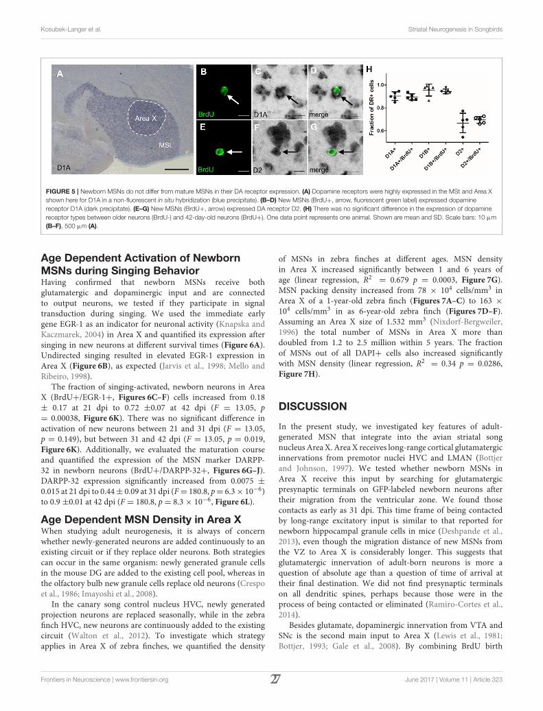
binding to D1-like receptors rises the resting potential and henceincreases the chance of an action potential, whereas DA bindingto D2-like receptors has the opposite effect. Neurons in the avianstriatum express four types of dopamine receptors. Differentfrom mice, up to 50% of MSNs in songbirds express both D1and D2 receptor types (Kubikova et al., 2010). To test if newbornneurons differ from older, resident neurons in Area X in theirexpression of DA receptors, we combined in situ hybridization todetect DA receptor mRNA with BrdU labeling (Figures 5A–G).Because the majority of new neurons were mature at 42 days afterBrdU labeling (Figure 6L), we decided to analyze DA receptor
expression at that point. We found that a fraction of 0.89 ± 0.03of new neurons expressed D1A, 0.94± 0.02 D1B, and 0.69± 0.03D2 receptor mRNA (Figure 5H).
These results did not differ statistically from DA receptormRNA expression values we found in non-BrdU labeled cells(0.9 ± 0.08 D1A, 0.95 ± 0.05 D1B, and 0.66 ± 0.08 D2). Theaverages of single-labeled D1A and D2 cells added up to morethan 1, indicating that at least a fraction of 0.58 of BrdU+ cellsco-expressed both receptor types. The averages of single-labeledD1B and D2 indicate that at least a fraction of 0.63 of BrdU+ cellsco-expressed D1B and D2 receptors.
Frontiers in Neuroscience | www.frontiersin.org 26 June 2017 | Volume 11 | Article 323
Kosubek-Langer et al. Striatal Neurogenesis in Songbirds
FIGURE 5 | Newborn MSNs do not differ from mature MSNs in their DA receptor expression. (A) Dopamine receptors were highly expressed in the MSt and Area X
shown here for D1A in a non-fluorescent in situ hybridization (blue precipitate). (B–D) New MSNs (BrdU+, arrow, fluorescent green label) expressed dopamine
receptor D1A (dark precipitate). (E–G) New MSNs (BrdU+, arrow) expressed DA receptor D2. (H) There was no significant difference in the expression of dopamine
receptor types between older neurons (BrdU-) and 42-day-old neurons (BrdU+). One data point represents one animal. Shown are mean and SD. Scale bars: 10 µm
(B–F), 500 µm (A).
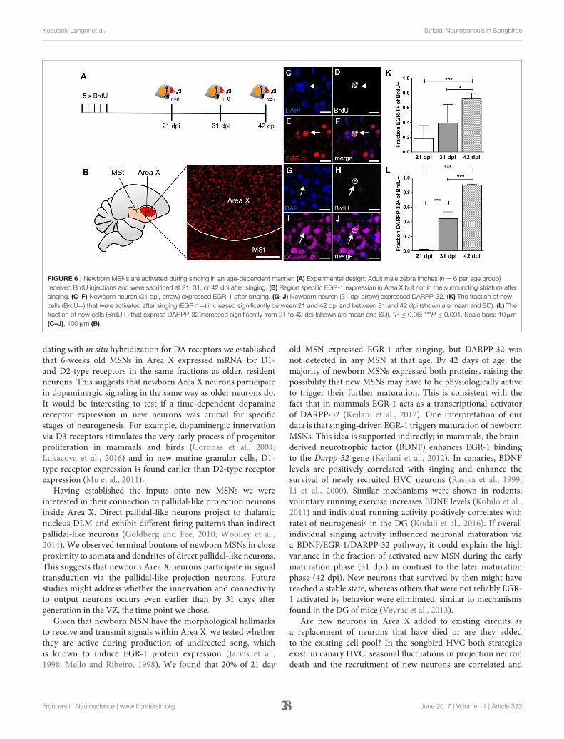
Age Dependent Activation of NewbornMSNs during Singing BehaviorHaving confirmed that newborn MSNs receive bothglutamatergic and dopaminergic input and are connectedto output neurons, we tested if they participate in signaltransduction during singing. We used the immediate earlygene EGR-1 as an indicator for neuronal activity (Knapska andKaczmarek, 2004) in Area X and quantified its expression aftersinging in new neurons at different survival times (Figure 6A).Undirected singing resulted in elevated EGR-1 expression inArea X (Figure 6B), as expected (Jarvis et al., 1998; Mello andRibeiro, 1998).
The fraction of singing-activated, newborn neurons in AreaX (BrdU+/EGR-1+, Figures 6C–F) cells increased from 0.18± 0.17 at 21 dpi to 0.72 ±0.07 at 42 dpi (F = 13.05, p= 0.00038, Figure 6K). There was no significant difference inactivation of new neurons between 21 and 31 dpi (F = 13.05,p = 0.149), but between 31 and 42 dpi (F = 13.05, p = 0.019,Figure 6K). Additionally, we evaluated the maturation courseand quantified the expression of the MSN marker DARPP-32 in newborn neurons (BrdU+/DARPP-32+, Figures 6G–J).DARPP-32 expression significantly increased from 0.0075 ±
0.015 at 21 dpi to 0.44± 0.09 at 31 dpi (F= 180.8, p= 6.3× 10⁻⁶)to 0.9±0.01 at 42 dpi (F = 180.8, p= 8.3× 10⁻⁶, Figure 6L).
Age Dependent MSN Density in Area XWhen studying adult neurogenesis, it is always of concernwhether newly-generated neurons are added continuously to anexisting circuit or if they replace older neurons. Both strategiescan occur in the same organism: newly generated granule cellsin the mouse DG are added to the existing cell pool, whereas inthe olfactory bulb new granule cells replace old neurons (Crespoet al., 1986; Imayoshi et al., 2008).
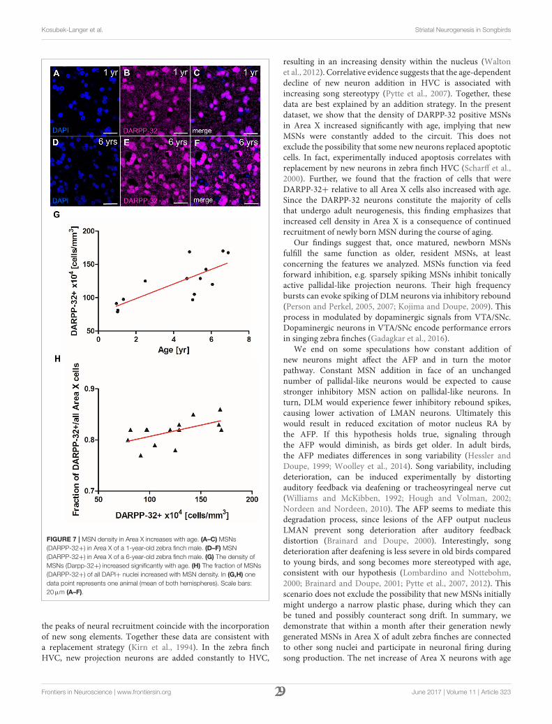
In the canary song control nucleus HVC, newly generatedprojection neurons are replaced seasonally, while in the zebrafinch HVC, new neurons are continuously added to the existingcircuit (Walton et al., 2012). To investigate which strategyapplies in Area X of zebra finches, we quantified the density
of MSNs in zebra finches at different ages. MSN densityin Area X increased significantly between 1 and 6 years ofage (linear regression, R2 = 0.679 p = 0.0003, Figure 7G).MSN packing density increased from 78 × 104 cells/mm3 inArea X of a 1-year-old zebra finch (Figures 7A–C) to 163 ×
104 cells/mm3 in as 6-year-old zebra finch (Figures 7D–F).Assuming an Area X size of 1.532 mm3 (Nixdorf-Bergweiler,1996) the total number of MSNs in Area X more thandoubled from 1.2 to 2.5 million within 5 years. The fractionof MSNs out of all DAPI+ cells also increased significantlywith MSN density (linear regression, R2 = 0.34 p = 0.0286,Figure 7H).
DISCUSSION
In the present study, we investigated key features of adult-generated MSN that integrate into the avian striatal songnucleus Area X. Area X receives long-range cortical glutamatergicinnervations from premotor nuclei HVC and LMAN (Bottjerand Johnson, 1997). We tested whether newborn MSNs inArea X receive this input by searching for glutamatergicpresynaptic terminals on GFP-labeled newborn neurons aftertheir migration from the ventricular zone. We found thosecontacts as early as 31 dpi. This time frame of being contactedby long-range excitatory input is similar to that reported fornewborn hippocampal granule cells in mice (Deshpande et al.,2013), even though the migration distance of new MSNs fromthe VZ to Area X is considerably longer. This suggests thatglutamatergic innervation of adult-born neurons is more aquestion of absolute age than a question of time of arrival attheir final destination. We did not find presynaptic terminalson all dendritic spines, perhaps because those were in theprocess of being contacted or eliminated (Ramiro-Cortes et al.,2014).
Besides glutamate, dopaminergic innervation from VTA andSNc is the second main input to Area X (Lewis et al., 1981;Bottjer, 1993; Gale et al., 2008). By combining BrdU birth
Frontiers in Neuroscience | www.frontiersin.org 27 June 2017 | Volume 11 | Article 323
Kosubek-Langer et al. Striatal Neurogenesis in Songbirds
FIGURE 6 | Newborn MSNs are activated during singing in an age-dependent manner. (A) Experimental design: Adult male zebra finches (n = 6 per age group)
received BrdU injections and were sacrificed at 21, 31, or 42 dpi after singing. (B) Region specific EGR-1 expression in Area X but not in the surrounding striatum after
singing. (C–F) Newborn neuron (31 dpi, arrow) expressed EGR-1 after singing. (G–J) Newborn neuron (31 dpi arrow) expressed DARPP-32. (K) The fraction of new
cells (BrdU+) that were activated after singing (EGR-1+) increased significantly between 21 and 42 dpi and between 31 and 42 dpi (shown are mean and SD). (L) The
fraction of new cells (BrdU+) that express DARPP-32 increased significantly from 21 to 42 dpi (shown are mean and SD). *P ≤ 0.05; ***P ≤ 0.001. Scale bars: 10µm
(C–J), 100µm (B).
dating with in situ hybridization for DA receptors we establishedthat 6-weeks old MSNs in Area X expressed mRNA for D1-and D2-type receptors in the same fractions as older, residentneurons. This suggests that newborn Area X neurons participatein dopaminergic signaling in the same way as older neurons do.It would be interesting to test if a time-dependent dopaminereceptor expression in new neurons was crucial for specificstages of neurogenesis. For example, dopaminergic innervationvia D3 receptors stimulates the very early process of progenitorproliferation in mammals and birds (Coronas et al., 2004;Lukacova et al., 2016) and in new murine granular cells, D1-type receptor expression is found earlier than D2-type receptorexpression (Mu et al., 2011).
Having established the inputs onto new MSNs we wereinterested in their connection to pallidal-like projection neuronsinside Area X. Direct pallidal-like neurons project to thalamicnucleus DLM and exhibit different firing patterns than indirectpallidal-like neurons (Goldberg and Fee, 2010; Woolley et al.,2014). We observed terminal boutons of newborn MSNs in closeproximity to somata and dendrites of direct pallidal-like neurons.This suggests that newborn Area X neurons participate in signaltransduction via the pallidal-like projection neurons. Futurestudies might address whether the innervation and connectivityto output neurons occurs even earlier than by 31 days aftergeneration in the VZ, the time point we chose.
Given that newborn MSN have the morphological hallmarksto receive and transmit signals within Area X, we tested whetherthey are active during production of undirected song, whichis known to induce EGR-1 protein expression (Jarvis et al.,1998; Mello and Ribeiro, 1998). We found that 20% of 21 day
old MSN expressed EGR-1 after singing, but DARPP-32 wasnot detected in any MSN at that age. By 42 days of age, themajority of newborn MSNs expressed both proteins, raising thepossibility that new MSNs may have to be physiologically activeto trigger their further maturation. This is consistent with thefact that in mammals EGR-1 acts as a transcriptional activatorof DARPP-32 (Keilani et al., 2012). One interpretation of ourdata is that singing-driven EGR-1 triggersmaturation of newbornMSNs. This idea is supported indirectly; in mammals, the brain-derived neurotrophic factor (BDNF) enhances EGR-1 bindingto the Darpp-32 gene (Keilani et al., 2012). In canaries, BDNFlevels are positively correlated with singing and enhance thesurvival of newly recruited HVC neurons (Rasika et al., 1999;Li et al., 2000). Similar mechanisms were shown in rodents;voluntary running exercise increases BDNF levels (Kobilo et al.,2011) and individual running activity positively correlates withrates of neurogenesis in the DG (Kodali et al., 2016). If overallindividual singing activity influenced neuronal maturation viaa BDNF/EGR-1/DARPP-32 pathway, it could explain the highvariance in the fraction of activated new MSN during the earlymaturation phase (31 dpi) in contrast to the later maturationphase (42 dpi). New neurons that survived by then might havereached a stable state, whereas others that were not reliably EGR-1 activated by behavior were eliminated, similar to mechanismsfound in the DG of mice (Veyrac et al., 2013).
Are new neurons in Area X added to existing circuits asa replacement of neurons that have died or are they addedto the existing cell pool? In the songbird HVC both strategiesexist: in canary HVC, seasonal fluctuations in projection neurondeath and the recruitment of new neurons are correlated and
Frontiers in Neuroscience | www.frontiersin.org 28 June 2017 | Volume 11 | Article 323
Kosubek-Langer et al. Striatal Neurogenesis in Songbirds
FIGURE 7 | MSN density in Area X increases with age. (A–C) MSNs
(DARPP-32+) in Area X of a 1-year-old zebra finch male. (D–F) MSN
(DARPP-32+) in Area X of a 6-year-old zebra finch male. (G) The density of
MSNs (Darpp-32+) increased significantly with age. (H) The fraction of MSNs
(DARPP-32+) of all DAPI+ nuclei increased with MSN density. In (G,H) one
data point represents one animal (mean of both hemispheres). Scale bars:
20µm (A–F).
the peaks of neural recruitment coincide with the incorporationof new song elements. Together these data are consistent witha replacement strategy (Kirn et al., 1994). In the zebra finchHVC, new projection neurons are added constantly to HVC,
resulting in an increasing density within the nucleus (Waltonet al., 2012). Correlative evidence suggests that the age-dependentdecline of new neuron addition in HVC is associated withincreasing song stereotypy (Pytte et al., 2007). Together, thesedata are best explained by an addition strategy. In the presentdataset, we show that the density of DARPP-32 positive MSNsin Area X increased significantly with age, implying that newMSNs were constantly added to the circuit. This does notexclude the possibility that some new neurons replaced apoptoticcells. In fact, experimentally induced apoptosis correlates withreplacement by new neurons in zebra finch HVC (Scharff et al.,2000). Further, we found that the fraction of cells that wereDARPP-32+ relative to all Area X cells also increased with age.Since the DARPP-32 neurons constitute the majority of cellsthat undergo adult neurogenesis, this finding emphasizes thatincreased cell density in Area X is a consequence of continuedrecruitment of newly born MSN during the course of aging.
Our findings suggest that, once matured, newborn MSNsfulfill the same function as older, resident MSNs, at leastconcerning the features we analyzed. MSNs function via feedforward inhibition, e.g. sparsely spiking MSNs inhibit tonicallyactive pallidal-like projection neurons. Their high frequencybursts can evoke spiking of DLM neurons via inhibitory rebound(Person and Perkel, 2005, 2007; Kojima and Doupe, 2009). Thisprocess in modulated by dopaminergic signals from VTA/SNc.Dopaminergic neurons in VTA/SNc encode performance errorsin singing zebra finches (Gadagkar et al., 2016).
We end on some speculations how constant addition ofnew neurons might affect the AFP and in turn the motorpathway. Constant MSN addition in face of an unchangednumber of pallidal-like neurons would be expected to causestronger inhibitory MSN action on pallidal-like neurons. Inturn, DLM would experience fewer inhibitory rebound spikes,causing lower activation of LMAN neurons. Ultimately thiswould result in reduced excitation of motor nucleus RA bythe AFP. If this hypothesis holds true, signaling throughthe AFP would diminish, as birds get older. In adult birds,the AFP mediates differences in song variability (Hessler andDoupe, 1999; Woolley et al., 2014). Song variability, includingdeterioration, can be induced experimentally by distortingauditory feedback via deafening or tracheosyringeal nerve cut(Williams and McKibben, 1992; Hough and Volman, 2002;Nordeen and Nordeen, 2010). The AFP seems to mediate thisdegradation process, since lesions of the AFP output nucleusLMAN prevent song deterioration after auditory feedbackdistortion (Brainard and Doupe, 2000). Interestingly, songdeterioration after deafening is less severe in old birds comparedto young birds, and song becomes more stereotyped with age,consistent with our hypothesis (Lombardino and Nottebohm,2000; Brainard and Doupe, 2001; Pytte et al., 2007, 2012). Thisscenario does not exclude the possibility that new MSNs initiallymight undergo a narrow plastic phase, during which they canbe tuned and possibly counteract song drift. In summary, wedemonstrate that within a month after their generation newlygenerated MSNs in Area X of adult zebra finches are connectedto other song nuclei and participate in neuronal firing duringsong production. The net increase of Area X neurons with age
Frontiers in Neuroscience | www.frontiersin.org 29 June 2017 | Volume 11 | Article 323
Kosubek-Langer et al. Striatal Neurogenesis in Songbirds
might provide a mechanism to achieve the equilibrium betweenplasticity and stereotypy needed to sustain adult song behavior.
ETHICS STATEMENT
This study was carried out in accordance with the governmentallaw (TierSchG). The protocol was approved by the LAGeSo,Berlin.
AUTHOR CONTRIBUTIONS
JK and CS planned experiments, JK and LS conductedexperiments, JK and CS wrote the manuscript.
FUNDING
JK was funded by the Elsa Neumann scholarship by the state ofBerlin.
REFERENCES
Alvarez-Buylla, A., Theelen, M., and Nottebohm, F. (1990). Proliferation “hot
spots” in adult avian ventricular zone reveal radial cell division. Neuron 5,
101–109.
Arvidsson, A., Collin, T., Kirik, D., Kokaia, Z., and Lindvall, O. (2002). Neuronal
replacement from endogenous precursors in the adult brain after stroke. Nat.
Med. 8, 963–970. doi: 10.1038/nm747
Bedard, A., Cossette, M., Levesque, M., and Parent, A. (2002). Proliferating
cells can differentiate into neurons in the striatum of normal adult
monkey. Neurosci. Lett. 328, 213–216. doi: 10.1016/S0304-3940(02)0
0530-X
Bottjer, S. W. (1993). The distribution of tyrosine hydroxylase immunoreactivity
in the brains of male and female zebra finches. J. Neurobiol. 24, 51–69.
doi: 10.1002/neu.480240105
Bottjer, S. W., and Johnson, F. (1997). Circuits, hormones, and learning: vocal
behavior in songbirds. J. Neurobiol. 33, 602–618.
Brainard, M. S., and Doupe, A. J. (2000). Interruption of a basal ganglia-forebrain
circuit prevents plasticity of learned vocalizations. Nature 404, 762–766.
doi: 10.1038/35008083
Brainard, M. S., and Doupe, A. J. (2001). Postlearning consolidation of birdsong:
stabilizing effects of age and anterior forebrain lesions. J Neurosci 21,
2501–2517.
Coronas, V., Bantubungi, K., Fombonne, J., Krantic, S., Schiffmann, S. N.,
and Roger, M. (2004). Dopamine D3 receptor stimulation promotes
the proliferation of cells derived from the post-natal subventricular
zone. J. Neurochem. 91, 1292–1301. doi: 10.1111/j.1471-4159.2004.0
2823.x
Crespo, D., Stanfield, B. B., and Cowan,W.M. (1986). Evidence that late-generated
granule cells do not simply replace earlier formed neurons in the rat dentate
gyrus. Exp. Brain Res. 62, 541–548.
Dayer, A. G., Cleaver, K. M., Abouantoun, T., and Cameron, H. A. (2005). New
GABAergic interneurons in the adult neocortex and striatum are generated
from different precursors. J. Cell Biol. 168, 415–427. doi: 10.1083/jcb.2004
07053
Deshpande, A., Bergami, M., Ghanem, A., Conzelmann, K. K., Lepier, A., Gotz,
M., et al. (2013). Retrograde monosynaptic tracing reveals the temporal
evolution of inputs onto new neurons in the adult dentate gyrus and olfactory
bulb. Proc. Natl. Acad. Sci. U.S.A. 110, E1152–1161. doi: 10.1073/pnas.12189
91110
Ernst, A., Alkass, K., Bernard, S., Salehpour, M., Perl, S., Tisdale, J., et al. (2014).
Neurogenesis in the striatum of the adult human brain. Cell 156, 1072–1083.
doi: 10.1016/j.cell.2014.01.044
Farries, M. A., Ding, L., and Perkel, D. J. (2005). Evidence for “direct” and
“indirect” pathways through the song system basal ganglia. J. Comp. Neurol.
484, 93–104. doi: 10.1002/cne.20464
Gadagkar, V., Puzerey, P. A., Chen, R., Baird-Daniel, E., Farhang, A. R., and
Goldberg, J. H. (2016). Dopamine neurons encode performance error in singing
birds. Science 354, 1278–1282. doi: 10.1126/science.aah6837
Gale, S. D., Person, A. L., and Perkel, D. J. (2008). A novel basal ganglia pathway
forms a loop linking a vocal learning circuit with its dopaminergic input. J.
Comp. Neurol. 508, 824–839. doi: 10.1002/cne.21700
Gerfen, C. R., and Surmeier, D. J. (2011). Modulation of striatal
projection systems by dopamine. Annu. Rev. Neurosci. 34, 441–466.
doi: 10.1146/annurev-neuro-061010-113641
Gerfen, C. R., and Wilson, C. J. (1996). “Chapter II: The basal ganglia,” in
Handbook of Chemical Neuroanatomy, eds A. B. L. W. Swanson and T. Hökfelt
(Amsterdam: Elsevier), 371–468.
Goldberg, J. H., Adler, A., Bergman, H., and Fee, M. S. (2010). Singing-
related neural activity distinguishes two putative pallidal cell types in the
songbird basal ganglia: comparison to the primate internal and external
pallidal segments. J. Neurosci. 30, 7088–7098. doi: 10.1523/JNEUROSCI.0168-1
0.2010
Goldberg, J. H., Farries, M. A., and Fee, M. S. (2013). Basal ganglia
output to the thalamus: still a paradox. Trends Neurosci. 36, 695–705.
doi: 10.1016/j.tins.2013.09.001
Goldberg, J. H., and Fee, M. S. (2010). Singing-related neural activity distinguishes
four classes of putative striatal neurons in the songbird basal ganglia. J.
Neurophysiol. 103, 2002–2014. doi: 10.1152/jn.01038.2009
Hessler, N. A., and Doupe, A. J. (1999). Singing-related neural activity in a
dorsal forebrain-basal ganglia circuit of adult zebra finches. J. Neurosci. 19,
10461–10481.
Hou, S. W., Wang, Y. Q., Xu, M., Shen, D. H., Wang, J. J., Huang, F.,
et al. (2008). Functional integration of newly generated neurons into
striatum after cerebral ischemia in the adult rat brain. Stroke 39, 2837–2844.
doi: 10.1161/STROKEAHA.107.510982
Hough, G. E. II, and Volman, S. F. (2002). Short-term and long-term effects
of vocal distortion on song maintenance in zebra finches. J. Neurosci. 22,
1177–1186.
Imayoshi, I., Sakamoto, M., Ohtsuka, T., Takao, K., Miyakawa, T., Yamaguchi,
M., et al. (2008). Roles of continuous neurogenesis in the structural and
functional integrity of the adult forebrain. Nat. Neurosci. 11, 1153–1161.
doi: 10.1038/nn.2185
Jarvis, E. D., Scharff, C., Grossman, M. R., Ramos, J. A., and Nottebohm, F.
(1998). For whom the bird sings: context-dependent gene expression. Neuron
21, 775–788.
Kaslin, J., Ganz, J., and Brand, M. (2008). Proliferation, neurogenesis and
regeneration in the non-mammalian vertebrate brain. Philos. Trans. R Soc.
Lond. B Biol. Sci. 363, 101–122. doi: 10.1098/rstb.2006.2015
Keilani, S., Chandwani, S., Dolios, G., Bogush, A., Beck, H., Hatzopoulos, A.
K., et al. (2012). Egr-1 induces DARPP-32 expression in striatal medium
spiny neurons via a conserved intragenic element. J. Neurosci. 32, 6808–6818.
doi: 10.1523/JNEUROSCI.5448-11.2012
Kempermann, G., Song, H., and Gage, F. H. (2015). Neurogenesis in
the Adult Hippocampus. Cold Spring Harb. Perspect. Biol. 7:a018812.
doi: 10.1101/cshperspect.a018812
Kirn, J., O’Loughlin, B., Kasparian, S., and Nottebohm, F. (1994). Cell death
and neuronal recruitment in the high vocal center of adult male canaries
are temporally related to changes in song. Proc. Natl. Acad. Sci. U.S.A. 91,
7844–7848.
Kirn, J. R., Fishman, Y., Sasportas, K., Alvarez-Buylla, A., and Nottebohm,
F. (1999). Fate of new neurons in adult canary high vocal center
during the first 30 days after their formation. J. Comp. Neurol. 411,
487–494.
Frontiers in Neuroscience | www.frontiersin.org 30 June 2017 | Volume 11 | Article 323
Kosubek-Langer et al. Striatal Neurogenesis in Songbirds
Kirn, J. R., and Nottebohm, F. (1993). Direct evidence for loss and
replacement of projection neurons in adult canary brain. J. Neurosci. 13,
1654–1663.
Knapska, E., and Kaczmarek, L. (2004). A gene for neuronal plasticity
in the mammalian brain: Zif268/Egr-1/NGFI-A/Krox-24/TIS8/ZENK? Prog.
Neurobiol. 74, 183–211. doi: 10.1016/j.pneurobio.2004.05.007
Kobilo, T., Liu, Q. R., Gandhi, K., Mughal, M., Shaham, Y., and van
Praag, H. (2011). Running is the neurogenic and neurotrophic stimulus in
environmental enrichment. Learn. Mem. 18, 605–609. doi: 10.1101/lm.22
83011
Kodali, M., Megahed, T., Mishra, V., Shuai, B., Hattiangady, B., and Shetty,
A. K. (2016). Voluntary running exercise-mediated enhanced neurogenesis
does not obliterate retrograde spatial memory. J. Neurosci. 36, 8112–8122.
doi: 10.1523/JNEUROSCI.0766-16.2016
Kojima, S., and Doupe, A. J. (2009). Activity propagation in an avian basal ganglia-
thalamocortical circuit essential for vocal learning. J. Neurosci. 29, 4782–4793.
doi: 10.1523/JNEUROSCI.4903-08.2009
Kubikova, L., Wada, K., and Jarvis, E. D. (2010). Dopamine receptors in a songbird
brain. J. Comp. Neurol. 518, 741–769. doi: 10.1002/cne.22255
Leblois, A., Wendel, B. J., and Perkel, D. J. (2010). Striatal
dopamine modulates basal ganglia output and regulates social
context-dependent behavioral variability through D1 receptors.
J. Neurosci. 30, 5730–5743. doi: 10.1523/JNEUROSCI.5974-0
9.2010
Lewis, J. W., Ryan, S. M., Arnold, A. P., and Butcher, L. L. (1981). Evidence for a
catecholaminergic projection to area X in the zebra finch. J. Comp. Neurol. 196,
347–354. doi: 10.1002/cne.901960212
Li, X. C., Jarvis, E. D., Alvarez-Borda, B., Lim, D. A., and Nottebohm,
F. (2000). A relationship between behavior, neurotrophin expression,
and new neuron survival. Proc. Natl. Acad. Sci. U.S.A. 97, 8584–8589.
doi: 10.1073/pnas.140222497
Lim, D. A., and Alvarez-Buylla, A. (2016). The adult ventricular-subventricular
zone (V-SVZ) and olfactory bulb (OB) neurogenesis. Cold Spring Harb.
Perspect. Biol. 8:pii: a018820. doi: 10.1101/cshperspect.a018820
Lois, C., Hong, E. J., Pease, S., Brown, E. J., and Baltimore, D. (2002). Germline
transmission and tissue-specific expression of transgenes delivered by lentiviral
vectors. Science 295, 868–872. doi: 10.1126/science.1067081
Lombardino, A. J., and Nottebohm, F. (2000). Age at deafening affects the stability
of learned song in adult male zebra finches. J. Neurosci. 20, 5054–5064.
Lukacova, K., Pavukova, E., Kostal, L., Bilcik, B., and Kubikova, L. (2016).
Dopamine D3 receptors modulate the rate of neuronal recovery, cell
recruitment in Area X, and song tempo after neurotoxic damage in songbirds.
Neuroscience 331, 158–168. doi: 10.1016/j.neuroscience.2016.06.032
Luzzati, F., De Marchis, S., Fasolo, A., and Peretto, P. (2006). Neurogenesis
in the caudate nucleus of the adult rabbit. J. Neurosci. 26, 609–621.
doi: 10.1523/JNEUROSCI.4371-05.2006
Mello, C. V., and Ribeiro, S. (1998). ZENK protein regulation by song in the brain
of songbirds. J. Comp. Neurol. 393, 426–438.
Mu, Y., Zhao, C., and Gage, F. H. (2011). Dopaminergic modulation of cortical
inputs during maturation of adult-born dentate granule cells. J. Neurosci. 31,
4113–4123. doi: 10.1523/JNEUROSCI.4913-10.2011
Nixdorf-Bergweiler, B. E. (1996). Divergent and parallel development
in volume sizes of telencephalic song nuclei in male
and female zebra finches. J. Comp. Neurol. 375, 445–456.
doi: 10.1002/(SICI)1096-9861(19961118)375:3<445::AID-CNE7>3.0.CO;2-2
Nordeen, K. W., and Nordeen, E. J. (1988). Projection neurons within a vocal
motor pathway are born during song learning in zebra finches. Nature 334,
149–151. doi: 10.1038/334149a0
Nordeen, K. W., and Nordeen, E. J. (2010). Deafening-induced vocal deterioration
in adult songbirds is reversed by disrupting a basal ganglia-forebrain circuit. J.
Neurosci. 30, 7392–7400. doi: 10.1523/JNEUROSCI.6181-09.2010
Paredes, M. F., Sorrells, S. F., Garcia-Verdugo, J. M., and Alvarez-Buylla, A. (2016).
Brain size and limits to adult neurogenesis. J. Comp. Neurol. 524, 646–664.
doi: 10.1002/cne.23896
Person, A. L., and Perkel, D. J. (2005). Unitary IPSPs drive precise
thalamic spiking in a circuit required for learning. Neuron 46, 129–140.
doi: 10.1016/j.neuron.2004.12.057
Person, A. L., and Perkel, D. J. (2007). Pallidal neuron activity increases during
sensory relay through thalamus in a songbird circuit essential for learning. J.
Neurosci. 27, 8687–8698. doi: 10.1523/JNEUROSCI.2045-07.2007
Pytte, C. L., George, S., Korman, S., David, E., Bogdan, D., and Kirn,
J. R. (2012). Adult neurogenesis is associated with the maintenance
of a stereotyped, learned motor behavior. J. Neurosci. 32, 7052–7057.
doi: 10.1523/JNEUROSCI.5385-11.2012
Pytte, C. L., Gerson, M., Miller, J., and Kirn, J. R. (2007). Increasing stereotypy in
adult zebra finch song correlates with a declining rate of adult neurogenesis.
Dev. Neurobiol. 67, 1699–1720. doi: 10.1002/dneu0.20520
Ramiro-Cortes, Y., Hobbiss, A. F., and Israely, I. (2014). Synaptic competition in
structural plasticity and cognitive function. Philos. Trans. R Soc. Lond. B. Biol.
Sci. 369:20130157. doi: 10.1098/rstb.2013.0157
Rasika, S., Alvarez-Buylla, A., and Nottebohm, F. (1999). BDNF mediates the
effects of testosterone on the survival of new neurons in an adult brain. Neuron
22, 53–62.
R Development Core Team (2013). R: A Language and Environment for Statistical
Computing. Vienna: R Foundation for Statistical Computing.
Reiner, A., Laverghetta, A. V., Meade, C. A., Cuthbertson, S. L., and Bottjer,
S. W. (2004). An immunohistochemical and pathway tracing study of the
striatopallidal organization of area X in the male zebra finch. J. Comp. Neurol.
469, 239–261. doi: 10.1002/cne.11012
Rochefort, C., He, X., Scotto-Lomassese, S., and Scharff, C. (2007). Recruitment
of FoxP2-expressing neurons to area X varies during song development. Dev.
Neurobiol. 67, 809–817. doi: 10.1002/dneu.20393
Sasaki, A., Sotnikova, T. D., Gainetdinov, R. R., and Jarvis, E. D. (2006). Social
context-dependent singing-regulated dopamine. J. Neurosci. 26, 9010–9014.
doi: 10.1523/JNEUROSCI.1335-06.2006
Scharff, C., Kirn, J. R., Grossman, M., Macklis, J. D., and Nottebohm, F. (2000).
Targeted neuronal death affects neuronal replacement and vocal behavior in
adult songbirds. Neuron 25, 481–492. doi: 10.1016/S0896-6273(00)80910-1
Scharff, C., and Nottebohm, F. (1991). A comparative-study of the behavioral
deficits following lesions of various parts of the zebra finch song system -
implications for vocal learning. J. Neurosci. 11, 2896–2913.
Schindelin, J., Arganda-Carreras, I., Frise, E., Kaynig, V., Longair, M., Pietzsch, T.,
et al. (2012). Fiji: an open-source platform for biological-image analysis. Nat.
Methods 9, 676–682. doi: 10.1038/nmeth.2019
Scott, B. B., and Lois, C. (2007). Developmental origin and identity of song
system neurons born during vocal learning in songbirds. J. Comp. Neurol. 502,
202–214. doi: 10.1002/cne.21296.
Sohrabji, F., Nordeen, E. J., and Nordeen, K. W. (1990). Selective impairment of
song learning following lesions of a forebrain nucleus in the juvenile zebra
finch. Behav. Neural Biol. 53, 51–63.
Song, J., Olsen, R. H. J., Sun, J., Ming, G. L., and Song, H. (2016). Neuronal
circuitry mechanisms regulating adult mammalian neurogenesis. Cold Spring
Harb. Perspect. Biol. 8. doi: 10.1101/cshperspect.a018937
Tattersfield, A. S., Croon, R. J., Liu, Y. W., Kells, A. P., Faull, R. L.,
and Connor, B. (2004). Neurogenesis in the striatum of the quinolinic
acid lesion model of Huntington’s disease. Neuroscience 127, 319–332.
doi: 10.1016/j.neuroscience.2004.04.061
Tchernichovski, O., Nottebohm, F., Ho, C. E., Pesaran, B., and Mitra, P. P. (2000).
A procedure for an automated measurement of song similarity. Anim. Behav.
59, 1167–1176. doi: 10.1006/anbe.1999.1416
Tepper, J. M., Tecuapetla, F., Koos, T., and Ibanez-Sandoval, O. (2010).
Heterogeneity and diversity of striatal GABAergic interneurons. Front.
Neuroanat. 4:150. doi: 10.3389/fnana.2010.00150
Tokarev, K., Boender, A. J., Claßen, G. A. E., and Scharff, C. (2016). Young, active
and well-connected: adult-born neurons in the zebra finch are activated during
singing. Brain Struct. Funct. 221, 1833–1843. doi: 10.1007/s00429-015-1006-y
Tonchev, A. B., Yamashima, T., Sawamoto, K., and Okano, H. (2005). Enhanced
proliferation of progenitor cells in the subventricular zone and limited neuronal
production in the striatum and neocortex of adult macaque monkeys after
global cerebral ischemia. J. Neurosci. Res. 81, 776–788. doi: 10.1002/jnr.20604
Veyrac, A., Gros, A., Bruel-Jungerman, E., Rochefort, C., Kleine Borgmann, F. B.,
Jessberger, S., et al. (2013). Zif268/egr1 gene controls the selection, maturation
and functional integration of adult hippocampal newborn neurons by learning.
Proc. Natl. Acad. Sci. U.S.A. 110, 7062–7067. doi: 10.1073/pnas.1220558110
Frontiers in Neuroscience | www.frontiersin.org 31 June 2017 | Volume 11 | Article 323
Kosubek-Langer et al. Striatal Neurogenesis in Songbirds
Walton, C., Pariser, E., and Nottebohm, F. (2012). The zebra finch paradox: song
is little changed, but number of neurons doubles. J. Neurosci. 32, 761–774.
doi: 10.1523/JNEUROSCI.3434-11.2012
Williams, H., and McKibben, J. R. (1992). Changes in stereotyped central motor
patterns controlling vocalization are induced by peripheral nerve injury. Behav.
Neural. Biol. 57, 67–78.
Woolley, S. C., Rajan, R., Joshua, M., and Doupe, A. J. (2014).
Emergence of context-dependent variability across a basal
ganglia network. Neuron 82, 208–223. doi: 10.1016/j.neuron.2014.
01.039
Conflict of Interest Statement: The authors declare that the research was
conducted in the absence of any commercial or financial relationships that could
be construed as a potential conflict of interest.
Copyright © 2017 Kosubek-Langer, Schulze and Scharff. This is an open-access article
distributed under the terms of the Creative Commons Attribution License (CC BY).
The use, distribution or reproduction in other forums is permitted, provided the
original author(s) or licensor are credited and that the original publication in this
journal is cited, in accordance with accepted academic practice. No use, distribution
or reproduction is permitted which does not comply with these terms.
Frontiers in Neuroscience | www.frontiersin.org 32 June 2017 | Volume 11 | Article 323

33

34
Publication B
Dynamic FoxP2 levels in male zebra finches are linked to morphology of adult-born Area X medium spiny neurons
35SCIENTIFIC REPORTS | (2020) 10:4787 | https://doi.org/10.1038/s41598-020-61740-6
www.nature.com/scientificreports
Dynamic FoxP2 levels in male zebra finches are linked to morphology of adult-born Area X medium spiny neuronsJennifer Kosubek-Langer* & Constance Scharff
The transcription factor FOXP2 is crucial for the formation and function of cortico-striatal circuits. FOXP2 mutations are associated with specific speech and language impairments. In songbirds, experimentally altered FoxP2 expression levels in the striatal song nucleus Area X impair vocal learning and song production. Overall FoxP2 protein levels in Area X are low in adult zebra finches and decrease further with singing. However, some Area X medium spiny neurons (MSNs) express FoxP2 at high levels (FoxP2high MSNs) and singing does not change this. Because Area X receives many new neurons throughout adulthood, we hypothesized that the FoxP2high MSNs are newly recruited neurons, not yet integrated into the local Area X circuitry and thus not active during singing. Contrary to our expectation, FoxP2 protein levels did not predict whether new MSNs were active during singing, assayed via immediate early gene expression. However, new FoxP2high MSNs had more complex dendrites, higher spine density and more mushroom spines than new FoxP2low MSNs. In addition, FoxP2 expression levels correlated positively with nucleus size of new MSNs. Together, our data suggest that dynamic FoxP2 levels in new MSNs shape their morphology during maturation and their incorporation into a neural circuit that enables the maintenance and social modulation of adult birdsong.
The forkhead box P2 transcription factor (FOXP2) is linked to speech and language disorders. Heterozygous FOXP2 mutations in humans affect both the coordination of fine orofacial movements and language percep-tion1–3. Because songbirds – like humans – need to learn most of their communicative vocalizations, they offer a unique model to study the role of FoxP2 (for nomenclature FOXP2/FoxP2 see Methods) for vocal learning and for the maintenance of learned vocalizations as adults4. Studying the relationship between FoxP2 and vocal learning in songbirds may inform the neurogenetic mechanism underlying the speech deficits in patients carrying FOXP2 mutations for the following reasons. The FoxP2 protein coding sequence is highly conserved between humans and songbirds as are the brain expression patterns, notably in the cerebellum and striatum5–7. Moreover, genetic manipulations of FoxP2 expression levels in the striatal song nucleus Area X during the critical phase of song learning lead to inaccurate and incomplete imitation of the tutor’s song and more variable vocal produc-tion3,8–10. This phenotype bears similarities to the specific speech deficits called developmental verbal dyspraxia, DVD (or childhood apraxia of speech), that patients carrying FOXP2 mutations suffer from. The core-phenotype of DVD consists of altered precision, consistency and sequencing of movements underlying speech in the absence of neuromuscular deficits11. In addition, altered FoxP2 levels in adult Area X affect the dopaminergic modulation of corticostriatal signaling important to song variability and affect song maintenance12,13, stressing the fact that tight regulation of FoxP2 expression is a prerequisite for correct neural transmission in differentiated neural cir-cuits. Additional effects of Foxp2 and its disruption on the embryonic development and the function of neural cir-cuits have been described in mice14–23. Further evidence for the biological relevance of tight regulation of FoxP2 expression levels comes from the following studies. FoxP2 expression levels in Area X transiently increase during song learning but are lower in adults7,24. Singing decreases overall FoxP2 levels in Area X but not in the surround-ing striatum and the degree of FoxP2 down regulation correlates with the amount of produced song25–27. This relationship is missing in deafened birds, pointing to an important role of auditory feedback for singing-driven FoxP2 down regulation27. How does singing affect FoxP2 expression at the cellular level? Medium spiny neurons
Department of Animal Behavior, Institute of Biology, Freie Universität Berlin, Berlin, Germany. *email: [email protected]
OPEN
36SCIENTIFIC REPORTS | (2020) 10:4787 | https://doi.org/10.1038/s41598-020-61740-6
www.nature.com/scientificreportswww.nature.com/scientificreports/
(MSNs), the most abundant cell type in the avian striatum, predominantly express FoxP2 at low levels (FoxP2low) while a subset expresses FoxP2 at very high levels (FoxP2high). Both subtypes are not equally affected by singing; the density of FoxP2high MSNs is not measurably different after singing, contrary to the decreasing density of FoxP2low MSNs24. The authors hypothesized that the difference might be due to the neuronal age. Adult Area X constantly receives new MSNs that originate at the ventricular zone28–33. FoxP2high MSNs colocalize more fre-quently with a marker for new neurons than FoxP2low MSNs24. Recently we showed that new MSNs mature and participate in singing activity – as measured by immediate early gene activation – within a timeframe of six weeks34. Whether FoxP2 influences not only the function but also the integration of new neurons into existing circuits is still an open question. Based on the results of Thompson et al. (ref. 21) we hypothesized that FoxP2high MSNs are newly recruited into Area X and need to become FoxP2low MSNs before they can participate in singing. To test this, we labelled neuronal progenitors in adult zebra finches. At different time points after these cells had migrated into Area X, we quantified their expression levels of FoxP2 and whether they also expressed the imme-diate early gene expression EGR-1 after singing.
In rodents, Foxp2 expression is associated with neurite outgrowth, neuronal morphology and synapse forma-tion in cortico-striatal circuits18,19,35–37. Foxp2 expression levels vary in striatal MNSs and these differences may be relevant for the morphology of striatal MSNs. Dopamine receptor 1 (D1) expressing MSNs38 express Foxp2 at higher levels than dopamine receptor 2 (D2) expressing MSNs23,35. These differences in Foxp2 levels may be linked to anatomical differences between D1 and D2 MSNs35,39. Furthermore, in mice carrying humanized Foxp2 alleles (Foxp2hum/hum mice), Foxp2high MSNs are more numerous in the dorsal striatum and their MSNs have longer dendrites than wildtype mice36,37. Based on the latter results we hypothesized that FoxP2 levels of new MSNs in adult songbirds correlate with their neural morphology. To test this, we virally labelled neural progen-itors and analyzed their FoxP2 expression, dendrite complexity and spine density after migration into Area X.
ResultsDynamic FoxP2 levels in new MSNs in Area X. To assess FoxP2 protein levels in individual newborn neurons we labelled progenitor cells with Bromodeoxyuridine (BrdU) and detected BrdU+/FoxP2+ cells after 21, 31 and 42 days post BrdU injection (dpi) in Area X of adult male zebra finches (Fig. 1a,b). We found that FoxP2 expression in Area X was very variable, with some neurons expressing FoxP2 at particularly high levels and some at low levels (Fig. 1c). At 21 dpi and at 31 dpi, the mean pixel intensities of all BrdU+/FoxP2+ cells formed a bimodal distribution, whereas at 42 dpi the distribution was unimodal and shifted to low FoxP2 expres-sion (Fig. 1d). We classified all neurons that had expression intensities within the top 30% of the measured pixel intensity distribution as FoxP2high neurons. Neurons within the bottom 30% of the measured pixel intensity dis-tribution were considered as FoxP2low. Because we were interested in the two extremes of the expression levels in this study, we did not analyze the new neurons with intermediate FoxP2 expression levels further (29.3% ± 9.4, SD, see Methods). At 21 dpi and at 31 dpi 36.17% ± 6.02 (SEM) and 34.91% ± 2.53 (SEM) percent of all BrdU+ cells were FoxP2high neurons, respectively. At 42 dpi the percentage of FoxP2high cells had significantly decreased to 12.95% ± 2.87 (SEM) on average (p = 0.0077, Kruskal-Wallis test, Fig. 1f,g). The percentage of FoxP2low neurons increased significantly from 21 dpi (34.59 ± 5.86, SEM) and 31 dpi (34.06 ± 3.35, SEM) to 42 dpi (59.19 ± 4.02, SEM, p = 0.013, ANOVA, Fig. 1e,g). We also noticed that BrdU+ cells varied in their nucleus size. Quantification revealed that the distribution of the nucleus size was shifted towards bigger nuclei at 31 dpi (data not shown). BrdU+/FoxP2+ cells at 31 dpi had significantly bigger nuclei (7.37 µm ± 0.94 (SD) than BrdU+/FoxP2+ cell at 21 dpi (6.88 µm ± 0.9 (SD), p = 0.00025, chi-square = 75.358, df = 2) or 42 dpi (6.74 µm ± 0.59 (SD), p = 2 × 10⁻⁶, chi-square = 75.358, df = 2, data not shown). Interestingly there was a significant positive relationship with a low effect size between nucleus size and FoxP2 expression levels in all three experimental groups (21 dpi: r2 = 0.086, p = 0.017, 31 dpi: r2 = 0.083, p = 5.2 × 10⁻⁷, 42 dpi = r2 = 0.039, p = 0.0012, Fig. 1h).
Singing induced activity of new MSNs is independent of FoxP2 levels. In adult zebra finches FoxP2 expression levels in Area X are behaviourally regulated. Undirected singing leads to downregulation of FoxP2 mRNA and protein24–26. Undirected singing is also associated with expression of the immediate early gene EGR-1 in Area X, which is therefore often used as a molecular readout of the neuronal activity associated with undirected song40–44. We hypothesized that FoxP2 levels in new FoxP2high neurons needed to be downregulated before activation by singing could occur, resulting in EGR-1 expression. Consequently, we did not expect to find BrdU+/FoxP2high/EGR-1+ MSNs in Area X. To test this, we analyzed BrdU+/EGR-1 ± /FoxP2+ cells in birds that had sung before sacrifice after 21 dpi, 31 dpi and 42 dpi.
Contrary to our hypothesis we found BrdU+/FoxP2high/EGR-1+ cells in Area X (Fig. 2a) in all groups, with large differences between birds. At 31 dpi, on average 41.45% ± 15.43 (SEM) of new neurons expressed FoxP2high and were also activated by singing (BrdU+/FoxP2high/EGR-1+) and 40.28% ± 13.34 (SEM) of the new neu-rons that expressed FoxP2high were not activated by singing (BrdU+/FoxP2high/EGR-1-) (Fig. 2b). At 42 dpi, the more birds had sung the fewer FoxP2high new neurons were found, resulting in a significant negative relation-ship between the number of BrdU+/FoxP2+/EGR-1+ neurons and the number of motifs sung before sacrifice (r2 = 0.753, p = 0.025, Fig. 2c) which was not the case at 21 dpi (r2 = 0.168, p = 0.493, data not shown) nor at 31 dpi (r2 = 0.164, p = 0.425, data not shown).
FoxP2 levels in new MSNs influence dendritic arborization and spine density. Because of the relationship of FoxP2 levels and nucleus size we wanted to further characterize the morphology of new neurons expressing different FoxP2 levels. We used a lentiviral approach to express Green Fluorescent Protein (GFP) in progenitor cells at their place of birth in the lateral wall of the lateral ventricle (Fig. 3a). After 31 and 42 dpi we found labeled neurons (GFP+) in Area X and the surrounding striatum (Fig. 3b). GFP+ neurons exhibited the typical morphology of medium spiny neurons with small somata and spiny dendrites (Fig. 3c) and expressed
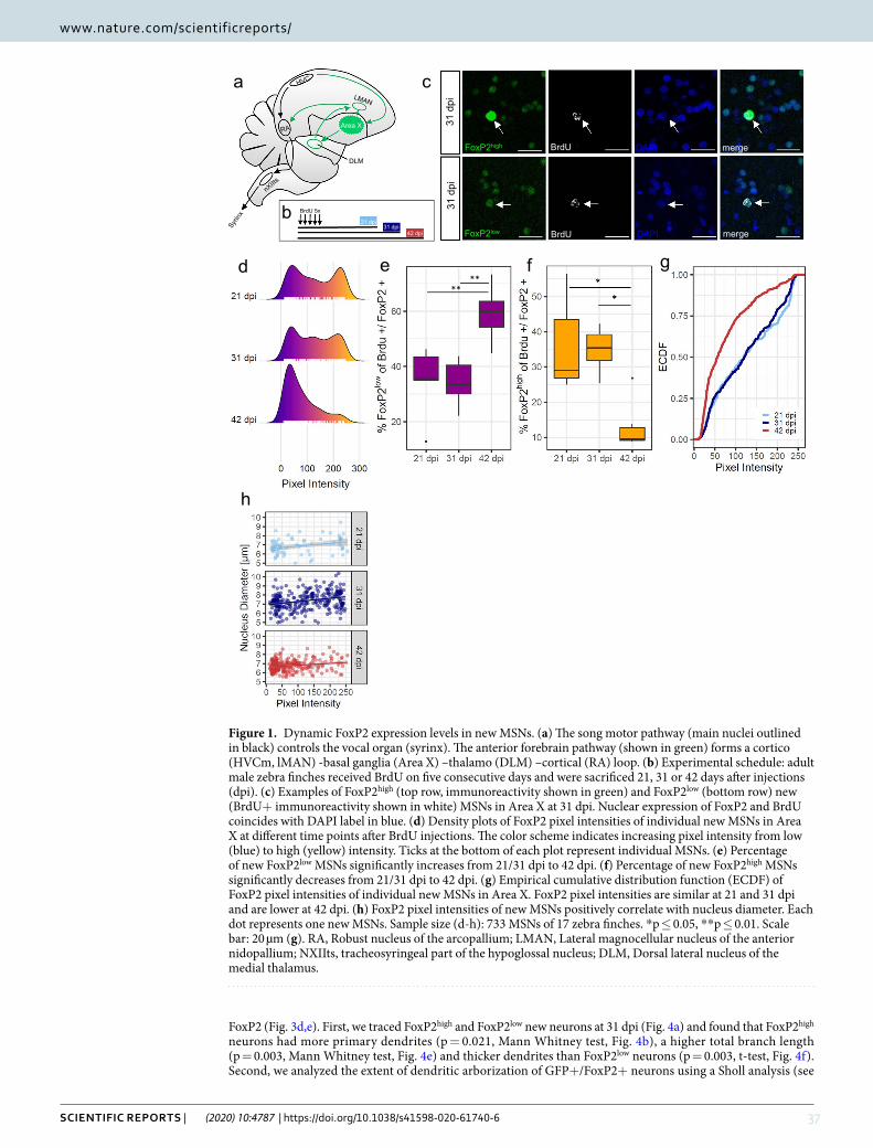
37SCIENTIFIC REPORTS | (2020) 10:4787 | https://doi.org/10.1038/s41598-020-61740-6
www.nature.com/scientificreportswww.nature.com/scientificreports/
FoxP2 (Fig. 3d,e). First, we traced FoxP2high and FoxP2low new neurons at 31 dpi (Fig. 4a) and found that FoxP2high neurons had more primary dendrites (p = 0.021, Mann Whitney test, Fig. 4b), a higher total branch length (p = 0.003, Mann Whitney test, Fig. 4e) and thicker dendrites than FoxP2low neurons (p = 0.003, t-test, Fig. 4f). Second, we analyzed the extent of dendritic arborization of GFP+/FoxP2+ neurons using a Sholl analysis (see
Figure 1. Dynamic FoxP2 expression levels in new MSNs. (a) The song motor pathway (main nuclei outlined in black) controls the vocal organ (syrinx). The anterior forebrain pathway (shown in green) forms a cortico (HVCm, lMAN) -basal ganglia (Area X) –thalamo (DLM) –cortical (RA) loop. (b) Experimental schedule: adult male zebra finches received BrdU on five consecutive days and were sacrificed 21, 31 or 42 days after injections (dpi). (c) Examples of FoxP2high (top row, immunoreactivity shown in green) and FoxP2low (bottom row) new (BrdU+ immunoreactivity shown in white) MSNs in Area X at 31 dpi. Nuclear expression of FoxP2 and BrdU coincides with DAPI label in blue. (d) Density plots of FoxP2 pixel intensities of individual new MSNs in Area X at different time points after BrdU injections. The color scheme indicates increasing pixel intensity from low (blue) to high (yellow) intensity. Ticks at the bottom of each plot represent individual MSNs. (e) Percentage of new FoxP2low MSNs significantly increases from 21/31 dpi to 42 dpi. (f) Percentage of new FoxP2high MSNs significantly decreases from 21/31 dpi to 42 dpi. (g) Empirical cumulative distribution function (ECDF) of FoxP2 pixel intensities of individual new MSNs in Area X. FoxP2 pixel intensities are similar at 21 and 31 dpi and are lower at 42 dpi. (h) FoxP2 pixel intensities of new MSNs positively correlate with nucleus diameter. Each dot represents one new MSNs. Sample size (d-h): 733 MSNs of 17 zebra finches. *p ≤ 0.05, **p ≤ 0.01. Scale bar: 20 µm (g). RA, Robust nucleus of the arcopallium; LMAN, Lateral magnocellular nucleus of the anterior nidopallium; NXIIts, tracheosyringeal part of the hypoglossal nucleus; DLM, Dorsal lateral nucleus of the medial thalamus.
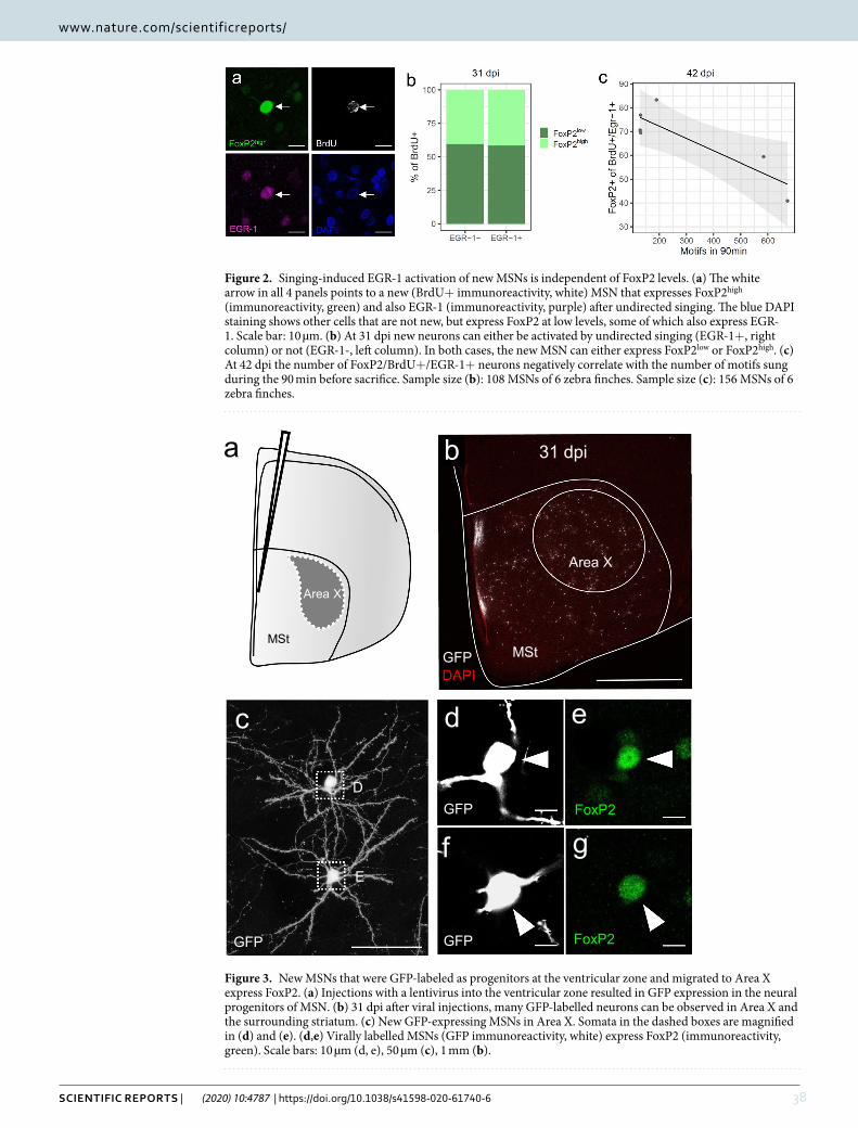
38SCIENTIFIC REPORTS | (2020) 10:4787 | https://doi.org/10.1038/s41598-020-61740-6
www.nature.com/scientificreportswww.nature.com/scientificreports/
Figure 2. Singing-induced EGR-1 activation of new MSNs is independent of FoxP2 levels. (a) The white arrow in all 4 panels points to a new (BrdU+ immunoreactivity, white) MSN that expresses FoxP2high (immunoreactivity, green) and also EGR-1 (immunoreactivity, purple) after undirected singing. The blue DAPI staining shows other cells that are not new, but express FoxP2 at low levels, some of which also express EGR-1. Scale bar: 10 µm. (b) At 31 dpi new neurons can either be activated by undirected singing (EGR-1+, right column) or not (EGR-1-, left column). In both cases, the new MSN can either express FoxP2low or FoxP2high. (c) At 42 dpi the number of FoxP2/BrdU+/EGR-1+ neurons negatively correlate with the number of motifs sung during the 90 min before sacrifice. Sample size (b): 108 MSNs of 6 zebra finches. Sample size (c): 156 MSNs of 6 zebra finches.
Figure 3. New MSNs that were GFP-labeled as progenitors at the ventricular zone and migrated to Area X express FoxP2. (a) Injections with a lentivirus into the ventricular zone resulted in GFP expression in the neural progenitors of MSN. (b) 31 dpi after viral injections, many GFP-labelled neurons can be observed in Area X and the surrounding striatum. (c) New GFP-expressing MSNs in Area X. Somata in the dashed boxes are magnified in (d) and (e). (d,e) Virally labelled MSNs (GFP immunoreactivity, white) express FoxP2 (immunoreactivity, green). Scale bars: 10 µm (d, e), 50 µm (c), 1 mm (b).
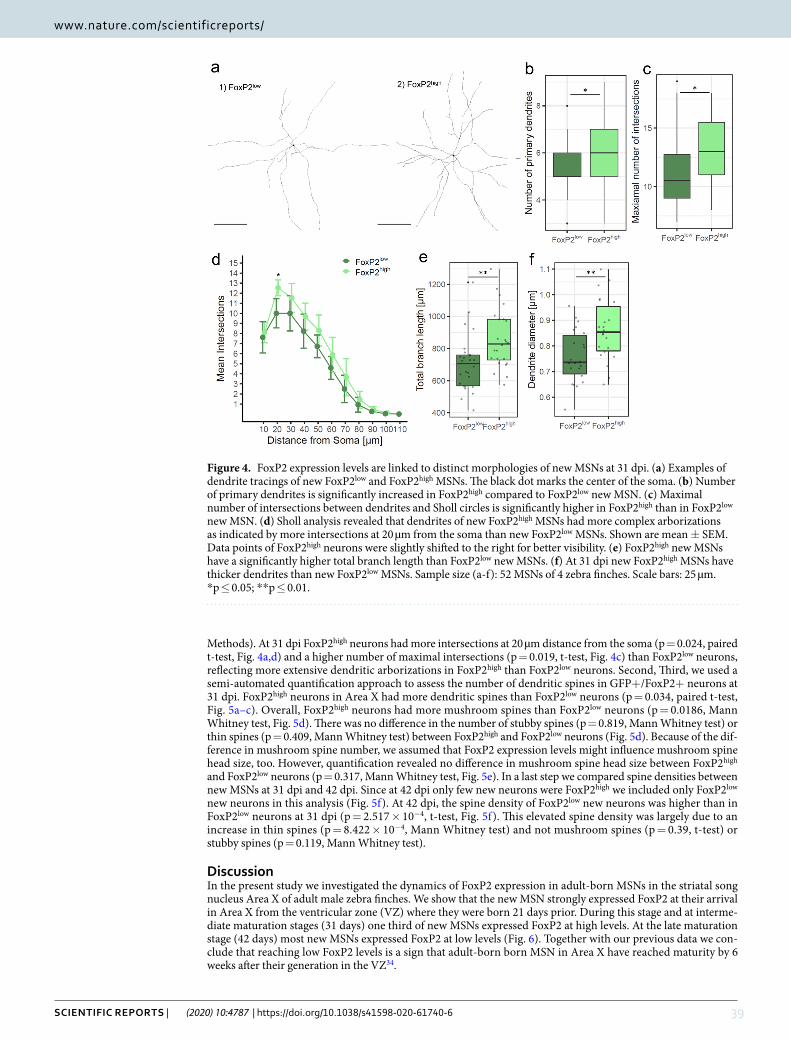
39SCIENTIFIC REPORTS | (2020) 10:4787 | https://doi.org/10.1038/s41598-020-61740-6
www.nature.com/scientificreportswww.nature.com/scientificreports/
Methods). At 31 dpi FoxP2high neurons had more intersections at 20 µm distance from the soma (p = 0.024, paired t-test, Fig. 4a,d) and a higher number of maximal intersections (p = 0.019, t-test, Fig. 4c) than FoxP2low neurons, reflecting more extensive dendritic arborizations in FoxP2high than FoxP2low neurons. Second, Third, we used a semi-automated quantification approach to assess the number of dendritic spines in GFP+/FoxP2+ neurons at 31 dpi. FoxP2high neurons in Area X had more dendritic spines than FoxP2low neurons (p = 0.034, paired t-test, Fig. 5a–c). Overall, FoxP2high neurons had more mushroom spines than FoxP2low neurons (p = 0.0186, Mann Whitney test, Fig. 5d). There was no difference in the number of stubby spines (p = 0.819, Mann Whitney test) or thin spines (p = 0.409, Mann Whitney test) between FoxP2high and FoxP2low neurons (Fig. 5d). Because of the dif-ference in mushroom spine number, we assumed that FoxP2 expression levels might influence mushroom spine head size, too. However, quantification revealed no difference in mushroom spine head size between FoxP2high and FoxP2low neurons (p = 0.317, Mann Whitney test, Fig. 5e). In a last step we compared spine densities between new MSNs at 31 dpi and 42 dpi. Since at 42 dpi only few new neurons were FoxP2high we included only FoxP2low new neurons in this analysis (Fig. 5f). At 42 dpi, the spine density of FoxP2low new neurons was higher than in FoxP2low neurons at 31 dpi (p = 2.517 × 10⁻⁴, t-test, Fig. 5f). This elevated spine density was largely due to an increase in thin spines (p = 8.422 × 10⁻⁴, Mann Whitney test) and not mushroom spines (p = 0.39, t-test) or stubby spines (p = 0.119, Mann Whitney test).
DiscussionIn the present study we investigated the dynamics of FoxP2 expression in adult-born MSNs in the striatal song nucleus Area X of adult male zebra finches. We show that the new MSN strongly expressed FoxP2 at their arrival in Area X from the ventricular zone (VZ) where they were born 21 days prior. During this stage and at interme-diate maturation stages (31 days) one third of new MSNs expressed FoxP2 at high levels. At the late maturation stage (42 days) most new MSNs expressed FoxP2 at low levels (Fig. 6). Together with our previous data we con-clude that reaching low FoxP2 levels is a sign that adult-born born MSN in Area X have reached maturity by 6 weeks after their generation in the VZ34.
Figure 4. FoxP2 expression levels are linked to distinct morphologies of new MSNs at 31 dpi. (a) Examples of dendrite tracings of new FoxP2low and FoxP2high MSNs. The black dot marks the center of the soma. (b) Number of primary dendrites is significantly increased in FoxP2high compared to FoxP2low new MSN. (c) Maximal number of intersections between dendrites and Sholl circles is significantly higher in FoxP2high than in FoxP2low new MSN. (d) Sholl analysis revealed that dendrites of new FoxP2high MSNs had more complex arborizations as indicated by more intersections at 20 µm from the soma than new FoxP2low MSNs. Shown are mean ± SEM. Data points of FoxP2high neurons were slightly shifted to the right for better visibility. (e) FoxP2high new MSNs have a significantly higher total branch length than FoxP2low new MSNs. (f) At 31 dpi new FoxP2high MSNs have thicker dendrites than new FoxP2low MSNs. Sample size (a-f): 52 MSNs of 4 zebra finches. Scale bars: 25 µm. *p ≤ 0.05; **p ≤ 0.01.
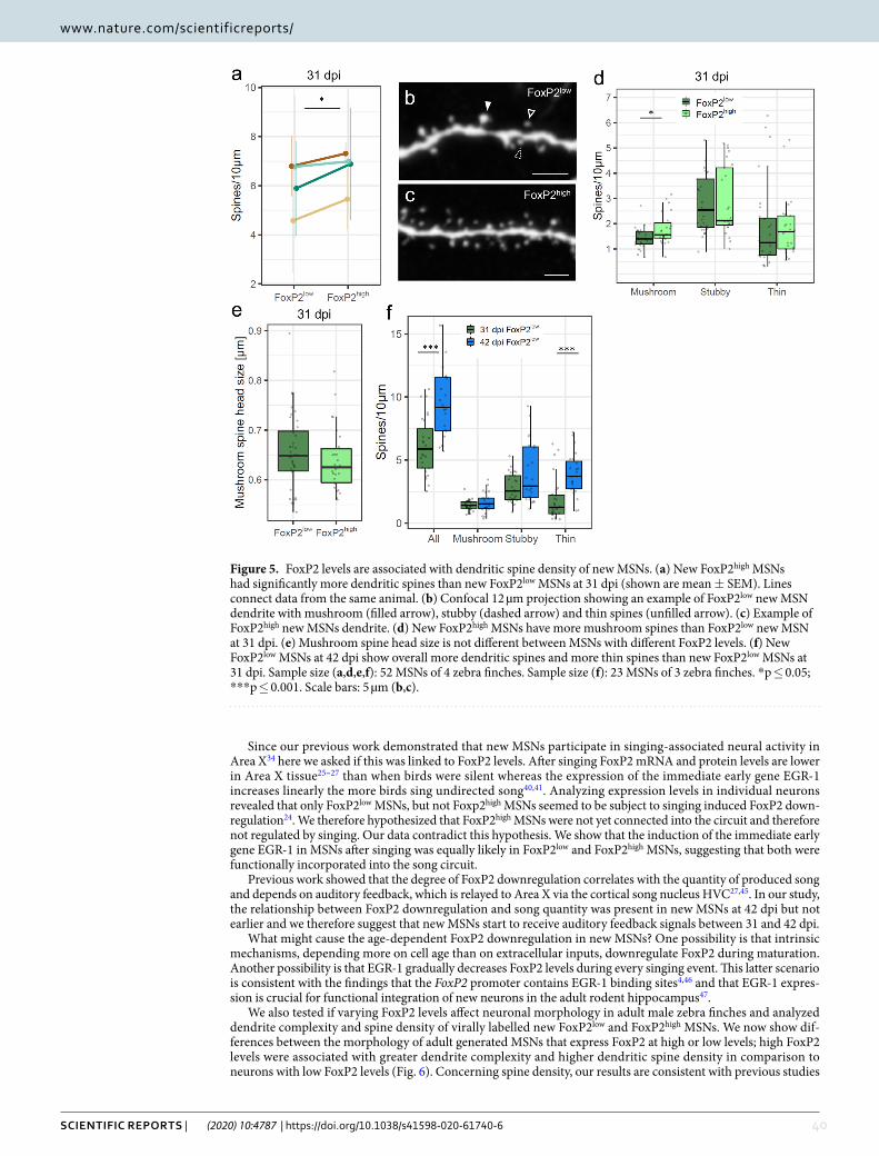
40SCIENTIFIC REPORTS | (2020) 10:4787 | https://doi.org/10.1038/s41598-020-61740-6
www.nature.com/scientificreportswww.nature.com/scientificreports/
Since our previous work demonstrated that new MSNs participate in singing-associated neural activity in Area X34 here we asked if this was linked to FoxP2 levels. After singing FoxP2 mRNA and protein levels are lower in Area X tissue25–27 than when birds were silent whereas the expression of the immediate early gene EGR-1 increases linearly the more birds sing undirected song40,41. Analyzing expression levels in individual neurons revealed that only FoxP2low MSNs, but not Foxp2high MSNs seemed to be subject to singing induced FoxP2 down-regulation24. We therefore hypothesized that FoxP2high MSNs were not yet connected into the circuit and therefore not regulated by singing. Our data contradict this hypothesis. We show that the induction of the immediate early gene EGR-1 in MSNs after singing was equally likely in FoxP2low and FoxP2high MSNs, suggesting that both were functionally incorporated into the song circuit.
Previous work showed that the degree of FoxP2 downregulation correlates with the quantity of produced song and depends on auditory feedback, which is relayed to Area X via the cortical song nucleus HVC27,45. In our study, the relationship between FoxP2 downregulation and song quantity was present in new MSNs at 42 dpi but not earlier and we therefore suggest that new MSNs start to receive auditory feedback signals between 31 and 42 dpi.
What might cause the age-dependent FoxP2 downregulation in new MSNs? One possibility is that intrinsic mechanisms, depending more on cell age than on extracellular inputs, downregulate FoxP2 during maturation. Another possibility is that EGR-1 gradually decreases FoxP2 levels during every singing event. This latter scenario is consistent with the findings that the FoxP2 promoter contains EGR-1 binding sites4,46 and that EGR-1 expres-sion is crucial for functional integration of new neurons in the adult rodent hippocampus47.
We also tested if varying FoxP2 levels affect neuronal morphology in adult male zebra finches and analyzed dendrite complexity and spine density of virally labelled new FoxP2low and FoxP2high MSNs. We now show dif-ferences between the morphology of adult generated MSNs that express FoxP2 at high or low levels; high FoxP2 levels were associated with greater dendrite complexity and higher dendritic spine density in comparison to neurons with low FoxP2 levels (Fig. 6). Concerning spine density, our results are consistent with previous studies
Figure 5. FoxP2 levels are associated with dendritic spine density of new MSNs. (a) New FoxP2high MSNs had significantly more dendritic spines than new FoxP2low MSNs at 31 dpi (shown are mean ± SEM). Lines connect data from the same animal. (b) Confocal 12 µm projection showing an example of FoxP2low new MSN dendrite with mushroom (filled arrow), stubby (dashed arrow) and thin spines (unfilled arrow). (c) Example of FoxP2high new MSNs dendrite. (d) New FoxP2high MSNs have more mushroom spines than FoxP2low new MSN at 31 dpi. (e) Mushroom spine head size is not different between MSNs with different FoxP2 levels. (f) New FoxP2low MSNs at 42 dpi show overall more dendritic spines and more thin spines than new FoxP2low MSNs at 31 dpi. Sample size (a,d,e,f): 52 MSNs of 4 zebra finches. Sample size (f): 23 MSNs of 3 zebra finches. *p ≤ 0.05; ***p ≤ 0.001. Scale bars: 5 µm (b,c).
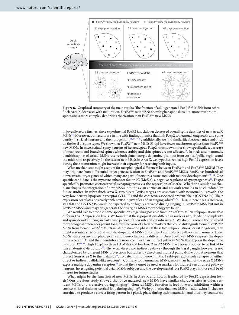
41SCIENTIFIC REPORTS | (2020) 10:4787 | https://doi.org/10.1038/s41598-020-61740-6
www.nature.com/scientificreportswww.nature.com/scientificreports/
in juvenile zebra finches, since experimental FoxP2 knockdown decreased overall spine densities of new Area X MSNs48. Moreover, our results are in line with findings in mice that link Foxp2 to neuronal outgrowth and spine density in striatal neurons and their progenitors18,19,35–37. Additionally, we find similarities between mice and birds on the level of spine types. We show that FoxP2low new MSNs 31 dpi have fewer mushroom spines than FoxP2high new MSNs. In mice, striatal spiny neurons of heterozygous Foxp2 knockdown mice show specifically a decrease of mushroom and branched spines whereas stubby and thin spines are not affected19. In birds and mammals, dendritic spines of striatal MSNs receive both glutamatergic dopaminergic input from cortical/pallial regions and the midbrain, respectively. In the case of new MSNs in Area X, we hypothesize that high FoxP2 expression levels during their maturation might increase their capacity for receiving both inputs.
What mechanisms might account for morphological differences between FoxP2low and FoxP2high MSNs? They may originate from differential target gene activation in FoxP2low and FoxP2high MSNs. FoxP2 has hundreds of downstream target genes of which many are part of networks associated with neurite development35,49–52. One specific candidate is the myocyte enhancer factor 2C (Mef2c), a negative regulator of synaptogenesis19. Foxp2 specifically promotes corticostriatal synaptogenesis via the repression of Mef2c. Whether a similar mecha-nism shapes the integration of new MSNs into the avian corticostriatal network remains to be elucidated by future studies. In zebra finch Area X, two direct FoxP2 targets are associated with neuronal outgrowth; the very-low-density-lipoprotein receptor (VLDLR) and the contactin-associated protein-like 2 (CNTNAP2). Their expression correlates positively with FoxP2 in juveniles and in singing adults53,54. Thus, in new Area X neurons, VLDLR and CNTNAP2 would be expected to be highly activated during singing in FoxP2high MSN but not in FoxP2low MSNs and may thus generate the diverging MSNs morphology we found.
We would like to propose some speculations regarding possible functions of two MSNs subpopulations that differ in FoxP2 expression levels. We found that these populations differed in nucleus size, dendritic complexity and spine density during an early time period of their integration into Area X. We do not know if the observed morphological differences persist long-term because of a lack of markers that could distinguish former FoxP2high MSNs from former FoxP2low MSNs in later maturation phases. If these two subpopulations persist long-term, they might resemble striato-nigral and striato-pallidal MSNs of the direct and indirect pathway in mammals. These MSNs subtypes are morphologically and neurochemically different. Direct pathway MSNs express the dopa-mine receptor D1 and their dendrites are more complex than indirect pathway MSNs that express the dopamine receptor D239,55. High Foxp2 levels in D1 MSNs and low Foxp2 in D2 MSNs have been proposed to be linked to this anatomical dichotomy35. The avian direct and indirect pathway through the basal ganglia however is not characterized by different MSN projections but rather by direct and indirect pallidal-like output neurons that project from Area X to the thalamus56. To date, it is not known if MSN subtypes exclusively synapse on either direct or indirect pallidal-like neurons57. Contrary to mammalian MSNs, more than half of the Area X MSNs express multiple dopamine receptors58 so that they cannot be used as markers for indirect versus direct pathway neurons. Investigating potential avian MSNs subtypes and the developmental role FoxP2 plays in those will be of interest for future studies.
What might be the function of new MSNs in Area X and how is it affected by FoxP2 expression lev-els? Our previous study showed that once matured, new MSNs have similar characteristics as older, res-ident MSNs and are active during singing34. General MSNs function is feed forward inhibition within a cortico-striatal-thalamo-cortical loop during singing59. We hypothesize that new MSNs in adult zebra finches are entrained to produce a correct firing pattern in a plastic phase during their maturation and thus may counteract
Figure 6. Graphical summary of the main results. The fraction of adult generated FoxP2high MSNs from zebra finch Area X decreases with maturation. FoxP2high new MSNs show higher spine densities, more mushroom spines and a more complex dendritic arborization than FoxP2low new MSNs.
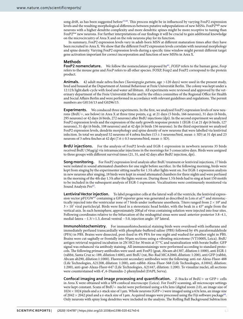
42SCIENTIFIC REPORTS | (2020) 10:4787 | https://doi.org/10.1038/s41598-020-61740-6
www.nature.com/scientificreportswww.nature.com/scientificreports/
song drift, as has been suggested before33,60. This process might be in influenced by varying FoxP2 expression levels and the resulting morphological differences between putative subpopulations of new MSNs. FoxP2high new neurons with a higher dendrite complexity and more dendritic spines might be more receptive to tuning than FoxP2low new neurons. For further interpretations of our findings it will be crucial to gain additional knowledge on the microcircuitry of Area X and on the role neurons play for its function.
In summary, FoxP2 expression levels vary in adult-born MSN at different maturation times after they have been recruited to Area X. We show that the different FoxP2 expression levels correlate with neuronal morphology and spine density. Varying FoxP2 expression levels during a specific time window might permit different target gene activation important for correct incorporation and function of new MSNs in Area X.
MethodsFoxP2 nomenclature. We follow the nomenclature proposed by61, FOXP refers to the human gene, Foxp refers to the mouse gene and FoxP refers to all other species. FOXP, Foxp2 and FoxP2 correspond to the protein product.
Animals. 42 adult male zebra finches (Taeniopygia guttata, age >120 days) were used in the present study, bred and housed at the Department of Animal Behaviour at Freie Universität Berlin. The colony was kept under a 12:12 h light:dark-cycle with food and water ad libitum. All experiments were reviewed and approved by the vet-erinary department of the Freie Universität Berlin and by the ethics committee of the Regional Office for Health and Social Affairs Berlin and were performed in accordance with relevant guidelines and regulations. The permit numbers are G0116/13 and G0296/15.
Experiments. We conduced three experiments. In the first, we analyzed FoxP2 expression levels of new neu-rons (BrdU+, see below) in Area X at three time points, e.g. at 21 days (5 birds, 166 neurons), 31 days (6 birds, 295 neurons) or 42 days (6 birds, 272 neurons) after BrdU injections (dpi). In the second experiment we analyzed FoxP2 expression levels and the expression of the early growth response protein 1 (EGR-1) at 21 dpi (6 birds, 127 neurons), 31 dpi (6 birds, 108 neurons) and at 42 dpi (6 birds 156 neurons). In the third experiment we analyzed FoxP2 expression levels, dendrite morphology and spine density of new neurons that were labelled via lentiviral infection. In total we analyzed 52 neurons of 4 zebra finches (13 ± 3 neurons/bird, mean ± SD) at 31 dpi and 23 neurons of 3 zebra finches at 42 dpi (7.6 ± 0.5 neurons/bird, mean ± SD).
BrdU injections. For the analysis of FoxP2 levels and EGR-1 expression in newborn neurons 35 birds received BrdU (50 µg/g) via intramuscular injections in the mornings for 5 consecutive days. Birds were assigned to three groups with different survival times (21, 31, and 42 days after BrdU injection, dpi).
Song monitoring. For FoxP2 expression level analysis after BrdU treatment or lentiviral injections, 17 birds were isolated in sound attenuated chambers for one night before sacrifice. In the following morning, birds were kept from singing by the experimenter sitting nearby for 1.5 h after lights went on. For EGR-1 expression analysis in new neurons after singing, 18 birds were kept in sound attenuated chambers for three nights and were perfused in the morning of the 4th day 1.5 h after the lights went on. During those 1.5 h birds had to sing at least 150 motifs to be included in the subsequent analysis of EGR-1 expression. Vocalizations were continuously monitored via Sound Analysis Pro62.
Lentiviral Vector injection. To label progenitor cells at the lateral wall of the ventricle, the lentiviral expres-sion vector pFUGW63 containing a GFP reporter gene was generated as described in Lois et al.63 and stereotac-tically injected into the ventricular zone of 7 birds under isoflurane anesthesia. Titers ranged from 2 × 10⁶ and 3 × 10⁷ viral particles/µl. Birds were fixed in a stereotaxic head holder, with the beak in a 45° angle from the vertical axis. In each hemisphere, approximately 200 µl of virus containing solution were injected into four sites. Following coordinates relative to the bifurcation of the midsagittal sinus were used: anterior-posterior 3.8–4.1, medial-latera −1.3/+1.3, dorsal-ventral −5.0, injection angle: 10° lateral.
Immunohistochemistry. For immunohistochemical staining birds were overdosed with isoflurane and immediately perfused transcardially with phosphate-buffered saline (PBS) followed by 4% paraformaldehyde (PFA) in PBS. Brains were dissected, post-fixed in 4% PFA for one night and washed for another night in PBS. Brains were cut sagitally or frontally into 50 µm sections using a vibrating microtome (VT1000S, Leica). BrdU antigen retrieval required incubation in 2N HCl for 30 min at 37 °C and neutralization with borate buffer. GFP signal was enhanced via antibody staining. All immunostainings were performed according to standard proto-cols. The following primary antibodies were used: anti FoxP2 (goat, Abcam ab1307, dilution:1:1000), anti EGR-1 (rabbit, Santa Cruz sc-189, dilution:1:600), anti BrdU (rat, Bio-Rad MCA2060, dilution: 1:200), anti GFP (rabbit, Abcam ab290, dilution:1:1000). Fluorescent secondary antibodies were the following: anti-rat-Alexa-Fluor-488 (Life Technologies, A21208, dilution: 1:200), anti-rabbit-Alexa-Fluor-568 (Life Technologies, A10042, dilution: 1:200), anti-goat-Alaxa-Fluor-647 (Life Technologies, A21447, dilution: 1:200). To visualize nuclei, all sections were counterstained with 4′, 6-Diamidin-2-phenylindol (DAPI, Serva).
Confocal imaging and image processing and quantification. Z-Stacks of BrdU+ or GFP+ cells in Area X were obtained with a SP8 confocal microscope (Leica). For FoxP2 scanning, all microscope settings were kept constant. Scans of BrdU+ nuclei were performed using a 63x lens (digital zoom 2.0), an image size of 1024 × 1024 pixels and a z-stack size of 1 µm. Whole neurons (GFP+) were imaged using a 63x lens, an image size of 2042 × 2042 pixel and a z-stack size of 1 µm. Acquired images were processed using the Fiji software package64. Only neurons with spiny long dendrites were included in the analysis. The Rolling Ball Background Subtraction
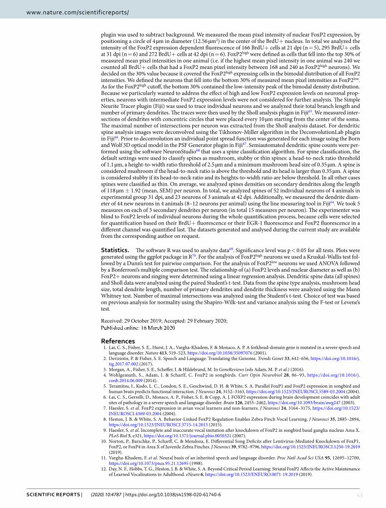
43SCIENTIFIC REPORTS | (2020) 10:4787 | https://doi.org/10.1038/s41598-020-61740-6
www.nature.com/scientificreportswww.nature.com/scientificreports/
plugin was used to subtract background. We measured the mean pixel intensity of nuclear FoxP2 expression, by positioning a circle of 4 µm in diameter (12.56 µm2) in the center of the BrdU+ nucleus. In total we analyzed the intensity of the FoxP2 expression dependent fluorescence of 166 BrdU+ cells at 21 dpi (n = 5), 295 BrdU+ cells at 31 dpi (n = 6) and 272 BrdU+ cells at 42 dpi (n = 6). FoxP2high were defined as cells that fell into the top 30% of measured mean pixel intensities in one animal (i.e. if the highest mean pixel intensity in one animal was 240 we counted all BrdU+ cells that had a FoxP2 mean pixel intensity between 168 and 240 as FoxP2high neurons). We decided on the 30% value because it covered the FoxP2high expressing cells in the bimodal distribution of all FoxP2 intensities. We defined the neurons that fell into the bottom 30% of measured mean pixel intensities as FoxP2low. As for the FoxP2high cutoff, the bottom 30% contained the low-intensity peak of the bimodal density distribution. Because we particularly wanted to address the effect of high and low FoxP2 expression levels on neuronal prop-erties, neurons with intermediate FoxP2 expression levels were not considered for further analysis. The Simple Neurite Tracer plugin (Fiji) was used to trace individual neurons and we analyzed their total branch length and number of primary dendrites. The traces were then used by the Sholl analysis plugin in Fiji65. We measured inter-sections of dendrites with concentric circles that were placed every 10 µm starting from the center of the soma. The maximal number of intersections per neuron was extracted from the Sholl analysis dataset. For dendritic spine analysis images were deconvolved using the Tikhonov-Miller algorithm in the DeconvolutionLab plugin in Fiji66. Prior to deconvolution an individual point spread function was generated for each image using the Born and Wolf 3D optical model in the PSF Generator plugin in Fiji67. Semiautomated dendritic spine counts were per-formed using the software NeuronStudio68 that uses a spine classification algorithm. For spine classification, the default settings were used to classify spines as mushroom, stubby or thin spines: a head-to-neck ratio threshold of 1.1 µm, a height-to-width ratio threshold of 2.5 µm and a minimum mushroom head size of 0.35 µm. A spine is considered mushroom if the head-to-neck ratio is above the threshold and its head is larger than 0.35 µm. A spine is considered stubby if its head-to-neck ratio and its heights-to-width ratio are below threshold. In all other cases spines were classified as thin. On average, we analyzed spines densities on secondary dendrites along the length of 118 µm ± 1.92 (mean, SEM) per neuron. In total, we analyzed spines of 52 individual neurons of 4 animals in experimental group 31 dpi, and 23 neurons of 3 animals at 42 dpi. Additionally, we measured the dendrite diam-eter of 44 new neurons in 4 animals (8–12 neurons per animal) using the line measuring tool in Fiji64. We took 5 measures on each of 3 secondary dendrites per neuron (in total 15 measures per neuron). The experimenter was blind to FoxP2 levels of individual neurons during the whole quantification process, because cells were selected for quantification based on their BrdU+ fluorescence or their EGR-1 fluorescence and FoxP2 fluorescence in a different channel was quantified last. The datasets generated and analysed during the current study are available from the corresponding author on request.
Statistics. The software R was used to analyze data69. Significance level was p < 0.05 for all tests. Plots were generated using the ggplot package in R70. For the analysis of FoxP2high neurons we used a Kruskal-Wallis test fol-lowed by a Dunn’s test for pairwise comparison. For the analysis of FoxP2low neurons we used ANOVA followed by a Bonferroni’s multiple comparison test. The relationship of (a) FoxP2 levels and nuclear diameter as well as (b) FoxP2+ neurons and singing were determined using a linear regression analysis. Dendritic spine data (all spines) and Sholl data were analyzed using the paired Student’s t-test. Data from the spine type analysis, mushroom head size, total dendrite length, number of primary dendrites and dendrite thickness were analyzed using the Mann Whitney test. Number of maximal intersections was analyzed using the Student’s t-test. Choice of test was based on previous analysis for normality using the Shapiro-Wilk-test and variance analysis using the F-test or Levene’s test.
Received: 29 October 2019; Accepted: 29 February 2020;
Published: xx xx xxxx
References1. Lai, C. S., Fisher, S. E., Hurst, J. A., Vargha-Khadem, F. & Monaco, A. P. A forkhead-domain gene is mutated in a severe speech and
language disorder. Nature 413, 519–523, https://doi.org/10.1038/35097076 (2001).2. Deriziotis, P. & Fisher, S. E. Speech and Language: Translating the Genome. Trends Genet 33, 642–656, https://doi.org/10.1016/j.
tig.2017.07.002 (2017).3. Morgan, A., Fisher, S. E., Scheffer, I. & Hildebrand, M. In GeneReviews (eds Adam, M. P. et al.) (2016).4. Wohlgemuth, S., Adam, I. & Scharff, C. FoxP2 in songbirds. Curr Opin Neurobiol 28, 86–93, https://doi.org/10.1016/j.
conb.2014.06.009 (2014).5. Teramitsu, I., Kudo, L. C., London, S. E., Geschwind, D. H. & White, S. A. Parallel FoxP1 and FoxP2 expression in songbird and
human brain predicts functional interaction. J Neurosci 24, 3152–3163, https://doi.org/10.1523/JNEUROSCI.5589-03.2004 (2004).6. Lai, C. S., Gerrelli, D., Monaco, A. P., Fisher, S. E. & Copp, A. J. FOXP2 expression during brain development coincides with adult
sites of pathology in a severe speech and language disorder. Brain 126, 2455–2462, https://doi.org/10.1093/brain/awg247 (2003).7. Haesler, S. et al. FoxP2 expression in avian vocal learners and non-learners. J Neurosci 24, 3164–3175, https://doi.org/10.1523/
JNEUROSCI.4369-03.2004 (2004).8. Heston, J. B. & White, S. A. Behavior-Linked FoxP2 Regulation Enables Zebra Finch Vocal Learning. J Neurosci 35, 2885–2894,
https://doi.org/10.1523/JNEUROSCI.3715-14.2015 (2015).9. Haesler, S. et al. Incomplete and inaccurate vocal imitation after knockdown of FoxP2 in songbird basal ganglia nucleus Area X.
PLoS Biol 5, e321, https://doi.org/10.1371/journal.pbio.0050321 (2007). 10. Norton, P., Barschke, P., Scharff, C. & Mendoza, E. Differential Song Deficits after Lentivirus-Mediated Knockdown of FoxP1,
FoxP2, or FoxP4 in Area X of Juvenile Zebra Finches. J Neurosci 39, 9782–9796, https://doi.org/10.1523/JNEUROSCI.1250-19.2019 (2019).
11. Vargha-Khadem, F. et al. Neural basis of an inherited speech and language disorder. Proc Natl Acad Sci USA 95, 12695–12700,https://doi.org/10.1073/pnas.95.21.12695 (1998).
12. Day, N. F., Hobbs, T. G., Heston, J. B. & White, S. A. Beyond Critical Period Learning: Striatal FoxP2 Affects the Active Maintenance of Learned Vocalizations in Adulthood. eNeuro 6, https://doi.org/10.1523/ENEURO.0071-19.2019 (2019).
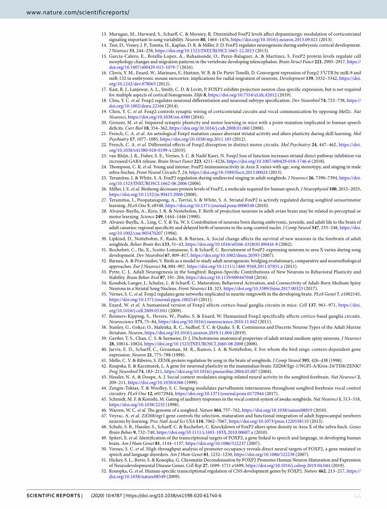
44SCIENTIFIC REPORTS | (2020) 10:4787 | https://doi.org/10.1038/s41598-020-61740-6
www.nature.com/scientificreportswww.nature.com/scientificreports/
13. Murugan, M., Harward, S., Scharff, C. & Mooney, R. Diminished FoxP2 levels affect dopaminergic modulation of corticostriatalsignaling important to song variability. Neuron 80, 1464–1476, https://doi.org/10.1016/j.neuron.2013.09.021 (2013).
14. Tsui, D., Vessey, J. P., Tomita, H., Kaplan, D. R. & Miller, F. D. FoxP2 regulates neurogenesis during embryonic cortical development.J Neurosci 33, 244–258, https://doi.org/10.1523/JNEUROSCI.1665-12.2013 (2013).
15. Garcia-Calero, E., Botella-Lopez, A., Bahamonde, O., Perez-Balaguer, A. & Martinez, S. FoxP2 protein levels regulate cellmorphology changes and migration patterns in the vertebrate developing telencephalon. Brain Struct Funct 221, 2905–2917, https://doi.org/10.1007/s00429-015-1079-7 (2016).
16. Clovis, Y. M., Enard, W., Marinaro, F., Huttner, W. B. & De Pietri Tonelli, D. Convergent repression of Foxp2 3′UTR by miR-9 and miR-132 in embryonic mouse neocortex: implications for radial migration of neurons. Development 139, 3332–3342, https://doi.org/10.1242/dev.078063 (2012).
17. Kast, R. J., Lanjewar, A. L., Smith, C. D. & Levitt, P. FOXP2 exhibits projection neuron class specific expression, but is not requiredfor multiple aspects of cortical histogenesis. Elife 8, https://doi.org/10.7554/eLife.42012 (2019).
18. Chiu, Y. C. et al. Foxp2 regulates neuronal differentiation and neuronal subtype specification. Dev Neurobiol 74, 723–738, https://doi.org/10.1002/dneu.22166 (2014).
19. Chen, Y. C. et al. Foxp2 controls synaptic wiring of corticostriatal circuits and vocal communication by opposing Mef2c. Nat Neurosci, https://doi.org/10.1038/nn.4380 (2016).
20. Groszer, M. et al. Impaired synaptic plasticity and motor learning in mice with a point mutation implicated in human speechdeficits. Curr Biol 18, 354–362, https://doi.org/10.1016/j.cub.2008.01.060 (2008).
21. French, C. A. et al. An aetiological Foxp2 mutation causes aberrant striatal activity and alters plasticity during skill learning. Mol Psychiatry 17, 1077–1085, https://doi.org/10.1038/mp.2011.105 (2012).
22. French, C. A. et al. Differential effects of Foxp2 disruption in distinct motor circuits. Mol Psychiatry 24, 447–462, https://doi.org/10.1038/s41380-018-0199-x (2019).
23. van Rhijn, J. R., Fisher, S. E., Vernes, S. C. & Nadif Kasri, N. Foxp2 loss of function increases striatal direct pathway inhibition via increased GABA release. Brain Struct Funct 223, 4211–4226, https://doi.org/10.1007/s00429-018-1746-6 (2018).
24. Thompson, C. K. et al. Young and intense: FoxP2 immunoreactivity in Area X varies with age, song stereotypy, and singing in male zebra finches. Front Neural Circuits 7, 24, https://doi.org/10.3389/fncir.2013.00024 (2013).
25. Teramitsu, I. & White, S. A. FoxP2 regulation during undirected singing in adult songbirds. J Neurosci 26, 7390–7394, https://doi.org/10.1523/JNEUROSCI.1662-06.2006 (2006).
26. Miller, J. E. et al. Birdsong decreases protein levels of FoxP2, a molecule required for human speech. J Neurophysiol 100, 2015–2025, https://doi.org/10.1152/jn.90415.2008 (2008).
27. Teramitsu, I., Poopatanapong, A., Torrisi, S. & White, S. A. Striatal FoxP2 is actively regulated during songbird sensorimotor learning. PLoS One 5, e8548, https://doi.org/10.1371/journal.pone.0008548 (2010).
28. Alvarez-Buylla, A., Kirn, J. R. & Nottebohm, F. Birth of projection neurons in adult avian brain may be related to perceptual or motor learning. Science 249, 1444–1446 (1990).
29. Alvarez-Buylla, A., Ling, C. Y. & Yu, W. S. Contribution of neurons born during embryonic, juvenile, and adult life to the brain of adult canaries: regional specificity and delayed birth of neurons in the song-control nuclei. J Comp Neurol 347, 233–248, https://doi.org/10.1002/cne.903470207 (1994).
30. Lipkind, D., Nottebohm, F., Rado, R. & Barnea, A. Social change affects the survival of new neurons in the forebrain of adultsongbirds. Behav Brain Res 133, 31–43, https://doi.org/10.1016/s0166-4328(01)00416-8 (2002).
31. Rochefort, C., He, X., Scotto-Lomassese, S. & Scharff, C. Recruitment of FoxP2-expressing neurons to area X varies during songdevelopment. Dev Neurobiol 67, 809–817, https://doi.org/10.1002/dneu.20393 (2007).
32. Barnea, A. & Pravosudov, V. Birds as a model to study adult neurogenesis: bridging evolutionary, comparative and neuroethological approaches. Eur J Neurosci 34, 884–907, https://doi.org/10.1111/j.1460-9568.2011.07851.x (2011).
33. Pytte, C. L. Adult Neurogenesis in the Songbird: Region-Specific Contributions of New Neurons to Behavioral Plasticity andStability. Brain Behav Evol 87, 191–204, https://doi.org/10.1159/000447048 (2016).
34. Kosubek-Langer, J., Schulze, L. & Scharff, C. Maturation, Behavioral Activation, and Connectivity of Adult-Born Medium SpinyNeurons in a Striatal Song Nucleus. Front Neurosci 11, 323, https://doi.org/10.3389/fnins.2017.00323 (2017).
35. Vernes, S. C. et al. Foxp2 regulates gene networks implicated in neurite outgrowth in the developing brain. PLoS Genet 7, e1002145, https://doi.org/10.1371/journal.pgen.1002145 (2011).
36. Enard, W. et al. A humanized version of Foxp2 affects cortico-basal ganglia circuits in mice. Cell 137, 961–971, https://doi.org/10.1016/j.cell.2009.03.041 (2009).
37. Reimers-Kipping, S., Hevers, W., Paabo, S. & Enard, W. Humanized Foxp2 specifically affects cortico-basal ganglia circuits.Neuroscience 175, 75–84, https://doi.org/10.1016/j.neuroscience.2010.11.042 (2011).
38. Stanley, G., Gokce, O., Malenka, R. C., Sudhof, T. C. & Quake, S. R. Continuous and Discrete Neuron Types of the Adult Murine Striatum. Neuron, https://doi.org/10.1016/j.neuron.2019.11.004 (2019).
39. Gertler, T. S., Chan, C. S. & Surmeier, D. J. Dichotomous anatomical properties of adult striatal medium spiny neurons. J Neurosci 28, 10814–10824, https://doi.org/10.1523/JNEUROSCI.2660-08.2008 (2008).
40. Jarvis, E. D., Scharff, C., Grossman, M. R., Ramos, J. A. & Nottebohm, F. For whom the bird sings: context-dependent geneexpression. Neuron 21, 775–788 (1998).
41. Mello, C. V. & Ribeiro, S. ZENK protein regulation by song in the brain of songbirds. J Comp Neurol 393, 426–438 (1998). 42. Knapska, E. & Kaczmarek, L. A gene for neuronal plasticity in the mammalian brain: Zif268/Egr-1/NGFI-A/Krox-24/TIS8/ZENK?
Prog Neurobiol 74, 183–211, https://doi.org/10.1016/j.pneurobio.2004.05.007 (2004). 43. Hessler, N. A. & Doupe, A. J. Social context modulates singing-related neural activity in the songbird forebrain. Nat Neurosci 2,
209–211, https://doi.org/10.1038/6306 (1999). 44. Zengin-Toktas, Y. & Woolley, S. C. Singing modulates parvalbumin interneurons throughout songbird forebrain vocal control
circuitry. PLoS One 12, e0172944, https://doi.org/10.1371/journal.pone.0172944 (2017). 45. Schmidt, M. F. & Konishi, M. Gating of auditory responses in the vocal control system of awake songbirds. Nat Neurosci 1, 513–518,
https://doi.org/10.1038/2232 (1998). 46. Warren, W. C. et al. The genome of a songbird. Nature 464, 757–762, https://doi.org/10.1038/nature08819 (2010).47. Veyrac, A. et al. Zif268/egr1 gene controls the selection, maturation and functional integration of adult hippocampal newborn
neurons by learning. Proc Natl Acad Sci USA 110, 7062–7067, https://doi.org/10.1073/pnas.1220558110 (2013). 48. Schulz, S. B., Haesler, S., Scharff, C. & Rochefort, C. Knockdown of FoxP2 alters spine density in Area X of the zebra finch. Genes
Brain Behav 9, 732–740, https://doi.org/10.1111/j.1601-183X.2010.00607.x (2010).49. Spiteri, E. et al. Identification of the transcriptional targets of FOXP2, a gene linked to speech and language, in developing human
brain. Am J Hum Genet 81, 1144–1157, https://doi.org/10.1086/522237 (2007). 50. Vernes, S. C. et al. High-throughput analysis of promoter occupancy reveals direct neural targets of FOXP2, a gene mutated in
speech and language disorders. Am J Hum Genet 81, 1232–1250, https://doi.org/10.1086/522238 (2007). 51. Hickey, S. L., Berto, S. & Konopka, G. Chromatin Decondensation by FOXP2 Promotes Human Neuron Maturation and Expression
of Neurodevelopmental Disease Genes. Cell Rep 27, 1699–1711 e1699, https://doi.org/10.1016/j.celrep.2019.04.044 (2019).52. Konopka, G. et al. Human-specific transcriptional regulation of CNS development genes by FOXP2. Nature 462, 213–217, https://
doi.org/10.1038/nature08549 (2009).
45SCIENTIFIC REPORTS | (2020) 10:4787 | https://doi.org/10.1038/s41598-020-61740-6
www.nature.com/scientificreportswww.nature.com/scientificreports/
53. Adam, I., Mendoza, E., Kobalz, U., Wohlgemuth, S. & Scharff, C. FoxP2 directly regulates the reelin receptor VLDLR developmentallyand by singing. Mol Cell Neurosci 74, 96–105, https://doi.org/10.1016/j.mcn.2016.04.002 (2016).
54. Adam, I., Mendoza, E., Kobalz, U., Wohlgemuth, S. & Scharff, C. CNTNAP2 is a direct FoxP2 target in vitro and in vivo in zebra finches: complex regulation by age and activity. Genes Brain Behav 16, 635–642, https://doi.org/10.1111/gbb.12390 (2017).
55. Calabresi, P., Picconi, B., Tozzi, A., Ghiglieri, V. & Di Filippo, M. Direct and indirect pathways of basal ganglia: a critical reappraisal. Nat Neurosci 17, 1022–1030, https://doi.org/10.1038/nn.3743 (2014).
56. Farries, M. A., Ding, L. & Perkel, D. J. Evidence for “direct” and “indirect” pathways through the song system basal ganglia. J Comp Neurol 484, 93–104, https://doi.org/10.1002/cne.20464 (2005).
57. Pidoux, M., Bollu, T., Riccelli, T. & Goldberg, J. H. Origins of basal ganglia output signals in singing juvenile birds. J Neurophysiol113, 843–855, https://doi.org/10.1152/jn.00635.2014 (2015).
58. Kubikova, L., Wada, K. & Jarvis, E. D. Dopamine receptors in a songbird brain. J Comp Neurol 518, 741–769, https://doi.org/10.1002/cne.22255 (2010).
59. Perkel, D. J., Farries, M. A., Luo, M. & Ding, L. Electrophysiological analysis of a songbird basal ganglia circuit essential for vocalplasticity. Brain Res Bull 57, 529–532, https://doi.org/10.1016/s0361-9230(01)00690-6 (2002).
60. Wilbrecht, L. & Kirn, J. R. Neuron addition and loss in the song system: regulation and function. Ann N Y Acad Sci 1016, 659–683, https://doi.org/10.1196/annals.1298.024 (2004).
61. Kaestner, K. H., Knochel, W. & Martinez, D. E. Unified nomenclature for the winged helix/forkhead transcription factors. Genes Dev 14, 142–146 (2000).
62. Tchernichovski, O., Nottebohm, F., Ho, C. E., Pesaran, B. & Mitra, P. P. A procedure for an automated measurement of songsimilarity. Anim Behav 59, 1167–1176, https://doi.org/10.1006/anbe.1999.1416 (2000).
63. Lois, C., Hong, E. J., Pease, S., Brown, E. J. & Baltimore, D. Germline transmission and tissue-specific expression of transgenesdelivered by lentiviral vectors. Science 295, 868–872, https://doi.org/10.1126/science.1067081 (2002).
64. Schindelin, J. et al. Fiji: an open-source platform for biological-image analysis. Nat Methods 9, 676–682, https://doi.org/10.1038/nmeth.2019 (2012).
65. Ferreira, T. A. et al. Neuronal morphometry directly from bitmap images. Nat Methods 11, 982–984, https://doi.org/10.1038/nmeth.3125 (2014).
66. Sage, D. et al. DeconvolutionLab2: An open-source software for deconvolution microscopy. Methods 115, 28–41, https://doi.org/10.1016/j.ymeth.2016.12.015 (2017).
67. Kirshner, H., Aguet, F., Sage, D. & Unser, M. 3-D PSF fitting for fluorescence microscopy: implementation and localizationapplication. J Microsc 249, 13–25, https://doi.org/10.1111/j.1365-2818.2012.03675.x (2013).
68. Rodriguez, A., Ehlenberger, D. B., Dickstein, D. L., Hof, P. R. & Wearne, S. L. Automated three-dimensional detection and shape classification of dendritic spines from fluorescence microscopy images. PLoS One 3, e1997, https://doi.org/10.1371/journal.pone.0001997 (2008).
69. R: A Language and Environment for Statistical Computing (R Foundation for Statistical Computing, 2013).70. Wickham, H. gglot2: Elegant Graphics for Data Analysis. (Springer, 2016).
AcknowledgementsJ.K.L. received funds from the Elsa Neumann scholarship by the state of Berlin, the Christiane Nüsslein-Volhard Foundation and the Max Planck School of Cognition.
Author contributionsJ.K.L. and C.S. designed experiments, J.K.L. performed experiments and data analysis, J.K.L. and C.S. wrote the manuscript.
Competing interestsThe authors declare no competing interests.
Additional informationCorrespondence and requests for materials should be addressed to J.K.-L.
Reprints and permissions information is available at www.nature.com/reprints.
Publisher’s note Springer Nature remains neutral with regard to jurisdictional claims in published maps and institutional affiliations.
Open Access This article is licensed under a Creative Commons Attribution 4.0 International License, which permits use, sharing, adaptation, distribution and reproduction in any medium or
format, as long as you give appropriate credit to the original author(s) and the source, provide a link to the Cre-ative Commons license, and indicate if changes were made. The images or other third party material in this article are included in the article’s Creative Commons license, unless indicated otherwise in a credit line to the material. If material is not included in the article’s Creative Commons license and your intended use is not per-mitted by statutory regulation or exceeds the permitted use, you will need to obtain permission directly from the copyright holder. To view a copy of this license, visit http://creativecommons.org/licenses/by/4.0/.
© The Author(s) 2020

46
General Discussion
47
General Discussion
Songbirds offer a model for studying functional adult striatal neurogenesis and its role for the maintenance of a learned behavior In this thesis, I present evidence for functional adult neurogenesis in the striatal song nucleus
Area X, which is implicated in song learning as well as in the long-term maintenance of song
(Sohrabji et al., 1990; Scharff and Nottebohm, 1991; Ali et al., 2013; Kubikova et al., 2014;
Kojima et al., 2018). In Publication A, I applied BrdU birth dating of new neurons and immuno-
histochemically analyzed their maturation course and singing induced activity in Area X after
varying survival times. Further, I virally labelled progenitor cells in the neurogenic niche
adjacent to the lateral ventricle and imaged the descending new medium spiny neurons (MSNs)
in Area X. To elucidate the connectivity of new MSNs, I retrogradely traced their postsynaptic
target cells, labelled presynaptic contacts from pallial nuclei and detected indicators of
dopaminergic signaling. The results presented in Publication A provide evidence that new
MSNs in Area X mature within a time frame of six weeks and are robustly active during singing.
Their connectivity within the local circuitry resembles the one of older, resident MSNs. By
analyzing MSNs densities in zebra finches up to seven years of age, I found that adult
neurogenesis is a process of constant addition, most likely increasing inhibitory signaling within
Area X. Altogether, the data presented in Publication A filled several knowledge gaps about
the processes of striatal adult neurogenesis and illustrate that adult born neurons in Area X
fulfill many essential prerequisites for a functional role in adult song maintenance.
Because adult neurogenesis in HVC has been intensively studied I will outline
similarities and differences between adult neurogenesis in HVC and Area X. One essential
similarity is that in both nuclei only one cell type is subject to adult neurogenesis; in HVC only
HVCRA but not HVCX projection neurons and in Area X only MSNs but not pallidal-like
neurons are recruited (Alvarez-Buylla et al., 1988a; Nordeen and Nordeen, 1988b; Rochefort
et al., 2007; Scotto-Lomassese et al., 2007). This indicates that the parts of the ventricular zone
(VZ) that give rise to pallial and striatal cells are either determined to produce a specific neuron
type like in the mammalian subventricular zone (Kelsch et al., 2007; Merkle et al., 2007; Merkle
et al., 2014) or that local cues in HVC and Area X only allow differentiation into one neuron
type. Since HVCRA neurons are mainly born post hatch, while most of HVCX neurons are
generated in ovo (Alvarez-Buylla et al., 1988a), it is conceivable that adult neurogenesis
represents a prolongation of juvenile neurogenesis. The fact that adult neurogenesis in HVC
decreases with age supports this notion (Wang et al., 2002; Wilbrecht et al., 2002; Pytte et al.,
General Discussion
48
2007). Striatal neurons are born during early embryonic development and after hatching.
Compared to other telencephalic regions, the striatum receives the highest number of new
neurons after hatching (Alvarez-Buylla et al., 1994) and Area X forms about 8-10 days post
hatch (Garcia-Calero and Scharff, 2013). Assuming that adult neurogenesis in Area X is an
extension of juvenile neurogenesis, I would expect that its rate decreased with age, like in HVC.
Interestingly, this is not the case and rates of Area X neurogenesis remain constant in adults
(Pytte et al., 2007).
Another similarity is that both new neuron types connect to their targets. New HCVRA
neurons connect to their target RA, e.g. are retrogradely labelled, by 21 days after they
originated in the pallial VZ (Kirn et al., 1999; Tokarev et al., 2015). Publication A shows that
new MSNs in Area X locally contacted pallidal-like neurons as early as 31 days after their
generation in the striatal ventricular zone. New MSNs migrate at least 1000 µm from the VZ to
Area X whereas new HVC neurons only migrate approximately less than 100 µm. Taking the
longer migration time of new MSNs from the VZ to Area X into account it is plausible that the
time between birth dating and connection to target neurons is very similar in new HVC neurons
and new MSNs.
Both new HVC and Area X neurons are activated by singing, as shown by the induction
of immediate early gene transcription (Tokarev et al., 2015; Kosubek-Langer et al., 2017). In
Publication A, I analyzed the percentage of new MSNs that expressed the immediate early
gene (IEG) EGR-1 after undirected singing . A methodologically similar study investigated IEG
expression in new HVCRA neurons after directed or undirected singing (Tokarev et al., 2015).
Comparing only the fraction of neurons that were new (BrdU+) and active during singing
(IEG+), it becomes clear that their proportion increased significantly with age. After 6 or 8
weeks, between 70-80% of new neurons were activated by singing, both in Area X and HVC
respectively (Kosubek-Langer et al., 2017; Tokarev et al., 2015). In HVC, a stable fraction of
new neurons was IEG+ without being retrogradely filled from RA, e.g. are probably not
connected (Tokarev et al., 2015). In new MSNs it is not clear if singing related activity depends
on coincident postsynaptic connections. Furthermore, the variability of how many new neurons
are active during singing decreases with survival time in both nuclei, i.e. the older the new
neurons become the more robust is their activation by singing across animals. EGR-1
expression is crucial for the survival of new granule neurons in the murine dentate gyrus of the
hippocampal formation (Veyrac et al., 2013). Further, EGR-1 induces the expression of
DARPP-32, a marker for mature MSNs, in primary striatal neurons (Keilani et al., 2012). Taken
General Discussion
49
together, singing-induced IEG expression most likely promotes both the maturation and
survival in new HVC and Area X neurons.
Another feature common between adult neurogenesis in HVC and Area X is that it is a process
of addition rather than of replacement. In both song nuclei the density of the added neuron type
increases with age and their total neuron number doubles within 11 or 5 years in HVC and
Area X, respectively (Walton et al., 2012; Kosubek-Langer et al., 2017). In contrast, in seasonal
breeding songbirds like canaries, new HVC neurons replace neurons that have died after the
preceding breeding season (Kirn et al., 1994; Thompson and Brenowitz, 2009). When neurons
are constantly added, as is the case in zebra finches, the anterior forebrain pathway faces two
challenges. First, all new MSNs have to be innervated by an unchanging number of HVCX and
LMAN neurons. Second, the pallidal-like neurons receive increasing inhibitory input from an
increasing number of MSNs. The same would be true for neurons in VTA/SNc that sent
dopaminergic innervation to Area X. Further experiments need to elucidate the concrete
timeline of all innervations on new MSNs and how pre- and postsynaptic neurons adapt to
increasing neuron numbers in Area X.
Many questions about the process of adult neurogenesis remain unanswered. What are
the cues that stop migration of new neurons and how do they become entrained to their specific
firing pattern, especially during singing? Adult-generated granule cells (GCs) in the
mammalian hippocampus receive input from mature GCs during a very restricted early time
window of their maturation. As their inputs from the entorhinal cortex become stronger, the
local inputs from mature GCs decrease (Vivar et al., 2012). If a similar scenario applied to new
MSNs in Area X they would be first entrained by older MSNs, before they would receive pallial
inputs from HVC and LMAN. Some evidence supports the hypothesis that new neurons are
entrained by local mature neurons the songbird brain. First, HVC neurons are often found in
clusters with direct soma-soma contact and new neurons can be part of these clusters (Burd and
Nottebohm, 1985). Second, new post migratory HVC neurons are found in closer proximity
than still migratory neurons (Scott et al., 2012). The notion that mature and new neurons form
clusters opens the possibility that they might use gap junctions for electrical coupling (Gahr and
Garcia-Segura, 1996; Alvarez-Buylla and Kirn, 1997). If neuronal clusters indeed are involved
in the maturation of new HVC and Area X neurons awaits further studies. In Area X,
dopaminergic signaling from VTA encodes performance errors (i.e. the matching of prediction
and actual song performance, Gadagkar et al., 2016; Kearney et al., 2019) and may be another
mechanism involved in the entrainment of new MSNs. Since new MSNs express Drd1A, Drd1B
General Discussion
50
and Drd2 dopamine receptors (Kosubek-Langer et al., 2017), their entrainment by VTA
dopaminergic signals is likely.
In the following section, the possible function of adult neurogenesis in Area X and HVC
will be discussed. Two lines of evidence suggest that in zebra finches new HVCRA neurons are
involved in adult song stereotypy (Pytte, 2016). First, specific ablation of HVCRA neurons
impairs song structure and the addition of new HVCRA neurons is associated with the recovery
of the stereotyped song (Scharff et al., 2000). Second, the recovery of distorted song after
paralysis of syringeal muscles or deafening correlates with the numbers of new neurons in HVC
(Pytte et al., 2011; Pytte et al., 2012). In Area X, I propose that adult neurogenesis also serves
to keep adult song from drifting and that it has two temporally segregated effects, an immediate
effect and a long-term effect. New neurons might provide a tool to counterbalance slight
changes in song features originating from physiological changes. Adult generated granule cells
in the dentate gyrus of mice undergo a sensitive phase between 1 and 1.5 months of their
maturation in which they exhibit enhanced synaptic plasticity that may contribute to behavioral
driven plasticity (Ge et al., 2007). Accordingly, each new MSN might have a sensitive time
window during maturation, in which it can be entrained to the song template, correct subtle
song changes and ultimately prevent song drift. Publication A shows that very young and
probably not fully matured new MSNs were active during singing and singing was shown to
enhance new neuron survival (Li et al., 2000). Therefore, it is conceivable that singing plays a
dual role for new neurons: (a) triggering the production of neurotrophins that secure their
survival and (b) entrainment to firing patterns during singing. Whether singing is relevant
during a sensitive period or if it affects the (electro-) physiological properties of new neuron
maturation and whether gap junctions between young and old MSNs are involved in this process
awaits further studies.
A long-term consequence of adult striatal neurogenesis would be song stabilization by
progressive “silencing” of the anterior forebrain pathway (AFP). In this scenario, the addition
of new MSNs would increase inhibition onto pallidal-like neurons within Area X and ultimately
lead to fewer excitatory outputs to the motor pathway. Two lines of evidence support this
hypothesis. First, specific loss of MSNs in an avian model for Huntington’s Disease leads to
decreased inhibition on pallidal-like neurons in Area X and abnormal firing in downstream
LMAN (Tanaka et al., 2016), indicating that the total number of MSNs in Area X does matter
for proper signaling in downstream nuclei of the AFP. Second, studies that tested the effect of
lacking auditory feedback on song production provide evidence that feedback from AFP to the
motor pathway decreases with age. Song deteriorates after the ablation of auditory feedback by
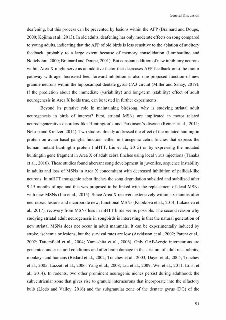
General Discussion
51
deafening, but this process can be prevented by lesions within the AFP (Brainard and Doupe,
2000; Kojima et al., 2013). In old adults, deafening has only moderate effects on song compared
to young adults, indicating that the AFP of old birds is less sensitive to the ablation of auditory
feedback, probably to a large extent because of memory consolidation (Lombardino and
Nottebohm, 2000; Brainard and Doupe, 2001). But constant addition of new inhibitory neurons
within Area X might serve as an additive factor that decreases AFP feedback onto the motor
pathway with age. Increased feed forward inhibition is also one proposed function of new
granule neurons within the hippocampal dentate gyrus-CA3 circuit (Miller and Sahay, 2019).
If the prediction about the immediate (variability) and long-term (stability) effect of adult
neurogenesis in Area X holds true, can be tested in further experiments.
Beyond its putative role in maintaining birdsong, why is studying striatal adult
neurogenesis in birds of interest? First, striatal MSNs are implicated in motor related
neurodegenerative disorders like Huntington’s and Parkinson’s disease (Reiner et al., 2011;
Nelson and Kreitzer, 2014). Two studies already addressed the effect of the mutated huntingtin
protein on avian basal ganglia function, either in transgenic zebra finches that express the
human mutant huntingtin protein (mHTT, Liu et al., 2015) or by expressing the mutated
huntingtin gene fragment in Area X of adult zebra finches using local virus injections (Tanaka
et al., 2016). These studies found aberrant song development in juveniles, sequence instability
in adults and loss of MSNs in Area X concomitant with decreased inhibition of pallidal-like
neurons. In mHTT transgenic zebra finches the song degradation subsided and stabilized after
9-15 months of age and this was proposed to be linked with the replacement of dead MSNs
with new MSNs (Liu et al., 2015). Since Area X recovers extensively within six months after
neurotoxic lesions and incorporate new, functional MSNs (Kubikova et al., 2014; Lukacova et
al., 2017), recovery from MSNs loss in mHTT birds seems possible. The second reason why
studying striatal adult neurogenesis in songbirds is interesting is that the natural generation of
new striatal MSNs does not occur in adult mammals. It can be experimentally induced by
stroke, ischemia or lesions, but the survival rates are low (Arvidsson et al., 2002; Parent et al.,
2002; Tattersfield et al., 2004; Yamashita et al., 2006). Only GABAergic interneurons are
generated under natural conditions and after brain damage in the striatum of adult rats, rabbits,
monkeys and humans (Bédard et al., 2002; Tonchev et al., 2003; Dayer et al., 2005; Tonchev
et al., 2005; Luzzati et al., 2006; Yang et al., 2008; Liu et al., 2009; Wei et al., 2011; Ernst et
al., 2014). In rodents, two other prominent neurogenic niches persist during adulthood; the
subventricular zone that gives rise to granule interneurons that incorporate into the olfactory
bulb (Lledo and Valley, 2016) and the subgranular zone of the dentate gyrus (DG) of the
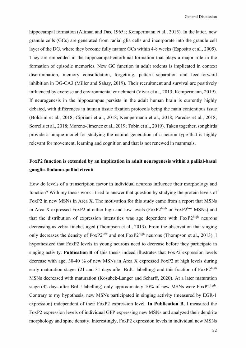
General Discussion
52
hippocampal formation (Altman and Das, 1965a; Kempermann et al., 2015). In the latter, new
granule cells (GCs) are generated from radial glia cells and incorporate into the granule cell
layer of the DG, where they become fully mature GCs within 4-8 weeks (Esposito et al., 2005).
They are embedded in the hippocampal-entorhinal formation that plays a major role in the
formation of episodic memories. New GC function in adult rodents is implicated in context
discrimination, memory consolidation, forgetting, pattern separation and feed-forward
inhibition in DG-CA3 (Miller and Sahay, 2019). Their recruitment and survival are positively
influenced by exercise and environmental enrichment (Vivar et al., 2013; Kempermann, 2019).
If neurogenesis in the hippocampus persists in the adult human brain is currently highly
debated, with differences in human tissue fixation protocols being the main contentious issue
(Boldrini et al., 2018; Cipriani et al., 2018; Kempermann et al., 2018; Paredes et al., 2018;
Sorrells et al., 2018; Moreno-Jimenez et al., 2019; Tobin et al., 2019). Taken together, songbirds
provide a unique model for studying the natural generation of a neuron type that is highly
relevant for movement, learning and cognition and that is not renewed in mammals.
FoxP2 function is extended by an implication in adult neurogenesis within a pallial-basal ganglia-thalamo-pallial circuit How do levels of a transcription factor in individual neurons influence their morphology and
function? With my thesis work I tried to answer that question by studying the protein levels of
FoxP2 in new MSNs in Area X. The motivation for this study came from a report that MSNs
in Area X expressed FoxP2 at either high and low levels (FoxP2high or FoxP2low MSNs) and
that the distribution of expression intensities was age dependent with FoxP2high neurons
decreasing as zebra finches aged (Thompson et al., 2013). From the observation that singing
only decreases the density of FoxP2low and not FoxP2high neurons (Thompson et al., 2013), I
hypothesized that FoxP2 levels in young neurons need to decrease before they participate in
singing activity. Publication B of this thesis indeed illustrates that FoxP2 expression levels
decrease with age; 30-40 % of new MSNs in Area X expressed FoxP2 at high levels during
early maturation stages (21 and 31 days after BrdU labelling) and this fraction of FoxP2high
MSNs decreased with maturation (Kosubek-Langer and Scharff, 2020). At a later maturation
stage (42 days after BrdU labelling) only approximately 10% of new MSNs were FoxP2high.
Contrary to my hypothesis, new MSNs participated in singing activity (measured by EGR-1
expression) independent of their FoxP2 expression level. In Publication B, I measured the
FoxP2 expression levels of individual GFP expressing new MSNs and analyzed their dendrite
morphology and spine density. Interestingly, FoxP2 expression levels in individual new MSNs
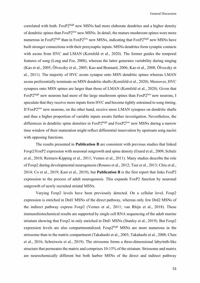
General Discussion
53
correlated with both. FoxP2high new MSNs had more elaborate dendrites and a higher density
of dendritic spines than FoxP2low new MSNs. In detail, the mature mushroom spines were more
numerous in FoxP2high than in FoxP2low new MSNs, indicating that FoxP2high new MSNs have
built stronger connections with their presynaptic inputs. MSNs dendrites form synaptic contacts
with axons from HVC and LMAN (Kornfeld et al., 2020). The former guides the temporal
features of song (Long and Fee, 2008), whereas the latter generates variability during singing
(Kao et al., 2005; Ölveczky et al., 2005; Kao and Brainard, 2006; Kao et al., 2008; Ölveczky et
al., 2011). The majority of HVC axons synapse onto MSN dendritic spines whereas LMAN
axons preferentially terminate on MSN dendritic shafts (Kornfeld et al., 2020). Moreover, HVC
synapses onto MSN spines are larger than those of LMAN (Kornfeld et al., 2020). Given that
FoxP2high new neurons had more of the large mushroom spines than FoxP2low new neurons, I
speculate that they receive more inputs form HVC and become tightly entrained to song timing.
If FoxP2low new neurons, on the other hand, receive more LMAN synapses on dendritic shafts
and thus a higher proportion of variable inputs awaits further investigation. Nevertheless, the
differences in dendritic spine densities in FoxP2high and FoxP2low new MSNs during a narrow
time window of their maturation might reflect differential innervation by upstream song nuclei
with opposing functions.
The results presented in Publication B are consistent with previous studies that linked
Foxp2/FoxP2 expression with neuronal outgrowth and spine density (Enard et al., 2009; Schulz
et al., 2010; Reimers-Kipping et al., 2011; Vernes et al., 2011). Many studies describe the role
of Foxp2 during developmental neurogenesis (Rousso et al., 2012; Tsui et al., 2013; Chiu et al.,
2014; Co et al., 2019; Kast et al., 2019), but Publication B is the first report that links FoxP2
expression to the process of adult neurogenesis. This expands FoxP2 function by neuronal
outgrowth of newly recruited striatal MSNs.
Varying Foxp2 levels have been previously detected. On a cellular level, Foxp2
expression is enriched in Drd1 MSNs of the direct pathway, whereas only few Drd2 MSNs of
the indirect pathway express Foxp2 (Vernes et al., 2011; van Rhijn et al., 2018). These
immunohistochemical results are supported by single cell RNA sequencing of the adult murine
striatum showing that Foxp2 in only enriched in Drd1 MSNs (Stanley et al., 2019). But Foxp2
expression levels are also compartmentalized; Foxp2high MSNs are more numerous in the
striosome than in the matrix compartment (Takahashi et al., 2003; Takahashi et al., 2008; Chen
et al., 2016; Schreiweis et al., 2019). The striosome forms a three-dimensional labyrinth-like
structure that permeates the matrix and comprises 10-15% of the striatum. Striosome and matrix
are neurochemically different but both harbor MSNs of the direct and indirect pathway
General Discussion
54
(Brimblecombe and Cragg, 2017). Dopamine levels are higher in the matrix than in the
striosomes (Salinas et al., 2016). In genetically engineered mice that carry humanized Foxp2
alleles (Foxp2hum/hum) the density of Foxp2high MSNs increased in the striosome of the dorsal
striatum in comparison to wildtype littermates. In the ventral striatum, however, the density of
Foxp2high MSNs increased in the matrix, not in the striosome. It is not clear which
(developmental) processes lead to the increase in Foxp2high MSNs, nor in which MSNs subtype
Foxp2 expression is elevated in Foxp2hum/hum mice. Further, it needs to be determined how
increased Foxp2 levels relate to the increased dendrite length found in striatal MSNs and other
neurons of the striatal-basal ganglia circuit of Foxp2hum/hum mice (Enard et al., 2009; Reimers-
Kipping et al., 2011).
In Publication B, I present data that FoxP2 expression levels distinguish fractions of
new MSNs in Area X during a short, specific time window of their maturation. Older, mature
MSNs cannot be distinguished by their FoxP2 expression levels. I argue that these early
differences in FoxP2 expression and concomitant morphological features may imprint young,
new MSNs and give rise to different MSNs subtypes, as is the case in mice. In mice, Drd1 direct
and Drd2 indirect pathway striatal MSNs (see Fig. 3 of the Introduction) cannot only be
segregated by the expression of dopamine receptors but by many other characteristics.
Dopamine modulates the response of MSNs to cortical inputs; Drd1s increase the intrinsic
excitability of MSNs, whereas Drd2s decrease the intrinsic excitability of MSNs (Surmeier et
al., 2014). Direct pathway activation promotes movement, while indirect pathway activation
suppresses motor behaviors (Kravitz et al., 2010). Further, direct and indirect MSNs differ in
electrophysiological properties (Cepeda et al., 2008; Day et al., 2008), morphology (Gertler et
al., 2008), synaptic plasticity (Kreitzer and Malenka, 2007; Shen et al., 2008), cortical inputs
(Wall et al., 2013) and brain-wide responses (Lee et al., 2016).
There is nothing known about the connectivity or electrophysiology of MSNs subtypes
in birds, although their existence has been presumed (Gale and Perkel, 2010; Pidoux et al.,
2015). But gene expression data provides hints for the existence of MSNs subtypes. Single
nucleus RNA sequencing identified five MSN cluster, of which two resembled the gene
expression profile of direct and indirect MSNs of the murine striatum (Xiao et al., 2020).
However, mRNA and protein abundance do not always correlate (Vogel and Marcotte, 2012)
and RNA profiles reflect one snapshot of a dynamic system, hence RNA profiling alone is
insufficient to determine neuronal subtypes. Corresponding to the murine basal ganglia, avian
indirect MSNs would exclusively innervate indirect pallidal-like neurons that inhibit the direct
pallidal-projection neurons. Following evidence underpin the hypothesis that early FoxP2
General Discussion
55
expression levels may indicate MSN subtype specification. First, mouse Drd1 direct MSNs and
corresponding direct MSNs in zebra finches both express FoxP2 at high levels whereas Drd2
indirect MSNs in mammals and birds express only low levels of Foxp2 (Vernes et al., 2011;
van Rhijn et al., 2018; Xiao et al., 2020). Second, both FoxP2high new MSNs in zebra finches
and Drd1 MSNs in mice had (a) more intersections in the Sholl analysis, which indicates a
higher dendritic complexity (b) a higher cumulative dendritic length, and (c) more primary
dendrites compared to FoxP2low new MSNs and Drd2 MSNs, respectively (Gertler et al., 2008;
Kosubek-Langer and Scharff, 2020). To test if this hypothesis holds true it requires suitable
markers, tracings and electrophysiological recordings. Unfortunately, a single avian MSNs
often expresses multiple dopamine receptors (Kubikova et al., 2010; Kosubek-Langer et al.,
2017; Xiao et al., 2020) and therefore MSNs in songbirds cannot be distinguished by their
dopamine receptors, which is possible in rodent MSNs. It is however possible that a
combination of several marker proteins will reveal MSN subtypes also in birds. Another
challenge in the detection of MSN subtypes is the fact that their targets, the direct and indirect
pallidal-like projection neurons, lie within Area X and this complicates their detection via
retrograde tracings. But since MSN subtypes may have different roles during song learning as
well as in the modulation of variability during directed and undirected singing, their detection
and how FoxP2 expression is involved in their subtype specification is of particular interest.
In summary, proper amounts of Foxp2 protein are crucial for a wide array of
neurobiological processes including brain development and motor learning in mammals
(French et al., 2007; Fujita et al., 2008; Groszer et al., 2008; Enard et al., 2009; Reimers-
Kipping et al., 2011; French et al., 2012; Kurt et al., 2012; Tsui et al., 2013; Chiu et al., 2014;
Chen et al., 2016; French et al., 2019; Kast et al., 2019). In zebra finches, the tight regulation
of FoxP2 protein levels is particularly important for its on-line function during song learning in
juveniles and for context-dependent song variability in adults (Haesler et al., 2004; Haesler et
al., 2007; Murugan et al., 2013; Heston and White, 2015; Day et al., 2019). In Publication B,
I present the first evidence that FoxP2 protein levels are as well relevant during the process of
striatal adult neurogenesis (Kosubek-Langer and Scharff, 2020), which broadens FoxP2
function in adult songbirds.

56
References
57
References Abdi, A., Mallet, N., Mohamed, F.Y., Sharott, A., Dodson, P.D., Nakamura, K.C., et al. (2015).
Prototypic and arkypallidal neurons in the dopamine-intact external globus pallidus. J
Neurosci 35(17), 6667-6688. doi: 10.1523/JNEUROSCI.4662-14.2015.
Adam, I., Mendoza, E., Kobalz, U., Wohlgemuth, S., and Scharff, C. (2016). FoxP2 directly regulates
the reelin receptor VLDLR developmentally and by singing. Mol Cell Neurosci 74, 96-105.
doi: 10.1016/j.mcn.2016.04.002.
Ali, F., Otchy, T.M., Pehlevan, C., Fantana, A.L., Burak, Y., and Olveczky, B.P. (2013). The basal
ganglia is necessary for learning spectral, but not temporal, features of birdsong. Neuron
80(2), 494-506. doi: 10.1016/j.neuron.2013.07.049.
Altman, J. (1962a). Are new neurons formed in the brains of adult mammals? Science 135(3509), 1127-1128. doi: 10.1126/science.135.3509.1127.
Altman, J. (1962b). Autoradiographic study of degenerative and regenerative proliferation of neuroglia
cells with tritiated thymidine. Experimental Neurology 5(4), 302-318. doi: 10.1016/0014-
4886(62)90040-7.
Altman, J. (1963). Autoradiographic investigation of cell proliferation in the brains of rats and cats.
Anat Rec 145, 573-591. doi: 10.1002/ar.1091450409.
Altman, J. (1969). Autoradiographic and histological studies of postnatal neurogenesis. IV. Cell
proliferation and migration in the anterior forebrain, with special reference to persisting
neurogenesis in the olfactory bulb. J Comp Neurol 137(4), 433-457. doi:
10.1002/cne.901370404.
Altman, J. (2011). "The Discovery of Adult Mammalian Neurogenesis," in Neurogenesis in the Adult
Brain I: Neurobiology, eds. T. Seki, K. Sawamoto, J.M. Parent & A. Alvarez-Buylla. (Tokyo:
Springer Japan), 3-46.
Altman, J., and Das, G.D. (1965a). Autoradiographic and histological evidence of postnatal
hippocampal neurogenesis in rats. J Comp Neurol 124(3), 319-335. doi:
10.1002/cne.901240303.
Altman, J., and Das, G.D. (1965b). Post-natal origin of microneurones in the rat brain. Nature
207(5000), 953-956. doi: 10.1038/207953a0.
References
58
Altman, J., and Das, G.D. (1967). Postnatal neurogenesis in the guinea-pig. Nature 214(5093), 1098-
1101. doi: 10.1038/2141098a0.
Alvarez-Borda, B., and Nottebohm, F. (2002). Gonads and singing play separate, additive roles in new
neuron recruitment in adult canary brain. J Neurosci 22(19), 8684-8690. doi:
10.1523/JNEUROSCI.22-19-08684.2002.
Alvarez-Buylla, A., and Kirn, J.R. (1997). Birth, migration, incorporation, and death of vocal control
neurons in adult songbirds. J Neurobiol 33(5), 585-601. doi: 10.1002/(SICI)1097-
4695(19971105)33:5<585::AID-NEU7>3.0.CO;2-0.
Alvarez-Buylla, A., Ling, C.Y., and Yu, W.S. (1994). Contribution of neurons born during embryonic,
juvenile, and adult life to the brain of adult canaries: regional specificity and delayed birth of
neurons in the song-control nuclei. J Comp Neurol 347(2), 233-248. doi:
10.1002/cne.903470207.
Alvarez-Buylla, A., and Nottebohm, F. (1988). Migration of young neurons in adult avian brain.
Nature 335(6188), 353-354. doi: 10.1038/335353a0.
Alvarez-Buylla, A., Theelen, M., and Nottebohm, F. (1988a). Birth of projection neurons in the higher
vocal center of the canary forebrain before, during, and after song learning. Proc Natl Acad
Sci U S A 85(22), 8722-8726. doi: 10.1073/pnas.85.22.8722.
Alvarez-Buylla, A., Theelen, M., and Nottebohm, F. (1988b). Mapping of radial glia and of a new cell
type in adult canary brain. J Neurosci 8(8), 2707-2712. doi: 10.1523/JNEUROSCI.08-08-
02707.1988.
Alvarez-Buylla, A., Theelen, M., and Nottebohm, F. (1990). Proliferation "hot spots" in adult avian
ventricular zone reveal radial cell division. Neuron 5(1), 101-109. doi: 10.1016/0896-
6273(90)90038-h.
Arvidsson, A., Collin, T., Kirik, D., Kokaia, Z., and Lindvall, O. (2002). Neuronal replacement from
endogenous precursors in the adult brain after stroke. Nat Med 8(9), 963-970. doi:
10.1038/nm747.
Barnea, A., and Pravosudov, V. (2011). Birds as a model to study adult neurogenesis: bridging
evolutionary, comparative and neuroethological approaches. Eur J Neurosci 34(6), 884-907.
doi: 10.1111/j.1460-9568.2011.07851.x.
References
59
Bédard, A., Cossette, M., Lévesque, M., and Parent, A. (2002). Proliferating cells can differentiate into
neurons in the striatum of normal adult monkey. Neuroscience Letters 328(3), 213-216. doi:
10.1016/s0304-3940(02)00530-x.
Boldrini, M., Fulmore, C.A., Tartt, A.N., Simeon, L.R., Pavlova, I., Poposka, V., et al. (2018). Human
Hippocampal Neurogenesis Persists throughout Aging. Cell Stem Cell 22(4), 589-599 e585.
doi: 10.1016/j.stem.2018.03.015.
Bolhuis, J.J., Okanoya, K., and Scharff, C. (2010). Twitter evolution: converging mechanisms in
birdsong and human speech. Nat Rev Neurosci 11(11), 747-759. doi: 10.1038/nrn2931.
Bottjer, S.W. (1993). The distribution of tyrosine hydroxylase immunoreactivity in the brains of male
and female zebra finches. J Neurobiol 24(1), 51-69. doi: 10.1002/neu.480240105.
Bottjer, S.W., Miesner, E.A., and Arnold, A.P. (1984). Forebrain lesions disrupt development but not
maintenance of song in passerine birds. Science 224(4651), 901-903. doi:
10.1126/science.6719123.
Bouyer, J.J., Park, D.H., Joh, T.H., and Pickel, V.M. (1984). Chemical and structural analysis of the
relation between cortical inputs and tyrosine hydroxylase-containing terminals in rat
neostriatum. Brain Research 302(2), 267-275. doi: 10.1016/0006-8993(84)90239-7.
Brainard, M.S., and Doupe, A.J. (2000). Interruption of a basal ganglia-forebrain circuit prevents
plasticity of learned vocalizations. Nature 404(6779), 762-766. doi: 10.1038/35008083.
Brainard, M.S., and Doupe, A.J. (2001). Postlearning consolidation of birdsong: stabilizing effects of
age and anterior forebrain lesions. J Neurosci 21(7), 2501-2517. doi:
10.1523/JNEUROSCI.21-07-02501.2001.
Brainard, M.S., and Doupe, A.J. (2002). What songbirds teach us about learning. Nature 417(6886), 351-358. doi: 10.1038/417351a.
Brainard, M.S., and Doupe, A.J. (2013). Translating birdsong: songbirds as a model for basic and
applied medical research. Annu Rev Neurosci 36, 489-517. doi: 10.1146/annurev-neuro-
060909-152826.
Brenowitz, E.A. (2014). Transsynaptic trophic effects of steroid hormones in an avian model of adult
brain plasticity. Front Neuroendocrinol. doi: 10.1016/j.yfrne.2014.09.003.
Brimblecombe, K.R., and Cragg, S.J. (2017). The Striosome and Matrix Compartments of the
Striatum: A Path through the Labyrinth from Neurochemistry toward Function. ACS Chem
Neurosci 8(2), 235-242. doi: 10.1021/acschemneuro.6b00333.
References
60
Burd, G.D., and Nottebohm, F. (1985). Ultrastructural characterization of synaptic terminals formed
on newly generated neurons in a song control nucleus of the adult canary forebrain. J Comp
Neurol 240(2), 143-152. doi: 10.1002/cne.902400204.
Burek, M.J., Nordeen, K.W., and Nordeen, E.J. (1994). Ontogeny of sex differences among newly-
generated neurons of the juvenile avian brain. Brain Res Dev Brain Res 78(1), 57-64. doi:
10.1016/0165-3806(94)90009-4.
Calabresi, P., Picconi, B., Tozzi, A., Ghiglieri, V., and Di Filippo, M. (2014). Direct and indirect
pathways of basal ganglia: a critical reappraisal. Nat Neurosci 17(8), 1022-1030. doi:
10.1038/nn.3743.
Campbell, P., Reep, R.L., Stoll, M.L., Ophir, A.G., and Phelps, S.M. (2009). Conservation and
diversity of Foxp2 expression in muroid rodents: functional implications. J Comp Neurol
512(1), 84-100. doi: 10.1002/cne.21881.
Cepeda, C., Andre, V.M., Yamazaki, I., Wu, N., Kleiman-Weiner, M., and Levine, M.S. (2008).
Differential electrophysiological properties of dopamine D1 and D2 receptor-containing
striatal medium-sized spiny neurons. Eur J Neurosci 27(3), 671-682. doi: 10.1111/j.1460-
9568.2008.06038.x.
Chen, Y.C., Kuo, H.Y., Bornschein, U., Takahashi, H., Chen, S.Y., Lu, K.M., et al. (2016). Foxp2
controls synaptic wiring of corticostriatal circuits and vocal communication by opposing
Mef2c. Nat Neurosci. doi: 10.1038/nn.4380.
Chiu, Y.C., Li, M.Y., Liu, Y.H., Ding, J.Y., Yu, J.Y., and Wang, T.W. (2014). Foxp2 regulates
neuronal differentiation and neuronal subtype specification. Dev Neurobiol 74(7), 723-738.
doi: 10.1002/dneu.22166.
Cipriani, S., Ferrer, I., Aronica, E., Kovacs, G.G., Verney, C., Nardelli, J., et al. (2018). Hippocampal
Radial Glial Subtypes and Their Neurogenic Potential in Human Fetuses and Healthy and
Alzheimer's Disease Adults. Cereb Cortex 28(7), 2458-2478. doi: 10.1093/cercor/bhy096.
Citri, A., and Malenka, R.C. (2008). Synaptic plasticity: multiple forms, functions, and mechanisms.
Neuropsychopharmacology 33(1), 18-41. doi: 10.1038/sj.npp.1301559.
Co, M., Hickey, S.L., Kulkarni, A., Harper, M., and Konopka, G. (2019). Cortical Foxp2 Supports
Behavioral Flexibility and Developmental Dopamine D1 Receptor Expression. Cereb Cortex.
doi: 10.1093/cercor/bhz209.
References
61
Cooper, B.G., and Goller, F. (2006). Physiological insights into the social-context-dependent changes
in the rhythm of the song motor program. J Neurophysiol 95(6), 3798-3809. doi:
10.1152/jn.01123.2005.
Day, M., Wokosin, D., Plotkin, J.L., Tian, X., and Surmeier, D.J. (2008). Differential excitability and
modulation of striatal medium spiny neuron dendrites. J Neurosci 28(45), 11603-11614. doi:
10.1523/JNEUROSCI.1840-08.2008.
Day, N.F., Hobbs, T.G., Heston, J.B., and White, S.A. (2019). Beyond Critical Period Learning:
Striatal FoxP2 Affects the Active Maintenance of Learned Vocalizations in Adulthood.
eNeuro 6(2). doi: 10.1523/ENEURO.0071-19.2019.
Dayer, A.G., Cleaver, K.M., Abouantoun, T., and Cameron, H.A. (2005). New GABAergic
interneurons in the adult neocortex and striatum are generated from different precursors. J Cell
Biol 168(3), 415-427. doi: 10.1083/jcb.200407053.
Demontis, D., Walters, R.K., Martin, J., Mattheisen, M., Als, T.D., Agerbo, E., et al. (2019).
Discovery of the first genome-wide significant risk loci for attention deficit/hyperactivity
disorder. Nat Genet 51(1), 63-75. doi: 10.1038/s41588-018-0269-7.
Devanna, P., Middelbeek, J., and Vernes, S.C. (2014). FOXP2 drives neuronal differentiation by
interacting with retinoic acid signaling pathways. Front Cell Neurosci 8, 305. doi:
10.3389/fncel.2014.00305.
Doetsch, F., and Scharff, C. (2001). Challenges for brain repair: insights from adult neurogenesis in
birds and mammals. Brain Behav Evol 58(5), 306-322. doi: DOI: 10.1159/000057572.
Ehninger, D., Li, W., Fox, K., Stryker, M.P., and Silva, A.J. (2008). Reversing neurodevelopmental
disorders in adults. Neuron 60(6), 950-960. doi: 10.1016/j.neuron.2008.12.007.
Enard, W. (2011). FOXP2 and the role of cortico-basal ganglia circuits in speech and language
evolution. Curr Opin Neurobiol 21(3), 415-424. doi: 10.1016/j.conb.2011.04.008.
Enard, W., Gehre, S., Hammerschmidt, K., Holter, S.M., Blass, T., Somel, M., et al. (2009). A
humanized version of Foxp2 affects cortico-basal ganglia circuits in mice. Cell 137(5), 961-
971. doi: 10.1016/j.cell.2009.03.041.
Ernst, A., Alkass, K., Bernard, S., Salehpour, M., Perl, S., Tisdale, J., et al. (2014). Neurogenesis in
the striatum of the adult human brain. Cell 156(5), 1072-1083. doi:
10.1016/j.cell.2014.01.044.
References
62
Esposito, M.S., Piatti, V.C., Laplagne, D.A., Morgenstern, N.A., Ferrari, C.C., Pitossi, F.J., et al.
(2005). Neuronal differentiation in the adult hippocampus recapitulates embryonic
development. J Neurosci 25(44), 10074-10086. doi: 10.1523/JNEUROSCI.3114-05.2005.
Farries, M.A., Ding, L., and Perkel, D.J. (2005). Evidence for "direct" and "indirect" pathways through
the song system basal ganglia. J Comp Neurol 484(1), 93-104. doi: 10.1002/cne.20464.
Farries, M.A., and Perkel, D.J. (2000). Electrophysiological properties of avian basal ganglia neurons
recorded in vitro. J Neurophysiol 84(5), 2502-2513. doi: 10.1152/jn.2000.84.5.2502.
Farries, M.A., and Perkel, D.J. (2002). A telencephalic nucleus essential for song learning contains
neurons with physiological characteristics of both striatum and globus pallidus. J Neurosci
22(9), 3776-3787. doi: 20026269.
Fee, M.S., and Scharff, C. (2010). The Songbird as a Model for the Generation and Learning of
Complex Sequential Behaviors. ILAR 51, 362-377. doi: 10.1093/ilar.51.4.362.
Ferland, R.J., Cherry, T.J., Preware, P.O., Morrisey, E.E., and Walsh, C.A. (2003). Characterization of
Foxp2 and Foxp1 mRNA and protein in the developing and mature brain. J Comp Neurol
460(2), 266-279. doi: 10.1002/cne.10654.
French, C.A., Groszer, M., Preece, C., Coupe, A.M., Rajewsky, K., and Fisher, S.E. (2007).
Generation of mice with a conditional Foxp2 null allele. Genesis 45(7), 440-446. doi:
10.1002/dvg.20305.
French, C.A., Jin, X., Campbell, T.G., Gerfen, E., Groszer, M., Fisher, S.E., et al. (2012). An
aetiological Foxp2 mutation causes aberrant striatal activity and alters plasticity during skill
learning. Mol Psychiatry 17(11), 1077-1085. doi: 10.1038/mp.2011.105.
French, C.A., Vinueza Veloz, M.F., Zhou, K., Peter, S., Fisher, S.E., Costa, R.M., et al. (2019).
Differential effects of Foxp2 disruption in distinct motor circuits. Mol Psychiatry 24(3), 447-
462. doi: 10.1038/s41380-018-0199-x.
Freund, T.F., Powell, J.F., and Smith, A.D. (1984). Tyrosine hydroxylase-immunoreactive boutons in
synaptic contact with identified striatonigral neurons, with particular reference to dendritic
spines. Neuroscience 13(4), 1189-1215. doi: 10.1016/0306-4522(84)90294-x.
Frisen, J. (2016). Neurogenesis and Gliogenesis in Nervous System Plasticity and Repair. Annu Rev
Cell Dev Biol 32, 127-141. doi: 10.1146/annurev-cellbio-111315-124953.
Fujita, E., Tanabe, Y., Shiota, A., Ueda, M., Suwa, K., Momoi, M.Y., et al. (2008). Ultrasonic
vocalization impairment of Foxp2 (R552H) knockin mice related to speech-language disorder
References
63
and abnormality of Purkinje cells. Proc Natl Acad Sci U S A 105(8), 3117-3122. doi:
10.1073/pnas.0712298105.
Gadagkar, V., Puzerey, P.A., Chen, R., Baird-Daniel, E., Farhang, A.R., and Goldberg, J.H. (2016).
Dopamine neurons encode performance error in singing birds. Science 354(6317), 1278-1282.
doi: 10.1126/science.aah6837.
Gahr, M., and Garcia-Segura, L.M. (1996). Testosterone-dependent increase of gap-junctions in HVC
neurons of adult female canaries. Brain Res 712(1), 69-73. doi: 10.1016/0006-8993(95)01448-
9.
Gale, S.D., and Perkel, D.J. (2010). Anatomy of a songbird basal ganglia circuit essential for vocal
learning and plasticity. J Chem Neuroanat 39(2), 124-131. doi:
10.1016/j.jchemneu.2009.07.003.
Gale, S.D., Person, A.L., and Perkel, D.J. (2008). A novel basal ganglia pathway forms a loop linking
a vocal learning circuit with its dopaminergic input. The Journal of comparative neurology
508(5), 824-839. doi: 10.1002/cne.21700.
Garcia-Calero, E., Botella-Lopez, A., Bahamonde, O., Perez-Balaguer, A., and Martinez, S. (2016).
FoxP2 protein levels regulate cell morphology changes and migration patterns in the
vertebrate developing telencephalon. Brain Struct Funct 221(6), 2905-2917. doi:
10.1007/s00429-015-1079-7.
Garcia-Calero, E., and Scharff, C. (2013). Calbindin expression in developing striatum of zebra
finches and its relation to the formation of area X. J Comp Neurol 521(2), 326-341. doi:
10.1002/cne.23174.
Ge, S., Pradhan, D.A., Ming, G.L., and Song, H. (2007). GABA sets the tempo for activity-dependent
adult neurogenesis. Trends Neurosci 30(1), 1-8. doi: 10.1016/j.tins.2006.11.001.
Gertler, T.S., Chan, C.S., and Surmeier, D.J. (2008). Dichotomous anatomical properties of adult
striatal medium spiny neurons. J Neurosci 28(43), 10814-10824. doi:
10.1523/JNEUROSCI.2660-08.2008.
Gokce, O., Stanley, G.M., Treutlein, B., Neff, N.F., Camp, J.G., Malenka, R.C., et al. (2016). Cellular
Taxonomy of the Mouse Striatum as Revealed by Single-Cell RNA-Seq. Cell Rep 16(4),
1126-1137. doi: 10.1016/j.celrep.2016.06.059.
Goldberg, J.H., Adler, A., Bergman, H., and Fee, M.S. (2010). Singing-related neural activity
distinguishes two putative pallidal cell types in the songbird basal ganglia: comparison to the
References
64
primate internal and external pallidal segments. J Neurosci 30(20), 7088-7098. doi:
10.1523/JNEUROSCI.0168-10.2010.
Goldberg, J.H., and Fee, M.S. (2010). Singing-related neural activity distinguishes four classes of
putative striatal neurons in the songbird basal ganglia. J Neurophysiol 103(4), 2002-2014. doi:
10.1152/jn.01038.2009.
Goldman, S.A., and Nottebohm, F. (1983). Neuronal production, migration, and differentiation in a
vocal control nucleus of the adult female canary brain. Proc Natl Acad Sci U S A 80(8), 2390-
2394. doi: 10.1073/pnas.80.8.2390.
Gould, E. (2007). How widespread is adult neurogenesis in mammals? Nature Reviews Neuroscience
8(6), 481-488. doi: 10.1038/nrn2147.
Groszer, M., Keays, D.A., Deacon, R.M., de Bono, J.P., Prasad-Mulcare, S., Gaub, S., et al. (2008).
Impaired synaptic plasticity and motor learning in mice with a point mutation implicated in
human speech deficits. Curr Biol 18(5), 354-362. doi: 10.1016/j.cub.2008.01.060.
Haesler, S., Rochefort, C., Georgi, B., Licznerski, P., Osten, P., and Scharff, C. (2007). Incomplete
and inaccurate vocal imitation after knockdown of FoxP2 in songbird basal ganglia nucleus
Area X. PLoS Biol 5(12), e321. doi: 10.1371/journal.pbio.0050321.
Haesler, S., Wada, K., Nshdejan, A., Morrisey, E.E., Lints, T., Jarvis, E.D., et al. (2004). FoxP2
expression in avian vocal learners and non-learners. J Neurosci 24(13), 3164-3175. doi:
10.1523/JNEUROSCI.4369-03.2004.
Heston, J.B., and White, S.A. (2015). Behavior-Linked FoxP2 Regulation Enables Zebra Finch Vocal
Learning. J Neurosci 35(7), 2885-2894. doi: 10.1523/JNEUROSCI.3715-14.2015.
Ho, V.M., Lee, J.A., and Martin, K.C. (2011). The cell biology of synaptic plasticity. Science
334(6056), 623-628. doi: 10.1126/science.1209236.
Hurley, P., Pytte, C., and Kirn, J.R. (2008). Nest of origin predicts adult neuron addition rates in the
vocal control system of the zebra finch. Brain Behav Evol 71(4), 263-270. doi:
10.1159/000127046.
Janik, V.M., and Slater, P.J.B. (1997). "Vocal Learning in Mammals," in Advances in the Study of
Behavior, eds. P.J.B. Slater, J.S. Rosenblatt, C.T. Snowdon & M. Milinski. Academic Press),
59-99.
Janik, V.M., and Slater, P.J.B. (2000). The different roles of social learning in vocal communication.
Animal Behaviour 60(1), 1-11. doi: https://doi.org/10.1006/anbe.2000.1410.
References
65
Jarvis, E.D., Scharff, C., Grossman, M.R., Ramos, J.A., and Nottebohm, F. (1998). For whom the bird
sings: context-dependent gene expression. Neuron 21(4), 775-788. doi: 10.1016/s0896-
6273(00)80594-2.
Kaestner, K.H., Knochel, W., and Martinez, D.E. (2000). Unified nomenclature for the winged
helix/forkhead transcription factors. Genes Dev 14(2), 142-146.
Kao, M.H., and Brainard, M.S. (2006). Lesions of an avian basal ganglia circuit prevent context-
dependent changes to song variability. J Neurophysiol 96(3), 1441-1455. doi:
10.1152/jn.01138.2005.
Kao, M.H., Doupe, A.J., and Brainard, M.S. (2005). Contributions of an avian basal ganglia-forebrain
circuit to real-time modulation of song. Nature 433(7026), 638-643. doi:
10.1038/nature03127.
Kao, M.H., Wright, B.D., and Doupe, A.J. (2008). Neurons in a forebrain nucleus required for vocal
plasticity rapidly switch between precise firing and variable bursting depending on social
context. J Neurosci 28(49), 13232-13247. doi: 10.1523/JNEUROSCI.2250-08.2008.
Kast, R.J., Lanjewar, A.L., Smith, C.D., and Levitt, P. (2019). FOXP2 exhibits projection neuron class
specific expression, but is not required for multiple aspects of cortical histogenesis. Elife 8.
doi: 10.7554/eLife.42012.
Kearney, M.G., Warren, T.L., Hisey, E., Qi, J., and Mooney, R. (2019). Discrete Evaluative and
Premotor Circuits Enable Vocal Learning in Songbirds. Neuron. doi:
10.1016/j.neuron.2019.07.025.
Keilani, S., Chandwani, S., Dolios, G., Bogush, A., Beck, H., Hatzopoulos, A.K., et al. (2012). Egr-1
induces DARPP-32 expression in striatal medium spiny neurons via a conserved intragenic
element. J Neurosci 32(20), 6808-6818. doi: 10.1523/JNEUROSCI.5448-11.2012.
Kelsch, W., Mosley, C.P., Lin, C.-W., and Lois, C. (2007). Distinct Mammalian Precursors Are
Committed to Generate Neurons with Defined Dendritic Projection Patterns. PLOS Biology
5(11), e300. doi: 10.1371/journal.pbio.0050300.
Kempermann, G. (2019). Environmental enrichment, new neurons and the neurobiology of
individuality. Nat Rev Neurosci 20(4), 235-245. doi: 10.1038/s41583-019-0120-x.
Kempermann, G., Gage, F.H., Aigner, L., Song, H., Curtis, M.A., Thuret, S., et al. (2018). Human
Adult Neurogenesis: Evidence and Remaining Questions. Cell Stem Cell 23(1), 25-30. doi:
10.1016/j.stem.2018.04.004.
References
66
Kempermann, G., Song, H., and Gage, F.H. (2015). Neurogenesis in the Adult Hippocampus. Cold
Spring Harb Perspect Biol 7(9), a018812. doi: 10.1101/cshperspect.a018812.
Kirn, J., O'Loughlin, B., Kasparian, S., and Nottebohm, F. (1994). Cell death and neuronal recruitment
in the high vocal center of adult male canaries are temporally related to changes in song. Proc
Natl Acad Sci U S A 91(17), 7844-7848. doi: 10.1073/pnas.91.17.7844.
Kirn, J.R., Fishman, Y., Sasportas, K., Alvarez-Buylla, A., and Nottebohm, F. (1999). Fate of new
neurons in adult canary high vocal center during the first 30 days after their formation. J Comp
Neurol 411(3), 487-494.
Kirn, J.R., and Nottebohm, F. (1993). Direct evidence for loss and replacement of projection neurons
in adult canary brain. J Neurosci 13(4), 1654-1663.
Kojima, S., Kao, M.H., and Doupe, A.J. (2013). Task-related "cortical" bursting depends critically on
basal ganglia input and is linked to vocal plasticity. Proc Natl Acad Sci U S A 110(12), 4756-
4761. doi: 10.1073/pnas.1216308110.
Kojima, S., Kao, M.H., Doupe, A.J., and Brainard, M.S. (2018). The Avian Basal Ganglia Are a
Source of Rapid Behavioral Variation That Enables Vocal Motor Exploration. J Neurosci
38(45), 9635-9647. doi: 10.1523/JNEUROSCI.2915-17.2018.
Kornfeld, J., Januszewski, M., Schubert, P., Jain, V., Denk, W., and Fee, M.S. (2020). An anatomical
substrate of credit assignment in reinforcement learning. bioRxiv. doi:
10.1101/2020.02.18.954354.
Kosubek-Langer, J., and Scharff, C. (2020). Dynamic FoxP2 levels in male zebra finches are linked to
morphology of adult-born Area X medium spiny neurons. Scientific Reports 10(1). doi:
10.1038/s41598-020-61740-6.
Kosubek-Langer, J., Schulze, L., and Scharff, C. (2017). Maturation, Behavioral Activation, and
Connectivity of Adult-Born Medium Spiny Neurons in a Striatal Song Nucleus. Front
Neurosci 11, 323. doi: 10.3389/fnins.2017.00323.
Kravitz, A.V., Freeze, B.S., Parker, P.R., Kay, K., Thwin, M.T., Deisseroth, K., et al. (2010).
Regulation of parkinsonian motor behaviours by optogenetic control of basal ganglia circuitry.
Nature 466(7306), 622-626. doi: 10.1038/nature09159.
Kreitzer, A.C., and Malenka, R.C. (2007). Endocannabinoid-mediated rescue of striatal LTD and
motor deficits in Parkinson's disease models. Nature 445(7128), 643-647. doi:
10.1038/nature05506.
References
67
Kubikova, L., Bosikova, E., Cvikova, M., Lukacova, K., Scharff, C., and Jarvis, E.D. (2014). Basal
ganglia function, stuttering, sequencing, and repair in adult songbirds. Sci Rep 4, 6590. doi:
10.1038/srep06590.
Kubikova, L., Wada, K., and Jarvis, E.D. (2010). Dopamine receptors in a songbird brain. J Comp
Neurol 518(6), 741-769. doi: 10.1002/cne.22255.
Kurt, S., Fisher, S.E., and Ehret, G. (2012). Foxp2 mutations impair auditory-motor association
learning. PLoS One 7(3), e33130. doi: 10.1371/journal.pone.0033130.
Lai, C.S., Fisher, S.E., Hurst, J.A., Vargha-Khadem, F., and Monaco, A.P. (2001). A forkhead-domain
gene is mutated in a severe speech and language disorder. Nature 413(6855), 519-523. doi:
10.1038/35097076.
Larson, T.A., Wang, T.W., Gale, S.D., Miller, K.E., Thatra, N.M., Caras, M.L., et al. (2013).
Postsynaptic neural activity regulates neuronal addition in the adult avian song control system.
Proc Natl Acad Sci U S A 110(41), 16640-16644. doi: 10.1073/pnas.1310237110.
Leblois, A., Wendel, B.J., and Perkel, D.J. (2010). Striatal dopamine modulates basal ganglia output
and regulates social context-dependent behavioral variability through D1 receptors. J Neurosci
30(16), 5730-5743. doi: 10.1523/JNEUROSCI.5974-09.2010.
Lee, H.J., Weitz, A.J., Bernal-Casas, D., Duffy, B.A., Choy, M., Kravitz, A.V., et al. (2016).
Activation of Direct and Indirect Pathway Medium Spiny Neurons Drives Distinct Brain-wide
Responses. Neuron 91(2), 412-424. doi: 10.1016/j.neuron.2016.06.010.
Lewis, J.W., Ryan, S.M., Arnold, A.P., and Butcher, L.L. (1981). Evidence for a catecholaminergic
projection to area X in the zebra finch. J Comp Neurol 196(2), 347-354. doi:
10.1002/cne.901960212.
Li, X.C., Jarvis, E.D., Alvarez-Borda, B., Lim, D.A., and Nottebohm, F. (2000). A relationship
between behavior, neurotrophin expression, and new neuron survival. Proc Natl Acad Sci U S
A 97(15), 8584-8589. doi: 10.1073/pnas.140222497.
Liégeois, F., Baldeweg, T., Connelly, A., Gadian, D.G., Mishkin, M., and Vargha-Khadem, F. (2003).
Language fMRI abnormalities associated with FOXP2 gene mutation. Nat Neurosci 6(11), 1230-1237. doi: 10.1038/nn1138.
Liégeois, F.J., Hildebrand, M.S., Bonthrone, A., Turner, S.J., Scheffer, I.E., Bahlo, M., et al. (2016).
Early neuroimaging markers of FOXP2 intragenic deletion. Sci Rep 6, 35192. doi:
10.1038/srep35192.
References
68
Lipkind, D., Nottebohm, F., Rado, R., and Barnea, A. (2002). Social change affects the survival of
new neurons in the forebrain of adult songbirds. Behav Brain Res 133(1), 31-43. doi:
10.1016/s0166-4328(01)00416-8.
Liu, F., You, Y., Li, X., Ma, T., Nie, Y., Wei, B., et al. (2009). Brain injury does not alter the intrinsic
differentiation potential of adult neuroblasts. J Neurosci 29(16), 5075-5087. doi:
10.1523/JNEUROSCI.0201-09.2009.
Liu, W.C., Kohn, J., Szwed, S.K., Pariser, E., Sepe, S., Haripal, B., et al. (2015). Human mutant
huntingtin disrupts vocal learning in transgenic songbirds. Nat Neurosci 18(11), 1617-1622.
doi: 10.1038/nn.4133.
Lledo, P.M., and Valley, M. (2016). Adult Olfactory Bulb Neurogenesis. Cold Spring Harb Perspect
Biol 8(8). doi: 10.1101/cshperspect.a018945.
Lombardino, A.J., and Nottebohm, F. (2000). Age at deafening affects the stability of learned song in
adult male zebra finches. J Neurosci 20(13), 5054-5064. doi: 10.1523/jneurosci.20-13-
05054.2000.
London, S.E. (2017). Developmental song learning as a model to understand neural mechanisms that
limit and promote the ability to learn. Behav Processes 163, 13-23. doi:
10.1016/j.beproc.2017.11.008.
Long, M.A., and Fee, M.S. (2008). Using temperature to analyse temporal dynamics in the songbird
motor pathway. Nature 456(7219), 189-194. doi: 10.1038/nature07448.
Lukacova, K., Baciak, L., Pavukova, E., Pichova, K., Kasparova, S., and Kubikova, L. (2017).
Imaging of striatal injury in a songbird brain. Gen Physiol Biophys 36(1), 23-29. doi:
10.4149/gpb_2016025.
Luzzati, F., De Marchis, S., Fasolo, A., and Peretto, P. (2006). Neurogenesis in the caudate nucleus of
the adult rabbit. J Neurosci 26(2), 609-621. doi: 10.1523/JNEUROSCI.4371-05.2006.
MacDermot, K.D., Bonora, E., Sykes, N., Coupe, A.M., Lai, C.S., Vernes, S.C., et al. (2005).
Identification of FOXP2 truncation as a novel cause of developmental speech and language
deficits. Am J Hum Genet 76(6), 1074-1080. doi: 10.1086/430841.
Mallet, N., Micklem, B.R., Henny, P., Brown, M.T., Williams, C., Bolam, J.P., et al. (2012).
Dichotomous organization of the external globus pallidus. Neuron 74(6), 1075-1086. doi:
10.1016/j.neuron.2012.04.027.
References
69
Mandelblat-Cerf, Y., Las, L., Denisenko, N., and Fee, M.S. (2014). A role for descending auditory
cortical projections in songbird vocal learning. Elife 3. doi: 10.7554/eLife.02152.
Medvedeva, V.P., Rieger, M.A., Vieth, B., Mombereau, C., Ziegenhain, C., Ghosh, T., et al. (2019).
Altered social behavior in mice carrying a cortical Foxp2 deletion. Hum Mol Genet 28(5), 701-717. doi: 10.1093/hmg/ddy372.
Merkle, F.T., Fuentealba, L.C., Sanders, T.A., Magno, L., Kessaris, N., and Alvarez-Buylla, A.
(2014). Adult neural stem cells in distinct microdomains generate previously unknown
interneuron types. Nat Neurosci 17(2), 207-214. doi: 10.1038/nn.3610.
Merkle, F.T., Mirzadeh, Z., and Alvarez-Buylla, A. (2007). Mosaic organization of neural stem cells
in the adult brain. Science 317(5836), 381-384. doi: 10.1126/science.1144914.
Miller, J.E., Spiteri, E., Condro, M.C., Dosumu-Johnson, R.T., Geschwind, D.H., and White, S.A.
(2008). Birdsong decreases protein levels of FoxP2, a molecule required for human speech. J
Neurophysiol 100(4), 2015-2025. doi: 10.1152/jn.90415.2008.
Miller, S.M., and Sahay, A. (2019). Functions of adult-born neurons in hippocampal memory
interference and indexing. Nat Neurosci 22(10), 1565-1575. doi: 10.1038/s41593-019-0484-2.
Moreno-Jimenez, E.P., Flor-Garcia, M., Terreros-Roncal, J., Rabano, A., Cafini, F., Pallas-Bazarra,
N., et al. (2019). Adult hippocampal neurogenesis is abundant in neurologically healthy
subjects and drops sharply in patients with Alzheimer's disease. Nat Med. doi:
10.1038/s41591-019-0375-9.
Morgan, A., Fisher, S.E., Scheffer, I., and Hildebrand, M. (2016). "FOXP2-Related Speech and
Language Disorders," in GeneReviews, eds. M.P. Adam, H.H. Ardinger, R.A. Pagon, S.E.
Wallace, L.J.H. Bean, K. Stephens & A. Amemiya. (Seattle (WA)).
Mukamel, Z., Konopka, G., Wexler, E., Osborn, G.E., Dong, H., Bergman, M.Y., et al. (2011).
Regulation of MET by FOXP2, genes implicated in higher cognitive dysfunction and autism
risk. J Neurosci 31(32), 11437-11442. doi: 10.1523/JNEUROSCI.0181-11.2011.
Murphy, K., James, L.S., Sakata, J.T., and Prather, J.F. (2017). Advantages of comparative studies in
songbirds to understand the neural basis of sensorimotor integration. J Neurophysiol 118(2), 800-816. doi: 10.1152/jn.00623.2016.
Murugan, M., Harward, S., Scharff, C., and Mooney, R. (2013). Diminished FoxP2 levels affect
dopaminergic modulation of corticostriatal signaling important to song variability. Neuron
80(6), 1464-1476. doi: 10.1016/j.neuron.2013.09.021.
References
70
Nelson, A.B., and Kreitzer, A.C. (2014). Reassessing models of basal ganglia function and
dysfunction. Annu Rev Neurosci 37, 117-135. doi: 10.1146/annurev-neuro-071013-013916.
Nixdorf-Bergweiler, B.E. (1996). Divergent and parallel development in volume sizes of telencephalic
song nuclei in male and female zebra finches. J Comp Neurol 375(3), 445-456. doi:
10.1002/(SICI)1096-9861(19961118)375:3<445::AID-CNE7>3.0.CO;2-2.
Nordeen, E.J., and Nordeen, K.W. (1988a). Sex and regional differences in the incorporation of
neurons born during song learning in zebra finches. J Neurosci 8(8), 2869-2874. doi:
10.1523/JNEUROSCI.08-08-02869.1988.
Nordeen, K.W., and Nordeen, E.J. (1988b). Projection neurons within a vocal motor pathway are born
during song learning in zebra finches. Nature 334(6178), 149-151. doi: 10.1038/334149a0.
Nottebohm, F., Stokes, T.M., and Leonard, C.M. (1976). Central control of song in the canary, Serinus
canarius. J Comp Neurol 165(4), 457-486. doi: 10.1002/cne.901650405.
Odom, K.J., Hall, M.L., Riebel, K., Omland, K.E., and Langmore, N.E. (2014). Female song is
widespread and ancestral in songbirds. Nat Commun 5, 3379. doi: 10.1038/ncomms4379.
Ölveczky, B.P., Andalman, A.S., and Fee, M.S. (2005). Vocal experimentation in the juvenile
songbird requires a basal ganglia circuit. PLoS Biol 3(5), e153. doi:
10.1371/journal.pbio.0030153.
Ölveczky, B.P., Otchy, T.M., Goldberg, J.H., Aronov, D., and Fee, M.S. (2011). Changes in the neural
control of a complex motor sequence during learning. J Neurophysiol 106(1), 386-397. doi:
10.1152/jn.00018.2011.
Paredes, M.F., Sorrells, S.F., Cebrian-Silla, A., Sandoval, K., Qi, D., Kelley, K.W., et al. (2018). Does
Adult Neurogenesis Persist in the Human Hippocampus? Cell Stem Cell 23(6), 780-781. doi:
10.1016/j.stem.2018.11.006.
Parent, J.M., Vexler, Z.S., Gong, C., Derugin, N., and Ferriero, D.M. (2002). Rat forebrain
neurogenesis and striatal neuron replacement after focal stroke. Ann Neurol 52(6), 802-813.
doi: 10.1002/ana.10393.
Paton, J.A., and Nottebohm, F.N. (1984). Neurons generated in the adult brain are recruited into
functional circuits. Science 225(4666), 1046-1048. doi: 10.1126/science.6474166.
Pidoux, M., Bollu, T., Riccelli, T., and Goldberg, J.H. (2015). Origins of basal ganglia output signals
in singing juvenile birds. J Neurophysiol 113(3), 843-855. doi: 10.1152/jn.00635.2014.
References
71
Pytte, C., Yu, Y.L., Wildstein, S., George, S., and Kirn, J.R. (2011). Adult neuron addition to the zebra
finch song motor pathway correlates with the rate and extent of recovery from botox-induced
paralysis of the vocal muscles. J Neurosci 31(47), 16958-16968. doi:
10.1523/JNEUROSCI.2971-11.2011.
Pytte, C.L. (2016). Adult Neurogenesis in the Songbird: Region-Specific Contributions of New
Neurons to Behavioral Plasticity and Stability. Brain Behav Evol 87(3), 191-204. doi:
10.1159/000447048.
Pytte, C.L., George, S., Korman, S., David, E., Bogdan, D., and Kirn, J.R. (2012). Adult neurogenesis
is associated with the maintenance of a stereotyped, learned motor behavior. J Neurosci
32(20), 7052-7057. doi: 10.1523/JNEUROSCI.5385-11.2012.
Pytte, C.L., Gerson, M., Miller, J., and Kirn, J.R. (2007). Increasing stereotypy in adult zebra finch
song correlates with a declining rate of adult neurogenesis. Dev Neurobiol 67(13), 1699-1720.
doi: 10.1002/dneu.20520.
Rakic, P. (1974). Neurons in Rhesus Monkey Visual Cortex: Systematic Relation between Time of
Origin and Eventual Disposition. Science 183(4123), 425. doi: 10.1126/science.183.4123.425.
Ramón y Cajal, S. (1913). Degeneration and Regeneration of the Nervous System. London: Oxford
Univ Press.
Reimers-Kipping, S., Hevers, W., Paabo, S., and Enard, W. (2011). Humanized Foxp2 specifically
affects cortico-basal ganglia circuits. Neuroscience 175, 75-84. doi:
10.1016/j.neuroscience.2010.11.042.
Reiner, A., Dragatsis, I., and Dietrich, P. (2011). Genetics and neuropathology of Huntington's
disease. Int Rev Neurobiol 98, 325-372. doi: 10.1016/B978-0-12-381328-2.00014-6.
Rochefort, C., He, X., Scotto-Lomassese, S., and Scharff, C. (2007). Recruitment of FoxP2-expressing
neurons to area X varies during song development. Dev Neurobiol 67(6), 809-817. doi:
10.1002/dneu.20393.
Rodenas-Cuadrado, P.M., Mengede, J., Baas, L., Devanna, P., Schmid, T.A., Yartsev, M., et al.
(2018). Mapping the distribution of language related genes FoxP1, FoxP2, and CntnaP2 in the
brains of vocal learning bat species. J Comp Neurol 526(8), 1235-1266. doi:
10.1002/cne.24385.
Rousso, D.L., Pearson, C.A., Gaber, Z.B., Miquelajauregui, A., Li, S., Portera-Cailliau, C., et al.
(2012). Foxp-mediated suppression of N-cadherin regulates neuroepithelial character and
References
72
progenitor maintenance in the CNS. Neuron 74(2), 314-330. doi:
10.1016/j.neuron.2012.02.024.
Salinas, A.G., Davis, M.I., Lovinger, D.M., and Mateo, Y. (2016). Dopamine dynamics and cocaine
sensitivity differ between striosome and matrix compartments of the striatum.
Neuropharmacology 108, 275-283. doi: 10.1016/j.neuropharm.2016.03.049.
Satterstrom, F.K., Kosmicki, J.A., Wang, J., Breen, M.S., De Rubeis, S., An, J.Y., et al. (2020). Large-
Scale Exome Sequencing Study Implicates Both Developmental and Functional Changes in
the Neurobiology of Autism. Cell 180(3), 568-584 e523. doi: 10.1016/j.cell.2019.12.036.
Scharff, C., Kirn, J.R., Grossman, M., Macklis, J.D., and Nottebohm, F. (2000). Targeted neuronal
death affects neuronal replacement and vocal behavior in adult songbirds. Neuron 25(2), 481-
492. doi: 10.1016/s0896-6273(00)80910-1.
Scharff, C., and Nottebohm, F. (1991). A comparative study of the behavioral deficits following
lesions of various parts of the zebra finch song system: implications for vocal learning. J
Neurosci 11(9), 2896-2913. doi: 10.1523/jneurosci.11-09-02896.1991.
Schreiweis, C., Bornschein, U., Burguiere, E., Kerimoglu, C., Schreiter, S., Dannemann, M., et al.
(2014). Humanized Foxp2 accelerates learning by enhancing transitions from declarative to
procedural performance. Proc Natl Acad Sci U S A 111(39), 14253-14258. doi:
10.1073/pnas.1414542111.
Schreiweis, C., Irinopoulou, T., Vieth, B., Laddada, L., Oury, F., Burguiere, E., et al. (2019). Mice
carrying a humanized Foxp2 knock-in allele show region-specific shifts of striatal Foxp2
expression levels. Cortex 118, 212-222. doi: 10.1016/j.cortex.2019.01.008.
Schulz, S.B., Haesler, S., Scharff, C., and Rochefort, C. (2010). Knockdown of FoxP2 alters spine
density in Area X of the zebra finch. Genes Brain Behav 9(7), 732-740. doi: 10.1111/j.1601-
183X.2010.00607.x.
Scott, B.B., Gardner, T., Ji, N., Fee, M.S., and Lois, C. (2012). Wandering neuronal migration in the
postnatal vertebrate forebrain. J Neurosci 32(4), 1436-1446. doi: 10.1523/JNEUROSCI.2145-
11.2012.
Scott, B.B., and Lois, C. (2007). Developmental origin and identity of song system neurons born
during vocal learning in songbirds. J Comp Neurol 502(2), 202-214. doi: 10.1002/cne.21296.
References
73
Scotto-Lomassese, S., Rochefort, C., Nshdejan, A., and Scharff, C. (2007). HVC interneurons are not
renewed in adult male zebra finches. Eur J Neurosci 25(6), 1663-1668. doi: 10.1111/j.1460-
9568.2007.05418.x.
Shen, W., Flajolet, M., Greengard, P., and Surmeier, D.J. (2008). Dichotomous dopaminergic control
of striatal synaptic plasticity. Science 321(5890), 848-851. doi: 10.1126/science.1160575.
Sohrabji, F., Nordeen, E.J., and Nordeen, K.W. (1990). Selective impairment of song learning
following lesions of a forebrain nucleus in the juvenile zebra finch. Behav Neural Biol 53(1), 51-63. doi: 10.1016/0163-1047(90)90797-a.
Sorrells, S.F., Paredes, M.F., Cebrian-Silla, A., Sandoval, K., Qi, D., Kelley, K.W., et al. (2018).
Human hippocampal neurogenesis drops sharply in children to undetectable levels in adults.
Nature 555(7696), 377-381. doi: 10.1038/nature25975.
Sossinka, R., and Böhner, J. (1980). Song Types in the Zebra Finch Poephila guttata castanotis. Z.
Tierpsychol. (123-132), 123-132.
Spiteri, E., Konopka, G., Coppola, G., Bomar, J., Oldham, M., Ou, J., et al. (2007). Identification of
the transcriptional targets of FOXP2, a gene linked to speech and language, in developing
human brain. Am J Hum Genet 81(6), 1144-1157. doi: 10.1086/522237.
Stanley, G., Gokce, O., Malenka, R.C., Sudhof, T.C., and Quake, S.R. (2019). Continuous and
Discrete Neuron Types of the Adult Murine Striatum. Neuron. doi:
10.1016/j.neuron.2019.11.004.
Stenman, J., Toresson, H., and Campbell, K. (2003). Identification of two distinct progenitor
populations in the lateral ganglionic eminence: implications for striatal and olfactory bulb
neurogenesis. J Neurosci 23(1), 167-174. doi: 10.1523/jneurosci.23-01-00167.2003.
Surmeier, D.J., Ding, J., Day, M., Wang, Z., and Shen, W. (2007). D1 and D2 dopamine-receptor
modulation of striatal glutamatergic signaling in striatal medium spiny neurons. Trends
Neurosci 30(5), 228-235. doi: 10.1016/j.tins.2007.03.008.
Surmeier, D.J., Graves, S.M., and Shen, W. (2014). Dopaminergic modulation of striatal networks in
health and Parkinson's disease. Curr Opin Neurobiol 29C, 109-117. doi:
10.1016/j.conb.2014.07.008.
Takahashi, K., Liu, F.C., Hirokawa, K., and Takahashi, H. (2003). Expression of Foxp2, a gene
involved in speech and language, in the developing and adult striatum. J Neurosci Res 73(1), 61-72. doi: 10.1002/jnr.10638.
References
74
Takahashi, K., Liu, F.C., Oishi, T., Mori, T., Higo, N., Hayashi, M., et al. (2008). Expression of
FOXP2 in the developing monkey forebrain: comparison with the expression of the genes
FOXP1, PBX3, and MEIS2. J Comp Neurol 509(2), 180-189. doi: 10.1002/cne.21740.
Tanaka, M., Singh Alvarado, J., Murugan, M., and Mooney, R. (2016). Focal expression of mutant
huntingtin in the songbird basal ganglia disrupts cortico-basal ganglia networks and vocal
sequences. Proc Natl Acad Sci U S A 113(12), E1720-1727. doi: 10.1073/pnas.1523754113.
Tattersfield, A.S., Croon, R.J., Liu, Y.W., Kells, A.P., Faull, R.L., and Connor, B. (2004).
Neurogenesis in the striatum of the quinolinic acid lesion model of Huntington's disease.
Neuroscience 127(2), 319-332. doi: 10.1016/j.neuroscience.2004.04.061.
Teramitsu, I., Poopatanapong, A., Torrisi, S., and White, S.A. (2010). Striatal FoxP2 is actively
regulated during songbird sensorimotor learning. PLoS One 5(1), e8548. doi:
10.1371/journal.pone.0008548.
Teramitsu, I., and White, S.A. (2006). FoxP2 regulation during undirected singing in adult songbirds.
J Neurosci 26(28), 7390-7394. doi: 10.1523/JNEUROSCI.1662-06.2006.
Thompson, C.K., and Brenowitz, E.A. (2009). Neurogenesis in an adult avian song nucleus is reduced
by decreasing caspase-mediated apoptosis. J Neurosci 29(14), 4586-4591. doi:
10.1523/JNEUROSCI.5423-08.2009.
Thompson, C.K., Schwabe, F., Schoof, A., Mendoza, E., Gampe, J., Rochefort, C., et al. (2013).
Young and intense: FoxP2 immunoreactivity in Area X varies with age, song stereotypy, and
singing in male zebra finches. Front Neural Circuits 7, 24. doi: 10.3389/fncir.2013.00024.
Tobin, M.K., Musaraca, K., Disouky, A., Shetti, A., Bheri, A., Honer, W.G., et al. (2019). Human
Hippocampal Neurogenesis Persists in Aged Adults and Alzheimer's Disease Patients. Cell
stem cell 24(6), 974-982.e973. doi: 10.1016/j.stem.2019.05.003.
Toda, T., and Gage, F.H. (2018). Review: adult neurogenesis contributes to hippocampal plasticity.
Cell Tissue Res 373(3), 693-709. doi: 10.1007/s00441-017-2735-4.
Tokarev, K., Boender, A.J., Classen, G.A., and Scharff, C. (2015). Young, active and well-connected:
adult-born neurons in the zebra finch are activated during singing. Brain Struct Funct 221(4), 1833-1843. doi: 10.1007/s00429-015-1006-y.
Tonchev, A.B., Yamashima, T., Sawamoto, K., and Okano, H. (2005). Enhanced proliferation of
progenitor cells in the subventricular zone and limited neuronal production in the striatum and
References
75
neocortex of adult macaque monkeys after global cerebral ischemia. J Neurosci Res 81(6),
776-788. doi: 10.1002/jnr.20604.
Tonchev, A.B., Yamashima, T., Zhao, L., Okano, H.J., and Okano, H. (2003). Proliferation of neural
and neuronal progenitors after global brain ischemia in young adult macaque monkeys.
Molecular and cellular neurosciences 23(2), 292-301. doi: 10.1016/s1044-7431(03)00058-7.
Tsui, D., Vessey, J.P., Tomita, H., Kaplan, D.R., and Miller, F.D. (2013). FoxP2 regulates
neurogenesis during embryonic cortical development. J Neurosci 33(1), 244-258. doi:
10.1523/JNEUROSCI.1665-12.2013.
Ullrich, R., Norton, P., and Scharff, C. (2016). Waltzing Taeniopygia: integration of courtship song
and dance in the domesticated Australian zebra finch. Animal Behaviour 112, 285-300. doi:
10.1016/j.anbehav.2015.11.012.
Usui, N., Co, M., Harper, M., Rieger, M.A., Dougherty, J.D., and Konopka, G. (2017). Sumoylation of
FOXP2 Regulates Motor Function and Vocal Communication Through Purkinje Cell
Development. Biol Psychiatry 81(3), 220-230. doi: 10.1016/j.biopsych.2016.02.008.
van der Kooy, D., and Fishell, G. (1987). Neuronal birthdate underlies the development of striatal
compartments. Brain Res 401(1), 155-161. doi: 10.1016/0006-8993(87)91176-0.
van Rhijn, J.R., Fisher, S.E., Vernes, S.C., and Nadif Kasri, N. (2018). Foxp2 loss of function
increases striatal direct pathway inhibition via increased GABA release. Brain Struct Funct
223(9), 4211-4226. doi: 10.1007/s00429-018-1746-6.
Vargha-Khadem, F., Watkins, K.E., Price, C.J., Ashburner, J., Alcock, K.J., Connelly, A., et al.
(1998). Neural basis of an inherited speech and language disorder. Proc Natl Acad Sci U S A
95(21), 12695-12700. doi: 10.1073/pnas.95.21.12695.
Vates, G.E., Broome, B.M., Mello, C.V., and Nottebohm, F. (1996). Auditory pathways of caudal
telencephalon and their relation to the song system of adult male zebra finches. J Comp
Neurol 366(4), 613-642. doi: 10.1002/(SICI)1096-9861(19960318)366:4<613::AID-
CNE5>3.0.CO;2-7.
Vernes, S.C., Oliver, P.L., Spiteri, E., Lockstone, H.E., Puliyadi, R., Taylor, J.M., et al. (2011). Foxp2
regulates gene networks implicated in neurite outgrowth in the developing brain. PLoS Genet
7(7), e1002145. doi: 10.1371/journal.pgen.1002145.
References
76
Vernes, S.C., Spiteri, E., Nicod, J., Groszer, M., Taylor, J.M., Davies, K.E., et al. (2007). High-
throughput analysis of promoter occupancy reveals direct neural targets of FOXP2, a gene
mutated in speech and language disorders. Am J Hum Genet 81(6), 1232-1250. doi:
10.1086/522238.
Veyrac, A., Gros, A., Bruel-Jungerman, E., Rochefort, C., Kleine Borgmann, F.B., Jessberger, S., et
al. (2013). Zif268/egr1 gene controls the selection, maturation and functional integration of
adult hippocampal newborn neurons by learning. Proc Natl Acad Sci U S A 110(17), 7062-
7067. doi: 10.1073/pnas.1220558110.
Vivar, C., Potter, M.C., Choi, J., Lee, J.Y., Stringer, T.P., Callaway, E.M., et al. (2012). Monosynaptic
inputs to new neurons in the dentate gyrus. Nat Commun 3, 1107. doi: 10.1038/ncomms2101.
Vivar, C., Potter, M.C., and van Praag, H. (2013). All about running: synaptic plasticity, growth
factors and adult hippocampal neurogenesis. Curr Top Behav Neurosci 15, 189-210. doi:
10.1007/7854_2012_220.
Vogel, C., and Marcotte, E.M. (2012). Insights into the regulation of protein abundance from
proteomic and transcriptomic analyses. Nat Rev Genet 13(4), 227-232. doi: 10.1038/nrg3185.
Wall, N.R., De La Parra, M., Callaway, E.M., and Kreitzer, A.C. (2013). Differential innervation of
direct- and indirect-pathway striatal projection neurons. Neuron 79(2), 347-360. doi:
10.1016/j.neuron.2013.05.014.
Walton, C., Pariser, E., and Nottebohm, F. (2012). The zebra finch paradox: song is little changed, but
number of neurons doubles. J Neurosci 32(3), 761-774. doi: 10.1523/JNEUROSCI.3434-
11.2012.
Wang, N., Aviram, R., and Kirn, J.R. (1999). Deafening alters neuron turnover within the
telencephalic motor pathway for song control in adult zebra finches. J Neurosci 19(23), 10554-10561.
Wang, N., Hurley, P., Pytte, C., and Kirn, J.R. (2002). Vocal control neuron incorporation decreases
with age in the adult zebra finch. J Neurosci 22(24), 10864-10870. doi:
10.1523/JNEUROSCI.22-24-10864.2002.
Watkins, K.E., Vargha-Khadem, F., Ashburner, J., Passingham, R.E., Connelly, A., Friston, K.J., et al.
(2002). MRI analysis of an inherited speech and language disorder: structural brain
abnormalities. Brain 125(Pt 3), 465-478. doi: 10.1093/brain/awf057.
References
77
Wei, B., Nie, Y., Li, X., Wang, C., Ma, T., Huang, Z., et al. (2011). Emx1-expressing neural stem cells
in the subventricular zone give rise to new interneurons in the ischemic injured striatum. The
European journal of neuroscience 33(5), 819-830. doi: 10.1111/j.1460-9568.2010.07570.x.
Wilbrecht, L., Crionas, A., and Nottebohm, F. (2002). Experience affects recruitment of new neurons
but not adult neuron number. The Journal of neuroscience : the official journal of the Society
for Neuroscience 22(3), 825-831. doi: 10.1523/JNEUROSCI.22-03-00825.2002.
Woolley, S.C., and Kao, M.H. (2015). Variability in action: Contributions of a songbird cortical-basal
ganglia circuit to vocal motor learning and control. Neuroscience 296, 39-47. doi:
10.1016/j.neuroscience.2014.10.010.
Woolley, S.C., Rajan, R., Joshua, M., and Doupe, A.J. (2014). Emergence of context-dependent
variability across a basal ganglia network. Neuron 82(1), 208-223. doi:
10.1016/j.neuron.2014.01.039.
Xiao, L., Merullo, D.P., Cao, M., Co, M., Kulkarni, A., Konopka, G., et al. (2020). Functional
Identification of Specialized Basal Ganglia Circuits that Regulate Vocal Motor Sequences.
bioRxiv, 2020.2003.2014.991042. doi: 10.1101/2020.03.14.991042.
Yamashita, T., Ninomiya, M., Hernandez Acosta, P., Garcia-Verdugo, J.M., Sunabori, T., Sakaguchi,
M., et al. (2006). Subventricular zone-derived neuroblasts migrate and differentiate into
mature neurons in the post-stroke adult striatum. J Neurosci 26(24), 6627-6636. doi:
10.1523/JNEUROSCI.0149-06.2006.
Yang, Z., You, Y., and Levison, S.W. (2008). Neonatal hypoxic/ischemic brain injury induces
production of calretinin-expressing interneurons in the striatum. J Comp Neurol 511(1), 19-33.
doi: 10.1002/cne.21819.
List of Publications
78
List of Publications Publication A (S. 20)
Kosubek-Langer, J., Schulze, L., and Scharff, C. (2017). Maturation, Behavioral Activation,
and Connectivity of Adult-Born Medium Spiny Neurons in a Striatal Song Nucleus. Frontiers
in Neuroscience
https://doi.org/10.3389/fnins.2017.00323
Publication B (S. 34)
Kosubek-Langer, J., and Scharff, C. (2020). Dynamic FoxP2 levels in male zebra finches are
linked to morphology of adult-born Area X medium spiny neurons. Scientific Reports 10(1). https://doi.org/10.1038/s41598-020-61740-6

79
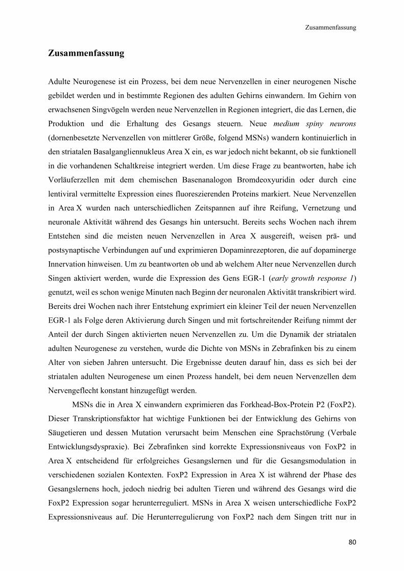
Zusammenfassung
80
Zusammenfassung
Adulte Neurogenese ist ein Prozess, bei dem neue Nervenzellen in einer neurogenen Nische
gebildet werden und in bestimmte Regionen des adulten Gehirns einwandern. Im Gehirn von
erwachsenen Singvögeln werden neue Nervenzellen in Regionen integriert, die das Lernen, die
Produktion und die Erhaltung des Gesangs steuern. Neue medium spiny neurons
(dornenbesetzte Nervenzellen von mittlerer Größe, folgend MSNs) wandern kontinuierlich in
den striatalen Basalgangliennukleus Area X ein, es war jedoch nicht bekannt, ob sie funktionell
in die vorhandenen Schaltkreise integriert werden. Um diese Frage zu beantworten, habe ich
Vorläuferzellen mit dem chemischen Basenanalogon Bromdeoxyuridin oder durch eine
lentiviral vermittelte Expression eines fluoreszierenden Proteins markiert. Neue Nervenzellen
in Area X wurden nach unterschiedlichen Zeitspannen auf ihre Reifung, Vernetzung und
neuronale Aktivität während des Gesangs hin untersucht. Bereits sechs Wochen nach ihrem
Entstehen sind die meisten neuen Nervenzellen in Area X ausgereift, weisen prä- und
postsynaptische Verbindungen auf und exprimieren Dopaminrezeptoren, die auf dopaminerge
Innervation hinweisen. Um zu beantworten ob und ab welchem Alter neue Nervenzellen durch
Singen aktiviert werden, wurde die Expression des Gens EGR-1 (early growth response 1)
genutzt, weil es schon wenige Minuten nach Beginn der neuronalen Aktivität transkribiert wird.
Bereits drei Wochen nach ihrer Entstehung exprimiert ein kleiner Teil der neuen Nervenzellen
EGR-1 als Folge deren Aktivierung durch Singen und mit fortschreitender Reifung nimmt der
Anteil der durch Singen aktivierten neuen Nervenzellen zu. Um die Dynamik der striatalen
adulten Neurogenese zu verstehen, wurde die Dichte von MSNs in Zebrafinken bis zu einem
Alter von sieben Jahren untersucht. Die Ergebnisse deuten darauf hin, dass es sich bei der
striatalen adulten Neurogenese um einen Prozess handelt, bei dem neuen Nervenzellen dem
Nervengeflecht konstant hinzugefügt werden.
MSNs die in Area X einwandern exprimieren das Forkhead-Box-Protein P2 (FoxP2).
Dieser Transkriptionsfaktor hat wichtige Funktionen bei der Entwicklung des Gehirns von
Säugetieren und dessen Mutation verursacht beim Menschen eine Sprachstörung (Verbale
Entwicklungsdyspraxie). Bei Zebrafinken sind korrekte Expressionsniveaus von FoxP2 in
Area X entscheidend für erfolgreiches Gesangslernen und für die Gesangsmodulation in
verschiedenen sozialen Kontexten. FoxP2 Expression in Area X ist während der Phase des
Gesangslernens hoch, jedoch niedrig bei adulten Tieren und während des Gesangs wird die
FoxP2 Expression sogar herunterreguliert. MSNs in Area X weisen unterschiedliche FoxP2
Expressionsniveaus auf. Die Herunterregulierung von FoxP2 nach dem Singen tritt nur in
Zusammenfassung
81
MSNs mit niedrigem FoxP2 Expressionsniveau und nicht in MSNs mit hohem FoxP2
Expressionsniveau auf. Daher postulierte ich, dass MSNs mit hohem FoxP2 Expressionsniveau
erst kürzlich in Area X eingewandert sind und deren FoxP2 Expression erst verringert werden
muss, bevor sie durch Singen aktiviert werden können. Diese Hypothese wurde getestet, indem
das FoxP2 Expressionsniveau und die EGR-1-Expression in einzelnen neuen MSNs von
singenden und nicht singenden Zebrafinken zu verschiedenen Zeitpunkten nach ihrer
Entstehung bestimmt wurde. Interessanterweise war die Aktivierung der neuen MSNs
unabhängig von ihrem FoxP2 Expressionsniveaus. Ein weiteres Ergebnis war, dass ein Drittel
der neuen MSNs in frühen Reifungsstadien FoxP2 in hohen Mengen exprimierte. Die Mehrheit
der gereiften MSNs exprimierte FoxP2 jedoch in geringen Mengen, was auf eine altersbedingte
Abnahme der FoxP2 Proteinmengen in einer Fraktion der neuen MSNs hinweist. Da gezeigt
wurde, dass Foxp2 das Wachstum und die Differenzierung von Neuronen fördert, habe ich die
Morphologie der Dendriten und die Dichte der Dornenfortsätze von neuen MSNs mit hohem
oder geringem FoxP2 Expressionsniveau analysiert. Im Vergleich hatten neue MSNs mit
hohem FoxP2 Expressionsniveau komplexere Dendriten und eine höhere Dichte an
pilzförmigen Dornenfortsätzen. Dies deutet darauf hin, dass sie während eines kurzen
Zeitraums ihrer Reifung mehr Verknüpfungen mit vorgeschalteten Gesangsnuklei ausbildeten.
Ein Verglich der hier präsentieren Ergebnisse mit den MSNs der direkten und indirekten
Verbindungen der Basalganglien von Nagetieren, lässt schlussfolgern, dass unterschiedliche
FoxP2 Expressionsniveaus und die damit einhergehende dichotome Morphologie neuer MSNs
auf die Existenz unterschiedlicher Subtypen ebendieser in Area X von Zebrafinken hindeuten.
Zusammenfassend zeigen die präsentierten Daten, dass die neuen MSNs, die in Area X von
erwachsenen Zebrafinken rekrutiert werden, funktionsfähig sind und für die Erhaltung des
Gesangs eine wichtige Rolle spielen könnten. Innerhalb der ersten sechs Wochen nach ihrer
Bildung weisen neue MSNs dynamische FoxP2 Expressionsniveaus auf. Letztere korrelieren
positiv mit der Komplexität der dendritischen Verzweigungen und der Dichte der
Dornenfortsätze von neuen MSNs, wodurch die Funktion von FoxP2 um eine Rolle in der
striatalen adulten Neurogenese erweitert wurde.

82
Danksagung
83
Danksagung Liebe Constance, ich möchte dir dafür danken, dass du mich mit offenen Armen in deiner
Arbeitsgruppe aufgenommen und mich stets unterstützt hast. Du hast mir die Freiheit gegeben
selbstbestimmt zu arbeiten und meinen eigenen Weg zu gehen. Wenn ich mal feststeckte, hast
du mich motiviert noch einen Schritt weiter zu gehen. Nach der Geburt von Loris hast du es
mir ermöglicht Familie und Forschung bestmöglich zu verbinden, ohne deine Flexibilität hatte
ich das nicht geschafft. Ich weiß es sehr zu schätzen, dass ich die Möglichkeit hatte
internationale Konferenzen und Meetings zu besuchen, was mich jedes Mal sehr inspiriert und
motiviert hat. Du bist eine großartige Mentorin und du hast mich gelehrt stets über den
Tellerrand zu schauen und alles zu hinterfragen. Ich danke Dir für Alles!
Herzlich bedanken möchte ich mich auch bei Mirjam, die die Zweitbegutachtung dieser
Arbeit angenommen hat. Meinen ehemaligen und aktuellen Kollegen danke ich für die
jahrelange Unterstützung und dafür, dass sie so eine so nette Atmosphäre im Institut geschaffen
haben: Marina, Ezequiel, Sina, Johanna, Marco, Maria, Fabian, Iris, Sandra, Philipp, Lydia und
Vanessa. Ganz besonders danke ich Janett und Doreen, für ihre Hilfe mit den Zebrafinken,
ebenso Ulla und Arpik für die Labororganisation und Anleitung bei allen möglichen Puffern,
Geräten und Labortechniken.
Liebe Adriana, ich glaube die Zeit, die wir zusammengearbeitet haben, war für mich die
Schönste während der Promotion. Wir hatten großen Spaß zusammen und viele lustige
Momente, das werde ich sehr vermissen. Auch wenn mal etwas nicht funktioniert hat, konnten
wir uns immer gegenseitig aufheitern. Ich habe wirklich Glück gehabt mit einer so tollen
Kollegin und Freundin zusammenarbeiten zu können, danke für die gemeinsame Zeit!
Liebe Mama, lieber Papa, viele Dank, dass ihr all die Jahre an mich geglaubt und mich
unterstützt habt! Ihr habt mir immer Freiraum gegeben und meine Wünsche ermöglicht, vielen
Dank für Alles. Herzlich bedanken möchte ich mich auch bei meinen Schwiegereltern Anne-
Bärbel und Klaus für ihre Unterstützung.
Lieber Thomas, ich danke dir, dass du mich so geduldig und stets ermutigend durch die
Höhen und Tiefen der Promotionszeit hindurch begleitet hast. Ich bin sehr glücklich darüber,
Loris und dich in meinem Leben zu haben.

84
Eidesstattliche Erklärung
85
Eidesstattliche Erklärung Hiermit versichere ich die vorliegende Dissertation selbstständig und ohne unerlaubte Hilfe
angefertigt zu haben. Bei der Verfassung der Dissertation wurden keine anderen als die im Text
angegebenen Quellen und Hilfsmittel verwendet. Ein Promotionsverfahren wurde zu keinem
früheren Zeitpunkt an einer anderen Hochschule oder bei einem anderen Fachbereich beantragt.
Berlin, den 16.04.2020
Jennifer Kosubek-Langer

![Hypnosis Antenatal Training for Childbirth (HATCh): a randomised controlled trial [NCT00282204]](https://img.pdfslide.net/doc/110x75/635a9be878abe3be930dbc65/hypnosis-antenatal-training-for-childbirth-hatch-a-randomised-controlled-trial.jpg)


