Embed Size (px)
Citation preview
583
Introduction
The oral cavity is an attractive site for the delivery of drugs; however, there are several shortcomings that should be overcome for achieving the efficient drug therapy namely, the intestinal and/or hepatic first pass elimination, high variance in bioavailability due to variable condition of gastrointestinal tract, difficulty in long-term and rate-regulated absorption, and impossibility of arbitrary drug input and its interruption (Amnuaikita et al., 2005). The transdermal administration of drugs is a viable alterna-tive to the oral route due to low metabolic activity of skin compared with that of the gastrointestinal tract and liver (Shams et al., 2010). Transdermal route is one of the
potent alternative routes, particularly, for the active that subjected to extensive hepatic first-pass metabolism and has a short biological half-life; however, the effectiveness of transdermal drug delivery depends on the ability of the drug to penetrate the skin in sufficient amounts to reach therapeutic levels. The main obstacle in the trans-dermal drug delivery is the stratum corneum (SC), the uppermost layer of the skin (Sinha and Kaur, 2000; Paudel et al., 2010). The SC consists of 10–15 layers of keratin-rich corneocytes embedded in a lipid matrix. The SC is con-sidered as the major contributor in the barrier properties of skin. Therefore, a deeper knowledge of the penetration pathway and effect on the biochemical composition and
OrIgInal arTIClE
Role of novel terpenes in transcutaneous permeation of valsartan: effectiveness and mechanism of action
Abdul Ahad, Mohammed Aqil, Kanchan Kohli, Yasmin Sultana, Mohd Mujeeb, and Asgar Ali
Faculty of Pharmacy, Hamdard University, M. B. Road New Delhi 110062, India
abstractContext: The greatest obstacle for transdermal delivery is the barrier property of the stratum corneum. Many approaches have been employed to breach the skin barrier; the most widely used one is that of chemical penetration enhancers. Of the penetration enhancers, terpenes are arguably the most highly advanced and proven category.
Objective: The aim of this investigation was to study effectiveness and mechanism of seven novel terpenes, namely iso-eucalyptol, β-citronellene, valencene, rose oxide, safranal, lavandulol acetate, and prenol, as potential penetration enhancers for improved skin permeation of valsartan through rat skin and human cadaver skin (HCS) with reference to established terpene eucalyptol.
Methods: Skin permeation studies were carried out using Automated Transdermal Diffusion Cell Sampling System (SFDC 6, LOGAN Instruments Corp., NJ) on rat skin and HCS. The mechanism of skin permeation enhancement of valsartan by terpenes treatment was evaluated by Fourier transform infrared spectroscopy (FT-IR) analysis, differential scanning calorimetry (DSC) thermogram, and histopathological examination.
Results and discussion: Among all study enhancers, iso-eucalyptol produced the maximum enhancement via rat skin [enhancement ratio (ER) = 7.4] and HCS (ER = 3.60) over control. FT-IR spectra and DSC thermogram of skin treated with aforesaid terpenes indicated that permeation occurred due to the disruption of lipid bilayers. No apparent skin irritation (erythema, edema) was observed on treatment with terpenes except β-citronellene, safranal, lavandulol acetate, and prenol, which caused mild irritation.
Conclusion: It is concluded that the iso-eucalyptol can be successfully used as safe and potential penetration enhancer for enhancement of skin permeation of lipophilic drug such as valsartan.
Keywords: Transdermal, terpenes, permeation enhancer, valsartan, hypertension
Address for Correspondence: Dr. Mohammed Aqil, Faculty of Pharmacy, Department of Pharmaceutics, Hamdard University, New Delhi 110 062, India. Tel.: +91–9811798725; Fax: +91-11-26059663. E-mail: [email protected]
(Received 31 July 2010; revised 13 September 2010; accepted 15 September 2010)
Drug Development and Industrial Pharmacy, 2011; 37(5): 583–596© 2011 Informa Healthcare USA, Inc.ISSN 0363-9045 print/ISSN 1520-5762 onlineDOI: 10.3109/03639045.2010.532219
Drug Development and Industrial Pharmacy
2011
37
5
583
596
31 July 2010
13 September 2010
15 September 2010
0363-9045
1520-5762
© 2011 Informa Healthcare USA, Inc.
10.3109/03639045.2010.532219
LDDI
532219
584 A. Ahad et al.
Drug Development and Industrial Pharmacy
structure across the entire SC, induced by penetrating molecules, is highly desirable. Several strategies have been employed to circumvent this natural barrier to avail of the great advantages offered by this route. One of the most widely used approaches employs the use of perme-ation enhancers also called as penetration enhancers, molecules that reduce the barrier properties of skin by act-ing on the different components of skin such as lipids and proteins (Williams and Barry, 2004). Extensive research during the past two decades has revealed considerable information on several classes of penetration enhancers, including surfactants (e.g. Tween), fatty acids/esters (e.g., oleic acid), solvents (e.g., dimethylsulfoxide, ethanol), and Azone® (Sapra et al., 2008; Karakatsani et al., 2010). Despite their fairly satisfactory performance in enhancing the permeation of drug molecule across the skin, chemical enhancers are viewed with suspicion in transdermal for-mulations due to their irritancy potential when employed at concentrations necessary for achieving useful levels of penetration enhancement. Efforts have been directed at identifying safe and effective enhancers from both natural products and synthetic chemicals. In particular, terpenes from natural sources have attracted great interest and found one of the promising groups of candidates to be employed as clinically acceptable permeation enhancers (Jain et al., 2008). Terpenes are a very safe and an effec-tive class of permeation enhancers obtained from natural sources. Further, quite a few terpenes are included in the list of generally recognized as safe (GRAS) agents issued by (US FDA) U.S. Food and Drug Administration (Sapra et al., 2008). They cause no skin toxicity or if any, only mild irritation. Even terpenes, which are considered to be skin irritants, do not cause lasting erythema (Ahad et al., 2009). Terpenes have been used for permeation enhancement of both hydrophilic and lipophilic drugs. The activity of terpenes as penetration enhancers is primarily related to their chemical structure as well as the physicochemical properties such as lipophilicity, size and chirality, boil-ing point (BP) and energy of vaporization, and degree of unsaturation. The readers are directed to a comprehensive review on the status of terpenes as penetration enhancers (Aqil et al., 2007). It is documented (Higaki et al., 2003) that for hydrophilic drugs, the primary effect of terpenes enhancer treatment is to increase drug diffusivity in the SC (i.e., to reduce the barrier properties of the skin). For lipophilic drugs, terpenes not only seem to increase drug diffusivity but also increase drug partitioning into the SC. Increases in partitioning are likely due to bulk solvent effects since many lipophilic drugs are moderately sol-uble in many of the terpenes (Cornwell et al., 1996). But the clear understanding of the mechanism of perturbed drug transport through SC by terpenes remains elusive. There are several analytical techniques used to determine the possible mechanism of action of penetration enhanc-ers such as electron paramagnetic resonance, electron diffraction, atomic force microscopy, small angle X-ray diffraction, transmission electron microscopy, and elec-tron spin resonance (Silva et al., 2006). Among these most
researched and highly suited techniques are differential scanning calorimetry (DSC) and Fourier transform infra-red spectroscopy (FT-IR) (Yamane et al., 1995; Cornwell et al., 1996; Vaddi et al., 2002), which specifically probes the outer layers of the skin, has proven to be a very useful method for describing the biochemical composition of the biological tissues, and as well facilitate to investigate the secondary structure composition insight into the protein.
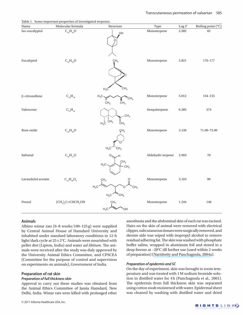
Valsartan, a lipophilic antihypertensive drug has a low oral bioavailability of about 25%. It has low molecular weight (435.5) and melting point (116–117°C) with a log partition coefficient of 4.5, and mean biological half life of 7.5 h. There are no reports of skin irritation attributed to valsartan. All the above characteristics make valsartan a good candidate for transdermal delivery. In a recent study, feasibility of valsartan for the transdermal delivery system was reported from our laboratory (Rizwan et al., 2008), and ethanol and isotonic phosphate buffer (IPB) pH 7.4 sol-vent system in the ratio of 40:60 v/v was reported a suitable vehicle for the transdermal delivery of valsartan. However, it was necessary to improve the permeation rate of val-sartan by using a suitable enhancement technique. Many overtures have been used to mitigate SC barrier property and the most commonly used approach is the use of sorp-tion promoters also known as permeation enhancers. In this study, we tried to improve the penetration of valsartan by using seven novel terpenes, namely iso-eucalyptol, β-citronellene, valencene, rose oxide, safranal, lavandulol acetate, and prenol, and compare it with an established terpene eucalyptol, and to elucidate the mechanism of skin permeation enhancement by FT-IR, DSC, and his-topathological studies. Some important properties of the studied terpenes are mentioned in Table 1.
Materials and methods
Valsartan was received as gratis sample from Ranbaxy Research Laboratories Ltd., Gurgaon, India. All terpenes namely iso-eucalyptol (≥98.5% purity), (+)β-citronellene (≥98.5% purity), (+)valencene (≥70.0% purity), (−)rose oxide (≥99.0% purity), safranal (≥70.0% purity), (±)-lavan-dulol acetate (≥98.5% purity), prenol (≥99.5% purity), and eucalyptol (≥99.0% purity) were purchased from authen-tic source (Sigma–Aldrich Chemicals Private Ltd., New Delhi, India). Sodium chloride, potassium sulfate, sodium azide and sodium bromide were purchased from S.D. Fine Chemicals, India. Sodium hydroxide and potassium dihydrogen orthophosphate were purchased from Merck India Ltd., India. Water for High Performance Liquid Chromatography (HPLC) was purchased from Thomas Baker (Chemicals) Ltd., Mumbai, India. Absolute etha-nol was purchased from Changshu Yangyuan Chemical Co. Ltd., China. Acetonitrile (HPLC grade) and methanol (HPLC grade) were purchased from Spectrochem Pvt. Ltd., Mumbai, India. All other chemicals used were of reagent grade. All materials were used as received. Double distilled water was used for all experiments.
Transcutaneous permeation of valsartan 585
© 2011 Informa Healthcare USA, Inc.
AnimalsAlbino wistar rats (6–8 weeks/100–125 g) were supplied by Central Animal House of Hamdard University and inhabited under standard laboratory conditions in 12-h light/dark cycle at 25 ± 2°C. Animals were nourished with pellet diet (Lipton, India) and water ad libitum. The ani-mals were received after the study was duly approved by the University Animal Ethics Committee, and CPSCEA (Committee for the purpose of control and supervision on experiments on animals), Government of India.
Preparation of rat skinPreparation of full thickness skinApproval to carry out these studies was obtained from the Animal Ethics Committee of Jamia Hamdard, New Delhi, India. Wistar rats were killed with prolonged ether
anesthesia and the abdominal skin of each rat was excised. Hairs on the skin of animal were removed with electrical clipper, subcutaneous tissues were surgically removed, and dermis side was wiped with isopropyl alcohol to remove residual adhering fat. The skin was washed with phosphate buffer saline, wrapped in aluminum foil and stored in a deep freezer at −20°C till further use (used within 2 weeks of preparation) (Narishetty and Panchagnula, 2004a).
Preparation of epidermis and SCOn the day of experiment, skin was brought to room tem-perature and was treated with 1 M sodium bromide solu-tion in distilled water for 4 h (Panchagnula et al., 2001). The epidermis from full thickness skin was separated using cotton swab moistened with water. Epidermal sheet was cleaned by washing with distilled water and dried
Table 1. Some important properties of investigated terpenes.Name Molecular formula Structure Type Log P Boiling point (°C)Iso-eucalyptol C
10H
18O
OH
O
Monoterpene 2.582 65
Eucalyptol C10
H18
O
O
CH3
CH3H3C
Monoterpene 2.821 176–177
β-citronellene C10
H18
CH3 CH3
CH3H3C Monoterpene 5.012 154–155
Valencene C15
H24
CH3
CH3
CH3H3C
Sesquiterpene 6.285 274
Rose oxide C10
H18
O CH3
CH3
O
H3C
Monoterpene 3.126 71.00–73.00
Safranal C10
H14
O
CH3
CH3
HO
H3C
Aldehydic terpene 2.903 70
Lavandulol acetate C12
H20
O2 CH3
CH3
CH2
O
O
H3C
H3C
Monoterpene 3.163 90
Prenol (CH3)
2C=CHCH
2OH CH3
OHH3C
Monoterpene 1.244 140
586 A. Ahad et al.
Drug Development and Industrial Pharmacy
under vacuum and examined for cuts or holes if any. SC samples were prepared by floating freshly prepared epidermis membrane on 0.1% trypsin solution for 12 h. Then, SC sheets were cleaned by washing with distilled water (Shakeel et al., 2008).
Preparation of HCSPreparation of human abdominal skinHuman abdominal skin was obtained post-mortem, sealed in evacuated polyethylene bags and stored at −20°C. The epidermal membranes of HCS were prepared by heat-separation technique (Yamane et al., 1995). The whole skin was immersed in water at 60°C for 2 min, fol-lowed by careful removal of the epidermis. The samples were stored at −20°C until used. Before the permeation experiments, the membranes with SC side up were floated over 0.9% (w/v) sodium chloride solution con-taining 0.002% (w/v) sodium azide solution for 3 days to ensure essentially full hydration of the SC (Vaddi et al., 2002; Rizwan et al., 2008).
Preparation of SCThe human SC was prepared by following the method reported by Vaddi et al. (2002). Briefly, epidermal mem-brane with SC side up was incubated in Petri dish over fil-ter paper imbibed with 0.1% (w/v) trypsin in 0.5% (w/v) sodium bicarbonate solution at 37 ± 1°C for 3 h. The SC was removed, thoroughly washed, and dried in a vacuum desiccator. Finally, the SC was dipped in acetone solution for 20 sec to remove sebaceous lipids and dried again.
Ex vivo skin permeation studiesEx vivo skin permeation studies were carried out in triplicate through rat skin and HCS using an Automated Transdermal Diffusion Cell Sampling System. The system consisted of three side-by-side cells with area of diffusion 0.636 cm2 and 4 ml of receptor cell volume. The water was warmed with the in built heater thermostated set at 37 ± 1°C throughout the experiments to provide a skin surface temperature of approximately 32 ± 1°C (Mura et al., 2009; Moghimi et al., 2009). A pump circulated the warmed water throughout the system. A Teflon coated mini-magnetic bead was kept in the receiver compart-ment for agitating the contained vehicle (ethanol:IPB, pH 7.4, 40:60) at 600g. The receptor compartment was filled with vehicle, containing 0.003% w/v sodium azide as a preservative (Narishetty and Panchagnula, 2004a,b). Receptor fluid was sonicated to remove dissolved gases and equilibrated at 37 ± 1°C before placing in the recep-tor compartment. The skin samples were mounted over the diffusion cells in such a way that SC side faced the donor compartment where as dermis faced the receiver compartment. The donor compartment was kept empty although the receiver compartment was filled with a mix-ture of ethanol:IPB (pH 7.4) (40:60) and stirred at 600g. The ethanol:IPB (pH 7.4) (40:60) solution was replaced every half an hour to stabilize the skin, which was evident by recording the UV absorption of the ethanol:IPB (pH
7.4) (40:60) solution. The zero absorption indicated the complete stabilization (Chisty et al., 2002; Amin et al., 2008).
After the stabilization of the skin, the donor chamber was filled with 4-ml solution of valsartan (20 mg; trans-dermal dose of valsartan) in vehicle (ethanol:IPB, pH 7.4, 40:60) with or without terpenes (0.5–5% w/v). The donor and receiver compartments were covered with Teflon plug to prevent evaporation of vehicle. After application of drug solution, 500-μl samples were withdrawn from the receptor compartment at different time intervals and analyzed for drug content in triplicate by HPLC method (Tatar and Saglik, 2002). The HPLC system consisted of a series LL-10AT VP Shimadzu pump, and SPD-10A Shimadzu UV–vis detector. Separation was achieved using Shiseido C-18 column (250 × 4.6 mm, i.d. 5 µm). The HPLC system was equipped with the software “Class-VP series (Shimadzu)”. Binary elution was carried out at a flow rate 1.3 ml/min with the mobile phase contain-ing 45% acetonitrile and 55% phosphate buffer solution (pH 3). Mobile phase was prepared daily, filtered by pass-ing through a 0.45-µm membrane filter and degassed. All chromatographic separations were performed at room temperature. Detection was carried out at 265 nm with UV detector. A standard curve was constructed for valsar-tan in the range of 1–10 µg/ml. A good linear relationship was observed between the concentration of valsartan and the peak area of valsartan with a correlation coef-ficient (r2 = 0.9984). The standard curve constructed as described previously was used for estimating valsartan in the skin permeation study. Receiver volume was immedi-ately replenished with the same amount of fresh vehicle maintained at 37 ± 1°C. Permeation parameters such as flux (J), permeability coefficient (Kp), enhancement ratio (ER), and lag time (t
lag) were calculated as given under
data analysis.
FT-IR studiesRat skin was prepared as mentioned earlier and SC was cut into small circular disc with approximate diameter of 1.5 cm. Sodium chloride (0.9% w/v) was prepared and 0.003% w/v sodium azide was added as antibacterial and antimycotic agent. Equal volume of sodium chloride (0.9% w/v) solution was placed in different conical flask and SC of approximate diameter 1.5 cm was floated over for 3 days. After 3 days of hydration, these discs were thoroughly blotted over filter paper and FT-IR (Perkin Elmer, Germany) was recorded before terpene treat-ment (control) in frequency range of 400–4000 cm−1, with resolution of 2 cm−1. Each spectrum was an aver-age of 60 scans. After taking FT-IR, the same discs were dipped into respective terpene solution (1% w/v) in 4 ml ethanol:IPB pH 7.4 (40:60) systems for 24 h, (equivalent to the permeation studies) at 21 ± 1°C. Each SC disc after treatment was washed, blotted dry, and then air-dried for 2 h. Samples were kept under vacuum in desicca-tors for 15 min to remove traces of solvent and enhancer completely (Krishnaiah et al., 2003). FT-IR spectra of all
Transcutaneous permeation of valsartan 587
© 2011 Informa Healthcare USA, Inc.
SC discs treated with enhancer were recorded again for comparison.
Differential scanning calorimeter studiesApproximately 20 mg of freshly prepared rat SC was taken and hydrated over saturated potassium sulfate solution for 3 days. Percent hydration was calculated using the formula:
% of hydration =Wt.of hydrated SC- Wt.of dry SC
Wt.of dry SC100
Hydrated rat SC samples were dipped into respective terpene solutions (1% w/v) in 4 ml ethanol:IPB pH 7.4 (40:60) systems for 24 h, at 21 ± 1°C. After the terpene treatment, SC was removed and blotted to attain hydra-tion of 20–25%, cut (5 mg) and sealed in aluminum hermatic pans, and equilibrated for 1 h before the DSC run. Then, the SC samples were scanned on a DSC6 dif-ferential scanning calorimeter (Perkin-Elmer, Germany). Scanning rate was 5°C/min over the temperature range of 30–200°C (Krishnaiah et al., 2002a; Vaddi et al., 2002).
Histopathological studiesApproval to carry out these studies was obtained from the Animal Ethics Committee of Jamia Hamdard, New Delhi, India. Histopathological studies were carried out for elucidation of mechanism of penetration enhance-ment and skin irritation potential of the investigated terpenes. In this study, abdominal skin of wistar rat was treated separately with 1% w/v putative enhancers, iso-eucalyptol, β-citronellene, valencene, rose oxide, safra-nal, lavandulol acetate, prenol, and with established enhancer eucalyptol (1% w/v) in ethanol:IPB pH 7.4 (40:60). After 24 h, rat was killed and the skin samples from treated and untreated (control) area were taken. Each specimen was stored in 10% formalin solution in phosphate buffer saline (pH 7.4). The specimen was cut into sections vertically. Each section was dehydrated using ethanol, embedded in paraffin for fixing, and stained with hematoxylin and eosin. These samples were then observed under light microscope (Motic, Japan) and compared with control sample. In each skin sample, three different sites were scanned and evaluated for elucidation of mechanism of penetration enhance-ment (Aqil et al., 2004)
Data analysisFlux is the rate of change of the cumulative amount of drug passes per unit area and time through the skin. The equation derived from Fick’s second law of diffusion can be used to calculate the steady-state flux (J).
JV dc/dt
A( g/cm /h)2
( )µ
where V (ml) is the volume of the receiver compartment, dc/dt is the steady-state slope from the plot of the amount of drug permeated through the skin versus time, and A is
the effective diffusional area. The permeability coefficient Kp (cm/h) was calculated as the quotient of flux and drug concentration in the donor compartment.
Lag time (tlag
) is obtained by extrapolating the linear portion of the same graph along the horizontal time (h) axis.
To determine the extent of enhancement, ER was cal-culated as follows:
ER =Flux of valsartan with enhancer
Flux of valsartan without enhanceer
One-way analysis of variance with Dunnett’s test was used for statistical analysis. The level of significance was taken as P < 0.05.
results and discussion
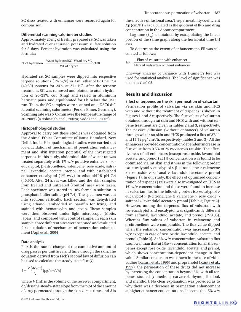
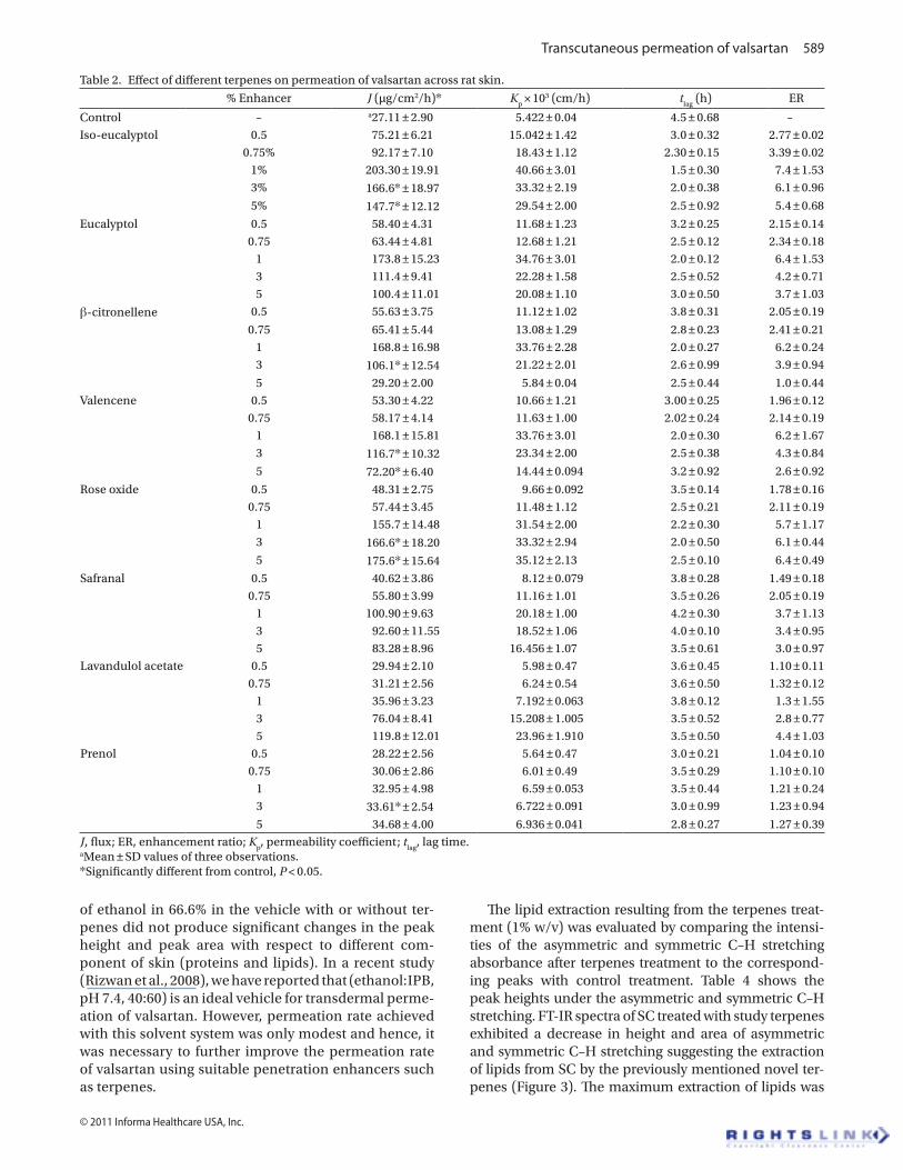
Effect of terpenes on the skin permeation of valsartanPermeation profile of valsartan via rat skin and HCS with and without the treatment of terpenes is shown in Figures 1 and 2 respectively. The flux values of valsartan obtained through rat skin and HCS with and without ter-pene treatment are given in Tables 2 and 3, respectively. The passive diffusion (without enhancer) of valsartan through wistar rat skin and HCS produced a flux of 27.11 and 17.72 µg/ cm2/h, respectively (Tables 2 and 3). All the enhancers provided concentration dependent increase in flux value from 0.5% to1% w/v across rat skin. The effec-tiveness of all enhancers (except rose oxide, lavandulol acetate, and prenol) at 1% concentration was found to be optimized via rat skin and it was in the following order: iso-eucalyptol > eucalyptol > β-citronellene ≥ valencene > rose oxide > safranal > lavandulol acetate > prenol (Figure 1). In our study, the effects of optimized concen-tration of terpenes (1%) were also investigated on HCS at 1% w/v concentration and these were found to increase in valsartan flux in the following order: iso-eucalyptol > eucalyptol > β-citronellene ≥ valencene > rose oxide > safranal > lavandulol acetate > prenol (Table 3; Figure 2). However, among the terpenes, flux of valsartan with iso-eucalyptol and eucalyptol was significantly different from safranal, lavandulol acetate, and prenol (P<0.05). Whereas flux values of valsartan in valencene and β-citronellene were comparable. The flux value dipped when the enhancer concentration was increased to 3% w/v except in case of rose oxide, lavandulol acetate, and prenol (Table 2). At 5% w/v concentration, valsartan flux was lower than that at 1%w/v concentration for all the ter-penes except rose oxide, lavandulol acetate, and prenol, which shows concentration-dependent change in flux value. Similar conclusion was drawn in the case of zido-vudine (Kararli et al., 1995) and propranolol (Kunta et al., 1997); the permeation of these drugs did not increase by increasing the concentration beyond 5%, with all ter-penes studied (t-anethole, carvacrol, thymol, linalool, and menthol). No clear explanation was provided as to why there was a decrease in permeation enhancement at higher enhancer concentrations. It seems that 5% w/v
588 A. Ahad et al.
Drug Development and Industrial Pharmacy
is the optimized enhancer concentration in case of rose oxide, lavandulol acetate, and prenol which produced 6.4-, 4.4-, and 1.27-fold enhancement over control (with-out enhancer), respectively (Table 2). All the terpene resulted in significant reduction of lag time (P < 0.05) relative to control and the order of terpene effect on lag time in decreasing order is as follows: control > safranal > lavandulol acetate > prenol > rose oxide > β-citronellene > valencene > eucalyptol > iso-eucalyptol (Table 2).
It is anticipated that lipophilic enhancers would pro-vide better penetration enhancement for a lipophilic permeant, whereas hydrophilic terpenes are more active toward promoting the permeation of hydrophilic drugs (Williams and Barry, 1991; Hori et al., 1991; Jain et al., 2001; Fujii et al., 2004; Aqil et al., 2007) such as valsartan. The results of our study are in agreement to the earlier hypothesis as iso-eucalyptol (Log P = 2.582; Table 1) and eucalyptol (Log P = 2.821; Table 1) being lipophilic compounds yielded higher flux (P < 0.005) for valsartan. Further more, prenol being the least lipophilic terpene produced the least penetration of valsartan. In contradic-tion, Ogiso et al. (1995) reported that oleyl oleate, a highly lipophilic enhancer, did not significantly increase indo-methacin flux, though latter is a highly lipophilic drug. In another report (El-Kattan et al., 2001), limonene provided higher enhancing effect for the permeation of nicardipine hydrochloride (hydrophilic calcium channel blocker) and hydrocortisone (a polar steroid) relative to fenchone and thymol (hydrophilic terpene). Similarly, p-menthan-3,8-diol, a more hydrophilic enhancer than menthol produced higher flux of indomethacin (a lipophilic drug) than antipyrine (a hydrophilic drug) (Fujii et al., 2004). In our study, valencene (sesquiterpene) showed better ER than prenol (simple chain hydrocarbon terpene) for the transdermal delivery of valsartan through rat skin, which is in agreement with the previous reports (Cornwell and Barry, 1994; Godwin and Michniak, 1999) that sesquiter-
pene compounds with polar head groups are generally more potent enhancers than pure hydrocarbons.
Of all the terpenes studied, b.p. of iso-eucalyptol (65°C) is the least (Table 1). This is a proposition of weak cohesive forces or self association of iso-eucalyptol mol-ecule which means that oxygen of functional ether and carbonyl group is free for interface. Therefore, the energy requisite for competitive hydrogen bonding in skin cer-amide is comparatively less for iso-eucalyptol, which can be associated to higher flux, Kp, ER, and shortest t
lag
found with iso-eucalyptol (Jain et al., 2001). On the other hand, in the case of prenol (BP = 140°C), β-citronellene (BP = 154–155°C) and valencene (BP = 274°C), additional energy is required to free the respective functional group from strong self-association, as reflected by higher BP (Table 1). It is reported that terpenes increase the drug percutaneous permeation mainly by disrupting the intercellular packing of the SC lipids. Hence, DSC and FT-IR studies were carried out to confirm such a hypoth-esis on the observed penetration enhancing effect of terpenes, on the permeability of valsartan through rat skin and HCS.
FT-IR spectral analysis of terpenes treated and untreated rat skinA typical FT-IR spectrum of rat SC shows separate lipid and protein peaks. The study of lipid biophysics by observing the peaks caused by C–H stretching vibra-tions would be helpful in identifying the influence of the terpenes proposed in the study. The SC lipid extraction leads to a decrease in the C–H stretching absorbance intensity. Preliminary FT-IR studies shows that vehicle (ethanol:IPB, pH 7.4, 40:60) did not cause any signifi-cant change in peak height or peak area of CH
2 stretch-
ing frequencies when compared with the untreated SC indicating that vehicle containing 40% ethanol do not extract lipids from SC. It is evident from the literature (Narishetty and Panchagnula, 2004a) that the presence
6000
5000
4000
3000
Cum
ulat
ive
drug
per
mea
ted
(µg/
cm2 )
2000
1000
00 5 10 15
Time (h)20 25 30
control-RSiso-eucalyptoleucalyptolβ-citronellenevalencenerose oxidesafranallavandulol acetateprenol
Figure 1. Permeation profile of valsartan across rat skin in the absence and presence of various terpenes (1% w/v) in vehicle. Experiments were conducted in triplicate.
Transcutaneous permeation of valsartan 589
© 2011 Informa Healthcare USA, Inc.
of ethanol in 66.6% in the vehicle with or without ter-penes did not produce significant changes in the peak height and peak area with respect to different com-ponent of skin (proteins and lipids). In a recent study (Rizwan et al., 2008), we have reported that (ethanol:IPB, pH 7.4, 40:60) is an ideal vehicle for transdermal perme-ation of valsartan. However, permeation rate achieved with this solvent system was only modest and hence, it was necessary to further improve the permeation rate of valsartan using suitable penetration enhancers such as terpenes.
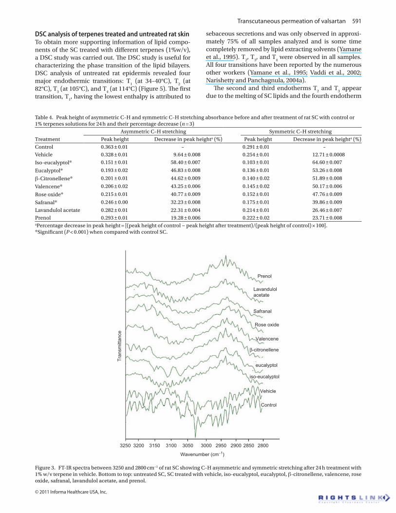
The lipid extraction resulting from the terpenes treat-ment (1% w/v) was evaluated by comparing the intensi-ties of the asymmetric and symmetric C–H stretching absorbance after terpenes treatment to the correspond-ing peaks with control treatment. Table 4 shows the peak heights under the asymmetric and symmetric C–H stretching. FT-IR spectra of SC treated with study terpenes exhibited a decrease in height and area of asymmetric and symmetric C–H stretching suggesting the extraction of lipids from SC by the previously mentioned novel ter-penes (Figure 3). The maximum extraction of lipids was
Table 2. Effect of different terpenes on permeation of valsartan across rat skin.
% Enhancer J (µg/cm2/h)* Kp × 103 (cm/h) t
lag (h) ER
Control – a27.11 ± 2.90 5.422 ± 0.04 4.5 ± 0.68 –Iso-eucalyptol 0.5 75.21 ± 6.21 15.042 ± 1.42 3.0 ± 0.32 2.77 ± 0.02 0.75% 92.17 ± 7.10 18.43 ± 1.12 2.30 ± 0.15 3.39 ± 0.02 1% 203.30 ± 19.91 40.66 ± 3.01 1.5 ± 0.30 7.4 ± 1.53 3% 166.6* ± 18.97 33.32 ± 2.19 2.0 ± 0.38 6.1 ± 0.96
5% 147.7* ± 12.12 29.54 ± 2.00 2.5 ± 0.92 5.4 ± 0.68
Eucalyptol 0.5 58.40 ± 4.31 11.68 ± 1.23 3.2 ± 0.25 2.15 ± 0.14 0.75 63.44 ± 4.81 12.68 ± 1.21 2.5 ± 0.12 2.34 ± 0.18 1 173.8 ± 15.23 34.76 ± 3.01 2.0 ± 0.12 6.4 ± 1.53 3 111.4 ± 9.41 22.28 ± 1.58 2.5 ± 0.52 4.2 ± 0.71 5 100.4 ± 11.01 20.08 ± 1.10 3.0 ± 0.50 3.7 ± 1.03
β-citronellene 0.5 55.63 ± 3.75 11.12 ± 1.02 3.8 ± 0.31 2.05 ± 0.19
0.75 65.41 ± 5.44 13.08 ± 1.29 2.8 ± 0.23 2.41 ± 0.21 1 168.8 ± 16.98 33.76 ± 2.28 2.0 ± 0.27 6.2 ± 0.24 3 106.1* ± 12.54 21.22 ± 2.01 2.6 ± 0.99 3.9 ± 0.94
5 29.20 ± 2.00 5.84 ± 0.04 2.5 ± 0.44 1.0 ± 0.44Valencene 0.5 53.30 ± 4.22 10.66 ± 1.21 3.00 ± 0.25 1.96 ± 0.12 0.75 58.17 ± 4.14 11.63 ± 1.00 2.02 ± 0.24 2.14 ± 0.19 1 168.1 ± 15.81 33.76 ± 3.01 2.0 ± 0.30 6.2 ± 1.67 3 116.7* ± 10.32 23.34 ± 2.00 2.5 ± 0.38 4.3 ± 0.84
5 72.20* ± 6.40 14.44 ± 0.094 3.2 ± 0.92 2.6 ± 0.92
Rose oxide 0.5 48.31 ± 2.75 9.66 ± 0.092 3.5 ± 0.14 1.78 ± 0.16 0.75 57.44 ± 3.45 11.48 ± 1.12 2.5 ± 0.21 2.11 ± 0.19 1 155.7 ± 14.48 31.54 ± 2.00 2.2 ± 0.30 5.7 ± 1.17 3 166.6* ± 18.20 33.32 ± 2.94 2.0 ± 0.50 6.1 ± 0.44
5 175.6* ± 15.64 35.12 ± 2.13 2.5 ± 0.10 6.4 ± 0.49
Safranal 0.5 40.62 ± 3.86 8.12 ± 0.079 3.8 ± 0.28 1.49 ± 0.18 0.75 55.80 ± 3.99 11.16 ± 1.01 3.5 ± 0.26 2.05 ± 0.19 1 100.90 ± 9.63 20.18 ± 1.00 4.2 ± 0.30 3.7 ± 1.13 3 92.60 ± 11.55 18.52 ± 1.06 4.0 ± 0.10 3.4 ± 0.95 5 83.28 ± 8.96 16.456 ± 1.07 3.5 ± 0.61 3.0 ± 0.97Lavandulol acetate 0.5 29.94 ± 2.10 5.98 ± 0.47 3.6 ± 0.45 1.10 ± 0.11 0.75 31.21 ± 2.56 6.24 ± 0.54 3.6 ± 0.50 1.32 ± 0.12 1 35.96 ± 3.23 7.192 ± 0.063 3.8 ± 0.12 1.3 ± 1.55 3 76.04 ± 8.41 15.208 ± 1.005 3.5 ± 0.52 2.8 ± 0.77 5 119.8 ± 12.01 23.96 ± 1.910 3.5 ± 0.50 4.4 ± 1.03Prenol 0.5 28.22 ± 2.56 5.64 ± 0.47 3.0 ± 0.21 1.04 ± 0.10 0.75 30.06 ± 2.86 6.01 ± 0.49 3.5 ± 0.29 1.10 ± 0.10 1 32.95 ± 4.98 6.59 ± 0.053 3.5 ± 0.44 1.21 ± 0.24 3 33.61* ± 2.54 6.722 ± 0.091 3.0 ± 0.99 1.23 ± 0.94
5 34.68 ± 4.00 6.936 ± 0.041 2.8 ± 0.27 1.27 ± 0.39J, flux; ER, enhancement ratio; K
p, permeability coefficient; t
lag, lag time.
aMean ± SD values of three observations.*Significantly different from control, P < 0.05.
590 A. Ahad et al.
Drug Development and Industrial Pharmacy
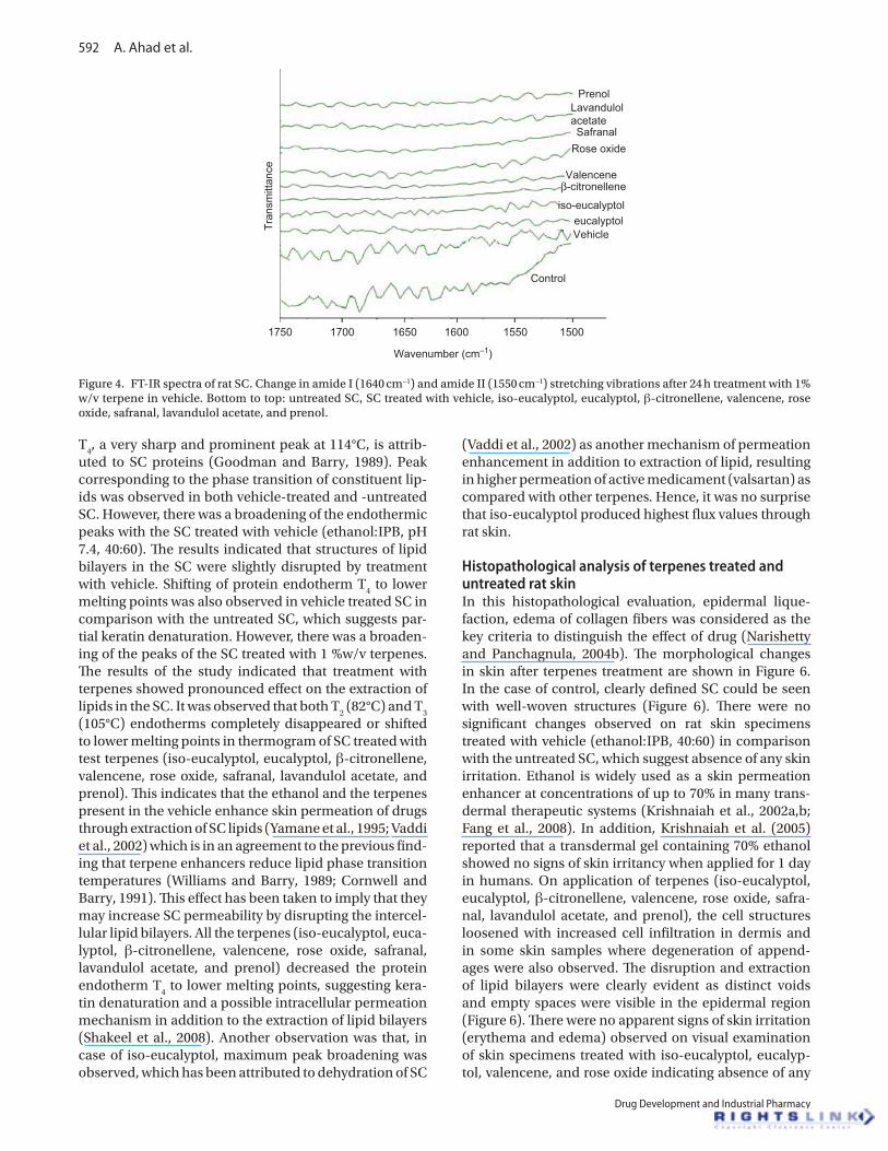
observed with iso-eucalyptol as the decrease in peak height and area was the highest. There was clear differ-ence in the FT-IR spectra of the control and the terpenes treated SC with prominent decrease in asymmetric and symmetric C–H stretching of peak height and area, lead-ing to conclude that study terpenes enhance permeation of valsartan by extracting SC lipids. Clearly, study ter-penes did not fluidize the SC lipids as the peak shift to a higher wave number was not observed (Jain et al., 2001; Krishnaiah et al., 2002b). The rate limiting step or main barrier of transdermal drug delivery is lipophilic part of SC in which lipids (ceramides) are tightly packed as bilay-ers due to high degree of hydrogen bonding. The amide I group of ceramide is hydrogen bounded to the amide I group of another ceramide, forming a tight network of hydrogen bonding at the head of ceramide. This hydrogen bonding lends strength and stability to lipids’ bilayers and thus imparts barrier property to SC (Jain et al., 2001). When skin is treated with terpenes, ceramides may get loosened because of competitive hydrogen bonding leading to breaking of hydrogen-bond network at the head of cer-amides. The tight hydrogen bonding between ceramides
causes split in the peak at 1650 cm−1 (amide I) as shown in the control skin spectrum (Figure 4). Treatment with terpenes resulted in either two or single peak at 1650 cm−1, which suggests breaking of hydrogen bonds by terpenes. There were no significant changes in the pattern of amide I peak when β-citronellene and valencene were used as enhancers suggesting that β-citronellene and valencene are not able to break hydrogen bonds. This is further sub-stantiated by the hypothesis (Jain et al., 2001) that break-ing or loosening of hydrogen bond network between ceramides head groups is caused by oxygen containing terpenes such as iso-eucalyptol, eucalyptol, rose oxide, lavundulol acetate, and rose oxide. Hence β-citronellene and valencene does not break hydrogen bond network between ceramides due to the absence of hydrogen-bond accepting or donating group.
It is concluded that greater extraction of the SC lipids by terpenes led to greater permeability of valsartan. The increase in permeability may be predominantly due to increased solute diffusivity in the partially delipidized SC (Yum et al., 1994). As expected the partially delipidized SC was highly permeable to the non-polar drug used in this study.
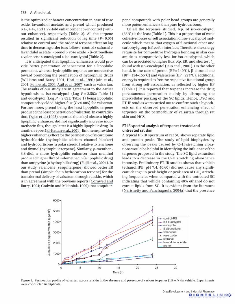
Table 3. Effect of different terpenes on permeation of valsartan across HCS.
% Enhancer J (µg/cm2/h)* Kp × 103 (cm/h) t
lag (h) ER
Control – 17.72a ± 1.56 3.54 ± 0.26 7.5 ± 0.65 –Iso-eucalyptol 1 63.83* ± 5.35 12.76 ± 1.75 3.0 ± 0.27 3.60 ± 0.028
Eucalyptol 1 44.29* ± 4.23 8.85 ± 0.64 4.0 ± 0.38 2.49 ± 0.021
β-Citronellene 1 30.65* ± 2.89 6.13 ± 0.56 4.0 ± 0.35 1.72 ± 0.016
Valencene 1 28.706* ± 2.45 5.74 ± 0.06 4.5 ± 0.41 1.61 ± 0.02
Rose oxide 1 27.64* ± 2.77 5.52 ± 0.50 5.5 ± 0.43 1.55 ± 0.01
Safranal 1 23.39* ± 2.11 4.67 ± 0.34 6.0 ± 0.55 1.31 ± 0.01
Lavandulol acetate 1 21.61* ± 1.99 4.32 ± 0.40 7.0 ± 0.63 1.21 ± 0.01
Prenol 1 19.49 ± 1.42 3.89 ± 0.028 7.0 ± 0.57 1.09 ± 0.01Abbreviations: J, flux; ER, enhancement ratio; K
p, permeability coefficient; t
lag, lag time.
aData are given as mean ± SD values.*Significantly different from control, P < 0.05.
1800
1600
1400
1200
1000
800
600
400
200
Cum
ulat
ive
amou
nt p
erm
eate
d (µ
g/cm
2 )
00 5 10 15
Time (hr)
20 25 30
control-HCSiso-eucalyptoleucalyptolβ-citronellenevalencenerose oxidesafranallavandulol acetateprenol
Figure 2. Permeation profile of valsartan across HCS in the absence and presence of various terpenes (1% w/v) in vehicle. Experiments were conducted in triplicate.
Transcutaneous permeation of valsartan 591
© 2011 Informa Healthcare USA, Inc.
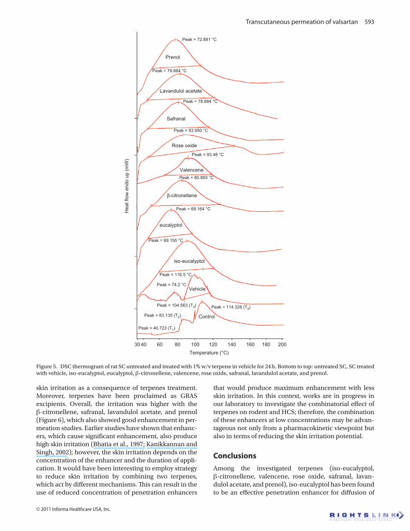
DSC analysis of terpenes treated and untreated rat skinTo obtain more supporting information of lipid compo-nents of the SC treated with different terpenes (1%w/v), a DSC study was carried out. The DSC study is useful for characterizing the phase transition of the lipid bilayers. DSC analysis of untreated rat epidermis revealed four major endothermic transitions: T
1 (at 34–40°C), T
2 (at
82°C), T3 (at 105°C), and T
4 (at 114°C) (Figure 5). The first
transition, T1, having the lowest enthalpy is attributed to
sebaceous secretions and was only observed in approxi-mately 75% of all samples analyzed and is some time completely removed by lipid extracting solvents (Yamane et al., 1995). T
2, T
3, and T
4 were observed in all samples.
All four transitions have been reported by the numerous other workers (Yamane et al., 1995; Vaddi et al., 2002; Narishetty and Panchagnula, 2004a).
The second and third endotherms T2 and T
3 appear
due to the melting of SC lipids and the fourth endotherm
Table 4. Peak height of asymmetric C–H and symmetric C–H stretching absorbance before and after treatment of rat SC with control or 1% terpenes solutions for 24 h and their percentage decrease (n = 3)
TreatmentAsymmetric C–H stretching Symmetric C–H stretching
Peak height Decrease in peak heighta (%) Peak height Decrease in peak heighta (%)Control 0.363 ± 0.01 – 0.291 ± 0.01 –Vehicle 0.328 ± 0.01 9.64 ± 0.008 0.254 ± 0.01 12.71 ± 0.0008
Iso-eucalyptol* 0.151 ± 0.01 58.40 ± 0.007 0.103 ± 0.01 64.60 ± 0.007
Eucalyptol* 0.193 ± 0.02 46.83 ± 0.008 0.136 ± 0.01 53.26 ± 0.008
β-Citronellene* 0.201 ± 0.01 44.62 ± 0.009 0.140 ± 0.02 51.89 ± 0.008
Valencene* 0.206 ± 0.02 43.25 ± 0.006 0.145 ± 0.02 50.17 ± 0.006
Rose oxide* 0.215 ± 0.01 40.77 ± 0.009 0.152 ± 0.01 47.76 ± 0.009
Safranal* 0.246 ± 0.00 32.23 ± 0.008 0.175 ± 0.01 39.86 ± 0.009
Lavandulol acetate 0.282 ± 0.01 22.31 ± 0.004 0.214 ± 0.01 26.46 ± 0.007Prenol 0.293 ± 0.01 19.28 ± 0.006 0.222 ± 0.02 23.71 ± 0.008aPercentage decrease in peak height = [(peak height of control − peak height after treatment)/(peak height of control) × 100].*Significant (P < 0.001) when compared with control SC.
3250 3200 3150 3100 3050
Wavenumber (cm−1)
Tran
smitt
ance
3000 2950 2900 2850 2800
Prenol
Lavandulolacetate
Safranal
Rose oxide
Valencene
eucalyptol
iso-eucalyptol
Vehicle
Control
β-citronellene
Figure 3. FT-IR spectra between 3250 and 2800 cm−1 of rat SC showing C–H asymmetric and symmetric stretching after 24 h treatment with 1% w/v terpene in vehicle. Bottom to top: untreated SC, SC treated with vehicle, iso-eucalyptol, eucalyptol, β-citronellene, valencene, rose oxide, safranal, lavandulol acetate, and prenol.
592 A. Ahad et al.
Drug Development and Industrial Pharmacy
T4, a very sharp and prominent peak at 114°C, is attrib-
uted to SC proteins (Goodman and Barry, 1989). Peak corresponding to the phase transition of constituent lip-ids was observed in both vehicle-treated and -untreated SC. However, there was a broadening of the endothermic peaks with the SC treated with vehicle (ethanol:IPB, pH 7.4, 40:60). The results indicated that structures of lipid bilayers in the SC were slightly disrupted by treatment with vehicle. Shifting of protein endotherm T
4 to lower
melting points was also observed in vehicle treated SC in comparison with the untreated SC, which suggests par-tial keratin denaturation. However, there was a broaden-ing of the peaks of the SC treated with 1 %w/v terpenes. The results of the study indicated that treatment with terpenes showed pronounced effect on the extraction of lipids in the SC. It was observed that both T
2 (82°C) and T
3
(105°C) endotherms completely disappeared or shifted to lower melting points in thermogram of SC treated with test terpenes (iso-eucalyptol, eucalyptol, β-citronellene, valencene, rose oxide, safranal, lavandulol acetate, and prenol). This indicates that the ethanol and the terpenes present in the vehicle enhance skin permeation of drugs through extraction of SC lipids (Yamane et al., 1995; Vaddi et al., 2002) which is in an agreement to the previous find-ing that terpene enhancers reduce lipid phase transition temperatures (Williams and Barry, 1989; Cornwell and Barry, 1991). This effect has been taken to imply that they may increase SC permeability by disrupting the intercel-lular lipid bilayers. All the terpenes (iso-eucalyptol, euca-lyptol, β-citronellene, valencene, rose oxide, safranal, lavandulol acetate, and prenol) decreased the protein endotherm T
4 to lower melting points, suggesting kera-
tin denaturation and a possible intracellular permeation mechanism in addition to the extraction of lipid bilayers (Shakeel et al., 2008). Another observation was that, in case of iso-eucalyptol, maximum peak broadening was observed, which has been attributed to dehydration of SC
(Vaddi et al., 2002) as another mechanism of permeation enhancement in addition to extraction of lipid, resulting in higher permeation of active medicament (valsartan) as compared with other terpenes. Hence, it was no surprise that iso-eucalyptol produced highest flux values through rat skin.
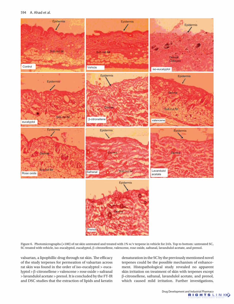
Histopathological analysis of terpenes treated and untreated rat skinIn this histopathological evaluation, epidermal lique-faction, edema of collagen fibers was considered as the key criteria to distinguish the effect of drug (Narishetty and Panchagnula, 2004b). The morphological changes in skin after terpenes treatment are shown in Figure 6. In the case of control, clearly defined SC could be seen with well-woven structures (Figure 6). There were no significant changes observed on rat skin specimens treated with vehicle (ethanol:IPB, 40:60) in comparison with the untreated SC, which suggest absence of any skin irritation. Ethanol is widely used as a skin permeation enhancer at concentrations of up to 70% in many trans-dermal therapeutic systems (Krishnaiah et al., 2002a,b; Fang et al., 2008). In addition, Krishnaiah et al. (2005) reported that a transdermal gel containing 70% ethanol showed no signs of skin irritancy when applied for 1 day in humans. On application of terpenes (iso-eucalyptol, eucalyptol, β-citronellene, valencene, rose oxide, safra-nal, lavandulol acetate, and prenol), the cell structures loosened with increased cell infiltration in dermis and in some skin samples where degeneration of append-ages were also observed. The disruption and extraction of lipid bilayers were clearly evident as distinct voids and empty spaces were visible in the epidermal region (Figure 6). There were no apparent signs of skin irritation (erythema and edema) observed on visual examination of skin specimens treated with iso-eucalyptol, eucalyp-tol, valencene, and rose oxide indicating absence of any
1750 1700 1650 1600 1550 1500
Wavenumber (cm−1)
Tran
smitt
ance
PrenolLavandulolacetateSafranal
Rose oxide
Valencene
eucalyptoliso-eucalyptol
Vehicle
Control
β-citronellene
Figure 4. FT-IR spectra of rat SC. Change in amide I (1640 cm−1) and amide II (1550 cm−1) stretching vibrations after 24 h treatment with 1% w/v terpene in vehicle. Bottom to top: untreated SC, SC treated with vehicle, iso-eucalyptol, eucalyptol, β-citronellene, valencene, rose oxide, safranal, lavandulol acetate, and prenol.
Transcutaneous permeation of valsartan 593
© 2011 Informa Healthcare USA, Inc.
skin irritation as a consequence of terpenes treatment. Moreover, terpenes have been proclaimed as GRAS excipients. Overall, the irritation was higher with the β-citronellene, safranal, lavandulol acetate, and prenol (Figure 6), which also showed good enhancement in per-meation studies. Earlier studies have shown that enhanc-ers, which cause significant enhancement, also produce high skin irritation (Bhatia et al., 1997; Kanikkannan and Singh, 2002); however, the skin irritation depends on the concentration of the enhancer and the duration of appli-cation. It would have been interesting to employ strategy to reduce skin irritation by combining two terpenes, which act by different mechanisms. This can result in the use of reduced concentration of penetration enhancers
that would produce maximum enhancement with less skin irritation. In this context, works are in progress in our laboratory to investigate the combinatorial effect of terpenes on rodent and HCS; therefore, the combination of these enhancers at low concentrations may be advan-tageous not only from a pharmacokinetic viewpoint but also in terms of reducing the skin irritation potential.
Conclusions
Among the investigated terpenes (iso-eucalyptol, β-citronellene, valencene, rose oxide, safranal, lavan-dulol acetate, and prenol), iso-eucalyptol has been found to be an effective penetration enhancer for diffusion of
30 40 60 80 100 120 140 160 180 200Temperature (°C)
Hea
t flo
w e
ndo
up (m
W)
Prenol
Lavandulol acetate
Safranal
Rose oxide
Valencene
eucalyptol
iso-eucalyptol
Vehicle
Control
β-citronellene
Peak = 72.881 °C
Peak = 79.884 °C
Peak = 78.884 °C
Peak = 83.950 °C
Peak = 93.48 °C
Peak = 80.893 °C
Peak = 69.164 °C
Peak = 89.155 °C
Peak = 116.5 °C
Peak = 74.2 °C
Peak = 104.563 (T3) Peak = 114.328 (T4)
Peak = 83.135 (T2)
Peak = 40.723 (T1)
Figure 5. DSC thermogram of rat SC untreated and treated with 1% w/v terpene in vehicle for 24 h. Bottom to top: untreated SC, SC treated with vehicle, iso-eucalyptol, eucalyptol, β-citronellene, valencene, rose oxide, safranal, lavandulol acetate, and prenol.
594 A. Ahad et al.
Drug Development and Industrial Pharmacy
valsartan, a lipophillic drug through rat skin. The efficacy of the study terpenes for permeation of valsartan across rat skin was found in the order of iso-eucalyptol > euca-lyptol > β-citronellene > valencene > rose oxide > safranal > lavandulol acetate > prenol. It is concluded by the FT-IR and DSC studies that the extraction of lipids and keratin
denaturation in the SC by the previously mentioned novel terpenes could be the possible mechanism of enhance-ment. Histopathological study revealed no apparent skin irritation on treatment of skin with terpenes except β-citronellene, safranal, lavandulol acetate, and prenol, which caused mild irritation. Further investigations,
EpidermisEpidermis
Epidermis
Epidermis Epidermis
EpidermisEpidermis
Epidermis
Epidermis
Epidermis
Sub.cut.fat
Sub.cut.fat
Sub.cut.fatSub.cut.fatSafranalRose oxide
Prenol
eucalyptolβ-citronellene
Lavandulolacetate
Dermis
Dermis
Dermis
Dermis Dermis
Dermis
Control Vehicle
Sub.cut.fat
Sub.cut.fat
iso-eucalyptol
DermalCollagen
valencene
Figure 6. Photomicrographs (×100) of rat skin untreated and treated with 1% w/v terpene in vehicle for 24 h. Top to bottom: untreated SC, SC treated with vehicle, iso-eucalyptol, eucalyptol, β-citronellene, valencene, rose oxide, safranal, lavandulol acetate, and prenol.
For
pers
onal
use
onl
y.
Transcutaneous permeation of valsartan 595
© 2011 Informa Healthcare USA, Inc.
including formulation development and characteriza-tion of valsartan using iso-eucalyptol and other terpenes, are in progress in our laboratory.
Declaration of interest
The authors acknowledge the financial support by All India Council for Technical Education (AICTE), New Delhi, India (Grant # 8023/BOR/RPS-157/2006-07). The author Abdul Ahad also thanks the Council for Scientific and Industrial Research (CSIR), India (File #09/591 (0084)/2009-EMR-I), for providing financial assistance in the form of a senior research fellowship. The authors declare that they have no conflicts of inter-est to disclose.
referencesAhad A, Aqil M, Kohli K, Chaudhary H, Sultana Y, Mujeeb M et al.
(2009).Chemical penetration enhancers: a patent review. Expert Opin Ther Pat, 19:969–988.
Amin S, Kohli K, Khar RK, Mir SR, Pillai KK. (2008).Mechanism of in vitro percutaneous absorption enhancement of carvedilol by penetration enhancers. Pharm Dev Technol, 13:533–539.
Amnuaikita C, Ikeuchia I, Ogawaraa K, Higakia K, Kimura T. (2005). Skin permeation of propranolol from polymeric film containing terpene enhancers for transdermal use. Int J Pharm, 289,167–178.
Aqil M, Ahad A, Sultana Y, Ali A. (2007). Status of terpenes as skin penetration enhancers. Drug Dis Today, 2007, 12, 1601–1607.
Aqil M, Ali A, Sultana Y, Parvez N. (2004). Matrix type transdermal drug delivery systems of metoprolol tartrate: skin toxicity and in vivo characterization. Eth Pharm J, 22, 55–50.
Bhatia KS, Gao S, Freeman TP, Singh J. (1997).Effect of penetration enhancers and iontophoresis on the ultrastructure and cholecystokinin-8 permeability through porcine skin. J Pharm Sci, 86:1011–1015.
Chisty MN, Bellantone RA, Taft DR, Plakogiannis FM. (2002).In vitro evaluation of the release of albuterol sulfate from polymer gels: effect of fatty acids on drug transport across biological membranes. Drug Dev Ind Pharm, 28:1221–1229.
Cornwell PA and Barry BW. (1991). The effects of a series of homologous terpene alcohols on the lipid structure of human stratum corneum as assessed by differential scanning calorimetry. In Scott RC, Guy RH, Hadgraft J and Boddr HE. (Eds), Prediction of Percutaneous Penetration, IBC Technical Services, London, Vol. 2, 394–400.
Cornwell PA, Barry BW, Bouwstra JA, and Gooris GS. (1996). Modes of action of terpene penetration enhancers in human skin; differential scanning calorimetry, smallangle X-ray diffraction and enhancer uptake studies. Int J Pharm, 127, 9–26.
Cornwell PA, Barry BW. (1994).Sesquiterpene components of volatile oils as skin penetration enhancers for the hydrophilic permeant 5-fluorouracil. J Pharm Pharmacol, 46:261–269.
El-Kattan AF, Asbill CS, Kim N, Michniak BB. (2001).The effects of terpene enhancers on the percutaneous permeation of drugs with different lipophilicities. Int J Pharm, 215:229–240.
Fang C, Liu Y, Ye X, Rong ZX, Feng XM, Jiang CB et al. (2008).Synergistically enhanced transdermal permeation and topical analgesia of tetracaine gel containing menthol and ethanol in experimental and clinical studies. Eur J Pharm Biopharm, 68:735–740.
Fujii M, Takeda Y, Yoshida M, Matsumoto M, Watanabe Y. (2004).Enhancement effect of p-menthane-3,8-diol on in vitro permeation of antipyrine and indomethacin through Yucatan micropig skin. Drug Dev Ind Pharm, 30:673–677.
Godwin DA, Michniak BB. (1999).Influence of drug lipophilicity on terpenes as transdermal penetration enhancers. Drug Dev Ind Pharm, 25:905–915.
Goodman M, Barry BW. (1989). Action of penetration enhancers on human stratum corneum as assessed by differential scanning calorimetry. In: Bronaugh RL, Maibach HI. (eds) Percutaneous absorption. Marcel Dekker, New York and Basel. 567–593.
Higaki K, Amnuaikit C, Kimura T. (2003). Strategies for overcoming the stratum corneum: chemical and physical approaches. Am J Drug Deliv, 1, 187–214.
Hori M, Satoh S, Maibach HI, Guy RH. (1991).Enhancement of propranolol hydrochloride and diazepam skin absorption in vitro: effect of enhancer lipophilicity. J Pharm Sci, 80:32–35.
Jain AK, Thomas NS, Panchagnula R. (2002).Transdermal drug delivery of imipramine hydrochloride. I. Effect of terpenes. J Control Release, 79:93–101.
Jain R, Aqil M, Ahad A, Ali A, Khar RK. (2008).Basil oil is a promising skin penetration enhancer for transdermal delivery of labetolol hydrochloride. Drug Dev Ind Pharm, 34:384–389.
Kanikkannan N, Singh M. (2002).Skin permeation enhancement effect and skin irritation of saturated fatty alcohols. Int J Pharm, 248:219–228.
Karakatsani M, Dedhiya M, Plakogiannis FM. (2010).The effect of permeation enhancers on the viscosity and the release profile of transdermal hydroxypropyl methylcellulose gel formulations containing diltiazem HCl. Drug Dev Ind Pharm, 36:1195–1206.
Kararli T, Kirchhoff CF, Penzotti Jr SC. (1995). Enhancement of transdermal transport of azidothymidine (AZT) with novel terpene and terpene like enhancers: in vivo-in vitro correlations. J Control Rel, 34, 43–51.
Krishnaiah YS, Al-Saidan SM, Chandrasekhar DV, Satyanarayana V. (2005).Bioavailability of nerodilol-based transdermal therapeutic system of nicorandil in human volunteers. J Control Release, 106:111–122.
Krishnaiah YS, Satyanarayana V, Bhaskar P. (2002a).Influence of limonene on the bioavailability of nicardipine hydrochloride from membrane-moderated transdermal therapeutic systems in human volunteers. Int J Pharm, 247:91–102.
Krishnaiah YS, Satyanarayana V, Bhaskar P. (2003).Enhanced percutaneous permeability of nicardipine hydrochloride by carvone across the rat abdominal skin. Drug Dev Ind Pharm, 29:191–202.
Krishnaiah YS, Satyanarayana V, Karthikeyan RS. (2002b).Effect of the solvent system on the in vitro permeability of nicardipine hydrochloride through excised rat epidermis. J Pharm Pharm Sci, 5:123–130.
Kunta JR, Goskonda VR, Brotherton HO, Khan MA, Reddy IK. (1997).Effect of menthol and related terpenes on the percutaneous absorption of propranolol across excised hairless mouse skin. J Pharm Sci, 86:1369–1373.
Moghimi HR, Makhmalzadeh BS, Manafi A. (2009).Enhancement effect of terpenes on silver sulphadiazine permeation through third-degree burn eschar. Burns, 35:1165–1170.
Mura S, Manconi M, Sinico C, Valenti D, Fadda AM. (2009).Penetration enhancer-containing vesicles (PEVs) as carriers for cutaneous delivery of minoxidil. Int J Pharm, 380:72–79.
Narishetty ST, Panchagnula R. (2004a).Transdermal delivery of zidovudine: effect of terpenes and their mechanism of action. J Control Release, 95:367–379.
Narishetty ST, Panchagnula R. (2004b).Transdermal delivery system for zidovudine: in vitro, ex vivo and in vivo evaluation. Biopharm Drug Dispos, 25:9–20.
Ogiso T, Iwaki M, Paku T. (1995).Effect of various enhancers on transdermal penetration of indomethacin and urea, and relationship between penetration parameters and enhancement factors. J Pharm Sci, 84:482–488.
Panchagnula R, Salve PS, Thomas NS, Jain AK, Ramarao P. (2001).Transdermal delivery of naloxone: effect of water, propylene glycol, ethanol and their binary combinations on permeation through rat skin. Int J Pharm, 219:95–105.
596 A. Ahad et al.
Drug Development and Industrial Pharmacy
Paudel KS, Hammell DC, Agu RU, Valiveti S, Stinchcomb AL. (2010).Cannabidiol bioavailability after nasal and transdermal application: effect of permeation enhancers. Drug Dev Ind Pharm, 36:1088–1097.
Rizwan M, Aqil M, Ahad A, Sultana Y, Ali MM. (2008).Transdermal delivery of valsartan: I. Effect of various terpenes. Drug Dev Ind Pharm, 34:618–626.
Sapra B, Jain S, Tiwary AK. (2008).Percutaneous permeation enhancement by terpenes: mechanistic view. Aaps J, 10:120–132.
Shakeel F, Baboota S, Ahuja A, Ali J, Shafiq S. (2008).Skin permeation mechanism and bioavailability enhancement of celecoxib from transdermally applied nanoemulsion. J Nanobiotechnology, 6:8.
Shams MS, Alam MI, Ali A, Sultana Y, Aqil M. (2010).Pharmacodynamics of a losartan transdermal system for the treatment of hypertension. Drug Dev Ind Pharm, 36:385–392.
Silva CL, Nunes SC, Eusébio ME, Sousa JJ, Pais AA. (2006).Study of human stratum corneum and extracted lipids by thermomicroscopy and DSC. Chem Phys Lipids, 140:36–47.
Sinha VR, Kaur MP. (2000).Permeation enhancers for transdermal drug delivery. Drug Dev Ind Pharm, 26:1131–1140.
Tatar S, Saglík S. (2002).Comparison of UV- and second derivative-spectrophotometric and LC methods for the determination of
valsartan in pharmaceutical formulation. J Pharm Biomed Anal, 30:371–375.
Vaddi HK, Ho PC, Chan SY. (2002).Terpenes in propylene glycol as skin-penetration enhancers: permeation and partition of haloperidol, Fourier transform infrared spectroscopy, and differential scanning calorimetry. J Pharm Sci, 91:1639–1651.
Williams AC and Barry BW. (1989). Permeation, FTIR and DSC investigations of terpene penetration enhancers in human skin. J Pharm Pharmacol, 41, 12P.
Williams AC and Barry BW. (1991). The enhancement index concept applied to terpene penetration enhancers for human skin and model lipophilic (oestradiol) and hydrophilic (5-fluorouracil) drugs. Int J Pharm, 74, 157–168.
Williams AC, Barry BW. (2004).Penetration enhancers. Adv Drug Deliv Rev, 56:603–618.
Yamane MA, Williams AC, Barry BW. (1995).Terpene penetration enhancers in propylene glycol/water co-solvent systems: effectiveness and mechanism of action. J Pharm Pharmacol, 47:978–989.
Yum S, Lee E, Taskovich L, Theeuwes F. (1994). Permeation Enhancement with Ethanol: Mechanism of Action through Skin. In Drug Permeation Enhancement; Hsich, D.S., Ed.; Marcel Dekker: New York, 143–170.


