Embed Size (px)
Citation preview
Role of Sorbitol Accumulation and Myo-inositol Depletion in ParanodalSwelling of Large Myelinated Nerve Fibersin the Insulin-deficient Spontaneously Diabetic Bio-breeding RatReversal by Insulin Replacement, an Aldose Reductase Inhibitor, and Myo-inositol
D. A. Greene, S. Chakrabarti,* S. A. Lattimer, and A. A. F. Sima*Diabetes Research Laboratories ofthe Department ofMedicine, University ofPittsburgh School ofMedicine, Pittsburgh, Pennsylvania15160; *Neuropathology Research Laboratory, Department ofPathology, University ofManitoba, Winnipeg, Canada
Abstract
Axo-glial dysjunction refers to the disruption of important junc-tional complexes that anchor terminal loops of myelin to theparanodal axolemma in diabetic human and animal peripheralnerve. Neither axo-glial dysjunction nor the preceeding acutelocalized paranodal swelling has been specifically attributed todiscrete metabolic consequences of insulin deficiency or hyper-glycemia. Two metabolic sequelae of hyperglycemia in diabeticnerve, sorbitol accumulation via aldose reductase, and (NaK)-ATPase deficiency related to myo-inositol depletion, were ex-plored as possible underlying causes of acute paranodal swellingin the spontaneously diabetic bio-breeding rat. 3 wk of insulinreplacement, or therapy with an aldose reductase inhibitor ormyo-inositol completely reversed paranodal swelling in suralnerve fibers after 3 wk of untreated insulin deficiency. Theseobservations suggest that insulin deficiency and hyperglycemiacause reversible paranodal swelling, and ultimately poorly re-versible axo-glial dysfunction, via the myo-inositol-related(Na,K)-ATPase defect rather than by the osmotic effects of sor-bitol accumulation within nerve fibers.
IntroductionIn the last decade, a cascade of metabolic abnormalities in di-abetic peripheral nerve, triggered by hyperglycemia and involvingmyo-inositol (MI)' and (Na,K)-ATPase, has to a large extentsatisfactorily explained the rapidly reversible slowing of nerveconduction in acutely diabetic rodents (1, 2); yet, the relationshipof this metabolic cascade to the chronic and more pathogenet-ically relevant structural abnormalities characterizing both mu-rine and human diabetic neuropathy remains to be clarified (2,3). Activation of the polyol pathway by hyperglycemia appearsto be an essential triggering event, since aldose reductase inhib-itors (ARI) prevent MI depletion (4-6), the associated (Na,K)-
Address reprint requests to Dr. Greene, 5510C MSRB-I, 1150 WestMedical Center Drive, University ofMichigan Medical Center, Ann Ar-bor, MI 48109.
Presented in part at the Annual Meeting of the American Societyfor Clinical Investigation, Washington DC, May 1986.
Receivedfor publication 18 September 1986.
1. Abbreviations used in this paper: ARI, aldose reductase inhibitor, BB,Bio-breeding; EMPA, evoked muscle potential amplitude; IDDM, in-sulin-dependent diabetes mellitus; MI, myo-inositol; MNCV, motor nerveconduction velocity; PZI, protamine zinc insulin.
ATPase defect (7), and the resultant reversible slowing of nerveconduction in diabetic animals (6); competitive inhibition byglucose of carrier-mediated MI uptake may also contribute tothe depletion ofnerve MI in diabetes (8). MI depletion diminishesnerve phosphoinositide turnover (9), interfering with (Na,K)-ATPase regulation (9-13) by a protein kinase-C-dependentmechanism (14), thereby producing a four- to fivefold increasein intraaxonal sodium concentration (1, 3, 15, 16), inactivationof nodal sodium channels, and a selective conduction block oflarge myelinated nerve fibers that acutely slows composite sal-tatory conduction (1, 3, 15, 16). Increased intracellular sodiumreduces the transmembrane gradient driving sodium-dependentMI uptake, further depleting MI and completing a self-reinforc-ing metabolic defect (8, 13).
The role of this metabolic and biophysical cascade in thedevelopment ofthe subsequent morphological abnormalities thatcharacterize human and murine diabetic neuropathy has notbeen satisfactorily explored (1, 2). The initial characteristic mor-phometric abnormality in the peripheral neuropathy of thespontaneously diabetic Bio-breeding (BB) rat occurs within thefirst 3 wk of diabetes and consists of localized swelling of largemyelinated nerve fibers in the region of the node of Ranvier (1,3). This swelling and the accompanying four- to fivefold increasein intraaxonal sodium are prevented by insulin replacementtherapy that normalizes blood glucose and nerve conductionvelocity (1, 3). With more prolonged hyperglycemia, nodalswelling gives rise to axo-glial dysfunction (17) accompanied byapparent loss offunctional sodium channels from the node, nei-ther ofwhich is acutely corrected by subsequent insulin replace-ment therapy despite rapid normalization of both MI contentand (Na,K)-ATPase activity (13, 17). Conceptually, the intra-cellular accumulation of either sorbitol (18) or sodium (3),through either aldose reductase activation or the MI-related(Na,K)-ATPase defect, respectively, could explain the early acuteparanodal swelling thought to underlie axo-glial dysfunction.Hence, the ability of insulin replacement, ARI administrationand MI supplementation to reverse paranodal swelling was ex-plored in the acutely diabetic BB rat, whose fidelity as a modelfor human disease is highlighted by the recent identification ofparanodal swelling and axo-glial dysfunction in sural nervebiopsies from patients with diabetic neuropathy (19). Insulinreplacement, ARI administration, or MI supplementation com-pletely reversed paranodal swelling as assessed by ratio of theparanodal fiber diameter to the internodal length. All three ther-apies correct MI depletion and (Na,K)-ATPase inactivation,whereas only insulin replacement and ARI treatment but notMI supplementation prevent sorbitol accumulation (6, 20);hence, paranodal swelling in diabetic nerve most likely reflectsthe intracellular accumulation of sodium due to decreased(Na,K)-ATPase activity rather than sorbitol accumulation.
Paranodal Swelling in the Spontaneously Diabetic Bio-breeding Rat 1479
J. Clin. Invest.© The American Society for Clinical Investigation, Inc.0021-9738/87/05/1479/07 $1.00Volume 79, May 1987, 1479-1485
Methods
Experimental design and animal model. Prediabetic male BB-rats andage-matched nondiabetes-prone male BB-rats obtained from the De-partment of Pathology, University of Massachusetts, Worchester, MA(courtesy of Dr. A. Like), were maintained in individual air-filtered met-abolic cages with ad lib. access to water and rat chow (Wayne Lab BloxF-6, Wayne Laboratory Animal Diets, Wayne Feed Division, Winnipeg,Manitoba [MI content 0.022% wt/wt]) (17). Body weight, urine volume,and glucosuria (Testape, Eli Lilly Canada, Inc., Toronto, Ontario) were
monitored daily, and glucose was measured in tail-vein blood samplesby Ames Eyetone (Miles Laboratory, Ltd., Rexdale, Ontario) every secondday between 2 and 4 p.m. (17). Upon detection of glucosuria and hy-perglycemia, the newly diabetic rats were immediately begun on smalldaily doses (0.5-3.0 U * d-') ofprotamine zinc insulin (PZI), (ConnaughtLaboratories, Ltd., Toronto, Ontario), designed to maintain blood glucoselevels between 350 and 450 mg* dl-' (17). 3 wk thereafter, age-matcheddiabetic animals were randomly distributed into four experimentalgroups. "Baseline" diabetics were sacrificed immediately in orderto obtainbaseline structural and morphometric parameters (3). The other exper-
imental groups were entered into a 3-wk study period during which:"insulin-deficient" diabetics were maintained hyperglycemic on smalldaily doses of PZI; "ARI-treated" diabetics were maintained insulin de-ficient on small doses of PZI but were given the ARI Statil (Stuart Phar-maceuticals, Wilmington, DE)25 mg* kg-' * d-' ina 10mg * ml- aqueous
solution by gastric intubation; and "MI-treated" diabetics were main-tained insulin deficient on small doses of PZI but in addition receivedMI, 670 mg kg-' d' in a 133-mg ml-' aqueous solution by gastricintubation (6). These groups were compared to a similarly aged previouslyreported group of "insulin-replaced" diabetics treated aggressively withlarger PZI doses (3.0-6.0 U d-') adjusted for changes in blood glucosein order to achieve and maintain euglycemia (3). End-point measurements(nerve conduction, morphological and biochemical assessments) were
performed by investigators unaware of the identity of the examined ma-
terial.Electrophysiological studies. Animals were lightly anesthetized with
ethyl ether (Fisher Scientific Company, Fair Lawn, NJ). Motor nerve
conduction velocity (MNCV) was determined noninvasively in the sciatic-posterior tibial conduction system in a temperature controlled environ-ment as previously described in detail (21). Briefly, the left sciatic-tibialnerve was stimulated proximally at the sciatic notch and distally at theankle via bipolar electrodes using supramaximal stimuli (8 V) from a
stimulater (TM 501, Tektronix, Inc., Beaverton, OR) at 20 Hz. Evokedmuscle potential amplitudes (EMPA) were collected from the first in-terosseous space of the hindpaw by a unipolar platinum recording elec-trode and displayed on a Tektronix 51 1 storage oscilloscope. Motor nerve
conduction velocity was calculated by subtracting the distal from theproximal latency measured in milliseconds from stimulus artifact to takeoff of the evoked muscle potential; the resultant difference was dividedinto the distance between the stimulating electrodes measured in milli-meters, yielding a value for MNCV in meters per second. MNCV andEMPA were measured on a weekly basis throughout the study (21).
Tissue collection. On the day after the final MNCV determination,nonfasted animals were anesthetized with Na pentobarbital (50 mg* kg).Mid-thigh segments of the left and right sciatic nerves were surgicallyremoved, weighed, and processed either for enzymatic ATPase mea-
surements or for gas-liquid chromatographic determination ofMI content
as previously described in detail (11). The right proximal sural nerve
(opposite to the side on which MNCV was performed) was fixed in situ
for 10 min by a cacodylate buffered (pH 7.40) 2.5% (vol/vol) glutaral-dehyde fixative adjusted to an osmolality of 300 mosmol with su-
crose (17).Biochemical measurements. Sciatic nerve MI was determined gas-
liquid chromatographically in protein-free Somogyi filtrates of sciatic
nerve homogenates as previously described (1 1, 20). Sciatic nerve (NaK)-ATPase activity was measured enzymatically as ouabain-inhibitedATPase activity in freshly prepared crude homogenates of whole sciatic
nerve as previously described in detail (11). Briefly, nerve segments were
minced and homogenized at 4VC in 2 ml of0.2 M sucrose-0.02 M Tris-HCI pH 7.5. 5-20 ,g ofhomogenate was assayed spectrophotometricallyin I ml of 100 mM NaCi, 10 mM KCa, 2.5 mM MgCI2, I mM Tris-ATP, I mM tri(cyclohexylammonium) phosphoenolpyruvate, 30 mMimidazole-HCI buffer (pH 7.3), 0.15 mM NADH, 50 ug of lactate de-hydrogenase and 30 sg of pyruvate kinase (22). ATPase activity in thepresence of 0.10 mM ouabain was defined as the ouabain-uninhibitableATPase fraction, since preliminary experiments had indicated that max-imum ouabain inhibition is obtained at this concentration (11). Ouabain-inhibitable ATPase activity, a precise measurement of (Na,K)-ATPaseactivity in rat sciatic nerve, was defined as the arithmetic difference be-tween composite and ouabain-uninhibitable ATPase activities (11).
Teasedfiber analysis. The proximal portion of the sural nerve waspost-fixed and stained in cacodylate-buffered 1% (vol/vol) osmium te-troxide, and 54±3 individual fibers were teased from each sural nervesegment in Epon (3). The teased fibers were examined under phase con-trast illumination at a magnification of X 320 and internodal lengthmeasured (3). The corresponding paranodal fiber diameter was deter-mined by measuring the maximum fiber width within a 30-Mm distanceof both nodes of Ranvier bordering each internodal segment, and themean value recorded for internode (3). The mean internodal length foreach fiber was plotted on the ordinate, and the paranodal fiber diameterwas plotted on the abscissa, and the slope of the linear regression wasused to define the paranodal fiber diameter to internodal length ratio (3).
Statistics. Data are mean±SEM, significance ofdifference is calculatedby Student's t test, and linear regessions are analyzed by the method ofleast squares.
Results
Clinical responses to insulin replacement, ARI, andMI therapyin BB diabetic rats (Table I). At baseline, following 3 wk ofinsulin deficiency, body weight was reduced, blood glucose waselevated, and tibial MNCV and EMPA were reduced similarlyin all five diabetic groups compared with nondiabetic BB controls(Table I). Body weight, blood glucose, and tibial MNCV andEMPA remained abnormal for the additional 3-wk study periodin insulin-deficient diabetics maintained on small doses of PZI(Table I). 3 wk of intensive insulin therapy in insulin replaceddiabetics normalized the rate of weight gain (Table I) as well asblood glucose, MNCV and EMPA. Administration of either anARI or MI for 3 wk to insulin-deficient BB rats did not affectbody weight or blood glucose, but nearly completely normalizedMNCV and EMPA in the ARI-treated and MI-treated diabetics.
Biochemical response in sciatic nerve to insulin replacement,ARIandMI therapy in insulin-deficient BB rats (Table II). Sciaticnerve MI content was significantly reduced in insulin-deficientdiabetics compared with nondiabetic controls. As in the strep-tozotocin- (20) and chronically diabetic BB (3, 13) rat, intensiveinsulin therapy in the "insulin replaced" diabetics corrected thenerve MI depletion resulting from insulin deficiency (Table II).Both ARI and MI treatment also corrected nerve MI depletionin the ART-treated and MI-treated BB diabetics as previouslyshown for the streptozotocin-induced diabetic rat (4-6, 20).
(Na,K)-ATPase activity. Neither 6 wk ofsustained hypergly-cemia in the insulin-deficient diabetics, nor 3 wk ofunmodifiedinsulin deficiency followed by 3 wk ofART or MI administrationstatistically significantly altered composite ATPase activity incrude homogenates of sciatic nerve although composite activitytended to be lowest in the insulin-deficient diabetics (data notshown). However, the ouabain-inhibitable component ofATPaseactivity was reduced - 28% in the insulin-deficient diabeticscompared with the nondiabetic controls (Table TI). 3 wk of in-tensive insulin therapy corrected this abnormality in the insulin-
1480 D. A. Greene, S. Chakrabarti, S. A. Lattimer, and A. A. F. Sima
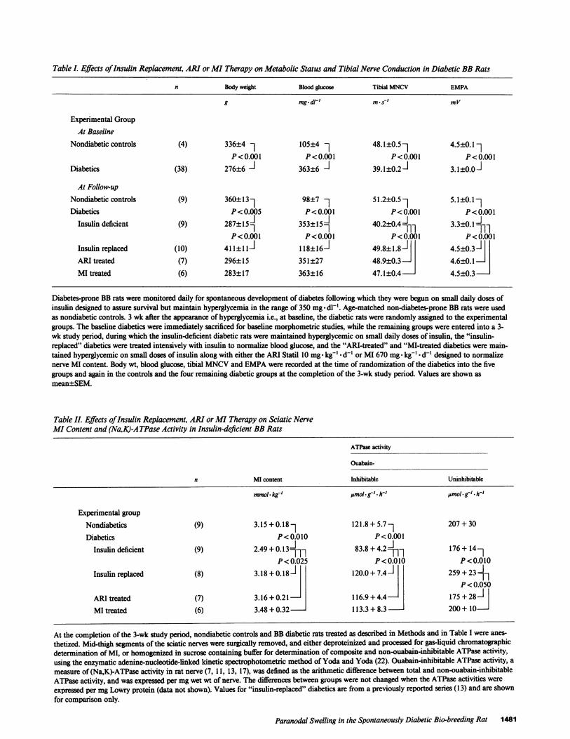
Table I. Effects ofInsulin Replacement, ARI or MI Therapy on Metabolic Status and Tibial Nerve Conduction in Diabetic BB Rats
n Body weight Blood glucose Tibial MNCV EMPA
g mgId1' ms' mV
Experimental GroupAt Baseline
Nondiabetic controls (4) 336±4 1 105±4 1 48.1±0.57 4.5±0.11P<0.001 P<0.001 P<0.001 P<0.001
Diabetics (38) 276±6 363±6 39.1±0.2-J 3.1±0.0
At Follow-upNondiabetic controls (9) 360±13] 98±7 1 51.2±0.5 5.1±0.17Diabetics P<0.005 P<0.001 P<0.001 P<0.001
Insulin deficient (9) 287±15353-15_ 40.2±0.4 1 3.3±0.11]]P<0.001 P<0.001 P<0.001 P<0.001
Insulin replaced (10) 411±1lli 118±16i 49.8±1.8 4.5±0.3ARI treated (7) 296±15 351±27 48.9±0.3 4.6_0.1MI treated (6) 283±17 363±16 47.1±0.4 4.5±0.3
Diabetes-prone BB rats were monitored daily for spontaneous development of diabetes following which they were begun on small daily doses ofinsulin designed to assure survival but maintain hyperglycemia in the range of 350 mg- dl-1. Age-matched non-diabetes-prone BB rats were usedas nondiabetic controls. 3 wk after the appearance of hyperglycemia i.e., at baseline, the diabetic rats were randomly assigned to the experimentalgroups. The baseline diabetics were immediately sacrificed for baseline morphometric studies, while the remaining groups were entered into a 3-wk study period, during which the insulin-deficient diabetic rats were maintained hyperglycemic on small daily doses of insulin, the "insulin-replaced" diabetics were treated intensively with insulin to normalize blood glucose, and the "ARI-treated" and "MI-treated diabetics were main-tained hyperglycemic on small doses of insulin along with either the ARI Statil 10 mg * kg-' d-' or MI 670 mg- kg-' - d-' designed to normalizenerve MI content. Body wt, blood glucose, tibial MNCV and EMPA were recorded at the time of randomization of the diabetics into the fivegroups and again in the controls and the four remaining diabetic groups at the completion of the 3-wk study period. Values are shown asmean±SEM.
Table II. Effects ofInsulin Replacement, ARI or MI Therapy on Sciatic NerveMI Content and (Na,K)-ATPase Activity in Insulin-deficient BB Rats
ATPase activity
Ouabain-
n MI content Inhibitable Uninhibitable
mmobl pg- mol g- *l h-' #mol -gr' * h-'
Experimental group
Nondiabetics (9) 3.15 + 0.18 121.8 + 5.7 207 + 30
Diabetics P<0.010 P<0.001
Insulin deficient (9) 2.49+0.13 'j 83.8+4.2-1 176+14P<0.025 P<0.010 P<0.010
Insulin replaced (8) 3.18+0.18 | 120.0+ 7.4 259 +23 j
P< 0.050
ARItreated (7) 3.16+0.21- 116.9+4.4 175+28 1
MI treated (6) 3.48 + 0.32 113.3 + 8.3 200 + 10
At the completion of the 3-wk study period, nondiabetic controls and BB diabetic rats treated as described in Methods and in Table I were anes-
thetized. Mid-thigh segments of the sciatic nerves were surgically removed, and either deproteinized and processed for gas-liquid chromatographicdetermination of MI, or homogenized in sucrose containing buffer for determination of composite and non-ouabain-inhibitable ATPase activity,using the enzymatic adenine-nucleotide-linked kinetic spectrophotometric method of Yoda and Yoda (22). Ouabain-inhibitable ATPase activity, a
measure of (Na,K)-ATPase activity in rat nerve (7, 11, 13, 17), was defined as the arithmetic difference between total and non-ouabain-inhibitableATPase activity, and was expressed per mg wet wt of nerve. The differences between groups were not changed when the ATPase activities wereexpressed per mg Lowry protein (data not shown). Values for "insulin-replaced" diabetics are from a previously reported series (13) and are shownfor comparison only.
Paranodal Swelling in the Spontaneously Diabetic Bio-breeding Rat 1481
replaced diabetics. Aldose reductase inhibitor and MI therapyin the hyperglycemic BB rats increased sciatic nerve ouabain-inhibitable ATPase activity in the ARI-treated and MI-treateddiabetics to 94 and 97%, respectively, ofthat in the nondiabeticcontrol (Table II). In contrast, residual ATPase activity in thepresence of inhibiting concentrations of ouabain ("ouabain un-inhibitable ATPase activity") was similar across experimentalgroups (except the insulin-replaced diabetics whose compositeactivity was identical to their contemporaneous nondiabeticcontrols [13]). Thus, the effects of insulin deficiency and ARIor MI administration were primarily confined to the ouabain-inhibitable (Na,K)-ATPase fraction, which in rat sciatic nerveis a reliable estimate of (Na,K)-ATPase activity (7, 11, 13, 17).
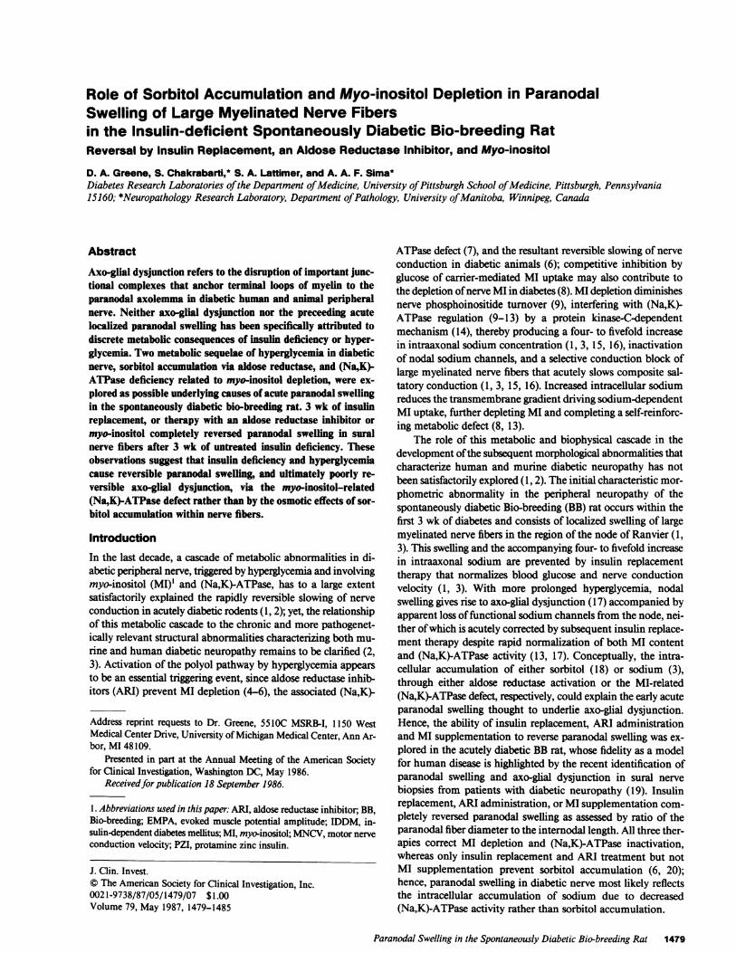
Qualitative assessment ofteasedfibers (Fig. 1). Teased my-elinated nerve fibers from baseline and insulin-deficient diabeticsexhibited marked swelling at or near the node of Ranvier com-pared with teased fibers from nondiabetic controls (3) (Fig. 1,compare panels a and b). Paranodal swelling appeared as distinctcuffings around the paranodal area, or as swellings that pro-gressively tapered off toward the internodal areas. This charac-
teristic abnormality was encountered much less frequently innerve fibers from insulin-replaced diabetics after 3 wk ofintensiveinsulin therapy, or from ARI-treated or MI-treated diabetics.Here the fibers had a much more normal appearance (Fig. 1 cand d).
Quantitative assessment of internodal length to paranodalfiber diameter ratio (Figs. 2 and 3). Linear regression analysisof internodal length vs. paranodal fiber diameter confirmed theimpression that paranodal fiber diameter was enlarged in un-treated insulin deficiency diabetes. Internodal fiber length andparanodal fiber diameter were linearly related in all six experi-mental groups (nondiabetic controls, and baseline, insulin-de-ficient, insulin-replaced, ARI-treated and MI-treated diabetics,Figs. 2 and 3). At baseline, the slope of the regression line wassignificantly shifted toward larger paranodal fiber diameters inthe baseline diabetics compared with their age-matched non-diabetic controls (P < 0.001) (Fig. 2, a and b).
At followup 3 wk later, the slope of the regression line wasnot significantly shifted in the older nondiabetic controls com-pared with the younger nondiabetic controls at baseline (compare
a
b Figure 1. Effect of insulin deficiency, ARItreatment and MI supplementation on theappearance of the node of Ranvier of largemyelinated fibers from the sural nerve ofspontaneously diabetic BB rats. BB ratswith spontaneous insulin deficiency of 3 wkduration were randomly distributed into ex-perimental groups that remained insulin de-ficient without treatment, or were treatedwith an ARI or MI supplementation for anadditional 3 wk. Photomicrographs of osmi-cated teased single fibers compare parano-
C dal fiber appearance in (a) a nondiabeticcontrol rat, (b) an insulin-deficient diabeticrat, (c) an ARI-treated diabetic rat, and (d)a MI-treated diabetic rat. Marked paranodalswelling that progressively tapered off to-ward the internodal areas was readily appar-ent in the untreated insulin-deficient dia-
1 betic rat (b) compared to the nondiabeticcontrol (a). Paranodal swelling was largelyameliorated after 3 wk of ARI- or MI-treat-
d ment (c and d), leaving fibers that resem-bled those of the nondiabetic control (a).
1482 D. A. Greene, S. Chakrabarti, S. A. Lattimer, and A. A. F. Sima
M lift, , -"::....
;.
bank
Non-diabetic controls(3 weeks); (n=4)
a = 0.140 t0.006
b = 0.090 t0.007
r2= 0.85 ± 0.03
a
5 10 15 20 25
Insulin-deficient diabetics(3 weeks); (n=4)
b =0.042+±0.002 p<0.001
b
5 10 15 ~~~~~~~2025
Juxtanodal fiber diameter (Aim)
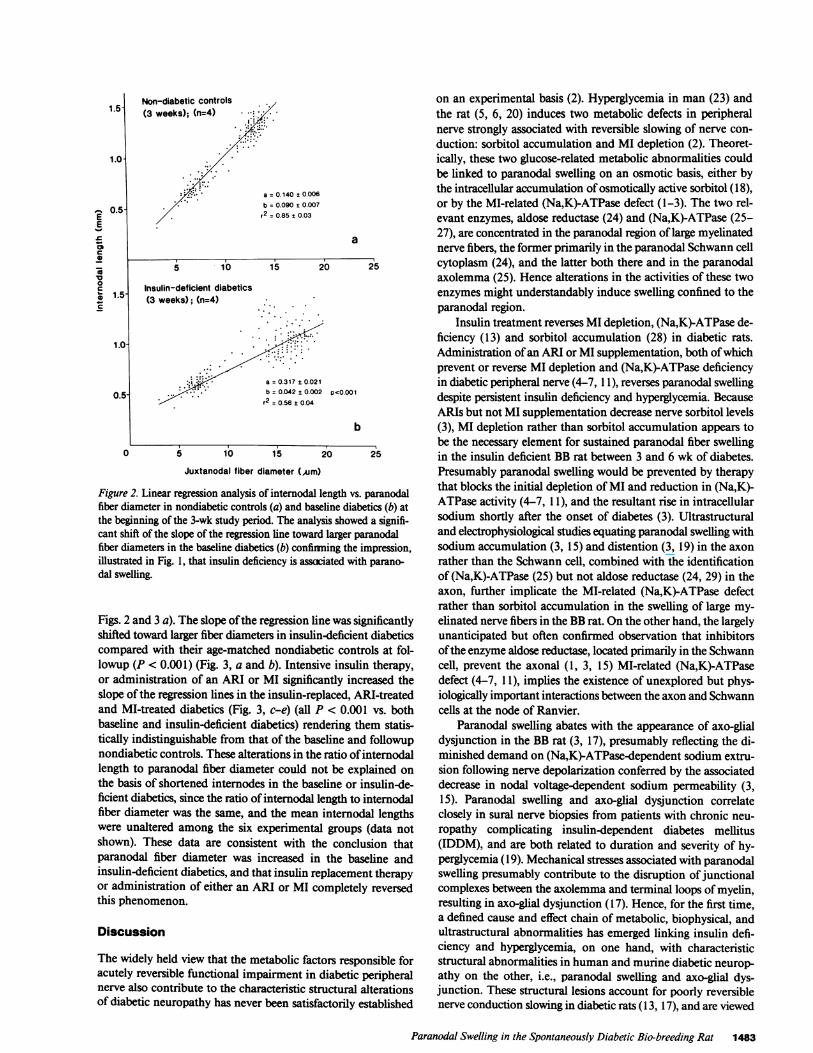
Figure 2. Linear regression analysis of internodal length vs. paranodalfiber diameter in nondiabetic controls (a) and baseline diabetics (b) at
the beginning of the 3-wk study period. The analysis showed a signifi-cant shift of the slope of the regression line toward larger paranodalfiber diameters in the baseline diabetics (b) confirming the impression,illustrated in Fig. 1, that insulin deficiency is assaciated with parano-dal swelling.
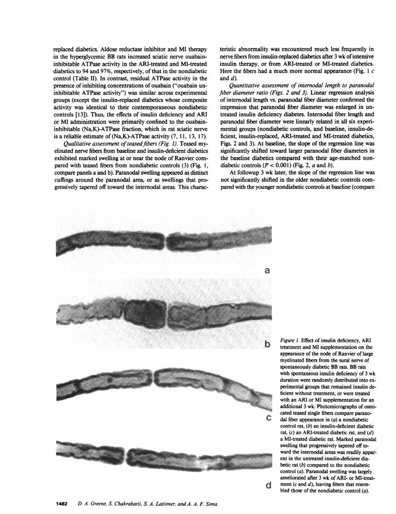
Figs. 2 and 3 a). The slope ofthe regression line was significantly
shifted toward larger fiber diameters in insulin-deficient diabetics
compared with their age-matched nondiabetic controls at fol-
lowup (P < 0.001) (Fig. 3, a and b). Intensive insulin therapy,or administration of an ARI or MI significantly increased the
slope of the regression lines in the insulin-replaced, ARI-treatedand MI-treated diabetics (Fig. 3, c-e) (all P < 0.001 vs. bothbaseline and insulin-deficient diabetics) rendering them statis-
tically indistinguishable from that of the baseline and followupnondiabetic controls. These alterations in the ratio ofinternodallength to paranodal fiber diameter could not be explained onthe basis of shortened internodes in the baseline or insulin-de-ficient diabetics, since the ratio of internodal length to internodalfiber diameter was the same, and the mean internodal lengthswere unaltered among the six experimental groups (data notshown). These data are consistent with the conclusion thatparanodal fiber diameter was increased in the baseline andinsulin-deficient diabetics, and that insulin replacement therapyor administration of either an ARI or MI completely reversedthis phenomenon.
Discussion
The widely held view that the metabolic factors responsible foracutely reversible functional impairment in diabetic peripheralnerve also contribute to the characteristic structural alterationsof diabetic neuropathy has never been satisfactorily established
on an experimental basis (2). Hyperglycemia in man (23) andthe rat (5, 6, 20) induces two metabolic defects in peripheralnerve strongly associated with reversible slowing of nerve con-
duction: sorbitol accumulation and MI depletion (2). Theoret-ically, these two glucose-related metabolic abnormalities couldbe linked to paranodal swelling on an osmotic basis, either bythe intracellular accumulation of osmotically active sorbitol (18),or by the MI-related (Na,K)-ATPase defect (1-3). The two rel-evant enzymes, aldose reductase (24) and (Na,K)-ATPase (25-27), are concentrated in the paranodal region of large myelinatednerve fibers, the former primarily in the paranodal Schwann cellcytoplasm (24), and the latter both there and in the paranodalaxolemma (25). Hence alterations in the activities of these twoenzymes might understandably induce swelling confined to theparanodal region.
Insulin treatment reverses MI depletion, (Na,K)-ATPase de-ficiency (13) and sorbitol accumulation (28) in diabetic rats.Administration ofan ARI or MI supplementation, both ofwhichprevent or reverse MI depletion and (Na,K)-ATPase deficiencyin diabetic peripheral nerve (4-7, 1 1), reverses paranodal swellingdespite persistent insulin deficiency and hyperglycemia. BecauseARIs but not MI supplementation decrease nerve sorbitol levels(3), MI depletion rather than sorbitol accumulation appears tobe the necessary element for sustained paranodal fiber swellingin the insulin deficient BB rat between 3 and 6 wk of diabetes.Presumably paranodal swelling would be prevented by therapythat blocks the initial depletion ofMI and reduction in (Na,K)-ATPase activity (4-7, 1 1), and the resultant rise in intracellularsodium shortly after the onset of diabetes (3). Ultrastructuraland electrophysiological studies equating paranodal swelling withsodium accumulation (3, 15) and distention (3, 19) in the axonrather than the Schwann cell, combined with the identificationof (Na,K)-ATPase (25) but not aldose reductase (24, 29) in theaxon, further implicate the MI-related (Na,K)-ATPase defectrather than sorbitol accumulation in the swelling of large my-elinated nerve fibers in the BB rat. On the other hand, the largelyunanticipated but often confirmed observation that inhibitorsofthe enzyme aldose reductase, located primarily in the Schwanncell, prevent the axonal (1, 3, 15) MI-related (Na,K)-ATPasedefect (4-7, 1 1), implies the existence of unexplored but phys-iologically important interactions between the axon and Schwanncells at the node of Ranvier.
Paranodal swelling abates with the appearance of axo-glialdysjunction in the BB rat (3, 17), presumably reflecting the di-minished demand on (NaK)-ATPase-dependent sodium extru-sion following nerve depolarization conferred by the associateddecrease in nodal voltage-dependent sodium permeability (3,15). Paranodal swelling and axo-glial dysjunction correlateclosely in sural nerve biopsies from patients with chronic neu-ropathy complicating insulin-dependent diabetes mellitus(IDDM), and are both related to duration and severity of hy-perglycemia (19). Mechanical stresses associated with paranodalswelling presumably contribute to the disruption ofjunctionalcomplexes between the axolemma and terminal loops ofmyelin,resulting in axo-glial dysjunction (17). Hence, for the first time,a defined cause and effect chain of metabolic, biophysical, andultrastructural abnormalities has emerged linking insulin defi-ciency and hyperglycemia, on one hand, with characteristicstructural abnormalities in human and murine diabetic neurop-athy on the other, i.e., paranodal swelling and axo-glial dys-junction. These structural lesions account for poorly reversiblenerve conduction slowing in diabetic rats (I13, 17), and are viewed
Paranodal Swelling in the Spontaneously Diabetic Bio-breeding Rat 1483
1.5-
1.0-
E 0.5-EE
ce
00
1.5O-c
1.0
0.5
I
Non-diabeticcontrols (n = 9)
a = 0. 147 + 0.007b = 0.082 ± 0.008r2 = 0.887 ± 0.037
1.5
a
5 lb 15 io 25
Insulin-deficientdiabetics (n= 9)(6 weeks)
b =0 039 + 0.002r2= 0.514 +0.039
E-
0)
-
~00Coa)c
P<0.001
b5 10 15 20 25
Insulin-replaceddiabetics (n = 10)(6 weeks)
a - 0179 t 0012b - 0.072 ± 0012 NSr2= 0.636 + 0053
1.0-
0.5-
1.5-
1.0-
0.5-
Ml-treated diabetics(6 weeks) (n=6)
a = 0.150 + 0.040b = 0.078 ± 0.005 N.S.r2 = 0.90 ± 0.03 d
5 10 15 20 25
ARI- treated diabetics(6 weeks) (n=6)
.I .
s
~ ~ ~ , _ *
,1.
*.: S 'I.{"
/.-..a = 0.103 ±0.0b = 0.083 ±0.Cr2 = 0.92 ± 0.0.
)29)04 N.S.2 e
5 10 15 20 25
Juxtanodal diameter (Mm)
C
5 10 15Juxtanodal diameter (Cm)
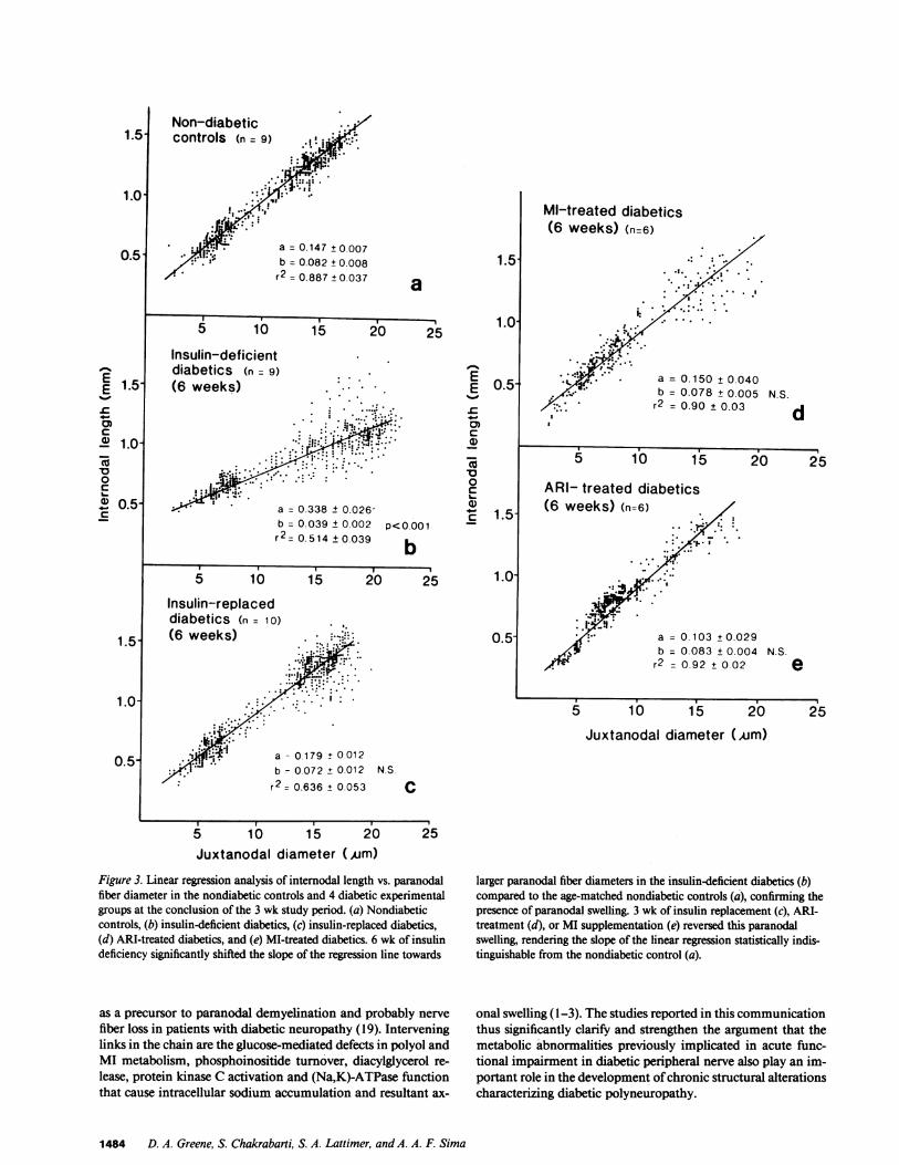
Figure 3. Linear regression analysis of internodal length vs. paranodalfiber diameter in the nondiabetic controls and 4 diabetic experimentalgroups at the conclusion of the 3 wk study period. (a) Nondiabeticcontrols, (b) insulin-deficient diabetics, (c) insulin-replaced diabetics,(d) ARI-treated diabetics, and (e) MI-treated diabetics. 6 wk of insulindeficiency significantly shifted the slope of the regression line towards
as a precursor to paranodal demyelination and probably nerve
fiber loss in patients with diabetic neuropathy (19). Interveninglinks in the chain are the glucose-mediated defects in polyol andMI metabolism, phosphoinositide turnover, diacylglycerol re-
lease, protein kinase C activation and (Na,K)-ATPase functionthat cause intracellular sodium accumulation and resultant ax-
larger paranodal fiber diameters in the insulin-deficient diabetics (b)compared to the age-matched nondiabetic controls (a), confirming thepresence of paranodal swelling. 3 wk of insulin replacement (c), ARI-treatment (d), or MI supplementation (e) reversed this paranodalswelling, rendering the slope of the linear regression statistically indis-tinguishable from the nondiabetic control (a).
onal swelling (1-3). The studies reported in this communicationthus significantly clarify and strengthen the argument that themetabolic abnormalities previously implicated in acute func-tional impairment in diabetic peripheral nerve also play an im-portant role in the development ofchronic structural alterationscharacterizing diabetic polyneuropathy.
1484 D. A. Greene, S. Chakrabarti, S. A. Lattimer, and A. A. F. Sima
1.5
1.0-
0.5
E 1.5-
-0
cd' 1.0-cR0c
4C 0.5-c
1.5
1.0
0.5
20I 25
20 25L-
II
AcknowledamentsThese studies were supported in part by U. S. Public Health Service grantRO- IAM-29892, the Harry Soffer Memorial Research Fund of the Uni-versity of Pittsburgh, the Canadian Diabetes Association, the MedicalResearch Council of Canada grant MA-7777, and a Research Grantfrom Stuart Pharmaceuticals, Wilmington, DE.
References
1. Sima, A. A. F. 1985. Annotation. Can the BB-rat help to unraveldiabetic neuropathy? Neuropathol. Appl. Neurobiol. 11:253-264.
2. Greene, D. A., S. Lattimer, J. Ulbrecht, and P. Carroll. 1985.Glucose-induced alterations in nerve metabolism: current perspectiveon the pathogenesis of diabetic neuropathy and future directions forresearch and therapy. Diabetes Care. 8:290-299.
3. Sima, A. A. F., and T. Brismar. 1985. Reversible diabetic nervedysfunction: structural correlates to electrophysiological abnormalities.Ann. Neurol. 18:21-29.
4. Finegold, D., S. Lattimer, S. Nolle, M. Bernstein, and D. A. Greene.1983. Polyol pathway activity and myo-inositol metabolism. Diabetes.32:988-992.
5. Gillon, K. R. W., and J. N. Hawthorne. 1983. Sorbitol, inositoland nerve conduction in diabetes. Life Sci. 32:1943-1947.
6. Mayer, J. H., and D. R. Tomlinson. 1983. Prevention of defectsof axonal transport and nerve conduction velocity by oral administrationof myo-inositol or an aldose reductase inhibitor in streptozotocin-diabeticrats. Diabetologia. 25:433-438.
7. Greene, D. A., and S. A. Lattimer. 1984. Action of sorbinil indiabetic peripheral nerve. Relationship of polyol (sorbitol) pathway in-hibition to a myo-inositol-mediated defect in sodium-potassium ATPaseactivity. Diabetes. 33:712-716.
8. Greene, D. A., and S. A. Lattimer. 1982. Sodium- and energy-dependent uptake of myo-inositol by rabbit peripheral nerve: competitiveinhibition by glucose and lack of an insulin effect. J. Clin. Invest. 70:1009-1018.
9. Simmons, D. A., A. I. Winegrad, and D. B. Martin. 1982. Signif-icance of tissue myo-inositol concentrations in metabolic regulation innerve. Science (Wash. DC). 217:848-851.
10. Das, P. K., G. Bray, J. A. Aquayo, and M. Rasminsky. 1976.Diminished ouabain-sensitive, sodium-potassium ATPase activity insciatic nerves of rats with streptozotocin-induced diabetes. Exp. Neurol.53:285-288.
11. Greene, D. A., and S. A. Lattimer. 1983. Impaired rat sciaticnerve sodium-potassium adenosine triphosphatase in acute streptozocindiabetes and its correction by dietary myo-inositol supplementation. J.Clin. Invest. 72:1058-1063.
12. Greene, D. A., and S. A. Lattimer. 1984. Impaired energy uti-lization and (Na,K)ATPase in diabetic peripheral nerve. Am. J. Physiol.246:E31 1-E318.
13. Greene, D. A., S. Yagihashi, S. A. Lattimer, and A. A. F. Sima.1984. Nerve Na+-K+-ATPase, conduction and myo-inositol in the insulindeficient BB rat. Am. J. Physiol. 247:E534-E539.
14. Greene, D. A., and S. A. Lattimer. 1986. Protein kinase C agonistsacutely normalize decreased ouabain-inhibitable respiration in diabeticrabbit nerve: implications for (Na,K)-ATPase regulation and diabeticcomplications. Diabetes. 35:242-245.
15. Brismar, T., and A. A. F. Sima. 1981. Changes in nodal functionin nerve fibers of the spontaneously diabetic BB-Wistar rat. Potentialclamp analysis. Acta Physiol. Scand. 113:499-506.
16. Brismar, T. 1983. Nodal function of pathological fibers. Exper-ientia (Basel). 39:946-953.
17. Sima, A. A. F., S. A. Lattimer, S. Yagihashi, and D. A. Greene.1986. Axo-glial dysjunction. A novel structural lesion that accounts forpoorly-reversible conduction slowing in the spontaneously-diabetic Bio-breeding rat. J. Clin. Invest. 77:474-485.
18. Gabbay, K. H. 1973. The sorbitol pathway and the complicationsof diabetes. N. Engl. J. Med. 288:831-836.
19. Sima, A. A. F., V. Bril, and D. A. Greene. 1986. A new char-acteristic ultrastructural abnormality, and morphologic evidence forpathogenetic heterogeneity in human diabetic neuropathy. Clin. Res.34:688a. (Abstr.)
20. Greene, D. A., P. V. DeJesus, and A. I. Winegrad. 1975. Effectsof insulin and dietary myoinositol on impaired peripheral motor nerveconduction velocity in acute streptozotocin diabetes. J. Clin. Invest. 55:1326-1336.
21. Sima, A. A. F., and K. Hay. 1981. Functional aspects and patho-genetic considerations of the neuropathy in the spontaneously diabeticBB-Wistar rat. Neuropathol. Appl. Neurobiol. 7:341-350.
22. Yoda, A., and S. Yoda. 1980. A new simple method for NaK-ATPase rich membrane fragments. Anal. Biochem. 110:82-88.
23. Mayhew, J. A., K. R. W. Gillon, and J. N. Hawthorne. 1983.Free and lipid inositol, sorbitol and sugars in sciatic nerve obtained post-mortem from diabetic patients and control subjects. Diabetologia. 24:13-15.
24. Chakrabarti, S., A. A. F. Sima, and S. Yagihashi. 1986. Aldosereductase in the BB rat; purification, immunological identification andlocalization in the retina and peripheral nerve. Diabetes. 35:101A.
25. Vorbrodt, A. W., A. S. Lossinsky, and H. M. Wisniewski. 1982.Cytochemical localization of ouabain-sensitive K'-dependent, p-nitro-phenylphosphatase (transport ATPase) in the mouse central and periph-eral nervous systems. Brain Res. 243:225-234.
26. Wood, J. G., D. H. Jean, J. N. Whitaker, B. J. McLaughlin, andR. W. Albers. 1977. Immunocytochemical localization of sodium, po-tassium activated ATPase in knifefish brain. J. Neurocytol. 6:571-581.
27. Schwartz, M., S. A. Ernst, G. J. Siegel, and B. W. Agranoff. 1981.Immunocytochemical localization of (Na+,K+)-ATPase in the goldfishoptic nerve. J. Neurochem. 36:107-115.
28. Stewart, M. A., W. R. Sherman, M. M. Kurien, G. I. Moonsammy,and M. Wisgerhof. 1967. Polyol accumulations in nervous tissue of ratswith experimental diabetes and galactosaemia. J. Neurochem. 14:1057-1066.
29. Ludvigson, M. A., and R. L. Sorenson. 1980. Immunohisto-chemical localization of aldose reductase. I. Enzyme purification andantibody preparation-localization in peripheral nerve, artery, and testis.Diabetes. 29:438-449.
Paranodal Swelling in the Spontaneously Diabetic Bio-breeding Rat 1485


