Embed Size (px)
Citation preview
Published Ahead of Print 6 April 2007. 2007, 6(7):1137. DOI: 10.1128/EC.00329-06. Eukaryotic Cell
Leandro Sastre and Christopher M. WestTalibah Metcalf, Hanke van der Wel, Ricardo Escalante,
Spore Coat Dictyostelium discoideumRole of SP65 in Assembly of the
http://ec.asm.org/content/6/7/1137Updated information and services can be found at:
These include:
REFERENCEShttp://ec.asm.org/content/6/7/1137#ref-list-1at:
This article cites 39 articles, 21 of which can be accessed free
CONTENT ALERTS more»articles cite this article),
Receive: RSS Feeds, eTOCs, free email alerts (when new
http://journals.asm.org/site/misc/reprints.xhtmlInformation about commercial reprint orders: http://journals.asm.org/site/subscriptions/To subscribe to to another ASM Journal go to:
on May 11, 2014 by guest
http://ec.asm.org/
Dow
nloaded from
on May 11, 2014 by guest
http://ec.asm.org/
Dow
nloaded from
EUKARYOTIC CELL, July 2007, p. 1137–1149 Vol. 6, No. 71535-9778/07/$08.00�0 doi:10.1128/EC.00329-06Copyright © 2007, American Society for Microbiology. All Rights Reserved.
Role of SP65 in Assembly of the Dictyostelium discoideum Spore Coat�
Talibah Metcalf,1† Hanke van der Wel,1 Ricardo Escalante,2Leandro Sastre,2 and Christopher M. West1*
Department of Biochemistry and Molecular Biology, University of Oklahoma Health Sciences Center, Oklahoma City, Oklahoma 73104,1
and Instituto de Investigaciones Biomedicas Alberto Sols, C.S.I.C./U.A.M., Arturo Duperier 4, 28029 Madrid, Spain2
Received 13 October 2006/Accepted 28 March 2007
Like the cyst walls of other protists, the spore coat of Dictyostelium discoideum is formed de novo to protectthe enclosed dormant cell from stress. Spore coat assembly is initiated by exocytosis of protein and polysac-charide precursors at the cell surface, followed by the infusion of nascent cellulose fibrils, resulting in anasymmetrical trilaminar sandwich with cellulose filling the middle layer. A molecular complex consisting ofcellulose and two proteins, SP85 and SP65, is associated with the inner and middle layers and is required forproper organization of distinct proteins in the outer layer. Here we show that, unlike SP85 and other proteinprecursors, which are stored in prespore vesicles, SP65 is, like cellulose, synthesized just in time. By taggingthe SP65 locus with green fluorescent protein, we find that SP65 is delivered to the cell surface via largelydistinct vesicles, suggesting that separate delivery of components of the cellulose-SP85-SP65 complex regulatesits formation at the cell surface. In support of previous in vivo studies, recombinant SP65 and SP85 are shownto interact directly. In addition, truncation of SP65 causes a defect of the outer layer permeability barrier asseen previously for SP85 mutants. These observations suggest that assembly of the cellulose-SP85-SP65 triadat the cell surface is biosynthetically regulated both temporally and spatially and that the complex contributesan essential function to outer layer architecture and function.
Cell walls protect cells from osmotic, physical, and predatorystress (32). The compositions of walls, which are found sur-rounding many free-living cells, the female gametes of mostanimals, and the somatic cells of plants, range from almostcompletely proteinaceous to almost completely polysaccha-ride. An important subclass of cell walls has a core layer con-sisting of cellulose fibrils, as found around somatic cells ofvascular plants; green algae; certain oomycetes and the watermold Achlya ambisexualis; and spore or cyst walls of the socialsoil amoeba Dictyostelium discoideum, the free-living amoebaeof the genus Acanthamoeba, the soil amoeba Hartmanella gle-bae, and the amoebae-flagellates Naegleria grubei and Schizo-pyrenus russelli. Much remains to be discovered about the roleof proteins in the assembly of cellulose-rich cell walls.
Dictyostelium is amoeboid during growth, and the absence ofa cell wall allows a phagocytic mode of feeding by wild-typecells. Axenic mutants, frequently used as a laboratory model,rely on constitutive fluid-phase endocytosis for nutrition (7). Inresponse to starvation, the amoebae aggregate and form mi-grating slugs that emerge from the soil to form fruiting bodies,which consist of a mass of spores supported aerially by a cel-lular stalk. Walls form de novo around each of the presporecells as they collectively rise to the top of the fruiting body andaround each of the stalk cells (32). The spore wall or coat isdistinct from the stalk cell wall and consists of three morpho-
logical layers. The major and central layer is 200 nm thick andcomposed of interlaced cellulose fibrils and a Gal/GalNAcpolysaccharide (GPS). It is bounded by an outer layer com-posed of proteins that form the major permeability barrier tomacromolecules and at the plasma membrane by an inner layercontaining SP85, a novel protein consisting of multiple Cys-rich and mucin-type domains.
The known coat protein precursors are synthesized duringslug migration and stored with the GPS in prespore vesicles(PSVs). The process of coat formation is first evidenced bytheir exocytosis and is followed by the formation of cellulosefibrils elaborated from transmembrane cellulose synthase com-plexes. The organization of the secreted proteins into morpho-logical layers depends on cellulose (37). Analysis of an SP85disruption strain and the effects of expressing discrete domainsof SP85 show that this protein influences the timing of coatformation as prespore cells rise up the stalk (35). SP85 alsoplays a critical role in outer layer assembly because overex-pression of SP85 domains exerts dominant-negative effects onouter layer morphology (17, 37). Some of these effects dependon SP85’s cellulose binding activity (17, 39) and possibly otherprotein interactions (16).
SP65, first identified in a two-dimensional (2-D) gel pro-teomic analysis of the coat (34), was later found to partiallycopurify with SP85 in urea extracts of spore coats (39). SP65was not incorporated into coats of SP85 mutant spores and wasalso selectively coimmunoprecipitated from the interspore ma-trix with exogenous SP85 by a mechanism that specificallyinvolved the C1 domain of SP85 (38). These findings reveal aphysiologically significant association between SP85 and SP65but do not show if the interaction is direct. This interactionmay mediate some functions of SP85. This possibility has nowbeen addressed by identifying the SP65 gene (named cotE),examining biochemical properties of the recombinant protein
* Corresponding author. Mailing address: Department of Biochem-istry and Molecular Biology, 940 Stanton L. Young Blvd., BMS 853,University of Oklahoma Health Sciences Center, Oklahoma City, OK73104. Phone: (405) 271-2227, ext. 1247. Fax: (405) 271-3139. E-mail:[email protected].
† Present address: Department of Molecular Microbiology and Im-munology, Johns Hopkins University School of Public Health, Balti-more, MD 21205.
� Published ahead of print on 6 April 2007.
1137
on May 11, 2014 by guest
http://ec.asm.org/
Dow
nloaded from
expressed separately from the complex milieu of the coat andinterspore matrix, and disrupting its gene. This has revealedthat SP65 is expressed after SP85 and other known coat pro-teins, suggesting that interaction with SP85 is under biosyn-thetic regulation. Disruption of the SP65 locus (cotE) yields acoat phenotype that partially overlaps with that of SP85-nullspores, supporting the importance of a cellulose-SP85-SP65complex for outer layer organization.
MATERIALS AND METHODS
Cells and cell culture. D. discoideum strains used are listed in Table 1. Cellswere grown axenically in HL-5 growth medium or in association with Klebsiellaaerogenes on SM agar (24). Axenically grown cells were induced to develop bycentrifugally washing cells in PDF buffer (24) and plating them on 0.45-�mMillipore filters or by washing them in KP (10 mM potassium phosphate, pH 6.5)and plating them on nonnutrient agar plates. Bacterially grown cells were cen-trifugally washed free of bacteria in KP and deposited on nonnutrient agarplates. Spores were harvested either by picking them with a loop into 0.2% NP-40in KP or slapping the inverted agar plate onto a countertop and recoveringspores by rinsing the lid with the same buffer. Spores were washed by centrifu-gation (10,000 g � 10 s). For pretreatments, spores were resuspended in thesolution indicated and then washed in KP by centrifugation. Plating efficiencywas determined by counting colonies formed by plating a serial dilution, pre-pared in a bacterial suspension, of a known number of spores, determined bycounting in a hemacytometer, on SM agar plates. Spore coats were isolated fromspores of axenically grown cells by density gradient centrifugation as describedpreviously (38).
Cloning the SP65 gene. The previously reported peptide sequence VRGNPTCLRNHDGI (38) was found to be encoded perfectly by a 309-nucleotide (nt)sequence present in a database of random sequences of shotgun-cloned Dictyo-stelium genomic DNA (gDNA) available at the time. Primers derived from thisnucleotide sequence were used to clone additional gDNA (unpublished data)using inverse PCR and linker-mediated PCR (20, 27, 28). Segments of the newsequences matched other gDNA sequences in the databases. This process wasreiterated until a full-length candidate open reading frame (ORF) for the proteinwas obtained and then extended until ORFs corresponding to the presumptiveneighboring genes were encountered to ensure that all potential exons wereidentified. The predicted coding region was deposited in GenBank as cotE(AF279135). The coding region was subsequently confirmed by automated geneprediction when sufficient sequence data for the locus were obtained by theDictyostelium Sequencing Consortium and is referenced as DDB0214991 at www.dictybase.org.
Expression of SP65 in vegetative cells. The sequence of processed full-lengthSP65, whose N terminus was inferred from Edman degradation analysis ofcyanogen bromide (CNBr) fragments of SP65 (38), was amplified in a PCR using65-CoS1 (5�-CGGGATCCAGTTATGATGCATGTTACAATGTAGT) and 65-CoAS1 (5�-CGGGATCCATTTGTCAAACCACCTATTGAATTGGCAG) asprimers (see Fig. 1 and 2), CsCl-purified strain Ax3 gDNA as template, and a 9:1ratio of Taq and Pfu polymerases. Bold letters indicate BamHI sites used forsubsequent cloning. The PCR products were ligated into pCR4TOPO andcloned into POP10 Escherichia coli cells (Invitrogen). The BamHI fragment froma plasmid whose insert had the correct sequence (pTOPO-SP65) was subclonedinto the unique BamHI site of the integrating plasmid pVS (38), yieldingpVSmycSP65.
pVSmycSP65 was electroporated into strain Ax3, and transformed cells wereselected at 10 �g/ml and then 120 �g/ml G418 (38). A high-level expression clone(HW210) was obtained by screening with monoclonal antibody (MAb) 9E10,which recognizes the myc epitope tag. SP65 was purified by pumping 3.8 liters ofHL-5 growth medium, from a cell culture that had achieved stationary phase,onto a 75-ml column containing SP-Sepharose High-Performance (Pharmacia)equilibrated in 50 mM HEPES-NaOH (pH 7.0), and eluting SP65 using a lineargradient of 0 to 1 M NaCl in the same buffer. SP65 eluted from the column as abroad peak at 100 to 200 mM NaCl based on dot blot and Western blot analysisusing MAb 9E10. Fractions were pooled and further purified on a Superose 12gel filtration column (Pharmacia) equilibrated with 50 mM sodium phosphatebuffer, pH 7.0. The protein eluted unexpectedly late as a broad peak, suggestingweak adsorption to the column.
Expression of SP85 in vegetative cells. SP85 coding DNA was amplified frompVSBW9M (39) using PCR primers SP85-Sdif (5�-AATGGATCCTCTAGAGGTACCTGATCAATGAAAATTTTAAAAAATTG) and SP85-AS (5�-AAGGGATCCGGTTAAAAACCATTGAGATCGTTTACGTCG) and then cloned intopCR4TOPO. pVSBW9M contains a version of SP85 in which the native signalpeptide is replaced by the celA signal peptide, and the myc tag was not includedin the construct. The SP85 DNA was excised using BamHI (bold in the primers)and cloned into the BglII site of the extrachromosomal vector pJK1 (9). Se-quencing showed that 13 of the 17 tetrapeptide repeats of mucin domain 1 weredeleted during cloning; instability has been observed previously for SP85 codingDNA (39). A clonal strain of Ax3 which secreted SP85�13 (HW219) was gen-erated by electroporation with pJK1sp85�13.
GFP tagging of the SP65/cotE locus. pVSmycSP65 was modified by the addi-tion of a green fluorescent protein (GFP) (S65T variant) coding sequence at theunique SacI site present in the vector immediately past the 3� end of the SP65coding region. The two successive stop codons of pVSmycSP65 were changed toSer residues by site-directed mutagenesis (nt changes are underlined) using thefollowing sense and antisense primers: 5�-GGTGGTTTGACAAATGGATCCTCATCATCAGAGCTC and 5�-GAGCTCTGATGATGAGGATCCATTTGTCAAACCACC, as previously described (17). Coding DNA (711 bp) of GFP wasamplified from pTX-GFP (generous gift of T. Egelhoff [12]) using GFP-SCS(5�-GAGCTCTCAGGTTCAGGTAGTAAAGGAGAAGAACTTTTCACTGGAGTTG) and GFP-SCAS (5�-GAGCTCTTATTTGTATAGTTCATCCATGCCATGTG) as primers in a PCR as described above. GFP-SCAS includes a stopcodon (underlined) resulting in a GFP C terminus of ELYK. The PCR productwas ligated into pTOPO and verified for the correct sequence at the OMRFDNA Sequencing Core using an ABI3730 capillary sequencer. The GFP codinginsert was excised with SacI (bold in sequences above) and ligated into theunique SacI site of pVSmycSP65 to yield pVS(mycSP65-GFP). This yielded, afterthe C terminus GLTN of SP65, GSSSSEL, followed by SGSG from the GFPexpression construct, followed by SKGE from the GFP N terminus. The bsrcassette was excised as a ClaI fragment from pbsr519 (21) and inserted into theunique ClaI site within the G418 resistance cassette of pVS(mycSP65-GFP).pVSbsr(mycSP65-GFP) was electroporated into strain Ax3, and transfectantswere selected using 10 �g/ml Blasticidin S. Clones containing the desired single-crossover insertion event were screened by Western blot analysis of developingplaque scrapes using anti-GFP rabbit Ab (Molecular Probes, Eugene, OR) forreactive bands at the expected Mr value and were retrieved at a frequency of10%. Genetic modification was confirmed by PCR analysis (38) as described inResults. Primer GFP-AS1 (5�-AAATTTAAGGGTAAGTTTTCCGTATGTTGCATACC) hybridizes at the 5� end of the GFP coding region (see Fig. 2E).
Interruption of the cotE locus. pTOPO-SP65 was modified by insertion of theBamHI fragment of pbsr519 (21) into the BglII site at nt 1306 with reverseorientation. The disruption DNA was excised with BamHI, purified on an aga-rose gel, recovered using a freeze-squeeze method (25), ethyl alcohol precipi-tated, and electroporated into strain Ax3 (38). Cells that grew in the presence of10 �g/ml Blasticidin S (ICN) were screened for modification of the cotE locususing PCR methods, yielding 16/22 clones with the desired replacement. Primer
TABLE 1. Strains and Abs used
Name Genotype (strain) ortarget (Ab)
Reference orsource
StrainsAx3 Normal (axenic derivative) 13HW70 (pspB-bsr) pspB/SP85� 38HW205 pspA::SP85-GFP UnpublishedHW207 (cotE-bsr) Truncated SP65 (SP65N1) This studyHW210 dscA::mycSP65 This studyHW211 cotE::SP65-GFP (clone 1) This studyHW212 cotE::SP65-GFP (clone 2) This studyHW219 act15::SP85 This studySrfA� srfA� 1
AbsMAb 5F5 SP85 peptide epitope 17MAb 16.1 SP85 O-glycan epitope 33MAb 4A11 SP65 This studyM2Bl5 and others SP65 This studyMAb 9E10 c-myc epitope tag 4MAb 83.5 SP96/SP75 glycan epitope 33Anti-SpiA (rabbit) SpiA (DD31) 22Anti-GFP (rabbit) SP65-GFP Molecular
Probes
1138 METCALF ET AL. EUKARYOT. CELL
on May 11, 2014 by guest
http://ec.asm.org/
Dow
nloaded from
bsr-S1 (5�-GAAAATCAAATCAAAAAGATAAAGCTGACCCGAAAGC) wasused in PCR studies to confirm gene replacement (see Results).
Northern blotting. cotE mRNA was detected by Northern blotting as de-scribed previously (1), using a probe generated from pTOPO-SP65 by PCR using65-CoS1 and 65-CoAS1 as primers.
Anti-SP65 Abs. A preparation of purified SP65 was mixed with either RIBIadjuvant (Ribi ImmunoChem Research, Hamilton, MT) or Immuneasy adjuvant(QIAGEN) and injected into 6-week-old BALB/c female mice. Preimmune andimmune sera were collected and tested by an enzyme-linked immunosorbentassay (ELISA) using purified SP65 adsorbed to microplate wells. Mouse 2,initially immunized three times using RIBI adjuvant, followed by two boostsusing Immuneasy adjuvant, was the best characterized as a result of its high titerafter the Immuneasy boosts. MAb clone 6H6 (4A11) was selected as a repre-sentative of �36 hybridoma clones which secreted ELISA- and immunofluores-cence-positive Abs. MAb 4A11 is an immunoglobulin G (IgG) and was purifiedfrom the medium of cell cultures grown in a CL350 chamber (Integra), using aprotein G column. Ascites were induced in other immunized mice after anintraperitoneal injection of pristane. Abs used are listed in Table 1.
SP65 binding assay. Anti-myc tag MAb 9E10 was expressed in cell culture,purified by protein G affinity chromatography, and coupled to CNBr-activatedSepharose 4B (Sigma). For immunoprecipitation (IP) studies, 32 �l of a 50%slurry of 9E10-Sepharose was diluted with 200 �l IP buffer, consisting of 50 mMTris-acetate, pH 7.5, 0.3 M NaCl, 1 mM EDTA, 1 mg/ml bovine serum albumin,0.5% (vol/vol) NP-40, and protease inhibitors (1 mM phenylmethylsulfonyl flu-oride, 10 �g/ml aprotinin, 10 �g/ml leupeptin), and centrifuged at 13,000 � g for1 min. The beads were resuspended with (i) 50 �l of a preparation of SP85 thathad been concentrated 10 times from the HL-5 culture medium of the SP85expression strain described above using a Vivaspin 2 centrifugal concentratorwith a polyethersulfone membrane (molecular weight cutoff, 30,000) and 50 �l ofHL-5 medium prepared in the same manner, (ii) 50 �l of mycSP65 prepared inthe same fashion and 50 �l of HL-5, or (iii) 50 �l of both SP85 and mycSP65. Themixtures, which were supplemented with protease inhibitors, were incubated for3 h at 4°C, with maintenance of the beads in suspension on a Vortex Genie 2. Thesupernatant was removed by centrifugation, and the beads were washed twicewith 1 ml IP wash buffer (IP buffer lacking bovine serum albumin). The beadsand supernatants were analyzed by Western blotting.
Immunofluorescence. Culminants were scraped from nonnutrient agar plates,resuspended in 25 mM EDTA in 10 mM KP (pH 6.5), dissociated by pipetting atroom temperature, deposited on coverslips freshly precoated with 1-mg/ml poly-L-lysine in H2O, and allowed to dry. After 5 to 10 min, unbound cells werewashed off with KP buffer. The coverslip was dipped in �20°C methanol for 30min, air dried, hydrated in 5% nonfat dried milk in TBS (20 mM Tris-HCl, pH7.4, 150 mM NaCl) for 30 min, incubated in primary Ab for 1 h at 22°C, washedfive times over 10 min with TBS, incubated in secondary Ab for 1 h, and washedagain as before. Coverslips were mounted onto slides in Vectashield (VectorLabs) with DAPI (4�,6�-diamidino-2-phenylindole) and sealed with fingernailpolish. Rabbit anti-GFP (1:1,000) was from Molecular Probes, and affinity-purified Texas Red-conjugated goat anti-rabbit Ab (1:100) and affinity-purifiedfluorescein isothiocyanate (FITC)-conjugated goat anti-mouse Ab (1:100) werefrom Jackson Laboratories. Epifluorescence images were collected through a60�-numerical-aperture 1.2 water-immersion Nikon lens mounted on a NikonEclipse TE2000 microscope via an ORCA charge-coupled device digital camerausing Improvision OpenLab software and processed in Photoshop.
Lectin incubation of spores. Spores from axenically grown cells were processedand examined using 4-�g/ml FITC-conjugated Ricinus communis agglutinin I(120) essentially as described previously (30).
Nucleotide sequence accession number. The predicted coding region was de-posited in GenBank as cotE (AF279135). The coding region was subsequentlyconfirmed by automated gene prediction when sufficient sequence data for thelocus were obtained by the Dictyostelium Sequencing Consortium and is refer-enced as DDB0214991 at www.dictybase.org.
RESULTS
Cloning the SP65 gene. SP65 was originally identified as aCoomassie blue- and silver-stained protein in 1-D and 2-Dsodium dodecyl sulfate (SDS)-polyacrylamide gels of highlypurified spore coats (34). The protein was purified in dena-tured form from coats by column chromatography and SDS-polyacrylamide gel electrophoresis (PAGE) (39) and cleavedwith CNBr. Two major fragments were recovered by SDS-
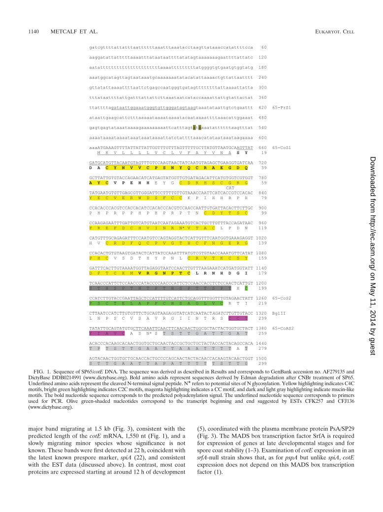
PAGE, and N-terminal sequence data were obtained byEdman degradation (38). A full-length ORF that encodedthese peptide sequences was obtained by a combination ofPCR from gDNA and sequence data from the Dictyosteliumgenomic databases available at the time. The predicted codingregion (Fig. 1) lacks introns and was deposited in GenBank ascotE (AF279135). As is typical for Dictyostelium coding DNA(27, 28), this ORF has a higher GC content than does flankingDNA and exhibits a favorable Kozak context for the predictedstart codon and a candidate polyadenylation signal after thestop codon. The predicted protein contains a single internalMet residue, consistent with the generation of only two bandsafter treatment with CNBr. Their Edman degradation-derivedsequences are highlighted in bold in Fig. 1.
The cotE/SP65 coding region was subsequently identified byautomated gene prediction when sufficient sequence data wereobtained by the Dictyostelium Sequencing Consortium and isassigned as DDB0214991 at www.dictybase.org. No expressedsequence tags (ESTs) are reported for this sequence in cDNAlibraries prepared from slug cells (26; www.dictybase.org). Thiswas initially surprising because known spore coat proteins ac-cumulate in prespore cells of the slug, and their mRNAs areabundantly expressed (29) and highly represented in the cDNAlibraries. However, two EST clones in a cDNA library fromculminating cells matched this locus, suggesting that cotE maybe expressed unusually late in development. The EST se-quences suggest possible transcription start and stop sites ashighlighted in brown in Fig. 1.
cotE lies on chromosome 2, the location of all but one of theknown coat protein genes, and is predicted to encode a 464-amino-acid protein. A signal peptide-like sequence is removedafter Ala17 based on the Edman degradation sequence data(Fig. 1), yielding a predicted protein with a calculated Mr of45,363 that is substantially smaller than the apparent Mr of65,000 based on SDS-PAGE. The occurrence of two N-glyco-sylation sequons (N* in Fig. 1) suggests that the apparent Mr
difference may be explained in part by N-glycosylation of thenative protein. The encoded protein has a predicted pI of 7.6compared to the pI range of 7.0 to 7.2 observed by 2-D gelelectrophoresis (34), and the difference might be due to anacidic posttranslational modification as suggested by thecharge stutter seen in the 2-D gels.
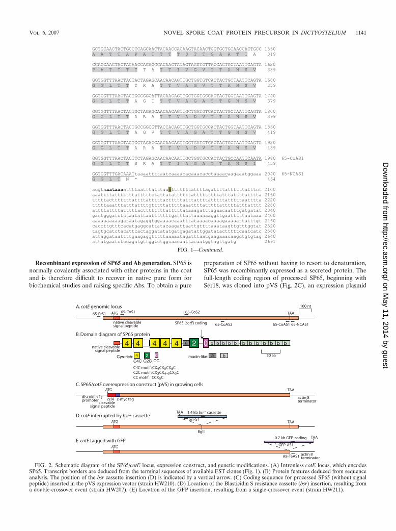
The predicted protein sequence contains motifs (Fig. 2B)characteristic of known spore coat proteins (34). The N-terminal half is composed of five Cys-rich C4C motifs (high-lighted in yellow in Fig. 1), each resembling the N-terminalsubdomain of the EGF motif, followed by a Cys-rich C2Cmotif (green) and a Cys-rich CC motif (pink). The evennumber of Cys residues is consistent with each participatingin an intramolecular disulfide bond. The C-terminal half,devoid of Cys residues, consists of 12 mucin-like motifs of 16residues each (light gray), which are potential targets ofextensive O-glycosylation (6, 30) that may, in addition toN-glycosylation, contribute to the difference between thepredicted and apparent Mr values.
Expression of SP65 mRNA. Northern blot analysis was per-formed to determine the time course of expression of cotE indeveloping cells collected every 2 h until fruiting bodies formedat 24 h. A nearly full-length probe against cotE detected a
VOL. 6, 2007 NOVEL SPORE COAT PROTEIN PRECURSOR IN DICTYOSTELIUM 1139
on May 11, 2014 by guest
http://ec.asm.org/
Dow
nloaded from
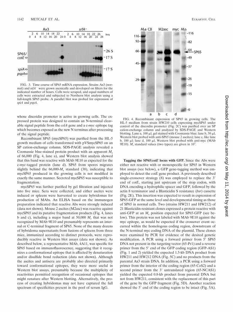
major band migrating at 1.5 kb (Fig. 3), consistent with thepredicted length of the cotE mRNA, 1,550 nt (Fig. 1), and aslowly migrating minor species whose significance is notknown. These bands were first detected at 22 h, coincident withthe latest known prespore marker, spiA (22), and consistentwith the EST data (discussed above). In contrast, most coatproteins are expressed starting at around 12 h of development
(5), coordinated with the plasma membrane protein PsA/SP29(Fig. 3). The MADS box transcription factor SrfA is requiredfor expression of genes at late developmental stages and forspore coat stability (1–3). Examination of cotE expression in ansrfA-null strain shows that, as for pspA but unlike spiA, cotEexpression does not depend on this MADS box transcriptionfactor (1).
CAT
FIG. 1. Sequence of SP65/cotE DNA. The sequence was derived as described in Results and corresponds to GenBank accession no. AF279135 andDictyBase DDB0214991 (www.dictybase.org). Bold amino acids represent sequences derived by Edman degradation after CNBr treatment of SP65.Underlined amino acids represent the cleaved N-terminal signal peptide. N* refers to potential sites of N glycosylation. Yellow highlighting indicates C4Cmotifs, bright green highlighting indicates C2C motifs, magenta highlighting indicates a CC motif, and dark and light gray highlighting indicate mucin-likemotifs. The bold nucleotide sequence corresponds to the predicted polyadenylation signal. The underlined nucleotide sequence corresponds to primersused for PCR. Olive green-shaded nucleotides correspond to the transcript beginning and end suggested by ESTs CFK257 and CFJ136(www.dictybase.org).
1140 METCALF ET AL. EUKARYOT. CELL
on May 11, 2014 by guest
http://ec.asm.org/
Dow
nloaded from
Recombinant expression of SP65 and Ab generation. SP65 isnormally covalently associated with other proteins in the coatand is therefore difficult to recover in native pure form forbiochemical studies and raising specific Abs. To obtain a pure
preparation of SP65 without having to resort to denaturation,SP65 was recombinantly expressed as a secreted protein. Thefull-length coding region of processed SP65, beginning withSer18, was cloned into pVS (Fig. 2C), an expression plasmid
FIG. 1—Continued.
FIG. 2. Schematic diagram of the SP65/cotE locus, expression construct, and genetic modifications. (A) Intronless cotE locus, which encodesSP65. Transcript borders are deduced from the terminal sequences of available EST clones (Fig. 1). (B) Protein features deduced from sequenceanalysis. The position of the bsr cassette insertion (D) is indicated by a vertical arrow. (C) Coding sequence for processed SP65 (without signalpeptide) inserted in the pVS expression vector (strain HW210). (D) Location of the Blasticidin S resistance cassette (bsr) insertion, resulting froma double-crossover event (strain HW207). (E) Location of the GFP insertion, resulting from a single-crossover event (strain HW211).
VOL. 6, 2007 NOVEL SPORE COAT PROTEIN PRECURSOR IN DICTYOSTELIUM 1141
on May 11, 2014 by guest
http://ec.asm.org/
Dow
nloaded from
whose discoidin promoter is active in growing cells. The ex-pressed protein was designed to contain an N-terminal cleav-able signal peptide from the celA gene and a c-myc epitope tagwhich becomes exposed as the new N terminus after processingof the signal peptide.
Recombinant SP65 (mycSP65) was purified from the HL-5growth medium of cells transformed with pVSmycSP65 on anSP cation-exchange column. SDS-PAGE analysis revealed aCoomassie blue-stained protein product with an apparent Mr
of 66,000 (Fig. 4, lane a), and Western blot analysis showedthat this band was reactive with MAb 9E10 as expected for thec-myc-tagged protein (lane d). SP65 from spores migratesslightly behind the 66,000-Mr standard (38), indicating thatmycSP65 produced in the growing cells is not modified inexactly the same manner. Secreted mycSP65 was susceptible tofragmentation.
mycSP65 was further purified by gel filtration and injectedinto five mice. Sera were collected, and either ascites wereinduced or spleens were harvested to create hybridomas forproduction of MAbs. An ELISA based on the immunogenpreparation indicated that reactive Abs were strongly induced(data not shown). Mouse 2 ascites (M2asc) was reactive againstmycSP65 and its putative fragmentation products (Fig. 4, lanesb and c), including a major band at 50,000 Mr that was notrecognized by MAb 9E10 and presumably represents an inter-nal or C-terminal fragment of SP65. None of the many dozensof hybridoma supernatants from fusions of spleens from threemice, immunized according to distinct protocols, were repro-ducibly reactive in Western blot assays (data not shown). Asdescribed below, a representative MAb, 4A11, was specific forSP65 based on immunofluorescence, suggesting that it recog-nizes a conformational epitope that is affected by denaturationand/or disulfide bond reduction (data not shown). Althoughthe ascites and antisera are probably also directed primarilytoward conformational epitopes, they were more useful inWestern blot assays, presumably because the multiplicity ofreactivities permitted recognition of occasional epitopes thatmight renature after Western blotting. Alternatively, the pro-cess of creating hybridomas may not have captured the fullspectrum of specificities present in the pool of serum IgG.
Tagging the SP65/cotE locus with GFP. Since the Abs wereeither not reactive with or monospecific for SP65 in Westernblot assays (see below), a GFP gene-tagging method was em-ployed to detect the cotE gene product. A previously describedsingle-crossover strategy (8) was employed to replace the 3�end of cotE, starting just upstream of the stop codon, withDNA encoding a hydrophilic spacer and GFP, followed by theactin 8 terminator and a Blasticidin S resistance (bsr) cassette(Fig. 2E). This insertion was expected to result in expression ofSP65-GFP at the same level and developmental timing as thoseof SP65 in normal cells. Two (strains HW211 and HW212) of21 Blasticidin-resistant clones expressed a protein reactive withanti-GFP at an Mr position expected for SP65-GFP (see be-low). This protein was not labeled with MAb 9E10 against themyc epitope, as would be expected if the crossover event oc-curred within the homologous coding region, downstream ofthe N-terminal myc coding DNA of the plasmid. These cloneswere examined by PCR for evidence of the desired geneticmodification. A PCR using a forward primer from 5� SP65DNA not present in the targeting vector (65-Pr1) and a reverseprimer from the 5� end of the GFP coding region (GFP-AS1)(Fig. 1 and 2) yielded the expected 1.5-kb DNA product fromHW211 and HW212 DNA (Fig. 5C) and no products from theparental Ax3 strain DNA. In addition, a PCR using a forwardprimer from the interior of the coding region (65-CoS2) and asecond primer from the 3� untranslated region (65-NCAS1)yielded the expected 0.8-kb product from parental DNA butnot from HW211, consistent with the replacement of this partof the gene by the GFP fragment (Fig. 5D). Another reactionshowed the 5� end of the coding region to be intact (Fig. 5A).
FIG. 4. Recombinant expression of SP65 in growing cells. TheHL-5 medium from strain HW210 cells expressing mycSP65 undercontrol of the discoidin promoter (Fig. 2C) was purified over an SPcation-exchange column and analyzed by SDS-PAGE and Westernblotting. Lane a, 100 �l, gel stained with Coomassie blue; lane b, 50 �l,Western blot probed with anti-SP65 (mouse 2 ascites); lane c, like laneb, 100 �l; lane d, 100 �l, Western blot probed with anti-myc (MAb9E10). Mr standard values (two types) are given in 103.
FIG. 3. Time course of SP65 mRNA expression. Strains Ax3 (nor-mal) and srfA� were grown axenically and developed on filters for theindicated number of hours. Cells were scraped, and equal numbers ofcells were extracted and subjected to Northern blot analysis using afull-length SP65 probe. A parallel blot was probed for expression ofspiA and pspA.
1142 METCALF ET AL. EUKARYOT. CELL
on May 11, 2014 by guest
http://ec.asm.org/
Dow
nloaded from
Interruption of the SP65/cotE locus by bsr. To study thefunction of SP65, the cotE locus was modified by insertion ofthe bsr cassette within the coding region using a double-cross-over strategy based on homologous recombination (Fig. 2D).Modified strains were identified based on PCR amplification ofan expected 1.0-kb band from the DNA of clonal strains usingprimers 65-Pr1, located upstream of the gene targeting DNA,and bsr-S1 (Fig. 1 and 2). An example comparing strainsHW207 and Ax3 is shown (Fig. 5B). In addition, amplificationproducts starting from the 5� end of the gene (Fig. 5A) or fromthe 3� end of the gene (Fig. 5D) were 1.4 kb larger in size asexpected due to the insertion. Both SP65-GFP and cotE-bsrstrains developed to form normal-appearing fruiting bodieswith elongate spores (see Fig. 11 below). Since the bsr cassetteof the targeting DNA was in reverse orientation relative tocotE sequences, its transcription terminator may also termi-nate a truncated SP65 mRNA encoding most of the Cys-richN-terminal half of SP65 (Fig. 2), up to R235 (Fig. 1) followedby SKLVFGSALSF-tga(stop) from the bsr cassette. This pro-tein would have an Mr of 25,672 and a pI of 7.6.
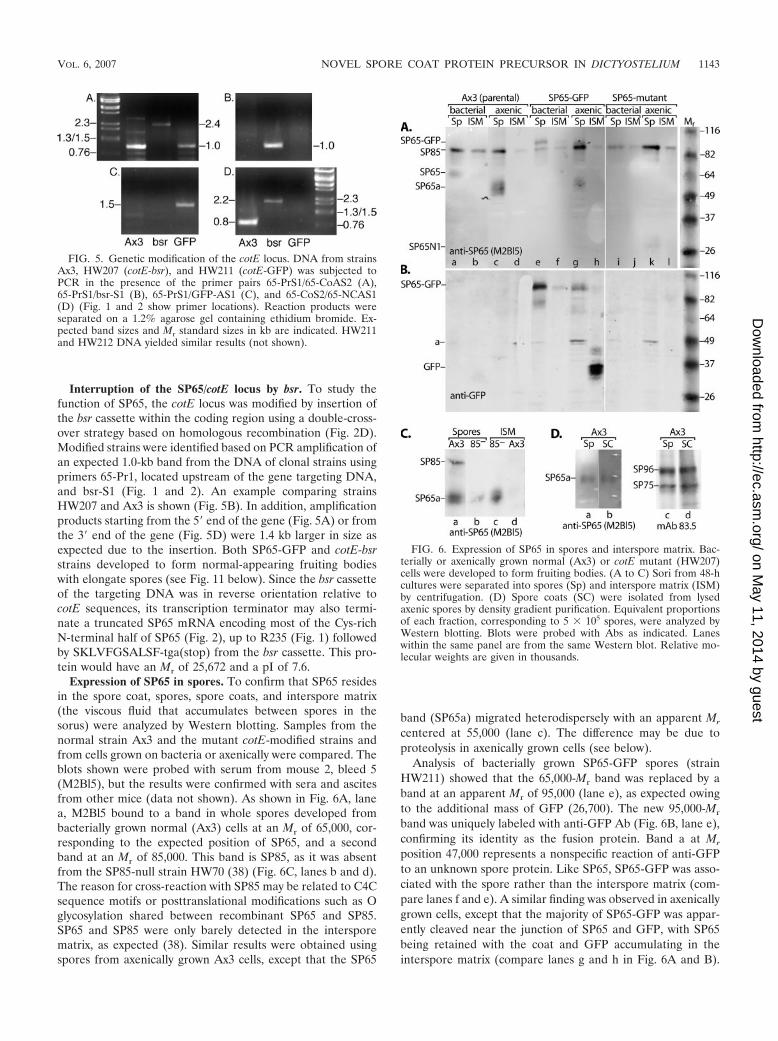
Expression of SP65 in spores. To confirm that SP65 residesin the spore coat, spores, spore coats, and interspore matrix(the viscous fluid that accumulates between spores in thesorus) were analyzed by Western blotting. Samples from thenormal strain Ax3 and the mutant cotE-modified strains andfrom cells grown on bacteria or axenically were compared. Theblots shown were probed with serum from mouse 2, bleed 5(M2Bl5), but the results were confirmed with sera and ascitesfrom other mice (data not shown). As shown in Fig. 6A, lanea, M2Bl5 bound to a band in whole spores developed frombacterially grown normal (Ax3) cells at an Mr of 65,000, cor-responding to the expected position of SP65, and a secondband at an Mr of 85,000. This band is SP85, as it was absentfrom the SP85-null strain HW70 (38) (Fig. 6C, lanes b and d).The reason for cross-reaction with SP85 may be related to C4Csequence motifs or posttranslational modifications such as Oglycosylation shared between recombinant SP65 and SP85.SP65 and SP85 were only barely detected in the intersporematrix, as expected (38). Similar results were obtained usingspores from axenically grown Ax3 cells, except that the SP65
band (SP65a) migrated heterodispersely with an apparent Mr
centered at 55,000 (lane c). The difference may be due toproteolysis in axenically grown cells (see below).
Analysis of bacterially grown SP65-GFP spores (strainHW211) showed that the 65,000-Mr band was replaced by aband at an apparent Mr of 95,000 (lane e), as expected owingto the additional mass of GFP (26,700). The new 95,000-Mr
band was uniquely labeled with anti-GFP Ab (Fig. 6B, lane e),confirming its identity as the fusion protein. Band a at Mr
position 47,000 represents a nonspecific reaction of anti-GFPto an unknown spore protein. Like SP65, SP65-GFP was asso-ciated with the spore rather than the interspore matrix (com-pare lanes f and e). A similar finding was observed in axenicallygrown cells, except that the majority of SP65-GFP was appar-ently cleaved near the junction of SP65 and GFP, with SP65being retained with the coat and GFP accumulating in theinterspore matrix (compare lanes g and h in Fig. 6A and B).
FIG. 5. Genetic modification of the cotE locus. DNA from strainsAx3, HW207 (cotE-bsr), and HW211 (cotE-GFP) was subjected toPCR in the presence of the primer pairs 65-PrS1/65-CoAS2 (A),65-PrS1/bsr-S1 (B), 65-PrS1/GFP-AS1 (C), and 65-CoS2/65-NCAS1(D) (Fig. 1 and 2 show primer locations). Reaction products wereseparated on a 1.2% agarose gel containing ethidium bromide. Ex-pected band sizes and Mr standard sizes in kb are indicated. HW211and HW212 DNA yielded similar results (not shown).
FIG. 6. Expression of SP65 in spores and interspore matrix. Bac-terially or axenically grown normal (Ax3) or cotE mutant (HW207)cells were developed to form fruiting bodies. (A to C) Sori from 48-hcultures were separated into spores (Sp) and interspore matrix (ISM)by centrifugation. (D) Spore coats (SC) were isolated from lysedaxenic spores by density gradient purification. Equivalent proportionsof each fraction, corresponding to 5 � 105 spores, were analyzed byWestern blotting. Blots were probed with Abs as indicated. Laneswithin the same panel are from the same Western blot. Relative mo-lecular weights are given in thousands.
VOL. 6, 2007 NOVEL SPORE COAT PROTEIN PRECURSOR IN DICTYOSTELIUM 1143
on May 11, 2014 by guest
http://ec.asm.org/
Dow
nloaded from
Other minor bands presumably represent other proteolyticevents.
Analysis of the cotE-bsr strain (HW207) confirmed the ab-sence of SP65 at the 65,000-Mr position (Fig. 6A, lane k).However, the mutant spores accumulated a new reactive bandat an apparent Mr of 28,000 (SP65N1), corresponding to theprotein expected if a cotE message truncated at the bsr inser-tion site were formed and translated (see above). This protein,concluded to represent the N-terminal half of SP65 (Fig. 2B),also appeared in spores from bacterially grown cells (data notshown) but is not evident in lane i because of underloading.The N-terminal half of SP65, comprised of C4C repeats, con-tains the information required for insertion into the coat, as itis not observed in the interspore matrix (lane l).
Examination of SP85 mutant cells (HW70 or 85�) showsthat, in contrast to Ax3, SP65 accumulates in the intersporematrix rather than the spore (Fig. 6C). This distribution, ob-served previously using Coomassie blue staining (38), confirmsthat SP65 depends on SP85 for incorporation into the coat.
To determine if SP65 is present in the spore coat, equalnumbers of spores and purified spore coats were compared byWestern blotting. Fig. 6D (lanes a and b) shows similar levelsof SP65 in the two fractions, indicating that the majority ofspore SP65 is in the coat. Lanes c and d show a similar distri-bution for two known coat proteins, SP96 and SP75, and serveas a loading control.
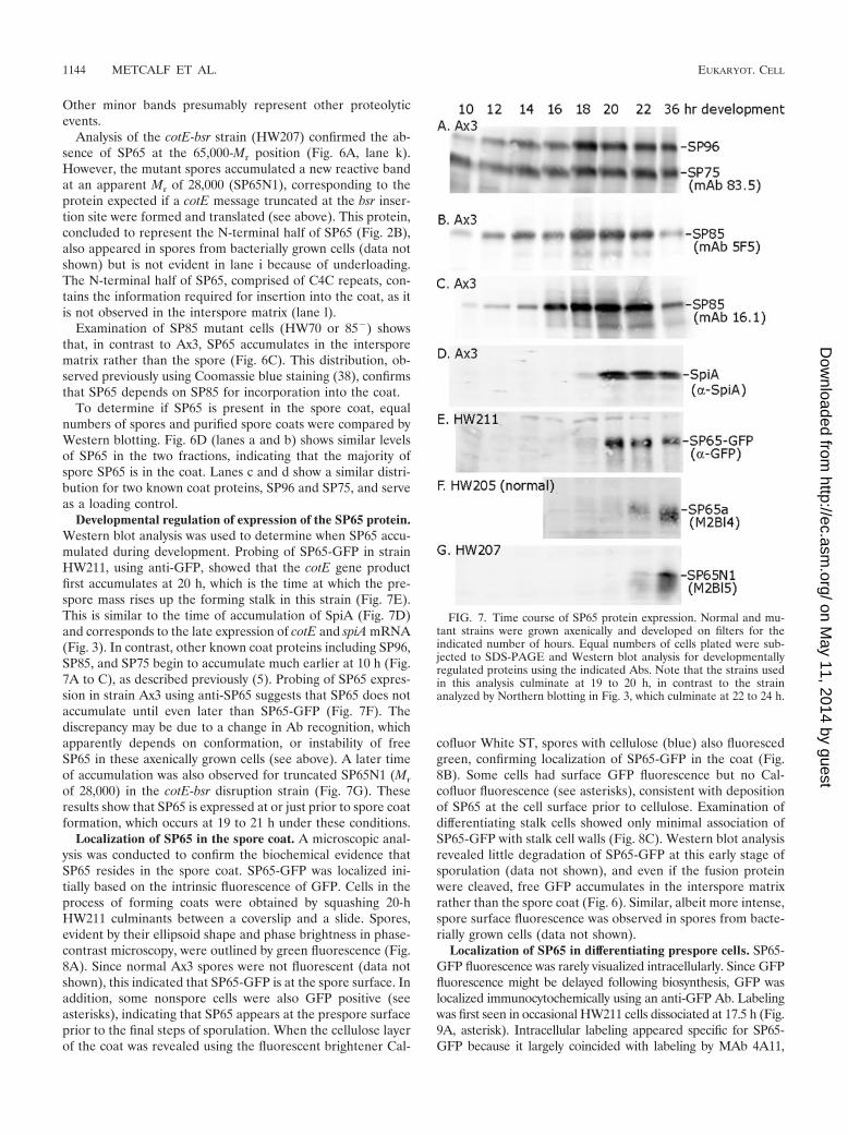
Developmental regulation of expression of the SP65 protein.Western blot analysis was used to determine when SP65 accu-mulated during development. Probing of SP65-GFP in strainHW211, using anti-GFP, showed that the cotE gene productfirst accumulates at 20 h, which is the time at which the pre-spore mass rises up the forming stalk in this strain (Fig. 7E).This is similar to the time of accumulation of SpiA (Fig. 7D)and corresponds to the late expression of cotE and spiA mRNA(Fig. 3). In contrast, other known coat proteins including SP96,SP85, and SP75 begin to accumulate much earlier at 10 h (Fig.7A to C), as described previously (5). Probing of SP65 expres-sion in strain Ax3 using anti-SP65 suggests that SP65 does notaccumulate until even later than SP65-GFP (Fig. 7F). Thediscrepancy may be due to a change in Ab recognition, whichapparently depends on conformation, or instability of freeSP65 in these axenically grown cells (see above). A later timeof accumulation was also observed for truncated SP65N1 (Mr
of 28,000) in the cotE-bsr disruption strain (Fig. 7G). Theseresults show that SP65 is expressed at or just prior to spore coatformation, which occurs at 19 to 21 h under these conditions.
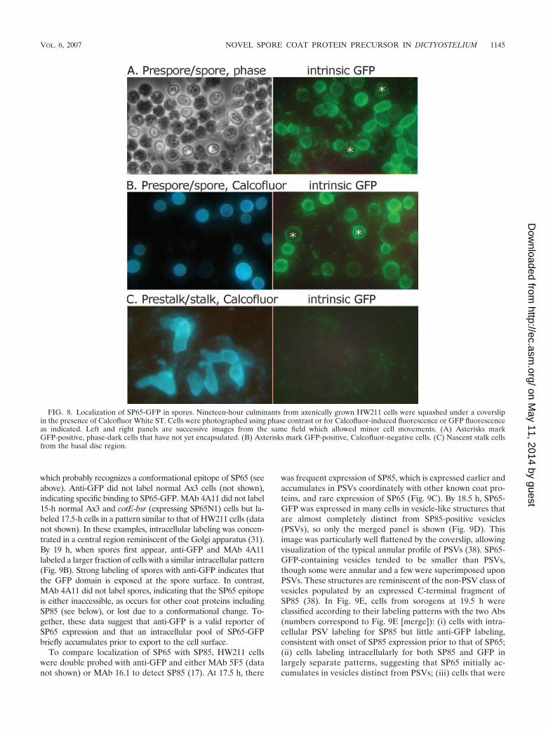
Localization of SP65 in the spore coat. A microscopic anal-ysis was conducted to confirm the biochemical evidence thatSP65 resides in the spore coat. SP65-GFP was localized ini-tially based on the intrinsic fluorescence of GFP. Cells in theprocess of forming coats were obtained by squashing 20-hHW211 culminants between a coverslip and a slide. Spores,evident by their ellipsoid shape and phase brightness in phase-contrast microscopy, were outlined by green fluorescence (Fig.8A). Since normal Ax3 spores were not fluorescent (data notshown), this indicated that SP65-GFP is at the spore surface. Inaddition, some nonspore cells were also GFP positive (seeasterisks), indicating that SP65 appears at the prespore surfaceprior to the final steps of sporulation. When the cellulose layerof the coat was revealed using the fluorescent brightener Cal-
cofluor White ST, spores with cellulose (blue) also fluorescedgreen, confirming localization of SP65-GFP in the coat (Fig.8B). Some cells had surface GFP fluorescence but no Cal-cofluor fluorescence (see asterisks), consistent with depositionof SP65 at the cell surface prior to cellulose. Examination ofdifferentiating stalk cells showed only minimal association ofSP65-GFP with stalk cell walls (Fig. 8C). Western blot analysisrevealed little degradation of SP65-GFP at this early stage ofsporulation (data not shown), and even if the fusion proteinwere cleaved, free GFP accumulates in the interspore matrixrather than the spore coat (Fig. 6). Similar, albeit more intense,spore surface fluorescence was observed in spores from bacte-rially grown cells (data not shown).
Localization of SP65 in differentiating prespore cells. SP65-GFP fluorescence was rarely visualized intracellularly. Since GFPfluorescence might be delayed following biosynthesis, GFP waslocalized immunocytochemically using an anti-GFP Ab. Labelingwas first seen in occasional HW211 cells dissociated at 17.5 h (Fig.9A, asterisk). Intracellular labeling appeared specific for SP65-GFP because it largely coincided with labeling by MAb 4A11,
FIG. 7. Time course of SP65 protein expression. Normal and mu-tant strains were grown axenically and developed on filters for theindicated number of hours. Equal numbers of cells plated were sub-jected to SDS-PAGE and Western blot analysis for developmentallyregulated proteins using the indicated Abs. Note that the strains usedin this analysis culminate at 19 to 20 h, in contrast to the strainanalyzed by Northern blotting in Fig. 3, which culminate at 22 to 24 h.
1144 METCALF ET AL. EUKARYOT. CELL
on May 11, 2014 by guest
http://ec.asm.org/
Dow
nloaded from
which probably recognizes a conformational epitope of SP65 (seeabove). Anti-GFP did not label normal Ax3 cells (not shown),indicating specific binding to SP65-GFP. MAb 4A11 did not label15-h normal Ax3 and cotE-bsr (expressing SP65N1) cells but la-beled 17.5-h cells in a pattern similar to that of HW211 cells (datanot shown). In these examples, intracellular labeling was concen-trated in a central region reminiscent of the Golgi apparatus (31).By 19 h, when spores first appear, anti-GFP and MAb 4A11labeled a larger fraction of cells with a similar intracellular pattern(Fig. 9B). Strong labeling of spores with anti-GFP indicates thatthe GFP domain is exposed at the spore surface. In contrast,MAb 4A11 did not label spores, indicating that the SP65 epitopeis either inaccessible, as occurs for other coat proteins includingSP85 (see below), or lost due to a conformational change. To-gether, these data suggest that anti-GFP is a valid reporter ofSP65 expression and that an intracellular pool of SP65-GFPbriefly accumulates prior to export to the cell surface.
To compare localization of SP65 with SP85, HW211 cellswere double probed with anti-GFP and either MAb 5F5 (datanot shown) or MAb 16.1 to detect SP85 (17). At 17.5 h, there
was frequent expression of SP85, which is expressed earlier andaccumulates in PSVs coordinately with other known coat pro-teins, and rare expression of SP65 (Fig. 9C). By 18.5 h, SP65-GFP was expressed in many cells in vesicle-like structures thatare almost completely distinct from SP85-positive vesicles(PSVs), so only the merged panel is shown (Fig. 9D). Thisimage was particularly well flattened by the coverslip, allowingvisualization of the typical annular profile of PSVs (38). SP65-GFP-containing vesicles tended to be smaller than PSVs,though some were annular and a few were superimposed uponPSVs. These structures are reminiscent of the non-PSV class ofvesicles populated by an expressed C-terminal fragment ofSP85 (38). In Fig. 9E, cells from sorogens at 19.5 h wereclassified according to their labeling patterns with the two Abs(numbers correspond to Fig. 9E [merge]): (i) cells with intra-cellular PSV labeling for SP85 but little anti-GFP labeling,consistent with onset of SP85 expression prior to that of SP65;(ii) cells labeling intracellularly for both SP85 and GFP inlargely separate patterns, suggesting that SP65 initially ac-cumulates in vesicles distinct from PSVs; (iii) cells that were
FIG. 8. Localization of SP65-GFP in spores. Nineteen-hour culminants from axenically grown HW211 cells were squashed under a coverslipin the presence of Calcofluor White ST. Cells were photographed using phase contrast or for Calcofluor-induced fluorescence or GFP fluorescenceas indicated. Left and right panels are successive images from the same field which allowed minor cell movements. (A) Asterisks markGFP-positive, phase-dark cells that have not yet encapsulated. (B) Asterisks mark GFP-positive, Calcofluor-negative cells. (C) Nascent stalk cellsfrom the basal disc region.
VOL. 6, 2007 NOVEL SPORE COAT PROTEIN PRECURSOR IN DICTYOSTELIUM 1145
on May 11, 2014 by guest
http://ec.asm.org/
Dow
nloaded from
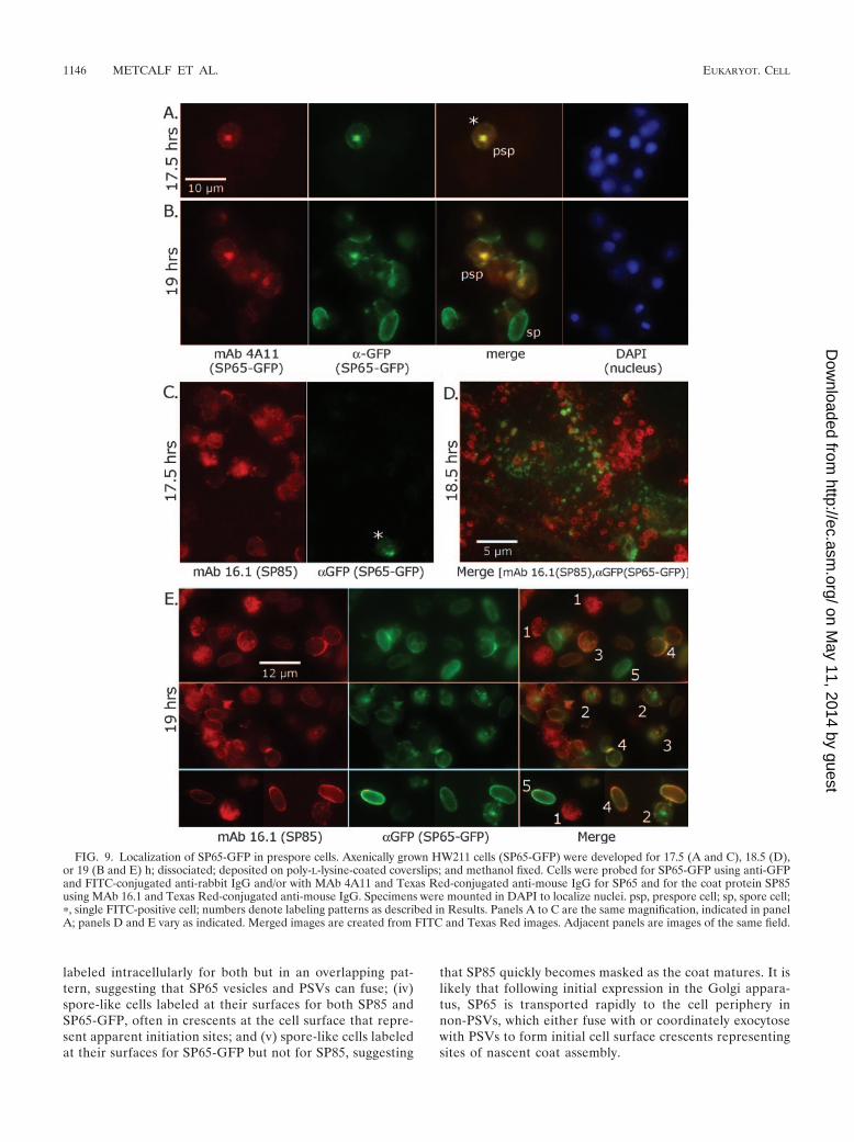
labeled intracellularly for both but in an overlapping pat-tern, suggesting that SP65 vesicles and PSVs can fuse; (iv)spore-like cells labeled at their surfaces for both SP85 andSP65-GFP, often in crescents at the cell surface that repre-sent apparent initiation sites; and (v) spore-like cells labeledat their surfaces for SP65-GFP but not for SP85, suggesting
that SP85 quickly becomes masked as the coat matures. It islikely that following initial expression in the Golgi appara-tus, SP65 is transported rapidly to the cell periphery innon-PSVs, which either fuse with or coordinately exocytosewith PSVs to form initial cell surface crescents representingsites of nascent coat assembly.
FIG. 9. Localization of SP65-GFP in prespore cells. Axenically grown HW211 cells (SP65-GFP) were developed for 17.5 (A and C), 18.5 (D),or 19 (B and E) h; dissociated; deposited on poly-L-lysine-coated coverslips; and methanol fixed. Cells were probed for SP65-GFP using anti-GFPand FITC-conjugated anti-rabbit IgG and/or with MAb 4A11 and Texas Red-conjugated anti-mouse IgG for SP65 and for the coat protein SP85using MAb 16.1 and Texas Red-conjugated anti-mouse IgG. Specimens were mounted in DAPI to localize nuclei. psp, prespore cell; sp, spore cell;�, single FITC-positive cell; numbers denote labeling patterns as described in Results. Panels A to C are the same magnification, indicated in panelA; panels D and E vary as indicated. Merged images are created from FITC and Texas Red images. Adjacent panels are images of the same field.
1146 METCALF ET AL. EUKARYOT. CELL
on May 11, 2014 by guest
http://ec.asm.org/
Dow
nloaded from
SP65 binds directly to SP85. The previously observed inter-action between SP65 and SP85 (38) was detected in the inter-spore matrix and coat extracts. To determine if the interactionoccurs independently of other coat factors, mycSP65 (Fig. 4)and �SP85 (lacking 13 mucin repeats) were expressed recom-binantly in the HL-5 growth medium of growing cells, whichlacks other coat components including the polysaccharides.MAb 9E10-Sepharose preferentially pulled down an apparent16,000-Mr N-terminal fragment of mycSP65 (mycSP65N2)from concentrated HL-5 containing mycSP65 (Fig. 10).mycSP65N2 is probably a degradation product that includes
most or all of its five Cys-rich C4C domains (155 amino acids).Though in the absence of mycSP65 the beads nonspecificallypulled down a small amount of �SP85, the amount was greatlyincreased if mycSP65 was present. This suggests that the Cys-rich N terminus of SP65 interacts directly with SP85, previouslyshown to involve its own Cys-rich C1 domain.
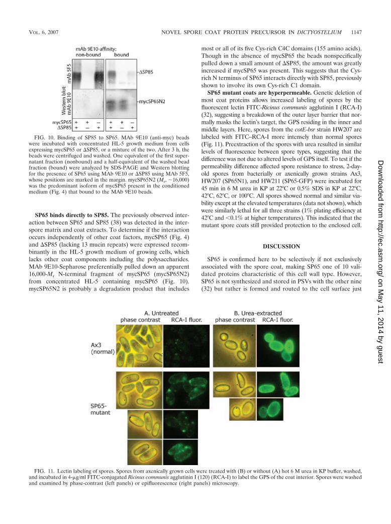
SP65 mutant coats are hyperpermeable. Genetic deletion ofmost coat proteins allows increased labeling of spores by thefluorescent lectin FITC-Ricinus communis agglutinin I (RCA-I)(32), suggesting a breakdown of the outer layer barrier that nor-mally masks the lectin’s target, the GPS residing in the inner andmiddle layers. Here, spores from the cotE-bsr strain HW207 arelabeled with FITC–RCA-I more intensely than normal spores(Fig. 11). Preextraction of the spores with urea resulted in similarlevels of fluorescence between spore types, suggesting that thedifference was not due to altered levels of GPS itself. To test if thepermeability difference affected spore resistance to stress, 2-day-old spores from bacterially or axenically grown strains Ax3,HW207 (SP65N1), and HW211 (SP65-GFP) were incubated for45 min in 6 M urea in KP at 22°C or 0.5% SDS in KP at 22°C,42°C, 62°C, or 100°C. All spores showed normal and similar via-bility except at the elevated temperatures (data not shown), whichwere similarly lethal for all three strains (1% plating efficiency at42°C and �0.1% at higher temperatures). This indicated that themutant spore coats still provided protection to the enclosed cell.
DISCUSSION
SP65 is confirmed here to be selectively if not exclusivelyassociated with the spore coat, making SP65 one of 10 vali-dated proteins characteristic of this cell wall type. However,SP65 is not synthesized and stored in PSVs with the other nine(32) but rather is formed and routed to the cell surface just
FIG. 10. Binding of SP85 to SP65. MAb 9E10 (anti-myc) beadswere incubated with concentrated HL-5 growth medium from cellsexpressing mycSP65 or �SP85, or a mixture of the two. After 3 h, thebeads were centrifuged and washed. One equivalent of the first super-natant fraction (nonbound) and a half-equivalent of the washed beadfraction (bound) were analyzed by SDS-PAGE and Western blottingfor the presence of SP65 using MAb 9E10 or �SP85 using MAb 5F5,whose positions are marked in the margin. mycSP65N2 (Mr, 16,000)was the predominant isoform of mycSP65 present in the conditionedmedium (Fig. 4) that bound to the MAb 9E10 beads.
FIG. 11. Lectin labeling of spores. Spores from axenically grown cells were treated with (B) or without (A) hot 6 M urea in KP buffer, washed,and incubated in 4-�g/ml FITC-conjugated Ricinus communis agglutinin I (120) (RCA-I) to label the GPS of the coat interior. Spores were washedand examined by phase-contrast (left panels) or epifluorescence (right panels) microscopy.
VOL. 6, 2007 NOVEL SPORE COAT PROTEIN PRECURSOR IN DICTYOSTELIUM 1147
on May 11, 2014 by guest
http://ec.asm.org/
Dow
nloaded from
prior to cellulose synthesis, the final known component to bedelivered to the cell surface during coat formation. The noveltiming and routing of SP65 suggest that the interaction of SP65with cellulose, via the intermediate PSV protein SP85, maycritically regulate coat assembly.
The conclusion that SP65 is a coat protein is based in part onuse of Abs generated against recombinant full-length mycSP65.Polyclonal Abs showed that SP65 is enriched in coats of sporesbased on Western blotting (Fig. 6), but their polyspecificity pre-cluded their use in microscopy for confirmation. The MAbs,which recognize conformational epitopes, did not bind sporecoats (Fig. 9B), probably because the epitopes were eithermasked or altered in coats. For similar reasons, DP87/SP75 andEB4/SP35 gene products were originally identified as intersporematrix proteins before other approaches identified them as coatproteins (32). Therefore, tagging the SP65/cotE locus with GFP toyield an SP65-GFP fusion protein was particularly informative.Intrinsic GFP fluorescence and anti-GFP Ab labeling are ob-served only at the surface of spores and not in slugs or in stalkcells (Fig. 8 and 9C to E). Functional evidence for coat localiza-tion emerged from the finding that truncation of SP65 renderedthe coat permeable to an exogenous macromolecular tracer(Fig. 11).
Western blotting showed that SP65-GFP is not expresseduntil within an hour of coat formation (Fig. 7), and this wascorroborated by immunofluorescence using anti-GFP andMAb 4A11 (Fig. 9A and B). Analysis of native SP65 confirmedthe late timing, but the strong bias of the Abs toward confor-mational epitopes, masking of the epitopes in the coat, andapparent ongoing processing including degradation thwartedprecise characterization of SP65 expression in the absence ofthe GFP tag. Protein expression was closely linked to messageaccumulation as determined by Northern blotting (Fig. 3),indicating that SP65 expression is under transcriptional con-trol. The timing is essentially identical to that of the previouslydescribed prespore-cell-specific, glycosylphosphatidylinositol-anchored protein SpiA, associated with the plasma membranethat is tightly bound to the coat. The transport pathway andfunction of SpiA are unknown (22). Recently, additional lategenes have been found to be expressed under the control of theGATA-type transcriptional factor StkA (14) or the MADS-box-type transcriptional factor SrfA (1, 2). SigD, regulated bySrfA, has amino acid sequence motifs found in other coatproteins and is a candidate for another coat protein with sim-ilar regulation. Unlike SigD and SpiA, however, SP65 does notdepend on SrfA (Fig. 3).
The intracellular localization of the SP65 precursor is dis-tinctive from that of known coat proteins. SP65-GFP was firstdetected in a central location (Fig. 9A and C) suggestive of theGolgi apparatus (31) and confirmed using MAb 4A11 for na-tive SP65. Subsequently, anti-GFP labeling was distributed in adispersed punctate manner consistent with vesicles. But dou-ble-labeling experiments showed that these are not equivalentto PSVs (Fig. 9D and E, number 2), where other coat proteinprecursors and the GPS accumulate (32). However, overlap ofSP65 and SP85 was occasionally observed (Fig. 9E, number 3),but it is not clear whether this reflects rare vesicle fusion or atemporal progression in which SP65 and SP85 vesicles fusetransiently close to the time of exocytosis consistent with evi-dence that PSVs undergo continual maturation (23). GFP flu-
orescence could not be used to dynamically track nascentSP65-GFP because vesicular GFP was rarely fluorescent. Flu-orescence acquisition by GFP depends on time and O2 (36),whose level in vesicles may be limiting. Initial formation of thecoat was evidenced by a fluorescent crescent at the cell surface.This arc was usually labeled for both SP65-GFP and SP85,which is most consistent with coordinate rather than sequentialsecretion of the two proteins. Distinct secretory trajectories fordiscrete cell wall precursors have been observed in other cellsystems, though this is most often associated with sequentiallydeposited wall layers (11, 15, 18). When cellulose is imagedusing Calcofluor, the presence of cells fluorescent for GFP butnot Calcofluor (Fig. 8B) suggests that SP65 is secreted to thecell surface before cellulose is deposited.
The dynamic properties of SP65, revealed by changes in Abbinding and Mr differences between axenically and bacteriallygrown cells, are unusual and may reflect a combination ofnatural protein maturation and local microenvironmental dif-ferences between axenically and bacterially derived sorogens.The transient appearance of the MAb 4A11 epitope might berelated to global disulfide rearrangements (34) or other post-translational modifications such as proteolytic processing (32,39) thought to occur during coat assembly. SP65 may be asso-ciated with the outer coat surface because the GFP of SP65-GFP is accessible at the spore surface based on Ab labelingand is likely exposed to proteases present within the spore coatand the interspore matrix (10, 19, 32). Indeed, SP65 is suscep-tible to proteolytic degradation when expressed in the growthmedium (Fig. 4). SP65-GFP was less stable in culminants fromaxenically grown cells than in culminants from bacteriallygrown cells, which correlates with the earlier time of encapsu-lation in the rising sorogen from axenically grown cells notedwhen nascent spores were collected (data not shown). A longerperiod of turbulent exposure to proteases experienced by theearlier-forming spores might explain the higher degree ofbreakdown of SP65 and SP65-GFP.
Previous studies suggested that SP85 simultaneously con-tacts cellulose and SP65 to form a structural unit of the coat.Domain mapping studies showed this to be mediated by its110-amino-acid C1 domain composed of four Cys-rich C4Cmotifs related to the N-terminal subdomain of EGF repeats(17, 35, 38). A direct SP65-SP85 interaction is confirmed hereusing recombinant proteins, which rules out a requirement fora coat-specific intermediary factor such as a coat polysaccha-ride. Interestingly, the in vitro interaction was detected using aspontaneously generated N-terminal fragment of SP65(mycSP65N2) that was captured by the MAb 9E10 beads usedto immunoprecipitate SP65. This region encompasses the C4Cmotifs of SP65. Full-length SP65 normally may fold in such away that the C-terminal domain masks accessibility of the Nterminus (where the myc epitope is located), but an effect of adistinct posttranslational modification of the recombinant pro-tein cannot be excluded. Together these results imply a generalfunction for C4C motifs in mediating protein-protein and pro-tein-carbohydrate interactions. This is consistent with the ob-servation that truncated SP65N1 (generated by the insertion ofbsr) accumulates in the coat rather than the interspore matrix(Fig. 6). The accumulation of SP65 in the interspore matrix ofSP85/pspB-bsr spores confirms previous findings that SP85 isrequired to retain SP65 in the coat in vivo (38).
1148 METCALF ET AL. EUKARYOT. CELL
on May 11, 2014 by guest
http://ec.asm.org/
Dow
nloaded from
The permeability and structural defects of SP65 and SP85mutant coats and the dependence of certain SP85 functions onthe cellulose binding activity of SP85 (17) suggest that theproposed cellulose-SP85-SP65 triad is essential for outer layerformation. Though the distinct properties of SP85 mutantspores, which tend to be spherical and to not rise to the top ofthe stalk (35), and SP65 mutant spores, which are more per-meable without urea pretreatment (compare Fig. 11 and ref-erence 38), suggest that the two proteins contribute separatefunctions, the comparison is complicated because the SP85mutation is a null whereas the SP65 mutant produces a poten-tially dominant-negative N-terminal fragment (SP65N1). Nev-ertheless, the new data suggest that some activities of SP85may be regulated by the availability of SP65, which is synthe-sized much later and transported to the cell surface via alargely distinct vesicle population.
ACKNOWLEDGMENTS
We are grateful to Scherwin Henry at the University of FloridaICBR Hybridoma Laboratory for preparation of the anti-SP65 Abs,the OMRF DNA Sequencing Facility for DNA sequencing, T.Egelhoff for pTX-GFP, and D. Fuller and W. F. Loomis for anti-SpiAAb. Scott Plafker and Margaret Clarke are thanked for access tofluorescence microscopes. Mandrin Shima is acknowledged for hishelp in the PCR studies.
This work was supported in part by National Science Foundationgrant no. 0350516, the SURE Program sponsored by the PresbyterianHealth Foundation, and grant BMC2002-01501 from the DireccionGeneral de Investigacion.
REFERENCES
1. Escalante, R., N. Moreno, and L. Sastre. 2003. Dictyostelium discoideumdevelopmentally regulated genes whose expression is dependent on MADSbox transcription factor SrfA. Eukaryot. Cell 2:1327–1335.
2. Escalante, R., N. Iranfar, L. Sastre, and W. F. Loomis. 2004. Identificationof genes dependent on the MADS-box transcription factor SrfA in Dictyo-stelium development. Eukaryot. Cell 3:564–566.
3. Escalante, R., Y. Yamada, D. Cotter, L. Sastre, and M. Sameshima. 2004.The MADS box transcription factor srfA is required for actin cytoskeletonorganization and spore coat stability during Dictyostelium sporulation. Mech.Dev. 121:51–56.
4. Evan, G. I., G. K. Lewis, G. Ramsay, and J. M. Bishop. 1985. Isolation ofmonoclonal antibodies specific for human c-myc proto-oncogene product.Mol. Cell. Biol. 5:3610–3616.
5. Fosnaugh, K. L., and W. F. Loomis. 1991. Coordinate regulation of the sporecoat genes in Dictyostelium discoideum. Dev. Genet. 12:123–132.
6. Gonzalez-Yanes, B., R. B. Mandell, M. Girard, S. Henry, O. Aparicio, M.Gritzali, R. D. Brown, G. W. Erdos, and C. M. West. 1989. The spore coat ofa fucosylation mutant in Dictyostelium discoideum. Dev. Biol. 133:576–587.
7. Hacker, U., R. Albrecht, and M. Maniak. 1997. Fluid-phase uptake by mac-ropinocytosis in Dictyostelium. J. Cell Sci. 110:105–112.
8. Hadwiger, J. A., and R. A. Firtel. 1992. Analysis of G alpha 4, a G-proteinsubunit required for multicellular development in Dictyostelium. Genes Dev.6:38–49.
9. Kim, J. Y., M. J. Caterina, J. L. Milne, K. C. Lin, J. A. Borleis, and P. N.Devreotes. 1997. Random mutagenesis of the cAMP chemoattractant recep-tor, cAR1, of Dictyostelium. J. Biol. Chem. 272:2060–2068.
10. Lenhard, J. M., A. Siegel, and S. J. Free. 1989. Developing Dictyostelium cellscontain the lysosomal enzyme -mannosidase in a secretory granule. J. CellBiol. 109:2761–2769.
11. Leucci, M. R., G. P. Di Sansebastiano, M. Gigante, G. Dalessandro, and G.Piro. 2006. Secretion marker proteins and cell-wall polysaccharides movethrough different secretory pathways. Planta 225:1001–1017.
12. Levi, S., M. Polyakov, and T. T. Egelhoff. 2000. Green fluorescent proteinand epitope tag fusion vectors for Dictyostelium discoideum. Plasmid 44:231–238.
13. Loomis, W. F. 1971. Sensitivity of Dictyostelium discoideum to nucleic acidanalogues. Exp. Cell Res. 64:484–486.
14. Loughran, G., K. Pinter, P. C. Newell, and J. D. Gross. 2000. Identification
of STKA-dependent genes in Dictyostelium discoideum. Differentiation 66:71–80.
15. Matese, J. C., S. Black, and D. R. McClay. 1997. Regulated exocytosis andsequential construction of the extracellular matrix surrounding the seaurchin zygote. Dev. Biol. 186:16–26.
16. McGuire, V., and S. Alexander. 1996. PsB multiprotein complex of Dictyo-stelium discoideum: demonstration of cellulose binding activity and order ofprotein subunit assembly. J. Biol. Chem. 271:14596–14603.
17. Metcalf, T., K. Kelley, G. W. Erdos, L. Kaplan, and C. M. West. 2003.Formation of the outer layer of the spore coat of Dictyostelium depends onthe inner layer protein SP85/PsB. Microbiology 149:305–317.
18. Nogueron, M. I., D. Mauzy-Melitz, and G. L. Waring. 2000. Drosophila dec-1eggshell proteins are differentially distributed via a multistep extracellularprocessing and localization pathway. Dev. Biol. 225:459–470.
19. North, M. J., K. Nicol, T. W. Sands, and D. A. Cotter. 1996. Acid-activatablecysteine proteinases in the cellular slime mold Dictyostelium discoideum.J. Biol. Chem. 271:14462–14467.
20. Pang, K. M., and D. A. Knecht. 1997. Partial inverse PCR: a technique forcloning flanking sequences. BioTechniques 22:1046–1048.
21. Puta, F., and C. Zeng. 1998. Blasticidin resistance cassette in symmetricalpolylinkers for insertional inactivation of genes in Dictyostelium. Folia Biol.(Praha) 44:185–188.
22. Richardson, D. L., W. F. Loomis, and A. R. Kimmel. 1994. Progression of aninductive signal activates sporulation in Dictyostelium discoideum. Develop-ment 120:2891–2900.
23. Srinivasan, S., H. Alexander, and S. Alexander. 1999. The prespore vesiclesof Dictyostelium discoideum. J. Biol. Chem. 274:35823–35831.
24. Sussman, M. 1987. Cultivation and synchronous morphogenesis of Dictyoste-lium under controlled experimental conditions. Methods Cell Biol. 28:9–29.
25. Thuring, R. W., J. P. Sanders, and P. Borst. 1975. A freeze-squeeze methodfor recovering long DNA from agarose gels. Anal. Biochem. 66:213–220.
26. Urushihara, H., T. Morio, T. Saito, Y. Kohara, E. Koriki, H. Ochiai, M.Maeda, J. G. Williams, I. Takeuchi, and Y. Tanaka. 2004. Analyses ofcDNAs from growth and slug stages of Dictyostelium discoideum. NucleicAcids Res. 32:1647–1653.
27. van der Wel, H., H. R. Morris, M. Panico, T. Paxton, A. Dell, L. Kaplan, andC. M. West. 2002. Molecular cloning and expression of a UDP-GlcNAc:hydroxyproline polypeptide GlcNAc-transferase that modifies Skp1 in thecytoplasm of Dictyostelium. J. Biol. Chem. 277:46328–46337.
28. van der Wel, H., H. R. Morris, M. Panico, T. Paxton, S. J. North, A. Dell,J. M. Thomson, and C. M. West. 2001. A non-Golgi 1,2-fucosyltransferasethat modifies Skp1 in the cytoplasm of Dictyostelium. J. Biol. Chem. 276:33952–33963.
29. van Driessche, N., C. Shaw, M. Katoh, T. Morio, R. Sucgang, M. Ibarra, H.Kuwayama, T. Saito, H. Urushihara, M. Maeda, I. Takeuchi, H. Ochiai, W.Eaton, J. Tollett, J. Halter, A. Kuspa, Y. Tanaka, and G. Shaulsky. 2002. Atranscriptional profile of multicellular development in Dictyostelium discoideum.Development 129:1543–1552.
30. Wang, F., T. Metcalf, H. van der Wel, and C. M. West. 2003. Initiation ofmucin-type O-glycosylation in Dictyostelium is homologous to the corre-sponding step in animals and is important for spore coat function. J. Biol.Chem. 278:51395–51407.
31. Weiner, O. H., J. Murphy, G. Griffiths, M. Schleicher, and A. A. Noegel.1993. The actin-binding protein comitin (p24) is a component of the Golgiapparatus. J. Cell Biol. 123:23–34.
32. West, C. M. 2003. Comparative analysis of spore coat formation, structureand function in Dictyostelium. Int. Rev. Cytol. 222:237–293.
33. West, C. M., G. W. Erdos, and R. Davis. 1986. Glycoantigen expression isregulated both temporally and spatially during development in the cellularslime molds Dictyostelium discoideum and D. mucoroides. Mol. Cell. Bio-chem. 72:121–140.
34. West, C. M., J. Mao, H. van der Wel, G. W. Erdos, and Y. Zhang. 1996. SP75is encoded by the DP87 gene, and belongs to a family of modular Dictyo-stelium outer layer spore coat proteins. Microbiology 142:2227–2243.
35. West, C. M., P. Zhang, A. C. McGlynn, and L. Kaplan. 2002. Outside-insignaling of cellulose synthesis by a spore coat protein in Dictyostelium.Eukaryot. Cell 1:281–292.
36. Zhang, L., H. N. Patel, J. W. Lappe, and R. M. Wachter. 2006. Reactionprogress of chromophore biogenesis in green fluorescent protein. J. Am.Chem. Soc. 128:4766–4772.
37. Zhang, P., A. C. McGlynn, W. F. Loomis, R. L. Blanton, and C. M. West.2001. Spore coat formation and timely sporulation depend on cellulose inDictyostelium. Differentiation 67:72–79.
38. Zhang, Y., P. Zhang, and C. M. West. 1999. A linking function for thecellulose-binding protein SP85 in the spore coat of Dictyostelium discoideum.J. Cell Sci. 112:4367–4377.
39. Zhang, Y., R. D. Brown, and C. M. West. 1998. Two proteins of the Dictyo-stelium spore coat bind to cellulose in vitro. Biochemistry 37:10766–10779.
VOL. 6, 2007 NOVEL SPORE COAT PROTEIN PRECURSOR IN DICTYOSTELIUM 1149
on May 11, 2014 by guest
http://ec.asm.org/
Dow
nloaded from


