Embed Size (px)
Citation preview

JOURNAL OF BACTERIOLOGY, Nov. 2011, p. 6187–6196 Vol. 193, No. 220021-9193/11/$12.00 doi:10.1128/JB.05930-11Copyright © 2011, American Society for Microbiology. All Rights Reserved.
RpiR Homologues May Link Staphylococcus aureus RNAIII Synthesisand Pentose Phosphate Pathway Regulation�†
Yefei Zhu,1 Renu Nandakumar,2 Marat R. Sadykov,1 Nandakumar Madayiputhiya,2 Thanh T. Luong,3Rosmarie Gaupp,1 Chia Y. Lee,3 and Greg A. Somerville1*
School of Veterinary Medicine and Biomedical Sciences, University of Nebraska—Lincoln, Lincoln, Nebraska 68583–09051;Proteomics and Metabolomics Core, Redox Biology Center, Department of Biochemistry, University of Nebraska,
Lincoln, Nebraska 685882; and Department of Microbiology and Immunology, University ofArkansas for Medical Sciences, Little Rock, Arkansas3
Received 3 August 2011/Accepted 7 September 2011
Staphylococcus aureus is a medically important pathogen that synthesizes a wide range of virulence deter-minants. The synthesis of many staphylococcal virulence determinants is regulated in part by stress-inducedchanges in the activity of the tricarboxylic acid (TCA) cycle. One metabolic change associated with TCA cyclestress is an increased concentration of ribose, leading us to hypothesize that a pentose phosphate pathway(PPP)-responsive regulator mediates some of the TCA cycle-dependent regulatory effects. Using bioinformat-ics, we identified three potential ribose-responsive regulators that belong to the RpiR family of transcriptionalregulators. To determine whether these RpiR homologues affect PPP activity and virulence determinantsynthesis, the rpiR homologues were inactivated, and the effects on PPP activity and virulence factor synthesiswere assessed. Two of the three homologues (RpiRB and RpiRC) positively influence the transcription of thePPP genes rpiA and zwf, while the third homologue (RpiRA) is slightly antagonistic to the other homologues.In addition, inactivation of RpiRC altered the temporal transcription of RNAIII, the effector molecule of theagr quorum-sensing system. These data confirm the close linkage of central metabolism and virulence deter-minant synthesis, and they establish a metabolic override for quorum-sensing-dependent regulation of RNAIIItranscription.
Staphylococcus aureus is an important human and animalpathogen that is capable of infecting nearly all host anatomicsites. The pathogenicity of S. aureus depends on its ability tosynthesize virulence factors that facilitate colonization, im-mune evasion, and nutrient acquisition. Virulence factor syn-thesis is controlled by a complex network of regulatory pro-teins, including the agr quorum-sensing system and the SarAfamily of regulators (6, 29). In addition, tricarboxylic acid(TCA) cycle activity is important for the regulation of staphy-lococcal virulence factor synthesis (33, 39–41, 48). Since thetwo most common types of regulation are genetic regulationand metabolic regulation, TCA cycle-dependent regulationmost likely occurs via one or both of these mechanisms. Ge-netic regulation occurs through the repression or induction ofenzyme synthesis, while metabolic regulation controls enzymeactivity through the availability of substrates and cofactors. Anexample of staphylococcal metabolic regulation is the synthesisof capsular polysaccharide, which is regulated by TCA cycleactivity through the supply of phosphoenolpyruvate for gluco-neogenesis (33). Other virulence factors, such as polysaccha-ride intercellular adhesin (PIA), are genetically regulated byTCA cycle activity through transcriptional repression of the
operon encoding the enzymes of PIA biosynthesis (i.e.,icaADBC) (34, 44). This TCA cycle-dependent genetic regula-tion likely depends on response regulators that react to meta-bolic changes associated with TCA cycle activity fluctuations(35, 41).
In Staphylococcus epidermidis, TCA cycle stress (i.e., anyenvironmental stressor, such as iron limitation, that is capableof altering TCA cycle activity) increases the intracellular riboseconcentration, indicating that carbon flow through the pentosephosphate pathway (PPP) is increased during TCA cycle stress(35). This suggests that if there is a regulator that can respondto the concentration of ribose, or another PPP metabolite, thenthe activity of that regulator will likely be altered. The PPP-responsive regulator prototype, RpiR, was first identified inEscherichia coli as a regulator of ribose-5-phosphate isomeraseB (rpiB), which catalyzes the reversible isomerization of ribu-lose-5-phosphate and ribose-5-phosphate (42). Members ofthe RpiR family often act as transcriptional regulators of sugarcatabolism, and RpiR homologues have been identified asrepressors and activators in both Gram-negative and Gram-positive bacteria, including E. coli, Pseudomonas putida, andBacillus subtilis (8, 42, 46). As sugar-responsive regulators,members of the RpiR family of proteins have N-terminal helix-turn-helix DNA binding motifs and C-terminal sugar isomer-ase binding (SIS) domains (1).
TCA cycle stress alters the intracellular ribose concentrationin S. epidermidis and also alters the temporal expression ofvirulence factors in S. epidermidis and S. aureus (34, 35, 39, 40).These observations led us to hypothesize that an RpiR homo-logue may link the PPP to virulence factor regulation in staph-
* Corresponding author. Mailing address: School of VeterinaryMedicine and Biomedical Sciences, University of Nebraska—Lincoln,155 VBS, Fair St. and East Campus Loop, Lincoln, NE 68583-0905.Phone: (402) 472-6063. Fax: (402) 472-9690. E-mail: [email protected].
† Supplemental material for this article may be found at http://jb.asm.org/.
� Published ahead of print on 16 September 2011.
6187

ylococci. A search of the S. aureus strain Mu50 genome (18)returned three open reading frames (ORFs) with significantamino acid homology to RpiR (21 to 23% amino acid identityand 45 to 46% amino acid similarity): SAV0317, SAV0193, andSAV2315. For simplicity, these homologues were designatedRpiRA (SAV0317), RpiRB (SAV0193), and RpiRC (SAV2315).To determine whether these RpiR homologues link the PPP tovirulence factor synthesis in S. aureus, three single mutants,three double mutants, and a triple mutant of the rpiR homo-logues were constructed in S. aureus strain UAMS-1, and theeffects on PPP activity, RNAIII transcription, capsular poly-saccharide biosynthesis, PIA accumulation, and the ability toform a biofilm were assessed.
MATERIALS AND METHODS
Bacterial strains and growth conditions. The strains and plasmids used in thisstudy are listed in Table 1. E. coli strains were grown in 2� YT broth (36) or on2� YT agar, and S. aureus strains were grown in tryptic soy broth (TSB) (BDBiosciences) or on TSB containing 1.5% agar. TSB is a complex medium thatcontains glucose (0.25%, wt/vol) and stachyose. Stachyose is a plant carbohydratethat S. aureus cannot catabolize. Unless otherwise stated, all bacterial cultureswere inoculated at 1:200 from an overnight culture (normalized for growth) intoTSB, incubated at 37°C, and aerated at 225 rpm with a flask-to-medium ratio of10:1. Antibiotics were purchased from Fisher Scientific or Sigma Chemical and,when used, were used at the following concentrations: for E. coli, ampicillin at100 �g/ml; for S. aureus, erythromycin at 8 or 10 �g/ml, chloramphenicol at 10to 15 �g/ml, and tetracycline at 10 �g/ml.
Construction of S. aureus rpiR mutants. To inactivate rpiRA (ORF SAV0317),a 2.559-kb fragment was PCR amplified using primers SAV0316-BamHI andSAV0318-SacI (Table 2), and the product was cloned into the SmaI site ofpBluescript II KS(�) (Stratagene) to generate plasmid pYF-7. The ermB cas-sette of pEC4 was amplified using primers pEC4ErmBNdeIF andpEC4ErmBNdeIR (Table 2) and was ligated into the NdeI site within rpiRA ofpYF-7 to yield plasmid pYF-8. The rpiRA::ermB fragment of pYF-8 was cloned
into the BamHI and SacI sites of pTS1 to create plasmid pYF-9. The tempera-ture-sensitive plasmid pYF-9 was isolated from S. aureus strain RN4220 and wasintroduced into strain UAMS-1 by electroporation. Transformed bacteria wereused to construct the rpiRA mutant using the temperature shift method of Foster(11).
To inactivate rpiRB (ORF SAV0193), the technique of gene splicing by over-lap extension (15) was used to replace a 741-bp internal region of rpiRB with thecat gene from plasmid pTS1. For PCR, genomic DNA from S. aureus strainUAMS-1 was used as a template for the amplification of regions flanking rpiRB.PCR primers BamHI-SAV0192-f and cat-SAV0193-r (Table 2) were used for theamplification of a 1.5-kb region upstream of rpiRB, and a 1.5-kb region of therpiRB downstream region was amplified using primers cat-SAV0193-f and SacI-SAV0195-r (Table 2). The cat gene was amplified from pTS1 using primersSAV0193-cat-f and SAV0193-cat-r (Table 2). The resulting 3.9-kb PCR productconsisted of an internal 816-bp cat gene with DNA flanking the rpiRB gene. The3.9-kb PCR product contained BamHI and SacI sites that were used for ligationinto pTS1-d digested with SacI and BamHI to generate pYM-4. Plasmid pYM-4was used to construct an rpiRB mutant (UAMS-1-rpiRB::cat) by using the tem-perature shift method of Foster (11).
Gene splicing by overlap extension was used to replace a 614-bp internalregion of rpiRC (ORF SAV2315) with the tetM gene from plasmid pJF-12 (Table1). A 1.5-kb region upstream of rpiRC was amplified using primers BamHI-SAV2312-f and tetM-SAV2315-r (Table 2), and primers tetM-SAV2315-f andKpnI-SAV2316-r (Table 2) were used for amplification of a 1.6-kb downstreamregion. tetM was amplified from pJF-12 using primers SAV2315-tetM-f andSAV2315-tetM-r (Table 2). A 5.4-kb PCR product consisting of the 2.3-kb tetMgene and DNA flanking the rpiRC gene with BamHI and KpnI sites was insertedinto pTS1-d digested with BamHI and KpnI to generate pYM-5. Plasmid pYM-5was used to construct a strain UAMS-1 rpiRC mutant (UAMS-1-rpiRC::tetM) byusing temperature shifts. To minimize the possibility that any phenotype(s) wasthe result of random mutations occurring during temperature shifts, all resultingmutations were back-crossed into wild-type strain UAMS-1 using transducingphage �85 (11). All mutants were verified by PCR and Southern blot analysis.The rpiR double mutants and triple mutant were constructed using transducingphage �85.
Construction of rpiR complementing plasmids. Plasmids pCL15 and pCL15-ermB (Table 1), containing a Pspac promoter, were used to construct the rpiRA,
TABLE 1. Strains and plasmids used in this study
Plasmid or strain Relevant genotype and/or characteristic(s)a Source or reference
PlasmidspBluescript II KS(�) E. coli phagemid cloning vector StratagenepTS1 S. aureus-E. coli temperature-sensitive shuttle vector; Ampr Camr 14pTS1-d Derivative of pTS1 with deletion of plasmid-encoded 3� region of ermC 34pEC4 pBluescript II KS(�) with ermB inserted into ClaI site 3pJF12 Plasmid pCR2.1 containing tetM; Ampr Minr J. Finan and G. ArcherpYF-7 pBluescript II KS(�) containing a portion of SAV0317 This studypYF-8 pYF-7 containing an ermB cassette inserted into the NdeI site of SAV0317 This studypYF-9 SAV0317::ermB product from pYF-8 inserted into BamHI/SacI-digested pTS1 This studypYM-4 Derivative of pTS1 with SAV0193::cat fragment This studypYM-5 Derivative of pTS1 with SAV2315::tetM fragment This studypCL15 Expression vector; derivative of pSI-1; Camr Chia LeepCL15-ermB Replacement of cat with ermB in expression plasmid pCL15; Ermr This studypYF-10 pCL15 with the SAV0317 gene under the control of the Pspac promoter; Camr This studypYF-11 pCL15-ermB with the SAV0193 gene under the control of the Pspac promoter; Ermr This studypYF-12 pCL15 with the SAV2315 gene under the control of the Pspac promoter; Camr This study
StrainsRN4220 Restriction-negative S. aureus 30DH5� E. coli cloning host InvitrogenUAMS-1 S. aureus clinical isolate 13UAMS-1-rpiRA SAV0317 insertion mutant of UAMS-1; Ermr This studyUAMS-1-rpiRB SAV0193 deletion mutant of UAMS-1; Camr This studyUAMS-1-rpiRC SAV2315 deletion mutant of UAMS-1; Minr This studyUAMS-1-rpiRAB SAV0317 SAV0193 double mutant of UAMS-1; Ermr Camr This studyUAMS-1-rpiRAC SAV0317 SAV2315 double mutant of UAMS-1; Ermr Minr This studyUAMS-1-rpiRBC SAV0193 SAV2315 double mutant of UAMS-1; Camr Minr This studyUAMS-1-rpiRABC SAV0317 SAV0193 SAV2315 triple mutant of UAMS-1; Ermr Camr Minr This study
aAmpr, ampicillin resistant; Camr, chloramphenicol resistant; Ermr, erythromycin resistant; Minr, minocycline resistant.
6188 ZHU ET AL. J. BACTERIOL.

rpiRB, and rpiRC complementation plasmids pYF-10, pYF-11, and pYF-12. Thepromoterless genes from S. aureus strain UAMS-1 were PCR amplified using theprimers listed in Table 2 and were ligated into plasmid pCL15 or pCL15-ermB.Plasmids were isolated from S. aureus strain RN4220 and were electroporatedinto the UAMS-1 rpiRA, rpiRB, and rpiRC mutants.
Northern blot analysis. To determine whether rpiR inactivation affected thetranscription of PPP genes, Northern blot analysis was performed on ribose5-phosphate isomerase A (rpiA) and glucose-6-dehydrogenase (G6PD) (zwf;ORF SAV1505). RNAIII transcript levels were also evaluated in order to de-termine the effect of rpiR inactivation on the agr system. Northern blotting wasperformed as described previously (36), except that total RNA was isolated usingthe FastRNA Pro Blue kit (Qbiogene) and was purified using an RNeasy kit(Qiagen). Probes for Northern blotting were generated by PCR amplification ofunique internal regions of RNAIII, rpiA, and zwf (Table 2) and were labeledusing the North2South random prime labeling kit (Pierce). Detection was per-formed using the chemiluminescent nucleic acid detection module (Pierce).
Glucose-6-dehydrogenase activity assay. To determine whether rpiR inactiva-tion affected PPP activity, G6PD activity was measured as described previously(7). Protein concentrations were determined using a modified Lowry assay(Pierce Chemical).
Western blot analysis. To determine whether rpiR inactivation affected proteinA biosynthesis, protein A was collected as described previously (45), and West-ern blot analysis was performed (43).
Hemolytic assay. Strain UAMS-1 is lysogenized with an hlb-converting phageand has a nonsense mutation in hla; hence, it does not produce the major
hemolysins alpha-toxin and beta-toxin (4). The mRNA for delta-toxin is con-tained within RNAIII (16). To determine whether inactivation of any rpiR ho-mologue altered delta-toxin accumulation, a semiquantitative microtiter plateassay was carried out as described previously (10). Briefly, horse red blood cells(RBCs; Colorado Serum Company) were washed three times in phosphate-buffered saline (PBS) (pH 7.2) and were suspended at 2% (vol/vol) in PBS.Bacteria were grown in TSB for 15 h and were then centrifuged at 16,100 � g for5 min; supernatants were collected, and 2-fold serial dilutions were made in PBS.Hemolytic assays were started by mixing 100 �l of freshly prepared 2% horseRBCs with 100 �l of serial 2-fold dilutions of the appropriate culture superna-tant. The microtiter plates were incubated at 37°C for 30 min, followed by 12 hat 4°C. After incubation, the supernatant fluids were collected, and hemoglobinrelease was measured at 595 nm. Each experiment was repeated three times, andthe mean and standard error of the mean (SEM) were calculated.
Polystyrene primary attachment assay. The primary attachment assay wasperformed as described by Lim et al. (19). Briefly, bacterial cultures (2 h post-inoculation) were diluted into TSB to yield approximately 300 CFU. Bacteriawere poured onto polystyrene petri dishes (Fisher Scientific) and were incubatedat 37°C for 30 min. Following incubation, the petri dishes were rinsed three timeswith sterile PBS (pH 7.5) and were covered with 15 ml of TSB containing 0.8%agar maintained at 48°C. The percentage of bacteria attached to the polystyrenewas defined as the number of CFU remaining in the petri dishes after washingcompared to the number of CFU in unwashed TSB plates. The experiment wasrepeated three times, and the mean and SEM were calculated.
TABLE 2. Primers used in this study
Primer target Primer designation Nucleotide sequence (5�–3�)
SAV0317 SAV0316-BamHI GCTGGATCCCGACTGAACAATGAACGCCTAAGTCSAV0318-SacI CCTGAGCTCATCAACGCCGGACAACAAAAGTG
ermB pEC4ErmBNdeI-f GCGCATATGCGTTAGATTAATTCCTACCAGTGACpEC4ErmBNdeI-r GCGCATATGCTCATAGAATTATTTCCTCCCG
SAV0193 BamHI-SAV0192-f CCAGGATCCAGAACGAATTATTGCTGCAGTAGGcat-SAV0193-r CCACTTTATCCAATTTTCGTTTGTTGTTCACCGTCATATCAATGATTTTATGTGG
cat SAV0193-cat-f CCACATAAAATCATTGATATGACGGTGAACAACAAACGAAAATTGGATAAAGTGGGSAV0193-cat-r GCAAGATGCTTCCGGTAATTATCAAGCGACTGTAAAAAGTACAGTCGGC
SAV0193 cat-SAV0193-f GCCGACTGTACTTTTTACAGTCGCTTGATAATTACCGGAAGCATCTTGCSacI-SAV0195-r GCAGAGCTCGTTGAATAAGTGCTTCTACCGCATAC
SAV2315 BamHI-SAV2312-f CCAGGATCCGATTCCTAAACTATGGAGTCGATGGGtetM-SAV2315-r CGTAAGAGCATATTTGTAAAGGAATCTCCGAGACCTCATTTTAATCACCTTTTGAGG
tetM SAV2315-tetM-f CCTCAAAAGGTGATTAAAATGAGGTCTCGGAGATTCCTTTACAAATATGCTCTTACGSAV2315-tetM-r GAAGTTGTTGCTCCCATATGCATCCGATCTCCTCCTTTCCACTTTAATTC
SAV2315 tetM-SAV2315-f GAATTAAAGTGGAAAGGAGGAGATCGGATGCATATGGGAGCAACAACTTCKpnI-SAV2316-r GAAGGTACCAATGGATTGTAGTTGGTATGAGTGAG
RNAIII SARNAIII-f GAAGGAGTGATTTCAATGGCACAAGSARNAIII-r GGCTCACGACCATACTTATTATTAAGGG
rpiA rpiaf GTGACATGACGCTGGGAATTGGrpiar GTATCCTGTCTCAAACACACCTGTCAG
SAV1505 SAV1505f GCACCACAATTCTTTGGCGTTATTTCSAV1505r AGTACGAATATAGAATGGTACACCAGCC
SAV0317 BamHI-SD-rpiR-f CAAGGATCCATTAAGATGAAGGGGTGACACAATGSacI-rpiR-r CAAGAGCTCAATCACGATGATTGTCTACAGTTGC
SAV0193 BamHI-SAV0193-f CTAGGATCCATGACAAATATTTTATATCGCATTGATAAACAGTTGAGSAV0193-r CAAACAACTGAATCACATCAAAAACTTCAATTG
SAV2315 BamHI-SAV2315-f CTAGGATCCATGTCAAACGTACTAACAGAAATAGATAGTCAATATCCSAV2315-r GCGTATGTTATACAAGATAAAAGACATGTAAGCTTTG
VOL. 193, 2011 RpiR AND STAPHYLOCOCCUS AUREUS VIRULENCE 6189

Capsule immunoblot assay. To determine whether rpiR inactivation alteredcapsule biosynthesis, capsule accumulation was quantified by immunoblotting asdescribed previously (22), except that immunoblots were developed using achemiluminescent horseradish peroxidase (HRP) substrate (Millipore). For thecapsule blots, bacteria (1.25 optical density at 660 nm [OD660] units) wereharvested after overnight growth in tryptic soy broth at 37°C, with a flask-to-medium ratio of 20:1, and were aerated at 225 rpm.
PIA immunoblot assay. PIA accumulation was determined after 2, 4, and 6 hof growth as described previously (47).
Biofilm formation in flow cell chambers. S. aureus strains were grown in flowcell chambers (Stovall Life Science) as described previously (47). To assessbacterial growth, at 12 h postinoculation and every 4 h thereafter, effluentsamples were collected, the pH was measured, and the chamber was photo-graphed.
Proteomic analyses. Bacterial cells (2 h and 6 h postinoculation) were har-vested by centrifugation and were suspended in 1.0 ml of lysis buffer containing50 mM ammonium bicarbonate, 8 M urea, and 1.5 mM phenylmethylsulfonylfluoride (PMSF). The samples were homogenized for 40 s at 6.0 m/s in aFastPrep instrument (MP Biomedical), and the lysate was centrifuged for 5 minat 20,800 � g and 4°C. Bacterial proteins were subjected to in-solution trypsindigestion as described previously (28). Briefly, the proteins were reduced with 10mM dithiothreitol and were alkylated with 40 mM iodoacetamide, followed bytrypsin (Roche) digestion (trypsin/protein ratio, 1:50) overnight at 37°C. Thetryptic peptides were desalted and concentrated using PepClean C18 spin col-umns according to the manufacturer’s instructions (Thermo Scientific).
Fully automated 2-dimensional (2D) chromatographic experiments were per-formed with an UltiMate 3000 Proteomics multidimensional liquid chromatog-raphy (MDLC) system (Dionex Corporation) integrated with a nanospray sourceand an LCQ (liquid chromatography quadrupole) Fleet ion trap mass spectrom-eter (Thermo Scientific). The first-dimension LC separation (strong cation-ex-change [SCX] chromatography) with fraction collection was followed by thesecond-dimension LC separation (reverse-phase chromatography) and detectionby tandem mass spectrometry (MS-MS). The first-dimension separation wasperformed on an SCX column (polysulfoethyl; inside diameter [i.d.], 1 mm;length, 15 cm; particle size, 5 �m; pore size, 300 Å; Dionex). Twenty microlitersof the sample was loaded onto the first-dimension SCX column and was elutedusing a salt gradient (0 to 600 mM) for 45 min. Based on the UV absorbance ofthe eluted peptides, selected fractions were subjected to second-dimension anal-ysis. The second-dimension separation included on-line sample preconcentrationand desalting using a monolithic C18 trap column (PepMap; i.d., 300 �m; length,5 mm; particle size, 5 �m; pore size, 100 Å; Dionex). The sample was loaded ontothe monolithic trap column at a flow rate of 300 nl/min. The desalted peptideswere then eluted and separated on a C18 PepMap column (i.d., 75 �m; length, 15cm; particle size, 3 �m; pore size, 100 Å) by applying an acetonitrile (ACN)gradient (ACN plus 0.1% formic acid; 90-min gradient at a flow rate of 300nl/min) and were introduced into the mass spectrometer using the nanospraysource. The LCQ Fleet mass spectrometer was operated with the followingparameters: nanospray voltage, 2.0 kV; heated capillary temperature, 200°C;full-scan m/z range, 400 to 2,000. The mass spectrometer was operated in thedata-dependent mode with 4 MS-MS spectra for every full scan, 5 microscansaveraged for full scans and MS-MS scans, a 3 m/z isolation width for MS-MSisolations, and 35% collision energy for collision-induced dissociation.
The MS-MS spectra were searched against the S. aureus MRSA252 databaseusing MASCOT (version 2.2; Matrix Science). The database search criteria wereas follows: enzyme, trypsin; missed cleavages, 2; mass, monoisotropic; fixedmodification, carbamidomethyl (C); peptide tolerance, 1.5 Da; MS-MS fragmention tolerance, 1 Da. Probability assessment of peptide assignments and proteinidentifications were accomplished by Scaffold (version 3.0; Proteome SoftwareInc.). Only peptides with �90% probability were considered. Criteria for proteinidentification included the detection of at least 2 unique identified peptides anda protein probability score of �90%. Relative quantitation of proteins was doneby use of the label-free method of spectral counting (20) using the normalizedspectral counts for each protein. For ease of reference, the NCBI GenInfoIdentifier (gi) numbers have been included in this report and in Tables S1 and S2in the supplemental material.
Hydrogen peroxide susceptibility assay. To determine if rpiR inactivationaffects hydrogen peroxide susceptibility, S. aureus strain UAMS-1 and all of therpiR mutant strains were grown in TSB for 15 h and were then diluted to anOD600 of 0.05 into sterile medium containing increasing concentrations of hy-drogen peroxide (Fisher Scientific). Cultures were grown at 37°C with shaking(225 rpm) for 4 h. Bacterial densities were determined by measuring the OD600.
RESULTS
Characterization of rpiR mutants. Analysis of the Mu50genome (18) revealed the presence of three RpiR homologues:RpiRA (SAV0317), RpiRB (SAV0193), and RpiRC(SAV2315). Each rpiR homologue was deleted either individ-ually or in tandem with one or both of the other rpiR homo-logues in strain UAMS-1 (Table 1). To assess the effects ofinactivation of the rpiR homologue genes on growth, the opti-cal densities and pH of the culture medium (TSB) were mea-sured over time (Fig. 1). Inactivation of any single rpiR homo-logue in UAMS-1 did not alter the growth rate, growth yield,or pH profile of the culture medium (Fig. 1A). Similarly, thedouble and triple mutants had growth rates and growth yieldsequivalent to those of the wild-type strain UAMS-1 (Fig. 1B).Of note, the pH profile of the culture medium for the triplemutant showed an increased rate of alkalization relative to thatfor the wild-type strain, suggesting that this strain had an in-creased rate of acetic acid utilization or an increase in ammo-nia generation due to amino acid catabolism (Fig. 1B). Theseresults demonstrate that the growth of the rpiR mutants isequivalent to that of the isogenic wild-type strain.
RpiR homologues regulate PPP activity. As stated above,RpiR was first identified in E. coli as a repressor of the PPPgene rpiB (42). Similarly, the Pseudomonas putida RpiR ho-
FIG. 1. Deletion of any rpiR homologue in strain UAMS-1 doesnot alter the growth profile in TSB medium. (A) Growth curves andculture medium pH profiles are shown for strain UAMS-1 and for theUAMS-1-rpiRA, UAMS-1-rpiRB, and UAMS-1-rpiRC mutants(A) and the UAMS-1-rpiRAB, UAMS-1-rpiRAC, UAMS-1-rpiRBC,and UAMS-1-rpiRABC mutants (B).
6190 ZHU ET AL. J. BACTERIOL.
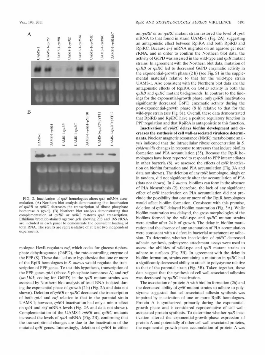
mologue HexR regulates zwf, which codes for glucose 6-phos-phate dehydrogenase (G6PD), the rate-controlling enzyme ofthe PPP (8). These data led us to hypothesize that one or moreof the RpiR homologues in S. aureus would regulate the tran-scription of PPP genes. To test this hypothesis, transcription ofthe PPP genes rpiA (ribose-5-phosphate isomerase A) and zwf(sav1505; coding for G6PD) in the rpiR mutant strains wasassessed by Northern blot analysis of total RNA isolated dur-ing the exponential phase of growth (2 h) (Fig. 2A and data notshown). Deletion of rpiRB or rpiRC decreased the transcriptionof both rpiA and zwf relative to that in the parental strainUAMS-1; however, rpiRA inactivation had only a minor effecton rpiA and zwf mRNA levels (Fig. 2A and data not shown).Complementation of the UAMS-1 rpiRB and rpiRC mutantsincreased the levels of rpiA mRNA (Fig. 2B), confirming thatthe transcriptional changes are due to the inactivation of themutated rpiR genes. Interestingly, deletion of rpiRA in either
an rpiRB or an rpiRC mutant strain restored the level of rpiAmRNA to that found in strain UAMS-1 (Fig. 2A), suggestingan antagonistic effect between RpiRA and both RpiRB andRpiRC. Because zwf mRNA migrates on an agarose gel nearrRNA, and in order to confirm the Northern blot data, theactivity of G6PD was assessed in the wild-type and rpiR mutantstrains. In agreement with the Northern blot data, mutation ofrpiRB or rpiRC led to decreased G6PD enzymatic activity inthe exponential-growth phase (2 h) (see Fig. S1 in the supple-mental material) relative to that for the wild-type strainUAMS-1. Also consistent with the Northern blot data are theantagonistic effects of RpiRA on G6PD activity in both therpiRB and rpiRC mutant backgrounds. In contrast to the find-ings for the exponential-growth phase, only rpiRB inactivationsignificantly decreased G6PD enzymatic activity during thepost-exponential-growth phase (8 h) relative to that for thewild-type strain (see Fig. S1). Overall, these data demonstratedthat RpiRB and RpiRC have a positive regulatory function inPPP regulation and that RpiRA is antagonistic to this function.
Inactivation of rpiRC delays biofilm development and de-creases the synthesis of cell wall-associated virulence determi-nants. Nuclear magnetic resonance (NMR) metabolomic anal-ysis indicated that the intracellular ribose concentration in S.epidermidis changes in response to stressors that induce biofilmformation and PIA accumulation (35). Because the RpiR ho-mologues have been reported to respond to PPP intermediatesin other bacteria (8), we assessed the effects of rpiR inactiva-tion on biofilm formation and PIA accumulation (Fig. 3A anddata not shown). The deletion of any rpiR homologue, singly orin tandem, did not significantly alter the accumulation of PIA(data not shown). In S. aureus, biofilms can form in the absenceof PIA biosynthesis (2); therefore, the lack of any significanteffect of rpiR inactivation on PIA accumulation did not pre-clude the possibility that one or more of the RpiR homologueswould affect biofilm formation. Consistent with this premise,deletion of rpiRC delayed biofilm maturation (Fig. 3A). Whilebiofilm maturation was delayed, the gross morphologies of thebiofilms formed by the wild-type and rpiRC mutant strainswere similar after 24 h of growth. The delay in biofilm matu-ration and the absence of any attenuation of PIA accumulationwere consistent with a defect in bacterial attachment or adhe-sion. To determine whether inactivation of rpiRC decreasedadhesin synthesis, polystyrene attachment assays were used toassess the abilities of wild-type and rpiR mutant strains toadhere to surfaces (Fig. 3B). In agreement with the delay inbiofilm formation, strains containing a mutation in rpiRC hada significantly decreased ability to attach to polystyrene relativeto that of the parental strain (Fig. 3B). Taken together, thesedata suggest that the synthesis of cell wall-associated adhesinswas decreased by rpiRC inactivation.
The association of protein A with biofilm formation (26) andthe decreased ability of rpiR mutant strains to adhere to poly-styrene suggested that cell-associated adhesin synthesis wasimpaired by inactivation of one or more RpiR homologues.Protein A is synthesized primarily during the exponential-growth phase and is considered representative of cell wall-associated protein synthesis. To determine whether rpiR inac-tivation altered the exponential-growth-phase expression ofprotein A and potentially of other cell wall-associated proteins,the exponential-growth-phase accumulation of protein A was
FIG. 2. Inactivation of rpiR homologues alters rpiA mRNA accu-mulation. (A) Northern blot analysis demonstrating that inactivationof rpiRB or rpiRC decreases the transcription of ribose phosphateisomerase A (rpiA). (B) Northern blot analysis demonstrating thatcomplementation of rpiRB or rpiRC restores rpiA transcription.Ethidium bromide-stained agarose gels showing 23S and 16S rRNAare included in each panel to demonstrate the equivalent loading oftotal RNA. The results are representative of at least two independentexperiments.
VOL. 193, 2011 RpiR AND STAPHYLOCOCCUS AUREUS VIRULENCE 6191
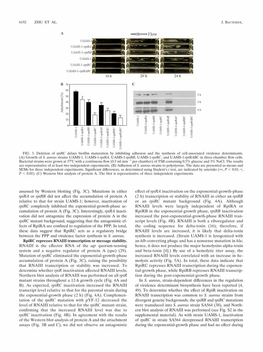
assessed by Western blotting (Fig. 3C). Mutations in eitherrpiRA or rpiRB did not affect the accumulation of protein Arelative to that for strain UAMS-1; however, inactivation ofrpiRC completely inhibited the exponential-growth-phase ac-cumulation of protein A (Fig. 3C). Interestingly, rpiRA inacti-vation did not antagonize the expression of protein A in therpiRC mutant background, suggesting that the antagonistic ef-fects of RpiRA are confined to regulation of the PPP. In total,these data suggest that RpiRC acts as a regulatory bridgebetween the PPP and virulence factor synthesis in S. aureus.
RpiRC represses RNAIII transcription or message stability.RNAIII is the effector RNA of the agr quorum-sensingsystem and a negative regulator of protein A (spa) (29).Mutation of rpiRC eliminated the exponential-growth-phaseaccumulation of protein A (Fig. 3C), raising the possibilitythat RNAIII transcription or stability was increased. Todetermine whether rpiR inactivation affected RNAIII levels,Northern blot analysis of RNAIII was performed on all rpiRmutant strains throughout a 12-h growth cycle (Fig. 4A andB). As expected, rpiRC inactivation increased the RNAIIItranscript level relative to that for the parental strain duringthe exponential-growth phase (2 h) (Fig. 4A). Complemen-tation of the rpiRC mutation with pYF-12 decreased thelevel of RNAIII relative to that for the rpiRC mutant strain,confirming that the increased RNAIII level was due torpiRC inactivation (Fig. 4B). In agreement with the resultsof the Western blot analysis of protein A and the attachmentassays (Fig. 3B and C), we did not observe an antagonistic
effect of rpiRA inactivation on the exponential-growth-phase(2 h) transcription or stability of RNAIII in either an rpiRBor an rpiRC mutant background (Fig. 4A). AlthoughRNAIII levels were largely independent of RpiRA orRpiRB in the exponential-growth phase, rpiRB inactivationincreased the post-exponential-growth-phase RNAIII tran-script levels (Fig. 4B). RNAIII is both a riboregulator andthe coding sequence for delta-toxin (16); therefore, ifRNAIII levels are increased, it is likely that delta-toxinsynthesis is increased. (Strain UAMS-1 is lysogenized withan hlb-converting phage and has a nonsense mutation in hla;hence, it does not produce the major hemolysins alpha-toxinand beta-toxin [4].) By use of a hemolytic titer assay, theincreased RNAIII levels correlated with an increase in he-molysis activity (Fig. 5A). In total, these data indicate thatRpiRC represses RNAIII transcription during the exponen-tial-growth phase, while RpiRB represses RNAIII transcrip-tion during the post-exponential-growth phase.
In S. aureus, strain-dependent differences in the regulationof virulence determinant biosynthesis have been reported (4,49). To determine whether the effect of RpiR inactivation onRNAIII transcription was common to S. aureus strains fromdivergent genetic backgrounds, the rpiRB and rpiRC mutationswere transduced into S. aureus strain SA564 (38), and North-ern blot analysis of RNAIII was performed (see Fig. S2 in thesupplemental material). As with strain UAMS-1, inactivationof rpiRC in strain SA564 derepressed RNAIII transcriptionduring the exponential-growth phase and had no effect during
FIG. 3. Deletion of rpiRC delays biofilm maturation by inhibiting adhesion and the synthesis of cell-associated virulence determinants.(A) Growth of S. aureus strains UAMS-1, UAMS-1-rpiRA, UAMS-1-rpiRB, UAMS-1-rpiRC, and UAMS-1-rpiRABC in three-chamber flow cells.Bacterial strains were grown at 37°C with a continuous flow (0.5 ml min�1 per chamber) of TSB containing 0.5% glucose and 3% NaCl. The resultsare representative of at least two independent experiments. (B) Adhesion of S. aureus strains to polystyrene. The data are presented as means andSEMs for three independent experiments. Significant differences, as determined using Student’s t test, are indicated by asterisks (��, P � 0.01; �,P � 0.05). (C) Western blot analysis of protein A. The blot is representative of three independent experiments.
6192 ZHU ET AL. J. BACTERIOL.

the post-exponential-growth phase (see Fig. S2). Similarly, in-activation of rpiRB in strain SA564 had a minimal effect onRNAIII transcript levels during the exponential-growth phase.In contrast to the finding for strain UAMS-1, inactivation ofrpiRB in strain SA564 had no apparent effect on RNAIII tran-scription during the post-exponential-growth phase. Thesedata demonstrate that RpiRC represses the exponential-growth-phase level of RNAIII in divergent genetic back-grounds.
Inactivation of rpiRC dramatically increases capsule accu-mulation. RNAIII is a positive regulator of capsule gene (cap)transcription (9, 21, 32); thus, an increase in RNAIII levelsshould correlate with an increase in capsule biosynthesis. Todetermine whether rpiR inactivation affects capsule biosynthe-sis, capsule accumulation was assessed by capsule immunoblot-ting. In agreement with the increased RNAIII levels, inactiva-tion of all three rpiR genes increased capsule accumulation(Fig. 5B); however, the increased accumulation of capsule was
FIG. 4. Deletion of rpiRC increases the transcription and/or stability of RNAIII. (A) Temporal Northern blot analysis of RNAIII. (B) Com-plementation of rpiR homologues moderately restores RNAIII levels after 2 h of growth. Ethidium bromide-stained agarose gels showing 23S and16S rRNA are included in each panel to demonstrate the equivalent loading of total RNA. All Northern blotting was performed at least twice usingindependently isolated total RNA.
VOL. 193, 2011 RpiR AND STAPHYLOCOCCUS AUREUS VIRULENCE 6193

most apparent in strains with a mutation in rpiRC. These datastrongly suggest that the RpiR-dependent derepression ofRNAIII facilitates virulence determinant expression and thatthe RpiR proteins act as a bridge between the PPP and viru-lence factor synthesis.
Inactivation of rpiRC alters the proteome. To identifychanges in cytosolic protein content in strain UAMS-1 andthe rpiRC mutant strains, cell-free lysates were preparedfrom strains UAMS-1, UAMS-1-rpiRC, and UAMS-1-rpiRABC grown to the exponential and post-exponentialphases of growth and were analyzed by 2D LC–MS-MS (seeTables S1 and S2 in the supplemental material). AlthoughrpiRC inactivation resulted in numerous proteomic changes,we were specifically interested in changes to PPP enzymesand proteins that might clarify the increased RNAIII tran-script levels. Proteomic analysis showed that the PPP en-zymes transaldolase (gi49242155) and ribose-phosphatepyrophosphokinase (gi49240856) were present at lower
concentrations in strains UAMS-1-rpiRC and UAMS-1-rpiRABC than in strain UAMS-1, consistent with regulationof the PPP by RpiRC. Interestingly, deletion of rpiRC in-creased the accumulation of ribosomal proteins (see Tables S1and S2 in the supplemental material); however, the reason forthis remains unknown. Proteomic analysis also suggested thatthere was an increase in the levels of proteins associated withB; specifically, inactivation of rpiRC increased the concentra-tions of the alkaline shock protein A (Asp23; gi49242531) andRsbU (gi49242422) (see Tables S1 and S2). Because asp23transcription is controlled exclusively by B, Asp23 is used asan indicator of B activity (17, 27). RsbU is a phosphatase thatdephosphorylates (activates) the anti-anti-sigma factor RsbV,which then binds the anti-sigma factor RsbW in a competitivemanner to increase the concentration of free B (12). In ad-dition to regulating the transcription of asp23, B regulates thetranscription of sarA from the sar P3 promoter (27). SarA is apositive effector of agrACDB and RNAIII transcription (5).Inactivation of rpiRC increased RNAIII levels relative to thosein the wild-type strain (Fig. 4), suggesting that rpiRC inactiva-tion might increase the availability of SarA. Consistent withthis suggestion, rpiRC inactivation increased the cytosolic con-centration of SarA (gi49240975) during both the exponential-and post-exponential-growth phases (see Tables S1 and S2).These data suggest that the increased RNAIII levels in therpiRC mutants are due to increased availability of B, whichincreases sarA transcription and translation, resulting in in-creased RNAIII transcription.
Inactivation of rpiRC decreases peroxide susceptibility. Insome strains of S. aureus, B has been implicated in suscepti-bility to oxidative stress (12, 17). This observation and the factthat strain UAMS-1-rpiRC had higher ferritin and catalaselevels than strain UAMS-1 (see Tables S1 and S2 in the sup-plemental material) led us to assess the susceptibilities ofstrain UAMS-1 and the rpiR mutants to peroxide stress (Fig.6). As expected, inactivation of rpiRC significantly decreasedthe susceptibilities of strains UAMS-1-rpiRC, UAMS-1-rpiRAC, UAMS-1-rpiRBC, and UAMS-1-rpiRABC to hydrogenperoxide relative to that of strain UAMS-1 (Fig. 6). Takentogether, these data demonstrate that the S. aureus RpiR fam-
FIG. 5. Inactivation of rpiR homologues alters virulence factor syn-thesis. (A) Hemolytic activities of culture supernatants from strainUAMS-1 and the rpiR mutant strains against washed rabbit erythro-cytes. The data are presented as the means and SEMs for three inde-pendent experiments. (B) Immunoblotting for capsule polysaccharide.The blot is representative of at least two independent experiments.
FIG. 6. Deletion of rpiRC decreases the susceptibility of S. aureusstrains to hydrogen peroxide. Data are presented as the means andSEMs for three independent experiments.
6194 ZHU ET AL. J. BACTERIOL.

ily of proteins functions in cell survival under conditions ofoxidative stress.
DISCUSSION
Three central metabolic pathways (i.e., glycolysis, the PPP,and the TCA cycle) provide the 13 biosynthetic intermediatesneeded to synthesize all macromolecules produced in bacteria.By default, virulence determinants are synthesized from these13 biosynthetic intermediates of central metabolism; hence,virulence determinant synthesis is dependent on the endoge-nous or exogenous availability of these intermediates or by-products of these intermediates. Because of the importance ofthese intermediates, bacteria have evolved metabolite-respon-sive regulators (e.g., CcpA, CodY) that “sense” the availabilityof these intermediates or compounds derived from them (41).Not only do these metabolite-responsive regulators function tomaintain metabolic homeostasis; many also regulate virulencedeterminant synthesis (41). Although metabolite-responsiveregulators that respond to changes in the levels of glycolyticand TCA cycle intermediates or derivatives have been identi-fied in S. aureus, none that respond to changes in the levels ofPPP intermediates have been identified. To that end, threeRpiR family members, RpiRA, RpiRB, and RpiRC, wereidentified and inactivated in S. aureus strain UAMS-1, and thephenotypic and regulatory changes associated with each RpiRhomologue were characterized.
PPP regulation. RpiRB and RpiRC positively regulate theexponential-growth-phase transcription of the PPP genes rpiAand zwf (Fig. 2A). In addition, RpiRC positively affects theexpression of transaldolase and ribose-phosphate pyrophos-phokinase (see Tables S1 and S2 in the supplemental mate-rial). Although RpiRB and RpiRC are paralogues, there ap-pears to be minimal overlap in function between the tworegulatory proteins, since inactivation of either rpiRB or rpiRCdecreases the transcription of rpiA and zwf to the same extent(Fig. 2A). In other words, RpiRB does not compensate for theloss of RpiRC, and RpiRC does not compensate for the loss ofRpiRB. Interestingly, RpiRA has only a slight effect on rpiAand zwf transcription; however, it does antagonize the regula-tory effects of both RpiRB and RpiRC (Fig. 2A). In doublemutants, inactivation of rpiRA restores the transcription of rpiAand, to a lesser extent, zwf to near-wild-type levels. Interest-ingly, this antagonism involves only RpiRB- and RpiRC-de-pendent regulation of rpiA and zwf, not RpiRC-dependentregulation of RNAIII (Fig. 4A and B). Taken together, thesedata confirm that the S. aureus RpiR homologues positivelyaffect PPP transcription and activity.
RNAIII regulation. Synthesis of RNAIII is under the controlof the agr cell density-sensing system (31); hence, RNAIIItranscription usually begins late in the exponential phase ofgrowth (�4 h) (Fig. 4A). The growth rates and growth yields ofthe UAMS-1 rpiRA, rpiRB, and rpiRC mutant strains are equiv-alent to those of the parental strain (Fig. 1A); thus, it wassurprising to find that the transcription of RNAIII was dere-pressed during the early-exponential-growth phase (2 h) instrain UAMS-1-rpiRC compared to that in strain UAMS-1(Fig. 4A). This RpiRC-dependent derepression persists intothe post-exponential-growth phase (4 to 6 h) but declinesthereafter (Fig. 4A). The more likely explanations for the
RpiRC-dependent derepression of RNAIII transcription areeither an increase in the level of expression of the agr celldensity-sensing system or an agr-independent increase in thelevel of RNAIII transcription. Proteomic analysis of the cyto-solic fractions of strains UAMS-1, UAMS-1-rpiRC, andUAMS-1-rpiRABC (see Tables S1 and S2 in the supplementalmaterial) demonstrated that rpiRC inactivation increased theintracellular SarA concentration during the exponential (2 h)-and post-exponential (6 h)-growth phases relative to that in theparental strain UAMS-1. The increased level of SarA is likelymediated by an increase in the level of free B due to enhancedRsbU phosphatase activity (gi49242422) (see Tables S1 and S2in the supplemental material). We speculate that the increasein the level of free B is a response to increased oxidativestress. This increase in oxidative stress would occur as carbonflow through glycolysis is increased due to the diversion ofcarbon away from the PPP. This leads to an increase in thereducing potential, which requires the oxidation of dinucle-otides via the electron transport chain to maintain redoxhomeostasis. An increase in electron transport chain activitywould result in an increase in the release of reactive oxygenspecies. This speculation is supported by proteomic analysis,which revealed increases in the levels of enzymes of glycol-ysis and the electron transport chain in the rpiRC andrpiRABC mutants relative to those in the wild-type strain.Consistent with an increase in free B levels, proteomicanalysis also revealed that rpiRC inactivation resulted in agreater accumulation of the B-regulated alkaline shockprotein A (gi49242531) in strain UAMS-1-rpiRC. These datasuggest that the increased level of RNAIII in strains lackingRpiRC is due to an increase in the SarA-mediated transcrip-tion of RNAIII. While these data form the basis for one ex-planation of how RpiRC can regulate virulence determinantsynthesis, it is an incomplete explanation, because data regard-ing known regulators, such as Rot (25), were not present in theproteomic analysis. That being said, these data confirm a directlinkage between central metabolism (i.e., the PPP) and threemajor virulence regulators (SarA, B, and RNAIII) in S. au-reus. Finally, these data demonstrate that putative metabolite-responsive regulators can override the normal quorum-sens-ing-dependent temporal pattern of virulence determinantsynthesis.
Conclusions. Richard Novick postulated in a “black-box”model (29) that an energy signal derived from intermediarymetabolism would, in an unknown (i.e., black-box) fashion,regulate the transcription of the agr cell density-sensing system.Since the introduction of this black-box model, several regu-lators (e.g., CcpA and CodY) that link metabolism to theregulation of virulence determinants have been identified (re-viewed in reference 41). In the present study, it was observedthat RNAIII synthesis is coregulated with central metabolism,specifically the PPP, through the direct or indirect action ofthree RpiR family regulators. Although the black-box model islargely accurate, based on data presented here and in otherstudies (23, 24, 37), the energy signal responsible for regulatingthe transcription of agr is more than likely a carbon signal.
In Pseudomonas putida, the DNA binding activity of theRpiR homologue HexR is modulated by the Entner-Doudoroffpathway intermediate 2-keto-3-deoxy-6-phosphogluconate (8).The three S. aureus RpiR homologues all have sugar isomerase
VOL. 193, 2011 RpiR AND STAPHYLOCOCCUS AUREUS VIRULENCE 6195
binding domains, suggesting that their regulatory activity maybe controlled by intermediates of the PPP. Collaborative stud-ies are under way to identify the metabolites to which the RpiRhomologues bind; hopefully, this information will fill in one ofthe black boxes in S. aureus virulence factor regulation.
ACKNOWLEDGMENTS
This article is a contribution of the University of Nebraska Agricul-tural Research Division, supported in part by funds provided throughthe Hatch Act and the National Institutes of Health (AI087668) toG.A.S. C.Y.L. was supported by funds provided by the National Insti-tutes of Health (AI-37027 and AI-67857).
We thank Markus Bischoff for critical review of the manuscript andhelpful discussions. Additionally, we thank the reviewers for helpfulsuggestions.
REFERENCES
1. Bateman, A. 1999. The SIS domain: a phosphosugar-binding domain. TrendsBiochem. Sci. 24:94–95.
2. Boles, B. R., M. Thoendel, A. J. Roth, and A. R. Horswill. 2010. Identificationof genes involved in polysaccharide-independent Staphylococcus aureus bio-film formation. PLoS One 5:e10146.
3. Bruckner, R. 1997. Gene replacement in Staphylococcus carnosus and Staph-ylococcus xylosus. FEMS Microbiol. Lett. 151:1–8.
4. Cassat, J., et al. 2006. Transcriptional profiling of a Staphylococcus aureusclinical isolate and its isogenic agr and sarA mutants reveals global differ-ences in comparison to the laboratory strain RN6390. Microbiology 152:3075–3090.
5. Cheung, A. L., M. G. Bayer, and J. H. Heinrichs. 1997. sar genetic determi-nants necessary for transcription of RNAII and RNAIII in the agr locus ofStaphylococcus aureus. J. Bacteriol. 179:3963–3971.
6. Cheung, A. L., K. A. Nishina, M. P. Trotonda, and S. Tamber. 2008. TheSarA protein family of Staphylococcus aureus. Int. J. Biochem. Cell Biol.40:355–361.
7. Clarke, P. M., and M. A. Payton. 1983. An enzymatic assay for acetate inspent bacterial culture supernatants. Anal. Biochem. 130:402–405.
8. Daddaoua, A., T. Krell, and J. L. Ramos. 2009. Regulation of glucosemetabolism in Pseudomonas: the phosphorylative branch and Entner-Dou-doroff enzymes are regulated by a repressor containing a sugar isomerasedomain. J. Biol. Chem. 284:21360–21368.
9. Dassy, B., T. Hogan, T. J. Foster, and J. M. Fournier. 1993. Involvement ofthe accessory gene regulator (agr) in expression of type 5 capsular polysac-charide by Staphylococcus aureus. J. Gen. Microbiol. 139(Pt. 6):1301–1306.
10. Fitzgerald, J. R., P. J. Hartigan, W. J. Meaney, and C. J. Smyth. 2000.Molecular population and virulence factor analysis of Staphylococcus aureusfrom bovine intramammary infection. J. Appl. Microbiol. 88:1028–1037.
11. Foster, T. J. 1998. Molecular genetic analysis of staphylococcal virulence.Methods Microbiol. 27:433–454.
12. Giachino, P., S. Engelmann, and M. Bischoff. 2001. B activity depends onRsbU in Staphylococcus aureus. J. Bacteriol. 183:1843–1852.
13. Gillaspy, A. F., et al. 1995. Role of the accessory gene regulator (agr) inpathogenesis of staphylococcal osteomyelitis. Infect. Immun. 63:3373–3380.
14. Greene, C., et al. 1995. Adhesion properties of mutants of Staphylococcusaureus defective in fibronectin-binding proteins and studies on the expres-sion of fnb genes. Mol. Microbiol. 17:1143–1152.
15. Horton, R. M., Z. L. Cai, S. N. Ho, and L. R. Pease. 1990. Gene splicing byoverlap extension: tailor-made genes using the polymerase chain reaction.Biotechniques 8:528–535.
16. Janzon, L., and S. Arvidson. 1990. The role of the delta-lysin gene (hld) inthe regulation of virulence genes by the accessory gene regulator (agr) inStaphylococcus aureus. EMBO J. 9:1391–1399.
17. Kullik, I., P. Giachino, and T. Fuchs. 1998. Deletion of the alternative sigmafactor B in Staphylococcus aureus reveals its function as a global regulatorof virulence genes. J. Bacteriol. 180:4814–4820.
18. Kuroda, M., et al. 2001. Whole genome sequencing of meticillin-resistantStaphylococcus aureus. Lancet 357:1225–1240.
19. Lim, Y., M. Jana, T. T. Luong, and C. Y. Lee. 2004. Control of glucose- andNaCl-induced biofilm formation by rbf in Staphylococcus aureus. J. Bacteriol.186:722–729.
20. Liu, H., R. G. Sadygov, and J. R. Yates III. 2004. A model for randomsampling and estimation of relative protein abundance in shotgun proteom-ics. Anal. Chem. 76:4193–4201.
21. Luong, T., S. Sau, M. Gomez, J. C. Lee, and C. Y. Lee. 2002. Regulation ofStaphylococcus aureus capsular polysaccharide expression by agr and sarA.Infect. Immun. 70:444–450.
22. Luong, T. T., S. W. Newell, and C. Y. Lee. 2003. Mgr, a novel global regulatorin Staphylococcus aureus. J. Bacteriol. 185:3703–3710.
23. Majerczyk, C. D., et al. 2010. Direct targets of CodY in Staphylococcusaureus. J. Bacteriol. 192:2861–2877.
24. Majerczyk, C. D., et al. 2008. Staphylococcus aureus CodY negatively regu-lates virulence gene expression. J. Bacteriol. 190:2257–2265.
25. McNamara, P. J., K. C. Milligan-Monroe, S. Khalili, and R. A. Proctor.2000. Identification, cloning, and initial characterization of rot, a locus en-coding a regulator of virulence factor expression in Staphylococcus aureus. J.Bacteriol. 182:3197–3203.
26. Merino, N., et al. 2009. Protein A-mediated multicellular behavior in Staph-ylococcus aureus. J. Bacteriol. 191:832–843.
27. Miyazaki, E., J. M. Chen, C. Ko, and W. R. Bishai. 1999. The Staphylococcusaureus rsbW (orf159) gene encodes an anti-sigma factor of SigB. J. Bacteriol.181:2846–2851.
28. Nandakumar, R., C. Espirito Santo, N. Madayiputhiya, and G. Grass. 2011.Quantitative proteomic profiling of the Escherichia coli response to metalliccopper surfaces. Biometals 24:429–444.
29. Novick, R. P. 2003. Autoinduction and signal transduction in the regulationof staphylococcal virulence. Mol. Microbiol. 48:1429–1449.
30. Novick, R. P. 1991. Genetic systems in staphylococci. Methods Enzymol.204:587–636.
31. Novick, R. P., et al. 1993. Synthesis of staphylococcal virulence factors iscontrolled by a regulatory RNA molecule. EMBO J. 12:3967–3975.
32. Pohlmann-Dietze, P., et al. 2000. Adherence of Staphylococcus aureus toendothelial cells: influence of capsular polysaccharide, global regulator agr,and bacterial growth phase. Infect. Immun. 68:4865–4871.
33. Sadykov, M. R., et al. 2010. Tricarboxylic acid cycle-dependent synthesis ofStaphylococcus aureus type 5 and 8 capsular polysaccharides. J. Bacteriol.192:1459–1462.
34. Sadykov, M. R., et al. 2008. Tricarboxylic acid cycle-dependent regulation ofStaphylococcus epidermidis polysaccharide intercellular adhesin synthesis. J.Bacteriol. 190:7621–7632.
35. Sadykov, M. R., et al. 2010. Using NMR metabolomics to investigate tricar-boxylic acid cycle dependent signal transduction in Staphylococcus epidermi-dis. J. Biol. Chem. 285:36616–36624.
36. Sambrook, J., E. F. Fritsch, and T. Maniatis. 1989. Molecular cloning: alaboratory manual, 2nd ed. Cold Spring Harbor Laboratory Press, ColdSpring Harbor, NY.
37. Seidl, K., et al. 2006. Staphylococcus aureus CcpA affects virulence determi-nant production and antibiotic resistance. Antimicrob. Agents Chemother.50:1183–1194.
38. Somerville, G. A., et al. 2002. In vitro serial passage of Staphylococcus aureus:changes in physiology, virulence factor production, and agr nucleotide se-quence. J. Bacteriol. 184:1430–1437.
39. Somerville, G. A., et al. 2002. Staphylococcus aureus aconitase inactivationunexpectedly inhibits post-exponential-phase growth and enhances station-ary-phase survival. Infect. Immun. 70:6373–6382.
40. Somerville, G. A., et al. 2003. Synthesis and deformylation of Staphylococcusaureus delta-toxin are linked to tricarboxylic acid cycle activity. J. Bacteriol.185:6686–6694.
41. Somerville, G. A., and R. A. Proctor. 2009. At the crossroads of bacterialmetabolism and virulence factor synthesis in staphylococci. Microbiol. Mol.Biol. Rev. 73:233–248.
42. Sorensen, K. I., and B. Hove-Jensen. 1996. Ribose catabolism of Escherichiacoli: characterization of the rpiB gene encoding ribose phosphate isomeraseB and of the rpiR gene, which is involved in regulation of rpiB expression. J.Bacteriol. 178:1003–1011.
43. Towbin, H., T. Staehelin, and J. Gordon. 1979. Electrophoretic transfer ofproteins from polyacrylamide gels to nitrocellulose sheets: procedure andsome applications. Proc. Natl. Acad. Sci. U. S. A. 76:4350–4354.
44. Vuong, C., et al. 2005. Staphylococcus epidermidis polysaccharide intercellu-lar adhesin production significantly increases during tricarboxylic acid cyclestress. J. Bacteriol. 187:2967–2973.
45. Vytvytska, O., et al. 2002. Identification of vaccine candidate antigens ofStaphylococcus aureus by serological proteome analysis. Proteomics 2:580–590.
46. Yamamoto, H., M. Serizawa, J. Thompson, and J. Sekiguchi. 2001. Regula-tion of the glv operon in Bacillus subtilis: YfiA (GlvR) is a positive regulatorof the operon that is repressed through CcpA and cre. J. Bacteriol. 183:5110–5121.
47. Zhu, Y., et al. 2007. Staphylococcus aureus biofilm metabolism and theinfluence of arginine on polysaccharide intercellular adhesin synthesis, bio-film formation, and pathogenesis. Infect. Immun. 75:4219–4226.
48. Zhu, Y., et al. 2009. Tricarboxylic acid cycle-dependent attenuation of Staph-ylococcus aureus in vivo virulence by selective inhibition of amino acid trans-port. Infect. Immun. 77:4256–4264.
49. Zielinska, A. K., et al. 2011. Defining the strain-dependent impact of thestaphylococcal accessory regulator (sarA) on the alpha-toxin phenotype ofStaphylococcus aureus. J. Bacteriol. 193:2948–2958.
6196 ZHU ET AL. J. BACTERIOL.


