Embed Size (px)
Citation preview

(R,S)-4-Phosphonophenylglycine, a Potent and SelectiveGroup III Metabotropic Glutamate Receptor Agonist,Is Anticonvulsive and Neuroprotective In Vivo
F. GASPARINI, V. BRUNO, G. BATTAGLIA, S. LUKIC, T. LEONHARDT, W. INDERBITZIN, D. LAURIE, B. SOMMER,M. A. VARNEY, S. D. HESS, E. C. JOHNSON, R. KUHN, S. URWYLER, D. SAUER, C. PORTET, M. SCHMUTZ,F. NICOLETTI, and P. J. FLOR
Novartis Pharma AG, Nervous System Research, Basel, Switzerland (F.G., S.L., T.L., W.I., D.L., B.S., R.K., S.U., D.S., C.P., M.S., P.J.F.);Istituto Mediterraneo di Neuroscienze “Neuromed,” Pozzilli, Italy (V.B., G.B., F.N.); SIBIA Neurosciences Incorporated, La Jolla, California(M.A.V., S.D.H., E.C.J.); and University of Catania, Catania, Italy (F.N.)
Accepted for Publication January 25, 1999 This paper is available online at http://www.jpet.org
ABSTRACTGroup III metabotropic glutamate receptors (mGluRs) arethought to modulate neurotoxicity of excitatory amino acids, viamechanisms of presynaptic inhibition, such as regulation ofneurotransmitter release. Here, we describe (R,S)-4-phospho-nophenylglycine (PPG) as a novel, potent, and selective agonistfor group III mGluRs. In recombinant cell lines expressing thehuman receptors hmGluR4a, hmGluR6, hmGluR7b, orhmGluR8a, EC50 values for (R,S)-PPG of 5.2 6 0.7 mM, 4.7 60.9 mM, 185 6 42 mM, and 0.2 6 0.1 mM, respectively, weremeasured. The compound showed EC50 and IC50 values of$200 mM at group I and II hmGluRs and was inactive at clonedhuman N-methyl-D-aspartate, a-amino-3-hydroxy-5-methyl-isoxazole-4-propionate, and kainate receptors (.300 mM). Onthe other hand, it showed micromolar affinity for a Ca21/Cl2-dependent L-glutamate binding site in rat brain, similar to otherphosphono-substituted amino acids like L-2-amino-4-phos-
phonobutyrate. In cultured cortical neurons, (R,S)-PPG pro-vided protection against a toxic pulse of N-methyl-D-aspartate(EC50 5 12 mM), which was reversed by the group III mGluRantagonist (R,S)-a-methylserine-O-phosphate but not by thegroup II antagonist (2S)-a-ethylglutamate. Moreover, (R,S)-PPGprotected against N-methyl-D-aspartate- and quinolinic acid-induced striatal lesions in rats and was anticonvulsive in themaximal electroshock model in mice. In contrast to the group IIImGluR agonists L-2-amino-4-phosphonobutyrate and L-serine-O-phosphate, (R,S)-PPG showed no proconvulsive effects(2200 nmol i.c.v.). These data provide novel in vivo evidence forgroup III mGluRs as attractive targets for neuroprotective andanticonvulsive therapy. Also, (R,S)-PPG represents an attrac-tive tool to analyze the roles of group III mGluRs in nervoussystem physiology and pathology.
The neurotoxicity of excitatory amino acids such as L-glutamate and some of its analogs, e.g., kainate and N-methyl-D-aspartic acid (NMDA), is well established in thecentral nervous system (Lipton and Rosenberg, 1994). Ana-logs of L-glutamate, which have been investigated in neuralsystems, share with their parent compound the a-amino acidmoiety and a distal, negatively ionizable group (Watkins etal., 1990). These structural features are considered essentialfor L-glutamate to interact with each member of its largefamily of ionotropic and metabotropic neurotransmitter re-ceptors (Hollmann and Heinemann, 1994).
In an effort to discover new agents interfering with the
glutamatergic system, a large panel of phosphono-substi-tuted a-amino acid derivatives has been generated. D-2-ami-no-5-phosphonopentanoic acid (D-AP5), D-(E)-2-amino-4-methyl-5-phosphono-3-pentenoic acid (CGP 40116; Fig. 1),D-(E)-4-(3-phosphonoprop-2-enyl)piperazine-2-carboxylicacid (D-CPPene), and 2-amino-3-(29-chloro-5-phosphonom-ethyl-biphenyl-3-yl)-propionic acid) (SDZ 220-581), for in-stance, are potent, selective, and competitive antagonists forNMDA receptors, which constitute one pharmacologicalgroup within the class of ionotropic glutamate receptors(iGluRs). Those and many related compounds served as toolsto elucidate the role of NMDA receptors in brain disorders,such as neurodegenerative processes following ischemia andepileptic seizures (Sauer et al., 1992; Urwyler et al., 1996a).Received for publication October 19, 1998.
ABBREVIATIONS: (1S,3R)-ACPD, 1-aminocyclopentane-1S,3R-dicarboxylic acid; CHO, Chinese hamster ovary; EGlu, (2S)-a-ethylglutamic acid;HEK, human embryonic kidney; iGluR, ionotropic glutamate receptor; L-AP4, L-2-amino-4-phosphonobutyrate; mGluR, metabotropic glutamatereceptor; MSOP, (R,S)-a-methylserine-O-phosphate; PCR, polymerase chain reaction; PPG, 4-phosphonophenylglycine; MES, maximal electro-shock test; GAD, glutamate decarboxylase; NMDA, N-methyl-D-aspartic acid; GABA, g-aminobutyric acid.
0022-3565/99/2901-1678$03.00/0THE JOURNAL OF PHARMACOLOGY AND EXPERIMENTAL THERAPEUTICS Vol. 290, No. 1Copyright © 1999 by The American Society for Pharmacology and Experimental Therapeutics Printed in U.S.A.JPET 290:1678–1687, 1999
1678

On the other hand, many phosphono-substituted a-aminoacids, like L-2-amino-4-phosphonobutyrate (L-AP4), L-serine-O-phosphate (L-SOP), and 4-phosphono-phenylglycine [(R,S)-PPG; Bigge et al., 1989], were found to be inactive as NMDAreceptor ligands.
L-AP4 and L-SOP, however, are potent and selective ago-nists at a group of metabotropic glutamate receptors(mGluRs). Eight mGluR subtypes are currently known,which are numbered according to the order of their moleculardiscovery, and are subdivided into three distinct groups(Tanabe et al., 1992; Conn and Pin, 1997). Group I mGluRs(mGluR1 and mGluR5) are positively coupled to the phospho-inositide/Ca21 cascade. Group II (mGluR2 and mGluR3) andgroup III (mGluR4, mGluR6, mGluR7, and mGluR8) recep-tors are both negatively coupled to adenylate cyclase in het-erologous expression assays. The three groups can be dis-criminated pharmacologically with the use of selectiveagonists. 3,5-Dihydroxyphenylglycine selectively activatesgroup I mGluRs, whereas 2R,4R-aminopyrrolidine-2,4-dicar-boxylate and LY-354740 are examples for group II selectiveagonists (Conn and Pin, 1997; Monn et al., 1997). L-AP4,L-SOP, and close analogs are the only selective agonistsknown for group III mGluRs, with low micromolar potency(EC50 values, 0.1–7 mM) at mGluR4, mGluR6, and mGluR8;mGluR7 can only be activated at concentrations higher than100 mM (Okamoto et al., 1994; Johansen et al., 1995; Connand Pin, 1997; Flor et al., 1997). Group III (and group II)mGluRs are thought to mediate presynaptic depression ofglutamatergic synaptic potentials in several brain areas,most likely via inhibition of voltage-gated calcium entry andregulation of glutamate release (Trombley and Westbrook,1992; Conn and Pin, 1997). Moreover, selective activation ofgroup III mGluRs results in neuroprotection in vitro; ago-nists like L-AP4 and L-SOP promote survival of rat cerebellargranule cells and protect cultured cortical and cerebellarneurons against toxic insults, such as prolonged b-amyloidpeptide exposure, transient iGluR activation, or mechanicaldamage (Graham and Burgoyne, 1994; Copani et al., 1995;Bruno et al., 1996; Faden et al., 1997). In contrast to thefindings with NMDA receptor antagonists, in vivo neuropro-
tection with group III mGluR agonists has not yet beenreported to our knowledge.
Both, proconvulsive and anticonvulsive effects of group IIImGluR agonists have been observed, depending not only onthe animal model used but also on timing and dosage of thedrug treatment (e.g., Graham and Burgoyne, 1994; Abdul-Ghani et al., 1997; Ghauri et al., 1996; Tang et al., 1997).
Here, we report in vitro and in vivo neuroprotective actionsof (R,S)-PPG, a compound with structural similarity to com-petitive NMDA receptor antagonists and group III mGluRagonists (Fig. 1). (R,S)-PPG was also tested for anticonvul-sive properties in the maximal electroshock-induced convul-sion model in mice, in comparison with L-AP4 and L-SOP. Inan effort to characterize activity and selectivity of (R,S)-PPGat the molecular level, the compound was tested for interac-tion with all eight mGluR subtypes, with a representativeselection of recombinant human iGluRs and with a Ca21/Cl2-dependent L-glutamate binding site of rat brain. Group IIImGluRs as attractive drug targets for the treatment of neu-rological disorders, such as epilepsy and Huntington’s dis-ease, will be discussed.
Experimental ProceduresChemical Synthesis of (R,S)-PPG. (R,S)-PPG was synthetized
in four steps starting from 4-hydroxybenzaldehyde using a differentsynthetic pathway than the one described (Bigge et al., 1989). Thestarting material was first esterified with trifluoromethanesulfon-icanhydride. The trifluoromethanesulfonate ester was then con-verted to the corresponding phosphonate ester using a palladium-catalyzed coupling. Conversion of the aldehyde to the amino nitrileand subsequent hydrolysis performed in concentrated HCl gave(R,S)-PPG with an overall yield of about 30%. Further details of thesynthesis will be published elsewhere.
Cloning of hmGluR8a cDNA. The sequence encoding the hu-man metabotropic glutamate receptor subtype 8a (hmGluR8a) wasconstructed from clones obtained by library screening in combinationwith polymerase chain reaction (PCR).
Library Screening. Five 3 105 plaques each of two human cDNAlibraries from whole adult brain (in lgt10; Clontech, Palo Alto, CA)and adult hippocampus (in lZAPII; Stratagene, Heidelberg, Ger-many) were screened with 59 and 39 probes from the rat mGluR4sequence (Tanabe et al., 1992) as described previously (Laurie et al.,1997). After a second round of screening, individual cDNA insertswere rescued into Bluescript SK(2) phagemids (Stratagene) by invitro (lgt10) or in vivo (lZAPII) excision. cDNA inserts were char-acterized by restriction enzyme mapping and DNA sequencing (ABIsystems, Langen, Germany). Two nonoverlapping clones were iden-tified as homologous to portions of the mouse mGluR8 sequence(Duvoisin et al., 1995): HMGBr7 (homologous to bases 576–799 ofmouse mGluR8) and HMGHi7 (homologous to bases 2059–2830 ofmouse mGluR8).
PCR. The 59 end of the hmGluR8 coding sequence was amplifiedfrom human retinal cDNA. Thermocycling conditions were: 94°C for1 min, 45°C for 1 min, 72°C for 1 min, 10 cycles, then 94°C for 30 s,55°C for 30 s, 72°C for 1 min, 38 cycles, using “Expand-High Fidelity”polymerase (Boehringer Mannheim, Mannheim, Germany). PCR oli-gos were 59-GTCGCTGACTGCAATACCACCTGCGGAGAAAATG-39[sense oligo from mouse mGluR8 sequence (Duvoisin et al., 1995);translation initiation codon underlined] and 59-CAACTATCTGAGC-CAATCCAG-39. The resultant 900-bp amplicon, “PCR5p8,” was sub-cloned into the PCR cloning vector pCRII (Invitrogen, San Diego,CA).
The missing sequence between clones HMGBr7 and HMGHi7 wasobtained by PCR from human retinal cDNA, and the resultant1570-bp amplicon was also subcloned into the vector pCRII. Ther-
Fig. 1. Chemical structures of phosphono-amino acid derivatives. (R,S)-PPG, D-(E)-2-amino-4-methyl-5-phosphono-3-pentenoic acid (CGP40116), and two reference agonists for group III mGluRs, L-AP4 andL-SOP, are depicted. The carbon atom backbone of each structure isnumbered in italics. All four compounds contain the amino acid functionand a distal phosphonic acid. CGP 40116, a classical competitive NMDAreceptor antagonist, and (R,S)-PPG have a considerably larger distancebetween the two acidic groups than the glutamic acid analogs L-AP4 andL-SOP.
1999 Neuroprotection by (R,S)-PPG, a Group III mGluR Agonist 1679

mocycling conditions were 94°C for 1 min, 60°C for 1 min, 72°C for 2min, 38 cycles, using Expand-High Fidelity polymerase (BoehringerMannheim). PCR oligos were 59-GACTCCTACCAAGCCCAAGC-CATG-39 and 59-CGCTGCTCTCCATAGTCAATGATG-39. This inter-vening sequence, “PCRint8”, was digested into three fragments withBamHI and the largest (1214-bp) fragment, “39PCRint8” ligated toHMGHi7 by a common BamHI site, forming 39PCRint8-HMGHi7. Aparallel digest of PCRint8 with MunI released the 420-bp fragment“59PCRint8”.
The full-length hmGluR8a sequence was constructed by ligating39PCRint8-HMGHi7 to 59PCRint8 via a common MunI site, ligatingthe product to PCR5p8 via an NcoI site and finally ligating the wholesequence into the expression vector pCIneo (Clontech) using XbaIand SalI sites. The coding region of the assembled hmGluR8a clone(hmGluR8a.pCIneo) was sequenced on both strands. In comparisonwith the very recently published hmGluR8a sequence (Wu et al.,1998), we find only one amino acid difference: Asp-768 to Ile, whichis encoded by the library-derived clone HMGHi7.
Stable Expression of hmGluR8a in HEK293 Cells. The con-struct hmGluR8a.pCIneo was linearized by digestion with Asp-700.One microgram of the linearized DNA was used to transfect 107
HEK293 cells. Selection for stable integration was made by additionof 0.8 mg/ml G418 (Life Technologies, Basel, Switzerland) to themedium (minimal essential medium with 2 mM L-glutamine supple-mented with 10% dialyzed fetal calf serum; Life Technologies), and30 G418-resistant clonal cell lines were isolated as described previ-ously (Laurie et al., 1995). Further selection was performed by mea-suring the glutamate (0.1 mM)-induced depression of forskolin (10mM)-elevated cAMP accumulation in cells grown in collagen-coatedwells. Responses ranged from no depression to about 85% depres-sion. Using this approach, two cell lines, HEK-hmGluR8a-2 andHEK-hmGluR8a-20, were identified as giving consistently good re-sponses for up to at least 20 passages of subculturing.
Stable Mammalian Cell Lines for Cloned mGluR1 tomGluR7 and Ionotropic Glutamate Receptors. Generation, cul-ture, and pharmacological characterization of stable cell lines forhmGluR1b, -2, -4a, -5a, -6, -7b, rat mGluR3, hNMDAR1A/2A,hNMDAR1A/2B, hGluR3i, and hGluR6 have been described recently(Knopfel et al., 1995; Laurie et al., 1995, 1997; Daggett et al., 1996;Varney et al., 1996, 1998; Flor et al., 1997 and references therein;Lin et al., 1997).
In Vitro Pharmacological Assays for Cloned Glutamate Re-ceptors. Measurements of cyclic AMP accumulation (Flor et al.,1997), inositol monophosphate formation (Knopfel et al., 1995), andcytoplasmic calcium elevation (Flor et al., 1996) were performed asdescribed previously.
L-[3H]Glutamate-Binding Assay for mGluR3. HEK293 cellsstably transfected with the cDNA encoding rat mGluR3 were cul-tured and harvested as described previously (Laurie et al., 1995).Membranes from these cells were washed by five cycles of centrifu-gation (10 min at 50,000g, 4°C) and resuspension in assay bufferbefore being frozen and stored at 280°C until their use in the bindingexperiments. After thawing, membranes were washed five times bycentrifugation and resuspension in ice-cold assay buffer as above.The L-[3H]glutamate-binding assay was performed in 0.6 ml of 50mM Tris-HCl buffer (pH 7.5 at 0°C) containing an aliquot of themembrane suspension (about 50 mg of protein), 5 nM L-[3H]gluta-mate (NEN, Regensdorf, Switzerland), 2.5 mM CaCl2, and the testcompounds at the appropriate concentrations. Nonspecific bindingwas determined by including 0.5 mM unlabeled L-glutamate. Thesamples were incubated at 0°C for 4 h before bound and free radio-ligand were separated by centrifugation at 4°C for 20 min at approx-imately 10,000g. The supernatant was decanted, and the pelletswere quickly and superficially rinsed with ice-cold assay buffer andthen added to scintillation fluid containing tissue solubilizer (Solv-able; NEN). After solubilization at 50°C overnight, the radioactivitywas measured by liquid scintillation counting. Competition curveswere analyzed, and IC50 values were determined by nonlinear curve
fitting using the program GraphPad Prism (GraphPad Software,Inc., San Diego, CA).
Ca21/Cl2-Dependent L-[3H]Glutamate Binding to Rat BrainMembranes. This assay was essentially performed as describedpreviously (Urwyler et al., 1996b). In brief, the assay mixture (in afinal volume of 1.1 ml) contained 50 mM Tris-HCl (pH 7.4), 2.5 mMCaCl2, extensively washed rat hippocampal membranes (freshly pre-pared on the day of the experiment) corresponding to approximately3 mg of original tissue (wet weight), 5 nM L-[3H]glutamate, and thetest compounds at the desired concentrations. Nonspecific bindingwas defined with 0.2 mM DL-AP7. The samples were incubated for 25min at 37°C before bound and free radioligand were separated bycentrifugation at 12,000g for 4 min. The pellets were quickly andsuperficially rinsed with 100 ml of ice-cold incubation buffer and thenadded to scintillation fluid containing tissue solubilizer (Solvable;NEN). After solubilization at 50°C overnight, the radioactivity wasmeasured by liquid scintillation counting.
Preparation of Cultured Cortical Cells and Examination ofNMDA Toxicity. Mixed cultures of cortical cells were prepared fromfetal mice (14–16 days of gestation), as described (Bruno et al., 1996),and used 13 to 14 days after plating. Cultures were exposed to 100mM NMDA for 10 min at room temperature in a HEPES-bufferedsalt solution. After extensive washing, cultures were incubated for18 to 24 h at 37°C in minimal essential medium-Eagle’s buffer (LifeTechnologies) supplemented with 25 mM NaHCO3 and 21 mM glu-cose. Neuronal toxicity was examined by phase-contrast microscopyand quantitated after staining with trypan blue. Stained neuroneswere counted from three random fields per well. Lactate dehydroge-nase release into the medium was also measured as described pre-viously (Bruno et al., 1996).
Examination of Neuronal Toxicity after Intrastriatal Infu-sion with NMDA and Quinolinic Acid. Male Sprague-Dawleyrats (250–300 g) were anesthetized with pentobarbital (50 mg/kg,i.p.) and infused with NMDA (100 nmol/0.5 ml/2 min) or NMDA 1group III mGluR agonists (balanced to neutral pH) in the left cau-date nucleus, at 12.0 mm AP, 2.6 mm L, and 5 mm V, according tothe Pellegrino and Cushman atlas. The injection was repeated at asecond site (1 mm posterior to the first site) to increase the extent ofstriatal toxicity. Seven days later, animals were sacrificed, and neu-ronal damage was assessed by either histological analysis or mea-surements of striatal glutamate decarboxylase (GAD) activity. Forhistological analysis, the brains were rapidly frozen in isopentane at240°C and then stored at 0°C. Twenty-micrometer cryostat sectionswere Nissl-stained and examined in light microscopy. For measure-ments of GAD activity, the corpus striatum was dissected bilaterallyand homogenized in 5 mM imidazol buffer containing 0.2% TritonX-100 and 10 mM dithiothreitol. An aliquot of the homogenate wasincubated in 400 ml of 10 mM phosphate buffer (pH 7.0) containing10 mM 2-mercaptoethanol, 0.02 mM pyridoxalphosphate, and 1 mCiof L-[3H]glutamate (Amersham Intl., Buckinghamshire, UK; specificactivity 46 Ci/mmol) for 1 h at 37°C; the reaction was stopped with 15ml of ice-cold 11.8 N HClO4. After centrifugation in a microfuge atmaximal speed, 10 ml of the supernatant were diluted with 0.01 NHCl and derivatized with O-phtalaldehyde and mercaptoethanol for1 min at room temperature before injection into HPLC. The HPLCapparatus consisted of a programmable solvent module 126 (Beck-man Instruments, Inc., Fullerton, CA), an analytical C18 reversed-phase column kept at 30°C (Ultrasphere ODS 5 mm spherical, 80 Åpore, 2 mm 3 15 cm; Beckman Instruments Inc.), and an RF-551spectrofluorimetric detector (Shimadzu Corp., Tokyo, Japan). Exci-tation and emission were set at 360 and 450 nm, respectively. Themobile phase consisted of 50 mM sodium phosphate, 10% methanol,pH 7.2 (A), and 50 mM sodium phosphate, 70% methanol, pH 7.2 (B).After 8 min of isocratic conditions with 98% (A) and 2% (B), (B) wasincreased up to 40% within 30 min and then to 98% within 1 min andthen maintained at 98% for 11 min before returning to the initialconditions. The radioactivity coeluting with g-aminobutyric acid(GABA) was collected and counted by scintillation spectrometry.
1680 Gasparini et al. Vol. 290
Protein concentrations in the original samples were determined byusing a commercially available kit (Bio-Rad, Richmond, CA).
Intrastriatal injection of quinolinic acid and evaluation of excito-toxic neurodegeneration by magnetic resonance imaging was per-formed as previously described (Sauer et al., 1992).
Maximal Electroshock Test (MES). Experiments were con-ducted on 19- to 25-g male mice [Tif:MAGf (SpF)] at 21–22°C. Gen-eralized tonic-clonic convulsions of the hind extremities were in-duced by passing alternating electrical currents of 50 Hz and 18 mAthrough corneal electrodes (for reference, see Kupferberg andSchmutz, 1997). L-AP4, L-SOP, and (R,S)-PPG were dissolved in0.9% saline, the pH corrected to 7.0 and administered i.c.v., i.p., ori.v. with pretreatment times of 15 or 30 min. Five to 10 animals perdose were used; ED50 values were calculated on the basis of at leastfive doses, and each experiment was done at least twice. The numberof animals protected from tonic hind limb extension seizure and theduration of hind limb tonus were determined in each dose group.
Statistics. Significant differences were estimated using the two-tailed Dunnett’s t test by comparing test-drug groups with the con-trol group. Values of 2P , 0.05 were considered as statisticallysignificant.
Materials. Molecular biology reagents and enzymes were pur-chased from Amersham, Bio-Rad, Boehringer Mannheim, Invitro-gen, New England Biolabs, and Stratagene. Tissue culture reagentswere from Life Technologies and Sigma. Forskolin, 3-isobutyl-1-methylxanthine, and L-SOP were obtained from Sigma. All otherreference agonists and antagonists were purchased from Tocris(Anawa Trading SA, Zurich, Switzerland). CGP 40116 was synthe-sized within Novartis Pharma AG by Dr. Roland Heckendorn. Allother chemicals were of reagent grade and were obtained from Fluka(Buchs, Switzerland), Merck (Darmstadt, Germany), Serva (Heidel-berg, Germany), or Sigma.
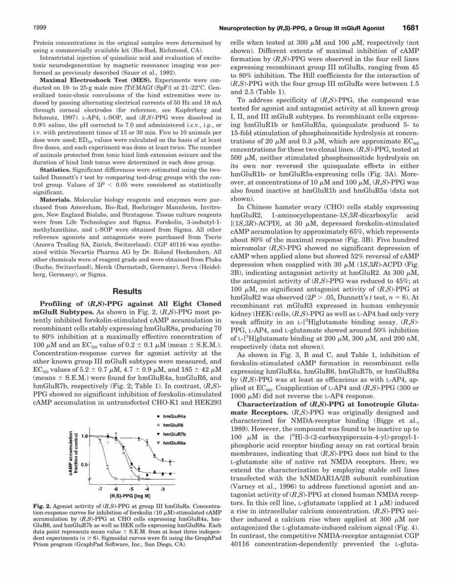
ResultsProfiling of (R,S)-PPG against All Eight Cloned
mGluR Subtypes. As shown in Fig. 2, (R,S)-PPG most po-tently inhibited forskolin-stimulated cAMP accumulation inrecombinant cells stably expressing hmGluR8a, producing 70to 80% inhibition at a maximally effective concentration of100 mM and an EC50 value of 0.2 6 0.1 mM (mean 6 S.E.M.).Concentration-response curves for agonist activity at theother known group III mGluR subtypes were measured, andEC50 values of 5.2 6 0.7 mM, 4.7 6 0.9 mM, and 185 6 42 mM(means 6 S.E.M.) were found for hmGluR4a, hmGluR6, andhmGluR7b, respectively (Fig. 2; Table 1). In contrast, (R,S)-PPG showed no significant inhibition of forskolin-stimulatedcAMP accumulation in untransfected CHO-K1 and HEK293
cells when tested at 300 mM and 100 mM, respectively (notshown). Different extents of maximal inhibition of cAMPformation by (R,S)-PPG were observed in the four cell linesexpressing recombinant group III mGluRs, ranging from 45to 80% inhibition. The Hill coefficients for the interaction of(R,S)-PPG with the four group III mGluRs were between 1.5and 2.5 (Table 1).
To address specificity of (R,S)-PPG, the compound wastested for agonist and antagonist activity at all known groupI, II, and III mGluR subtypes. In recombinant cells express-ing hmGluR1b or hmGluR5a, quisqualate produced 5- to15-fold stimulation of phosphoinositide hydrolysis at concen-trations of 20 mM and 0.3 mM, which are approximate EC80
concentrations for these two clonal lines. (R,S)-PPG, tested at500 mM, neither stimulated phosphoinositide hydrolysis onits own nor reversed the quisqualate effects in eitherhmGluR1b- or hmGluR5a-expressing cells (Fig. 3A). More-over, at concentrations of 10 mM and 100 mM, (R,S)-PPG wasalso found inactive at hmGluR1b and hmGluR5a (data notshown).
In Chinese hamster ovary (CHO) cells stably expressinghmGluR2, 1-aminocyclopentane-1S,3R-dicarboxylic acid[(1S,3R)-ACPD], at 30 mM, depressed forskolin-stimulatedcAMP accumulation by approximately 65%, which representsabout 80% of the maximal response (Fig. 3B). Five hundredmicromolar (R,S)-PPG showed no significant depression ofcAMP when applied alone but showed 52% reversal of cAMPdepression when coapplied with 30 mM (1S,3R)-ACPD (Fig.3B), indicating antagonist activity at hmGluR2. At 300 mM,the antagonist activity of (R,S)-PPG was reduced to 45%; at100 mM, no significant antagonist activity of (R,S)-PPG athmGluR2 was observed (2P . .05, Dunnett’s t test, n 5 8). Atrecombinant rat mGluR3 expressed in human embryonickidney (HEK) cells, (R,S)-PPG as well as L-AP4 had only veryweak affinity in an L-[3H]glutamate binding assay. (R,S)-PPG, L-AP4, and L-glutamate showed around 50% inhibitionof L-[3H]glutamate binding at 200 mM, 300 mM, and 200 nM,respectively (data not shown).
As shown in Fig. 3, B and C, and Table 1, inhibition offorskolin-stimulated cAMP formation in recombinant cellsexpressing hmGluR4a, hmGluR6, hmGluR7b, or hmGluR8aby (R,S)-PPG was at least as efficacious as with L-AP4, ap-plied at EC80. Coapplication of L-AP4 and (R,S)-PPG (300 or1000 mM) did not reverse the L-AP4 response.
Characterization of (R,S)-PPG at Ionotropic Gluta-mate Receptors. (R,S)-PPG was originally designed andcharacterized for NMDA-receptor binding (Bigge et al.,1989). However, the compound was found to be inactive up to100 mM in the [3H]-3-(2-carboxypiperazin-4-yl)-propyl-1-phosphoric acid receptor binding assay on rat cortical brainmembranes, indicating that (R,S)-PPG does not bind to theL-glutamate site of native rat NMDA receptors. Here, weextend the characterization by employing stable cell linestransfected with the hNMDAR1A/2B subunit combination(Varney et al., 1996) to address functional agonist and an-tagonist activity of (R,S)-PPG at cloned human NMDA recep-tors. In this cell line, L-glutamate (applied at 1 mM) induceda rise in intracellular calcium concentration. (R,S)-PPG nei-ther induced a calcium rise when applied at 300 mM norantagonized the L-glutamate-induced calcium signal (Fig. 4).In contrast, the competitive NMDA-receptor antagonist CGP40116 concentration-dependently prevented the L-gluta-
Fig. 2. Agonist activity of (R,S)-PPG at group III hmGluRs. Concentra-tion-response curves for inhibition of forskolin (10 mM)-stimulated cAMPaccumulation by (R,S)-PPG at CHO cells expressing hmGluR4a, hm-GluR6, and hmGluR7b as well as HEK cells expressing hmGluR8a. Eachdata point represents mean value 6 S.E.M. from at least three indepen-dent experiments (n $ 6). Sigmoidal curves were fit using the GraphPadPrism program (GraphPad Software, Inc., San Diego, CA).
1999 Neuroprotection by (R,S)-PPG, a Group III mGluR Agonist 1681

mate-evoked rise in intracellular calcium (Fig. 4). Similarlack of activity of 300 mM (R,S)-PPG was observed in calciumassays for cloned hNMDAR1A/2A (data not shown, Table 1).
Moreover, stable cell lines expressing human AMPA-(GluR3) or kainate(GluR6) receptors were utilized in thesame assay (Daggett et al., 1996; Varney et al., 1998). Appli-cation of L-glutamate resulted in robust increases of cytoplas-mic calcium, whereas (R,S)-PPG (300 mM) did neither evokecalcium signals on its own nor antagonize L-glutamate ineither of the two cell lines (data not shown; Table 1).
Ca21/Cl2-Dependent L-[3H]Glutamate Binding to RatBrain Membranes. L-AP4, L-SOP, and (R,S)-PPG displacedL-[3H]glutamate binding at rat hippocampal membranes,measured in the presence of CaCl2, with IC50 values of 0.5mM, 3.1 mM, and 1.9 mM, respectively (Table 2). Thus, L-AP4was the most potent among the three compounds, having a4-fold higher affinity than (R,S)-PPG, which was about equi-potent with L-SOP.
Neuroprotective Activity of (R,S)-PPG in NeuronalCulture. In primary cultures of cortical neurons coculturedwith glia, (R,S)-PPG was highly neuroprotective when ap-plied during the NMDA-induced excitotoxic pulse (10 min).The action of (R,S)-PPG was concentration-dependent, withan apparent EC50 value of 12 mM (Fig. 5A). Maximally effec-tive concentrations of (R,S)-PPG rescued slightly more than50% of the neuronal population from excitotoxic degeneration(Fig. 5). (R,S)-a-Methylserine-O-phosphate (MSOP), a pref-erential antagonist of group III mGluRs (Thomas et al.,1996), prevented the neuroprotective activity of (R,S)-PPG.In contrast, the selective group II mGluR antagonist (2S)-a-ethylglutamic acid (EGlu) (Jane et al., 1996; Thomas et al.,1996) did not affect the action of (R,S)-PPG (Fig. 5B). Neither(R,S)-PPG nor MSOP or EGlu had any effect on neuronalviability when applied to the cultures in the absence ofNMDA (not shown).
Protection by (R,S)-PPG against NMDA- and Quino-linic Acid-Induced Striatal Lesions. Intrastriatal infu-sion of various ionotropic glutamate receptor agonists resultsin neurochemical and neuropathological changes resemblingHuntington’s disease (DiFiglia, 1990). First, we have used
infusion of 100 nmol of NMDA which induces an extendedarea of necrosis characterized by neuronal loss, reactive gli-osis, edema, and neuronal pyknosis (Fig. 6A). Neuronal dam-age was visible across the extension of the caudate nucleus,up to 3 mm posterior to the injection site. Coinfusion of 250nmol of (R,S)-PPG with NMDA resulted in efficacious neu-roprotection against excitotoxic neuronal damage; particu-larly in the more lateral parts of the caudate nucleus, dras-tically reduced neuronal loss and pyknosis as well as lessedema formation were seen (Fig. 6B). To quantitate the pro-tective effect of (R,S)-PPG, we have measured striatal GADactivity as a biochemical marker of viable GABAergic neu-rones (Fig. 7), which has been widely used as post-mortemassessment of lesion size (e.g., Urwyler et al., 1996a). NMDAinfusion led to a 45 to 50% decrease in GAD activity ascompared with the respective contralateral side. No reduc-tion in GAD activity was observed in animals coinfused withNMDA plus (R,S)-PPG. The protective activity of (R,S)-PPGagainst NMDA toxicity was mimicked by L-AP4 (50 or 250nmol, Fig. 7).
Furthermore, we tested (R,S)-PPG for protection againstintrastriatal infusion of quinolinic acid (200 nmol), whichproduced larger lesions than NMDA. Lesion size was quan-titated by magnetic resonance imaging as described (Sauer etal., 1992). Compared with the control group receiving 200nmol of quinolinic acid (11 animals treated), coinfusion of(R,S)-PPG (250 nmol) showed significant protection (2P ,.005, Dunnett’s t test) with a reduction of the lesion size by58.4% 6 13% (mean 6 S.E.M., n 5 13 animals).
Protection by (R,S)-PPG against MES-Induced Con-vulsions in Mice. MES is commonly used as a basic in vivotest for anticonvulsive compounds (Kupferberg and Schmutz,1997 and references therein). In this test, with i.c.v. injec-tions of L-AP4 and L-SOP, a pretreatment time of 15 min, anddoses between 60 and 220 nmol, no anticonvulsive effectswere seen. When doses were increased to approximately 2000nmol, L-AP4 and L-SOP induced clonic/clonic-tonic seizures at5 to 10 min after drug administration in 40 to 60% of thetreated animals (Table 3). In contrast, (R,S)-PPG up to 2200nmol did not show any proconvulsant effect. Moreover, (R,S)-
TABLE 1Activity of (R,S)-PPG on metabotropic and ionotropic glutamate receptors expressed in recombinant mammalian cellsConcentration-response curve fitting and statistical analysis were done using the GraphPad Prism program (GraphPad Software, Inc.). EC50 values are given 6 S.E.M. (n $3); nH, Hill coefficient.
Assay Glutamate Receptor Clone EC50 (Agonist Activity) IC50 (Antagonist Activity)
mM
Group I mGluRs1 PI hydrolysis Human mGluR1b .500 .500
Human mGluR5a .500 .500Group II mGluRs
2 Forskolin-stimulated cAMP Human mGluR2 .300 $300L-[3H] Glutamate binding (displacement) Rat mGluR3 $200a
Group III mGluRs2 Forskolin-stimulated cAMP Human mGluR4a 5.2 6 0.7, nH 5 1.5 .300
Human mGluR6 4.7 6 0.9, nH 5 1.7 .300Human mGluR7b 185 6 42, nH 5 2.2 .1000Human mGluR8a 0.21 6 0.1, nH 5 2.5 .300
Ionotropic GluRs1 Cytoplasmic Ca21 Human NMDAR1A/2A .300 .300
Human NMDAR1A/2B .300 .300Human AMPAR(GluR3) .300 .300Human KainateR(GluR6) .300 .300
a These binding data do not discriminate between agonist and antagonist activity; all other data are from functional receptor assays.
1682 Gasparini et al. Vol. 290

PPG when applied at 173 nmol (i.c.v.) produced 100% protec-tion against MES with an ED50 value of 78 nmol (Table 3). Atdoses above 2000 nmol (i.c.v.), all three compounds werelethal in 20 to 60% of the animals. (R,S)-PPG given i.p. or i.v.was inactive against MES-induced convulsions up to 100mg/kg and 10 mg/kg, respectively.
DiscussionIn Vitro Pharmacology. Recent cDNA cloning and re-
combinant expression of the heterogeneous family of G pro-tein-coupled (metabotropic) glutamate receptors has createda large gap between the molecular knowledge of mGluRsubtypes and the understanding of their role in brain func-tion and dysfunction. Thus, the discovery of subtype-selectivecompounds and their testing in experimental models for ner-vous system physiology and pathology has become increas-ingly important.
In the present study, using 12 different cell lines stablyexpressing cloned ionotropic and metabotropic glutamate re-ceptor subtypes, we have characterized the pharmacologicalprofile of (R,S)-PPG. This compound behaved as a potentagonist at hmGluR8a (EC50 5 200 nM), hmGluR6 (EC50 54.7 mM), and hmGluR4a (EC50 5 5.2 mM), with no apprecia-ble activity (up to 200 mM) at hmGluR1, hmGluR2, ratmGluR3, hmGluR5, and hNMDAR1A/2B, hNMDAR1A/2A,AMPAR(hGluR3), and kainateR(hGluR6).
Fig. 3. Activity of (R,S)-PPG at group I, II, and III hmGluRs. A, inositolmonophosphate formation of CHO cells expressing hmGluR1b and Ltk2
cells expressing hmGluR5a. Exposure to buffer was taken as control andset to 1.0; for submaximal and maximal stimulation of hmGluR1b, 20 mMquisqualate and 1000 mM quisqualate were used, respectively; 0.3 mMquisqualate gave submaximal stimulation of hmGluR5a, and 10 mMquisqualate showed the maximal effect. To test for agonist activity atgroup I mGluRs, (R,S)-PPG was applied at 500 mM to CHO-hmGluR1band Ltk2-hmGluR5a cells. Antagonist activity of 500 mM (R,S)-PPG athmGluR1b and hmGluR5a was determined by coapplication of the sub-maximal concentrations of quisqualate (around EC80). Mean values 6S.E.M. from at least two independent experiments are shown (n $ 4). B,inhibition of forskolin-stimulated cAMP accumulation in CHO cells ex-pressing either hmGluR2 or hmGluR4a. Forskolin (10 mM) stimulatedcAMP formation about 40-fold (taken as control). All values are given asfraction of control. The effect of forskolin is inhibited by 30 mM (1S,3R)-ACPD in hmGluR2-expressing cells and by 1 mM L-AP4 in hmGluR4a-expressing cells. These submaximal agonist concentrations representapproximately EC80. To test for agonist activity, (R,S)-PPG was appliedto forskolin-stimulated CHO-hmGluR2 and CHO-hmGluR4a cells. An-tagonist activity of (R,S)-PPG at hmGluR2 and hmGluR4a was deter-mined by coapplication of submaximal concentrations of (1S,3R)-ACPDand L-AP4, respectively. Columns represent mean values 6 S.E.M. of atleast two independent experiments (n $ 5). Asterisks indicate statisti-cally significant antagonism or agonist activity of (R,S)-PPG (2P , .01;Dunnett’s t test). C, inhibition of forskolin (10 mM)-stimulated cAMPaccumulation in CHO cells expressing hmGluR7b and HEK cells express-ing hmGluR8a. Forskolin-stimulated cAMP formation was taken as con-trol. To obtain submaximal depression of the forskolin-stimulated cAMPaccumulation, 500 mM L-AP4 and 1 mM L-AP4 were used for hmGluR7band hmGluR8a, respectively (this represents about EC80). (R,S)-PPG wastested for agonist activity as well as antagonist activity of submaximalL-AP4. Asterisks indicate statistically significant agonist activity of (R,S)-PPG (2P , .01; Dunnett’s t test, n $ 4).
Fig. 4. Lack of activity at NMDA-type ionotropic glutamate receptors by(R,S)-PPG. Elevation of cytoplasmic calcium ion concentrations, [Ca21]i,in Ltk2 cells expressing cloned human NMDA receptors (subunit combi-nation 1A/2B). A, increase in the fluorescent response ratio F340/F380corresponds to an elevation in [Ca21]i. Three single calcium measure-ments are shown: application of 1 mM L-glutamate (L-Glu) alone, coap-plication of L-Glu (1 mM) and (R,S)-PPG (300 mM), and 300 mM (R,S)-PPGalone. The duration of the applications is indicated with a column. B, eachcolumn represents calcium signals (mean values 6 S.E.M.) of at least sixmeasurements of two independent experiments. The response of 1 mML-Glu was taken as control and set to 100%. Asterisks indicate statisti-cally significant antagonist activity (2P , .01; Dunnett’s t test).
TABLE 2Activity of group III mGluR agonists at a Ca21/CI2-dependentL-glutamate binding site of rat brainThe affinities of L-AP4, L-SOP, and (R,S)-PPG as displacers of L-[3H]glutamatebinding to rat hippocampal membranes were measured in the presence of calciumchloride as described (Urwyler et al., 1997b). The results shown are means 6 S.E.M.from three independent experiments, each performed in triplicate; nH, Hill coeffi-cient.
Compound IC50 nH
mM
L-AP4 0.5 6 0.1 1.28 6 0.13L-SOP 3.1 6 0.6 1.32 6 0.10(R,S)-PPG 1.9 6 0.2 1.02 6 0.01
1999 Neuroprotection by (R,S)-PPG, a Group III mGluR Agonist 1683

(R,S)-PPG therefore exhibited approximately 1000-fold se-lectivity for hmGluR8 and $40-fold selectivity for hmGluR4aand hmGluR6 versus group I/II mGluRs and all testediGluRs. (R,S)-PPG activated hmGluR7b only at high micro-molar concentrations (EC50 5 185 mM), which is only slightlymore potent than the compound’s weak antagonist activity athmGluR2 and its activity against glutamate binding at ratmGluR3 (Table 1). In addition to its potent activity at groupIII mGluRs, (R,S)-PPG, as well as L-AP4 and L-SOP, dis-played micromolar affinity for a Ca21/Cl2-dependentL-[3H]glutamate binding site in rat brain (Fagg et al., 1982;Urwyler et al., 1996b). The functional role of this binding sitewas proposed to be that of a glutamate transporter, butalternative explanations are also possible (see Urwyler et al.,1996b and references therein for detailed discussion). Theaffinity of (R,S)-PPG at this site was found to be 4-fold lowerthan the affinity of L-AP4. Whether this difference is re-flected at the level of endogenous glutamate accumulation,and thus could contribute to explain the discrepancy we havefound between L-AP4 and (R,S)-PPG in the MES model in
mice (see below), is currently unclear, especially because theexplanation cannot be extended to L-SOP.
In summary, (R,S)-PPG represents a novel group IIImGluR agonist, with in vitro pharmacological properties in-distinguishable from the current agonists L-SOP, L-AP4, andcyclopropyl-AP4 (Johansen et al., 1995; Okamoto et al., 1994;Conn and Pin, 1997; Flor et al., 1997;). Interestingly, how-ever, (R,S)-PPG differs structurally from all of these com-pounds but shares close similarity with classical competitiveNMDA receptor antagonists like CGP 40116, as the distancebetween the amino acid moiety and the phosphonate group iscomparable. Because this distance is substantially smaller inL-AP4 and L-SOP, the potency and selectivity of (R,S)-PPGfor group III mGluRs is quite surprising and suggests aparticular mode of binding of (R,S)-PPG, involving the phe-
Fig. 5. Protection of cultured cortical neurons against NMDA-inducedexcitotoxic degeneration by (R,S)-PPG. A, concentration-protection rela-tionship of (R,S)-PPG against NMDA (100 mM)-induced degeneration inmixed cortical cultures assessed by extracellular lactate dehydrogenaseactivity (measured as mOD/min). The value measured with 100 mMNMDA was taken as 100%. To determine EC50 and Hill coefficient (nH),a sigmoidal curve was fit using the GraphPad Prism program (GraphPadSoftware, Inc.). B, neuronal degeneration in mixed cortical cultures wasinduced by 100 mM NMDA (resulting in approximately 85% of maximalNMDA toxicity) and assessed by extracellular lactate dehydrogenaseactivity (measured as mOD/min, set to 100%). Statistically significantneuroprotection by (R,S)-PPG is indicated by asterisks (2P , .01; Dun-nett’s t test, n $ 4, at least two independent experiments). (R,S)-PPGneuroprotection can be antagonized by MSOP but not by EGlu.
Fig. 6. (R,S)-PPG protects against NMDA-induced striatal lesions invivo. Nissl staining of 20-mm cryostat sections from rat caudate nucleuslateral to the site of NMDA (100 nmol) injection (A); note the large areaof extensive necrotic damage at the left side of the micrograph (whitearrowheads) and widespread neuronal pyknosis; black arrowheads pointat two pyknotic neurones. When (R,S)-PPG (250 nmol) is coinjected withNMDA (B), necrotic damage and neuronal pyknosis is drastically re-duced; open arrowheads indicate two healthy neurons. Also, note thehigher neuronal density in B compared with A.
1684 Gasparini et al. Vol. 290

nyl spacer between the a-amino acid and phosphonic acidmoieties.
a-Methylphosphonophenyl glycine, a mixed antagonist ofgroup II and III mGluRs (Bedingfield et al., 1996), is struc-turally the most closely related compound and differs from(R,S)-PPG only by a methyl group in the a-position. Thus,replacement of that group with the hydrogen of (R,S)-PPGhas changed the antagonistic properties into selective ago-nist activity. Similar properties have been shown for linearL-glutamate analogs and cyclopropylglycine derivatives(Jane et al., 1996; Thomas et al., 1996), where the introduc-tion of a methyl group at the a-position changed agonists intoantagonists.
Neuroprotection and Anticonvulsive Actions Medi-ated by (R,S)-PPG. Neuroprotective effects of L-AP4 andL-SOP observed in several in vitro paradigms (see Introduc-tion) prompted us to examine (R,S)-PPG, as a structurallydifferent but pharmacologically indistinguishable compound,in the model of NMDA-induced degeneration of mouse corti-cal neurons cocultured with glia (Bruno et al., 1996). Here,we found (R,S)-PPG highly neuroprotective, and its actionwas antagonized by MSOP, which is a group III mGluRantagonist (Thomas et al., 1996), but not by the group IImGluR antagonist EGlu. These results therefore strengthenthe suggestion that activation of group III mGluRs is neuro-protective in vitro (e.g., Copani et al., 1995; Bruno et al.,1996; Faden et al., 1997).
To investigate neuroprotective effects of (R,S)-PPG also invivo, we analyzed striatal degeneration following local infu-sion of NMDA and quinolinic acid into the rat caudate nu-cleus. The use of such excitotoxic injury models, to produceneuronal depletion, reactive gliosis, and alterations of neu-rotransmitter levels, has been highly valuable for examiningpathological patterns reminiscent of Huntington’s disease(HD). Even if the primary cause of HD is unrelated, excito-toxic injury mediated by iGluR activation may play a role inprogressive neuronal depletion (DiFiglia, 1990).
We found (R,S)-PPG protective against NMDA- and quino-linic acid-induced striatal lesions; to our knowledge, thisprovides the first in vivo evidence that activation of group IIImGluRs is neuroprotective in animal models.
Inhibition of glutamate release by presynaptic mGluR4, -7and/or -8 (Shigemoto et al., 1997) may represent a commonmechanism of neuroprotection in vitro and in vivo. Accord-ingly, an enhanced release of endogenous glutamate has beenshown to facilitate the progression of NMDA toxicity in cor-tical cultures (Monyer et al., 1992). Additionally, in vivostriatal toxicity induced by kainate or NMDA receptor ago-nists such as quinolinic acid and NMDA critically involvesthe presence of cortical glutamatergic fibers afferent to thecaudate nucleus of striatum (Colwell et al., 1996 and refer-ences therein). Although little recurrent excitation exists inthe striatum, the endogenously released L-glutamate mayhave a permissive role on NMDA and quinolinic acid toxicity,perhaps by activating postsynaptic group I mGluRs or otherfacilitatory receptors.
Upon depletion of the caudate neuronal population duringthe progression of HD, the resulting “excess” of corticostriatalglutamatergic input may cause further neuronal loss (Di-Figlia, 1990), and inhibition of this input via group IIImGluRs may provide protection. In addition to the regulationof presynaptic glutamate release, an inhibition of NMDA
receptors by postsynaptic group III mGluRs via a proteinphosphorylation cascade (Martin et al., 1997) may also beinvolved in their neuroprotective effects. Thus, activation ofgroup III mGluRs could open several novel strategies tointerfere with the progressive course of neurodegenerativedisorders.
The potency at cloned mGluRs relative to the protectiveactivity of (R,S)-PPG in primary culture (EC50 5 12 mM)suggests that the neuroprotective action of (R,S)-PPG pref-erentially involves mGluR4, mGluR6, and/or mGluR8, whichare all expressed by cultured cortical neurons (Faden et al.,1997). For in vivo neuroprotection, however, mGluR6 is prob-ably irrelevant because of its restricted expression in theretinal bipolar cells layer. A more detailed examination of therelative contribution of individual group III mGluR subtypesto in vitro and in vivo neuroprotection awaits the discovery ofmore selective agonists and the utilization of group IIImGluR subtype-deficient mice.
Modulation of epileptic seizures by group III mGluRs hasbeen frequently reported (e.g., Ghauri et al., 1996; Abdul-Ghani et al., 1997; Tang et al., 1997). Thus, we were temptedto test (R,S)-PPG, in comparison with L-AP4 and L-SOP, inthe MES model in mice. MES is a basic screening test foranticonvulsive drugs, it is indicative of drug activity primar-ily against generalized tonic-clonic and, secondarily, also par-tial seizures, and it led to the discovery of several clinicallyapproved antiepileptics, e.g., carbamazepine, oxcarbazepine,and phenytoin (Kupferberg and Schmutz, 1997 and refer-ences therein). Surprisingly, our results for (R,S)-PPG ob-tained with this model revealed important differences ascompared with the prototypic group III mGluR agonists L-AP4 and L-SOP. In agreement with previous results (Ghauriet al., 1996; Tang et al., 1997), both L-AP4 and L-SOP wereproconvulsive at high doses (around 2000 nmol) and did notprotect against MES-induced seizures at any of the givendoses (60–2400 nmol). In contrast, (R,S)-PPG exhibited sub-stantial anticonvulsive activity with an ED50 value of 78nmol, full protection at 173 nmol, and did not exhibit anyproconvulsive effect up to 2200 nmol. These discrepanciescould possibly be explained by physicochemical propertiesand/or divergent in vivo metabolism of the structurally quitedifferent group III mGluR agonists L-AP4, L-SOP, and (R,S)-PPG. Although we cannot exclude that anticonvulsive prop-erties of (R,S)-PPG are mediated by a mechanism distinctfrom group III mGluRs, other reports also support group IIImGluRs as mediators of anticonvulsive and antiepileptogeniceffects. Abdul-Ghani et al. (1997) reported protective effectsof L-AP4 on development of electrical kindling and also infully kindled rats. Furthermore, Tang et al. (1997) reportedfor L-SOP an immediate, transient (,10 min) proconvulsiveeffect followed by a prolonged (.1 day) anticonvulsive effectagainst sound-induced seizures with an anticonvulsant ED50
value of 36 nmol. Moreover, recent experimental evidencefrom mGluR7-deficient mice that exhibit spontaneous epilep-tic seizures upon certain olfactory stimuli indicates that atleast one group III mGluR is critically involved in maintain-ing the delicate balance between neuronal inhibition andexcitation (H. van der Putten, personal communication).
In conclusion, (R,S)-PPG represents a novel pharmacolog-ical tool to analyze the role of group III mGluRs in nervoussystem physiology and pathology, and our in vivo data sup-port a neuroprotective and anticonvulsive role for group III
1999 Neuroprotection by (R,S)-PPG, a Group III mGluR Agonist 1685

mGluRs and encourage the search for systemically activegroup III mGluR agonists as promising drugs for the treat-ment of neurological disorders, such as Huntington’s diseaseand epilepsies.
Acknowledgments
We thank H. Allgeier for critically reading the manuscript andhelpful discussion, R. Heckendorn for providing CGP 40116, H. vander Putten for sharing unpublished data, and P. Schoeffter for initialcAMP measurements with the HEK-hmGluR8a cell lines.
ReferencesAbdul-Ghani AS, Attwell PJ, Singh-Kent N, Bradford HF, Croucher MJ and Jane DE
(1997) Anti-epileptogenic and anticonvulsant activity of L-2-amino-4-phospho-nobutyrate, a presynaptic glutamate receptor agonist. Brain Res 755:202–212
Bedingfield JS, Jane DE, Kemp MC, Toms NJ and Roberts PJ (1996) Novel potentselective phenylglycine antagonists of metabotropic glutamate receptors. EurJ Pharmacol 309:71–78.
Bigge CF, Drummond JT, Johnson G, Malone T, Probert AW Jr, Marcoux FW,Coughenour LL and Brahce LJ (1989) Exploration of phenyl-spaced 2-amino-(5–9)-phosphonoalkanoic acids as competitive N-methyl-D-aspartic acid antagonists.J Med Chem 32:1580–1590.
Bruno V, Copani A, Bonanno L, Knopfel T, Kuhn R, Roberts PJ and Nicoletti F (1996)Activation of group III metabotropic glutamate receptors is neuroprotective incortical cultures. Eur J Pharmacol 310:61–66.
Colwell CS, Altemus KL and Levine MS (1996) Metabotropic glutamate receptoractivation selectively limits excitotoxic damage in the intact neostriatum. BrainRes 726:223–226.
Conn PJ and Pin JP (1997) Pharmacology and functions of metabotropic glutamatereceptors. Annu Rev Pharmacol Toxicol 37:205–237.
Copani A, Bruno V, Battaglia G, Leanza G, Pellitteri R, Russo A, Stanzani S andNicoletti F (1995) Activation of metabotropic glutamate receptors protects culturedneurons against apoptosis induced by beta-amyloid peptide. Mol Pharmacol 47:890–897.
Daggett LP, Jachec C, Lin FF, Deal C, Varney MA, Hess SD, Velicelebi G and
Johnson EC (1996) Functional characterization of two isoforms of the humanGluR6 receptor and distribution of GluR6 RNA editing sites. Soc Neurosci Abst(236.11).
DiFiglia M (1990) Excitotoxic injury of the neostriatum: A model for Huntington’sdisease. Trends Neurol Sci 13:286–289.
Duvoisin RM, Zhang C and Ramonell K (1995) A novel metabotropic glutamatereceptor expressed in the retina and olfactory bulb. J Neurosci 15:3075–3083.
Faden AI, Ivanova SA, Yakovlev AG and Mukhin AG (1997) Neuroprotective effectsof group III mGluR in traumatic neuronal injury. J Neurotrauma 14:885–895.
Fagg GE, Foster AC, Mena EE and Cotman CW (1982) Chloride and calcium ionsreveal a pharmacologically distinct population of L-glutamate binding sites insynaptic membranes: Correspondence between biochemical and electrophysiolog-ical data. J Neurosci 2:958–965.
Flor PJ, Gomeza J, Tones MA, Kuhn R, Pin JP and Knopfel T (1996) The C-terminaldomain of the mGluR1 metabotropic glutamate receptor affects sensitivity toagonists. J Neurochem 67:58–63.
Flor PJ, Van Der Putten H, Ruegg D, Lukic S, Leonhardt T, Bence M, Sansig G,Knopfel T and Kuhn R (1997) A novel splice variant of a metabotropic glutamatereceptor, human mGluR7b. Neuropharmacology 36:153–159.
Ghauri M, Chapman AG and Meldrum BS (1996) Convulsant and anticonvulsantactions of agonists and antagonists of group III mGluRs. Neuroreport 7:1469–1474.
Graham ME and Burgoyne RD (1994) Activation of metabotropic glutamate recep-tors by L-AP4 stimulates survival of rat cerebellar granule cells in culture. EurJ Pharmacol 288:115–123.
Hollmann M and Heinemann S (1994) Cloned glutamate receptors. Annu Rev Neu-rosci 17:31–108.
Jane DE, Thomas NK, Tse HW and Watkins JC (1996) Potent antagonists at theL-AP4- and (1S,3S)-ACPD-sensitive presynaptic metabotropic glutamate receptorsin the neonatal rat spinal cord. Neuropharmacology 35:1029–1035.
Johansen PA, Chase LA, Sinor AD, Koerner JF, Johnson RL and Robinson MB(1995) Type 4a metabotropic glutamate receptor: Identification of new potentagonists and differentiation from the L-(1)-2-amino-4-phosphonobutanoic acid-sensitive receptor in the lateral perforant pathway in rats. Mol Pharmacol 48:140–149.
Knopfel T, Sakaki J, Flor PJ, Baumann P, Sacaan AI, Velicelebi G, Kuhn R andAllgeier H (1995) Profiling of trans-azetidine-2,4-dicarboxylic acid at the humanmetabotropic glutamate receptors mGlu1b, -2, -4a and -5a. Eur J Pharmacol288:389–392.
Kupferberg HJ and Schmutz M (1997) Screening of new compounds and the role ofthe pharmaceutical industry, in Epilepsy: A Comprehensive Textbook (Engel J andPedley TA eds) vol 2, pp 1417–1434, Lippincott-Raven Publishers, Philadelphia.
Laurie DJ, Danzeisen M, Boddeke HW and Sommer B (1995) Ligand binding profileof the rat metabotropic glutamate receptor mGluR3 expressed in a transfected cellline. Naunyn Schmiedeberg’s Arch Pharmacol 351:565–568.
Laurie DJ, Schoeffter P, Wiederhold KH and Sommer B (1997) Cloning, distributionand functional expression of the human mGlu6 metabotropic glutamate receptor.Neuropharmacology 36:145–152.
Lin FF, Varney M, Sacaan AI, Jachec C, Daggett LP, Rao S, Flor P, Kuhn R, KernerJA, Standaert D, Young AB and Velicelebi G (1997) Cloning and stable expressionof the mGluR1b subtype of human metabotropic receptors and pharmacologicalcomparison with the mGluR5a subtype. Neuropharmacology 36:917–931.
Lipton SA and Rosenberg PA (1994) Excitatory amino acids as a final commonpathway for neurologic disorders. N Engl J Med 330:613–622.
Martin G, Nie Z and Siggins GR (1997) Metabotropic glutamate receptors regulateN-methyl-D-aspartate-mediated synaptic transmission in nucleus accumbens.J Neurophysiol 78:3028–3038.
Monn JA, Valli MJ, Massey SM, Wright RA, Salhoff CR, Johnson BG, Howe T, AltCA, Rhodes GA, Robey RL, Griffey KR, Tizzano JP, Kallman MJ, Helton DR andSchoepp DD (1997) Design, synthesis, and pharmacological characterization of(1)-2-aminobicyclo[3.1.0]hexane-2,6-dicarboxylic acid (LY354740): A potent, selec-tive, and orally active group 2 metabotropic glutamate receptor agonist possessinganticonvulsant and anxiolytic properties. J Med Chem 40:528–537.
Monyer H, Giffard RG, Hartley DM, Dugan LL, Goldberg MP and Choi DW (1992)Oxygen or glucose deprivation-induced neuronal injury in cortical cell cultures isreduced by tetanus toxin. Neuron 8:967–973.
Okamoto N, Hori S, Akazawa C, Hayashi Y, Shigemoto R, Mizuno N and NakanishiS (1994) Molecular characterization of a new metabotropic glutamate receptormGluR7 coupled to inhibitory cyclic AMP signal transduction. J Biol Chem 269:1231–1236.
Sauer D, Allegrini PR, Thedinga KH, Massieu L and Fagg GE (1992) Evaluation ofquinolinic acid-induced excitotoxic neurodegeneration in rat striatum by quanti-tative magnetic resonance imaging in vivo. J Neurosci Methods 42:69–74.
Shigemoto R, Kinoshita A, Wada E, Nomura S, Ohishi H, Takada M, Flor PJ, NekiA, Abe T, Nakanishi S and Mizuno N (1997) Differential presynaptic localizationof metabotropic glutamate receptor subtypes in the rat hippocampus. J Neurosci17:7503–7522.
Tanabe Y, Masu M, Ishii T, Shigemoto R and Nakanishi S (1992) A family ofmetabotropic glutamate receptors. Neuron 8:169–179.
Tang E, Yip PK, Chapman AG, Jane DE and Meldrum BS (1997) Prolonged anti-convulsant action of glutamate metabotropic receptor agonists in inferior collicu-lus of genetically epilepsy-prone rats. Eur J Pharmacol 327:109–115.
Thomas NK, Jane DE, Tse HW and Watkins JC (1996) a-Methyl derivatives ofserine-O-phosphate as novel, selective competitive metabotropic glutamate recep-tor antagonists. Neuropharmacology 35:637–642.
Fig. 7. Quantitation of in vivo neuroprotection by (R,S)-PPG. Degenera-tion of GABAergic neurones in rat corpus striatum was induced byNMDA (100 nmol) injection and assessed by measuring the GAD activity.GAD activity of rat corpus striatum contralateral to the site of NMDAinjection was taken as control and set to 100%. Each column representsmean values 6 S.E.M. Statistically significant neuroprotection by (R,S)-PPG and L-AP4 is indicated by asterisks (2P , .01, Dunnett’s t test; thenumber of tested animals in each group is indicated within the columns;for each group, at least two independent experiments were performed).
TABLE 3Activity of L-AP4, L-SOP, and (R,S)-PPG in the maximal electroshocktest (mouse)n.d., ED50 values were not determined. The results shown are derived from at leasttwo independent experiments, each performed with at least five animals per group.
Compound PretreatmentTime
% Protection (Dose), Modeof Application
ED50, Mode ofApplication
min
L-AP4 15 0% (223 nmol), i.c.v.a n.d.L-SOP 15 0% (218 nmol), i.c.v.a n.d.(R,S)-PPG 15 100% (173 nmol), i.c.v.b 78 nmol, i.c.v.b
(R,S)-PPG 30 0% (100 mg/kg), i.p. n.d.(R,S)-PPG 30 0% (10 mg/kg), i.v. n.d.
a At 10-fold higher doses (2200–2400 nmol), proconvulsive effects were observedin 40 to 60% of the compound-treated animals (n $ 10).
b No proconvulsive effects were observed at any of the given doses (10–2200nmol).
1686 Gasparini et al. Vol. 290
Trombley PQ and Westbrook GL (1992) L-AP4 inhibits calcium currents and synaptictransmission via a G-protein-coupled glutamate receptor. J Neurosci 12:2043–2050.
Urwyler S, Campbell E, Fricker G, Jenner P, Lemaire M, McAllister KH, Neijt HC,Park CK, Perkins M, Rudin M, Sauter A, Smith L, Wiederhold KH and Muller W(1996a) Biphenyl-derivatives of 2-amino-7-phosphono-heptanoic acid, a novel classof potent competitive N-methyl-D-aspartate receptor antagonists–II: Pharmaco-logical characterization in vivo. Neuropharmacology 35:655–669.
Urwyler S, Laurie D, Lowe DA, Meier CL and Muller W (1996b) Biphenyl-derivatives of 2-amino-7-phosphonoheptanoic acid, a novel class of potent compet-itive N-methyl-D-aspartate receptor antagonist–I: Pharmacological characteriza-tion in vitro. Neuropharmacology 35:643–654.
Varney MA, Jachec C, Deal C, Hess SD, Daggett LP, Skvoretz R, Urcan M, MorrisonJH, Moran T, Johnson EC and Velicelebi G (1996) Stable expression and charac-terization of recombinant human heteromeric N-methyl-D-aspartate receptor sub-types NMDAR1A/2A and NMDAR1A/2B in mammalian cells. J Pharmacol ExpTher 279:367–378.
Varney MA, Jachec C, Rao S, Lin FF, Daggett L, Hess S, Velicelebi G and Johnson EC(1998) Pharmacological characterization of the recombinant human AMPA receptorsubtype 3 stably expressed in HEK293 cells. J Pharmacol Exp Ther 285:358–370.
Watkins JC, Krogsgaard Larsen P and Honore T (1990) Structure-activity relation-ships in the development of excitatory amino acid receptor agonists and competi-tive antagonists. Trends Pharmacol Sci 11:25–33.
Wu S, Wright RA, Rockey PK, Burgett SG, Arnold JS, Rosteck PR Jr, Johnson BG,Schoepp DD and Belagaje RM (1998) Group III human metabotropic glutamatereceptors 4, 7 and 8: Molecular cloning, functional expression, and comparison ofpharmacological properties in RGT cells. Mol Brain Res 53:88–97.
Send reprint requests to: Dr. Peter J. Flor, K-125.6.08, Nervous SystemResearch, Novartis Pharma AG, CH-4002 Basel, Switzerland. E-mail:peter–[email protected]
1999 Neuroprotection by (R,S)-PPG, a Group III mGluR Agonist 1687


