Embed Size (px)
Citation preview
INTRODUCTION
The larval abdominal body wall musculature of Drosophilaconsists of a regular, repetitive array of 30 different musclesper hemisegment. Each muscle has a unique morphology,orientation and position (Bate, 1990; Bate, 1993), and consistsof one single polynucleate myotube, while vertebrate musclesconsist of bundles of several myotubes. Common to both, themyotubes are formed by fusion of mesodermal cells (Baylieset al., 1998). Therefore, Drosophila is a well suited modelorganism in which to study the genetics of myoblast fusion.
Specification of the muscle pattern starts during earlyembryonic development. After formation of the mesodermalcell layer, the mesoderm becomes subdivided into different celltypes based on levels of twist expression: low levels markfuture visceral and heart mesoderm, high levels prospectivesomatic mesoderm (Baylies and Bate, 1996).
In the prospective somatic mesoderm, four cell groups arespecified dorsally, dorsolaterally, ventrolaterally and ventrallyby the expression of lethal of scute. From each of these groups,one muscle progenitor cell is singled out by lateral inhibition(Carmena et al., 1995). These cells divide asymmetrically andprovide the embryo with a subpopulation of myoblasts, themuscle founder cells. The expression of genes such as S59andkrüppel, which specifically affect subsets of muscles, is already
switched on in corresponding founder cell subsets. Therefore,the unique character of each future muscle is already‘programmed’ into individual founder cells (Frasch, 1999).
The syncytial myotubes form during stages 11-14, when thefounder cells fuse with myoblasts of a second class, the fusion-competent myoblasts. These cells do not contribute to theidentity of an individual muscles, but rather provide cellmaterial for the outgrowing muscle (Bate, 1990; Rushton et al.,1995).
Based on mutational analysis, several steps in the myoblastfusion process can be distinguished (Paululat et al., 1999):attraction and adhesion of fusion-competent myoblasts tofounder cells (blocked in mbc, snsand Df(1)w67k30 mutants(Rushton et al., 1995; Bour et al., 2000; Ruiz-Gomez et al.,2000); alignment of fusion-competent myoblasts and foundercells with paired vesicles on either side of the adjoiningmembranes (the prefusion complex); the formation of electron-dense plaques; and, finally, vesiculation and membranebreakdown.
For attraction, binding and the initiation of fusion, fusion-competent myoblasts and founder cells need an asymmetricequipment of extracellular membrane-bound components thatallows for attraction and discrimination; fusion-competentmyoblasts and founder cells fuse only with cells from the othergroup, rather than with themselves (Baylies et al., 1998).
4229Development 128, 4229-4239 (2001)Printed in Great Britain © The Company of Biologists Limited 2001DEV7904
The polynucleate myotubes of vertebrates andinvertebrates form by fusion of myoblasts. We report theinvolvement of the Drosophila melanogasterRoughest (Rst)protein as a new membrane-spanning component in thisprocess. Rst is strongly expressed in mesodermal tissuesduring embryogenesis, but rst null mutants display onlysubtle embryonic phenotypes. Evidence is presented thatthis is due to functional redundancy between Rst andits paralogue Kirre. Both are highly related single-pass transmembrane proteins with five extracellularimmunoglobulin domains and three conserved motifs in the
intracellular domain. The expression patterns of kirre andrst overlap during embryonic development in musclefounder cells. Simultaneous deletion of both genes causesan almost complete failure of fusion between musclefounder cells and fusion-competent myoblasts. This defectcan be rescued by one copy of either gene. Moreover, Rst,like Kirre is a myoblast attractant.
Key words:Drosophila, kirre, duf, rst, irreC, Fusion-competentmyoblast, Muscle founder cell, Myoblast fusion
SUMMARY
rst and its paralogue kirre act redundantly during embryonic muscle
development in Drosophila
Martin Strünkelnberg 1,*, Bernhard Bonengel 1,*, Livia M. Moda 2, Alexander Hertenstein 1, H. Gert de Couet 1,3,Ricardo G. P. Ramos 2 and Karl-Friedrich Fischbach 1,‡
1Institut für Biologie III, Schänzlestr.1, Albert-Ludwigs-Universität, D-79104 Freiburg im Breisgau, Germany2Departmento de Biologia Celular, Molecular e Bioagentes Patogênicos, Faculdade de Medicina de Ribeirão Preto, Universidadede São Paulo, Av. Bandeirantes 3900, 14.049-900 Ribeirão Preto-SP, Brazil3Department of Zoology, University of Hawaii at Manoa, 2538 McCarthy Mall, Honolulu, HI 96822, USA*These two authors contributed equally to this work‡Author for correspondence (e-mail: [email protected])
Accepted 25 July 2001

4230
Recently, two genes encoding members of the immunoglobulinsuperfamily, snsand kirre (kirre was also named dumbfounded;Ruiz-Gomez et al., 2000), have been reported to show theexpected features. Both encode transmembrane cell adhesionmolecules involved in myoblast attraction and/or fusion. WhileSns is expressed only on fusion-competent myoblasts, but noton founder cells (Bour et al., 2000), kirre is expressedselectively in the latter, but not in fusion-competent myoblasts.We present new data concerning the role of kirre and theinvolvement of its paralogue rst (Ramos et al., 1993; Reiter etal., 1996) in myoblast fusion.
MATERIALS AND METHODS
Fly stocks and genetic methodsAll stocks used in this work have been previously described.Maintenance and manipulation of Drosophilastocks was performedat 25°C on standard-cornmeal-molasses-agar food. Staging ofembryos was performed according to Campos-Ortega and Hartenstein(Campos-Ortega and Hartenstein, 1997).
For misexpression and rescue experiments, we used Df(1)w67k30,Nfa-g (courtesy of S. Artavanis-Tsakonas). The cross for the rescuewas Df(1)w67k30, Nfa-g/FM7;+/+;UAS-rst/+ × FM7/Y;twi-Gal4/+;twi-Gal4/+. Lack of epidermal Rst expression was used as a marker forthe Df(1)w67k30 chromosome, and remnants of ectopic Rst in themidgut region served as a marker for UAS-rst driven bytwi-Gal4.
Cloning of the kirre cDNADuring isolation of the D. virilis orthologue of rst (M. S.,unpublished) we serendipitously isolated a related, paralogoussequence. We subsequently identified and cloned the correspondingD. melanogastersequence, which we named kirre, using RT-PCR based on data of the EDGP (Cosmid 163A10– AccessionNumber, AL035436; Benos et al., 2000). The complete kirre cDNAsequence is available in GenBank under the Accession NumberAF196553.
RT-PCR Using the primers 5′-AGCACACCGCTTGAATCAGA-3′ and 5′-ACAGATGCAGCACAGCACTTA-3′ specific for sequencesupstream from the two possible adenylation sites of the annotatedkirre open reading frame (Benos et al., 2000), we performed reversetranscription (Superscript II, Life Industries) of 5 µg of total D.melanogasterpupal RNA followed by a RNase H (Life Industries)digestion as described by the manufacturer. Using Pfu turbo DNA-polymerase (Stratagene), 10 µl of the resulting cDNA was used in a100 µl PCR with 60 pmol of primers that flank the complete kirreORF (5′-TTAAACATGAGTGGCCAGAGG-3′ and 5′-TTGCCGC-TGAAAATGAAGCG-3′). PCR conditions were 94°C for 3 minutes,35 cycles of 94°C for 1 minute, 55°C for 1 minute and 72°C for 12minutes, and finally 15 minutes at 72°C. PCR was performed in aRobocycler (Stratagene).
The untranslated regions (UTRs) were cloned using the RapidAmplification of cDNA ends (RACE) protocol as described elsewhere(Dieffenbach and Dveksler, 1995). For the 3′UTR, the adapter primerCCAGTGAGCAGAGTGACGAT15 was used in RT of 5 µg of totalpupal RNA as above. Primers for PCR were 5′-CAGAGTCCG-TCCGGTCAGTT-3′ and 5′-CCAGTGAGCAGAGTGACG-3′, and ina nested PCR 5′-GACCTCTGGCCACTCATGTT-3′ and 5′-GAGG-ACTCGAGCTCAAGC-3′.
For the 5′-UTR, the primer 5′-GGCGGAACGAGAACGGTTAG-3′ was used in RT of 0.5 µg commercial embryonal polyA-RNA(Clontech) as above. Nested PCRs were performed using 5′-GC-GGATAAATGTCCAATGAG-3′ and 5′-TGCCGACCATCGAGTAG-CGT-3′ with adapter primers as above. Amplification products were
subcloned into the pCRII-TOPO vector (Invitrogen) and sequenceduntil all ambiguities were resolved.
Single fly PCROne to three flies of the relevant genotype were frozen in liquidnitrogen and incubated overnight at −80°C. After addition of 50 µl 1×PCR buffer per fly, the mixture was heated to 94°C for 3 minutes andtissue was mechanically disrupted. The resulting solution (5 µl) wasused in a standard 50 µl PCR reaction. Primers specific for kirre were5′-CTGATCCTGACGCTGCTCCT-3′ and 5′-GGCGGAACGAGAA-CGGTTAG-3′. Primers spanning the presumed transcriptional startsite of rst were 5′-CACTCTGACTAATTCACAATG-3′ and 5′-GAGTTGAGATCAAAGAGCCCAG-3′. PCR conditions were: 94°Cfor 3 minutes, 5 cycles of 94°C for 1 minute, 58°C for 2 minutes and72°C for 40 seconds, and then 30 cycles of 94°C for 30 seconds, 56°Cfor 40 seconds and 72°C for 40 seconds, followed by 72°C for 10minutes.
In-situ hybridizationEmbryo dechorionation, devitellination and fixation was carried outas previously described (Ashburner, 1989). In situ hybridization wasperformed as described previously (Oxtoby and Jowett, 1993) and, forprobe [K], according to Tautz and Pfeifle (Tautz and Pfeifle, 1989).Probes were selected on the basis of least nucleotide sequencesimilarity to exclude potential cross-hybridization. For kirre twodifferent probes were used with identical results: probe [K] wasisolated using the primers 5′-CAAGAGCGAACAGAGCAAGA-3′and 5′-CATCTGAACATCGGGCATCG-3′. Probe [K intra] wasisolated using the primers 5′-GTAATACCGGAGGCATCACG-3′ and5′-TTAAACATGAGTGGCCAGAGG-3′. As for rst, we used a probeisolated using the primers 5′CTGTAAGAAGCGCACCAAGC3′ and5′-GCTAAGTGCCTAACCTAAGC-3′.
Immunocytochemistry and microscopyImmunocytochemistry was performed as described (Schneider et al.,1995). Antibodies used were against myosin heavy chain (1:1000-1:10,000; Kiehart and Feghali, 1986), Rst (1:10, mAB24A5.1;Schneider et al., 1995), β-3-tubulin (1:1000, provided by R.Renkawitz-Pohl), β-galactosidase (1:1000, abcam, Cambridge, UK)and GFP (1:250, abcam). Laser scanning microscopy was carried outwith Leica TCS-NT and Leica TCS4D microscopes. Data wereprocessed using AMIRA-2.2 (Indeed, Berlin, Germany) andPhotoshop (Adobe).
Heat shock treatmentHomozygous pCa18Z∆3.1 flies (Moda et al., 2000) were placed ongrape juice plates for 1 hour to lay eggs. Eggs were allowed to developat 25°C for 7 hours and then incubated in a waterbath at 34.5°C for4 hours. This means that individual embryos were heat-shockedstarting at late stage 11 to late stage 12 until late stage 14 to mid stage15. Controls were treated at 25°C instead of 34.5°C.
RESULTS
Overexpression of rst interferes with muscledevelopmentAlthough the embryonic rst expression pattern is highlydynamic, none of the known loss-of-function mutations islethal (Ramos et al., 1993). We used pCa18Z∆3.1 flies (Modaet al., 2000) to overexpress a secretable extracellular versionof the protein by heat shock induction. Heat shocks appliedduring stages 12 to 15 induced embryonic lethality. Analysisof these embryos revealed muscle defects (Fig. 1A-D).Secretable Rst interfered with myoblast fusion. In heat shockedpCa18Z∆3.1 embryos, the muscles were thinner and more
M. Strünkelnberg and others

4231Rst in muscle development
unfused myoblasts could be observed than inheatshocked yw control embryos.
Additionally, we used the UAS/Gal4 system (Brandand Perrimon, 1993) to express a full-length rst-construct under transcriptional control of the da-Gal4driver (Wodarz et al., 1995). The genotype da-Gal4/+;UAS-rst/+, caused embryonic lethality andinduced defects in myoblast fusion: muscles wereconspicuously thin and unfused myoblasts were presentin large numbers (Fig. 1E, asterisk). Moreover, somemuscles seemed to be unable to find their properattachment sites within the epidermis (Fig. 1E, arrow).Mesodermal overexpression of rst mediated by twist-Gal4 (Baylies and Bate, 1996), 24B-Gal4 (Brand andPerrimon, 1993) or twist-Gal4; 24B-Gal4 driversneither caused lethality nor scorable musclephenotypes. Ectodermal expression using 69B-Gal4(Castelli-Gair et al., 1994) did not cause obviousphenotypes, except for some unfused myoblastsattached to the epidermis still present in stage 16 (datanot shown).
Removal or truncation of Rst causes mildmuscle defects: rst is not essential for muscledevelopmentAs overexpression of Rst causes dramatic muscledefects and lethality, we analysed rst mutants in detail.Loss of Rst protein in rstirreC1 (Schneider et al., 1995)was not lethal. However, embryos homozygous forrstirreC1 did show mild muscle defects. Individualmuscles were missing in some, but not in all segments(Fig. 1F, arrowheads). Embryos homozygous for theallele rst6, carrying a truncated version of theintracellular domain of Rst (Ramos et al., 1993),showed thin muscles and sometimes lacked thementirely (Fig. 1G, arrowhead) in some segments.Furthermore, rst6 embryos exhibited muscles in ectopicpositions (Fig. 1G, arrow).
kirre , a locus highly related to rstThe kirre locus maps cytogenetically to region 3C6 and lies 3kb distal to Notch (Fig. 2A). The rst and kirre loci are separatedby 127 kb and are transcribed from opposite strands with their5′ flanking regions towards each other (see Fig. 5A). The kirrecDNA consists of 3295 residues and contains a single longopen reading frame encoding a protein of 959 amino acids.Algorithms by Sonnhammer et al. (Sonnhammer et al., 1998)and Nielsen et al. (Nielsen et al., 1997) identify a signal peptidesequence (amino acids 7-31) and one putative transmembraneregion (amino acids 575-597).
The conceptual Kirre sequence shows an overall similarityof 45% to Rst (BLAST algorithm; Altschul et al., 1997). LikeRst, the predicted extracellular portion of the Kirre proteindisplays an array of five immunoglobulin (Ig) domains (Fig.2B; Walsh and Doherty, 1997). Stretches of high conservationwith Rst reside primarily in the region of the five Ig domains.Within these domains, the degree of conservation successivelydecreases from the N terminus to the transmembrane domain(Figs 2B,C; boundaries of Ig domains as in Ramos et al.(Ramos et al., 1993)). Both proteins contain stretches of aminoacids with short side chains at differing positions (Fig. 2B,
arrows): Rst contains a stretch of glycines between the secondand third immunoglobulin-domain and Kirre harbours an arrayof 18 serines interrupted by a single glycine residue at the Nterminus.
The intracellular domain of Kirre is considerably longer thanthat of Rst and displays only low overall homology with theone of Rst. However, three highly conserved motifs weredetected (Fig. 2C): One is located close to the transmembranedomain consisting of the sequence PADVI. The second motif,R[Y/F]SAIYGNPYLR(S)[S/T]NSSLLPP, corresponds to theconsensus sequence of autophosphorylation domains ofreceptor tyrosine kinases (Yarden and Ullrich, 1988). The thirdmotif, T[A/H]V, resides at the C terminus of both sequencesand corresponds to the consensus sequence of the PDZ-bindingmotif ([T/S]XV; Garner et al., 2000). In addition to the sitecontained in the putative autophosphorylation domain, oneputative tyrosine and one putative serine phosphorylation siteare conserved between Rst and Kirre (NetPhos 2.0 algorithm)(Blom et al., 1999). A conspicuous difference between theKirre and Rst proteins is the lack of the opa-like repeat of Rstin Kirre (Ramos et al., 1993).
Fig. 1. Rst overexpression and mutant phenotypes in the stage 16 abdominalmuscle pattern. Three dimensional reconstructions of confocal stacks ofantibody staining against β-3-tubulin. Anterior is to the left and dorsal isupwards. (A) Heatshocked ywembryo. Arrowheads indicate ventral acute (va)and ventral oblique (vo) muscles. (B) Heatshocked pCa18Z∆3.1embryo.Muscles are thin and some are missing (e.g. ventral acute muscles).(C) Heatshocked ywembryo. Single confocal plane interior to the muscles.(D) Heatshocked pCa18Z∆3.1 1embryo. Single confocal plane as in C,showing a large amount of unfused fusion-competent myoblasts. (E)da-Gal4/+;UAS-rst/+ embryo showing unfused fusion-competent myoblasts(asterisk), thin muscles and some muscles with ectopic projections (arrow).Several muscles are missing. lt, lateral transversal muscle. (F)rstirreC1 mutantembryo showing missing and thin muscles (arrowheads). (G)rst6 mutantembryo with misled (arrow) and thin muscles (arrowhead). Asterisks markunfused fusion-competent myoblasts. Scale bars: 20 µm.

4232
Similarity searches using the BLAST algorithm showed thatthe four N-terminal Ig domains of Kirre, Rst, Sns (Bour et al.,2000) and Hibris (GenBank Accession Number, AF210316)
are closely related (Fig. 2C). Sns and Hibris have been shownto be involved in muscle development (Bour et al., 2000) (H.A. Dworak and H. Sink, personal communication).
M. Strünkelnberg and others
Fig. 2.The kirre and rst genes and proteins. (A) Physical map of the kirre locus. The first exon of kirre resides about 30 kb distal and is notdepicted. Unbroken lines symbolize genomic DNA present in the depicted deficiency chromosomes and the cosP479BEtransgene. Hatchedbars refer to the closest possible breakpoint predictions. Arrowheads symbolize kirre-specific primers used in single fly PCR. Open readingframes are depicted as black bars and orientation of transcription by arrows. Restriction sites: B, BglII; E, EcoRI; H, HindIII; S, SacI; Xb, XbaI;Xh, XhoI. (B) Schematic comparison of the Rst and Kirre proteins. Numbers refer to sizes of Immunoglobulin (Ig) domains and to percentagesof sequence identities of paralogous Ig domains, respectively. Arrows indicate the serine- and glycine-rich repeats of Kirre and Rst,respectively. An asterisk marks the sequence stretch separating the autophosphorylation domain and the PDZ-binding motif in Kirre.(C) Alignment of Rst, Kirre, Sns and Hibris. Residues identical in Rst and Kirre are on green background, residues identical within all foursequences are boxed in red. Borders of Ig domains (Ramos et al., 1993) are marked by a vertical bar and an inverted triangle. Arrows indicatecysteines involved in forming a disulphide bond. Serine- and glycine-rich repeats of Kirre and Rst, respectively, are underlined. Putativephosphorylation sites conserved within Kirre and Rst are marked by P in an inverted triangle. Unconserved sites are boxed. APD,autophosphorylation domain with consensus sequence below; IC, intracellular domain; IG, Immunoglobulin domain; PADVI, conserved motif;opa, opa-like repeat; PDZ, PDZ-binding motif; SP, signal peptide; TM, transmembrane domain. Boxed sequence stretches contain thecorresponding patterns.

4233Rst in muscle development
The spatial expression patterns of rst and kirreoverlap in muscle founder cells We performed in situ hybridization using probes specific forkirre and rst, respectively (see Materials and Methods), toidentify temporal and spatial expression domains for bothgenes.
Expression of rst mRNA could be detected in embryonicstages 4 to 14 (Fig. 3A-F) (Ramos et al., 1993). Duringstage 12, the rst transcript was detected in the majorityof mesodermal cells (Fig. 3E). During stages 13 to 14mesodermal expression of rst could be detected close to theepidermis at positions where muscle founder cells reside (Fig.
3F′out), as well as immediately interior of the founder cellswhere fusion-competent myoblasts can be found (Fig. 3F,asterisk, Fig. 3F′ in). Unlike for kirre, we were never able toidentify individual muscle precursors based on rst labelling.
In comparison with rst, the expression of kirre is morerestricted and switched on later during development. The kirremRNA was detected from stage 11 through to stage 16 (Fig. 3G-L). During stages 12-13, the kirre probe labelled segmentalclusters of mesodermal cells close to the epidermis. Based onposition and morphology, this suggests that kirre is expressed inmuscle founder cells (Fig. 3I,I′). During stages 13 to 14, kirrelabelled outgrowing founder cells and muscle precursors (Fig.
Fig. 3. Comparative analysis of rst andkirre expression patterns. Anterior istowards the left. Dorsal is upwards forA,I,K (lateral view) and F,H,L(ventrolateral view). (B-E,G) Dorsalview. (A-F) rst in situ hybridization.(A) Expression ofrst starts during stage 4in seven stripes enveloping the embryo.Soon thereafter this pattern fades and rstis expressed dorsally in procephalicregions and in the future amnioserosa,while ventrally the striped patternremains present during invagination ofthe ventral furrow (data not shown).(B) At stage 8, rst labels a segmentalpattern in mesectodermal cells.(C) During stage 9, rst is expressed in themidline (arrow) and mesodermal cellclusters (arrowhead). (D) At stages 10 to11, the staining in the midline increasesand rst starts to label progenitors of thevisceral muscles (arrowhead). (E) Atstage 12, the midline staining fades andrst is expressed strongly in the majorityof mesodermal cells. (F) At stages 13-14,rst labels segmental stripes ofmesodermal cells close to the epidermis.(F′out) Focal plane close to the epidermis:rst expression includes regions wherefounder cells reside. dl, dorsolateral; l,lateral; v, ventral clusters (d, dorsalcluster out of focus in F’out). (F’in) Focalplane somewhat further interior withinthe mesoderm: rst is expressed wherefusion-competent myoblasts reside.(G-L) kirre in situ hybridization.(G) Expression of kirre starts duringstage 11 in the progenitors of the visceralmuscles (arrowhead). (H) kirreexpression can be detected up to stage 16, when kirre labels a ventral u-shaped structure at the posterior end of the head region (arrowhead) andtwo regions more anterior (arrow). (I) During stages 12 to 13, kirre is expressed in segmental clusters close to the epidermis at positions wherefounder cells reside. (I’) Higher magnification of the boxed region in I, showing kirre expression in the dorsal group of muscle founder cells(d). (J) Scheme of the larval muscle pattern. Depicted in red are muscle groups that can be identified in K′,L′. (K,K′,L,L′) Expression of kirre inoutgrowing muscle founder cells/precursors in stage 13-14. lt, lateral transversal muscle precursor; vt, ventral transversal precursor. (M-O) Rstantibody staining of abdominal hemisegments of embryos of the rP298lacZenhancer trap line. Green labels β-galactosidase-expressing nucleiand red highlights Rst signal. (M) Single confocal plane within somatic mesoderm of a stage 13-14 rP298lacZembryo. Arrowheads indicatecells labelled for Rst and β-galactosidase. (M′) Reconstruction of one hemisegment viewed from anterior. Position and morphology of Rst-labelled cells that do not express β-galactosidase strongly suggests that these cells are fusion-competent myoblasts. vm, visceral muscles.(N,N′) Staining of stage 13 to 14 rP298lacZembryos in a mbcC1 background are roughly comparable with M,M′. (O) In older embryos of thesame genotype, Rst expression can be detected in fibrous cells expressing β-galactosidase (founder cells), while it is very weak in fusion-competent myoblasts. v, ventral founder cells; sb, founder cell of segment border muscle. Scale bars: 20 µm.

4234
3K,K′,L,L′). Our data regarding kirre localization reconfirmresults reported by Ruiz-Gomez et al. (Ruiz-Gomez et al., 2000).
A monoclonal antibody against Rst (Schneider et al., 1995)was used to address protein expression in more detail.Crossreactivity of this antibody with Kirre was ruled out by thecomplete absence of staining of embryos homozygous for therst null allele rstirreC2 (see Fig. 5A). As the proximal breakpointof this inversion lies within the second intron of the rst gene,it should leave expression of kirre unimpaired, which residesabout 127 kb more proximal. Moreover, staining of wild-type embryos using this antibody reconfirmed rst in situhybridization patterns.
To determine the myogenic cell types expressing Rst, weused the muscle founder cell-specific enhancer trap line rP298-lacZ (Nose et al., 1998). During embryonic stages 13 to 14, allcells expressing β-galactosidase also showed Rst staining intheir periphery (Fig. 3M), indicating that Rst is expressed bymuscle founder cells. As predicted by in situ hybridization, Rstwas also detected in mesodermal cells that did not express β-galactosidase. Morphology and position of these cells suggestthat they are fusion-competent myoblasts (Fig. 3M′). Thelocalization of Rst within the membranes of myogenic cellswas restricted to discrete spots (Fig. 3M,N).
In rP298-lacZ embryos, fusion-competent myoblasts thathave started to fuse with founder cells begin to express β-galactosidase. This complicates the distinction between the twocell types. To determine whether Rst is expressed in isolatedfounder cells, we crossed rP298-lacZ into a mbcC1 geneticbackground (Fig. 3N-O). In mbcC1 embryos, myoblast fusionis almost completely blocked and by stage 16 these embryosdisplay a pattern of isolated, globular, fusion-competentmyoblasts and stretched out, fibrous muscle founder cells(Rushton et al., 1995). By stages 13 to 14, antibody stainingfor β-galactosidase and Rst revealed a pattern comparable withstaining in a wild-type background (Fig. 3N,N′). However,during stages 15 to 16, Rst expression on fusion-competentmyoblasts almost completely disappeared, while labelling waspronounced on the cytoplasmic extensions of founder cells(Fig. 3O). Moreover, as rP298lacZ mirrors kirre expression(Ruiz-Gomez et al., 2000), it follows that the expressionpatterns of rst and kirre overlap.
Muscles attach at specific sites in the epidermis, theapodemes. Rst is also expressed in the apodemes (Fig. 4), asshown by immunodetection of Rst in embryos of the apodeme-specific lacZ-reporter Wβ1HI-lacZ (Buttgereit, 1993).
Deletion of kirre does not cause obvious muscledefects: kirre is not essential for muscledevelopmentWe used combinations of available chromosomalrearrangements to generate flies deficient for the kirre gene.Several deletions are available that remove the Notchlocus andvarying stretches of the genomic area 5′ of Notch(Fig. 5A). Inorder to obtain kirre-deficient males, we combined Df(1)N54l9
(Lefevre and Green, 1972; Perlman, 1983) with the transgenecosP479BE(Fig. 5A) that rescues the Notch function (Ramoset al., 1989). Similarly, kirre-deficient females were generatedby placing Df(1)N54l9 in trans over Df(1)w67k30. The latterdeficiency removes the genomic region, including subdivisions3C2 to 3C6 that containw, rst and vt (Fig. 5A) (Lefevre andGreen, 1972). The proximal breakpoint is located just proximalto the region where kirre is located and leaves Notch intact(Fig. 2A) (Grimwade et al., 1985). The progeny of both crossescarry at least one wild-type copy of Notch and hence rescuethe Notch-dependent lethality.
As reported previously (Lefevre and Green, 1972; Ramos etal., 1989), both crosses give rise to viable adult flies. We
M. Strünkelnberg and others
Fig. 4.Rst is expressed in the apodemes. Double staining ofabdominal hemisegments of the Wβ1HI-lacZapodeme specificenhancer trap line shows overlap during stage 14: (A) Rst channel.(B) Overlay of Rst (red) and β-galactosidase (green). a, anterior;iapo, intersegmental apodemes; p, posterior. Scale bar: 20 µm.
Fig. 5.kirre is not essential for muscle development. (A) Schematic depiction of the white-Notchregion and deficiencies used in this study (seetext). Df(1)w67c23is a viable deletion removing 3C2-5 (Lefevre and Green, 1972; see discussion). (B) Single fly PCR proves that kirre is notpresent in the genome of Df(1)N54l9/Y;cosP479BE/+ flies. Lanes 1,3,5: kirre-specific primers. Lanes 2,4,6:rst-specific primers. Lanes 1,2:Df(1)N54l9/Y;cosP479BE/+as template. Lanes 3,4: wild-type Berlin males as template. Lanes 5,6: no template. (C) Abdominal muscle patternof Df(1)N54l9/Y;cosP479BE/+embryo stained for muscle Myosin is indistinguishable from wild type. Anterior is towards the left, dorsal isupwards. Scale bar: 20 µm.

4235Rst in muscle development
performed single fly PCR on Df(1)N54l9/Y;cosP479BE/+malesto confirm the absence of the kirre locus in Df(1)N54l9. Theexperiments reproducibly failed to reveal an amplificationproduct using kirre-specific primers (Fig. 2A, arrowheads; Fig.5B, lane 1), while the same primers gave rise to PCR productson wild-type males (Fig. 5B, lane 3). PCR with rst specificprimers used as controls for the integrity of the DNApreparation always showed amplification products on the sameDf(1)N54l9/Y; cosP479BE/+DNA preparations used with thekirre-specific primers (Fig. 5B, lane2) and also on wild-typecontrol males (Fig. 5B, lane 4).
To reveal a putative muscle phenotype of kirre-deficientembryos, we analysed the muscles of non-balanced embryos.We were unable to find defects (Fig. 5C). The GFP balancerwe used for selection of the appropriate genotypes(FM7i,P{w+mC=ActGFP} JMR3) does not allow cleardistinction between balanced and non-balanced embryosbefore stage 16-17 because of a strong maternal contributionof GFP. To rule out the possibility of kirre-deficient embryosdisplaying a muscle phenotype that arrests development beforethese stages, we analysed 110 embryos resulting from a crossDf(1)N54l9/FM6 x X/Y;cosP479BE by anti-Myosin staining andconfocal microscopy. Aside from the expected Notchphenotypes, no defects were detectable with this antibody (datanot shown). The deficiency Df(1)N54l9 does not extend into therst locus, as Df(1)N54l9/rst6 females do not display the rst eyephenotype of rst6 (data not shown).
A kirre, rst double mutant is lethal and shows severemuscle defectsThe deficiency Df(1)w67k30 causes embryonic lethality anddisplays an almost complete lack of myoblast fusion (Fig. 6;Ruiz-Gomez et al., 2000). The genomic interval removed byDf(1)w67k30 extends from white to kirre. As yet, there is nosingle embryonic lethal locus known within this region. Hence,the Df(1)w67k30phenotype could be caused by the removal oftwo or several loci. Kirre has been shown to be a myoblastattractant expressed on founder cells and reintroduction of kirrecan partially rescue the Df(1)w67k30 phenotype (Ruiz-Gomezet al., 2000). Therefore, removal of kirre is partly responsiblefor the Df(1)w67k30 phenotype. However, as shown above,embryos deficient for a smaller genomic region including kirredo not show a defect in myoblast fusion. Therefore, removalof kirre alone cannot be responsible for theDf(1)w67k30
phenotype. As the situation for rst is similar – rst is involvedin but not essential for myoblast fusion – we conclude that thephenotype of Df(1)w67k30 is caused by the simultaneousremoval of the rst and kirre loci.
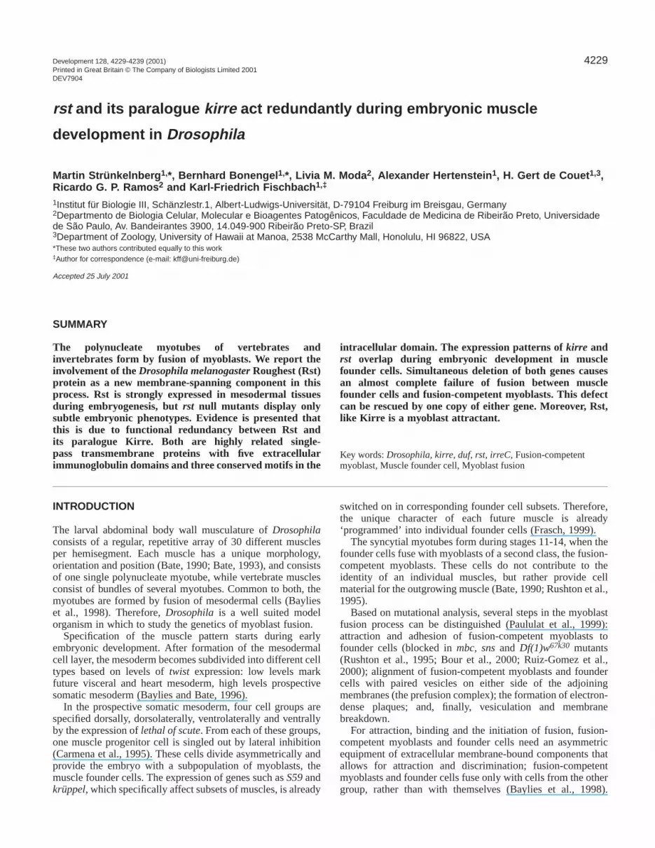
Reintroduction of rst rescues the Df(1)w67k30
phenotypeTo test the hypothesis that the Df(1)w67k30 phenotype ispartially caused by the removal of the rst locus, wereintroduced rst into the mesoderm of Df(1)w67k30 embryos(Fig. 7) using the UAS/Gal4-system. We used twi-Gal4 as adriver, which mediates expression within the entire mesodermfrom gastrulation up to late stage 11, when the expressionbecomes restricted to somatic muscles and to the heart (Bayliesand Bate, 1996). Expression of rst under the control of the twi-Gal4 driver in the background of Df(1)w67k30 almostcompletely restored the wild-type muscle pattern (Fig. 7B).
The muscle pattern of the rescued embryos displayed onlysmall defects such as ectopically projecting or thinner muscles(Fig. 7B, arrow and asterisk). We conclude that rst alone issufficient to rescue the Df(1)w67k30phenotype. As the same hasbeen shown for kirre (Ruiz-Gomez et al., 2000), this supportsthe hypothesis that the Df(1)w67k30phenotype is caused by thesimultaneous removal of rst and kirre.
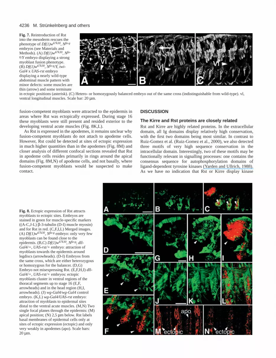
Rst is a myoblast attractantEctopically expressed Kirre recruits fusion-competentmyoblasts to ectopic sites in a background null for bothrstand kirre (Ruiz-Gomez et al., 2000). If Rst and Kirre arefunctionally equivalent during myoblast fusion, Rst should alsobe sufficient for ectopic recruitment of fusion-competentmyoblasts. We tested this hypothesis (Fig. 8) using the samedriver lines as were used for Kirre, wg-Gal4(Glise and Noselli,1997) and dll-Gal4 (Calleja et al., 1996).
The cross Df(1)w67k30/w; dll-Gal4/+ x w/Y; +/+; UAS-rstyielded progeny hemi- and heterozygous for Df(1)w67k30. In aDf(1)w67k30background, overexpression of Rst driven by dll-Gal4 recruited myoblasts to the epidermis analogous to theresults for Kirre. Fusion-competent myoblasts that wouldnormally reside further inside the embryo are attracted towardsthe sites of ectopic Rst expression in the leg discs and in thehead region (Fig. 8B,C). Embryos that carry at least one non-deficiency X-chromosome, albeit not defective in myoblastfusion, also displayed attraction of fusion-competentmyoblasts to sites of ectopic Rst expression (Fig. 8D-I). Thesemyoblasts were still present in late stages (around stage 16),when embryos from the same cross that did not express Rstectopically displayed none or only few unfused myoblasts.Misexpression of Rst under the control of the wg-Gal4line ina wild-type genetic background yielded comparable results:
Fig. 6. Df(1)w67k30is a rst, kirredouble mutant and shows severemuscle defects. (A) Wild-type abdominal muscle pattern as revealedby staining against β-3-tubulin. vl, ventral longitudinal muscles; sb,segment border muscle. (B)Df(1)w67k30embryos display a drasticmyoblast fusion phenotype with thin outgrowing muscle founder cellsand many unfused myoblasts (asterisks). (C) Detailed view of twoabdominal hemisegments of the same embryo. Scale bars: 20 µm.
4236
fusion-competent myoblasts were attracted to the epidermis inareas where Rst was ectopically expressed. During stage 16these myoblasts were still present and resided exterior to thedeveloping ventral acute muscles (Fig. 8K,L).
As Rst is expressed in the apodemes, it remains unclear whyfusion-competent myoblasts do not attach to apodeme cells.However, Rst could be detected at sites of ectopic expressionin much higher quantities than in the apodemes (Fig. 8M) andcloser analysis of different confocal sections revealed that Rstin apodeme cells resides primarily in rings around the apicaldomains (Fig. 8M,N) of apodeme cells, and not basally, wherefusion-competent myoblasts would be suspected to makecontact.
DISCUSSION
The Kirre and Rst proteins are closely relatedRst and Kirre are highly related proteins. In the extracellulardomain, all Ig domains display relatively high conservation,with the first two domains being most similar. In contrast toRuiz-Gomez et al. (Ruiz-Gomez et al., 2000), we also detectedthree motifs of very high sequence conservation in theintracellular domain. Interestingly, two of these motifs may befunctionally relevant in signalling processes: one contains theconsensus sequence for autophosphorylation domains ofligand-dependent tyrosine kinases (Yarden and Ullrich, 1988).As we have no indication that Rst or Kirre display kinase
M. Strünkelnberg and others
Fig. 7. Reintroduction of Rstinto the mesoderm rescues thephenotype of Df(1)w67k30, Nfa-g
embryos (see Materials andMethods). (A)Df(1)w67k30, Nfa-
g/Y embryo displaying a strongmyoblast fusion phenotype.(B) Df(1)w67k30, Nfa-g/Y, twi-Gal4 x UAS-rst embryodisplaying a nearly wild-typeabdominal muscle pattern withminor defects: some muscles arethin (arrow) and some terminatein ectopic positions (asterisk). (C) Hetero- or homozygously balanced embryo out of the same cross (indistinguishable from wild-type). vl,ventral longitudinal muscles. Scale bar: 20 µm.
Fig. 8.Ectopic expression of Rst attractsmyoblasts to ectopic sites. Embryos arestained in green for muscle-specific markers((A-C,J-L) β-3-tubulin (D-I) muscle myosin)and for Rst in red. (C,F,I,L) Merged images.(A) Df(1)w67k30, Nfa-g embryo: only very fewmyoblasts can be found close to theepidermis. (B,C) Df(1)w67k30, Nfa-g; dll-Gal4/+, UAS-rst/+embryo: attraction ofmyoblasts towards the epidermis aroundlegdiscs (arrowheads). (D-I) Embryos fromthe same cross, which are either heterozygousor homozygous for the balancer. (D,G)Embryo not misexpressing Rst. (E,F,H,I) dll-Gal4/+, UAS-rst/+ embryos: ectopicmyoblasts cluster in ventral regions of thethoracal segments up to stage 16 (E,F,arrowheads) and in the head region (H,I,arrowheads). (J) wg-Gal4/wg-Gal4controlembryo. (K,L) wg-Gal4/UAS-rstembryo:attraction of myoblasts to epidermal sitesdistal to the ventral acute muscles. (M,N) Twosingle focal planes through the epidermis: (M)apical position; (N) 2,5 µm below. Rst labelsbasal membranes of epidermal cells only atsites of ectopic expression (ectopic) and onlyvery weakly in apodemes (apo). Scale bars:20 µm.

4237Rst in muscle development
activity, we propose a putative as yet unknown interactionpartner to do so. Additionally, a C-terminal stretch correspondsto the PDZ-binding motif, which may have implications for thesubcellular localization of the proteins (Garner et al., 2000).Indeed, Rst distribution in all tissues studied so far showsstrong subcellular localisation.
The expression patterns of rst and kirre overlap, butare not identicalExpression patterns of rst and kirre overlap in muscle foundercells. With regard to kirre, our results confirm those of Ruiz-Gomez et al. (Ruiz-Gomez et al., 2000), showing expressionin founder cells and muscle precursors. As for rst, theexpression is more widespread and includes, in addition toexpression in founder cells, fusion-competent myoblasts.
Although Rst immunoreactivity is present in founder cells,we could not unequivocally identify individual outgrowingprecursors based on Rst staining, because of subcellularlocalization and the strong expression of Rst in fusion-competent myoblasts directly adjacent. However, onidentification of precursors by staining for β-3-tubulin, a weakresidual Rst immunoreactivity can initially be detected inprecursors until the end of stage 13, which fades soonthereafter (data not shown). With regard to the muscleprecursors, this downregulation of Rst seems to be dependenton the occurrence of muscle fusion. In contrast to wild type,Rst is still expressed on founder cells at stage 16 in a mbcC1
background (Fig. 3O), where myoblast adhesion is almostcompletely blocked (Rushton et al., 1995). In fusion-competentmyoblasts, however, Rst is downregulated in a mbcC1
background, with large numbers of unfused fusion-competentmyoblasts still being present at that stage. This asymmetry inregulation of Rst expression in both cell types is noteworthy.Fusion-dependent downregulation of Rst in muscle precursorscould function in size regulation of muscles.
Rst overexpression and mutant phenotypesAlthough the rst gene is not essential for muscle fusion, smalldefects like thinner and missing muscles can be detected in rst6
and rstirreC1 individuals, indicating the involvement of rst inmuscle development.
Overexpression of a secretable, extracellular version of Rstduring stages when myoblast fusion occurs (stages 12-15)leads to embryonic lethality and defects in myoblast fusion.Mechanistically, the extracellular part of the protein maycompete with endogenous Rst for an as yet unknownextracellular ligand or, as the Rst protein has been shown tomediate homophilic cell adhesion (Schneider et al., 1995), theextracellular domain could also bind to endogenous Rst andthereby disturb its function.
Ubiquitous overexpression of the full-length Rst protein alsocaused embryonic lethality and a severe muscle fusionphenotype. Ectodermal overexpression of Rst did not causedefects in the muscle pattern but ectopic localization andprolonged occurrence of myoblasts at sites of ectopic Rstexpression. Mesodermal expression did not induce anydetectable phenotype. The reason why global misexpression ofRst differs from misexpression in the mesoderm alone (in mostof which Rst is expressed anyway) and from misexpression inthe ectoderm alone appears to be the increase of Rst expressingsticky surfaces: the withdrawal of fusion-competent myoblasts
from recruiting founders and precursors may considerablylower the probability for these cell types to contact each other.
Some of the defects observed in rst mutants concern musclesin ectopic positions (Fig. 1E,G, arrows). Even though Rst isexpressed in the apodemes, our data do not point to an essentialrole for kirre and/or rst in myotube guidance or attachment:analysis of the subcellular localization shows accumulation ofRst primarily around the apical borders of the apodemes, ratherthan basally, where outgrowing muscles would be expected tomake contact. Moreover, apodeme specification is also notblocked in individuals lacking ectodermal Rst and Kirre, asjudged by the muscle pattern (Fig. 7B). Hence, a putativefunction of Rst in apodeme specification would be redundantlysafeguarded by additional as yet unknown factors. Apodemespecification is also not disrupted in da-Gal4/+;UAS-rst/+embryos, as revealed by antibody staining against Alien(Goubeaud et al., 1996; data not shown). This clearly rules outthe possibility that the strong muscle phenotype observed inthese embryos is due to defects in specification of the muscleattachment sites, and argues that restricted expression of Rst isnot essential for normal apodeme specification to occur. Thisis underlined by the fact that 69B-Gal4/+;UAS-rst/+embryosthat express Rst only in the ectoderm do not show attachmentdefects (data not shown).
Simultaneous deletion of kirre and rst blocksmyoblast fusionThe deficiencyDf(1)w67k30deletes the genomic region 3C2-3C6, including rst and kirre. It has been reported previouslythat Df(1)w67k30 is associated with a lethal myoblast fusionphenotype (Ruiz-Gomez et al., 2000). Our analysis of fliesdeficient for kirre, but not for rst, revealed that neither kirrenor rst alone are essential for myoblast fusion. Moreover,reintroduction of rst into the mesoderm restored theDf(1)w67k30phenotype to almost wild-type, as has been shownfor kirre (Ruiz-Gomez et al., 2000). Hence, one copy of eitherrst or kirre is sufficient to restore the wild-type muscle pattern.Furthermore, ectopic expression of Rst mediated ectopicrecruitment of fusion-competent myoblasts as has been shownwith Kirre (Ruiz-Gomez et al., 2000). Given the overlappingmesodermal expression patterns of rst and kirre, and thesignificant structural similarity between the two proteins, weconclude that rst and kirre have at least partially redundantfunctions during muscle development.
Our findings may provide a molecular basis for the findingsof Lefevre and Green (Lefevre and Green, 1972), whoproposed a genetic duplication between 3C2-5 (the rst locusresides at 3C5) and 3C6: individuals are viable when either oneof the regions 3C2-5 or 3C6 is deleted; however, they die whenboth are missing (Fig. 5A). Even though kirre is now mappedto 3C7 (FlyBase – http://flybase.bio.indiana.edu), the mappingdata of Lefevre and Green can account for kirre, since othermapping data put the kirre locus into 3C6: the 5′ end of Notchwas mapped in between 3C6 and 3C7 (Rykowski et al., 1988)and the faswb deletion, which resides in between kirre andNotch(Fig. 2A) (Ramos et al., 1989), has been shown to fuse3C6 and 3C7 (Keppy and Welshons, 1976).
Rst, Kirre and the fusion machineryRst expression in fusion-competent myoblasts is not essentialfor their attraction towards ectopic Kirre or Rst: myoblasts can

4238
be attracted to ectopic sites in a Df(1)w67k30background, whereRst is only present at ectopic sites and not in fusion-competentmyoblasts – thus strongly suggesting a heterophilic trans-interaction. However, as Rst has been shown to mediatehomophilic cell adhesion (Schneider et al., 1995), a homophilictrans-interaction of Rst may also contribute to the fusionprocess.
At present, the data do not allow a prediction of themolecular mechanisms in which Rst and Kirre take part;however, it is conceivable that they include the related celladhesion molecules Sns and Hbs that are expressed on fusion-competent myoblasts (Bour et al., 2000) (H. A. Dworak andH. Sink, personal communication). A model of the fusionmachinery may include assembly of adhesion moleculeswithin heteromeric complexes with differing compositions onthe side of the fusion-competent myoblasts (including Sns,Hbs and Rst) and on the founder cells (including Kirre andRst). These complexes may still function after loss of singlecomponents. It will need further analysis and binding assaysto elucidate how these membrane proteins play together andhow they are connected to the other components of the fusionmachinery.
We acknowledge the technical assistance by M. S. A. Costa, M.Böhler, W. Brinkmann and M. S. Z. Graeff. Thanks to D. P. Kiehart,R. Renkawitz-Pohl, S. Artavanis-Tsakonas, M. Bate, M. Calleja andS. Noselli for antibodies and/or fly strains, and G. Igloi forsequencing. M. S. and B. B. are indebted to all members of theDepartment of Morphology, USP-Ribeirao Preto, Brazil for greathospitality and to M. Hoehne for careful reading of the manuscriptand discussion. This work was supported by the SFB 388 (to K. F.F.), FAPESP (Grants #96/06235-2 and #00/07874-3 to R. G. P. R.)and DAAD/CAPES (PROBRAL 2000) to M. S., B. B. and K. F. F.
REFERENCES
Altschul, S. F., Madden, T. L., Schäffer, A. A., Zhang, J., Zhang, Z., Miller,W. and Lipman, D. J. (1997). Gapped BLAST and PSI-BLAST: a newgeneration of protein database search programs. Nucleic Acids Res. 25,3389-3402.
Ashburner, M. (1989). Drosophila– A Laboratory Manual. Cold SpringHarbor, New York: Cold Spring Harbor Laboratory Press.
Bate, M. (1990). The embryonic development of larval muscles in Drosophila.Development110, 791-804.
Bate, M. (1993). The mesoderm and its derivatives. In The development ofDrosophila melanogaster. Vol. 2 (ed. M. Bate and A. Martinez-Arias), p.1013. Cold Spring Harbor, New York: Cold Spring Harbor Laboratory Press.
Baylies, M. K. and Bate, M.(1996). twist: a myogenic switch in Drosophila.Science 272, 1481-1484.
Baylies, M. K., Bate, M. and Ruiz-Gómez, M. (1998). Myogenesis: a viewfrom Drosophila. Cell 93, 921-927.
Benos, P. V., Gatt, M. K., Ashburner, M., Murphy, L., Harris, D., Barrell,B., Ferraz, C., Vidal, S., Brun, C., Demailles, J. et al. (2000). Fromsequence to chromosome: the tip of the X chromosome of D. melanogaster.Science.287, 2220-2222.
Blom, N., Gammeltoft, S. and Brunak, S. (1999). Sequence- and structure-based prediction of eukaryotic protein phosphorylation sites. J. Mol. Biol.294, 1351-1362.
Bour, B., Chakravarti, M., West, J. M. and Abmayr, S. M. (2000).Drosophila SNS, a member of the immunoglobulin superfamily that isessential for myoblast fusion Genes Dev.14, 1498-1511.
Brand, A. H. and Perrimon, N. (1993). Targeted gene expression as a meansof altering cell fates and generating dominant phenotypes. Development118,401-441.
Buttgereit, D. (1993). Redundant enhancer elements guide beta 1 tubulingeneexpression in apodemes during Drosophilaembryogenesis. J. Cell Sci.105,721-727.
Calleja, M., Morena, E., Pelaz, S. and Morata, G. (1996). Visualization ofgene expression in living adult Drosophila. Science274, 252-255.
Campos-Ortega, J. A. and Hartenstein, V. (1997). The EmbryonicDevelopment of Drosophila melanogaster. 2nd edn. Berlin-Heidelberg:Springer Verlag.
Carmena, A., Bate, M. and Jiménez, F. (1995). lethal of scute, a proneuralgene, participates in the specification of muscle progenitors duringDrosophilaembryogenesis. Genes Dev.9, 2373-2383.
Castelli-Gair, J., Greig, S., Micklem, G. and Akam, M. (1994). Dissectingthe temporal requirements for homeotic gene function. Development120,1983-1995.
Dieffenbach, C. W. and Dveksler, G. S. (1995). PCR primer: A LaboratoryManual. Cold Spring Harbor, New York: Cold Spring Harbor LaboratoryPress.
Frasch, M. (1999). Controls in patterning and diversification of somaticmuscles during Drosophilaembryogenesis. Curr. Opin. Genet. Dev. 9, 522-529.
Garner, C. C., Nash, J. and Huganir, R. L. (2000). PDZ domains in synapseassembly and signalling. Trends Cell Biol. 10, 274-80.
Glise, B. and Noselli, S. (1997). Coupling of Jun amino-terminal kinase anddecapentaplegic signaling pathways in Drosophila morphogenesis. GenesDev.11, 1738-1747.
Goubeaud, A., Knirr, S., Renkawitz-Pohl, R. and Paululat, A. (1996). TheDrosophila gene alien is expressed in the muscle attachment sites duringembryogenesis and encodes a protein highly conserved between plants,Drosophilaand vertebrates. Mech. Dev.57, 59-68.
Grimwade, B., Muskavitch, M. A. T., Welshons, W. J., Yedvobnick, B. andArtavanis-Tsakonas, S. (1985). The molecular genetics of the Notchlocusin Drosophila melanogaster. Dev. Biol.107, 503-519.
Keppy, D. O. and Welshons, W. J. (1976). The cytogenetics of a recessivevisible mutant associated with a deficiency adjacent to the Notch locus inDrosophila melanogaster. Genetics85, 497-506.
Kiehart, D. P. and Feghali, R. (1986). Cytoplasmic myosin from Drosophilamelanogaster. J. Cell Biol.103, 1517-1525.
Lefevre, G. Jr. and Green, M. M. (1972). Genetic duplication in the white-split interval of the X chromosome in Drosophila melanogaster.Chromosoma36, 391-412.
Moda, L., Machado, R. and Ramos, R. G. P. (2000). Ubiquitousoverexpression of a transgene encoding the extracellular portion of theDrosophilaRoughest-Irregular Chiasm C protein induces early embryoniclethality. An. Acad. Bras. Cienc.72, 381-388.
Nielsen, H., Engelbrecht, J., Brunak, S. and von Heijne, G. (1997).Identification of prokaryotic and eukaryotic signal peptides and predictionof their cleavage sites. Protein Eng.10, 1-6.
Nose, A., Isshiki, T. and Takeichi, M. (1998). Regional specification ofmuscle progenitors in Drosophila: the role of the msh homeobox gene.Development125, 215-223.
Oxtoby, E. and Jowett, T. (1993). Cloning of the zebrafish krox-20gene (krx-20)and its expression during hindbrain development. Nucleic Acids Res.21,1087-1095
Paululat, A., Holz, A. and Renkawitz-Pohl, R. (1999). Essential genes formyoblast fusion in Drosophila embryogenesis. Mech. Dev. 83, 17-26.
Perlman, J. (1983). One side of a deletion breakpoint from the Drosophilamelanogastergenome contains a transposable element. Gene21, 87-94.
Ramos, R. G. P., Grimwade, B. G., Wharton, K. A., Scottgale, T. N. andArtavanis-Tsakonas, S. (1989). Physical and functional definition of theDrosophila Notchlocus by P element transformation. Genetics123, 337-348.
Ramos, R. G. P., Igloi, G. L., Lichte, B., Baumann, U., Maier, D.,Schneider, T., Brandstätter, J. H., Fröhlich, A. and Fischbach, K.-F.(1993). The irregular chiasm C-roughestlocus of Drosophila, which affectsaxonal projections and programmed cell death, encodes a novelimmunoglobulin-like protein. Genes Dev. 7, 2533-2547.
Reiter, C., Schimansky, T., Nie, Z. and Fischbach, K. F. (1996).Reorganization of membrane contacts prior to apoptosis in the Drosophilaretina: the role of the IrreC-rst protein. Development122, 1931-1940.
Ruiz-Gómez, M., Coutts, N., Price, A., Taylor, M. V. and Bate, M. (2000).Drosophila dumbfounded: a myoblast attractant essential for fusion. Cell102, 189-198.
Rushton, E., Drysdale, F., Abmayr, S. M., Michelson, A. M. and Bate, M.(1995). Mutations in a novel gene, myoblast city, provide evidence insupport of the founder cell hypothesis for Drosophilamuscle development.Development121, 1979-1988.
Rykowski, M. C., Parmelee, S. J., Agard, D. A. and Sedat, J. W. (1988).
M. Strünkelnberg and others

4239Rst in muscle development
Precise determination of the molecular limits of a polytene chromosomeband: Regulatory sequences for the Notch gene are in the interband. Cell54, 461-472.
Schneider, T., Reiter, C., Eule, E., Bader, B., Lichte, B., Nie, Z.,Schimansky, T., Ramos, R. G. P. and Fischbach, K. F. (1995). Restrictedexpression of the Rst protein is required for normal axonal projections ofcolumnar visual neurons. Neuron15, 259-271.
Sonnhammer, E. L. L., van Heijne, G. and Krogh, A. (1998). A hiddenMarkov model for predicting transmembrane helices in protein sequences.In Proceedings of the Sixth International Conference on Intelligent Systemsfor Molecular Biology(ed. J. Glasgow, T. Littlejohn, F. Major, R. Lathrop,D. Sankoff and C. Sensen), pp. 175-182. Menlo Park, CA: AAAI Press.
Tautz, D. and Pfeifle, C. (1989). A non-radioactive in situ hybridizationmethod for the localization of specific RNAs in Drosophilaembryos revealstranslational control of the segmentation gene hunchback. Chromosoma 98,81-85.
Walsh, F. S. and Doherty, P. (1997). Neural cell adhesion molecules of theimmunoglobulin superfamily: role in axon growth and guidance. Annu. Rev.Cell Dev. Biol. 13, 425-456.
Wodarz, A., Hinz, U., Engelbert, M. and Knust, E. (1995). Expression ofcrumbs confers apical character on plasma membrane domains ofectodermal epithelia of Drosophila. Cell 82, 67-76.
Yarden, Y. and Ullrich, A. (1988) Growth factor receptor tyrosine kinases.Annu. Rev. Biochem.57, 443-478.


