Embed Size (px)
Citation preview

Running Header: FUNCTIONAL ANALYSIS OF THE CONSERVED TRANSCRIPTION ELONGATION FACTOR SPT5
FUNCTIONAL ANALYSIS OF THE CONSERVED TRANSCRIPTION ELONGATION
FACTOR SPT5
Emily Schwenger
McGill University
Author Note
This paper was prepared under the expert supervision of Dr. Jason Tanny of the Department of Pharmacology and Therapeutics at McGill University.
FUNCTIONAL ANALYSIS OF THE CONSERVED TRANSCRIPTION ELONGATION FACTOR SPT5
2
ABSTRACT INTRODUCTION: The passage of RNA polymerase II (RNAPII) during transcript elongation is
dependent upon accessory proteins, histone modifications, and large enzymatic complexes that
communicate with each other to regulate transcription rate and processivity. The elongation step
is a critical target for regulation at many genes in metazoans, and aberrant elongation is
associated with a variety of diseases, including cancer, HIV, and cardiac hypertrophy (Cherrier et
al. 2013, Le Douce et al., 2012). The transcription elongation factor Spt5 is directly bound to
RNAPII and is important for coupling chromatin modification states and RNA processing to
RNAPII elongation. Spt5 is phosphorylated by Cdk9, the kinase subunit of the positive
elongation factor (P-TEFb). Phosphorylation of Spt5 is critical for its functions in elongation but
the relevant downstream pathways have not been elucidated. Our lab has provided genetic
evidence that Spt5 phosphorylation (Spt5-P) has a close functional relationship with co-
transcriptional histone modifications such as monoubiquitylation of histone H2B (H2Bub1) and
methylation of lysine 4 of histone H3 (H3K4) (Sanso et al., 2012). METHODS: Here we further
extend our genetic analysis of the role of Spt5 in transcript elongation through genome-wide
synthetic genetic analysis in the model eukaryote Schizosaccharomyces pombe . Observed
growth defects, or synthetic lethal and synthetic sick (SSL) genetic interactions, were followed
up with phenotypic analysis by fluorescence microscopy. RESULTS: Spt5 mutants exhibited
genetic interactions with mutants affecting the histone variant H2A.Z, its chaperone the SNF2-
related helicase Swr1 complex comprised of Swr1, Swc2, and Msc1, and nucleoporin Mex67. In
particular, deletion of H2A.Z+ and msc1+ combined with mutation of the phosphorylation site of
Spt5’s CTR created a striking synthetic interaction bordering on lethality. CONCLUSION:
Taking into account that Spt5 is one of RNAPII’s only accessory proteins that is universally

FUNCTIONAL ANALYSIS OF THE CONSERVED TRANSCRIPTION ELONGATION FACTOR SPT5
3
conserved across all domains of life, an implied overlapping function with a non-canonical
histone variant could be translated into humans and manipulated into an effective and specific
therapy for diseases involving aberrant gene expression, such as cancer (Hartzog et al., 2013).
INTRODUCTION
Eukaryotes have evolved means to regulate gene expression for a wide range of purposes,
including cell maintenance, delineation, and development (Campos et al., 2009). Central to gene
regulation is the nucleosome, composed of a histone octamer, 147 base pairs of DNA, and a
linker histone. While the typical histone core consists of two H2A-H2B dimers flanked by two
H3-H4 dimers, a variety of histone variants exist that can substitute in for any of the canonical
histones, serving functions such as centromeric silencing, activation of highly transcribed genes,
and DNA repair (Chen et al., 2014).
In addition to the central nucleosome, layers of protein-DNA and protein-protein
interactions form a complex network that oversees the regulation and processing of the mRNA
transcript. Paramount to this regulation is the recruitment and passage of RNA polymerase II
(RNAPII), which is associated with highly conserved patterns of histone modifications. These
patterns include, but are not limited to, methylation of histone H3 Lys4 (H3K4me) and
acetylation of histones H3 and H4 at 5’ ends, methylation of histone H3 Lys36 (H3K36me)
towards 3′ ends, and mono-ubiquitylation of histone H2B at a conserved site in the carboxyl-
terminus (H2Bub1) throughout coding regions of genes (Sanso et al., 2012). While some of these
modifications exert their main effects on transcription by altering the intrinsic structure of
chromatin, such as the charge neutralizing effects of histone acetylation, many function

FUNCTIONAL ANALYSIS OF THE CONSERVED TRANSCRIPTION ELONGATION FACTOR SPT5
4
predominantly through the creation or elimination of binding sites. Though much is known about
how chromatin modifications regulate recruitment of RNA polymerase II and general
transcription factors to the promoter region during transcription initiation, their roles in transcript
elongation and the precise spatial and temporal arrangement of the overlapping processes of
generating a mature mRNA, such as capping and splicing, are poorly understood.
Transcription elongation factors Spt4 and Spt5 are bound directly to RNAPII and are its
only accessory proteins universally conserved across all domains of life. Spt5 has been shown to
have an essential function in the activation of transcription elongation and its deletion is lethal to
both budding and fission yeast (Hartzog et al., 2013). Importantly, Spt5 plays a vital role in
relieving RNAPII from its transcriptionally engaged but paused state just downstream of the
promoter. This process of promoter-proximal halting was originally thought to have unique
involvement in rapid activation of stress-induced heat shock proteins (HSPs) in Drosophila, but
is now known to take place across the genome (Gilchrist et al., 2012). Transcription machinery
and transcription factors are pre-assembled at a promoter-proximal region, thus making it
possible for elongation to be activated almost instantaneously in response to transcription
activators such as c-Myc (Rahl et al., 2010). Moreover, pausing facilitates recruitment of
mRNA-processing factors, and can be rate-determining for expression of tightly regulated genes
(Sanso et al., 2012, Core et al., 2008).
Spt5 is known to associate with RNAPII until its release near the 3’ end of the gene,
therefore, release from pausing does not encompass its full function. Spt5 has been shown to
interact with the RNA polymerase II associated factor 1 (Paf1) complex which itself associates
with elongating RNAPII and coordinates a variety of histone modifications (Squazzo et al.,
2002). Furthermore, it has known functional interactions with H3K4me3, H3K36me3, H2Bub1,

FUNCTIONAL ANALYSIS OF THE CONSERVED TRANSCRIPTION ELONGATION FACTOR SPT5
5
and subunits of the Rpd3S histone deacetylase complex, whose function in occluding cryptic
promoters is dependent on H3K36 methylation (Quan et al., 2010).
Spt5 is phosphorylated by Cdk9, the kinase subunit of the essential positive elongation
factor P-TEFb, at threonine 1 of its CTR. This residue was mutated to create two of the query
strains used in this study, spt5T1A and spt5T1E, constituting ablation and constitutive activation
of the Cdk9 phosphorylation site, respectively. The CTD of RNAPII is similarly phosphorylated
by Cdk9 and other cyclin-dependent kinases that regulate transcription. These phosphorylations
are critical for RNAPII elongation, pre-mRNA-processing, histone modifications, and the release
from promoter-proximal pausing described above. Cdk9 activity is required for generation of co-
transcriptional H2Bub1, H3K4me, and H3K36me, although the molecular mechanisms have yet
to be determined.
Synthetic Genetic Analysis and the Cdk9-spt5-H2Bub1 Pathway
We have recently shown that Cdk9 and phosphorylated Spt5 (Spt5-P) have an intimate
functional connection with co-transcriptional histone modifications in the fission yeast S. pombe
through establishment of a positive feedback loop with H2Bub1. Our study also suggests that
Spt5-P and H3K4me may have a shared function comprising that of H2Bub1. We sought to gain
further insight into this function by collaborating with the lab of Dr. Nevan Krogan (University
of California, San Francisco) who had previously developed a high-throughput screen designed
for epistatic mapping of the entire S. pombe genome. The screen consisted of a system of genetic
manipulations combined with introduction of binary combinations of double mutants through
genetic crossing (Roguev et al., 2007). Selection strategies were four-fold in order to provide a
robust and efficient way of selecting for double mutants amongst a mixture of parent cells,
diploids, and recombinant cells. Their strategies included anti-diploid selection, mating type

FUNCTIONAL ANALYSIS OF THE CONSERVED TRANSCRIPTION ELONGATION FACTOR SPT5
6
selection, and antibiotic and auxotrophic markers for mutant selection. Spt5T1A, spt5T1E, and
spt5∆C were the query strains of interest, and candidate mutants inducing observable growth
defects when mated with any of the query strains were further investigated through tetrad
analysis.
This report describes the characterization of a selection of genetic interactions identified
by the high-throughput screen. These analyses validated some key interactions and suggested
potentially novel functions for Spt5 in regulation of gene expression.
MATERIALS & METHODS
Schizosaccharomyces Pombe
The S. pombe strains used are listed in Table 1 below.
Table 1. List of S. pombe strains used
Strain Name Strain Information Reference Wild type ade6-M216 ura4+ leu+ Tanny et al., 2007 h2b-K119R htb1-K119R::kanMX6; ade6; ura+; h+ Tanny et al., 2007 h2b-K119R htb1-K119R::hphMX6; ade6; ura+; h+ This study spt5-WT spt5-WT(7)::ura4+ ade6-M210 leu1-32 ura4-D18
his3-D1 h- Fisher et al., 2006
spt5-T1A spt5-T1A(7)::ura4+ ade6-M210 leu1-32 ura4-D18 his3-D1 h-
Fisher et al., 2006
spt5-T1E spt5-T1E(7)::ura4+ ade6-M210 leu1-32 ura4-D18 his3-D1 h-
Fisher et al., 2006
spt5∆C
spt5∆C::ura4+ ade6-M210 leu1-32 ura4-D18 his3-D1 h-
Fisher et al., 2006
spt5-WT spt5-WT(7)::natMX6 (3 isolates) ade6-M210 leu1-32 ura4D-18 mat1_m-cyhS smt0 rpl42::cyhR (sP56Q) h-
Roguev et al., 2007
spt5-T1A spt5-T1A::natMX6 (3 isolates) ade6-M210 leu1-32 ura4D-18 mat1_m-cyhS smt0 rpl42::cyhR (sP56Q) h-
Roguev et al., 2007
spt5-T1E spt5-T1E::natMX6 (3 isolates) ade6-M210 leu1-32 ura4D-18 mat1_m-cyhS smt0 rpl42::cyhR
Roguev et al., 2007

FUNCTIONAL ANALYSIS OF THE CONSERVED TRANSCRIPTION ELONGATION FACTOR SPT5
7
(sP56Q) h- pht1∆ pht1∆::hphMX4; ade6 ura4-D18 h- Tanny et al., 2007 msc1∆ msc1∆::kanMX6; leu1-32; ura- h- Tanny et al., 2007 sir2∆ sir2∆::kanMX6 h- Ekwall et al., 2005
clr3∆ clr3∆::kanMX h- Ekwall et al., 2005
pmt3∆ pmt3∆::hphMX6; ade6 h- Schneider et al., 2010 hos2∆ hos2∆::leu2; leu1-32 h- Ekwall et al., 2005
rtt109∆ rtt109∆::kanMX6 h- Schneider et al., 2010 msc1∆::kanR; leu1-32; h+ Ahmed et al., 2004
pht1∆ pht1∆::hphMX4; ade6; h+ Tanny et al., 2007 swr1∆ swr1∆::kanMX4; ade6-M216; ura4-D18; leu1-32;
h- Bioneer, Inc.
swc2∆ swc2∆::kanMX4; ade6-M216; ura4-D18; leu1-32; h-
Bioneer, Inc.
nrc1∆ nrc1∆::kanMX4; ade6-M216; ura4-D18; leu1-32; h-
Bioneer, Inc.
nto1∆ nto1∆::kanMX4; ade6-M216; ura4-D18; leu1-32 Bioneer, Inc. nup132∆ nup132∆::kanMX4; ade6-M216; ura4-D18; leu1-
32 Bioneer, Inc.
mst2∆ mst2∆::kanMX4; ade6-M216; ura4-D18; leu1-32 Bioneer, Inc. epe1∆ epe1∆::kanMX4; ade6-M216; ura4-D18; leu1-32 Bioneer, Inc. sgf29∆ sgf29∆::kanMX4; ade6-M216; ura4-D18; leu1-32 Bioneer, Inc. mug183∆ mug183∆::kanMX4; ade6-M216; ura4-D18; leu1-
32 Bioneer, Inc.
png2∆ png2∆::kanMX4; ade6-M216; ura4-D18; leu1-32 Bioneer, Inc. mex67∆ mex67∆::kanMX4; ade6-M216; ura4-D18; leu1-
32 Bioneer, Inc.
ulp2∆ ulp2∆::kanMX4; ade6-M216; ura4-D18; leu1-32 Bioneer, Inc. elp1∆ elp1∆::kanMX4; ade6-M216; ura4-D18; leu1-32 Bioneer, Inc. cxr1∆ cxr1∆::kanMX4; ade6-M216; ura4-D18; leu1-32 Bioneer, Inc. spt2∆ spt2∆::kanMX4; ade6-M216; ura4-D18; leu1-32 Bioneer, Inc. dbl8∆ dbl8∆::kanMX4; ade6-M216; ura4-D18; leu1-32 Bioneer, Inc. kap114∆ kap114∆::kanMX4; ade6-M216; ura4-D18; leu1-
32 Bioneer, Inc.
nap2∆ nap2∆::kanMX4; ade6-M216; ura4-D18; leu1-32 Bioneer, Inc. clr2∆ clr2∆::kanMX4; ade6-M216; ura4-D18; leu1-32 Bioneer, Inc. lsg1∆ lsg1∆::kanMX4; ade6-M216; ura4-D18; leu1-32 Bioneer, Inc.

FUNCTIONAL ANALYSIS OF THE CONSERVED TRANSCRIPTION ELONGATION FACTOR SPT5
8
Tetrad Dissection
S. pombe frozen glycerol stocks were streaked onto agar plates with yeast extract
supplement (YES) containing 5 g/L yeast extract, 30g/L glucose, and 250mg/L each of histidine,
leucine, adenine and uracil. These were grown at 30˚C in a dry box incubator until colonies were
visible, usually overnight for one to two days.
Two strains of opposite mating type, one h+ and the other h-, were subsequently mated.
A colony from each strain was spread, one on top of the other, onto a small patch on a standard
SPA mating plate (Moreno et al., 1991). The plates were then kept at room temperature for two
to four days, during which sporulation was induced through means of nitrogen starvation, as per
standard S. pombe mating protocol (Moreno et al., 1991).
Prior to preparing the tetrad dissection, the patch of mixed cells was inspected for the
presence of tetrads under a standard compound microscope at a magnification of 40x. If tetrads
were present, a swab of cells was diluted in 1 mL distilled water with thorough mixing. 20 µL
was subsequently plated in a bead near the edge of an agar YES plate, which was gently tilted to
form a line of cells down the middle of the plate.
After allowing five minutes to dry, the plate was placed upside-down on a staging
platform above a mounted micromanipulator, all part of the Zeiss Axioskop 40 tetrad dissection
microscope. 14 tetrads were selected and placed in a vertical line parallel to the central line of
cells, 7 on each side. They were then left at 37˚C to encourage breakdown of the ascus wall and
checked every few hours. Tetrads with broken down ascii were subsequently dissected, with
each of the four individual cells placed 3 mm apart in a horizontal line.
If only a minority of asci broke down before the end of the day, the plate was left
overnight at room temperature or in a 20˚C dry box incubator and checked for broken down
FUNCTIONAL ANALYSIS OF THE CONSERVED TRANSCRIPTION ELONGATION FACTOR SPT5
9
tetrads the following morning. Leaving the plate at 37˚C for more than 5 hours is discouraged
due to the possibility of cell doubling, which would make it impossible to accurately isolate the
cells into the four individual haploid products of meiosis.
This cycling of incubation and dissection was repeated until the majority of the 14
selected tetrads were successfully dissected. The plate was then incubated at 30˚C for a few days
until colonies grew to about 1 mm in diameter.
Thereafter, the tetrad colonies were replica plated onto selective media according to the
respective antibiotic resistance or auxotrophic markers of the parent cells and incubated
overnight at 30˚C. Agar plates were made with 200 mg/L geneticin (G418, Sigma-Aldrich), 200
mg/L hygromycin (Sigma-Aldrich), 200 mg/L nourseothricin (clonNAT, Werner BioAgents),
and EMM –ura, as per standard protocol (Moreno et al., 1991).
Microscopy
S. pombe cells were fixed and stained with diamino-phenylindole (DAPI) and calcofluor as
described previously (Viladevall et al., 2009). Cells were viewed using a Leica DM5000b
microscope and photographed with a CCD camera. Images were processed using Volocity
software.
Creation of h2b1-K119R::hygromycin Mutant Strain
To introduce a hygromycin marker in place of a G418 marker into a G418R h2b1-K119R
mutant, the primer sequences 5’-CGGATCCCCGGGTTAATTAAGGCG-3’ and 5’-
GAATTCGAGCTCGTTTAAACACTGGATGGCGGCGTTAGTATCG-3’ were used to
amplify a hygromycin B (hygB) cassette with flanking homology regions from plasmid pFA6a-

FUNCTIONAL ANALYSIS OF THE CONSERVED TRANSCRIPTION ELONGATION FACTOR SPT5
10
hphMX6 (Sato et al., 2005). Synthesis was catalyzed by Taq DNA Polymerase (Invitrogen) in
reactions containing PCR Reaction Buffer (Invitrogen), MgCl2 (Invitrogen),
deoxyribonucleotides (Invitrogen) as per standard PCR protocol (Azuma et al., 1991).
10 mL liquid cultures were grown overnight, spun down in a Thermo tabletop centrifuge
at 3000 rpm for two minutes and washed with water and a solution of 100 mM lithium acetate,
10 mM Tris, and 1 mM EDTA (LiAc/TE), consecutively. They were then resuspended in the
LiAc/TE at a concentration of 5 x 10exp8 cells per mL. 100 µL cells was mixed with 1 µL
salmon sperm DNA (Invitrogen) and 2 µg transforming DNA, and the mixture was left to rest at
room temperature for ten minutes. A solution of LiAc/TE and Polyethylene glycol was added
prior to a one hour incubation at 30˚C, followed by addition of 43 µL dimethyl sulfoxide and 20
minute heat shock in a 42˚C water bath. Cells were recovered by incubating on non-selective
YES plates and replica plated onto hygromycin B media when colonies were easily observable.
Western Immunoblotting
S. pombe cells were grown as per standard procedure (Moreno et al., 1991). 10 mL
cultures were grown in a shaking incubator at 30˚C until they reached an optical density between
0.4 and 0.6, indicative of logarithmic growth. These were then spun down, washed in 5-10 ml of
20% trichloroacetic acid (TCA), and stored at -80˚C.
Cells were lysed through agitation with cold-acid-washed glass beads (Sigma, 425-600
microns) using the MiniBeadBeater-16 (Biospec) for four thirty-second intervals with one
minute breaks in between. 1 mL 5% TCA was added and the liquid retrieved and spun down for
ten minutes at maximum speed using a standard tabletop centrifuge. The supernatant was

FUNCTIONAL ANALYSIS OF THE CONSERVED TRANSCRIPTION ELONGATION FACTOR SPT5
11
discarded, samples were resuspended in Laemmli buffer (50µM Tris pH 6.8, 2% SDS, 10%
glycerol, 5% β-mercaptoethanol, bromophenol blue), and neutralized with 2M Tris base.
15% acrylamide (BioRad) gels were set and samples run at a voltage of 120 V for fifteen
minutes, followed by 180V until the front of the loading dye ran into the buffer. The running
buffer used contained 3 g/L of Tris base, 20g/L glycine, and 0.1% SDS.
Transfer was performed at a constant current of 200 mA on ice in cold transfer buffer
containing 3.6g/L Tris, 15g/L Glycine, 0.4g/L of sodium dodecyl sulfate (SDS) and 2%
methanol. Nitrocellulose membranes were blocked for one hour on a shaking platform with 5%
skim milk powder dissolved in Tris-buffered saline with Tween-20 (TBST). Primary antibody
sources and concentrations can be found in Table 2. Membranes were incubated overnight at 4˚C
in primary antibody followed by one hour with anti-rabbit antibody conjugated with HRP (GE
Healthcare UK Limited) at a dilution of 1 to 4000 in TBST, with three five minute washes in
TBST after each incubation. Finally, the ECL Prime detection kit (BioRad) procedure was
followed, and the nitrocellulose membrane exposed on film.
Table 2. Western Blotting Conditions
Proteins Primary Antibody Concentration
Source of Primary Antibody
!-H3
H3K4me3
H3K36me3
1:5000
1:3000
1:2000
Abcam
Abcam
Abcam

FUNCTIONAL ANALYSIS OF THE CONSERVED TRANSCRIPTION ELONGATION FACTOR SPT5
12
RESULTS
In order to further elucidate the mechanisms behind
the cdk9-spt5-H2Bub1 pathway in governing
chromatin states and transcription, we analyzed
putative SSL interactions from a high-throughput
screen involving genome-wide epistasis mapping.
The screen was carried out by our collaborator Dr.
Nevan Krogan (UCSF, Roguev et al., 2007). We
sought to confirm genetic interactions from this screen of candidate knockout mutants
queried against htb1-K119R and three mutant spt5 strains.
Two of the spt5 strains used are characterized by point mutations at the phosphoacceptor
threonine residue (Thr1) in the CTR. Spt5∆C has its entire CTR deleted, and therefore exhibits a
wider array of genetic interactions than the point mutants. To conduct the screen we re-
constructed the spt5T1A and spt5T1E mutants, as well as a wild-type spt5+ control, in the
standard strain used for screening by the Krogan lab (Roguev et al., 2007). DNA sequences
encoding the variant Spt5 C-termini were introduced into yeast and recombinants were selected
using a linked antibiotics resistance marker. PCR analysis of the transformants confirmed correct
integration of the new spt5 sequences (see Figure 1).
Tetrad dissection is a powerful genetic technique unique to yeast and was our primary
follow-up to the genetic interactions displayed in the screen. It is based on the premise that
diploid cells undergo meiosis and package the four haploid nuclei into spores, with each haploid
cell containing a distinct genotype (Moreno et al., 1991). The most common segregation pattern
ura4+ -‐2.7 kb
NATMX6 -‐2.8 kb Figure 1. Confirmation of spt5 CTR by PCR Spt5 CTR was amplified. The two sets of transformants exhibit correct size of PCR product, representing the length of the CTR added to the flanking marker, ura4+ or clonNAT.
spt5-‐W
T
spt5-‐T1A
spt5-‐T1E
FUNCTIONAL ANALYSIS OF THE CONSERVED TRANSCRIPTION ELONGATION FACTOR SPT5
13
is tetratype, with two cells identical to each of the single mutant parent cells, and two
recombinant cells, one wild-type and one double mutant. The four cells are manually separated
and the double mutant is isolated after antibiotic and/or auxotrophic selection. This acts as a
robust and efficient strategy for performing genetic manipulations and observing synthetic
interactions that provide useful insight into protein functionality, and was used to validate the
results from the high-throughput screen (summarized in Table 3).
We confirmed a subset of strong negative and positive interactions identified by the
screen. We crossed previously analyzed spt5 mutant strains (marked with ura4+ conferring
uracil prototrophy) with individual gene knockout strains provided by the Krogan lab (marked
with a G418R cassette). We also tested interactions with the spt5∆C mutant and the functionally
related htb1-K119R mutant, both of which display a stronger growth phenotype than the
phosphoacceptor mutants. We reasoned these crosses might more sensitively reveal genetic
interactions. Tetrads were dissected and plated on selective media containing G418, hygB,
and/or EMM-ura to isolate the double mutants. Observed growth patterns are summarized in
Table 3.
Tetrad dissections with the spt5 mutants revealed six novel genetic interactions. The most
striking of these were pht1+ and msc1+ knockout mutants, which produced synthetic interactions
bordering on lethality when crossed with all three spt5 mutants (Figure 4). The interactions with
spt5T1A and spt5T1E are indicative of a specific, shared function of both Pht1 and Msc2 with
Cdk9, the upstream kinase responsible for phosphorylating the substituted threonine residue.

FUNCTIONAL ANALYSIS OF THE CONSERVED TRANSCRIPTION ELONGATION FACTOR SPT5
14
Pht1 is the S. pombe homolog of the histone variant H2A.Z, which plays an important
role in governing boundaries between chromatin states (Campos et al., 2009). Interestingly,
Msc1 is a key regulator in the Swr1 complex, the chaperone responsible for loading H2A.Z. By
htb1-‐K119R x HTS
Result Tetrad Analysis Result
pmt3∆ (-‐) N/IT hos2∆ (-‐) N/IT clr3∆ (-‐) N/IT sir2∆ (-‐) C rtt109∆ (-‐) C
spt5WT spt5T1A spt5T1E spt5∆C x HTS
Result Tetrad Analysis Result
HTS Result
Tetrad Analysis Result
HTS Result
Tetrad Analysis Result
HTS Result
Tetrad Analysis Result
pht1∆ (-‐) C (-‐) SSL (-‐) SSL SSL pht1∆ C C (-‐) (-‐) (-‐) (-‐) n/a (-‐) msc1∆ C C (-‐) (-‐) (-‐) (-‐) n/a (-‐) swr1∆ C C (-‐) C (-‐) C n/a (-‐) swc2∆ C C (-‐) C (-‐) C n/a (-‐) nrc1∆ C N (-‐) N (-‐) N/IT n/a (-‐) nto1∆ C N/IT (-‐) N/IT (-‐) N/IT n/a N/IT nup132∆ C C (-‐) C (-‐) C n/a N/IT mst2∆ C N/IT (-‐) N/IT (-‐) N/IT n/a N/IT epe1∆ C C (-‐) N/IT (-‐) N/IT n/a N/IT sgf29∆ C C (-‐) N (-‐) N n/a N mug183∆ C C (-‐) N (-‐) N/IT n/a N/IT png2∆ C C (-‐) C (-‐) C n/a C mex67∆ C C (-‐) C (-‐) C n/a (-‐) ulp2∆ C C (-‐) N (-‐) N n/a N/IT elp1∆ C N/IT (-‐) N/IT (-‐) N/IT n/a N/IT cxr1∆ C C (-‐) C (-‐) C n/a C spt2∆ C C (+) C (+) C n/a C dbl8∆ C C (+) C (+) C n/a C kap114∆ C C (+) C (+) C n/a C nap2∆ C C (+) C (+) C n/a C clr2∆ C N/IT (+) N/IT (+) N/IT n/a N/IT lsg1∆ C C (+) N (+) N n/a N
Table 3. Results of Tetrad Analyses Candidate strains were crossed with either htb1-K119R or spt5 mutant query strains. Double mutant progeny were examined for growth defects or enhancements after plating on EMM-ura G418, and/or HygB selective media; N=no pattern; IT=incomplete tetrads; C=consistent colony sizes for all four haploids; (-)=synthetic sick or synthetic lethal; (+)=double mutants are observably healthier than single mutants. N/A=

FUNCTIONAL ANALYSIS OF THE CONSERVED TRANSCRIPTION ELONGATION FACTOR SPT5
15
displaying similar patterns in growth defects, Msc1 corroborates our finding that H2A.Z
genetically interacts with Spt5 and Cdk9.
Staining with DAPI and calcofluor revealed wild-type phenotypes across all double
mutants with the exception of the pht1∆spt5∆C mutant, which exhibited marked septation
defects resembling those observed in the htb1-K119R single mutant (Figure 5). This implies a
greater functional overlap of pht1 with the truncation mutant over the phosphorylation site
mutants, which can be attributed to the added functions of other residues along Spt5’s CTR that
may serve as docking sites for various transcriptional regulators and chromatin remodelers.
However, it is important to note that spt5 mutants lacking a CTR display some minor septation
defects on their own (data not shown).
We further studied the
genetic interaction between
Spt5 and H2A.Z by performing
tetrad dissections of the histone
exchange chaperones Swr1 and
Swc2, responsible for
facilitating incorporation of the
histone variant H2A.Z. Both swr1∆ and swc2∆, of the swr1 complex, formed pronounced
synthetic sick interactions with spt5∆C, but not with either of the spt5 point mutants.
Accordingly, this reaffirms a shared function between H2A.Z and Spt5. Despite this, it appears
that the Swr1 complex lacks a specific interaction with Cdk9 and, rather, interacts with another
facet of Spt5.
pht1∆
msc1∆
spt5WT spt5T1A spt5T1E spt5∆C Figure 2. Effects of different mutations in the CTR of spt5 Pht1 and msc1 deletion mutants were crossed with spt5 query strains. The smallest colonies represent the double mutants, as confirmed by plating on minimal media lacking uracil and G418.

FUNCTIONAL ANALYSIS OF THE CONSERVED TRANSCRIPTION ELONGATION FACTOR SPT5
16
Furthermore, nrc1∆ and mex67∆ exhibited synthetic interactions with the spt5 truncation
mutant. However, it is important to note that the wild-type control for the nrc1∆ cross did not
show four healthy colonies, as expected. For this reason, this cross should be repeated to reaffirm
swr1 x spt5C swc1 x spt5C nrc1 x spt5C mex67 x spt5C Figure 3. Four confirmed genetic interactions with spt5C Tetrad analysis of four candidate strains. In all four crosses, the double mutant is represented by the smallest colony or no colony due to synthetic lethality.
set1spt5-‐T1A
sir2h2b1-‐K119R
wt
pht1spt5-‐T1A pht1spt5-‐T1E pht1spt5C
rtt109h2b1-‐K119R
Figure 4. Phenotypic Analysis of Septation Defects Micrographs of putative enhancers of spt5 and h2b-K119R mutants. Samples of cells were fixed and stained with DAPI and calcofluor to visualize DNA and division septa, respectively. The pht1∆spt5∆C mutants appear to have slightly enhanced septation defects, indicated by the arrows.
set1spt5T1A
clr3h2b1-‐K119R
FUNCTIONAL ANALYSIS OF THE CONSERVED TRANSCRIPTION ELONGATION FACTOR SPT5
17
that the observed growth defects in the truncation mutant are a result of a synthetic genetic
interaction, and not the effects of the nrc1+ knockout alone.
Mex67∆, on the other hand, crossed successfully with all strains, and, noteably, displayed
consistent SSL interactions with the spt5 truncation mutant. In line with all other crosses, the
double mutant was verified via plating on G418 and EMM -ura selective media, reaffirming that
Mex67 has a redundant function with Spt5, independent of Cdk9.
Many of the crosses exhibited consistent colony sizes, indicating that some of the
putative enhancers were false positives. Moreover, they appeared phenotypically wild-type after
staining with DAPI and
calcofluor (data not shown),
suggesting that no genetic
interaction exists and, as a result,
they do not share any appreciable
function with the transcription
elongation factor Spt5. It is
suggested that all tetrad analyses
with either no observable pattern
and/or incomplete tetrads are
repeated to attain a greater sample size and increase the likelihood of a discernible pattern.
We examined whether some of the knockouts that positively interact with the spt5
mutants might show a similar positive effect on the htb1-K119R phenotypes, which are more
severe. Crosses done with htb1-K119R produced markedly sick tetrads that were often
incomplete sets of two or three, making it difficult to identify any synthetic interactions. In order
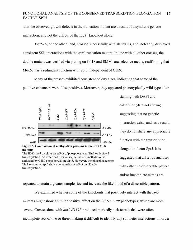
H3K36me3 -‐15 kDa
H3K4me3 -‐15 kDa
!-‐H3 -‐15 kDa Figure 5. Comparison of methylation patterns in the spt5 CTR mutants The H3K4me3 displays an effect of phosphorylated Thr1 on lysine 4 trimethylation. As described previously, lysine 4 trimethylation is activated by Cdk9 phosphorylating Spt5. However, the phosphoacceptor Thr1 residue of Spt5 shows no significant effect on H3K36 trimethylation.
Wild ty
pe
H2b1
-‐K11
9R
Set2
Spt5-‐W
T
Spt5-‐T1A
Spt5-‐T1E
Spt5C

FUNCTIONAL ANALYSIS OF THE CONSERVED TRANSCRIPTION ELONGATION FACTOR SPT5
18
to discern a reliable pattern, these crosses should be repeated in larger numbers, as well. The
sir2+ and rtt109+ knockouts exhibited no added growth defects in combination with htb1-K119R.
Subsequent staining with DAPI and calcofluor revealed familiar septation defects in the double
mutants seen previously in the htb1-K119R alone, usually in the form of cells with multiple
division septa (Figure 5). This, combined with the lack of defect in growth, indicates that these
strains do not interact genetically.
Finally, immunoblot analysis was performed to characterize the dependency of
trimethylation of lysines 4 and 36 of H3 on Spt5 phosphorylation state. Previous knowledge that
Spt5 phosphorylation promotes H3K4me3 was reiterated, along with the absolute requirement of
H2Bub1 for H3K4me3. H3K36me3, on the other hand, does not require H2Bub1. Notably, there
is no apparent difference between H3K36me3 levels between the wild-type strain and the
spt5T1A mutant. Due to prior research showing that Cdk9 activates trimethylation of H3K36, we
suggest that a different substrate exists for Cdk9 that functions to promote H3K36me3. In
addition, H3K36me3 is less dependent on Cdk9 phosphorylation of the spt5 CTR, but appears to
rely upon other functions of the CTR, independent of Cdk9 action. Therefore, the downstream
effect of the Spt5 CTR on lysine 36 trimethylation and subsequent recruitment of histone
modulators provides a potential explanation for the genetic interactions described above
observed solely with the spt5 truncation mutant. However, it is important to note that the
H3K36me3 antibody did not bind efficiently, and the H3K36me3 immunoblot should be
repeated for more accurate results.

FUNCTIONAL ANALYSIS OF THE CONSERVED TRANSCRIPTION ELONGATION FACTOR SPT5
19
DISCUSSION
These results confirm the novel genetic interactions involving the histone variant H2A.Z,
its chaperones Msc1, Swr1, and Swc2, and Spt5 or its upstream modulator Cdk9. Previous
studies have shown that H2A.Z functions in enforcing heterochromatin boundaries. With the
facilitation of the Swr1 complex (Swr1C) as chaperone, a H2A.Z-H2B dimer can substitute into
the nucleosome and restrict chromatin states to their respective domains. Our data suggest that
H2A.Z, and the Swr1C components Msc1, Swr1 and Swc2 display redundancy with Spt5. Msc1
appears to exert a regulatory function on the chaperone complex, as previous work has shown
that H2A.Z is ectopically found in the inner centromere and in subtelomeric chromatin in msc1+
deletion strains (Buchanan et al., 2009). This demonstrates that Msc1 acts as a negative regulator
of H2A.Z loading in these regions, which normally lie past the heterochromatin boundary and
are not actively transcribed. On the other hand, ablation of either Swr1 or Swc2 disrupts the
ability of Swr1C to bind and load H2A.Z into promoter regions of euchromatin (Weber et al.,
2010).
It is possible that aberrant loading of H2A.Z into condensed chromatin and consequent
transcription of inactive genes has more functional overlap with Cdk9 function than the loss of
H2A.Z observed in swr1∆ and swc2∆ mutants. This could account for the specific interaction
observed between the Msc1 and the strains mutated at the Cdk9 phosphorylation site on Spt5’s
CTR. However, this is paradoxical with the finding that loss of H2A.Z itself mimics the growth
defects of msc1∆. To better explain this discrepancy, experiments should be done to further
characterize the nature of the role of Spt5’s CTR in establishing boundaries between chromatin
states.

FUNCTIONAL ANALYSIS OF THE CONSERVED TRANSCRIPTION ELONGATION FACTOR SPT5
20
Generation of point mutations at other putative phosphoacceptor residues along the CTR
of Spt5 would further narrow down the functional overlap between H2A.Z and Spt5, and
minimize the likelihood of structural divergence induced by a truncation mutant. Moreover,
genetic analyses with pht1+ and cdk9+ knockouts would give some insight into what fraction of
the interaction, if any, can be attributed to Cdk9 activity upstream of Spt5 activity.
Furthermore, chromatin boundaries are marked by conserved consensus sequences,
therefore, transformation of a marker adjacent to a boundary consensus sequence makes it
possible to design chromatin boundary assays based on the aberrant spreading of
heterochromatin. Transforming the spt5 strains in this study in such a way and measuring
differences in expression of the marker gene could give insight into whether the CTR of Spt5
plays a role in separating euchromatin from heterochromatin (Weber et al., 2014).
The deletion of nuclear mRNA export receptor mex67 combined with the spt5∆C mutant
formed a distinct synthetic interaction independent of the four proteins described above. Mex67
acts primarily to prevent the export of unspliced pre-mRNAs until intron removal is completed to
avoid the expression of aberrant and potentially harmful proteins (Hackmann et al., 2014). This
loosely implies a shared function in mRNA quality control between the Spt5 CTR and Mex67.
Interestingly, Mex67 and the family of nucleoporins have been shown to interact with
transcription machinery. Certain export factors, such as TREX, bind co-transcriptionally to the
nascent mRNA and facilitate its packaging into a messenger ribonucleoprotein particle (mRNP,
Chanarat et al., 2012). This demonstrates that transcription and nuclear export are intrinsically
intertwined, and that the elaborate complexes forming the nuclear pore are spatially arranged
with nucleosomes in a highly organized manner. Future experiments investigating the exact
function and binding partners of Mex67 could perhaps unveil a role beyond that of mRNA
FUNCTIONAL ANALYSIS OF THE CONSERVED TRANSCRIPTION ELONGATION FACTOR SPT5
21
exporter for Mex67. The possibility of a role in chromatin boundary mechanisms for Mex67
should also be considered, given the consistent pattern in our findings.
Our results also displayed a pattern with relation to the high-throughput screen based on
the magnitude of the initial interactions (data not shown). The genetic interactions that we
successfully recapitulated showed very severe growth defects in the high-throughput screen,
whereas the double mutants that showed no difference in phenotype in the tetrad analysis
exhibited quite minor growth defects in the original screen. We attribute this to two possible
reasons. It is plausible that the high-throughput screen is more sensitive than tetrad analysis, due
to the competitive environment imposed upon the double mutants. In the initial screen, a pool of
cells were induced to mate and sequentially plated on three rounds of selective media, including
cycloheximide on EMM -ura, geneticin (G418), and nourseothricin (NAT). This requires the
double mutants to survive amongst healthier diploid cells and single mutants until their
respective selections, forcing them to contend for nutrients and space. Subtle growth defects may
be augmented as a result. On the other hand, the colonies plated from tetrad analyses are clones
of one cell, therefore no competition between strains exists.
It is, however, more likely that the succession of cycloheximide (a highly toxic protein
synthesis inhibitor) combined with minimal media and subsequent antibiotic treatment inflicted
excessive amounts of stress on the cells, contributing to the detrimental effects of the existing
mutations. This combined with potential interference between each selection is conducive to
generating false positives, not uncommon for a high-throughput screen of this nature (Wiren et
al., 2005).
This type of analysis provides a platform for in-depth investigation of a number of
different Spt5-dependent mechanisms. While much follow-up work is needed to substantiate

FUNCTIONAL ANALYSIS OF THE CONSERVED TRANSCRIPTION ELONGATION FACTOR SPT5
22
these suggested interactions, we have provided good evidence of significant functional overlap
with Spt5 and the five confirmed candidate mutants. Additional tetrad dissections should be
conducted to increase the sample size of the crosses that had incomplete tetrads and/or did not
exhibit a pattern with the aim of discerning a consistent pattern.
ACKNOWLEDGEMENTS
These studies were supported by the Canadian Institute for Health Research (CIHR). I
would like to thank Dr. Jason Tanny for his guidance, and for offering me this opportunity to
participate in this project, Dr. Jean Mbogning for his guidance and advice, and Viviane Pagé for
her technical support.

FUNCTIONAL ANALYSIS OF THE CONSERVED TRANSCRIPTION ELONGATION FACTOR SPT5
23
REFERENCES Ahmed S, Palermo C, Wan S, Walworth NC (2004) A novel protein with similarities to Rb binding
protein 2 compensates for loss of Chk1 function and affects histone modification in fission yeast. Mol Cell Biol 24: 3660-3669.
Azuma Y, Yamagishi M, Ueshima R, Ishihama A (1991) Cloning and sequence determination of the
Schizosaccharomyces pombe rpb1 gene encoding the largest subunit of RNA polymerase II. Nucleic Acids Res 19: 461-468.
Buchanan L, Durand-Dubief M, Roguev A, Sakalar C, Wilhelm B, et al. (2009) The
Schizosaccharomyces pombe JmjC-protein, Msc1, prevents H2A.Z localization in centromeric and subtelomeric chromatin domains. PLoS Genet 5: e1000726.
Campos EI, Reinberg D (2009) Histones: annotating chromatin. Annu Rev Genet 43: 559-599. Chanarat S, Burkert-Kautzsch C, Meinel DM, Strasser K (2012) Prp19C and TREX: interacting to
promote transcription elongationand mRNA export. Transcription 3: 8-12. Chen P, Wang Y, Li G (2014) Dynamics of histone variant H3.3 and its coregulation with H2A.Z at
enhancers and promoters. Nucleus 5. Cherrier T, Le Douce V, Eilebrecht S, Riclet R, Marban C, et al. (2013) CTIP2 is a negative regulator of
P-TEFb. Proc Natl Acad Sci U S A 110: 12655-12660. Core LJ, Lis JT (2008) Transcription regulation through promoter-proximal pausing of RNA polymerase
II. Science 319: 1791-1792. Dimitriadis EK, Weber C, Gill RK, Diekmann S, Dalal Y (2010) Tetrameric organization of vertebrate
centromeric nucleosomes. Proc Natl Acad Sci U S A 107: 20317-20322. Gilchrist DA, Adelman K (2012) Coupling polymerase pausing and chromatin landscapes for precise
regulation of transcription. Biochim Biophys Acta 1819: 700-706. Guo M, Xu F, Yamada J, Egelhofer T, Gao Y, et al. (2008) Core structure of the yeast spt4-spt5
complex: a conserved module for regulation of transcription elongation. Structure 16: 1649-1658.
Hackmann A, Wu H, Schneider UM, Meyer K, Jung K, et al. (2014) Quality control of spliced mRNAs
requires the shuttling SR proteins Gbp2 and Hrb1. Nat Commun 5: 3123. Hartzog GA, Fu J (2013) The Spt4-Spt5 complex: a multi-faceted regulator of transcription elongation.
Biochim Biophys Acta 1829: 105-115. Le Douce V, Colin L, Redel L, Cherrier T, Herbein G, et al. (2012) LSD1 cooperates with CTIP2 to
promote HIV-1 transcriptional silencing. Nucleic Acids Res 40: 1904-1915.

FUNCTIONAL ANALYSIS OF THE CONSERVED TRANSCRIPTION ELONGATION FACTOR SPT5
24
Pei Y, Du H, Singer J, Stamour C, Granitto S, et al. (2006) Cyclin-dependent kinase 9 (Cdk9) of fission
yeast is activated by the CDK-activating kinase Csk1, overlaps functionally with the TFIIH-associated kinase Mcs6, and associates with the mRNA cap methyltransferase Pcm1 in vivo. Mol Cell Biol 26: 777-788.
Quan TK, Hartzog GA (2010) Histone H3K4 and K36 methylation, Chd1 and Rpd3S oppose the
functions of Saccharomyces cerevisiae Spt4-Spt5 in transcription. Genetics 184: 321-334. Rahl PB, Lin CY, Seila AC, Flynn RA, McCuine S, et al. (2010) c-Myc regulates transcriptional pause
release. Cell 141: 432-445. Saiz JE, Fisher RP (2002) A CDK-activating kinase network is required in cell cycle control and
transcription in fission yeast. Curr Biol 12: 1100-1105. Sanso M, Lee KM, Viladevall L, Jacques PE, Page V, et al. (2012) A positive feedback loop links
opposing functions of P-TEFb/Cdk9 and histone H2B ubiquitylation to regulate transcript elongation in fission yeast. PLoS Genet 8: e1002822.
Sato M, Dhut S, Toda T (2005) New drug-resistant cassettes for gene disruption and epitope tagging in
Schizosaccharomyces pombe. Yeast 22: 583-591. Squazzo SL, Costa PJ, Lindstrom DL, Kumer KE, Simic R, et al. (2002) The Paf1 complex physically
and functionally associates with transcription elongation factors in vivo. EMBO J 21: 1764-1774. Tanny JC, Erdjument-Bromage H, Tempst P, Allis CD (2007) Ubiquitylation of histone H2B controls
RNA polymerase II transcription elongation independently of histone H3 methylation. Genes Dev 21: 835-847.
Viladevall L, St Amour CV, Rosebrock A, Schneider S, Zhang C, et al. (2009) TFIIH and P-TEFb
coordinate transcription with capping enzyme recruitment at specific genes in fission yeast. Mol Cell 33: 738-751.
Weber CM, Henikoff JG, Henikoff S (2010) H2A.Z nucleosomes enriched over active genes are
homotypic. Nat Struct Mol Biol 17: 1500-1507. Weber CM, Ramachandran S, Henikoff S (2014) Nucleosomes Are Context-Specific, H2A.Z-Modulated
Barriers to RNA Polymerase. Mol Cell 53: 819-830. Wiren M, Silverstein RA, Sinha I, Walfridsson J, Lee HM, et al. (2005) Genomewide analysis of
nucleosome density histone acetylation and HDAC function in fission yeast. EMBO J 24: 2906-2918.


