Embed Size (px)
Citation preview
Oral Oncology 49 (2013) 845–853
Contents lists available at SciVerse ScienceDirect
Oral Oncology
journal homepage: www.elsevier .com/locate /ora loncology
Review
Salivary gland cancer stem cells
1368-8375/$ - see front matter � 2013 Elsevier Ltd. All rights reserved.http://dx.doi.org/10.1016/j.oraloncology.2013.05.013
⇑ Corresponding author. Address: University of Michigan School of Dentistry,1011 N., University Rm. 2309, Ann Arbor, MI 48109-1078, United States. Tel.: +1(734) 936 9300.
E-mail address: [email protected] (J.E. Nör).
April Adams a, Kristy Warner a, Jacques E. Nör a,b,c,⇑a Department of Restorative Sciences, University of Michigan School of Dentistry, United Statesb Department of Biomedical Engineering, University of Michigan College of Engineering, United Statesc Department of Otolaryngology, University of Michigan School of Medicine, United States
a r t i c l e i n f o s u m m a r y
Article history:Received 13 March 2013Received in revised form 22 May 2013Accepted 31 May 2013Available online 28 June 2013
Keywords:Mucoepidermoid carcinomaAdenoid cystic carcinomaPerivascular nicheChemoresistanceTumor initiating cells
Emerging evidence suggests the existence of a tumorigenic population of cancer cells that demonstratestem cell-like properties such as self-renewal and multipotency. These cells, termed cancer stem cells(CSC), are able to both initiate and maintain tumor formation and progression. Studies have shown thatCSC are resistant to traditional chemotherapy treatments preventing complete eradication of the tumorcell population. Following treatment, CSC are able to re-initiate tumor growth leading to patient relapse.Salivary gland cancers are relatively rare but constitute a highly significant public health issue due to thelack of effective treatments. In particular, patients with mucoepidermoid carcinoma or adenoid cysticcarcinoma, the two most common salivary malignancies, have low long-term survival rates due to thelack of response to current therapies. Considering the role of CSC in resistance to therapy in other tumortypes, it is possible that this unique sub-population of cells is involved in resistance of salivary glandtumors to treatment. Characterization of CSC can lead to better understanding of the pathobiology of sal-ivary gland malignancies as well as to the development of more effective therapies. Here, we make a briefoverview of the state-of-the-science in salivary gland cancer, and discuss possible implications of thecancer stem cell hypothesis to the treatment of salivary gland malignancies.
� 2013 Elsevier Ltd. All rights reserved.
Introduction
Salivary gland cancer is a relatively rare yet deadly disease. Onaverage, 3300 new cases are diagnosed every year in the USA. Dueto limited mechanistic understanding of the disease and lack ofeffective regimens for chemotherapy, surgery is still the main treat-ment option of these patients. As a consequence, treatment for thesetumor is generally accompanied by significant morbidity and debil-itating facial disfigurement. Malignant tumors are generally fatal.This is reflected in the 5-year survival rate that drops drastically from78% for stage I tumors to 25%, 21%, and 23% for stages II–IV, respec-tively.1 Of much concern is the fact that the survival of patients hasnot improved over the last 3 decades, which is in contrast with thesignificant improvement in survival observed in other glandular tu-mors. Such data suggest that focused research efforts on the under-standing of the pathobiology of these tumors could lead tosignificant improvements in patient survival and quality of life.
Mounting evidence supports the existence of a sub-populationof tumorigenic cells that possess stem cell-like characteristics inmany tumor types (e.g. breast cancer, pancreatic cancer, head
and neck squamous cell carcinomas). These cells, termed cancerstem cells (CSC), are capable of self-renewal and also to differenti-ate into cells that make up the bulk of the tumor. Cancer stem cellsare resilient cells that play a major role in resistance to chemother-apy and radiation therapy in other cancer types.2–4 While suchstudies are unveiling the mechanisms of resistance to therapy inother malignancies, very little is known about the resistance of sal-ivary gland tumors. Indeed, one of the most pressing clinical issuesin salivary gland cancer is the poor response to therapy.5 It is cer-tainly possible that low proliferation rates contribute to resistanceto therapy in a group of salivary gland tumors but another possibil-ity is that cancer stem cells play a role in the resistance to therapyobserved in these tumors. Characterization of stem cells in thesetumors might lead to the identification of novel pathways thatcould be targeted to sensitize these tumors to chemotherapy.
Salivary gland structure and function
Salivary glands play an essential role in protection and mainte-nance of health in the oral cavity, lubrication of food, taste of food,and speech. Saliva is produced in secretory cells called acini. Thereare three different types of acini and each is characterized by thecomposition of the cell secretions. Serous cells release saliva thatis abundant in several proteins but lacks mucin protein. Mucouscells secrete saliva-containing mucin proteins attached to carbohy-

846 A. Adams et al. / Oral Oncology 49 (2013) 845–853
drates.6 Seromucous cells secrete a combination of both mucousand serous saliva. Once the saliva is secreted from these cells, itis transported through intercalated ducts, small excretory ducts,and then through a larger excretory duct that opens into themouth.6 Excretory ducts are lined with columnar epithelium,cuboidal cells surround the intercalated ducts, and columnar cellsmake up the striated duct. As the saliva passes through these ducts,additional proteins, such as Immunoglobulin A and lysozyme, fromthe ductal cells are secreted into the saliva. Myoepithelial cells con-tract and help secretory cells release the saliva and also promotesalivary flow through the ducts.
Salivary glands are subdivided into the major and minor glands.The major salivary glands consist of three pairs of glands that are lo-cated around the oral cavity. The largest are the parotid glands thatare located in directly below the ears along the jaw. Saliva is ex-ported from the gland directly across from the crowns of the secondmaxillary molars via the Stensen’s duct, a 5 cm duct connecting thegland to the oral cavity. Secretions from the parotid glands are exclu-sively serous. The sublingual gland is located underneath the floor ofthe mouth and are the smallest of the major salivary glands. Theseglands open to the oral cavity via 8–20 excretory ducts and secreteonly mucous saliva.6 The submandibular glands are also located inthe floor of the month but are adjacent to the mandibular bone. Sal-iva is secreted via the Warthon’s duct that opens into the floor of themouth. This gland secrets seromucous saliva but contains a higherpercentage of serous acini then mucous acini. The oral cavity alsocontains 600–1000 minor salivary glands that can be found on thetongue, inside of the cheek, lips, floor of the mouth, and the hard pal-ate.6 Secretions from these glands are predominately mucous withthe exception of von Ebner’s glands, which are exclusively serous.
Salivary gland cancer
Salivary gland cancers are rare accounting for 2–6.5% of all headand neck cancers with annual incidence of 2.2–3.0 cases per100,000 people in the United States.7–9 Tumors can originate ineither the major or minor salivary glands. Approximately 80% ofthese tumors arise in the parotid gland, 15% arise in the subman-dibular gland, and 5% arise in the minor and sublingual salivaryglands.10 Males have a 51% higher rate of incidence over females,although both tend to develop the cancer within the fifth decadeof life.11 While little is known about the pathogenesis of salivarygland cancers, research has shown that radiation exposure is a riskfactor and suggests that occupation exposures, viruses, UV light,alcohol, and tobacco may also be involved.12–14 As much as 75%of salivary masses are benign. However, presentation of bothmalignant and benign tumors is similar making diagnosis andtreatment very challenging. Malignant salivary gland tumors aremarkedly heterogeneous including 24 histologic subtypes, gener-ating significant challenges in diagnosis, prognosis, and treat-ment.9 The following discussion is centered on mucoepidermoidcarcinomas and adenoid cystic carcinomas (Fig. 1), the two mostcommon salivary gland malignancies.
Mucoepidermoid carcinoma
Mucoepidermoid carcinoma (MEC) is the most common sali-vary gland malignancy and represents approximately 5–15% ofall salivary gland tumors and 30–35% of all malignant salivarygland tumors.8,15–21 These tumors occur in both the major andminor salivary gland glands and are mostly comprised of epider-moid, mucous, and intermediate cells types. The epidermoid cellsare polygonal in shape and characterized by keratinization andintercellular bridges. Mucous cells vary in size but all stain posi-tively for mucin proteins. Intermediate cells are thought to func-
tions as progenitor cells for epidermoid and mucous cells and areoften basal-like in appearance. Mucoepidermoid carcinomas alsocontain a variety of other cell types including squamous, clear,columnar, and other uncommon cell types.22–26 They are extralob-ular tumors and are believed to originate in the excretory duct.22,24
Diagnosis of mucoepidermoid carcinoma is based on the pres-ence of both, histological and cytogenetic abnormalities. These tu-mors are categorized into three grades depending on the amount ofcyst formation, the degree of cytological mutation, and the relativenumber of epidermoid, mucous, and intermediate cell types. Low-grade tumors tend to have a minimal amount of cytological muta-tion, a high population of mucous cell, and noticeable cyst forma-tion. High-grade tumors contain large areas of intermediate andsquamous cells that demonstrate increased mitotic activity. Inter-mediate-grade tumors manifest a combination of both low andhigh-grade characteristics. Additional unfavorable histologic fac-tors include perineural invasion, necrosis, increased mitotic rate,angiolymphatic invasion, anaplasia, infiltrative growth pattern,and the presence of a cystic component.27 However, this gradingsystem is often variable making reproducibility difficult.28 Lowand intermediate-grade tumors are treated using surgical resec-tions while treatment for high-grade tumors includes neck dissec-tion and radiation therapy.27 While surgical removal and radiationis often successful, a significant number of patients have a recur-rence of the disease years later.29 For these patients, few treat-ments options are available as mucoepidermoid carcinomas arehighly chemo-resistant.12 As a result, chemotherapy is used for pa-tient palliation, although ineffective for actual treatment.30 Im-proved understanding of the pathobiology of the disease leadingto rationally designed targeted therapies are necessary to improvethe outcome of patients with mucoepidermoid carcinoma.
The most common cytogenetic abnormality in mucoepidermoidcarcinoma is a recurrent translocation between chromosomes 11and 19 creating the CRTC1-MAML2 fusion protein. This transloca-tion is found in 38–81% of mucoepidermoid carcinomas and is ex-pressed in all cell types. CREB-regulated transcription coactivator 1(CRTC1) protein activates transcription mediated by cAMP re-sponse element-binding (CREB) protein.26,31 CREB activated genesregulate cell differentiation and proliferation.32 Abnormal expres-sion of these genes has been shown to lead to cancer develop-ment.32 MAML2 is a coactivator for Notch transcriptional activitythat regulates cellular differentiation and proliferation.32,33 In thefusion protein, the intracellular Notch-binding domain of MAML2is replaced by the CREB binding domain of CRTC1.12
Many studies have shown that presence of CRTC–MAML2 trans-location has prognostic and diagnostic value.12 Patients with tu-mors expressing CRTC1–MAML2 have a greater overall survivalas well as a lower risk of recurrence and metastasis when com-pared with fusion-negative tumors.34,35 However, there is a subsetof high-grade tumors that express CRTC1–MAML2. Studies by Anz-ick and colleagues found that in these high-grade tumors express-ing CRTC1–MAML2 and additional deletion or hypermethylation ofCDKN2A was often found suggesting that the presence or absenceof both of these abnormalities may serve as a better diagnosticmarker.36 The role this protein plays in the pathogenesis of muco-epidermoid carcinoma is not known, however, research suggeststhat this mutation occurs early on during tumor initiation.12,27
Studies have shown that this translocation also appears in a subsetof Warthin’s tumors and may be linked to the development ofthese tumors to malignant MEC tumors.34,35,37
Adenoid cystic carcinoma
Adenoid cystic carcinoma (ACC) is the second most commonmalignant salivary gland cancer accounting for 10–25% of pa-tients.37–40 Tumors can occur in both parotid and submandibular

Figure 1. Schematic representation of a salivary gland indicating putative areas of origin for mucoepidermoid carcinomas and adenoid cystic carcinomas. Adapted by authorsfrom Bell et al. Salivary gland cancers: biology and molecular targets for therapy. Curr Oncol Rep 2012;14(2):166–174.
A. Adams et al. / Oral Oncology 49 (2013) 845–853 847
glands as well as the minor salivary glands but are believed to arisefrom the intercalated duct reserve cells found in each of theseglands.22,41 Adenoid cystic carcinoma is histologically a biphasiccancer indicating that it is composed of both epithelial and myoep-ithelial cells.22,42,43 Although growth of these tumors is slow, thelong-term prognosis of these patients is poor. The 5-year survivalrates are very favorable at 70–90%.39 However, the 15 and 20-yearsurvival rates are rather poor at 35–40% and 10% respec-tively.39,40,43 Patients with distant metastasis have a 5-year sur-vival rate as low as 20%.38 Overall low survival is primarily dueto the persistence of tumor growth, late recurrence after initialtreatment, perineural invasion, hematogenous spread and invasionto distant and neighboring tissues.44–47
Diagnosis and determination of tumor grade is based solely onthe predominance of one of three histologic growth patterns. Thecribriform pattern is easily characterized as having numerouspseudocysts giving a ‘‘Swiss cheese’’ like appearance. These pseud-ocysts are mostly made of myoepithelial cells. Ductal areas withinthis pattern are composed of basophilic mucoid material. Tumorcells are cuboidal in shape and are small in size. The tubular pat-tern has similar shaped tumors cells, however, they constitutesmall ducts in this case. The inside of these ducts is lined with bothbasal myoepithelial cells and luminal ductal cells. Tumors contain-ing a solid growth pattern demonstrate large islands of tumorscells with no appearance of cysts of tubules. These areas have high-er rates of mitosis, necrosis, and variability in cellular shape. Pre-dominance of tubular and cribriform growth patterns isassociated with less aggressive progression and overall longer sur-vival time.27,40,43 Tumors consisting of >30% solid pattern have thehighest grading, and are associated with increased aggressivenessand lower survival.43 Though regional metastasis is rare, solid tu-mors have a greater likelihood of metastasizing to the lymphnodes.43 Biomarkers of epithelial to mesenchymal transition(EMT) such as Snail1 and Slug have also emerged as being associ-ated with increased tumor aggressiveness and may be useful indiagnosing adenoid cystic carcinoma.39,40 As all grades are consid-ered aggressive, all ACC patients are treated with surgery and radi-ation.43 Chemotherapy treatment has low response rate. However,improved understanding of the biology of adenoid cystic carcino-mas is leading to clinical trials using target therapies known towork in other cancer types.43
The most common cytological abnormality is a translocationbetween chromosomes 6 and 9 resulting in the MYB–NFIB fusionprotein.48 This translocation occurs in 33–100% of primary ACCsamples and expression is not correlated with aggressiveness orgrade.31,48,49 Human nuclear factor 1 (NFI) transcription factor con-tains domains that enable dimerization and DNA-binding. MYB is atranscription factor that regulates genes involved in proliferation,differentiation, and apoptosis. The t(6;9) translocation typically re-sults in loss of exon 15 in the MYB protein, a site shown to bind mi-cro-RNAs, which in turn negatively regulate expression of MYB.48
This leads to overexpression of the fusion protein and overexpres-sion of MYB induced genes, which are involved in cell cycle control,angiogenesis, and apoptosis.48
Recent work has shown that c-Kit and epidermal growth factor(EGF) tyrosine kinase receptors are also over expressed in adenoidcystic carcinoma.50–55 Although it is uncertain the exact mecha-nism by which overexpression of c-Kit influences cancer growthand progression, it has been suggested that it may influence vari-ous genetic and epigenetic processes. EGFR overexpression is com-monly found in cancer and promotes cancer development throughinhibition of apoptosis and stimulation of angiogenesis.10 Imatinibmethylate has been successfully used to inhibit tyrosine kinasereceptors in other cancers such as chronic myelogenous leukemiaand gastrointestinal stromal tumors.56–58 However, little or no re-sponse has been observed in adenoid cystic carcinoma.59 Furtherstudies in c-Kit and EGFR may have uncovered the reason for thislack of response. Work by the Bell laboratory demonstrated thatexpression of c-Kit is found in the inner ductal cells but negativein the myoepithelial cells. Interestingly, the myoepithelial cellsshowed strong expression of EGFR while the ductal cells showedvery little expression. This difference in expression between celltypes could lead to complex patterns of drug response. Further re-search will be necessary to determine the full therapeutic potentialof these targets.27
Cancer stem cell hypothesis
The cancer stem cell hypothesis states that tumors are initiatedand maintained by a sub-population of tumorigenic cells capableof continuous self-renewal and differentiation. The idea that stemcells could initiate cancer progression was first suggested over
848 A. Adams et al. / Oral Oncology 49 (2013) 845–853
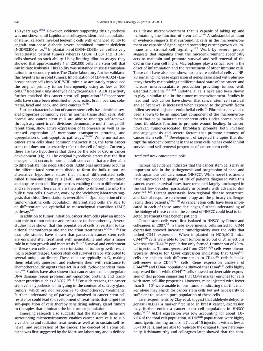
150 years ago.60,61 However, evidence supporting this hypothesiswas not shown until Lapidot and colleagues identified a populationof stem-like acute myeloid leukemic cells with enhanced ability toengraft non-obese diabetic severe combined immune-deficient(NOD/SCID) mice.62 Implantation of CD34+ CD38� cells effectivelyrecapitulated patient tumors whereas CD34+ CD38+ and CD34�cells showed no such ability. Using limiting dilution assays, theyshowed that approximately 1 in 250,000 cells is a stem cell thatcan initiate leukemia. This ability was sustained in serial transplan-tation into secondary mice. The Clarke laboratory further validatedthis hypothesis in solid tumors. Implantation of CD44+/CD24�Lin-breast cancer cells into NOD/SCID mice also accurately reproducedthe original primary tumor heterogeneity using as few as 100cells.63 Isolation using aldehyde dehydrogenase 1 (ALDH1) activityfurther enriched this cancer stem cell population.64 Cancer stemcells have since been identified in pancreatic, brain, ovarian, colo-rectal, head and neck, and liver cancers.65–96
Further characterization of cancer stem cells has identified sev-eral properties commonly seen in normal tissue stem cells. Bothnormal and cancer stem cells are able to undergo self-renewalthrough asymmetric cell divisions, demonstrate multi-lineage dif-ferentiation, show active expression of telomerase as well as in-creased expression of membrane transporter proteins, andupregulation of anti-apoptotic pathways.97–100 While normal andcancer stem cells share common characteristics, the term cancerstem cell does not necessarily refer to the cell of origin. Currentlythere are two hypotheses that describe the role of CSC in cancerdevelopment (Fig. 2). The original hypothesis states that the firstoncogenic hit occurs in normal adult stem cells that are then ableto differentiate into neoplastic cells. Additional mutations occur asthe differentiated stem cells divide to form the bulk tumor. Analternative hypothesis states that normal differentiated cells,called tumor-initiating cells (T-IC), undergo oncogenic mutationand acquire stem cell-like properties enabling them to differentiateand self-renew. These cells are then able to differentiate into thebulk tumor cells. However, work by the Weinberg laboratory sug-gests that this differentiation is reversible.101 Upon depletion of thetumor-initiating cells population, differentiated cells are able tode-differentiate via epithelial to mesenchymal transition (EMT)pathway.102
In addition to tumor initiation, cancer stem cells play an impor-tant role in tumor relapse and resistance to chemotherapy. Severalstudies have shown that this population of cells is resistant to tra-ditional chemotherapeutic and radiation treatments.3,4,103–106 Forexample, studies have shown that pancreatic cancer stem cellsare enriched after treatment with gemcitabine and play a majorrole in tumor growth and metastasis.65,107 Survival and enrichmentof these stem cells allows for re-initiation of tumor growth result-ing in patient relapse. Cancer stem cell survival can be attributed toseveral unique attributes. These cells are typically in G0 makingthem relatively quiescent and endowing them with resistance tochemotherapeutic agents that act in a cell cycle-dependent man-ner.108 Studies have also shown that cancer stem cells upregulateDNA damage repair proteins, anti-apoptotic proteins, and trans-porter proteins such as ABCG2.109–115 For such reasons, the cancerstem cells hypothesis is intriguing in the context of salivary glandtumors, which are not responsive to chemotherapy treatments.Further understanding of pathways involved in cancer stem cellresistance could lead to development of treatments that target thissub-population of cells thereby sensitizing salivary gland tumorsto therapies that eliminate the bulk tumor population.
Emerging research also suggests that the stem cell niche andsurrounding microenvironment enables cancer stem cells to sur-vive chemo and radiation treatments as well as to sustain self-re-newal and progression of the cancer. The concept of a stem cellniche was first suggested by the Morrison laboratory and is defined
as a tissue microenvironment that is capable of taking up andmaintaining the function of stem cells.116 A substantial amountof evidence suggests that surrounding cells in the microenviron-ment are capable of signaling and promoting cancer growth via im-mune and stromal cell signaling.117 Work by several groupssuggests that signaling from the microenvironment specificallyacts to maintain and promote survival and self-renewal of theCSC in the stem cell niche. Macrophages play a critical role in theonset of inflammation and the recruitment of other immune cells.These cells have also been shown to activate epithelial cells via NF-kB signaling, increase expression of genes associated with pluripo-tency thereby maintaining undifferentiated state of the cancer, andincrease microvasculature production providing tumors withessential nutrients.118–121 Endothelial cells have also been shownto play a similar role in the tumor microenvironment. Studies inhead and neck cancer have shown that cancer stem cell survivaland self-renewal is increased when exposed to the growth factormilieu secreted adjacent endothelial cells.93 Fibroblasts have alsobeen shown to be an important component of the microenviron-ment that helps maintain cancer stem cells. Under normal condi-tions, fibroblasts function to promote wound healing. In cancer,however, tumor-associated fibroblasts promote both invasionand angiogenesis and secrete factors that promote stemness ofcancer stem cells.122 Development of targeted therapies that dis-rupt the microenvironment in these stem cells niches could inhibitsurvival and self-renewal properties of cancer stem cells.
Head and neck cancer stem cells
Increasing evidence indicates that the cancer stem cells play animportant role in the pathogenesis and progression of head andneck squamous cell carcinomas (HNSCC). While novel treatmentshave improved the quality of life of patients diagnosed with thiscancer, overall survival rates have remained largely unchanged inthe last few decades, particularly in patients with advanced dis-ease.123–125 Distant metastasis, loco-regional disease recurrence,and lack of response to chemotherapy are the primary challengesfacing these patients.126–130 As cancer stem cells have been impli-cated in each of these same challenges, further understanding ofthe biology of these cells in the context of HNSCC could lead to tar-geted treatments that benefit patients.
Cancer stem cells were first isolated in HNSCC by Prince andcolleagues in 2007.88 In these experiments, cells sorted for CD44expression showed increased tumorigenicity over the cells thatlacked CD44 expression. When implanted in NOD/SCID mice,CD44high cells were able to form tumors in 20 out of 31 injectionswhereas the CD44low population only formed 1 tumor out of 40 to-tal injections. Tumors generated from CD44high cells were pheno-typically diverse for CD44 expression indicating that CD44high
cells are able to both differentiate in to CD44low cells but alsoself-renew into CD44high cells. Gene expression analysis ofCD44high and CD44- populations showed that CD44high cells highlyexpressed Bmi-1 while CD44low cells showed no detectable expres-sion of this protein suggesting that CD44 marker enriches for cellswith stem cell-like properties. However, mice injected with fewerthan 5 � 103 were unable to form tumors indicating that this mar-ker alone may enrich for cancer stem cells but not necessarily besufficient to isolate a pure population of these cells.
Later experiments by Clay et al. suggest that aldehyde dehydro-genase (ALDH), a marker first used in breast cancer, expressionmay further enrich a cancer stem cell population in HNSCCcells.64,131 ALDH expression was low accounting for about 1.0–7.8% of the total cell population. ALDHhigh populations were highlytumorigenic forming tumors in 7 out of 15 injections with as few as50–100 cells, and are able to replicate the original tumor heteroge-neity. Krishnamurthy and colleagues later showed that the com-

Figure 2. Schematic representation of two prevailing hypotheses for tumorigenesis, i.e. the cancer stem cell hypothesis and the tumor-initiating cell hypothesis. Adapted byauthors from Reya et al. Stem cells, cancer, and cancer stem cells. Nature 2001;414(6859):105–111.
A. Adams et al. / Oral Oncology 49 (2013) 845–853 849
bined expression of ALDH and CD44 further enhanced the ability toidentify the cancer stem cell population. Using 1000 ALDHhigh-
CD44high cells, tumors were formed in 13 out 15 injections whileonly 3 out of 15 tumors were seen when 10,000 ALDHlowCD44low
cells were injected.93
Further characterization of the microenvironment surroundinghead and neck cancer stem cells suggests the existence of a peri-vascular niche that supports stem cell maintenance and resistanceto anoikis.93 They observed that approximately 80% of ALDHhigh
cells are located within 100-lm radius of neighboring blood ves-sels. This area was identified as the perivascular niche as it was cal-culated to be the area of diffusion of oxygen and nutrients from theblood vessels. Exposure to endothelial-cell secreted factors en-hanced the expression of ALDH, CD44, and stemness markerBmi-1, as well as a 3-fold increase in the number of orospheresin low attachment conditions suggesting that endothelial cells playan important role in stem cell self-renewal. Ablation of endothelialcells via an artificial caspase-based death switch drastically de-creased the ALDHhighCD44high positive population. Later experi-ments indicated that endothelial cell regulation of cancer stemcells is in part mediated by IL-6. Although not as potent as fullendothelial cell-conditioned media, treatment with rhIL-6 in-creased in vitro orosphere formation and the tumorigenic potentialof cancer stem cells (unpublished observations).
Studies by Campos and colleagues found that upon induction byanchorage and serum starvation, cancer stem cells exposed toendothelial cell-conditioned media were more resistant to anoi-kis.132 This occurred via the PI-3/Akt pathway that is known to reg-ulate proliferation and cell survival. Endothelial cell-conditionedmedia induces phosphorylation of Akt. Blocking VEGF decreasedAkt phosphorylation. Together, these studies provide evidence ofa microenvironment that is capable of supporting and maintainingthe cancer stem cell population. Targeting the crosstalk betweencancer stem cells and other cells of their supportive niche may pro-vide an effective way to abrogate the tumorigenic function of thesecells.
Salivary gland cancer stem cells
The cancer stem cell hypothesis has yet to be fully explored insalivary gland tumors. However, initial experiments by Sun andcolleagues indicate that ALDH isolates cancer stem cells in adenoidcystic carcinomas.133 Using a patient derived xenograft model,
serially diluted ALDHhigh and ALDHlow cells were injected intoNOD/SCID mice. Under these conditions, 24 out of 60 injectionsof 100-1000 ALDHhigh cells were able to form tumors. Notably,injection of as few as 50 ALDHhigh cells was able to generate a tu-mor. No tumors were observed in ALDHlow injected mice at similardilutions of cells. In total, 56/122 injections of ALDHhigh cellsformed tumors were only 5/126 injections of ALDHlow cells wereable to form tumors. ALDHhigh cells also showed an increased abil-ity to form spheres when plated in low-attachment plates as wellas increased invasion in a Matrigel-coated Boyden chamber.ALDHhigh cells infected with luciferase vectors showed increasedability to metastasize when compared to ALDHlow cells. Collec-tively, these data suggest that ALDH may indeed isolate a moretumorigenic population of cells.
Zhou and colleagues further characterized the expression ofALDH in adenoid cystic carcinomas.134 Immunohistochemical anal-ysis of ALDH expression in human tumors indicated three stainingpatterns. Approximately 63% of patient’s samples showed stainingonly in the stromal cells, 26% had neither stromal nor epithelialstaining, and 11% had both epithelial and stromal staining. Normalsalivary gland showed staining for ALDH only in epithelial cells.However, these different patterns had no correlation to tumor size,perineural invasion, or overall survival. Additional experiments areneeded to further verify ALDH as a cancer stem cell marker in ade-noid cystic carcinoma. Studies by Fujita and colleagues found over-lapping populations of CD44 and CD133 markers in adenoid cysticcarcinomas. However, whether or not these markers isolate andmore tumorigenic population of cells has yet to be testing insphere and in vivo models.135
As previously stated, EGFR is commonly upregulated in adenoidcystic carcinoma tumors. Research in other cancer types has dem-onstrated the role of EGF signaling in the self-renewal of CSC sug-gesting this pathway may also play a role in the self-renewal of CSCin adenoid cystic carcinoma. A clinical trial in breast cancer deter-mined that treatment with lapatinib, an EGFR/HER2 inhibitor, sub-stantially reduced the CD44high/CD24low stem cell population. 136
In the same study, the authors showed that lapatinib decreasedmammosphere formation in vitro.136 Korkaya and colleagues fur-ther confirmed these results.137 They showed that HER2 signalingsignificantly increased the Aldefluor stem cell population of nor-mal mammary epithelial cells (NMECs) and increased mammo-sphere formation. Interestingly, HER2 positive but Aldefluornegative cells were unable to form mammospheres. Notably, this

Figure 3. Photomicrographs of spheres formed by mucoepidermoid carcinoma cells at passage 62 or 87 cultured in ultra-low attachment plates indicating the existence ofcells exhibiting stem-like behavior in this cell line.
850 A. Adams et al. / Oral Oncology 49 (2013) 845–853
concept was also confirmed in head and neck squamous cell carci-noma models.138 HNSCC cells lines transfected with EGFRvIII dem-onstrated increased proliferation, decreased sensitivity to cisplatintreatment, and increased the CD44high population. The role of EGFRin adenoid cystic carcinoma CSC has yet to be investigated but maybe important for understanding the self-renewal and tumorigenic-ity of these cells.
In addition to EGFR, c-kit (CD117) is also commonly upregu-lated in adenoid cystic carcinoma. Interestingly, c-kit tyrosinereceptor kinase binds stem cell factor (SCF) to trigger pathways in-volved in the maintenance of progenitor cells.139 Interestingly, thiscell surface receptor is commonly used to isolate progenitor cells insubmandibular glands.140 Expression of c-kit in these sample over-laps with expression of other commonly knows stem cell markers,(e.g. Nanog, Oct3/4), suggesting it plays an important role in themaintenance of stem cell properties.143 As a marker of stem cellsin normal salivary gland, c-kit could also be a potential markerfor CSC in adenoid cystic carcinoma as well. Studying the role ofthis protein in the context of CSC could provide useful insight intothe isolation and regulation of these cells.
Unpublished work for our group also suggests the presence ofCSC in mucoepidermoid carcinoma. Using the mucoepidermoidcarcinoma cells lines that our laboratory has generated, we are ableto generate orospheres (Fig. 3). These structures, first characterizedin head and neck squamous cell carcinoma CSC, exploit the factthat stem cells possess anchorage independent growth.141–143 Byculturing these cells in low attachment and serum free conditions,we are able to generate orospheres in unsorted cells suggestingthat these cell lines do indeed contain a unique stem cell popula-tion. Cells cultured in full 10% FBS were unable to form spheresand demonstrated lower viability in low attachment compared tocells cultured in serum free suggesting that our conditions do in-deed isolate a stem cell-like population.
Conclusions
The most pressing clinical challenges in treatment of salivarygland cancers are tumor resistance to chemotherapy and the lackof targeted treatments that are safe and effective in these tumors.While surgery and radiation treatment successfully cure a subsetof these patients, many present recurrent and/or metastatic dis-ease several years later leading to significant morbidity. Cancerstem cells have been shown to be resistant to chemotherapy andradiation treatments leading to tumor relapse. These cells are alsoimplicated in the progression and development of metastasis. It ispossible that cancer stem cells are involved in the processes thatresult in the late recurrence or metastases that are frequently ob-served in patients with salivary malignancies. Therefore, selective
targeting of this rare sub-population of tumorigenic cancer stemcells could inhibit tumor recurrence and metastasis, and improvepatient survival and quality of life.
Conflict of interest statement
The authors declare no conflict of interests associated with thismanuscript.
Acknowledgments
We thank Kenneth Rieger for his help with the illustrations in-cluded here. Support for this work was provided by Weathermaxfoundation, University of Michigan Comprehensive Cancer Center;grant from the Adenoid Cystic Carcinoma Research Foundation(AACRF); grant P50-CA-97248 (University of Michigan Head NeckSPORE) from the NIH/NCI, and R21-DE19279, R01-DE21139, andR01-DE23220 from the NIH/NIDCR.
References
[1] Luukkaa H, Klemi P, Leivo I, Koivunen P, Laranne J, Mäkitie A, et al. Salivarygland cancer in Finland 1991–96: an evaluation of 237 cases. ActaOtolaryngol 2005;125(2):207–14.
[2] Ailles LE, Weissman IL. Cancer stem cells in solid tumors. Curr OpinBiotechnol 2007;18(5):460–6.
[3] Diehn M, Cho RW, Clarke MF. Therapeutic implications of the cancer stem cellhypothesis. Semin Radiat Oncol 2009;19(2):78–86.
[4] Diehn M, Cho RW, Lobo NA, Kalisky T, Dorie MJ, Kulp AN, et al. Association ofreactive oxygen species levels and radioresistance in cancer stem cells.Nature 2009;458(7239):780–3.
[5] Laurie SA, Licitra L. Systemic therapy in the palliative management ofadvanced salivary gland cancers. J Clin Oncol 2006;24(17):2673–8.
[6] Miletich I. Introduction to salivary glands: structure, function and embryonicdevelopment. Front Oral Biol 2010;14:1–20.
[7] Speight PM, Barrett AW. Salivary gland tumors. Oral Dis 2002;8(5):229–40.[8] Spiro RH. Salivary neoplasms: overview of a 35-year experience with 2807
patients. Head Neck Surg 1986;8(3):177–84.[9] Gillespie MB, Albergotti WG, Eisele DW. Recurrent salivary gland cancer. Curr
Treat Options Oncol 2012;13(1):58–70.[10] Bell D, Hanna EY. Salivary gland cancers: biology and molecular targets for
therapy. Curr Oncol Rep 2012;14(2):166–74.[11] Boukheris H, Curtis RE, Land CE, Dores GM. Incidence of carcinoma of the
major salivary glands according to the WHO classification, 1992 to 2006: apopulation-based study in the United States. Cancer Epidemiol BiomarkersPrev 2009;18(11):2899–906.
[12] O’Neill ID. t(11;19) translocation and CRTC1–MAML2 fusion oncogene inmucoepidermoid carcinoma. Oral Oncol 2009;45(1):2–9.
[13] Land CE, Saku T, Hayashi Y, Takahara O, Matsuura H, Tokuoka S, et al.Incidence of salivary gland tumors among atomic bomb survivors, 1950–1987. Evaluation of radiation-related risk. Radiat Res 1996;146(1):28–36.
[14] Mayne ST, Morse DE, Winn DM. Cancer epidemiology and prevention. NewYork: Oxford University Press; 2006. p. 674–696.
[15] Eversole LR, Sabes WR, Rovin S. Aggressive growth and neoplastic potential ofodontogenic cysts: with special reference to central epidermoid andmucoepidermoid carcinomas. Cancer 1975;35(1):270–82.

A. Adams et al. / Oral Oncology 49 (2013) 845–853 851
[16] Ezsias A, Sugar AW, Milling MA, Ashley KF. Central mucoepidermoidcarcinoma in a child. J Oral Maxillofac Surg 1994;52(5):512–5.
[17] Ellis GL, Auclair PL. Tumors of the salivary glands—Atlas of tumor pathology.In: Fascicle 17 Washington: Armed Forces Institute of, Pathology; 1996.
[18] Gingell JC, Beckerman T, Levy BA, Snider LA. Central mucoepidermoidcarcinoma. Review of the literature and report of a case associated with anapical periodontal cyst. Oral Surg Oral Med Oral Pathol 1984;57(4):436–40.
[19] Ito FA, Ito K, Vargas PA, de Almeida OP, Lopes MA. Salivary gland tumors in abrazilian population: a retrospective study of 496 cases. Int J Oral MaxillofacSurg 2005;34(5):533–6.
[20] Luna MA. Salivary mucoepidermoid carcinoma: revisited. Adv Anat Pathol2006;13(6):293–307.
[21] Pires FR, de Almeida OP, de Araujo VC, Kolawski LP. Prognostic factors in headand neck mucoepidermoid carcinoma. Arch Otolaryngol Head Neck Surg2004;130(2):174–80.
[22] Akrish S, Peled M, Ben-Izhak O, Nagler RM. Malignant salivary gland tumorsand cyclo-oxygenase-2: a histopathological and immunohistochemicalanalysis with implications on histogenesis. Oral Oncol 2009;45(12):1044–50.
[23] Alos L, Lugan B, Castillo M, Nadal A, Carreras M, Caballero M, et al. Expressionof membrane-bound mucins (MUC1 and MUC4) and secreted mucins (MUC2,MUC5AC, MUC5B, MUC6 and MUC7) in mucoepidermoid carcinomas ofsalivary glands. Am J Surg Pathol 2005;29(6):806–13.
[24] Azevedo RS, de Almeida OP, Kowalski LP, Pires FR. Comparative cytokeratinexpression in the different cell types of salivary gland mucoepidermoidcarcinoma. Head Neck Pathol 2008;2(4):257–64.
[25] Coxon A, Rozenblum E, Park YS, Joshi N, Tsurutani J, Dennis PA, et al. Mect1–Maml2 fusion oncogene linked to the aberrant activation of cyclic AMP/CREBregulated genes. Cancer Res 2005;65(16):7137–44.
[26] Jaskoll T, Htet K, Abichaker G, Kaye FJ, Melnick M. CRTC1 expression duringnormal and abnormal salivary gland development supports a precursor cellorigin for mucoepidermoid cancer. Gene Expr Patterns 2011;11(1–2):57–63.
[27] Bell D, Holsinger CF, El-Naggar AK. CRTC1/MAML2 fusion transcript in centralmucoepidermoid carcinoma of mandible–diagnostic and histogeneticimplications. Ann Diagn Pathol 2010;14(6):396–401.
[28] Browand BC, Waldron CA. Central mucoepidermoid tumors of the jaws.Report of nine cases and review of the literature. Oral Surg Oral Med OralPathol 1975;40:631–43.
[29] Chen AM, Granchi PJ, Garcia J, Bucci MK, Fu KK, Eisele DW. Local-regionalrecurrence after surgery without postoperative irradiation for carcinomas ofthe major salivary glands: impli- cations for adjuvant therapy. Int J RadiatOncol Biol Phys 2007;67(4):982–7.
[30] Cai BL, Xu XF, Fu SM, Shen LL, Zhang J, Guan SM, et al. Nuclear translocation ofMRP1 contributes to multidrug resistance of mucoepidermoid carcinoma.Oral Oncol 2011;47(12):1134–40.
[31] Bhaijee F, Pepper DJ, Pitman KT, Bell D. New developments in the molecularpathogenesis of head and neck tumors: a review of tumor-specific fusiononcogenes in mucoepidermoid carcinoma, adenoid cystic carcinoma, andNUT midline carcinoma. Ann Diagn Pathol 2011;15(1):69–77.
[32] Wu L, Liu J, Gao P, Nakamura M, Cao Y, Shen H, et al. Transforming activity ofMECT1–MAML2 fusion oncoprotein is mediated by constitutive CREBactivation. EMBO J 2005;24(13):2391–402.
[33] Tonon G, Modi S, Wu L, Kubo A, Coxon AB, Komiya T, et al. t(11;19)(q21;p13)translocation in mucoepidermoid carcinoma creates a novel fusion productthat disrupts a Notch signaling pathway. Nat Genet 2003;33(2):208–430.
[34] Behboudi A, Enlund F, Winnes M, Andren Y, Nordkvist A, Leivo I, et al.Molecular classification of mucoepidermoid carcinomas- prognosticsignificance of the MECT1–MAML2 fusion oncogene. Genes ChromosomesCancer 2006;45(5):470–81.
[35] Okabe M, Miyabe S, Nagatsuka H, Terada A, Hanai N, Yokoi M, et al. MECT1–MAML2 fusion transcript defines a favorable subset of mucoepidermoidcarcinoma. Clin Cancer Res 2006;12(13):3902–7.
[36] Anzick SL, Chen WD, Park Y, Meltzer P, Bell D, El-Naggar AK, et al. Unfavorableprognosis of CRTC1–MAML2 positive mucoepidermoid tumors with CDKN2Adeletions. Genes Chromosomes Cancer 2010;49(1):59–69.
[37] Bell D, Luna MA, Weber RS, Kaye FJ, El-Naggar AK. CRTC1/MAML2 fusiontranscript in Warthin’s tumor and mucoepidermoid carcinoma: evidence fora common genetic association. Genes Chromosomes Cancer2008;47(4):309–14.
[38] Cai Y, Wang R, Zhao YF, Jia J, Sun ZJ, Chen XM. Expression of Neuropilin-2 insalivary adenoid cystic carcinoma: its implication in tumor progression andangiogenesis. Pathol Res Pract 2010;206(12):793–9.
[39] Jiang J, Tang Y, Zhu G, Zheng M, Yang J, Liang X. Correlation betweentranscription factor Snail1 expression and prognosis in adenoid cysticcarcinoma of salivary gland. Oral Surg Oral Med Oral Pathol Oral RadiolEndod 2010;110(6):764–9.
[40] Tang Y, Liang X, Zhu G, Zheng M, Yang J, Chen Y. Expression and importance ofzinc-finger transcription factor Slug in adenoid cystic carcinoma of salivarygland. J Oral Pathol Med 2010;39(10):775–80.
[41] Zhang J, Peng B, Chen X. Expressions of nuclear factor kappaB, inducible nitricoxide synthase, and vascular endothelial growth factor in adenoid cysticcarcinoma of salivary glands: correlations with the angiogenesis and clinicaloutcome. Clin Cancer Res 2005;11(20):7334–43.
[42] Hunt JL. An update on molecular diagnostics of squamous and salivary glandtumors of the head and neck. Arch Pathol Lab Med 2011;135(5):602–9.
[43] Seethala RR. An update on grading of salivary gland carcinomas. Head NeckPathol 2009;3(1):69–77.
[44] Kim KH, Sung MW, Chung PS, Rhee CS, Park CI, Kim WH. Adenoid cysticcarcinoma of the head and neck. Arch Otolaryngol Head Neck Surg1994;120(7):721–6.
[45] Matsuba HM, Spector GJ, Thawley SE, Simpson JR, Mauney M, Pikul FJ.Adenoid cystic salivary gland carcinoma. A histopathologic review oftreatment failure patterns. Cancer 1986;57(3):519–24.
[46] Rapidis AD, Givalos N, Gakiopoulou H, Faratzis G, Stavrianos SD, Vilos GA,et al. Adenoid cystic carcinoma of the head and neck. Clinicopathologicalanalysis of 23 patients and review of the literature. Oral Oncol2005;41(3):328–35.
[47] Xue F, Zhang Y, Liu F, Jing J, Ma M. Expression of IgSF in salivary adenoidcystic carcinoma and its relationship with invasion and metastasis. J OralPathol Med 2005;34(5):295–7.
[48] Persson M, Andrèn Y, Mark J, Horlings HM, Persson F, Stenman G. Recurrentfusion of MYB and NFIB transcription factor genes in carcinomas of the breastand head and neck. Proc Natl Acad Sci USA 2009;106(44):18740–4.
[49] Skálová A, Vanecek T, Sima R, Laco J, Weinreb I, Perez-Ordonez B, et al.Mammary analogue secretory carcinoma of salivary glands, containing theETV6-NTRK3 fusion gene: a hitherto undescribed salivary gland tumor entity.Am J Surg Pathol 2010;34(5):599–608.
[50] Edwards PC, Bhuiya T, Kelsch RD. C-kit expression in the salivary glandneoplasms adenoid cystic carcinoma, polymorphous low-gradeadenocarcinoma, and monomorphic adenoma. Oral Surg Oral Med OralPathol Oral Radiol Endod 2003;95(5):586–93.
[51] Fukuoka M, Yano S, Giaccone G, Tamura T, Nakagawa K, Douillard JY. Multi-institutional randomized phase II trial of gefitinib for previously treatedpatients with advanced non-small-cell lung cancer (The IDEAL 1 Trial). J ClinOncol 2003;21(12):2237–46.
[52] Holst VA, Marshall CE, Moskaluk CA, Frierson Jr HF. KIT protein expressionand analysis of c-kit gene mutation in adenoid cystic carcinoma. Mod Pathol1999;12(10):956–60.
[53] Kris MG, Natale RB, Herbst RS, Lynch Jr TJ, Prager D, Belani CP, et al. Efficacy ofgefitinib, an inhibitor of the epidermal growth factor receptor tyrosine kinase,in symptomatic patients with non-small cell lung cancer: a randomized trial.JAMA 2003;290(16):2149–58.
[54] Mino M, Pilch BZ, Faquin WC. Expression of KIT (CD117) in neoplasms of thehead and neck: an ancillary marker for adenoid cystic carcinoma. Mod Pathol2003;16(12):1224–31.
[55] Perez-Soler R, Phase II. Clinical trial data with the epidermal growth factorreceptor tyrosine kinase inhibitor erlotinib (OSI-774) in non-small-cell lungcancer. Clin Lung Cancer 2004;6(Suppl 1):S20–3.
[56] Heinrich MC, Blanke CD, Druker BJ, Corless CL. Inhibition of KIT tyrosinekinase activity: a novel molecular approach to the treatment of KIT-positivemalignancies. J Clin Oncol 2002;20(6):1692–703.
[57] O’Brien SG, Guilhot F, Larson RA, Gathmann I, Baccarani M, Cervantes F, et al.Imatinib compared with interferon and low-dose cytarabine for newlydiagnosed chronic-phase chronic myeloid leukemia. N Engl J Med2003;348(11):994–1004.
[58] Verweij J, Casali PG, Zalcberg J, LeCesne A, Reichardt P, Blay JY, et al.Progression-free survival in gastrointestinal stromal tumours with high-doseimatinib: randomised trial. Lancet 2004;364(9440):1127–34.
[59] Hotte SJ, Winquist EW, Lamont E, MacKenzie M, Vokes E, Chen EX, et al.Imatinib mesylate in patients with adenoid cystic cancers of the salivaryglands expressing c-kit: a Princess Margaret Hospital phase II consortiumstudy. J Clin Oncol 2005;23(3):585–90.
[60] Cohnheim J. Ueber entzundung und eiterung. Path Anat Physiol Kiln Med1867;40(1–79):63.
[61] Durante F. Nesso fisio-pathologico tra la struttura dei nei materni e la genesidi alcuni tumori maligni. Arch Memor Observ Chir Pract 1874;11:217–26.
[62] Lapidot T, Sirard C, Vormoor J, Murdoch B, Hoanq T, Caceres-Cortes J, et al. Acell initiating human acute myeloid leukaemia after transplantation intoSCIDmice. Nature 1994;367(6464):645–8.
[63] Al-Hajj M, Wicha MS, Benito-Hernandez A, Morrison SJ, Clarke MF.Prospective identification of tumorigenic breast cancer cells. Proc Natl AcadSci USA 2003;100(7):3983–8.
[64] Ginestier C, Hur MH, Charafe-Jauffret E, Monville F, Dutcher J, Brown M, et al.ALDH1 is a marker of normal and malignant human mammary stem cells anda predictor of poor clinical outcome. Cell Stem Cell 2007;1(5):555–67.
[65] Hermann PC, Huber SL, Herrler T, Aicher A, Ellwart JW, Guba M, et al. Distinctpopulations of cancer stem cells determine tumor growth and metastaticactivity in human pancreatic cancer. Cell Stem Cell 2007;1(3):313–23.
[66] Li C, Heidt DG, Dalerba P, Burant CF, Zhang L, Adsay V, et al. Identification ofpancreatic cancer stem cells. Cancer Res 2007;67(3):1030–7.
[67] Li C, Wu JJ, Hynes M, Dosch J, Sarkar B, Welling TH, et al. c-Met is a marker ofpancreatic cancer stem cells and therapeutic target. Gastroenterology2011;141(6):2218–2227e5.
[68] Rasheed ZA, Yang J, Wang Q, Kowalski J, Freed I, Murter C, et al. Prognosticsignificance of tumorigenic cells with mesenchymal features in pancreaticadenocarcinoma. J Natl Cancer Inst 2010;102(5):340–51.
[69] Beier D, Hau P, Proescholdt M, Lohmeier A, Wischhusen J, Oefner PJ, et al.CD133(1) and CD133(–) glioblastoma-derived cancer stem cells showdifferential growth characteristics and molecular profiles. Cancer Res2007;67(9):4010–5.
[70] He J, Liu Y, Zhu T, Zhu J, Dimeco F, Vescovi AL, et al. CD90 is identified as amarker for cancer stem cells in primary high-grade gliomas using tissuemicroarrays. Mol Cell Proteomics 2012;11(6):M111.

852 A. Adams et al. / Oral Oncology 49 (2013) 845–853
[71] Patrawala L, Calhoun T, Schneider-Broussard R, Zhou J, Claypool K, Tang DG.Side population is enriched in tumorigenic, stem-like cancer cells, whereasABCG21 and ABCG22 cancer cells are similarly tumorigenic. Cancer Res2005;65(14):6207–19.
[72] Shu Q, Wong KK, Su JM, Adesina AM, Yu LT, Tsang YT, et al. Direct orthotopictransplantation of fresh surgical specimen preserves CD1331 tumor cells inclinically relevant mouse models of medulloblastoma and glioma. Stem Cells2008;26(6):1414–24.
[73] Singh SK, Clarke ID, Terasaki M, Boon VE, Hawkins C, Squire J, et al.Identification of a cancer stem cell in human brain tumors. Cancer Res2003;63(18):5821–8.
[74] Singh SK, Hawkins C, Clark ID, Squir JA, Bayani L, Hide T, et al. Identification ofhuman brain tumour initiating cells. Nature 2004;432(7015):396–401.
[75] Son MJ, Woolard K, Nam DH, Lee J, Fine HA. SSEA-1 is an enrichment markerfor tumor-initiating cells in human glioblastoma. Cell Stem Cell2009;4(5):440–52.
[76] Baba T, Convery PA, Matsumura N, Whitaker RS, Kondoh E, Perry T, et al.Epigenetic regulation of CD133 and tumorigenicity of CD1331 ovarian cancercells. Oncogene 2009;28(2):209–18.
[77] Bapat SA, Mali AM, Koppikar CB, Kurrey NK. Stem and progenitor-like cellscontribute to the aggressive behavior of human epithelial ovarian cancer.Cancer Res 2005;65(8):3025–9.
[78] Fong MY, Kakar SS. The role of cancer stem cells and the side population inepithelial ovarian cancer. Histol Histopathol 2010;25(1):113–20.
[79] Ferrandina G, Bonanno G, Pierelli L, Perillo A, Procoli A, Mariotti A, et al.Expression of CD133-1 and CD133-2 in ovarian cancer. Int J Gynecol Cancer2008;18(3):506–14.
[80] Szotek PP, Pieretti-Vanmarcke R, Masiakos PT, Dinulescu DM, Connolly D,Foster R, et al. Ovarian cancer side population defines cells with stem cell-likecharacteristics and Mullerian Inhibiting Substance responsiveness. Proc NatlAcad Sci USA 2006;103(30):11154–9.
[81] Wang L, Mezencev R, Bowen NJ, Matyunina LV, McDonald JF. Isolation andcharacterization of stem-like cells from a human ovarian cancer cell line. MolCell Biochem 2012;363(1–2):257–68.
[82] Zhang S, Balch C, Chan MW, Lai HC, Matei D, Schilder JM, et al. Identificationand characterization of ovarian cancer-initiating cells from primary humantumors. Cancer Res 2008;68(11):4311–20.
[83] Dalerba P, Dylla SJ, Part IK, Liu R, Wang X, Cho RW, et al. Phenotypiccharacterization of human colorectal cancer stem cells. Proc Natl Acad SciUSA 2007;104(24):10158–63.
[84] Huang EH, Hynes MJ, Zhang T, Ginestier C, Dontu G, Appelman H, et al.Aldehyde dehydrogenase 1 is a marker for normal and malignant humancolonic stem cells (SC) and tracks SC overpopulation during colontumorigenesis. Cancer Res 2009;69(8):3382–9.
[85] O’Brien CA, Pollett A, Gallinger S, Dick JE. A human colon cancer cell capableof initiating tumour growth in immunodeficient mice. Nature2007;445(7123):106–10.
[86] Ricci-Vitiani L, Lombardi DG, Pilozzi E, Biffoni M, Todaro M, Peschle C, et al.Identification and expansion of human colon-cancer-initiating cells. Nature2007;445(7123):111–5.
[87] Todaro M, Alea MP, Di Stefano AB, Cammareri P, Vermeulen L, Iovino F, et al.Colon cancer stem cells dictate tumor growth and resist cell death byproduction of interleukin-4. Cell Stem Cell 2007;1(4):389–402.
[88] Prince ME, Sivanandan R, Kaczorowski A, Wolf GT, Kaplan MJ, Delerba P, et al.Identification of a subpopulation of cells with cancer stem cell properties inhead and neck squamous cell carcinoma. Proc Natl Acad Sci USA2007;104(3):973–8.
[89] Chen YC, Chen YW, Hsu HS, Tseng LM, Huang PI, Lu KH, et al. Aldehydedehydrogenase 1 is a putative marker for cancer stem cells in head andneck squamous cancer. Biochem Biophys Res Commun2009;385(3):307–13.
[90] Krishnamurthy S, Dong Z, Vodopyanov D, Imai A, Helman JI, Prince ME, et al.Endothelial cell-initiated signaling promotes the survival and self-renewal ofcancer stem cells. Cancer Res 2010;70(23):9969–78.
[91] Cheung PF, Cheng CK, Wong NC, Ho JC, Yip CW, Lui VC, et al. Granulin-epithelin precursor is an oncofetal protein defining hepatic cancer stem cells.PLoS One 2011;6(12):e28246.
[92] Ma S, Chan KW, Hu L, Lee TK, Wo JY, Ng IO, et al. Identification andcharacterization of tumorigenic liver cancer stem/progenitor cells.Gastroenterology 2007;132(7):2542–56.
[93] Ma S, Chan KW, Lee TK, Tang KH, Wo JY, Zheng BJ, et al. Aldehydedehydrogenase discriminates the CD133 liver cancer stem cell populations.Mol Cancer Res 2008;6(7):1146–53.
[94] Yang ZF, Ho DW, Ng MN, Lau CK, Yu WC, Ngai P, et al. Significance of CD90+cancer stem cells in human liver cancer. Cancer Cell 2008;13(2):153–66.
[95] Yang ZF, Ngai P, Ho DW, Yu WC, Ng MN, Lau CK, et al. Identification of localand circulating cancer stem cells in human liver cancer. Hepatology2008;47:919–28.
[96] Yin S, Li J, Hu C, Chen X, Yao M, Yan M, et al. CD133 positive hepatocellularcarcinoma cells possess high capacity for tumorigenicity. Int J Cancer2007;120(7):1444–50.
[97] Cheng L, Ramesh AV, Flesken-Nikitin A, Choi J, Nikitin AY. Mouse models forcancer stem cell research. Toxicol Pathol 2010;38(1):62–71.
[98] Dontu G, Abdallah WM, Foley JM, Jackson KW, Clarke MF. Kawamura, et al. Invitro propagation and transcriptional profiling of human mam- mary stem/progenitor cells. Genes Dev 2003;17(10):1253–70.
[99] Reynolds BA, Weiss S. Clonal and population analyses demonstrate that anEGF-responsive mamma- lian embryonic CNS precursor is a stem cell. DevBiol 1996;175(1):1–13.
[100] Weiss S, Reynolds BA, Vescovi AL, Morshead C, Craig CG, van der Kooy D. Isthere a neural stem cell in the mammalian forebrain? Trends Neurosci1996;19(9):387–93.
[101] Mani SA, Guo W, Liao MJ, Eaton EN, Ayyanan A, Zhou AY, et al. The epithelial–mesenchymal transition generates cells with properties of stem cells. Cell2008;133(4):704–15.
[102] Chaffer CL, Brueckmann I, Scheel C, Kaestli AJ, Wiggins PA, Rodrigues LO, et al.Normal and neoplastic nonstem cells can spontaneously convert to a stem-like state. Proc Natl Acad Sci USA 2011;108(19):7950–5.
[103] Hambardzumyan D, Squatrito M, Holland EC. Radiation resistance and stem-like cells in brain tumors. Cancer Cell 2006;10(6):454–6.
[104] Korkaya H, Paulson A, Charafe-Jauffret E, et al. Regulation of mammary stem/progenitor cells by PTEN/Akt/beta-catenin signaling. PLoS Biol2009;7(6):e1000121.
[105] Reya T, Morrison SJ, Clarke MF, Weissman IL. Stem cells, cancer, and cancerstem cells. Nature 2001;414(6859):105–11.
[106] Shafee N, Smith CR, Wei S, Kim Y, Mills GB, Hortobagyi, et al. Cancer stemcells contribute to cisplatin resistance in Brca1/p53-mediated mousemammary tumors. Cancer Res 2008;68(9):3243–50.
[107] Simeone DM. Pancreatic cancer stem cells: implications for the treatment ofpancreatic cancer. Clin Cancer Res 2008;14(18):5646–8.
[108] Venezia TA, Merchant AA, Ramos CA, Whitehouse NL, Young AS, Shaw CA,et al. Molecular signatures of proliferation and quiescence in hematopoieticstem cells. PLoS Biol 2004;2(10):e301.
[109] Cai J, Weiss ML, Rao MS. In search of ‘‘stemness’’. Exp Hematol2004;32(7):585–98.
[110] Cairns J. The cancer problem. Sci Am 1975;233(5):64–72. 68–77.[111] Cairns J. Somatic stem cells and the kinetics of mutagenesis and
carcinogenesis. Proc Natl Acad Sci USA 2002;99(16):10567–70.[112] Park Y, Gerson SL. DNA repair defects in stem cell function and aging. Annu
Rev Med 2005;56:495–508.[113] Potten CS, Owen G, Booth D. Intestinal stem cells protect their genome by
selective segregation of template DNA strands. J Cell Sci 2002;115(11):2381–8.[114] Wang S, Yang D, Lippman ME. Targeting Bcl-2 and Bcl-XL with
nonpeptidic small-molecule antagonists. Semin Oncol 2003;30(5 Suppl16):133–42.
[115] Wicha MS, Liu S, Dontu G. Cancer stem cells: an old idea–a paradigm shift.Cancer Res 2006;66(4):1883–90.
[116] Morrison SJ, Spradling AC. Stem cells and niches: mechanisms that promotestem cell maintenance throughout life. Cell 2008;132(4):598–611.
[117] Tlsty TD, Coussens LM. Tumor stroma and regulation of cancer development.Annu Rev Pathol 2006;1:119–50.
[118] Polverini PJ, Cotran PS, Gimbrone Jr MA, Unanue ER. Activated macrophagesinduce vascular proliferation. Nature 1977;269:804–6.
[119] Cao JX, Cui YX, Long ZJ, Dai ZM, Lin JY, Liang Y, et al. Pluripotency-associated genes in human nasopharyngeal carcinoma CNE-2 cells arereactivated by a unique epigenetic sub-microenvironment. BMC Cancer2010;10:68.
[120] Kim SJ, Kim JS, Papadopoulos J, WookKim S, Maya M, Zhang F, et al.Circulating monocytes expressing CD31: implications for acute and chronicangiogenesis. Am J Pathol 2009;174(5):1972–80.
[121] Oguma K, Oshima H, Aoki M, Uchio R, Naka K, Nakamura S, et al. Activatedmacrophages promote Wnt signalling through tumour necrosis factor-alphain gastric tumour cells. EMBO J 2008;27(12):1671–81.
[122] Galiè M, Konstantinidou G, Peroni D, Scambi I, Marchini C, Lisi V, et al.Mesenchymal stem cells share molecular signature with mesenchymaltumor cells and favor early tumor growth in syngeneic mice. Oncogene2008;27(18):2542–51.
[123] Ferlay J, Parkin DM, Steliarova-Foucher E. Estimates of cancer incidence andmortality in Europe in 2008. Eur J Cancer 2010;46(4):765–81.
[124] Haddad RI, Shin DM. Recent advances in head and neck cancer. N Engl J Med2008;359(11):1143–54.
[125] Jemal A, Bray F, Center MM, Ferlay J, Ward E, Forman D. Global cancerstatistics. CA Cancer J Clin 2011;61(2):69–90.
[126] Allegra E, Baudi F, La Boria A, Fagiani F, Garozzo A, Costanzo FS. Multiple headand neck tumours and their genetic relationship. Acta Otorhinolaryngol Ital2009;29(5):237–41.
[127] Franchin G, Minatel E, Gobitti C, Talamini R, Vaccher E, Sartor G, et al.Radiotherapy for patients with early-stage glottic carcinoma: univariate andmultivariate analyses in a group of consecutive, unselected patients. Cancer2003;98(4):765–72.
[128] Jones AS, Morar P, Phillips DE, Field JK, Husband D, Helliwell TR. Secondprimary tumors in patients with head and neck squamous cell carcinoma.Cancer 1995;75(6):1343–53.
[129] Leemans CR, Tiwari R, Nauta JJ, van der Waal I, Snow GB. Recurrence at theprimary site in head and neck cancer and the significance of neck lymph nodemetastases as a prognostic factor. Cancer 1994;73(1):187–90.
[130] Sjögren EV, Wiggenraad RG, Le Cessie S, Snijder S, Pomp J, Baatenburg de JongRJ. Outcome of radiotherapy in T1 glottic carcinoma: a population-basedstudy. Eur Arch Otorhinolaryngol 2009;266(5):735–44.
[131] Clay MR, Tabor M, Owen JH, Carey TE, Bradford CR, Wolf GT, et al. Single-marker identification of head and neck squamous cell carcinoma cancer stemcells with aldehyde dehydrogenase. Head Neck 2010;32(9):1195–201.

A. Adams et al. / Oral Oncology 49 (2013) 845–853 853
[132] Campos MS, Neiva KG, Meyers KA, Krishnamurthy S, Nör JE. Endothelialderived factors inhibit anoikis of head and neck cancer stem cells. Oral Oncol2012;48(1):26–32.
[133] Sun S, Wang Z. ALDH high adenoid cystic carcinoma cells display cancer stemcell properties and are responsible for mediating metastasis. BiochemBiophys Res Commun 2010;396(4):843–8.
[134] Zhou JH, Hanna EY, Roberts D, Weber RS, Bell D. ALDH1immunohistochemical expression and its significance in salivary adenoidcystic carcinoma. Head Neck 2013;35(4):575–8.
[135] Fujita S, Ikeda T. Cancer stem-like cells in adenoid cystic carcinoma ofsalivary glands: relationship with morphogenesis of histological variants. JOral Pathol Med 2012;41(3):207–13.
[136] Li X, Lewis MT, Huang J, Gutierrez C, Osborne CK, Wu MF, et al. Intrinsicresistance of tumorigenic breast cancer cells to chemotherapy. J Natl CancerInst 2008;100(9):672–9.
[137] Korkaya H, Paulson A, Iovino F, Wicha MS. HER2 regulates the mammarystem/progenitor cell population driving tumorigenesis and invasion.Oncogene 2008;27(47):6120–30.
[138] Abhold EL, Kiang A, Rahimy E, Kuo SZ, Wang-Rodriguez J, Lopez JP, et al. EGFRkinase promotes acquisition of stem cell-like properties: a potentialtherapeutic target in head and neck squamous cell carcinoma stem cells.PLoS One 2012;7(2):e32459.
[139] Cheng M, Qin G. Progenitor cell mobilization and recruitment: SDF-1, CXCR4,a4-integrin, and c-kit. Prog Mol Biol Transl Sci 2012;111:243–64.
[140] Lombaert IM, Hoffman MP. Epithelial stem/progenitor cells in the embryonicmouse submandibular gland. Front Oral Biol 2010;14:90–106.
[141] Dontu G, Wicha MS. Survival of mammary stem cells in suspension culture:implications for stem cell biology and neoplasia. J Mammary Gland BiolNeoplasia 2005;10(1):75–86.
[142] Reynolds BA, Weiss S. Generation of neurons and astrocytes from isolatedcells of the adult mammalian central nervous system. Science1992;255(5052):1707–10.
[143] Krishnamurthy S, Nör JE. Orosphere assay: a method for propagation of headand neck cancer stem cells. Head Neck 2012. http://dx.doi.org/10.1002/hed.23076 [Epub ahead of print].


