Embed Size (px)
Citation preview
Capsaicin-Induced Activation of p53-SMAR1 Auto-Regulatory Loop Down-Regulates VEGF in Non-Small CellLung Cancer to Restrain AngiogenesisSamik Chakraborty1, Arghya Adhikary1., Minakshi Mazumdar1., Shravanti Mukherjee1,
Pushpak Bhattacharjee1, Deblina Guha1, Tathagata Choudhuri2, Samit Chattopadhyay3,
Gaurisankar Sa1, Aparna Sen4, Tanya Das1*
1 Division of Molecular Medicine, Bose Institute, Kolkata, West Bengal, India, 2 Department of Biotechnology, Viswa-Bharati, Santiniketan, West Bengal, India, 3 National
Center for Cell Science, Pune University, Pune, Maharashtra, India, 4 Department of Microbiology, Lady Brabourne College, Kolkata, West Bengal, India
Abstract
Lung cancer is the leading cause of cancer-related deaths worldwide. Despite decades of research, the treatment options forlung cancer patients remain inadequate, either to offer a cure or even a substantial survival advantage owing to its intrinsicresistance to chemotherapy. Our results propose the effectiveness of capsaicin in down-regulating VEGF expression in non-small cell lung carcinoma (NSCLC) cells in hypoxic environment. Capsaicin-treatment re-activated p53-SMAR1 positive feed-back loop in these cells to persuade p53-mediated HIF-1a degradation and SMAR1-induced repression of Cox-2 expressionthat restrained HIF-1a nuclear localization. Such signal-modulations consequently down regulated VEGF expression tothwart endothelial cell migration and network formation, pre-requisites of angiogenesis in tumor micro-environment. Theabove results advocate the candidature of capsaicin in exclusively targeting angiogenesis by down-regulating VEGF intumor cells to achieve more efficient and cogent therapy of resistant NSCLC.
Citation: Chakraborty S, Adhikary A, Mazumdar M, Mukherjee S, Bhattacharjee P, et al. (2014) Capsaicin-Induced Activation of p53-SMAR1 Auto-Regulatory LoopDown-Regulates VEGF in Non-Small Cell Lung Cancer to Restrain Angiogenesis. PLoS ONE 9(6): e99743. doi:10.1371/journal.pone.0099743
Editor: A. R. M. Ruhul Amin, Winship Cancer Institute of Emory University, United States of America
Received December 31, 2013; Accepted May 16, 2014; Published June 13, 2014
Copyright: � 2014 Chakraborty et al. This is an open-access article distributed under the terms of the Creative Commons Attribution License, which permitsunrestricted use, distribution, and reproduction in any medium, provided the original author and source are credited.
Funding: SC- University Grants Commission- UGC 2-8/2002(SA I)(SA-I)dated 06.11.2008. AA- Department of Science and Technology- DST R/3393/06 dated16.10.2006. MM- Department of Biotechnology-DBT-RA dated 05.06.2009. SM- Council of Scientific and Industrial Research- CSIR 09/015(0386)/2010 EMR-1 dated22.02.2010. PB- Department of Biotechnology-DBT-RA dated 01.06.2010. DG- University Grants Commission- UGC 2-8/2002(SA I) dated 21.04.2011. The fundershad no role in study design, data collection and analysis, decision to publish, or preparation of the manuscript.
Competing Interests: The authors have declared that no competing interests exist.
* E-mail: [email protected]
. These authors contributed equally to this work.
Introduction
Highly resistant non small-cell lung carcinoma (NSCLC) that
comprises 80% of all lung cancers is intrinsically resistant to
chemotherapy and/or irradiation therapy. Since, angiogenesis is
essential for NSCLC growth and metastasis, therefore controlling
tumor-associated angiogenesis can be a promising tactic in limiting
NSCLC progression. Several pro-angiogenic factors such as
vascular endothelial growth factor (VEGF) are highly expressed in
the tumor microenvironment and strongly induce tumor angio-
genesishttp://www.ncbi.nlm.nih.gov/pmc/articles/PMC3827603/
- b3 [1]. This shift of the tumor microenvironment to an
angiogenic state, or ‘‘angiogenic switch’’ [1,2], is an important
rate limiting factor in tumor development.
Expression of the VEGF gene has been shown to be upregulated
by hypoxia [3–5] and turnover of VEGF is mediated by the
hypoxia-inducible factor-1 (HIF-1) [2,3]. Under normoxic condi-
tions, HIF-1a levels are strongly regulated by oxygen tension
through hydroxylation of prolyl residues, while hypoxic conditions
hinder prolyl hydroxylation of HIF-1a [4] and the protein is
stabilized, enabling it to transactivate target genes like VEGF [3].
A wealth of reports tightly link HIF-1a to p53 in an inverse
relationship in which p53 inhibits HIF-1a transcription [6] and
induces its degradation under several sub-cellular conditions of
stress [7] thereby http://www.jbc.org/search?author1 = Joanna+Zawacka-Pankau&sortspec = date&submit = Submitresulting in its
potent repression. Interestingly, p53 is stabilized by SMAR1, a
scaffold matrix-associated region-binding protein, through dis-
placement of Mdm2 from p53 N-terminal pocket and hence
rescuing p53 from the Mdm2-mediated proteasomal degradation
[8]. Contemporary reports [9,10] demonstrate that on mild DNA
damage SMAR1 promotes p53 deacetylation through recruitment
of HDAC1 and specifically represses Bax and Puma expression
thereby inhibiting apoptosis. These reports not only attest the
candidature of SMAR1 in modulating the activity of p53 but also
raise the possibility of involvement of p53 in other cellular
functions in the mild DNA-damaging micro-environment of the
cell. Importantly, several studies have also identified complex
cross-talks between p53 and Cox-2, whereby Cox-2 suppresses
p53-network in cancer cells [11,12] and vice versa [12]. All these
information tempted us to render insight into the existence of an
interactive relationship between SMAR1, p53, Cox-2 and HIF1-a,
if any, that decides fate of VEGF expression during tumor
angiogenesis in NSCLC. Inhibition of tumor growth by anti-
angiogenic agents has been achieved where promising antitumor
responses have been reported for a variety of anti-VEGF agents.
PLOS ONE | www.plosone.org 1 June 2014 | Volume 9 | Issue 6 | e99743
However, toxicity of most of these drugs as well as development of
resistance towards those agents signify the necessity of identifying
alternative non-toxic agents to achieve the continuous inhibition of
angiogenesis for effective NSCLC therapy.
Increasing evidence indicated the anticancer effects of capsaicin
(8-methyl-N-vanillyl-6-nonenamide), the active component of chili
pepper, against various cancers [13–18]. Capsaicin has been
reported to provoke apoptosis and restricts benzo(a)pyrene
induced lung tumorigenesis in Swiss albino mice [19] and
alleviates the imbalance in xenobiotic metabolizing enzymes and
tumor markers [20]. According to Anandakumar et al. [20]
capsaicin modulates pulmonary antioxidant defense system during
benzo(a)pyrene-induced lung cancer in Swiss albino mice [19].
Furthermore, capsaicin displays anti-proliferative activity against
human small cell lung cancer in cell culture and nude mice models
via the E2F pathway [21]. Recent report from our laboratory has
shown the apoptogenic effect of capsaicin on human NSCLC cells
[22]. However, there is no report on the role of this phytochemical
on NSCLC-induced angiogenesis. In addition, although capsacin
has been shown to suppress human fibrosarcoma-induced
angiogenesis in chick chorioallantoic membrane assay [23] by
inhibiting VEGF-induced proliferation, and capillary-like tube
formation of primary cultured human endothelial cells, the effect
of this phytochemical on the VEGF expression in NSCLC cell has
not yet been explored in detail.
Our results signify that while impediment of p53-SMAR1 loop
induced VEGF expression in NLCSC cells thereby favoring
endothelial cell (EC) migration and network formation in tumor
environment, reframing of these pro-angiogenic signals by
capsaicin blocked VEGF production even under hypoxic condi-
tion, thereby restraining NSCLC-induced net work formation by
the endothelial cells. Such development in understanding may
offer the panorama of exclusively targeting pro-angiogenic factors
and pathways to achieve more efficient and cogent lung cancer
therapy.
Materials and Methods
Cell cultureHuman cancer cell lines, A549, MCF-7, HeLa, HCT-15, and
normal lung fibroblast WI-38, were obtained from National
Centre for Cell Science, India, and maintained at 37uC and 5%
CO2 in DMEM medium supplemented with 10% FBS (Lonza,
NH), L-glutamine (2 mM), sodium pyruvate (100 mg/ml), nones-
sential amino acids (100 mM), streptomycin (100 mg/ml), penicillin
(50 U/ml; Invitogen, CA) [24]. Human umbilical vein endothelial
cells (HUVEC) were obtained from HIMEDIA Cell Culture
(Mumbai, India), and maintained at 37uC and 5% CO2 in M199
(Gibco, Grand Island, NY) supplemented with 20% FBS and EC
growth supplement (ECGS; BD Biosciences, Bedford, MA). All
experiments were performed with HUVECs between passages 2–
5. Viable cell numbers were determined by Trypan blue exclusion
test [11].
To mimic hypoxia, A549 cells were grown in presence of
100 mmol/L CoCl2 (Sigma, St Louis). Cells were treated with
capsaicin (Sigma, St. Louis) as required in experiment. When
required, A549 cells were treated with Brefeldin A (1 mg/ml,
Calbiochem, NJ) and/or MG 132 (10 mM, Sigma, St Louis) and/
or recombinant VEGF (0.8 ng/ml, to keep equivalent amount of
VEGF that was present in Hy-A549 spent media which was used
at 1:1 ratio with M199 on endothelial cell; Peprotech, CA) and/or
VEGF neutralizing antibody (25 ng/ml, R&D Biosystems, MN),
6 hours before harvesting cells.
Wound healing assayWound healing assay was performed to assess the migration of
HUVECs. Cells were seeded in 12-well plates and allowed to form
confluent monolayers. The monolayers were then scratched
horizontally using a sterile 100 ml pipette tip. Then cells were
cultivated in medium containing 0.5% FBS overnight and exposed
to different concentration of agents. Cells were washed with PBS
and cultivated in DMEM medium. Bright field images of the
randomly selected views along the scraped line were taken.
Migration was quantified by a semi-automated computer-assisted
procedure by a person blinded with respect to the experimental-
treatment. The data from triplicate wells were calculated as the
mean +/2 SEM. The migration rate of control cells was taken as
100% and that of other plates were compared with control cells
[25].
Sprout formation assayMatrigel Matrix (BD Biosciences, Heidelberg, Germany) were
thawed at 4uC overnight. 12-well plate was coated with 500 ml
Matrigel/per well and incubated at 37uC for 20 min for
polymerization. HUVECs (16105) were re-suspended in 500 ml
M199 containing 2% FBS and different concentration of agents,
and seeded onto the Matrigel-coated plate. After 6–8 h incuba-
tion, HUVECs in control group formed tube-like structure, which
were defined as endothelial cord formations that connected at both
ends. Tube formation was visualized by microscopically (Olympus
IX71) and photographs of 10 randomly selected fields were
captured by CCD camera.
Western blotting and co-immunoprecipitationCells were homogenized in Hepes buffer (20 mM Hepes,
pH 7.5, 10 mM KCl, 1.5 mM MgCl2, 1 mM Na-EDTA, 1 mM
Na-EGTA, and 1 mM DTT) supplemented with protease and
phosphatase inhibitor mixtures [24]. Total protein concentration
of cell lysate was estimated by Lowry’s method. Equal amounts of
protein were subjected to SDS-PAGE, and then electrically
transferred onto PVDF membranes (Millipore, Darmstadt,
Germany). Subsequently, the membrane was blocked for 1 h with
5% non-fat milk in TBS-0.1% Tween 20 (TBST) and probed with
specific antibodies like, anti-VEGF, anti-TGF-b, anti-EGF, anti
bFGF, anti-HIF-1a, anti-p53, anti-Cox-2,anti-SMAR1/BANP,
anti-H1-Histone and anti-a-Actin (Santa Cruz, CA), anti-phos-
pho-ser15-p53, anti-EGFR (Cell signalling, MA). The protein of
interest was visualized by chemiluminescence (GE Biosciences, NJ)
[24]. For co-immunoprecipitation, immunocomplexes from whole
cell lysate were purified using specific antibody and protein A-
Sepharose beads (Invitrogen, MD) and immunoblotted. The
protein of interest was visualized by chemi-luminescence. Equiv-
alent protein loading was verified using anti-a-actin antibodies.
Flow cytometryFor determination of apoptotic cell death, cells were stained
with propidium iodide (PI) and Annexin-V-FITC (BD Pharmin-
gen, CA) and analyzed on flow cytometer (FACS Callibur, BD
Biosciences, CA). Electronic compensation of the instrument was
done to exclude overlapping of the emission spectra. Total 10,000
events were acquired for analysis using CellQuest software (BD
Biosciences, CA) [26]. Annexin-V-positive cells were regarded as
early apoptotic cells [24]. For quantification of intracellular
VEGF, cells pre-treated with Brefelidin A were harvested, washed
with PBS, fixed with 4% para-formaldehyde for 10 min and
permeabilized with 0.1% Triton-X-100 for 5 min and incubated
with anti-VEGF antibody followed by TRITC-conjugated sec-
Capsaicin Downregulates VEGF-Mediated Angiogenesis
PLOS ONE | www.plosone.org 2 June 2014 | Volume 9 | Issue 6 | e99743
ondary antibody. Fluorescence was determined flow cytometrically
using CellQuest software (Becton Dickinson, San Jose, CA).
Fluorescence imagingCells grown on a cover slip were fixed with 4% para-
formaldehyde and were stained with specific antibody after
permeabilization with Triton X100, followed by TRITC/FITC-
conjugated secondary antibody and visualized with confocal
microscope (Carl Zeiss, Germany).
ELISAThe conditioned medium of the cells was collected and clarified
by centrifugation at 2,000 rpm for 10 min. Concentration of the
protein of interest was measured using the ELISA kit according to
the manufacturer’s instruction (Ray Biotech, GA).
RT-PCR assayFollowing Saha et al. [11], 2 mg of the total RNAs extracted with
Trizol reagent (Invitrogen, CA, USA) was reverse transcribed and
subjected to PCR using RTplus PCR system (Eppendorf,
Hamburg, Germany) an GeneAmp PCR system 2720 (Applied
Biosystems; Foster city, CA). The resulting cDNAs were amplified
with primers specific for VEGF 59-GAGATGAGCTTCCTA-
CAGCAC-39(forward) and 59-TCACCGCCTCGGCTTGTCA-
CAT-39(reverse), p53 59-CCCACTCACCGTACTAA-39 (for-
ward) and 59-GTGGTTTCAAGGCCAGATGT-39 (reverse),
HIF-1a 59-TGGTGACATGATTTACATTTCTGA-39 (forward)
and 59-AAGGCCATTTCTGTGTGTAAGC-39 (reverse),
SMAR1, 59-GCATTGAGGCCAAGCTGAA-AGCTC-39 (for-
ward) and 59-GGAGTTCAGGGTGATGAGTGTGA C-39(re-
verse), Cox-2 59-TGAT-CGAAGACTACGTGCAACA-39 (for-
ward) and 59-GCGGATGCCAGTGATAGAGTG-39 (reverse)
and GAPDH (internal standard) 59-CAGAACAT-
CATCCCTGC-CTCT-39 (forward), 59-GCTT-GA-
CAAAGTGGTCGTTGA-G-39 (reverse).
Plasmids and siRNA transfectionspcDNA3.1 p53, pcDNA3.1 SMAR1 and pcDNA3.0 Cox-2 or
SMAR1-shRNA (300 pmole/million cells),and control pcDNA3.0
vectors (2 mg/million cells) were introduced into exponentially
growing cancer cells using lipofectamine-2000 (Invitrogen, CA)
according to the protocol provided by the manufacturer. Stably
expressing clones were isolated by limiting dilution by selection
with G418 (400 mg/ml; Cellgro, USA) and puromycin (1 mg/ml;
Cellgro, USA) for 14 days, and cells surviving this treatment were
cloned and assessed for p53, SMAR1 and Cox-2 by immunoblot-
ting. For endogenous silencing of specific genes, cells were
transfected with 300 pmol of HIF-1a-/Cox-2 -siRNA (Santa
Cruz, CA) and p53 shRNA (Santa Cruz, CA) using lipofectamine-
2000 for 12 h. The mRNA and protein levels were determined by
RT-PCR and western blotting.
Chromatin Immunoprecipitation and PCRThe ChIP assay was performed as previously reported by our
laboratory [9]. Briefly, agarose beads were blocked with BSA and,
following washing, the beads were pre-incubated with antibody
against SMAR1/BANP (BTG-3 associated nuclear protein; Santa
Cruz, CA). The cell lysates were sonicated to shear the DNA to
lengths between 200 and 1000 base pairs and then centrifuged at
13,000 rpm for 10 min at 4uC. Supernatants were diluted 10-fold
in ChIP dilution buffer and added to the pelleted agarose beads
that were pre-incubated with antibodies. Following overnight
incubation at 4uC, the beads were washed with low salt, high salt,
LiCl and Tris/EDTA buffers. Finally, the chromatin was eluted by
incubating the beads with 5 M NaCl at 65uC and proteins were
removed by treatment with proteinase K. ChIP DNA was then
purified using an appropriate purification kit and stored at 220uC.
SMAR1-linked ChIP DNA was amplified using PCR. The
sequences of probable SMAR1 binding sites on Cox-2 promoter
are as follows: site-1: 59-TGA-CCAGCATCCCAAATGTA-39
(forward) and 59-TGAGGGA-AAAACAGGGCATA-39 (reverse);
site-2 59-CAAAAAGAAAATGA-TCCACGC-39 (forward) and
59-TCATGAGACACGGATGCCTA-39 (reverse); site-3 59-
CCGTGTCTCA-TGAGGAATCA-39 (forward) and 59-AT-
CATGGGTAGTGCTCAGGG-39 (reverse); site-4 59-TGCT-
GTCATTTTCCTGAATGC-39 (forward) and 59-
GGGGATTTTGACAGTTGGAA-39 (reverse); site-5 59-
GCCCAGGCA-ACTGAAAAGTA-39 (forward) and 59-
CTCCCTGATGCGTGGATTAT-39 (reverse); site-6 59-TTT-
TGGACATTTAGCG-TCCC-39 (forward) and 59-
TGTTCTCCGTACCTTCA -CCC-39 (reverse); site-7 59-
TACCTTTCCC-GCCTCTCTTT-39 (forward) and 59-
TGGGGCGAGTA-AGGTTAAGA-39 (reverse); site-8 59-AAC-
CTTACTCGCCCCAGTCT-39 (forward) and 59- CAGA-AG-
GACACTTGG-CTTCC-39 (reverse).
Quantitative real-time PCRQuantitative real-time PCR was done to quantify the time
dependent expression levels of VEGF and HIF-1a in capsaicin-
treated Hy-A549 cells (22). Quantitative real time PCR was
performed in Master cycler gradient (an Applied Biosystems 7500
Sequence Detection System) using SYBR-green Rox mix (ABgene,
Epsom, United Kingdom). Primers used for VEGF are 59-
TCACAGGTACAGGGATGAGGACAC-39 (forward) and 59-
CAAAGCACAG-CAATGTCCTGAAG-39(reverse) and for HIF-
1a 59-TGGTGACATGATTTACATTTCTGA-39 (forward) and
59-AAGGCCATTTCTGTGTGTAAGC-39 (reverse).
Statistical analysesValues have been shown as standard error of mean (SEM) or
representative of typical experiment except otherwise indicated.
Data were analyzed, and when appropriate, significance of the
differences between mean values was determined by Student’s t
test. Results were considered significant at p,0.05. All experi-
ments were performed independently three times.
Results
Capsaicin inhibits NSCLC-induced EC migration andnetwork formation
Since EC migration is a pivotal step for tumor-induced
angiogenesis; we investigated the migration of HUVECs in the
presence of the spent media from cultures of both normal cells and
NSCLC. In gist, HUVECs were cultured in the absence or
presence of spent media of normal lung fibroblast cells (WI-38)
and NSCLC cells (A549) as well as CoCl2-stimulated A549 cells
(Hy-A549). Results of wound healing assay exhibited non-
significant migration of HUVECs in the presence of spent media
of WI-38 cells, whereas, significant migration was observed in the
presence of A549 spent media, a situation mimicking the tumor-
bearing condition (Figure 1A). Percent migration of HUVECs was
even more in Hy-A549 spent media (Figure 1A) indicating that the
hypoxic condition favors angiogenesis. Hy-A549 cells were,
therefore, used for rest of the experiments to specify hypoxic
condition.
Interestingly, spent-media of capsaicin-treated Hy-A549 signif-
icantly inhibited tumor-induced HUVEC migration in a capsaicin
Capsaicin Downregulates VEGF-Mediated Angiogenesis
PLOS ONE | www.plosone.org 3 June 2014 | Volume 9 | Issue 6 | e99743
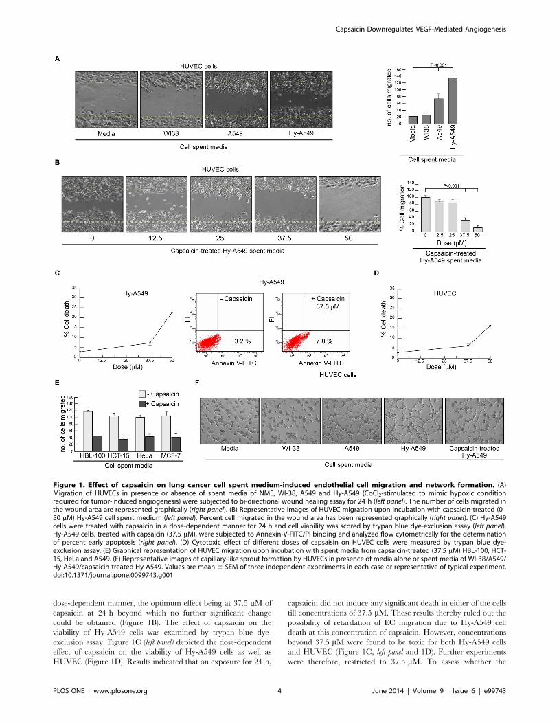
dose-dependent manner, the optimum effect being at 37.5 mM of
capsaicin at 24 h beyond which no further significant change
could be obtained (Figure 1B). The effect of capsaicin on the
viability of Hy-A549 cells was examined by trypan blue dye-
exclusion assay. Figure 1C (left panel) depicted the dose-dependent
effect of capsaicin on the viability of Hy-A549 cells as well as
HUVEC (Figure 1D). Results indicated that on exposure for 24 h,
capsaicin did not induce any significant death in either of the cells
till concentrations of 37.5 mM. These results thereby ruled out the
possibility of retardation of EC migration due to Hy-A549 cell
death at this concentration of capsaicin. However, concentrations
beyond 37.5 mM were found to be toxic for both Hy-A549 cells
and HUVEC (Figure 1C, left panel and 1D). Further experiments
were therefore, restricted to 37.5 mM. To assess whether the
Figure 1. Effect of capsaicin on lung cancer cell spent medium-induced endothelial cell migration and network formation. (A)Migration of HUVECs in presence or absence of spent media of NME, WI-38, A549 and Hy-A549 (CoCl2-stimulated to mimic hypoxic conditionrequired for tumor-induced angiogenesis) were subjected to bi-directional wound healing assay for 24 h (left panel). The number of cells migrated inthe wound area are represented graphically (right panel). (B) Representative images of HUVEC migration upon incubation with capsaicin-treated (0–50 mM) Hy-A549 cell spent medium (left panel). Percent cell migrated in the wound area has been represented graphically (right panel). (C) Hy-A549cells were treated with capsaicin in a dose-dependent manner for 24 h and cell viability was scored by trypan blue dye-exclusion assay (left panel).Hy-A549 cells, treated with capsaicin (37.5 mM), were subjected to Annexin-V-FITC/PI binding and analyzed flow cytometrically for the determinationof percent early apoptosis (right panel). (D) Cytotoxic effect of different doses of capsaisin on HUVEC cells were measured by trypan blue dye-exclusion assay. (E) Graphical representation of HUVEC migration upon incubation with spent media from capsaicin-treated (37.5 mM) HBL-100, HCT-15, HeLa and A549. (F) Representative images of capillary-like sprout formation by HUVECs in presence of media alone or spent media of WI-38/A549/Hy-A549/capsaicin-treated Hy-A549. Values are mean 6 SEM of three independent experiments in each case or representative of typical experiment.doi:10.1371/journal.pone.0099743.g001
Capsaicin Downregulates VEGF-Mediated Angiogenesis
PLOS ONE | www.plosone.org 4 June 2014 | Volume 9 | Issue 6 | e99743
reduction in cell number is due to early apoptosis, the number of
Annexin-V positive cells was determined by flow cytometry.
Results of Figure 1C (right panel) demonstrated 37.5 mM dose of
capsaicin as sub-apoptotic dose for Hy-A549 cells. These results
together ensured the absence of ‘interference’ of apoptotic cells
at 37.5 mM dose of capsaicin used in the subsequent experi-
ments. That the effect of capsaicin is not restricted to a specific
type, was validated using a battery of cell lines, i.e., HBL-100,
HCT-15, HeLa and, MCF-7 (Figure 1E). However, for detailed
mechanistic studies experiments were performed with Hy-A549
cells.
Next, the sprout formation assay of ECs was performed on
Matrigel, a well-established angiogenesis assay. HUVECs formed
tube-like networks within 8 h (Figure 1F). Angiogenic sprout
formation of HUVECs was highest in the presence of spent media
of Hy-A549 cells followed by that of A549 cells and WI-38 cells
(Figure 1F). However, capsaicin pre-treatment of Hy-A549
effectively hindered sprout formation by HUVECs (Figure 1F)
where tube-like structures were reduced both in width and in
length and showed incomplete as well as broken network
structures (Figure 1F).
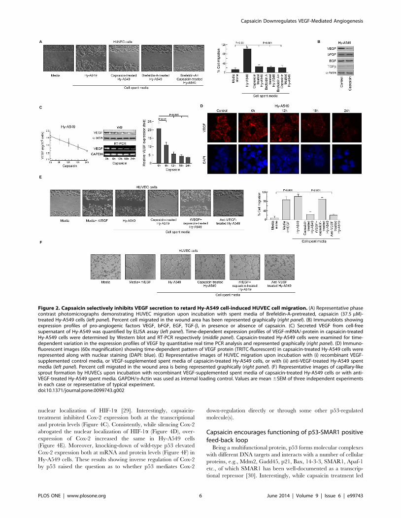
Capsaicin inhibits VEGF to retard cancer cell-induced ECmigration
All the reactions so far defined occurred independent of direct
contact of HUVEC cells with tumor cells or even proximity,
thereby raising the possibility of the presence of tumor-shed
soluble pro-migratory factors in the spent media. Supporting this,
spent medium of Brefeldin-A-treated Hy-A549 failed to induce
significant EC migration (Figure 2A) and capsaicin could not
introduce any further effect (Figure 2A). To re-confirm our
hypothesis, effect of capsaicin in regulating pro-angiogenic factors
like VEGF, bFGF, EGF, TGF-b, was monitored in Brefeldin-A
pre-treated Hy-A549 cells. Although capsaicin failed to alter
bFGF, EGF and TGF-b levels it significantly down-regulated
VEGF expression (Figure 2B). Further studies revealed that the
spent media of Hy-A549 cells (106 cells/ml of media) contained an
average of 1.6 ng VEGF that decreased significantly after
capsaicin treatment (Figure 2C, left panel). To adjudge the effect
of capsaicin in regulating VEGF in Hy-A549 cells, we adopted few
approaches. In the first approach, when Brefeldin A-pretreated
Hy-A549 cells were treated with capsaicin for 24 h, intracellular
VEGF was abrogated both at mRNA and protein levels
(Figure 2C, middle panel). These observations were further
supported by our quantitative real time PCR data depicting a
decrease in relative VEGF mRNA expression in Hy-A549 cells
following capsaicin treatment (Figure 2C, right panel) and re-
confirmed by confocal microscopy (Figure 2D). In the second
approach, control media supplemented with recombinant VEGF
(0.8 ng/ml) increased endothelial cell migration and sprout
formation (Figure 2E & 2F). In the third approach, when
capsaicin-pretreated Hy-A549 spent media was supplemented
with recombinant VEGF protein (0.8 ng/ml), reversal of capsaicin
effect on EC migration and sprout formation was observed
(Figure 2E & 2F). In the fourth approach, HUVECs showed
significant reduction in migration (Figure 2E) and sprout
formation (Figure 2F) when Hy-A549 cell spent medium was
pre-treated with anti-VEGF antibody. However, anti-VEGF
antibody failed to furnish any significant additional inhibitory
effect on HUVEC migration and sprout formation in capsaicin
pre-treated Hy-A549 spent media (Figure 2E & 2F). These results
together validated the hypothesis that Hy-A549 cell-shed VEGF
plays a crucial role in cancer-induced EC migration and sprout
formation. Afore-furnished observations tempted us to demarcate
the complete mechanism underlying the regulation of tumor
angiogenesis by capsaicin.
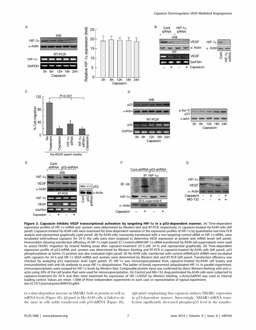
Capsaicin inhibits VEGF transcription by targeting HIF-1aAs capsaicin reduces VEGF both at the transcriptional as well as
translational levels we hypothesized that this phytochemical might
have a regulatory effect on HIF-1a, the main transcription factor
of VEGF during hypoxia [4]. Interestingly, capsaicin reduced
HIF-1a at protein level but not at mRNA level in Hy-A549 cells
(Figure 3A, left panel), which was further confirmed by our
quantitative real time PCR data (Figure 3A, right panel). Further-
more, HIF-1a-siRNA-transfected Hy-A549 cells furnished de-
crease in VEGF expression (Figure 3B) and spent media of these
transfectants demonstrated significantly less EC migration
(Figure 3C). These results indicated that capsaicin inhibited
VEGF by down-modulating its key transcription factor HIF-1a(Figure 3C). However, since capsaicin treatment of these
transfectants demonstrated additional decrease in VEGF, involve-
ment of HIF-1a-independent pathway(s) of VEGF inhibition by
capsaicin cannot be negated.
Capsaicin targets HIF-1a in a p53-dependent mannerSince p53 directly targets HIF-1a for proteosomal degradation
[9], we assessed the role of p53, if any, in capsaicin-induced
regulation of HIF-1a and VEGF. Capsaicin treatment resulted in
a time-dependent elevation of p53 in Hy-A549 cells (Figure 3D)
though this augmentation in p53 level was much lower than that
induced by the apoptotic dose (,50 mM) of capsaicin [27,28].
Additionally, capsaicin treatment increased the level of p-Ser15-
p53 suggesting stability and functional activation of p53
(Figure 3D). Next, an increase in HIF-1a protein expression
along with up-regulation in VEGF expression was observed in
p53-shRNA-transfected Hy-A549 cells (Figure 3E), thereby
indicating the possibility of involvement of p53 in capsaicin-
induced anti-angiogenicity. Results of Figure 3F depicted
significant HIF-1a ubiquitination in capsaicin-treated Hy-A549
cells while silencing p53 decreased the same (Figure 3F).
Moreover, addition of the proteosome blocker MG-132 to Hy-
A549 prior to capsaicin treatment partially increased the level of
HIF-1a, although the effect was not comparable to that of p53-
shRNA-transfected Hy-A549 cells (Figure 3G). These results
tempted us to hypothesize that p53-dependent degradation of
HIF-1a had a crucial role in capsaicin-mediated decrease in
VEGF expression. Above results also indicate an additional role
of p53 in maintaining transcriptional activity of HIF-1aTherefore, all these findings leave a room for exploring the
status of the factors responsible for transporting HIF-1a to the
nucleus, up on capsaicin treatment.
Capsaicin inhibits the nuclear localization of HIF-1a bydown regulating Cox-2 in a p53-dependent manner
To understand the role of p53, if any, in controlling nuclear
translocation of the transcription factor HIF-1a, the nucleus to
cytoplasmic ratio of HIF-1a was checked up on capsaicin-
treatment. Results of Figure 4A demonstrated a sharp decrease
in nuclear HIF-1a level with its increase in cytoplasm, indicating
the inhibition in the transcriptional activity of HIF-1a in capsaicin-
treated Hy-A549 cells. Confocal microscopic data authenticated
these results (Figure 4B). Silencing p53 reversed this effect
(Figure 4A) indicating that p53 is responsible also for obstructing
HIF-1a transport to the nucleus up on capsaicin treatment. It is
well acknowledged that intra-cellular PGE2, which is synthesized
by the pro-inflammatory enzyme Cox-2 [5], is a determinant of
Capsaicin Downregulates VEGF-Mediated Angiogenesis
PLOS ONE | www.plosone.org 5 June 2014 | Volume 9 | Issue 6 | e99743
nuclear localization of HIF-1a [29]. Interestingly, capsaicin-
treatment inhibited Cox-2 expression both at the transcriptional
and protein levels (Figure 4C). Consistently, while silencing Cox-2
abrogated the nuclear localization of HIF-1a (Figure 4D), over-
expression of Cox-2 increased the same in Hy-A549 cells
(Figure 4E). Moreover, knocking-down of wild-type p53 elevated
Cox-2 expression both at mRNA and protein levels (Figure 4F) in
Hy-A549 cells. These results showing inverse regulation of Cox-2
by p53 raised the question as to whether p53 mediates Cox-2
down-regulation directly or through some other p53-regulated
molecule(s).
Capsaicin encourages functioning of p53-SMAR1 positivefeed-back loop
Being a multifunctional protein, p53 forms molecular complexes
with different DNA targets and interacts with a number of cellular
proteins, e.g., Mdm2, Gadd45, p21, Bax, 14-3-3, SMAR1, Apaf-1
etc., of which SMAR1 has been well-documented as a transcrip-
tional repressor [30]. Interestingly, while capsaicin treatment led
Figure 2. Capsaicin selectively inhibits VEGF secretion to retard Hy-A549 cell-induced HUVEC cell migration. (A) Representative phasecontrast photomicrographs demonstrating HUVEC migration upon incubation with spent media of Brefeldin-A-pretreated, capsaicin (37.5 mM)-treated Hy-A549 cells (left panel). Percent cell migrated in the wound area has been represented graphically (right panel). (B) Immunoblots showingexpression profiles of pro-angiogenic factors VEGF, bFGF, EGF, TGF-b, in presence or absence of capsaicin. (C) Secreted VEGF from cell-freesupernatant of Hy-A549 was quantified by ELISA assay (left panel). Time-dependent expression profiles of VEGF-mRNA/-protein in capsaicin-treatedHy-A549 cells were determined by Western blot and RT-PCR respectively (middle panel). Capsaicin-treated Hy-A549 cells were examined for time-dependent variation in the expression profiles of VEGF by quantitative real time PCR analysis and represented graphically (right panel). (D) Immuno-fluorescent images (60x magnification) showing time-dependent pattern of VEGF protein (TRITC-fluorescent) in capsaicin-treated Hy-A549 cells wererepresented along with nuclear staining (DAPI: blue). (E) Representative images of HUVEC migration upon incubation with (i) recombinant VEGF-supplemented control media, or VEGF-supplemented spent media of capsaicin-treated Hy-A549 cells, or with (ii) anti-VEGF-treated Hy-A549 spentmedia (left panel). Percent cell migrated in the wound area is being represented graphically (right panel). (F) Representative images of capillary-likesprout formation by HUVECs upon incubation with recombinant VEGF-supplemented spent media of capsaicin-treated Hy-A549 cells or with anti-VEGF-treated Hy-A549 spent media. GAPDH/a-Actin was used as internal loading control. Values are mean 6SEM of three independent experimentsin each case or representative of typical experiment.doi:10.1371/journal.pone.0099743.g002
Capsaicin Downregulates VEGF-Mediated Angiogenesis
PLOS ONE | www.plosone.org 6 June 2014 | Volume 9 | Issue 6 | e99743
to a time-dependent increase in SMAR1 both at protein as well as
mRNA levels (Figure 4G, left panel) in Hy-A549 cells, it failed to do
the same in cells stably transfected with p53-shRNA (Figure 4G,
right panel) emphasizing that capsaicin induces SMAR1 expression
in p53-dependent manner. Interestingly, SMAR1-shRNA trans-
fection significantly decreased phospho-p53 level in the transfec-
Figure 3. Capsaicin inhibits VEGF transcriptional activation by targeting HIF-1a in a p53-dependent manner. (A) Time-dependentexpression profiles of HIF-1a mRNA and -protein were determined by Western blot and RT-PCR, respectively, in capsaicin-treated Hy-A549 cells (leftpanel). Capsaicin-treated Hy-A549 cells were examined for time-dependent variation in the expression profiles of HIF-1a by quantitative real time PCRanalysis and represented graphically (right panel). (B) Hy-A549 cells, transiently transfected with a non-targeting control-siRNA or HIF-1a-siRNA, wereincubated with/without capsaicin for 24 h; the cells were then analyzed to determine VEGF expression at protein and mRNA levels (left panel).Immunoblot showing transfection efficiency of HIF-1a (right panel) (C) Control-siRNA/HIF-1a-siRNA-transfected Hy-A549 cell-supernatants were usedto assess HUVEC migration by wound healing assay after capsaicin-treatment (37.5 mM; 24 h) and represented graphically. (D) Time-dependentexpression profile of p53-mRNA and -protein was determined by Western blotting and RT-PCR in capsaicin-treated Hy-A549 cells (left panel). p53phosphorylation at Serine-15 position was also evaluated (right panel). (E) Hy-A549 cells, transfected with control-shRNA/p53-shRNA were incubatedwith capsaicin for 24 h and HIF-1a VEGF-mRNA and -protein were determined by Western blot and RT-PCR (left panel). Transfection efficiency waschecked by analyzing p53 expression level (right panel). (F) HIF-1a was immunoprecipitated from capsaicin-treated Hy-A549 cell lysates andimmunoblotted with anti-Ub antibody to assay HIF-1a ubiquitination. The ladder of bands represented ubiquitinated HIF-1a. In parallel experiment,immunoprecipitates were assayed for HIF-1a levels by Western blot. Comparable protein input was confirmed by direct Western blotting with anti-a-actin using 20% of the cell lysates that were used for immunoprecipitation. (G) Control and MG-132 drug-pretreated Hy-A549 cells were subjected tocapsaicin-treatment for 24 h and then were examined for expression of HIF-1a/VEGF by Western blotting. a-Actin/GAPDH was used as internalloading control. Values are mean 6SEM of three independent experiments in each case or representative of typical experiment.doi:10.1371/journal.pone.0099743.g003
Capsaicin Downregulates VEGF-Mediated Angiogenesis
PLOS ONE | www.plosone.org 7 June 2014 | Volume 9 | Issue 6 | e99743
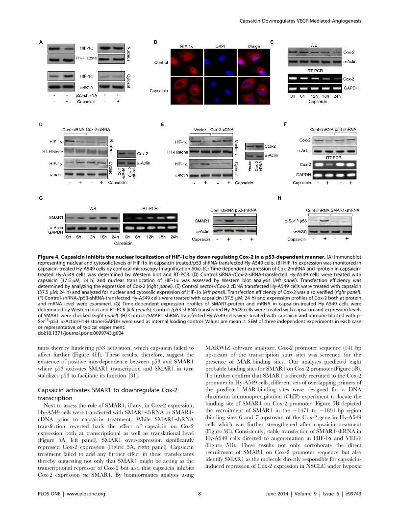
tants thereby hindering p53 activation, which capsaicin failed to
affect further (Figure 4H). These results, therefore, suggest the
existence of positive interdependence between p53 and SMAR1
where p53 activates SMAR1 transcription and SMAR1 in turn
stabilizes p53 to facilitate its function [31].
Capsaicin activates SMAR1 to downregulate Cox-2transcription
Next to assess the role of SMAR1, if any, in Cox-2 expression,
Hy-A549 cells were transfected with SMAR1-shRNA or SMAR1-
cDNA prior to capsaicin treatment. While SMAR1-shRNA
transfection reverted back the effect of capsaicin on Cox2
expression both at transcriptional as well as translational level
(Figure 5A, left panel), SMAR1 over-expression significantly
repressed Cox-2 expression (Figure 5A, right panel). Capsaicin
treatment failed to add any further effect in these transfectants
thereby suggesting not only that SMAR1 might be acting as the
transcriptional repressor of Cox-2 but also that capsaicin inhibits
Cox-2 expression via SMAR1. By bioinformatics analysis using
MARWIZ software analyzer, Cox-2 promoter sequence (141 bp
upstream of the transcription start site) was screened for the
presence of MAR-binding sites. Our analyses predicted eight
probable binding sites for SMAR1 on Cox-2 promoter (Figure 5B).
To further confirm that SMAR1 is directly recruited to the Cox-2
promoter in Hy-A549 cells, different sets of overlapping primers of
the predicted MAR-binding sites were designed for a DNA
chromatin immunoprecipitation (ChIP) experiment to locate the
binding site of SMAR1 on Cox-2 promoter. Figure 5B depicted
the recruitment of SMAR1 in the 21471 to 21891 bp region
(binding sites 6 and 7) upstream of the Cox-2 gene in Hy-A549
cells which was further strengthened after capsaicin treatment
(Figure 5C). Consistently, stable transfection of SMAR1-shRNA in
Hy-A549 cells directed to augmentation in HIF-1a and VEGF
(Figure 5D). These results not only corroborate the direct
recruitment of SMAR1 on Cox-2 promoter sequence but also
identify SMAR1 as the molecule directly responsible for capsaicin-
induced repression of Cox-2 expression in NSCLC under hypoxic
Figure 4. Capsaicin inhibits the nuclear localization of HIF-1a by down regulating Cox-2 in a p53-dependent manner. (A) Immunoblotrepresenting nuclear and cytosolic levels of HIF-1a in capsaicin-treated/p53-shRNA-transfected Hy-A549 cells. (B) HIF-1a expression was monitored incapsaicin-treated Hy-A549 cells by confocal microscopy (magnification 60x). (C) Time-dependent expression of Cox-2-mRNA and -protein in capsaicin-treated Hy-A549 cells was determined by Western blot and RT-PCR. (D) Control siRNA-/Cox-2-siRNA-transfected Hy-A549 cells were treated withcapsaicin (37.5 mM; 24 h) and nuclear translocation of HIF-1a was assessed by Western blot analysis (left panel). Transfection efficiency wasdetermined by analyzing the expression of Cox-2 (right panel). (E) Control vector-/Cox-2 cDNA transfected Hy-A549 cells were treated with capsaicin(37.5 mM; 24 h) and analyzed for nuclear and cytosolic expression of HIF-1a (left panel). Transfection efficiency of Cox-2 was also verified (right panel).(F) Control-shRNA-/p53-shRNA-transfected Hy-A549 cells were treated with capsaicin (37.5 mM; 24 h) and expression profiles of Cox-2 both at proteinand mRNA level were examined. (G) Time-dependent expression profiles of SMAR1-protein and mRNA in capsaicin-treated Hy-A549 cells weredetermined by Western blot and RT-PCR (left panels). Control-/p53-shRNA transfected Hy-A549 cells were treated with capsaicin and expression levelsof SMAR1 were checked (right panel). (H) Control-/SMAR1-shRNA transfected Hy-A549 cells were treated with capsaicin and immune-blotted with p-Ser15-p53. a-Actin/H1-Histone/GAPDH were used as internal loading control. Values are mean 6 SEM of three independent experiments in each caseor representative of typical experiment.doi:10.1371/journal.pone.0099743.g004
Capsaicin Downregulates VEGF-Mediated Angiogenesis
PLOS ONE | www.plosone.org 8 June 2014 | Volume 9 | Issue 6 | e99743
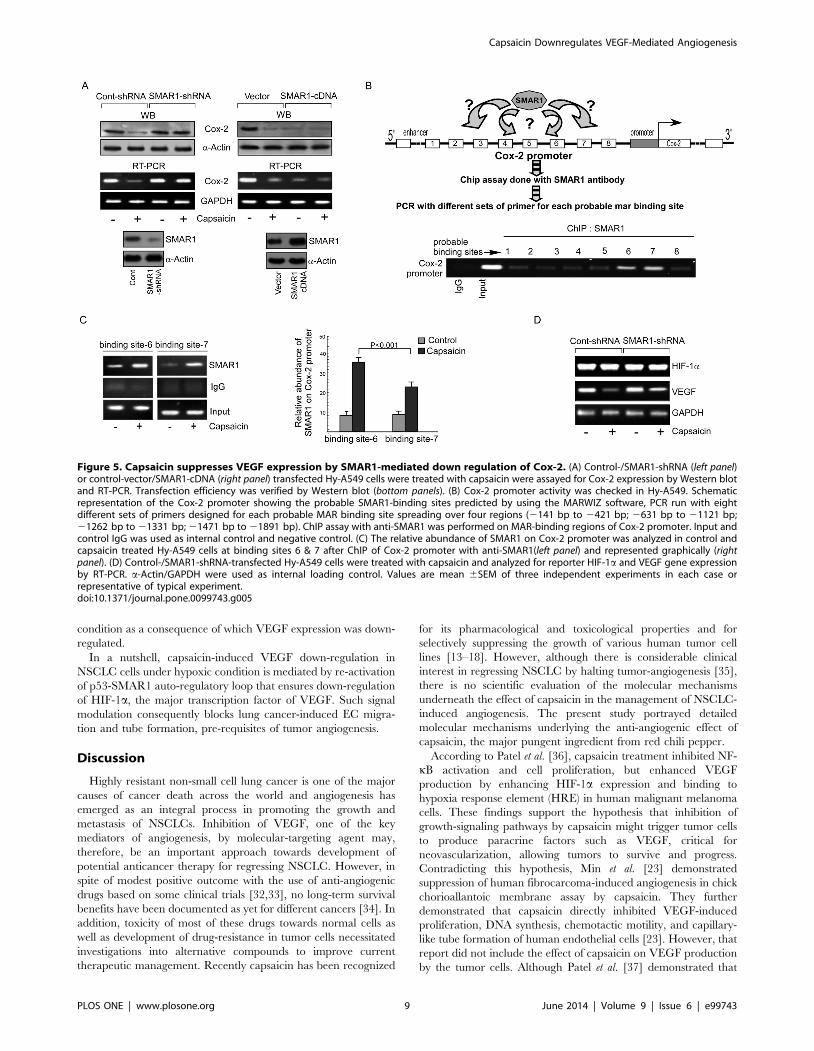
condition as a consequence of which VEGF expression was down-
regulated.
In a nutshell, capsaicin-induced VEGF down-regulation in
NSCLC cells under hypoxic condition is mediated by re-activation
of p53-SMAR1 auto-regulatory loop that ensures down-regulation
of HIF-1a, the major transcription factor of VEGF. Such signal
modulation consequently blocks lung cancer-induced EC migra-
tion and tube formation, pre-requisites of tumor angiogenesis.
Discussion
Highly resistant non-small cell lung cancer is one of the major
causes of cancer death across the world and angiogenesis has
emerged as an integral process in promoting the growth and
metastasis of NSCLCs. Inhibition of VEGF, one of the key
mediators of angiogenesis, by molecular-targeting agent may,
therefore, be an important approach towards development of
potential anticancer therapy for regressing NSCLC. However, in
spite of modest positive outcome with the use of anti-angiogenic
drugs based on some clinical trials [32,33], no long-term survival
benefits have been documented as yet for different cancers [34]. In
addition, toxicity of most of these drugs towards normal cells as
well as development of drug-resistance in tumor cells necessitated
investigations into alternative compounds to improve current
therapeutic management. Recently capsaicin has been recognized
for its pharmacological and toxicological properties and for
selectively suppressing the growth of various human tumor cell
lines [13–18]. However, although there is considerable clinical
interest in regressing NSCLC by halting tumor-angiogenesis [35],
there is no scientific evaluation of the molecular mechanisms
underneath the effect of capsaicin in the management of NSCLC-
induced angiogenesis. The present study portrayed detailed
molecular mechanisms underlying the anti-angiogenic effect of
capsaicin, the major pungent ingredient from red chili pepper.
According to Patel et al. [36], capsaicin treatment inhibited NF-
kB activation and cell proliferation, but enhanced VEGF
production by enhancing HIF-1a expression and binding to
hypoxia response element (HRE) in human malignant melanoma
cells. These findings support the hypothesis that inhibition of
growth-signaling pathways by capsaicin might trigger tumor cells
to produce paracrine factors such as VEGF, critical for
neovascularization, allowing tumors to survive and progress.
Contradicting this hypothesis, Min et al. [23] demonstrated
suppression of human fibrocarcoma-induced angiogenesis in chick
chorioallantoic membrane assay by capsaicin. They further
demonstrated that capsaicin directly inhibited VEGF-induced
proliferation, DNA synthesis, chemotactic motility, and capillary-
like tube formation of human endothelial cells [23]. However, that
report did not include the effect of capsaicin on VEGF production
by the tumor cells. Although Patel et al. [37] demonstrated that
Figure 5. Capsaicin suppresses VEGF expression by SMAR1-mediated down regulation of Cox-2. (A) Control-/SMAR1-shRNA (left panel)or control-vector/SMAR1-cDNA (right panel) transfected Hy-A549 cells were treated with capsaicin were assayed for Cox-2 expression by Western blotand RT-PCR. Transfection efficiency was verified by Western blot (bottom panels). (B) Cox-2 promoter activity was checked in Hy-A549. Schematicrepresentation of the Cox-2 promoter showing the probable SMAR1-binding sites predicted by using the MARWIZ software, PCR run with eightdifferent sets of primers designed for each probable MAR binding site spreading over four regions (2141 bp to 2421 bp; 2631 bp to 21121 bp;21262 bp to 21331 bp; 21471 bp to 21891 bp). ChIP assay with anti-SMAR1 was performed on MAR-binding regions of Cox-2 promoter. Input andcontrol IgG was used as internal control and negative control. (C) The relative abundance of SMAR1 on Cox-2 promoter was analyzed in control andcapsaicin treated Hy-A549 cells at binding sites 6 & 7 after ChIP of Cox-2 promoter with anti-SMAR1(left panel) and represented graphically (rightpanel). (D) Control-/SMAR1-shRNA-transfected Hy-A549 cells were treated with capsaicin and analyzed for reporter HIF-1a and VEGF gene expressionby RT-PCR. a-Actin/GAPDH were used as internal loading control. Values are mean 6SEM of three independent experiments in each case orrepresentative of typical experiment.doi:10.1371/journal.pone.0099743.g005
Capsaicin Downregulates VEGF-Mediated Angiogenesis
PLOS ONE | www.plosone.org 9 June 2014 | Volume 9 | Issue 6 | e99743
capsaicin enhanced production of VEGF by melanoma cells we
did not observe similar induction of VEGF following capsaicin
treatment. Our results demonstrated that capsaicin acted directly
on tumor angiogenesis by suppressing VEGF expression in
hypoxic NSCLC cells while having minimal toxic effects on
normal cells. This difference might be due to differences in the cell
lines used in these studies. In fact, the anti-proliferative and anti-
tumor effects of capsaicin on in vivo lung cancer models [38] failed
to support the role of capsaicin as a VEGF inducer for neo-
vascularization that allows lung cancer to survive and progress.
Our work established the mechanism of capsaicin-induced down-
regulation in VEGF expression in our NSCLC model. It is well
acknowledged that hypoxia-conducive environment, resulting
from the increasing distance between the growing tumor cells
and the capillaries or from the inefficiency of new vessels, resulted
in rapid up-regulation of VEGF [39] and its receptor via HIF-1a[40]. We observed that in hypoxic NSCLC cells, capsaicin re-
activated the auto-regulatory p53-SMAR1 signaling loop that in
turn ensured down-regulation of HIF-1a, the major transcription
factor of VEGF. Effect of such signal modulation consequently
blocked tumor-induced endothelial cell migration and tube
formation, pre-requisites of tumor angiogenesis.
Scaffold/matrix attachment regions (S/MARs) are regulatory
DNA sequences mostly present upstream of various promoters.
Matrix attachment region-binding proteins (MARBPs), which
bind to such regulatory sequences, interact with numerous
chromatin modifying factors and facilitate transcription in
response to diverse stimuli [41]. SMAR1 is an MARBP identified
in mouse double positive thymocytes, wherein it binds to MARbsequence at TCRb locus and affects V(D)J recombination [42,43].
Subsequently, SMAR1 has been characterized as a tumor
suppressor by virtue of its ability to interact with p53 and delay
tumor growth in mouse melanoma model [38,39]. In fact, p53
target gene SMAR1 [44] activates and stabilizes p53 [8] with
which it acts synergistically during DNA damage [44]. Coordi-
nated regulation of p53 apoptotic targets by SMAR1 has also been
documented [10]. However, the biological and functional signif-
icances of p53 and SMAR1 cross-talk in the context to VEGF
expression by non-small cell lung cancer are not known.
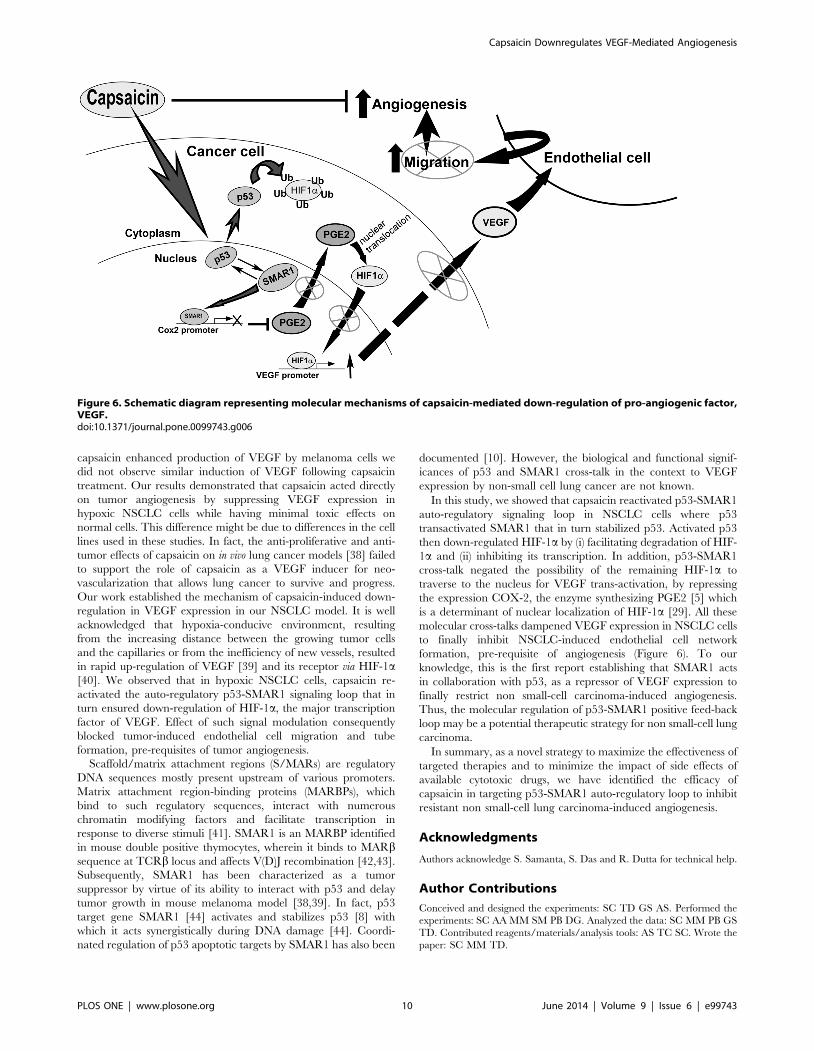
In this study, we showed that capsaicin reactivated p53-SMAR1
auto-regulatory signaling loop in NSCLC cells where p53
transactivated SMAR1 that in turn stabilized p53. Activated p53
then down-regulated HIF-1a by (i) facilitating degradation of HIF-
1a and (ii) inhibiting its transcription. In addition, p53-SMAR1
cross-talk negated the possibility of the remaining HIF-1a to
traverse to the nucleus for VEGF trans-activation, by repressing
the expression COX-2, the enzyme synthesizing PGE2 [5] which
is a determinant of nuclear localization of HIF-1a [29]. All these
molecular cross-talks dampened VEGF expression in NSCLC cells
to finally inhibit NSCLC-induced endothelial cell network
formation, pre-requisite of angiogenesis (Figure 6). To our
knowledge, this is the first report establishing that SMAR1 acts
in collaboration with p53, as a repressor of VEGF expression to
finally restrict non small-cell carcinoma-induced angiogenesis.
Thus, the molecular regulation of p53-SMAR1 positive feed-back
loop may be a potential therapeutic strategy for non small-cell lung
carcinoma.
In summary, as a novel strategy to maximize the effectiveness of
targeted therapies and to minimize the impact of side effects of
available cytotoxic drugs, we have identified the efficacy of
capsaicin in targeting p53-SMAR1 auto-regulatory loop to inhibit
resistant non small-cell lung carcinoma-induced angiogenesis.
Acknowledgments
Authors acknowledge S. Samanta, S. Das and R. Dutta for technical help.
Author Contributions
Conceived and designed the experiments: SC TD GS AS. Performed the
experiments: SC AA MM SM PB DG. Analyzed the data: SC MM PB GS
TD. Contributed reagents/materials/analysis tools: AS TC SC. Wrote the
paper: SC MM TD.
Figure 6. Schematic diagram representing molecular mechanisms of capsaicin-mediated down-regulation of pro-angiogenic factor,VEGF.doi:10.1371/journal.pone.0099743.g006
Capsaicin Downregulates VEGF-Mediated Angiogenesis
PLOS ONE | www.plosone.org 10 June 2014 | Volume 9 | Issue 6 | e99743
References
1. Bergers G, Benjamin LE (2003) Tumorigenesis and the angiogenic switch.
Nature reviews cancer 3: 401–410.2. Folkman J (1995) Angiogenesis in cancer, vascular, rheumatoid and other
disease. Nature Medicine 1: 27–31.3. Semenza GL (2003) Targeting HIF-1 for cancer therapy. Nature reviews cancer
3: 721–732.
4. Pugh CW, Ratcliffe PJ (2003) Regulation of angiogenesis by hypoxia: role of theHIF system. Nature medicine 9: 677–684.
5. Papadimitriou A, King AJ, Jones PM, Persaud SJ (2007) Anti-apoptotic effects ofarachidonic acid and prostaglandin E2 in pancreatic beta-cells. Cellular
physiology and biochemistry 20: 607–616.
6. Blagosklonny MV, An WG, Romanova LY, Trepel J, Fojo T, et al. (1998) p53inhibits hypoxia-inducible factor-stimulated transcription. The journal of
biological chemistry 273: 11995–11998.7. Ravi R, Mookerjee B, Bhujwalla ZM, Sutter CH, Artemov D, et al. (2000)
Regulation of tumor angiogenesis by p53-induced degradation of hypoxia-inducible factor 1alpha. Genes & Development 14: 34–44.
8. Jalota A, Singh K, Pavithra L, Kaul-Ghanekar R, Jameel S, et al. (2005) Tumor
suppressor SMAR1 activates and stabilizes p53 through its arginine-serine-richmotif. The journal of biological chemistry 280: 16019–16029.
9. Sen GS, Mohanty S, Hossain DM, Bhattacharyya S, Banerjee S, et al. (2011)Curcumin enhances the efficacy of chemotherapy by tailoring p65NFkB-p300
cross-talk in favor of p53-p300 in breast cancer. The journal of biological
chemistry 286: 42232–42247.10. Sinha S, Malonia SK, Mittal SP, Singh K, Kadreppa S, et al. (2010)
Coordinated regulation of p53 apoptotic targets BAX and PUMA by SMAR1through an identical MAR element. EMBO Journal 29: 830–842.
11. Saha B, Adhikary A, Ray P, Saha S, Chakraborty S, et al. (2012) Restoration oftumor suppressor p53 by differentially regulating pro- and anti-p53 networks in
HPV-18-infected cervical cancer cells. Oncogene 31: 173–186.
12. De-Moraes E, Dar NA, de-Moura-Gallo CV, Hainaut P (2007) Cross-talksbetween cyclooxygenase-2 and tumor suppressor protein p53: Balancing life and
death during inflammatory stress and carcinogenesis. International journal ofcancer 121: 929–937.
13. Zhang J, Nagasaki M, Tanaka Y, Morikawa S (2003) Capsaicin inhibits growth
of adult T-cell leukemia cells. Leukemia research 27: 275–283.14. Jung MY, Kang HJ, Moon A (2001) Capsaicin-induced apoptosis in SK-Hep-1
hepatocarcinoma cells involves Bcl-2 downregulation and caspase-3 activation.Cancer Letters 165: 39–45.
15. Chen D, Yang Z, Wang Y, Zhu G, Wang X (2012) Capsaicin induces cyclearrest by inhibiting cyclin-dependent-kinase in bladder carcinoma cells.
International journal of urology 19: 662–668.
16. Thoennissen NH, O’Kelly J, Lu D, Iwanski GB, La DT, et al. (2010) Capsaicincauses cell-cycle arrest and apoptosis in ER-positive and -negative breast cancer
cells by modulating the EGFR/HER-2 pathway. Oncogene 29: 285–296.17. Amantini C, Ballarini P, Caprodossi S, Nabissi M, Morelli MB, et al. (2009)
Triggering of transient receptor potential vanilloid type 1 (TRPV1) by capsaicin
induces Fas/CD95-mediated apoptosis of urothelial cancer cells in an ATM-dependent manner. Carcinogenesis 30: 1320–1329.
18. Shin DH, Kim OH, Jun HS, Kang MK (2008) Inhibitory effect of capsaicin onB16-F10 melanoma cell migration via the phosphatidylinositol 3-kinase/Akt/
Rac1 signal pathway. Experimental & molecular medicine 40: 486–494.19. Anandakumar P, Kamaraj S, Jagan S, Ramakrishnan G, Devaki T (2013)
Capsaicin provokes apoptosis and restricts benzo(a)pyrene induced lung
tumorigenesis in Swiss albino mice. International immunopharmacology17:254–259.
20. Anandakumar P, Kamaraj S, Jagan S, Ramakrishnan G, Vinodhkumar R, et al.(2008) Capsaicin modulates pulmonary antioxidant defense system during
benzo(a)pyrene-induced lung cancer in Swiss albino mice. Phytotherapy
research 22:529–533.21. Brown KC, Witte TR, Hardman WE, Luo H, Chen YC, et al. (2010) Capsaicin
displays anti-proliferative activity against human small cell lung cancer in cellculture and nude mice models via the E2F pathway. PLoS one 5:e10243.
22. Chakraborty S, Mazumdar M, Mukherjee S, Bhattacharjee P, Adhikary A, et al.
(2014) Restoration of p53/miR-34a regulatory axis decreases survival advantageand ensures Bax-dependent apoptosis of non-small cell lung carcinoma cells.
FEBS letters 588: 549–559.
23. Min JK, Han KY, Kim EC, Kim YM, Lee SW, et al. (2004) Capsaicin inhibits
in vitro and in vivo angiogenesis. Cancer research 64: 644–651.24. Mazumdar M, Adhikary A, Chakraborty S, Mukherjee S, Manna A, et al. (2013)
Targeting RET to induce medullary thyroid cancer cell apoptosis: anantagonistic interplay between PI3K/Akt and p38MAPK/caspase-8 pathways.
Apoptosis 18: 589–604.
25. Adhikary A, Mohanty S, Lahiry L, Hossain DM, Chakraborty S, et al. (2010)Theaflavins retard human breast cancer cell migration by inhibiting NF-kappaB
via p53-ROS cross-talk. FEBS letters 584: 7–14.26. Hossain DM, Panda AK, Manna A, Mohanty S, Bhattacharjee P, et al. (2013)
FoxP3 acts as a cotranscription factor with STAT3 in tumor-induced regulatory
T cells. Immunity 39:1057–1069.27. Chou CC, Wu YC, Wang YF, Chou MJ, Kuo SJ, et al. (2009) Capsaicin-
induced apoptosis in human breast cancer MCF-7 cells through caspase-independent pathway. Oncology reports 21: 665–671.
28. Gallo O, Schiavone N, Papucci L, Sardi I, Magnelli L, et al. (2003) Down-regulation of nitric oxide synthase-2 and cyclooxygenase-2 pathways by p53 in
squamous cell carcinoma. The american journal of pathology 163: 723–732.
29. Liu XH, Kirschenbaum A, Lu M, Yao S, Dosoretz A, et al. (2002) ProstaglandinE2 induces hypoxia-inducible factor-1alpha stabilization and nuclear localiza-
tion in a human prostate cancer cell line. The journal of biological chemistry277: 50081–50086.
30. Malonia SK, Sinha S, Lakshminarasimhan P, Singh K, Jalota-Badhwar A, et al.
(2011) Gene regulation by SMAR1: Role in cellular homeostasis and cancer.Biochimica et biophysica acta 1815: 1–12.
31. Oyagbemi AA, Saba AB, Azeez OI (2010) Capsaicin: a novel chemopreventivemolecule and its underlying molecular mechanisms of action. Indian journal of
cancer 47: 53–58.32. Yang JC, Haworth L, Sherry RM, Hwu P, Schwartzentruber DJ, et al. (2003) A
randomized trial of bevacizumab, an anti–vascular endothelial growth factor
antibody, for metastatic renal cancer. The new england journal of medicine 349:427–434.
33. Mayer RJ (2004) Two steps forward in the treatment of colorectal cancer. Thenew england journal of medicine 350: 2406–2408.
34. Hurwitz H, Fehrenbacher L, Novotny W (2004) Bevacizumab plus Irinotecan,
Fluorouracil, and Leucovorin for Metastatic Colorectal Cancer. The newengland journal of medicine 350: 2335–2342.
35. Folkman J (1995) Seminars in medicine of the Beth Israel Hospital, Boston.Clinical applications of research on angiogenesis. The new england journal of
medicine 333: 1757–1763.36. Patel PS, Yang S, Li A, Varney ML, Singh RK (2002) Capsaicin regulates
vascular endothelial cell growth factor expression by modulation of hypoxia
inducing factor-1alpha in human malignant melanoma cells. Journal of cancerresearch and clinical oncology 128: 461–468.
37. Patel PS, Varney ML, Dave BJ, Singh RK (2002) Regulation of constitutive andinduced NF-kappaB activation in malignant melanoma cells by capsaicin
modulates interleukin-8 production and cell proliferation. Journal of interferon
& cytokine research 22: 427–435.38. Teel RW, Huynh HT (1999) Lack of the inhibitory effect of intragastrically
administered capsaicin on NNK-induced lung tumor formation in the A.Jmouse. In Vivo 13: 231–234.
39. Bottaro DP, Liotta LA (2003) Cancer: Out of air is not out of action. Nature 423593–595.
40. Blagosklonny MV (2004) Antiangiogenic therapy and tumor progression.
Cancer cell 5: 13–17.41. Zaidi SK, Young DW, Choi JY, Pratap J, Javed A, et al. (2005) The dynamic
organization of gene-regulatory machinery in nuclear microenvironments.EMBO reports 6: 128–133. Review.
42. Chattopadhyay S, Kaul R, Charest A, Housman D, Chen J (2000) SMAR1, a
novel, alternatively spliced gene product, binds the Scaffold/Matrix-associatedregion at the T cell receptor beta locus. Genomics 68: 93–96.
43. Kaul-Ghanekar R, Majumdar S, Jalota A, Gulati N, Dubey N, et al. (2005)Abnormal V(D)J recombination of T cell receptor beta locus in SMAR1
transgenic mice. The journal of biological chemistry. 280: 9450–9459.
44. Singh K, Mogar D, Giridharagopalan RO, Gogiraju R, Pande G, et al. (2007)p53 target gene SMAR1 is dysregulated in breast cancer: its role in cancer cell
migration and invasion. PloS one 2: e660.
Capsaicin Downregulates VEGF-Mediated Angiogenesis
PLOS ONE | www.plosone.org 11 June 2014 | Volume 9 | Issue 6 | e99743


