Embed Size (px)
Citation preview
JOURNAL OF RAMAN SPECTROSCOPYJ. Raman Spectrosc. 2006; 37: 1086–1097Published online in Wiley InterScience(www.interscience.wiley.com) DOI: 10.1002/jrs.1592
Scientific investigations of the Tokhung-Ri tomb muralpaintings (408 A.D.) of the Koguryo era, DemocraticPeople’s Republic of Korea
R. Mazzeo,1∗ E. Joseph,1 V. Minguzzi,2 G. Grillini,2 P. Baraldi3 and D. Prandstraller4
1 University of Bologna, Microchemistry and Microscopy Art Diagnostic Laboratory (M2ADL), Ravenna, Italy2 University of Bologna, Earth Science Department, Bologna, Italy3 University of Modena, Chemistry Department, Modena, Italy4 University of Bologna, Institute of Metallurgy, Bologna, Italy
Received 2 September 2005; Accepted 23 April 2006
In the framework of the UNESCO workshops on the Conservation and Preservation of the Koguryomural paintings, which were held in Pyongyang in 2004 and 2005, paint samples were collected from theTokhung-Ri tomb located in suburban Pyongyang and analyzed by optical, polarized and FTIR microscopy,scanning electron microscopy coupled with energy-dispersive X-ray analysis (SEM-EDX), X-ray diffraction(XRD) as well as Raman spectroscopy, in order to characterize the composition of pigments, the executiontechnique adopted and the state of conservation of the murals. The first scientific results seem to confirmthe suggestion of local conservators about the adoption of a fresco technique, even though it is not yet clearwhether this was intentionally achieved by the North Korean painters. As regards this, analyses of moresamples as well as confirmations from a survey of the historical literature are needed. Copyright 2006John Wiley & Sons, Ltd.
KEYWORDS: Koguryo era; Tokhung-Ri tomb; murals; fresco technique; analytical investigations
INTRODUCTION
The interest of the conservation science community instudying painting materials and techniques used in FarEast Asian art has grown in the last decade thanks to theavailability and use of integrated analytical approaches.1 – 6
This paper, which makes use of such an integrated approach,presents the results of scientific examinations carried outfor the first time on North Korean mural paintings.Samples (Table 1) collected from the Tokhung-Ri tombof the Koguryo era (37 B.C.–668 A.D.), which is locatedin suburban Pyongyang, Democratic People’s Republic ofKorea, have been analyzed in the framework of the UNESCOworkshops on the Conservation and Preservation of theKoguryo mural paintings, organized in Pyongyang in 2004and 2005. The research was aimed at characterizing thematerial constitution, the state of conservation and thepainting technique of the murals, in order to plan for themost appropriate restoration procedures to be adopted.
ŁCorrespondence to: R. Mazzeo, University of Bologna,Microchemistry and microscopy art diagnostic laboratory(M2ADL), via Tombesi dall’Ova 55, 48100 Ravenna, Italy.E-mail: [email protected]
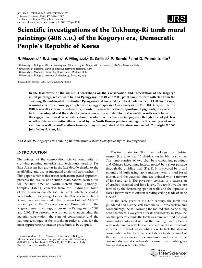
The tomb dates to 408 A.D. and belongs to a ministernamed Jing who had 13 districts under his jurisdiction.The tomb consists of two chambers containing paintingsand Chinese ideograms, interconnected by a short passagethrough the dividing wall (Fig. 1). It is covered by a turfmound and built using stone masonry with a mud-basedmortar, and the external joints are pointed with a mixtureof lime and sand. The pavement consists of a successionof crushed charcoal and lime layers. The tomb’s vaults areformed by the decreasing span of walls and the topmost isclosed by two tiers in caisson receding ceiling made of stoneblocks.7
In the early years of the 20th century, the tomb wasplundered and a stone slab from the vault was broken, andconsequently, the soil forming the mound slid inside fillingboth chambers. Two years after its rediscovery in 1976, thesoil that penetrated into the tomb was removed, and the localconservators informed us that the paintings were cleanedwith water and vegetable soap, and a cement slab was builtin order to prevent water infiltration. In fact, the state ofconservation is bad because of salt deposits, detachment ofthe paint layers caused by compression and cracks of theconcrete dome and condensation caused by a double glassbarrier that was built in 1994.7
Copyright 2006 John Wiley & Sons, Ltd.
Investigations of the Tokhung-Ri tomb mural paintings 1087
Table 1. Position and description of the analyzed samples
Sample Color Location/description
DPRK1 Dark red West wall, inner passage connecting the twochambers south side, upper section. Sample was takenfrom the external dark red band from ¾30 cm fromthe top above a lacuna. The sampling area was alsocovered with a salt deposit
DPRK2 Yellow East wall of inner passage connecting the twochambers, north side, upper section. Sample wastaken from the yellow band, just beneath the ceiling.This area is slightly covered with mud
DPRK3 Green 1st chamber: south wall, west section, lower area offrieze and 40 cm away from the corner. The greenpaint layer appears bright and the sample was takenfrom close to a nearby lacuna
DPRK4 Black/red 1st chamber West wall, south section, upper area offrieze near the corner
DPRK5 Yellow 1st chamber West wall, south section, upper row ofdignitaries (south side of 3rd in sequence from south)
DPRK6 White deposit onyellow paint layer
1st chamber West wall, 4th dignitary from south,upper row of dignitaries
DPRK7 Brown East wall of inner passage, sample of stone andpreparation layer for the painted decorations
DPRK8 Black North wall, 1st chamber: sample from the backgroundbehind the main figure
DPRK9 Red North wall, 1st chamber: upper part of east section.
EXPERIMENTAL
The samples collected were submitted to a complement ofanalytical methods (Table 2): (1) optical (OM)), ultravioletfluorescence (UV) and polarized light (PLM) microscopy,(2) scanning electron microscopy (SEM) coupled with elec-tron probe microanalysis (EDX), (3) X-ray diffraction (XRD),(4) thermogravimetric (TG), derivative thermogravimetric(DTG) and differential thermal analyses (DTA), (5) Fouriertransform infrared micro-spectroscopy (µ-FTIR) coupledwith attenuated total reflectance (ATR) and (6) micro Raman-spectroscopy (µ-Raman).
Optical microscopyApart from sample DPRK6, the other samples were embed-ded in a polyester resin support and they were cross-sectioned and polished with conventional methods usingsilicon carbide cards of successive grades, 120, 400, 800and 1000. Dark-field observation of the cross-sectioned sam-ples was performed using an optical microscope, OlympusBX51M, and photomicrographs were recorded with a scan-ner digital camera, Olympus DP70. Mineralogical, structuraland textural data of the materials were obtained by opti-cal analyses on thin sections of the whole sample (sampleDPRK7).
Scanning electron microscopyA scanning electron microscope, Philips XL 20 model SEM-EDX equipped with an energy-dispersive X-ray analyzer,was used on the cross-sectioned samples DPRK1, DPRK2,DPRK3, DPRK4, DPRK5 and DPRK7. The elemental com-position determination was carried out at an accelerationvoltage of 25–30 keV, a lifetime >50 s, CPS ³ 2000 and aworking distance of 34 mm. EDX-4 software equipped witha ZAF correction procedure for bulk specimens was used forsemiquantitative analyses of X-ray intensities.
Samples DPRK8 and DPRK9 were analyzed using anextended pressure scanning electron microscope, Zeiss EVO50 EP, equipped with an INCA EDX detector run with theINCA energy software, in the variable pressure mode at aninternal chamber pressure of 70 Pa. The determination ofthe elemental composition was carried out at an accelerationvoltage of 20–25 keV, a lifetime of 400 s, CPS ³ 12 500 and aworking distance of 8 mm.
X-ray diffractionSemiquantitative mineralogical analyses were performedon powdered samples by XRD using a Philips PW 1710diffractometer. Instrumental and measuring conditions were:Cu K˛ radiation; 40 kV/30 mA; divergence and detector slitsof 1°; 2� step size of 0.02°; and time for step of 1 s.
Copyright 2006 John Wiley & Sons, Ltd. J. Raman Spectrosc. 2006; 37: 1086–1097DOI: 10.1002/jrs
1088 R. Mazzeo et al.
(a)
(b)
(c)
Figure 1. Tokhung-Ri tomb: (a) drawing of the front and main chambers seen from the west, (b) portrait of the deceased master ofthe tomb, (c) detail of knights.
Thermogravimetric, derivative thermogravimetricand differential thermal analysesIdentification and quantitative analysis of carbonate contentswere carried out by TG, DTG and DTA analyses usinga Setaram TAG 24 apparatus. Operative conditions were:heating rate 20 °C/min; CO2 atmosphere; heating range from20 to 1000 °C.
Micro-FTIR spectroscopyDepending on the sample size available, FTIR analyses wereperformed with the KBr pellet technique in transmissionmode, or by using a Thermo Nicolet Continuµm FTIRmicroscope equipped with a slide-on micro ATR (Si crystal)device and the spectra recorded in the reflection mode, orusing diamond/NaCl compression cells with the spectra
Copyright 2006 John Wiley & Sons, Ltd. J. Raman Spectrosc. 2006; 37: 1086–1097DOI: 10.1002/jrs
Investigations of the Tokhung-Ri tomb mural paintings 1089
Table 2. Types of analyses performed on samples
Type of analysis
SampleStereo
microscope OM PLM SEM-EDXExtended pressure
SEM-EDX XRDTG,DTG,
DTAFTIR,
µ-FTIR µ-Raman
DPRK1 ð ð ð ð ðDPRK2 ð ð ð ð ðDPRK3 ð ð ð ð ðDPRK4 ð ð ð ð ðDPRK5 ð ð ð ð ðDPRK6 ð ðDPRK7 ð ð ð ð ð ðDPRK8 ð ð ðDPRK9 ð ð ð
recorded in the transmission mode. The FTIR microscope,fitted with a MCT type A detector and cooled by liquidnitrogen, uses a 15ð Thermo-Electron Infinity Reflachromatobjective and a tube factor of 10ð. Single-point ATRmeasurements were performed by placing cross-sectionedsamples directly on a motorized XYZ microscope stage,which was also used for the ATR mapping; the latter wasperformed directly on selected areas of the cross-sectionof sample DPRK3. For each analyzed area, a total of 16spectra were collected with a step size of 60 microns. Fortransmittance measurements, either a micro-diamond orNaCl compression cell on fragments of whole sampleswas used. An FTIR Thermo Nicolet Avatar 370 was usedto record the spectra from 4000 to 650 cm�1 (ATR andcompression cells) and from 4000 to 400 cm�1 (KBr pellettechnique). A total of 64 scans were recorded and theresulting interferogram averaged. The mirror velocity was
Figure 2. PLM microphotograph of the thin section of sampleDPRK7: arrows indicate the location of the recrystallizedcalcite.
(a)
(b)
(c)
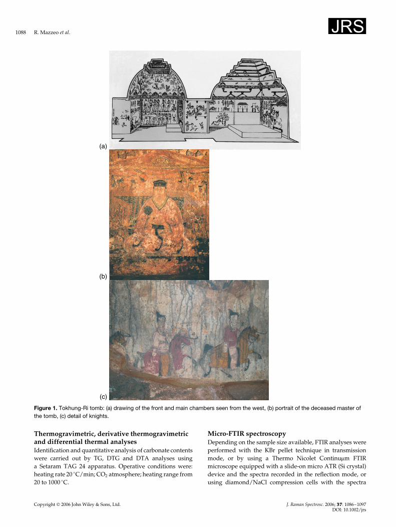
Figure 3. Stereo-microphotograph of a thin section of sampleDPRK7: (a) stone substrate, (b) white preparation layer (calcite,quartz and clay) and (c) clay and calcium carbonateconcretion/deposit.
1.8988 cm s�1, with a resolution of 4 cm�1 and triangularapodization. Acquisition and postrun processing werecarried out using the Nicolet ‘Omnic’ software.
Micro-Raman spectroscopyThe micro-Raman analyses of the paint samples wereperformed directly on the fragment surface or by placingthe cross-sectioned samples on the microscope stage anddirecting the laser light through a 50ð objective of anOlympus microscope onto the different paint layers visibleunder the cross-section. The Raman analyses were carriedout with a Jobin Yvon-Horiba Labram instrument, andwith a laser of 632.8 nm at powers ranging from 0.5 to 5mW (slit: 5 cm�1) according to the thermal stability of thecompounds to be investigated. A CCD (330 ð 1100 pixels)
Copyright 2006 John Wiley & Sons, Ltd. J. Raman Spectrosc. 2006; 37: 1086–1097DOI: 10.1002/jrs
1090 R. Mazzeo et al.
detector cooled by the Peltier effect was used at 200 K. Forcomplete investigation, a Jasco 2000 spectrometer combinedwith an Olympus microscope was used. This instrument wascooled with liquid nitrogen and used the 488 nm laser linewith a power that was always lower than 1 mW. The spectralinterval was changed according to the type of compounds,but was generally between 1100 and 100 cm�1. The organic
compounds were studied in the high wavenumber range,whereas the inorganic ones, the oxides and sulfides inparticular, at a lower wavenumber range. The spectracollection time was also varied according to the magnitudeof the scattering signal. Each paint sample was investigatedby analyzing an average of 30 particles distributed in themass of the sample analyzed, and in the subsequent layers
(a)
(b)
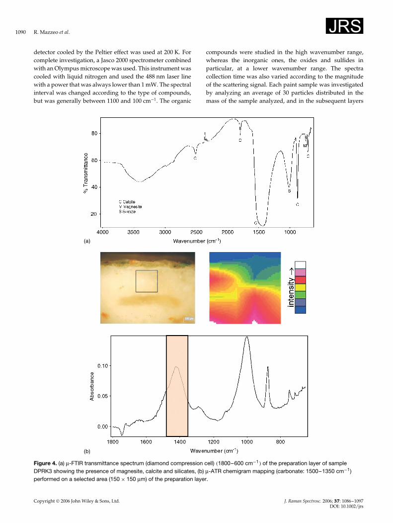
Figure 4. (a) µ-FTIR transmittance spectrum (diamond compression cell) �1800–600 cm�1� of the preparation layer of sampleDPRK3 showing the presence of magnesite, calcite and silicates, (b) µ-ATR chemigram mapping (carbonate: 1500–1350 cm�1)performed on a selected area (150 ð 150 µm) of the preparation layer.
Copyright 2006 John Wiley & Sons, Ltd. J. Raman Spectrosc. 2006; 37: 1086–1097DOI: 10.1002/jrs
Investigations of the Tokhung-Ri tomb mural paintings 1091
in the case of cross sections. If the sample was stronglyheterogeneous, up to 100 measurements were carried out.The spectra recorded were elaborated and prepared forreporting by using the program Grams by Thermogalactic.
RESULTS AND DISCUSSION
Stone substrateThe stone has a grayish color and is made up of graniterock with a granular texture characterized by a partialpseudo-parallel orientation such that an exfoliation withpreferential detachments is produced within the stonestructure.
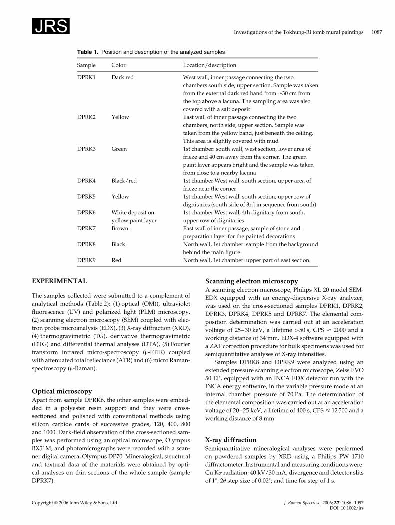
Optical observations and XRD analyses revealed thepresence of polycrystalline quartz, potassium feldspar,plagioclase, mica (mainly biotite with traces of muscovite)and additional minerals such as apatite, zircon and opaqueminerals (probably sulfides and/or iron oxy-hydroxide).From the petrographic point of view, the stone can beclassified as a quartz monzonite with an iso-orientedstructure. The PLM examination of sample DPRK7 alsoshowed that recrystallized calcite crystals with a ‘secretion’structure are present between the stone substrate andthe preparation layer (Fig. 2). Their fiber-like texture isresponsible for the observed detachment of the preparationlayer from the stone substrate, even though some contactpoints still exist between the two.
As far as the mechanisms of formation of calcite crystalsare concerned, probably different factors are to be takeninto consideration: (1) the very low porosity of the stonesubstrate that does not allow water to be absorbed, (2) therelatively high porosity of the preparation layer and itspoor original adhesion to the stone substrate, and (3) thetomb’s environment, which is characterized by relative
humidity (RH) values close to 100%. Therefore, a possiblemechanism could be as follows: (1) During the periods inwhich the RH is very high, water penetrates the preparationlayer and partially dissolves the constituent calcite. (2) Thecalcite recrystallization happens at the interface with thestone substrate as a kind of sub-fluorescence phenomenonfavored by RH changes towards more dry environmentalcondition. For this, a one-year monitoring campaign aimedat evaluating the seasonal RH changes inside the tomb wouldbe indispensable in order to validate this hypothesis of themechanism of formation of the calcite crystals.
Preparation layersThe only available information from local conservator-restorers refers to the use of two preparatory layers composedof lime, sand and organic aggregates, which were applied tothe vaults and walls: the first layer (5 cm thick) composed oflime and sand/chopped straw in the proportion of 1 : 3; andthe second layer composed of a mixture of lime and sand inequal proportion, with a thickness of about 1.5 cm. A limewash (0.8–1 mm thick) was then applied as a primer for thepainted areas.
Only sample DPRK7 was collected in such a way thatthe stone substrate was included, thus showing the overallstratigraphic morphology. Optical microscopy observationsshowed that only one white preparation layer is presentover the stone substrate (Fig. 3). Its thickness varies fromabout 0.5 to 1.5 mm, and vegetal filaments and black carbonelements are embedded within the layer. Transverse crevicesare present, which are re-cemented by limpid spar calcite.
The SEM-EDX results indicated the presence of notonly pure calcite, together with traces of quartz and claymineral components, but also of calcite rich in magnesiumor dolomite, CaMg�CO3�2, as confirmed by TG and DTA
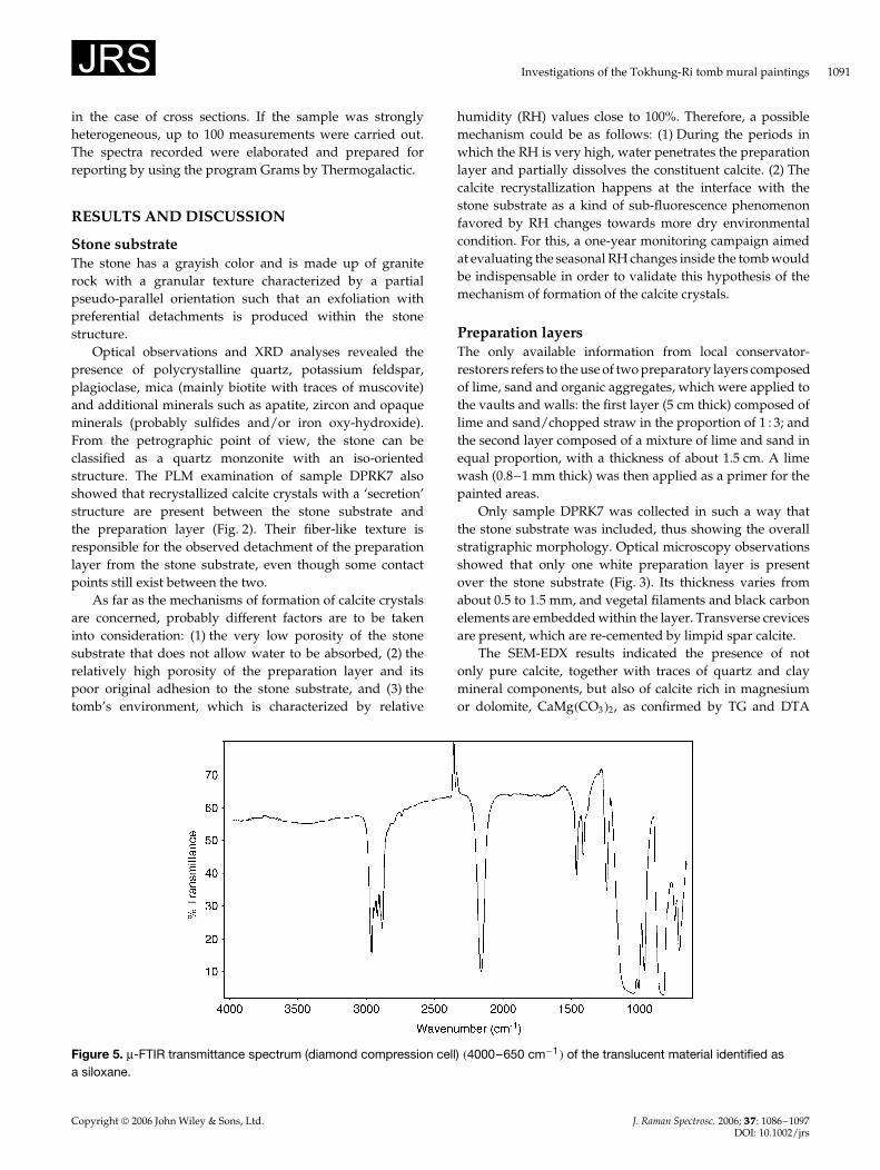
Figure 5. µ-FTIR transmittance spectrum (diamond compression cell) �4000–650 cm�1� of the translucent material identified asa siloxane.
Copyright 2006 John Wiley & Sons, Ltd. J. Raman Spectrosc. 2006; 37: 1086–1097DOI: 10.1002/jrs
1092 R. Mazzeo et al.
analyses. µ-FTIR examination of the DPRK3 preparation layer(Fig. 4) indicated the contemporary presence of magnesite�MgCO3�, calcite and silicates. The preparation layers of theother samples are constituted of the same compounds. Inaddition, stereo microscopic observations of the preparationlayer of samples DPRK4, DPRK5 and DPRK3 showedthe presence of traces of a translucent material that wasconstituted of a siloxane material. In fact, its FTIR spectrum(Fig. 5) is characterized by a strong infrared band in the1130–1000 cm�1 region due to the asymmetric Si–O–Sistretching vibration, the strong Si–H stretching vibrationat 2158 cm�1 as well as a band at 1242 cm�1 associated withthe symmetric Si–Et deformation.8 This material, which isscarce and randomly present within the preparation layers,may hardly represent the residue of the application of asiloxane-based protective coating or consolidant applied onthe painted surfaces in the past. In fact, if this was thecase, it should be more evenly distributed along the overallpainted cross-section and a thin layer should also be presenton the outermost external surface and easily detectable by µ-FTIR. More probably, this translucent material represents theresidue of antifoaming agents that are added to detergents,and its presence is linked to the use of surface cleaningprocedures that were carried out in the past and mentionedby local conservators.
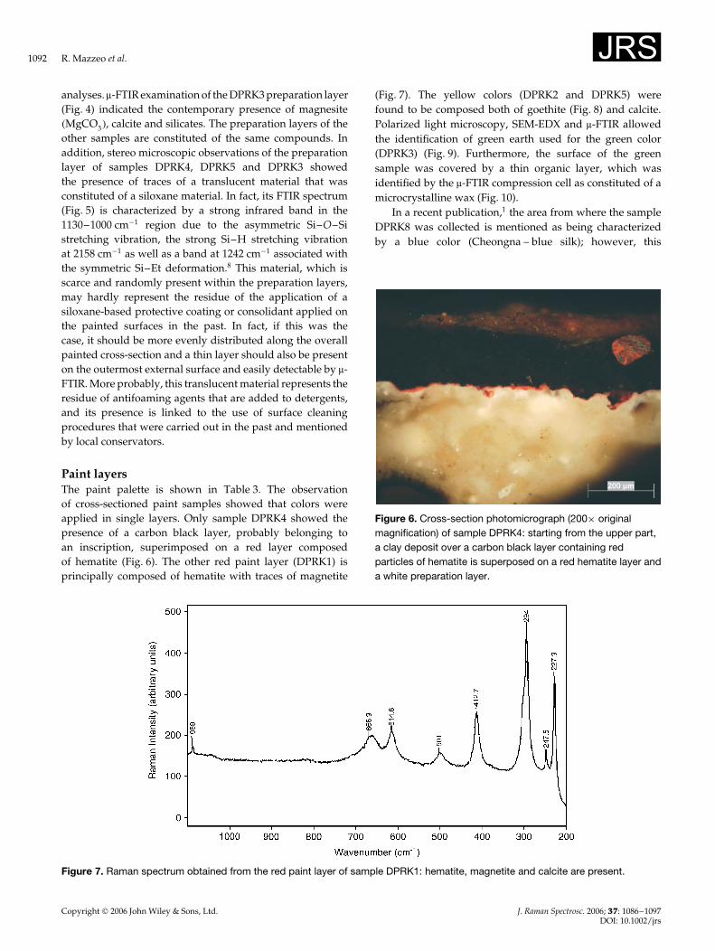
Paint layersThe paint palette is shown in Table 3. The observationof cross-sectioned paint samples showed that colors wereapplied in single layers. Only sample DPRK4 showed thepresence of a carbon black layer, probably belonging toan inscription, superimposed on a red layer composedof hematite (Fig. 6). The other red paint layer (DPRK1) isprincipally composed of hematite with traces of magnetite
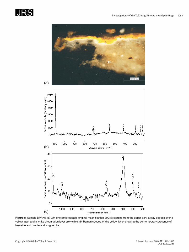
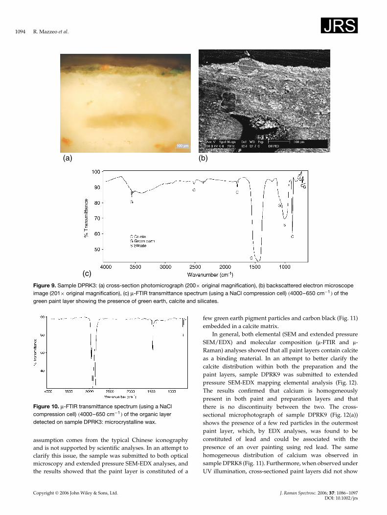
(Fig. 7). The yellow colors (DPRK2 and DPRK5) werefound to be composed both of goethite (Fig. 8) and calcite.Polarized light microscopy, SEM-EDX and µ-FTIR allowedthe identification of green earth used for the green color(DPRK3) (Fig. 9). Furthermore, the surface of the greensample was covered by a thin organic layer, which wasidentified by the µ-FTIR compression cell as constituted of amicrocrystalline wax (Fig. 10).
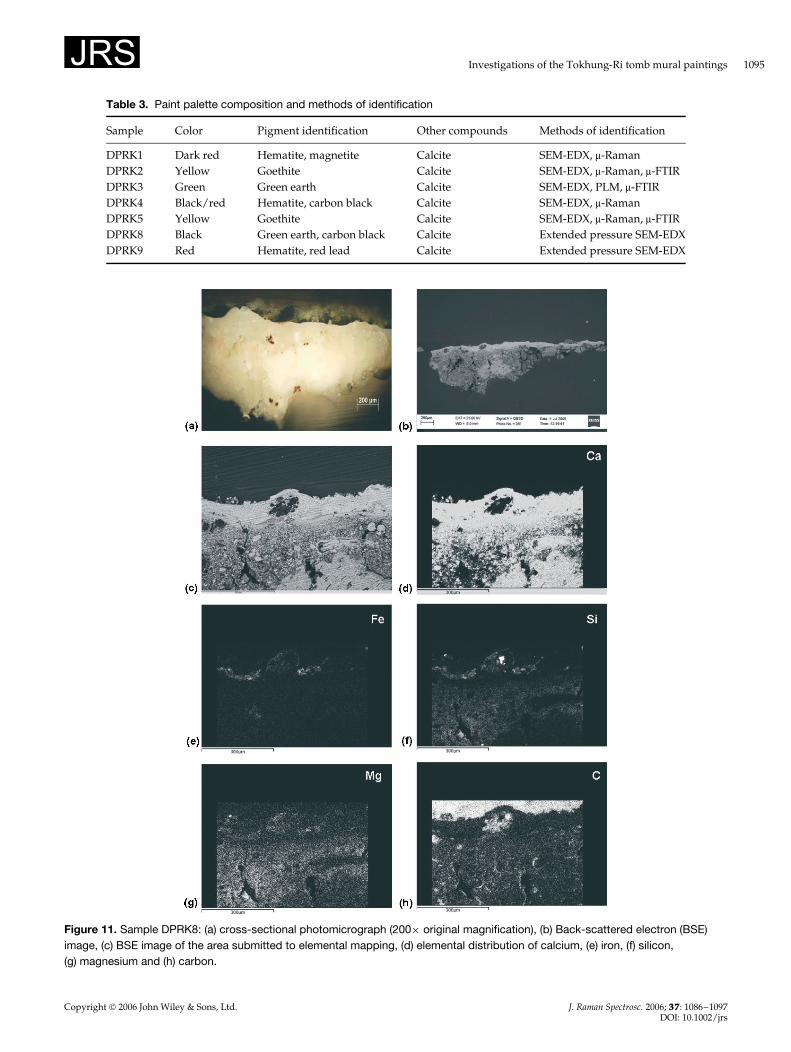
In a recent publication,1 the area from where the sampleDPRK8 was collected is mentioned as being characterizedby a blue color (Cheongna – blue silk); however, this
Figure 6. Cross-section photomicrograph (200ð originalmagnification) of sample DPRK4: starting from the upper part,a clay deposit over a carbon black layer containing redparticles of hematite is superposed on a red hematite layer anda white preparation layer.
Figure 7. Raman spectrum obtained from the red paint layer of sample DPRK1: hematite, magnetite and calcite are present.
Copyright 2006 John Wiley & Sons, Ltd. J. Raman Spectrosc. 2006; 37: 1086–1097DOI: 10.1002/jrs
Investigations of the Tokhung-Ri tomb mural paintings 1093
(a)
(b)
(c)
Figure 8. Sample DPRK5: (a) OM photomicrograph (original magnification 200ð): starting from the upper part, a clay deposit over ayellow layer and a white preparation layer are visible, (b) Raman spectra of the yellow layer showing the contemporary presence ofhematite and calcite and (c) goethite.
Copyright 2006 John Wiley & Sons, Ltd. J. Raman Spectrosc. 2006; 37: 1086–1097DOI: 10.1002/jrs
1094 R. Mazzeo et al.
(c)
(b)(a)
Figure 9. Sample DPRK3: (a) cross-section photomicrograph (200ð original magnification), (b) backscattered electron microscopeimage (201ð original magnification), (c) µ-FTIR transmittance spectrum (using a NaCl compression cell) �4000–650 cm�1� of thegreen paint layer showing the presence of green earth, calcite and silicates.
Figure 10. µ-FTIR transmittance spectrum (using a NaClcompression cell) �4000–650 cm�1� of the organic layerdetected on sample DPRK3: microcrystalline wax.
assumption comes from the typical Chinese iconographyand is not supported by scientific analyses. In an attempt toclarify this issue, the sample was submitted to both opticalmicroscopy and extended pressure SEM-EDX analyses, andthe results showed that the paint layer is constituted of a
few green earth pigment particles and carbon black (Fig. 11)embedded in a calcite matrix.
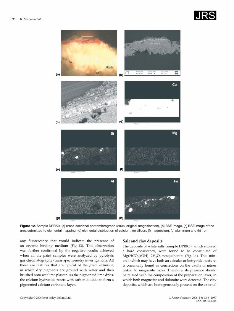
In general, both elemental (SEM and extended pressureSEM/EDX) and molecular composition (µ-FTIR and µ-Raman) analyses showed that all paint layers contain calciteas a binding material. In an attempt to better clarify thecalcite distribution within both the preparation and thepaint layers, sample DPRK9 was submitted to extendedpressure SEM-EDX mapping elemental analysis (Fig. 12).The results confirmed that calcium is homogeneouslypresent in both paint and preparation layers and thatthere is no discontinuity between the two. The cross-sectional microphotograph of sample DPRK9 (Fig. 12(a))shows the presence of a few red particles in the outermostpaint layer, which, by EDX analyses, was found to beconstituted of lead and could be associated with thepresence of an over painting using red lead. The samehomogeneous distribution of calcium was observed insample DPRK8 (Fig. 11). Furthermore, when observed underUV illumination, cross-sectioned paint layers did not show
Copyright 2006 John Wiley & Sons, Ltd. J. Raman Spectrosc. 2006; 37: 1086–1097DOI: 10.1002/jrs
Investigations of the Tokhung-Ri tomb mural paintings 1095
Table 3. Paint palette composition and methods of identification
Sample Color Pigment identification Other compounds Methods of identification
DPRK1 Dark red Hematite, magnetite Calcite SEM-EDX, µ-RamanDPRK2 Yellow Goethite Calcite SEM-EDX, µ-Raman, µ-FTIRDPRK3 Green Green earth Calcite SEM-EDX, PLM, µ-FTIRDPRK4 Black/red Hematite, carbon black Calcite SEM-EDX, µ-RamanDPRK5 Yellow Goethite Calcite SEM-EDX, µ-Raman, µ-FTIRDPRK8 Black Green earth, carbon black Calcite Extended pressure SEM-EDXDPRK9 Red Hematite, red lead Calcite Extended pressure SEM-EDX
Figure 11. Sample DPRK8: (a) cross-sectional photomicrograph (200ð original magnification), (b) Back-scattered electron (BSE)image, (c) BSE image of the area submitted to elemental mapping, (d) elemental distribution of calcium, (e) iron, (f) silicon,(g) magnesium and (h) carbon.
Copyright 2006 John Wiley & Sons, Ltd. J. Raman Spectrosc. 2006; 37: 1086–1097DOI: 10.1002/jrs
1096 R. Mazzeo et al.
Figure 12. Sample DPRK9: (a) cross-sectional photomicrograph (200ð original magnification), (b) BSE image, (c) BSE image of thearea submitted to elemental mapping, (d) elemental distribution of calcium, (e) silicon, (f) magnesium, (g) aluminum and (h) iron.
any fluorescence that would indicate the presence ofan organic binding medium (Fig. 13). This observationwas further confirmed by the negative results achievedwhen all the paint samples were analyzed by pyrolysisgas chromatography/mass spectrometry investigations. Allthese are features that are typical of the fresco technique,in which dry pigments are ground with water and thenbrushed onto wet lime plaster. As the pigmented lime dries,the calcium hydroxide reacts with carbon dioxide to form apigmented calcium carbonate layer.
Salt and clay depositsThe deposits of white salts (sample DPRK6), which showeda hard consistency, were found to be constituted ofMg�HCO3�(OH) Ð 2H2O, nesquehonite (Fig. 14). This min-eral, which may have both an acicular or botryoidal texture,is commonly found as concretions on the vaults of mineslinked to magnesite rocks. Therefore, its presence shouldbe related with the composition of the preparation layer, inwhich both magnesite and dolomite were detected. The claydeposits, which are homogeneously present on the external
Copyright 2006 John Wiley & Sons, Ltd. J. Raman Spectrosc. 2006; 37: 1086–1097DOI: 10.1002/jrs
Investigations of the Tokhung-Ri tomb mural paintings 1097
Figure 13. Sample DPRK5: cross-section observed under UVlight illumination (original magnification 200ð). The only whitishfluorescence observable is due to the typical fluorescence ofthe calcite.
Figure 14. FTIR transmittance spectrum �4000–400 cm�1� ofsample DPRK6: nesquehonite.
surfaces of all samples (Fig. 2) and show a yellowish/browncolor, are mainly constituted of quartz, K-feldspars and tracesof mica cemented by a micritic calcite. From the point of viewof the procedures for surface cleaning, the presence of calcitein the outermost external clayish deposits makes these layersvery hard to be removed either mechanically or chemically.
CONCLUSIONS
The complement of analytical investigations carried out onthe Tokhung-Ri mural paintings allowed us to draw somepreliminary conclusions. All the identified pigments aresuitable for use with a fresco technique. Calcite is presentin both preparation and paint layers, and the stratigraphicmorphology of the cross-sectioned samples show that theyare strictly connected to each other without any visiblediscontinuity. These observations may represent a clear
evidence of the fact that paint colors were applied overa white preparation layer when it was still in a wet limeplaster condition. Furthermore, there is no evidence of thepresence of any organic binding media. At the present stateof knowledge, these observations greatly favor the viewthat a fresco technique was used for the execution of themural painting. It is not yet clear whether this techniquewas intentionally achieved by the North Korean painters.However, before drawing any decisive conclusions, the useof a fresco technique in DPRK should be confirmed by asurvey in the historical literature. In case of further scientificinvestigations and historical information confirming the useand existence of a fresco technique, this site could representone of the first examples of fresco painting in Far East Asia,as the majority of the wall paintings studied so far6,9 werefound to have been painted with secco techniques. It wouldalso be advisable to perform similar scientific investigationson the mural paintings located in China and belonging to thesame Koguryo era.
As far as the state of conservation of the Tokhung-Ri mural paintings is concerned, they are not in a goodstate of conservation, as revealed by the detachment ofthe preparation and paint layers from the stone substrate.Furthermore, the double-glass barriers installed inside thetomb has to be regarded just as a protection againstvandalism and not as a safeguard for conservation. In fact,the levels of RH measured inside and outside the double-glass barriers are of the same order of magnitude. The waterinfiltration from the surrounding turf mound as well as thehigh RH levels may be responsible for the detachment of thepaint layers that was observed.
REFERENCES1. Lena K. Koguryo Tomb Murals. ICOMOS Korea: Seoul, Korea, 2004.2. Yi Y, Yu H, Kim S, Kang H, Jo Y, Aoki S, Ohbayashi K. Mun. hwa.
jae. 2003; 4(12): 1.3. Moon W-S, Hong J-O, Hwang J-J, Kim S-K, Cho N-C. Annual
review in cultural properties studies 2002; 35: 160.4. Chen Q, Sinkai T, Inaba M, Sugisita R, Huang G. Bunkazai Hozon
Shufuku Gakkai shi : kobunkazai no kagaku 1997; 41: 78.5. Kang H, Yi Y, Yu H, Kim Y, Jo Y, Aoki S, Yamamoto N,
Ohbayashi K. Conserv. Sci. Museum 2001; 3: 43.6. Mazzeo R, Baraldi P, Lujan R, Fagnano C. J. Raman Spectrosc. 2004;
35: 678.7. Lujan Lunsford R. In UNESCO Symposium on the Conservation
of Koguryo Tombs, Scientific and Methodological Approach, Seoul,Republic of Korea, October 2004; 100.
8. Lee Smith A. Analysis of Silicones. R.E. Krieger PublishingCompany: Malabar, Florida, 1983.
9. Mazzeo R, Joseph E, Minguzzi V, Modugno F, Prati S. Indaginiscientifiche sui dipinti murali della dinastia Yuan (1279–1368d.C.) situati nel sito archeologico di Yao Wang Shan, Cina. InFar Est Asian mural paintings: diagnosis, conservation and restoration.When east and west encounter and exchange, Mazzeo R (ed.). LongoEditore: Ravenna, 2006; 65.
Copyright 2006 John Wiley & Sons, Ltd. J. Raman Spectrosc. 2006; 37: 1086–1097DOI: 10.1002/jrs


