Embed Size (px)
Citation preview
Naučni časopis Medicinskog fakulteta Univerziteta u Nišu i
Podružnice Srpskog lekarskog društva u Nišu
Scientific journal of the University of Niš Faculty of Medicine and
the Department of the Serbian Medical Society in Niš
Acta Medica Medianae
Vol 59, No 1, March 2020 UDK 61 ISSN 0365-4478 (Printed version)
ISSN 1821-2794 (Online)
http://www.medfak.ni.ac.rs/amm
Izvršni urednik Executive Editor Prof. Boris Đinđić, MD, PhD (Niš, Serbia)
Izvršni urednik za farmaciju Executive Editor for Pharmacy Prof. Andrija Šmelcerović, PhD (Niš, Serbia)
Sekreterijat uređivačkog odbora Editorial assisstants Jelena Milenković, MD, PhD (Niš, Serbia), sekretar (chief assistant) Assist. Prof. Voja Pavlović, MD, PhD (Niš, Serbia) Assist. Prof. Zoran Bojanić, MD, PhD (Niš,Serbia) Assist. Prof. Jasmina Đorđević-Jocić, MD, PhD (Niš, Serbia) Assist. Prof.Jelena Lazarević, PhD (Niš, Serbia) Dr Rade R. Babić, MD, PhD (Niš, Serbia) Assist. Prof. Nataša Milosavljević, PhD (Niš, Serbia) Nataša Bakić-Mirić, University lecturer of English, PhD (Niš, Serbia) Assist. Prof. Tomislav Kostić, MD, PhD (Niš, Serbia) Danica Marković, MD (Niš, Serbia) Slavica Stojnev, MD (Niš, Serbia) Denitsa Yancheva, PhD (Sofia, Bulgaria) Assist. Prof. Ivana Damnjanović, PharmD, PhD(Niš, Serbia) Assist. Prof. Nikola Stefanović, PharmD, PhD (Niš, Serbia) Dane Krtinić, MD (Niš, Serbia) Milovan Stojanović, MD (Niš, Serbia) Assist. Milica Kostić, PharmD (Niš, Serbia) Assist. Milica Milutinović, PharmD (Niš, Serbia) Assist. Prof. Bojana Miladinović, PharmD, PhD (Niš, Serbia) Assist. Dragan Zlatanović, MD, PhD (Niš, Serbia) Assist. Bobana Milojković, MD, PhD (Niš, Serbia) Assist. Prof. Tanja Džopalić, MD (Niš, Serbia) Assist. Aleksandar Ranković, MD, PhD (Niš, Serbia) Dr Ana Spasić, PharmD (Niš, Serbia) Dr Dušan Radomirović, MD (Niš, Serbia) Dr Sonja Janković, MD (Niš, Serbia) Dr Igor Zivković, MD (Belgrade, Serbia) Tehnička i internet obrada Technical and Internet Editing Topić Goran, BA Lektor za engleski jezik Proofreading Bojana Marjanović, BA in English language and literature Milena Đorđević, BA in English language and literature Lektori za srpski jezik Proofreading Ana Višnjić, BA in Serbian language and literature Neda Pavlović, Phd, Linguistics:Serbian language Nikola Đorđević, BA in Serbian language and literature
Uređivački savet Advisory Editors Prof. Dobrila Stanković-Đorđević, MD, PhD (Niš, Serbia) Prof. Dragan Veselinović, MD, PhD (Niš, Serbia)
Uređivački odbor Editorial Board Prof. Milan Višnjić, MD, PhD (Niš, Serbia) Prof. Dušica Pavlović, MD, PhD (Niš, Serbia) Prof. Miroslav Stojanović, MD, PhD (Niš, Serbia Prof. Dušan Sokolović, MD, PhD (Niš, Serbia) Prof. Marija Daković-Bjelaković, MD, PhD (Niš, Serbia Prof. Dušanka Kitic, MD, PhD (Niš, Serbia) Prof. Ivan Micić, MD, PhD (Niš, Serbia) Prof. Dušan Milisavljević, MD, PhD (Niš, Serbia) Prof. Biljana Đorđević, MD, PhD (Niš, Serbia) Prof. Maja Milojković, MD, PhD (Niš, Serbia) Prof. dr Eugene N. Myers (Pittsburgh, USA) Prof. dr Helmut Roskamm (Bad Krozingen, Austria) Prof. dr Waldemar Kozuschek (Bochum, Germany) Prof. dr Raimond Ardaillou (Paris, France) Prof. dr Milan Dimitrijević (Houston, USA) Prof. dr Robin Leake (Glasgow, UK) Academician Aleksej Prijmak (Moscow, Russia) Academician Mihail Pereljman (Moscow, Russia) Prof. Miodrag Jevtić, MD, PhD (MMA, Belgrade, Serbia) Prof. dr Žernakova Nina Ivanovna (Belgorod, Russia) Academician Petrija Vasileva (Sofia, Bulgaria) Prof. dr Badr Eldin Mostafa (Cairo, Egypt) Prof. dr Dan M. Fliss (Tel-Aviv, Israel) Prof. Takanori Hattori, MD, PhD (Shiga, Japan) Prof. Savevski Jordan, MD, PhD (Skopje, RN Macedonia) Prof. Davran Gaipov, PhD (Almaty, Kazakhstan) Assoc. Prof. Ilko Getov, PhD (Sofia, Bulgaria) Prof. Vladmila Bojanić, MD, PhD (Niš, Serbia) Prof. Aleksandra Stankovic, MD, PhD (Niš, Serbia) Prof. Dragan Veselinović, MD, PhD (Niš, Serbia) Academician. Milorad Mitković, MD, PhD (Niš, Serbia) Prof. Nebojša Đorđević, MD, PhD (Niš, Serbia) Prof. Stojan Radić, MD, PhD (Niš, Serbia) Prof. Saša Živić, MD, PhD (Niš, Serbia) Prof. Zorica Stanojević, MD, PhD (Niš, Serbia) Prof. Dušica Stojanović, MD, PhD (Niš, Serbia) Prof. Stevo Najman, PhD (Niš, Serbia) Prof. Zoran Radovanovic MD, PhD (Niš, Serbia)
Acta Medica Medianae (UDK 61; ISSN 0365-4478 štampana verzija; ISSN 1821-2794 elektronska verzija) je zvanični časopis Medicinskog fakulteta Univerziteta u Nišu i Podružnice Srpskog lekarskog društva u Nišu pod pokroviteljstvom Ministarstva za nauku i tehnološki razvoj Republike Srbije. Časopis izlazi četiri puta godišnje od 1962 godine. Izdavač je Medicinski fakultet Univerziteta u Nišu, Bulevar dr Zorana Đinđića 81, 18000 Niš, Srbija. Sadržaj i celokupan tekst časopisa dostupan je na sajtu Medicinskog fakulteta http://www.medfak.ni.ac.rs/amm. Godišnja pretplata: za inostranstvo 60 USA dolara, za ustanove 2500 dinara i za pojedince 1500 dinara. Sredstva uplatiti na žiro račun Medicinskog fakulteta u Nišu br. 840-1681666-03, sa naznakom za Acta Medica Medianae. Uputstvo autorima se objavljuje u svakom broju, pri čemu je autor dužan da se pridržava navedenih uputstava prilikom predaje ruko-pisa. Radovi se mogu slati u elektronskom formatu na adresu: [email protected]. Naknada za štampanje rada iznosi 1000 dinara za autora, a 500 dinara za koautore, za svaki prihvaćeni rad. Acta Medica Medianae zadržava pravo dalje distribucije i štampanja radova. Kontakt adresa: Časopis Acta Medica Medianae, Medicinski fakultet, Bulevar dr Zorana Đinđića 81, 18000 Niš, Srbija E-mail: [email protected] Tel+381-18-4533001 lok. 122 fax. +381-18-4534336 Tiraž 200 primeraka. Štampa: “Sven”, Niš, Srbija. Acta Medica Medianae je trenutno indeksirana na Index Copernicus-u, Srpskom citatnom indeksu, DOAJ i EBSCO Copyright © by University of Niš Faculty of Medicine
Acta Medica Medianae (UDK 61; ISSN 0365-4478 printed version; ISSN 1821-2794 online) is the official Journal of the University of Niš Faculty of Medicine and the Department of the Serbian Medical Society in Niš published with the help of the Ministry of Science and Technological Development of the Republic of Serbia. The Journal has been published four times a year since 1962. The publisher is the University of Niš Faculty of Medicine, Institutional address: dr Zoran Đinđić 81, 18000 Niš, Serbia. Table of contents and full texts of articles are available on the Institutional Home Page at http://www.medfak.ni.ac.rs/amm. Prices are subject to change. All subscriptions start with the first issue of the current year. For payment details contact the Secreteriat at [email protected]. Instructions for authors appear in every issue. Manuscripts accepted for publication are not returned to the author(s). Acta Medica Medianae retains the right for further distribution and printing of the articles. Editorial correspodence: Journal Acta Medica Medianae, Faculty of Medicine, Dr Zoran Đinđić 81, 18000 Niš, Serbia. Electronic submission of the papers: [email protected] Phone: +381-18-4533001 lok. 113 fax. +381-18-4534336 Printed on acid-free paper; 200 issues. Press: “Sven”, Niš, Serbia Acta Medica Medianae is currently indexed in Index Copernicus, Serbian Citation Index, DOAJ and EBSCO Copyright © by University of Niš Faculty of Medicine

Naučni časopis Medicinskog fakulteta Univerziteta u Nišu i
Podružnice Srpskog lekarskog društva u Nišu
Scientific journal of the University of Niš Faculty of Medicine and
the Department of the Serbian Medical Society in Niš
Acta Medica Medianae
Vol 59, No 1, March 2020 UDK 61 ISSN 0365-4478 (Printed version)
ISSN 1821-2794 (Online)
http://www.medfak.ni.ac.rs/amm
Autor slike na prednjoj stranici: Bojana Marjanović Lektor za engleski jezik, Medicinski fakultet, Univerzitet u Nišu
Vol 59, No 1, March, 2020
PREVALENCE AND DETERMINANTS OF HEMOGLOBIN VARIABILITY AND ITS IMPACT ON MORTALITY IN PATIENTS ON MAINTENANCE HEMODIALYSIS Zorica Dimitrijević, Branka Mitić, Karolina Paunović, Sonja Šalinger-Martinović
5
ТHE JUSTIFICATION OF CLINICAL PHARMACY SKILLS AND KNOWLEDGE FOR MODERN COMMUNITY PHARMACIST Maja Koraćević, Aleksandra Catić-Đorđević, Nikola Stefanović, Ivana Damnjanović, Ivana Stošić, Radmila Veličković-Radovanović
14
DETERMINING THE CONTENT OF Cd, Cu, Pb AND Zn IN THE LEAVES OF DANDELION (TARAXACUM OFFICINALE WEBB.) AND IN THE SOIL BY ICP-OES Dragan Velimirović, Biljana Kaličanin, Milan Stojković, Snežana Tošić
23
ANALYSIS OF GENE AMPLIFICATION IN PAPILLARY THYROID CARCINOMAS Aleksandar Milićević, Dragan Mihailović, Žaklina Mijović
31
EARLY POSTOPERATIVE OUTCOMES OF SURGICALLY TREATED AORTIC DISSECTION IN MARFAN SYNDROME PATIENTS Ivan Nešić, Jelena Dotlić, Igor Živković, Aleksandra Šljivić, Petar Vuković, Slobodan Mićović, Petar Milačić, Miroslav Miličić, Djordje Zdravković, Milan Ćirković, Miodrag Perić
36
RELATIONSHIPS BETWEEN QUALITY OF SLEEP AND INSOMNIA WITH DEPRESSION AND ANXIETY SYMPTOMS IN MEDICAL UNIVERSITY STUDENTS IN SERBIA Aleksandar Višnjić, Snežana Miljković, Dragan Nikolić, Tamara Jovanović, Katarina Bulatović, Marko Ristić, Dragan Toskić
44
DISTRIBUTION OF PDGFR+ CELLS AND INTERSTITIAL CELLS OF CAJAL IN THE HUMAN
FETAL GUT Goran Radenković, Aleksandra Veličkov, Vladimir Petrović, Miloš Dičić, Marko Gmijović
51
EARLY DETECTION OF POTENTIAL CHRONIC ALCOHOLISM BY DETERMINING THE LEVEL OF IGA, MCV AND TRANSFERRIN Muhamed Sarvan, Sonja Ketin, Radmila Maksimović, Rade Biočanin
60
RELATIONSHIP BETWEEN BODY COMPOSITION AND VERTICAL JUMP PERFORMANCE AMONG ADOLESCENTS Darko Stojanović, Zoran Savić, Hadži Miloš Vidaković, Tijana Stojanović, Zoran Momčilović, Toplica Stojanović
64
CHARACTERISTICS OF CHRONIC VENOUS ULCERATION RELATED TO AGE Vesna Karanikolić, Maša Golubović, Nataša Djindjić, Nila Kučar
71
SIGNIFICANCE OF GENOTYPIC ALPHA GALACTOSIDASE A MUTATIONS IN FABRY DISEASE TREATMENT Jelena Randjelović, Mina Cvetković, Tamara Vrećić, Andriana Jovanović, Marina Randjelović, Tatjana Cvetković
76
GLOBE-PRESERVING SURGERY FOR TREATMENT OF ADVANCED EYELID CARCINOMA INFILTRATING ANTERIOR PERIORBITAL FAT TISSUE: CASE REPORT Nina Vujošević, Predrag Kovačević
83
INVASIVE DUCTAL BREAST CANCER METASTASIS INTO THE TEMPOROMANDIBULAR JOINT TWO YEARS AFTER THE INITIAL TREATMENT: A CASE REPORT Predrag Radović, Milovan Papović, Miloš Trajković, Nikola Živković, Andrija Ćosić
90
INTERNAL HERNIA ASSOCIATED WITH MECKEL’S DIVERTICULUM IN GERIATRIC PATIENT Aleksandar Karanikolić, Miodrag Djordjević, Ivan Pešić, Lidija Djordjević, Nebojša Ignjatović, Aleksandar Zlatić, Toplica Bojić
96
PATHOPHYSIOLOGICAL MECHANISMS OF ALUMINIUM TOXICITY Novica Bojanić, Jelena Milenković, Dijana Stojanović, Maja Milojković, Nataša Djindjić, Marko Gmijović
100
Vol 59, No 1, March, 2020
USE OF BOTULINUM TOXIN A AND SUBSEQUENT REHABILITATION IN AMBULATORY CHILDREN WITH SPASTIC CEREBRAL PALSY – EFFECTS AND DILEMMAS Hristina Čolović, Vesna Živković, Dragan Zlatanović, Nadica Milošević-Milenović, Dragana Janošević-Radović
110
RADIOLOGICAL CHARACTERISTICS OF PERIOSTEAL REACTIONS Nemanja Rančić, Aleksandar Jovanovski, Jelena Stevanović, Ratko Stamatović, Dejan Kostić, Igor Sekulić, Berislav Vekić, Jasenka Vasić-Vilić
116
THE ASSOCIATION OF GENETIC POLYMORPHISMS WITH DIABETES MELLITUS TYPE 1 Maja Jović, Vesna Cvetković, Milena Despotović, Tatjana Jevtović-Stoimenov
125
RETINAL PIGMENT EPITHELIUM TEARS IN YOUNGER PATIENTS: CAUSES AND CONSEQUENCES Sonja Cekić, Jasmina Djordjević-Jocić, Branislav Tomašević, Ivan Jovanović, Predrag Jovanović, Milena Vujanović
133
CYSTIC DUPLICATION OF STOMACH: A CASE REPORT Ivona Djordjević, Andjelka Slavković, Zoran Marjanović, Dragoljub Živanović, Ana Kostić, Danijela Djerić
139
Nd: YAG LASER ANTERIOR CAPSULOTOMY IN CAPSULAR PHIMOSIS IN THE EYE WITH PEX AND ZONULAR LESION Aleksandar Veselinović, Marija Cvetanović, Dragan Veselinović
144
ACUTE DACRYOADENITIS ASSOCIATED WITH INFECTIOUS MONONUCLEOSIS Marija Cvetanović, Aleksandar Veselinović, Marija Trenkić-Božinović, Kristina Stojanović, Ivona Trajković, Dragan Veselinović
149
SURGICAL TREATMENT OF GIANT PERICARDIAL CYST THROUGH THE LATERAL THORACOTOMY Stefan Timčić, Igor Živković, Staša Krasić, Marko Andjelković, Ivan Nešić, Milan Ćirković, Miodrag Perić
153
GESTALT PSYCHOTHERAPY: SCIENCE OR QUASI-SCIENCE? Jana Milić
158
THE APPLICATION OF INFORMATION TECHNOLOGIES IN THE PROCESS OF NURSING CARE Мilena Cvetković-Jovanović, Sunčica Ivanović, Sanja Trgovčević, Tatjana Kilibarda, Maja Stanković, Suzana Milutinović
165
Secretariat GUIDELINES FOR PAPER SUBMISSION TO ACTA MEDICA MEDIANAE 173
Vol 59, No 1, Mart, 2020
PREVALENCIJA I DETERMINANTE VARIJABILNOSTI HEMOGLOBINA I NJEN UTICAJ NA MORTALITET KOD BOLESNIKA NA HRONIČNOM PROGRAMU HEMODIJALIZE Zorica Dimitrijević, Branka Mitić, Karolina Paunović, Sonja Šalinger-Martinović
5
OPRAVDANOST STICANJA VEŠTINA I ZNANJA IZ KLINIČKE FARMACIJE TOKOM OBRAZOVANJA SAVREMENOG FARMACEUTA Maja Koraćević, Aleksandra Catić-Đorđević, Nikola Stefanović, Ivana Damnjanović, Ivana Stošić, Radmila Veličković-Radovanović
14
ODREĐIVANJE SADRŽAJA KADMIJUMA, BAKRA, OLOVA I CINKA U LISTOVIMA MASLAČKA (TARAXACUM OFFICINALE WEBB.) I ZEMLJIŠTU ICP-OES METODOM Dragan Velimirović, Biljana Kaličanin, Milan Stojković, Snežana Tošić
23
ANALIZA GENSKIH AMPLIFIKACIJA U PAPILARNOM KARCINOMU ŠTITNE ŽLEZDE Aleksandar Milićević, Dragan Mihailović, Žaklina Mijović
31
RANI POSTOPERATIVNI ISHODI HIRURŠKI TRETIRANE AORTNE DISEKCIJE KOD BOLESNIKA SA MARFANOVIM SINDROMOM Ivan Nešić, Jelena Dotlić, Igor Živković, Aleksandra Šljivić, Petar Vuković, Slobodan Mićović, Petar Milačić, Miroslav Miličić, Đorđe Zdravković, Milan Ćirković, Miodrag Perić
36
POVEZANOST KVALITETA SNA I NESANICE SA SIMPTOMIMA DEPRESIJE I ANKSIOZNOSTI KOD STUDENATA MEDICINE U SRBIJI Aleksandar Višnjić, Snežana Miljković, Dragan Nikolić, Tamara Jovanović, Katarina Bulatović, Marko Ristić, Dragan Toskić
44
DISTRIBUCIJA PDGFR+ ĆELIJA I INTERSTICIJALNIH ĆELIJA KAHALA U CREVU
FETUSA ČOVEKA Goran Radenković, Aleksandra Veličkov, Vladimir Petrović, Miloš Dičić, Marko Gmijović
51
RANO OTKRIVANJE POTENCIJALNOG HRONIČNOG ALKOHOLIZMA ODREĐIVANJEM NIVOA IGA, MCV I TRANSFERINA Muhamed Sarvan, Sonja Ketin, Radmila Maksimović, Rade Biočanin
60
RELACIJE IZMEĐU TELESNE KOMPOZICIJE I VISINE VERTIKALNOG SKOKA KOD ADOLESCENATA Darko Stojanović, Zoran Savić, Hadži Miloš Vidaković, Tijana Stojanović, Zoran Momčilović, Toplica Stojanović
64
KARAKTERISTIKE HRONIČNE VENSKE ULCERACIJE POVEZANE SA STAROŠĆU Vesna Karanikolić, Maša Golubović, Nataša Đinđić, Nila Kučar
71
ZNAČAJ GENOTIPSKIH MUTACIJA ALFA GALAKTOZIDAZE A U TERAPIJI FABRIJEVE BOLESTI Jelena Ranđelović, Mina Cvetković, Tamara Vrećić, Andriana Jovanović, Marina Ranđelović, Tatjana Cvetković
76
HIRURŠKO LEČENJE UZNAPREDOVALOG KARCINOMA KAPKA KOJI INFILTRIRA PREDNJE PERIBULBARNO MASNO TKIVO UZ OČUVANJE OČNE JABUČICE - PRIKAZ SLUČAJA Nina Vujošević, Predrag Kovačević
83
METASTAZA INVAZIVNOG DUKTALNOG KARCINOMA DOJKE U PREDELU TEMPOROMANDIBULARNOG ZGLOBA DVE GODINE NAKON INICIJALNOG TRETMANA: PRIKAZ SLUČAJA Predrag Radović, Milovan Papović, Miloš Trajković, Nikola Živković, Andrija Ćosić
90
UNUTRAŠNJA INKARCERACIJA UZROKOVANA MECKELOVIM DIVERTIKULUMOM KOD BOLESNIKA STARIJEG ŽIVOTNOG DOBA Aleksandar Karanikolić, Miodrag Đorđević, Ivan Pešić, Lidija Đorđević, Nebojša Ignjatović, Aleksandar Zlatić, Toplica Bojić
96
PATOFIZIOLOŠKI MEHANIZMI ALUMINIJUMSKE TOKSIČNOSTI Novica Bojanić, Jelena Milenković, Dijana Stojanović, Maja Milojković, Nataša Đinđić, Marko Gmijović
100
Vol 59, No 1, Mart, 2020
UPOTREBA BOTULINUM TOKSINA A I NAREDNA REHABILITACIJA AMBULANTNA KOD DECE SA SPASTIČNOM CEREBRALNOM PARALIZOM – EFEKTI I DILEME Hristina Čolović, Vesna Živković, Dragan Zlatanović, Nadica Milošević-Milenović, Dragana Janošević-Radović
110
RADIOGRAFSKE KARAKTERISTIKE PERIOSTNIH REAKCIJA Nemanja Rančić, Aleksandar Jovanovski, Jelena Stevanović, Ratko Stamatović, Dejan Kostić, Igor Sekulić, Berislav Vekić, Jasenka Vasić-Vilić
116
UDRUŽENOST GENSKIH POLIMORFIZAMA SA POJAVOM DIABETES MELITUSA TIP 1 Maja Jović, Vesna Cvetković, Milena Despotović, Tatjana Jevtović-Stoimenov
125
RASCEP PIGMENTNOG EPITELA RETINE KOD MLADIH PACIJENTA — UZROCI I POSLEDICE Sonja Cekić, Jasmina Đorđević-Jocić, Branislav Tomašević, Ivan Jovanović, Predrag Jovanović, Milena Vujanović
133
CISTIČNA DUPLIKACIJA ŽELUCA - PRIKAZ SLUČAJA Ivona Đorđević, Anđelka Slavković, Zoran Marjanović, Dragoljub Živanović, Ana Kostić, Danijela Đerić
139
Nd: YAG LASER KAPSULOTOMIJA PREDNJE KAPSULE KOD KAPSULOFIMOZE U OKU SA PEX-OM I LEZIJOM ZONULA Aleksandar Veselinović, Marija Cvetanović, Dragan Veselinović
144
AKUTNI DAKRIOADENITIS UDRUŽEN SA INFEKTIVNOM MONONUKLEOZOM Marija Cvetanović, Aleksandar Veselinović, Marija Trenkić-Božinović, Kristina Stojanović, Ivona Trajković, Dragan Veselinović
149
HIRURŠKO LEČENJE GIGANTSKE PERIKARDNE CISTE KROZ LATERALNU TORAKOTOMIJU Stefan Timčić, Igor Živković, Staša Krasić, Marko Anđelković, Ivan Nešić, Milan Ćirković, Miodrag Perić
153
GEŠTALT PSIHOTERAPIJA: NAUKA ILI KVAZINAUKA? Jana Milić
158
PRIMENA INFORMACIONIH TEHNOLOGIJA U PROCESU ZDRAVSTVENE NEGE Мilena Cvetković-Jovanovi1, Sunčica Ivanović, Sanja Trgovčević, Tatjana Kilibarda, Maja Stanković, Suzana Milutinović
164
Uredništvo JEDINSTVENI KRITERIJUMI ZA OBJAVLJIVANJE NAUČNIH RADOVA U BIOMEDICINSKIM ČASOPISIMA
170
PROPOZICIJE ZA PISANJE RADOVA U ACTA MEDICA MEDIANAE 172
5 www.medfak.ni.ac.rs/amm
Original article UDC: 616.155.16:616.61-78-036.8
doi:10.5633/amm.2020.0101
PREVALENCE AND DETERMINANTS OF HEMOGLOBIN VARIABILITY AND ITS IMPACT ON MORTALITY IN PATIENTS ON
MAINTENANCE HEMODIALYSIS
Zorica Dimitrijević1,2, Branka Mitić1,2, Karolina Paunović1, Sonja Šalinger-Martinović2
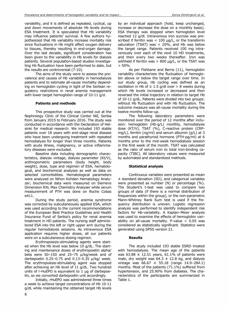
Treatment with erythrоpoiesis-stimulating agents (ESA) is the optimal therapy for renal
anemia. However, maintaining hemoglobin (Hb) within narrow targets remains a significant clinical problem because during ESA treatment, the Hb levels usually fluctuate widely; this phe-nomenon is termed "hemoglobin variability" and is associated with higher mortality. Our study aimed to determine the prevalence and cause of hemoglobin variability in patients on chronic hemodialysis (HD) treatment and to estimate the association of Hb variability with all-cause mortality.
A prospective study was conducted on 193 chronic HD patients treated with ESA. Hemoglobin cycling was defined as Hb variability throughout at least eight weeks and amplitude of more than 1.5 g/dl from the Serbian target range of 10-11 g/dl.
During the one-year follow-up, there was 5.6 ESA dose modification per patient. 23.4% of patients had never experienced Hb cycling during the study period. The total number of 460 hemoglobin excursions were recorded in 76.6% of patients, with 2.42 ± 2.7 Hb excursions per year, mean amplitude of 2.13 ± 0.76 g/dL, and the average length of Hb excursion of 8.2 ± 2.7 weeks. The Hb cycling was not affected by the gender, age, weekly ESA dose, or the presence of diabetes or hypertension. However, Hb variability was associated with ESA dose change, CRP, and HD vascular access type. The odds ratio for 1-year all-cause mortality was 1.424 (95% CI: 1.231–1.682, P < 0.001).
Hemoglobin cycling frequently occurs in ESA treated HD patients as a result of current practice in ESA dosing, the presence of infection, and the type of vascular access for HD and these fluctuations predicted overall mortality.
Acta Medica Medianae 2020;59(1):05-13. Key words: hemoglobin variability, erythropoiesis-stimulating agents, hemodialysis
1Clinic of Nephrology, Clinical Center Niš, Niš, Serbia 2University of Niš, Faculty of Medicine, Niš, Serbia
Contact: Zorica Dimitrijević,
Njegoševa 52, 18000 Niš, Serbia
E-mail: [email protected]
Introduction Anemia is a common complication that is as-
sociated with adverse cardiovascular complications
and poor outcomes in patients with chronic kidney disease (CKD) (1). The introduction of erythropo-iesis-stimulating agents (ESA) has revolutionized the management of anemia in CKD, leading to substan-tial reductions in the blood transfusion requirements, improvement in energy, and physical function (2) and improvements in health-related quality of life
(3). Even though the optimal target hemoglobin (Hb) concentration in hemodialysis (HD) patients continues to be a substantial dilemma, the European
Best Practice Guidelines (EBPG) recommended that the target hemoglobin level should be determined on an individual basis, having in mind gender, age, ethnicity, activity, and comorbid conditions (4). Nevertheless, with the publication of CREATE and
CHOIR studies, both the upper and lower limits for
target Hb concentration was set to 10-12 g/dL (5, 6) as it was shown that targeting normal Hb levels did not result in better survival, but rather in increased cardiovascular events and mortality in HD patients. Keeping patients’ Hb levels in such a narrow range is difficult considering the loss of physiological regula-tion of red cell generation and many other factors,
such as iron deficiency, chronic inflammation, secon-dary hyperparathyroidism, malnutrition, and inade-quate dialysis dose. The data confirm that only 30% of patients will belong to this hemoglobin range at any point in time because fluctuations in the Hb level result in frequent under-and overreaching the target level (7). This phenomenon is known as Hb
Prevalence and determinants of hemoglobin variability and its impact... Zorica Dimitrijević et al.
6
variability, and it is defined as repeated, cyclical, up
and down movements of absolute Hb levels during
ESA treatment. It is speculated that Hb variability may influence patients' survival. A few authors hy-pothesized that Hb variability increase mortality risk since fluctuations in Hb might affect oxygen delivery to tissues, thereby resulting in end-organ damage.
Over the last decade, significant consideration has been given to the variability in Hb levels for dialysis patients. Several population‐based studies investiga-
ting Hb fluctuation have been performed to date, but the results are controversial (7-10).
The aims of the study were to assess the pre-valence and causes of Hb variability in hemodialysis patients and to estimate all-cause mortality depend-
ing on hemoglobin cycling in light of the Serbian re-gulatory restrictions in renal anemia management
with lower target hemoglobin range of 10-11 g/d. Patients and methods
This prospective study was carried out at the Nephrology Clinic of the Clinical Center Niš, Serbia from January 2015 to February 2016. The study was conducted in accordance with the Declaration of Hel-sinki for medical research. We included 193 stable patients over 18 years with end-stage renal disease who have been undergoing treatment with repeated
hemodialysis for longer than three months. Patients with acute illness, malignancy, or active inflamma-tory diseases were excluded.
Baseline data including demographic charac-teristics, dialysis vintage, dialysis parameter (Kt/V),
anthropometric parameters (body height, body weight), dose, type and regimen of ESA, hematolo-
gical, and biochemical analyses as well as data on selected comorbidities. Hematological parameters were analyzed on Nihon Kohden Hematology Analy-zer; biochemical data were measured on Siemens Dimension RXL Max Chemistry Analyser while serum measurement of PTH was done on Roche Cobas
e411. During the study period, anemia syndrome
was corrected by subcutaneously applied ESA, which was used according to the current recommendations of the European Best Practice Guidelines and Health Insurance Fund of Serbia's policy for renal anemia treatment in HD patients. The nursing staff adminis-
tered ESA into the left or right upper arm during the
regular hemodialysis sessions. As intravenous ESA application requires higher doses, all our patients were on a subcutaneous dosing regimen.
Erythropoiesis-stimulating agents were start-ed when the Hb level was below 10 g/dL. The start-ing and maintenance doses of erythropoietin alpha/
beta were 50–150 and 25–75 μ/kg/week and of darbepoetin 0.25–0.75 and 0.13–0.35 μ/kg/ week. The erythropoiesis-stimulating agent was stopped after achieving an Hb level of 11 g/dL. Two hundred units of r-HuEPO is equivalent to 1 μg of darbepoe-tin, so we converted darbepoetin unit accordingly.
Initially, rHuEPO was administered three times a week to achieve target concentrations of Hb 10-11 g/dL while maintaining the obtained target Hb levels
by an individual approach (hold, keep unchanged,
increase or decrease the dose on a monthly basis).
ESA therapy was stopped when hemoglobin level reached 12 g/dl. Intravenous iron sucrose was pre-scribed if ferritin was < 100 μg/L, or the transferrin saturation (TSAT) was < 20%, and Hb was below the target range. Patients received 100 mg intra-
venously over each of the next 10 HD treatments, and then every two weeks thereafter. Iron was withheld if ferritin was > 800 μg/L, or the TSAT was > 50%.
As per Fishbane and Berns (11), hemoglobin variability characterizes the fluctuation of hemoglo-bin above or below the target range over time. In
our study group, Hb cycling was defined as an oscillation in Hb of ≥ 1.5 g/dl over > 8 weeks during which Hb levels increased or decreased and then
reversed the initial trajectory in relation to target Hb of 10-11 g/dL. Patients were divided into two groups: without Hb fluctuation and with Hb fluctuation. The outcome measure was all-cause mortality during the
twelve months follow-up. The following laboratory parameters were
monitored over the period of 12 months after inclu-sion: hemoglobin (Hb-g/L) monthly, hemodialysis dose (KT/V), TSAT (%), C-reactive protein (CRP-mg/L), ferritin (ng/ml) and serum albumin (g/L) at 3
months and parathyroid hormone (PTH-pg/ml) at 6 months prior to the mid-week hemodialysis session in the first week of the month. TSAT was calculated as the ratio of serum iron to total iron-binding ca-pacity (TIBC). All laboratory values were measured by automated and standardized methods.
Statistical analysis Continuous variables were presented as mean
± standard deviation (SD), and categorical variables were presented as number (N) or percentage (%). The Student's t-test was used to compare two groups of data (if there is a normal distribution of
frequencies within the group), or the non-parametric Mann-Whitney Rank Sum test is used if the fre-quency distribution is uneven. Logistic regression analysis was performed to identify independent risk factors for Hb-variability. A Kaplan–Meier analysis was used to examine the effects of hemoglobin vari-
ability on all-cause mortality. P-value < 0.05 was considered as statistically significant. Statistics were
generated using SPSS version 21. Results The study included 193 stable ESRD treated
with hemodialysis. The mean age of the patients was 63.88 ± 12.51 years, 61.1% of patients were male, dry weight was 64.3 ± 12.8 kg, and dialysis vintage was 66.67 ± 55.18 (range 14.9–284.1) months. Most of the patients (71.1%) suffered from hypertension, and 25.90% from diabetes. The cha-racteristics of the participants are summarized in
Table 1.
Acta Medica Medianae 2020, Vol.59(1) Prevalence and determinants of hemoglobin variability and its impact...
7
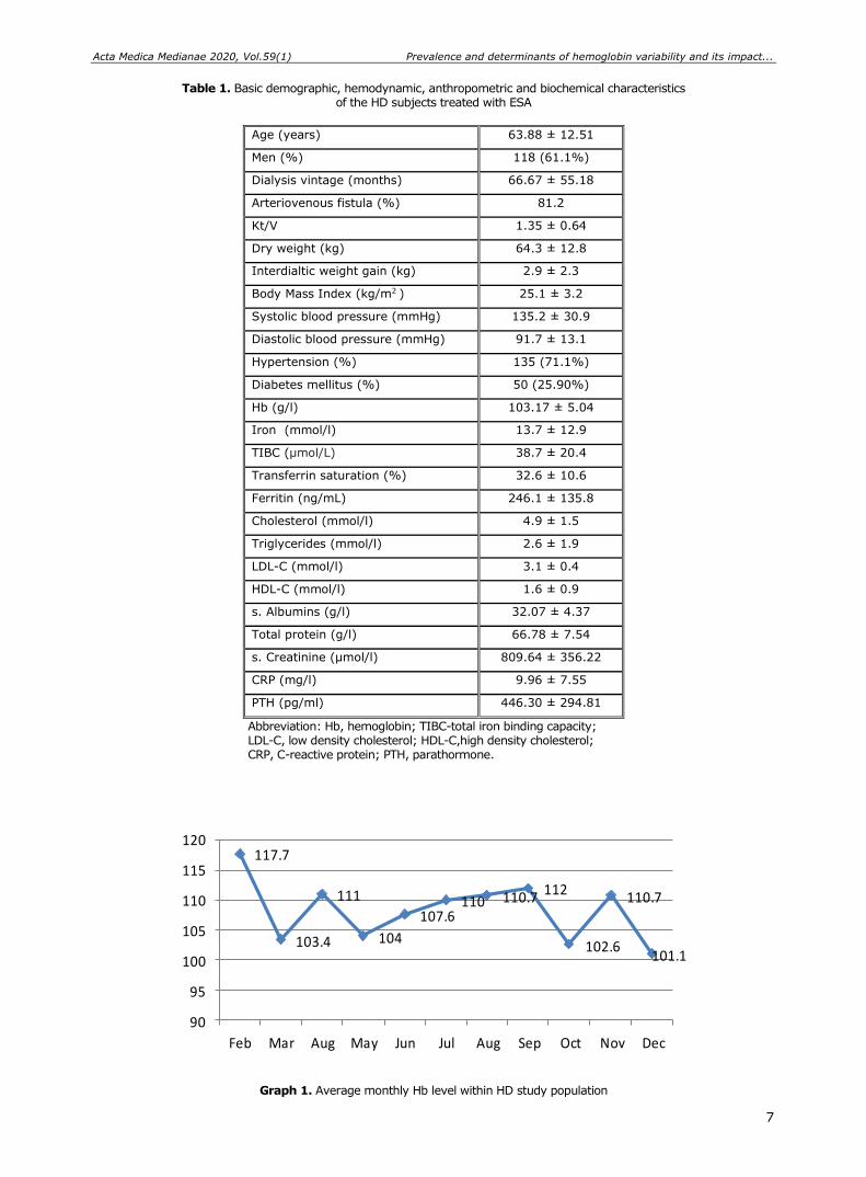
Table 1. Basic demographic, hemodynamic, anthropometric and biochemical characteristics of the HD subjects treated with ESA
Age (years) 63.88 ± 12.51
Men (%) 118 (61.1%)
Dialysis vintage (months) 66.67 ± 55.18
Arteriovenous fistula (%) 81.2
Kt/V 1.35 ± 0.64
Dry weight (kg) 64.3 ± 12.8
Interdialtic weight gain (kg) 2.9 ± 2.3
Body Mass Index (kg/m2 ) 25.1 ± 3.2
Systolic blood pressure (mmHg) 135.2 ± 30.9
Diastolic blood pressure (mmHg) 91.7 ± 13.1
Hypertension (%) 135 (71.1%)
Diabetes mellitus (%) 50 (25.90%)
Hb (g/l) 103.17 ± 5.04
Iron (mmol/l) 13.7 ± 12.9
TIBC (μmol/L) 38.7 ± 20.4
Transferrin saturation (%) 32.6 ± 10.6
Ferritin (ng/mL) 246.1 ± 135.8
Cholesterol (mmol/l) 4.9 ± 1.5
Triglycerides (mmol/l) 2.6 ± 1.9
LDL-C (mmol/l) 3.1 ± 0.4
HDL-C (mmol/l) 1.6 ± 0.9
s. Albumins (g/l) 32.07 ± 4.37
Total protein (g/l) 66.78 ± 7.54
s. Creatinine (μmol/l) 809.64 ± 356.22
CRP (mg/l) 9.96 ± 7.55
PTH (pg/ml) 446.30 ± 294.81
Abbreviation: Hb, hemoglobin; TIBC-total iron binding capacity; LDL-C, low density cholesterol; HDL-C,high density cholesterol; CRP, C-reactive protein; PTH, parathormone.
117.7
103.4
111
104
107.6110 110.7
112
102.6
110.7
101.1
90
95
100
105
110
115
120
Feb Mar Aug May Jun Jul Aug Sep Oct Nov Dec
Graph 1. Average monthly Hb level within HD study population
Prevalence and determinants of hemoglobin variability and its impact... Zorica Dimitrijević et al.
8
Graph 1 displays the mean hemoglobin values
during one-year follow-up. Considering the whole
study population, mean Hb was maintained within the target range most of the time.
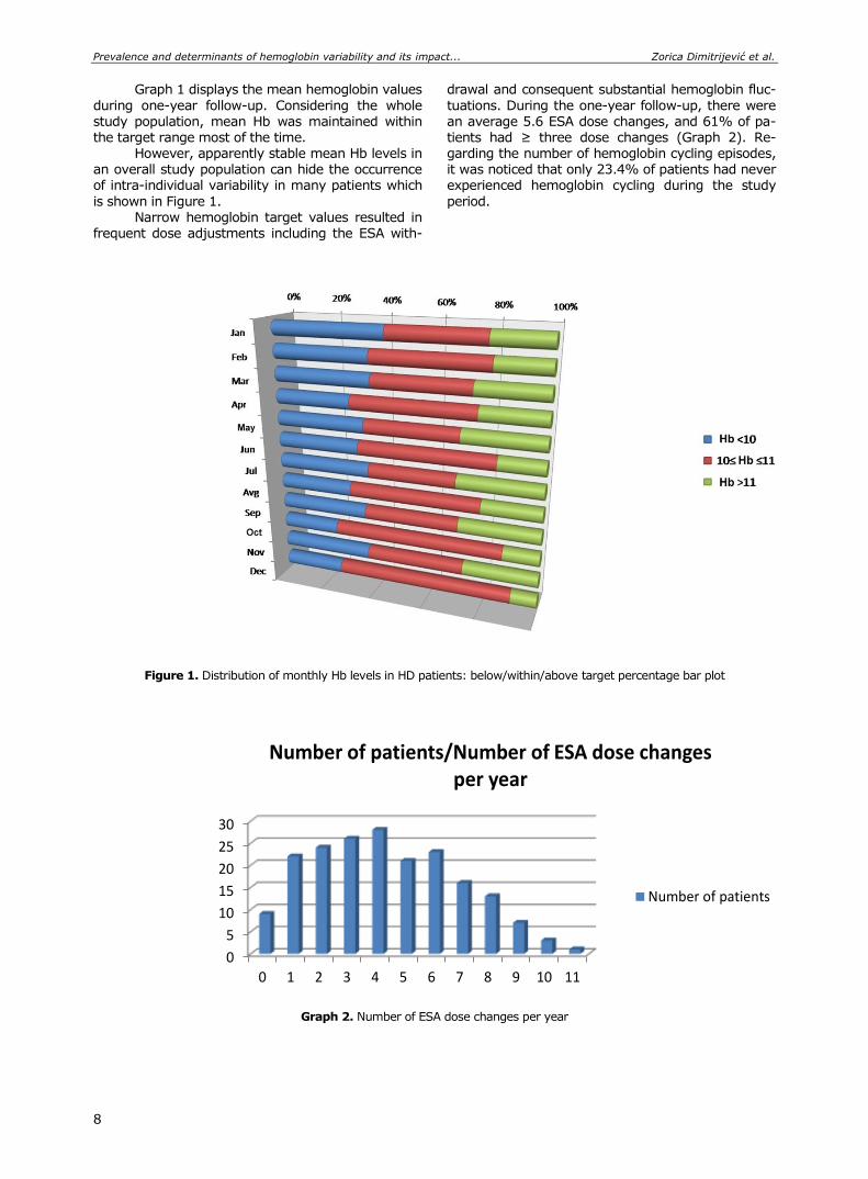
However, apparently stable mean Hb levels in an overall study population can hide the occurrence of intra-individual variability in many patients which
is shown in Figure 1. Narrow hemoglobin target values resulted in
frequent dose adjustments including the ESA with-
drawal and consequent substantial hemoglobin fluc-
tuations. During the one-year follow-up, there were
an average 5.6 ESA dose changes, and 61% of pa-tients had ≥ three dose changes (Graph 2). Re-garding the number of hemoglobin cycling episodes, it was noticed that only 23.4% of patients had never experienced hemoglobin cycling during the study
period.
Figure 1. Distribution of monthly Hb levels in HD patients: below/within/above target percentage bar plot
0
5
10
15
20
25
30
0 1 2 3 4 5 6 7 8 9 10 11
Number of patients/Number of ESA dose changes per year
Number of patients
Graph 2. Number of ESA dose changes per year
Acta Medica Medianae 2020, Vol.59(1) Prevalence and determinants of hemoglobin variability and its impact...
9
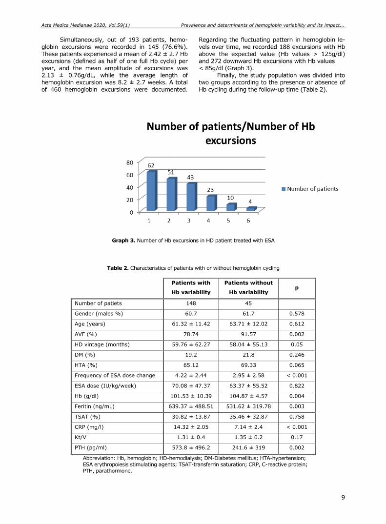
Simultaneously, out of 193 patients, hemo-
globin excursions were recorded in 145 (76.6%).
These patients experienced a mean of 2.42 ± 2.7 Hb excursions (defined as half of one full Hb cycle) per year, and the mean amplitude of excursions was 2.13 ± 0.76g/dL, while the average length of hemoglobin excursion was 8.2 ± 2.7 weeks. A total
of 460 hemoglobin excursions were documented.
Regarding the fluctuating pattern in hemoglobin le-
vels over time, we recorded 188 excursions with Hb
above the expected value (Hb values > 125g/dl) and 272 downward Hb excursions with Hb values < 85g/dl (Graph 3).
Finally, the study population was divided into two groups according to the presence or absence of
Hb cycling during the follow-up time (Table 2).
Graph 3. Number of Hb excursions in HD patient treated with ESA
Table 2. Characteristics of patients with or without hemoglobin cycling
Patients with
Hb variability
Patients without
Hb variability p
Number of patiets 148 45
Gender (males %) 60.7 61.7 0.578
Age (years) 61.32 ± 11.42 63.71 ± 12.02 0.612
AVF (%) 78.74 91.57 0.002
HD vintage (months) 59.76 ± 62.27 58.04 ± 55.13 0.05
DM (%) 19.2 21.8 0.246
HTA (%) 65.12 69.33 0.065
Frequency of ESA dose change 4.22 ± 2.44 2.95 ± 2.58 < 0.001
ESA dose (IU/kg/week) 70.08 ± 47.37 63.37 ± 55.52 0.822
Hb (g/dl) 101.53 ± 10.39 104.87 ± 4.57 0.004
Feritin (ng/mL) 639.37 ± 488.51 531.62 ± 319.78 0.003
TSAT (%) 30.82 ± 13.87 35.46 ± 32.87 0.758
CRP (mg/l) 14.32 ± 2.05 7.14 ± 2.4 < 0.001
Kt/V 1.31 ± 0.4 1.35 ± 0.2 0.17
PTH (pg/ml) 573.8 ± 496.2 241.6 ± 319 0.002
Abbreviation: Hb, hemoglobin; HD-hemodialysis; DM-Diabetes mellitus; HTA-hypertension; ESA erythropoiesis stimulating agents; TSAT-transferrin saturation; CRP, C-reactive protein; PTH, parathormone.
Prevalence and determinants of hemoglobin variability and its impact... Zorica Dimitrijević et al.
10
The change in Hb level was not affected by
the gender, age, weekly ESA dose or the presence
of diabetes or hypertension. However, the frequency of ESA dose change (p < 0,001), inflammation (p < 0.001), type of vascular access (p = 0.002) and secondary hyperparathyroidism (p = 0.002) signi-ficantly influenced hemoglobin variability.
Six variables with the highest correlation co-efficient in the univariate analysis were included in the multiple linear regression analysis to determine the significant predictors of Hb variability. The re-sults show that Hb variability was associated with ESA dose change (OR 1.56;95% CI 1.29–2.04, p < 0.001), CRP (OR 1.73; 95% CI 1.22–1.99, p <
0.001) and vascular access type (OR 2.13; 95% CI 1.56–3.18, p = 0.033) (Table 3).
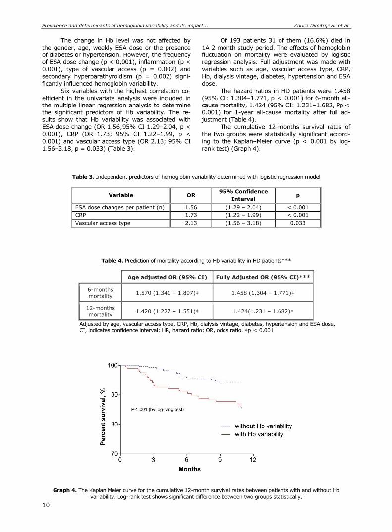
Of 193 patients 31 of them (16.6%) died in
1A 2 month study period. The effects of hemoglobin
fluctuation on mortality were evaluated by logistic regression analysis. Full adjustment was made with variables such as age, vascular access type, CRP, Hb, dialysis vintage, diabetes, hypertension and ESA dose.
The hazard ratios in HD patients were 1.458 (95% CI: 1.304–1.771, p < 0.001) for 6-month all-cause mortality, 1.424 (95% CI: 1.231–1.682, Pp <
0.001) for 1-year all-cause mortality after full ad-justment (Table 4).
The cumulative 12-months survival rates of the two groups were statistically significant accord-ing to the Kaplan–Meier curve (p < 0.001 by log-
rank test) (Graph 4).
Table 3. Independent predictors of hemoglobin variability determined with logistic regression model
Variable OR 95% Confidence
Interval p
ESA dose changes per patient (n) 1.56 (1.29 – 2.04) < 0.001
CRP 1.73 (1.22 – 1.99) < 0.001
Vascular access type 2.13 (1.56 – 3.18) 0.033
Table 4. Prediction of mortality according to Hb variability in HD patients***
Age adjusted OR (95% CI) Fully Adjusted OR (95% CI)***
6-months mortality
1.570 (1.341 – 1.897)‡ 1.458 (1.304 – 1.771)‡
12-months mortality
1.420 (1.227 – 1.551)‡ 1.424(1.231 – 1.682)‡
Adjusted by age, vascular access type, CRP, Hb, dialysis vintage, diabetes, hypertension and ESA dose, CI, indicates confidence interval; HR, hazard ratio; OR, odds ratio. ‡p < 0.001
Graph 4. The Kaplan Meier curve for the cumulative 12-month survival rates between patients with and without Hb variability. Log-rank test shows significant difference between two groups statistically.
Acta Medica Medianae 2020, Vol.59(1) Prevalence and determinants of hemoglobin variability and its impact...
11
Discussion
Keeping the constant Hb levels is obligatory
to ensure continuous and sufficient oxygen delivery to tissues. In healthy subjects, individual variation in the Hb level occurs within the range of normal va-lues, usually does not exceed 1 g/dL and have no clinical significance. However, for hemodialysis pa-tients, substantial variability in the Hb level over time is not uncommon. Fluctuations in the Hb levels provoke repeated episodes of relative ischemia and tissue hypoxia, which may result in organ dysfunc-tion or injury (10).
The key finding of this study is confirming that Hb cycling frequently occurs in hemodialysis pa-tients. Over a one-year period, only 25% of patients using ESA had stable Hb levels within a target range of 10-11 g/dL. This reflects the difficulty of maintain-ing Hb levels within a narrow range as recommend-ed by the most recent guidelines and hemoglobin management still remained a substantial challenge in the care of hemodialysis patients, with almost all patients moving between categories over fairly short time periods. The finding that patients receiving ESA had high variability agrees with previous studies and points to the current practice of prescribing ESA as one of the causes of Hb variability (12-14). The other possible factors that might affect patients’ Hb variability were summarized in a review by Kalantar-Zadeh and Aronoff (15). These authors concluded that drug‐related factors, patient demographics, iron deficiency, infections, inflammation, malignancies, and reimbursement‐related factors all had an impact on Hb variability. Of these multiple factors, the ESA dose was the most actionable factor in the mana-gement of anemia for patients on dialysis therapy.
In the present study, we observed three major determinants of Hb fluctuation. The first was a frequent change in ESA dose. A positive correlation was seen between ESA dose change and amplitude of Hb excursion, implying that dose changes were causal, rather than reactive. That finding has also been published by others (16) and strongly impli-cates current dosing strategies and anemia manage-ment protocols in the pathogenesis of Hb cycling. Interestingly, compared with dose increases, dose reductions seemed to be a stronger predictor of cycling. We noted 272 downward Hb excursions. The Hb decline was mostly the consequence of ESA withdrawal (in 78.3% of cases) and dose reduction in 15%.
Evidence suggests that inflammation is an important factor associated with Hb variability. In a retrospective study of 225 hemodialysis patients, high CRP values were associated with less stable Hb levels (17). Likewise, Barany et al. reported a significant correlation between Hb variability and CRP levels (18). Similarly to these findings, we ob-served that higher CRP values significantly influence Hb variability. These results provide supporting evi-dence that inflammation can trigger hemoglobin va-riability. Thus, ESA dosage should be regularly re-viewed, and patients should be monitor closely in the presence of inflammatory conditions.
Whereas the weekly dose of ESA was compa-rable regardless of the vascular access used, the
weekly dose of ESA used in the patients with central vein catheter (CVC) was significantly higher than that used in those with AVF. This observation is consistent with other studies that indicate that CVC use as vascular accesses is associated with the need for higher doses of ESA secondly to blood loss during dialysis and possible catheter-related infections (19). Besides, the type of vascular access had an impact on Hb variability, possibly via intercurrent inflam-mation.
Studies about the clinical significance of Hb variability have been increased but results were conflicting. Regidor et al. (20) noticed that patients with Hb fall greater than 2 g/dL had the greatest mortality risk when compared with patients who showed Hb fall lower than 0.8 g/dL. In a cohort of 34,963 prevalent HD patients, Yang et al. (21) de-monstrated that per every 1 g/dL increase of Hb variability, there is a 33% increase in mortality risk. On the contrary, Zeynep et al. found that hemo-globin variability has a modest association with mor-bidity and all-cause mortality in ESA treated dialysis patients (22). Persistently or transiently low Hb levels have also been associated with hospitalization and death (9, 23, 24, 25), as have downward Hb excursions (25). In our study, we likewise observed that Hb fluctuation was an independent determinant of mortality, which is in accordance with the recent study of Lin et al. (26). They also demonstrated that high Hb variability is an independent risk factor for cardiovascular mortality in HD patients and might influence the cardiac function.
Although the direct effects of Hb variation on patient outcome are still not fully understood, it is evident that large or frequent fluctuations are un-desirable. Low Hb levels have a negative impact on symptoms and quality of life for patients; they also increase the requirement for blood transfusions. The myocardium may be particularly vulnerable to he-moglobin fluctuation because it has to compensate for periods of reduced oxygen delivery with increa-sed output and myocardial cell growth. Hemoglobin levels higher than current target ranges may be associated with worse cardiovascular outcomes (7, 8), and higher Hb levels maintained with higher ESA doses have a significant cost implication. More fre-quent Hb fluctuations outside of target ranges re-quire more clinician time to determine response in terms of ESA dose adjustment or of intravenous iron dosing.
Conclusion Hemoglobin management remained a sub-
stantial challenge in the care of hemodialysis pa-tients, with almost all patients moving between dif-ferent hemoglobin categories over fairly short time periods. Our study demonstrates that both inflam-mation and the frequent changes of ESA dose were the major predictors of hemoglobin variability. The
current ESA reimbursement practice demands con-stant adjustments of the ESA doses. The question is whether modification of treatment policies can con-tribute to reducing cycling and whether this influ-ences the outcome. To answer this question, further studies are needed.
Prevalence and determinants of hemoglobin variability and its impact... Zorica Dimitrijević et al.
12
References
1. KDIGO clinical practice guideline for anemia in chronic kidney disease. Kidney Int Suppl 2012;2:279-335.
2. Baris A. The Relationship between Depressive Symp-toms and Erythropoietin Resistance in Stable Hemo-dialysis Patients with Adequate Iron Stores. Int J Artif Organs 2013;36:314-19.[CrossRef] [PubMed]
3. Spinowitz B, Pecoits-Filho R, Winkelmayer WC, Per-gola PE, Rochette S, Thompson-Leduc P et al. Econo-mic and quality of life burden of anemia on patients with CKD on dialysis: a systematic review. J Med Econ 2019;[CrossRef] [PubMed]
4. Locatelli F, Bárány P, Covic A, De Francisco A, Del Vecchio L, Goldsmith D et al. Kidney disease: improv-ing global outcomes guidelines on anaemia manage-ment in chronic kidney disease: a European renal best practice position statement. Nephrol Dial Transplant 2013;28:1346-59. [CrossRef] [PubMed]
5. Macdougall IC. Steering Committee of the CREATE trial; CREATE Study Group: CREATE: new strategies for early anaemia management in renal insufficiency. Nephrol Dial Transplant 2003;18:13-16.[PubMed]
6. Singh AK, Szczech L, Tang KL, Barnhart H, Sapp S, Wolfson M et al; CHOIR Investigators: Correction of anemia with epoetin alfa in chronic kidney disease. N Engl J Med 2006;355:2085-98.[CrossRef] [PubMed]
7. Brunelli SM, Lynch KE, Ankers ED, Joffe MM, Yang W, Thadhani RI et al. Association of hemoglobin variability and mortality among contemporary incident hemodia-lysis patients. Clin J Am Soc Nephrol 2008; 3:1733-40.[CrossRef] [PubMed]
8. Weinhandl ED, Peng Y, Gilbertson DT, Bradbury BD, Collins AJ. Hemoglobin variability and mortality: Con-founding by disease severity. Am J Kidney Dis 2011; 57:255-65.[CrossRef] [PubMed]
9. Ebben JP, Gilbertson DT, Foley RN, Collins AJ. Hemoglobin level variability: Associations with comor-bidity, intercurrent events, and hospitalizations. Clin J Am Soc Nephrol 2006;1:1205-10. [CrossRef] [PubMed]
10. Zhao L, Hu C, Cheng J, Zhang P, Jiang H, Chen J. Haemoglobin variability and all‐cause mortality in
haemodialysis patients: A systematic review and meta‐analysis. Nephrology 2019;24(12):1265-1272.
[CrossRef] [PubMed] 11. Fishbane S, Berns JS. Evidence and implications of
haemoglobin cycling in anemia management. Nephrol Dial Transplant 2007;22:2129-32. [CrossRef] [PubMed]
12. Spiegel DM. Hemoglobin variability in chronic kidney disease: A cross-sectional study. Am J Med Sci 2009; 337:340-3.[CrossRef] [PubMed]
13. Minutolo R, Chiodini P, Cianciaruso B, Pota A, Bellizzi V, Avino D et al. Epoetin therapy and hemoglobin level variability in nondialysis patients with chronic kidney disease. Clin J Am Soc Nephrol 2009;4:552-9. [CrossRef] [PubMed]
14. Boudville NC, Djurdjev O, Macdougall IC, de Francisco AL, Deray G, Besarab A et al. Hemoglobin variability in nondialysis chronic kidney disease: examining the
association with mortality. Clin J Am Soc Nephrol 2009;4:1176-82.[CrossRef] [PubMed]
15. Kalantar-Zadeh K, Aronoff GR. Hemoglobin variability in anemia of chronic kidney disease. J Am Soc Nephrol 2009;20:479-87.[CrossRef] [PubMed]
16. Fishbane S, Berns JS. Hemoglobin cycling in hemo-dialysis patients treated with recombinant human erythropoietin. Kidney Int 2005;68:1337-43. [CrossRef] [PubMed]
17. de Francisco A, Stenvinkel P, Vaulont S. Inflammation and its impact on anaemia in chronic kidney disease: from haemoglobin variability to hyporesponsiveness. NDT Plus 2009;2:i18-i26.[CrossRef] [PubMed]
18. Barany P, Carrero JJ, Snaedal J'onsd'ottir S. Variability of hemoglobin (Hb) levels in relation to inflammatory status and iron metabolism in hemodialysis (HD) patients (Pts). Proceedings of the American Society of Nephrology Annual Congress; 2006; San Diego, USA.
19. Roberts TL, Obrador GT, St Peter WL, Pereira BJ, Collins AJ. Relationship among catheter insertions, vascular access infections, and anemia management
in hemodialysis patients. Kidney Int 2004;66:2429-36. [CrossRef] [PubMed]
20. Regidor DL, Kopple JD, Kovesdy CP, Kilpatrick RD, McAllister CJ, Aronovitz J et al. Associations between changes in hemoglobin and administered erythro-poiesis-stimulating agent and survival in hemodialysis patients. J Am Soc Nephrol 2006;17:1181-91. [CrossRef] [PubMed]
21. Yang W, Israni RK, Brunelli SM, Joffe MM, Fishbane S, Feldman HI. Hemoglobin variability and mortality in ESRD. J Am Soc Nephrol 2007; 18:3164-70. [CrossRef] [PubMed]
22. Zeynep B, Bahar GD, Suleyman K, Emre T, Mehtap EU, Nurhan OAal. Factors Influencing Hemoglobin Variability and Its Association with Mortality in Hemo-dialysis Patients. Scientific World Journal 2018;18. [CrossRef] [PubMed]
23. Regidor DL, Kopple JD, Kovesdy CP, Kilpatrick RD, McAllister CJ, Aronovitz J, et al. Associations between changes in hemoglobin and administered erythro-poiesis-stimulating agent and survival in hemodialysis patients. J Am Soc Nephrol 2006;17:1181-91. [CrossRef] [PubMed]
24. Gilbertson DT, Ebben JP, Foley RN, Weinhandl ED, Bradbury BD, Collins AJ. Hemoglobin level variability: associations with mortality. Clin J Am Soc Nephrol 2008;3:133-8.[CrossRef] [PubMed]
25. Ishani A, Solid CA, Weinhandl ED, Gilbertson DT, Foley RN, Collins AJ. Association between number of months below K/DOQI haemoglobin target and risk of hospi-talization and death. Nephrol Dial Transplant 2008; 23:1682-9. [CrossRef] [PubMed]
26. Lin FJ, Zhang X, Huang LS, Ji G, Huang HD, Xie Yet al. Impact of hemoglobin variability on cardiovascular mortality in maintenance hemodialysis patients. Int Urol Nephrol 2018;50:1703-12.[CrossRef] [PubMed]
Acta Medica Medianae 2020, Vol.59(1) Prevalence and determinants of hemoglobin variability and its impact...
13
Originalni rad UDC: 616.155.16:616.61-78-036.8
doi:10.5633/amm.2020.0101
PREVALENCIJA I DETERMINANTE VARIJABILNOSTI HEMOGLOBINA I NJEN UTICAJ NA MORTALITET KOD BOLESNIKA NA HRONIČNOM
PROGRAMU HEMODIJALIZE
Zorica Dimitrijević1,2, Branka Mitić1,2, Karolina Paunović1, Sonja Šalinger-Martinović2
1Klinika za nefrologiju, Klinički centar Niš, Niš, Srbija 2Univerzitet u Nišu, Medicinski fakultet, Niš, Srbija
Kontakt: Zorica Dimitrijević
Njegoševa 52, 18000 Niš, Srbija E-mail: [email protected]
Terapija agensima stimulacije eritropoeze (ASE) predstavlja optimalno lečenje renalne
anemije. Međutim, održavanje hemoglobina (Hb) u okviru uskih ciljnih vrednosti ostaje značajan klinički problem, s obzirom na to da tokom primene ASE nivoi Hb obično značajno osciliraju; ovaj fenomen poznat je kao "varijabilnost hemoglobina", a udružen je sa poveća-nom smrtnošću bolesnika. Naše istraživanje imalo je za cilj da analizira učestalost i uzroke na-stanka varijabilnosti hemoglobina kod bolesnika lečenih hemodijalizom (HD) i da proceni njen uticaj na mortalitet bolesnika.
Prospektivnom studijom obuhvaćeno je 193 bolesnika na hroničnoj HD, koji su lečeni ASE. Varijabilnost hemoglobina definisana je kao oscilacija koncentracije Hb u periodu od najmanje osam nedelja sa amplitudom većom od 1,5 g/dl od zadatih ciljnih vrednosti hemo-
globina, koji u Srbiji za bolesnike na HD trenutno iznosi 10 g/dl — 11 g/dl.
Tokom jednogodišnjeg praćenja, bilo je 5,6 modifikacija doze ASE po bolesniku. 23,4% bolesnika nije imalo značajne oscilacije Hb tokom studijskog perioda. Ukupno 460 oscilacija (ekskurzija) hemoglobina zabeleženo je kod 76,6% bolesnika, sa 2,42 ekskurzije ± 2,7 ekskurzija godišnje, prosečne amplitude 2,13 g/dL ± 0,76 g/dL i prosečne dužine trajanja 8,2 nedelje ± 2,7 nedelja. Na osilaciju Hb nije uticala starost, pol, nedeljna doza ASE, kao ni prisustvo dijabetesa ili hipertenzije. Međutim, varijabilnost Hb zavisila je od promena doze ASE, CRP-a i tipa vaskularnog pristupa za HD.
Varijabilnost hemoglobina često se javlja kod bolesnika na HD lečenih ASE, kao posledica prakse učestalih promena doze ASE, prisustva infekcija i vrste vaskularnog pristupa za HD. Ove fluktuacije hemoglobina uticale su na povećanje mortaliteta kod naših bolesnika. Procena rizika za jednogodišnji mortalitet bila je 1,424 (95% CI: 1,231 – 1,682; P < 0,001).
Acta Medica Medianae 2020;59(1):05-13.
Ključne reči: varijabilnost hemoglobina, agensi stimulacije eritropoeze,
hemodijaliza
14 www.medfak.ni.ac.rs/amm
Original article UDC: 615.01/.03:615.851.4
doi:10.5633/amm.2020.0102
ТHE JUSTIFICATION OF CLINICAL PHARMACY SKILLS AND KNOWLEDGE FOR MODERN COMMUNITY PHARMACIST
Maja Koraćević1,2, Aleksandra Catić-Djordjević1, Nikola Stefanović1, Ivana Damnjanović1,
Ivana Stošić1, Radmila Veličković-Radovanović1,3
The course of pharmacy education has undergone a radical change as it aims to
become more patient oriented. The primary objective of this study was to assess the asso-ciation between clinical pharmacy education and daily activities of the community pharmacist (CP) including recognition and solution of drug-related problems (DRP). Furthermore, we wanted to investigate CPs’ attitudes regarding knowledge and skills that can be required for routine community pharmacy practice. The simple questionnaire was formed to evaluate the significance of the implementation of clinical pharmacy course in the education of CPs. The questionnaires were sent by post or email or they were provided directly by one of the
researcher. Data acquired from 234 CPs were divided into two groups: clinical pharmacy education group (CPEG) and non-clinical pharmacy education group (NCEG). The most frequent DRP recognized by respondents (CPEG and NCEG) were drug interactions, followed by suboptimal efficacy of the treatment and inappropriate dosage selection. Additionally, CPEG statistically more frequently than NCEG recognized low adherence (p < 0.05), while NCEG more frequently recognized inappropriate dosing interval (p < 0.05) and omitted drugs, that should have been prescribed (p < 0.05). The respondents agreed that knowledge of drug therapy, therapeutic planning skills and critical evaluation of drug information skills were the most important clinical pharmacy skills and knowledge required for modern community pharmacy practice. Still, CPEG gave advantage to the knowledge of laboratory and diagnostic skills compared to NCEG (p < 0.05). This study indicated that clinical pharmacy education can move the focus of the CPs towards a patient, but still positions drug in the center of their activity. The pharmacists with the clinical pharmacy education considered the knowledge of laboratory and diagnostic skills to be of significant importance, which confirms the ongoing change in pharmacist orientation.
Acta Medica Medianae 2020;59(1):14-22. Key words: clinical pharmacy, pharmacy practice, community pharmacy,
pharmacy education
1University of Niš, Faculty of Medicine, Department of Pharmacy, Niš, Serbia 2University of Niš, Innovation Center, Niš, Serbia 3Clinic of Nephrology, Clinical Center Niš, Niš, Serbia
Contact: Aleksandra Catić-Đorđević
81 Dr. Zoran Djindjić Blvd., 18000 Niš, Serbia
E-mail: [email protected]
Introduction
A community pharmacist (CP) has numerous
responsibilities regarding patients and their medica-tions in everyday pharmacy practice. A medication review is assumed to be the first activity, which is done by the pharmacists during their routine work in the pharmacy (1, 2). This activity implies recognition
of potential or existing drug-related problems (DRP), such as no indication for a drug, the choice of drug, formulation of a drug, dosage regimen, drug inter-actions, adverse effects and drug’s contraindication.
Pharmacists' roles are evolving and therefore they are meant to deal not only with DRPs, connected to
drug per se, but also with patients’ adherence, qua-lity of life, pharmacoeconomy issues and giving an appropriate and acceptable solution and a piece of advice as well (2, 3). Furthermore, the increasing number of new medications and dietary supplem-
ents requires critical evaluation, processing and sharing the information regarding that novelty from the pharmacists towards other healthcare professio-nals and patients (3, 4). To sum up, this all repre-sents a real challenge for the modern pharmacist, which requires multidisciplinary skills and knowledge
in order to handle it. Skills and knowledge that can be acquired in undergraduate or postgraduate course of clinical pharmacy, seem to be important and help-ful for accurate, effective and prompt response to
Тhe justification of clinical pharmacy skills and knowledge for ... Maja Koraćević et al.
15
defined problems (5, 6). The course of pharmacy
education has undergone a radical change as it aims
at become a more patient oriented profession (7). Therefore, required knowledge and skills of a mo-dern CP are communication skills, knowledge of drug therapy, nondrug therapy and complementary medi-cine, knowledge of the disease, laboratory and dia-
gnostic skills, physical assessment skills, therapeutic planning skills and critical evaluation of drug infor-mation skills (8, 9). The successful communication seems to be the crucial regarding patient counseling, a leading tool in everyday pharmacy practice (10, 11). Additionally, academic pharmacy program can develop stronger collaborative relationships with
practice sites, resulting in access to diverse patient care environments (12). Lots of information avail-able nowadays enables patients to be more involved
in their treatments, which gives an emphasis on the self-care/self-medication and consequently CP in-volvement. Self-medication risks may be severe un-less they are controlled by a healthcare professional,
who can notice potential harmful effects in appropri-ate time frame (13, 14). Also, there is always an urge of the patients to solve their problem as soon as possible in an acceptable manner, which makes community pharmacy first place to step in. The healthcare system needs an emphatic CP, who pos-
sesses an active knowledge and could provide pharmaceutical care for achieving this goal (15-17).
The aim of this study was to assess the asso-ciation between clinical pharmacy education and daily activities of the CP including recognition and solution of drug-related problems. Furthermore, we
wanted to investigate CPs’ attitudes regarding know-
ledge and skills that can be required for routine community pharmacy practice.
Novelty of the Work
• This article indicated that clinical pharmacy skills and knowledge should have more important
role in the curriculum of undergraduate studies of pharmacy and continuous education for community pharmacist as well.
• The course of clinical pharmacy may shape patient-focused pharmacist, who should be able more effectively to recognize and solve drug-related
problems in everyday community pharmacy prac-tice.
• The skills acquired through clinical pharmacy course will improve inter-professional co-operation, but will not transform pharmacist into physician.
Respondents and methods
The investigation was performed among CPs
in the region of southeastern Serbia. The simple questionnaire was formed to investigate the sig-nificance of the implementation of clinical pharmacy course in education of the CPs. The questionnaire contained questions regarding demographic charac-
teristics (gender, age) of the CPs and their education (clinical pharmacy course or not), the frequency of DRP recognized by CP and pharmaceutical inter-
ventions provided to handle DRP and pharmacists’
attitudes about required knowledge and skills for
community pharmacy practice. The research was conducted between January and March 2015. Data were acquired from 234 Serbian CPs affiliated to Pharmaceutical Chamber of Serbia. The sample re-presents 22.7% of all CP in the region of south-
eastern Serbia (1030 members, data were reviewed on 24.03.2016). The pharmacists were supposed to voluntarily fill and return the questionnaire in two weeks. The questionnaires were sent to 282 CPs by post or email or they were provided directly by one of the researcher as well. Of all sent questionnaires, 238 were returned, but only 234 were valid for sta-
tistical analysis and included into study. Respond-ents were divided into two groups based on their education of clinical pharmacy: clinical pharmacy
education group (CPEG), who had undergraduate or postgraduate education in clinical pharmacy and nonclinical pharmacy education group (NCEG), who did not have any previous education in clinical phar-
macy. The chi-square test was used to compare data between CPEG and NCEG. All analyses were performed with SPSS statistical analysis software, version 16.0 (SPSS, Chicago, IL, United States). The significance level was set at p < 0.05.
Ethics approval: This type of study does not
have ethics approval due to it was conducted on voluntary basis.
Results Demographic data regarding respondents and
number questionnaires are given in Table 1.
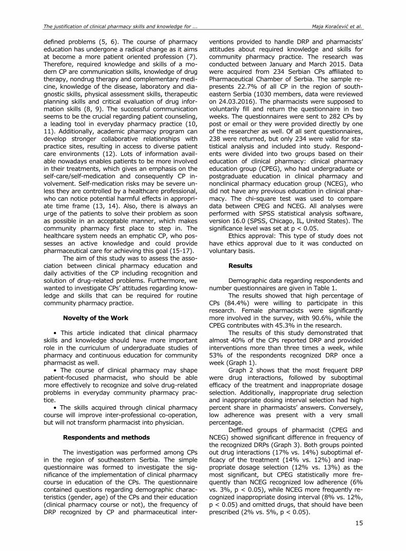
The results showed that high percentage of CPs (84.4%) were willing to participate in this research. Female pharmacists were significantly more involved in the survey, with 90.6%, while the CPEG contributes with 45.3% in the research.
The results of this study demonstrated that almost 40% of the CPs reported DRP and provided
interventions more than three times a week, while 53% of the respondents recognized DRP once a week (Graph 1).
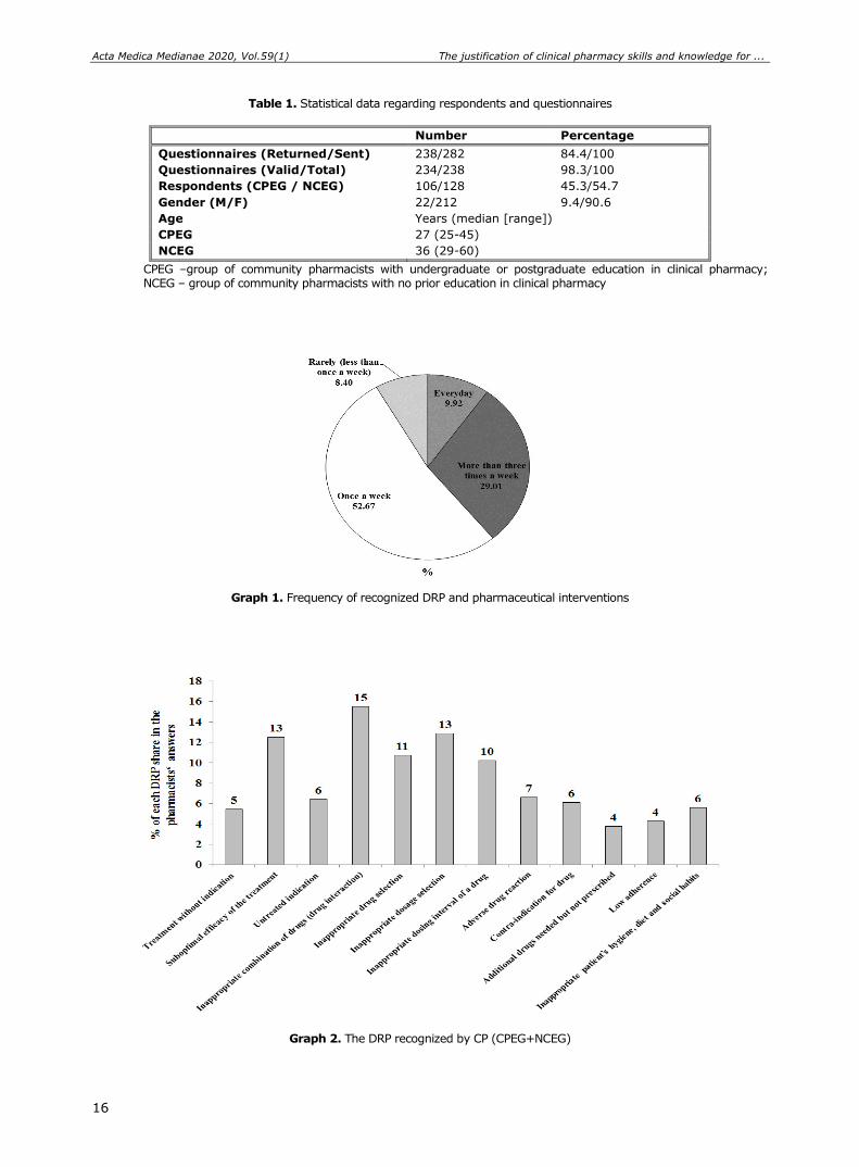
Graph 2 shows that the most frequent DRP were drug interactions, followed by suboptimal efficacy of the treatment and inappropriate dosage
selection. Additionally, inappropriate drug selection and inappropriate dosing interval selection had high
percent share in pharmacists’ answers. Conversely, low adherence was present with a very small percentage.
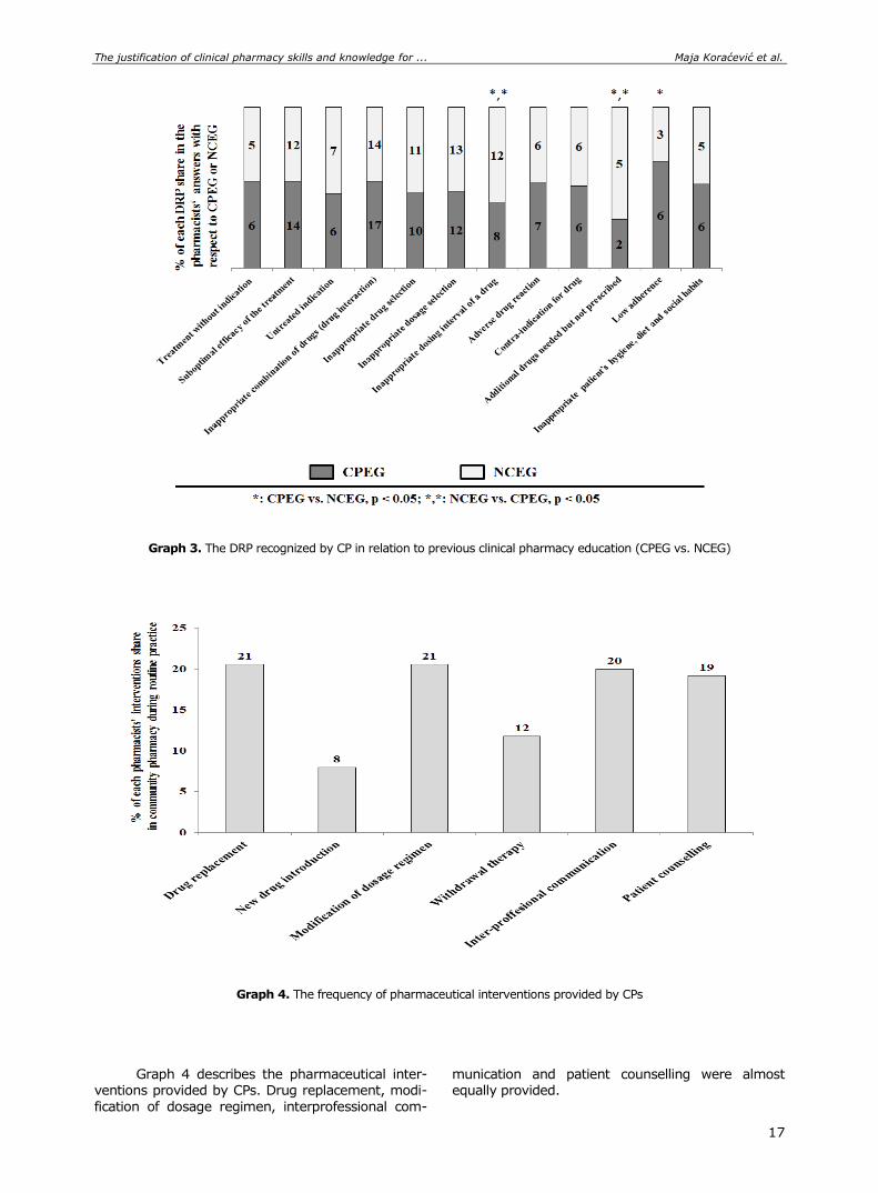
Deffined groups of pharmacist (CPEG and NCEG) showed significant difference in frequency of
the recognized DRPs (Graph 3). Both groups pointed out drug interactions (17% vs. 14%) suboptimal ef-ficacy of the treatment (14% vs. 12%) and inap-propriate dosage selection (12% vs. 13%) as the most significant, but CPEG statistically more fre-quently than NCEG recognized low adherence (6% vs. 3%, p < 0.05), while NCEG more frequently re-
cognized inappropriate dosing interval (8% vs. 12%, p < 0.05) and omitted drugs, that should have been prescribed (2% vs. 5%, p < 0.05).
Acta Medica Medianae 2020, Vol.59(1) Тhe justification of clinical pharmacy skills and knowledge for ...
16
Table 1. Statistical data regarding respondents and questionnaires
Number Percentage
Questionnaires (Returned/Sent) 238/282 84.4/100
Questionnaires (Valid/Total) 234/238 98.3/100
Respondents (CPEG / NCEG) 106/128 45.3/54.7
Gender (M/F) 22/212 9.4/90.6
Age Years (median [range])
CPEG 27 (25-45)
NCEG 36 (29-60)
CPEG –group of community pharmacists with undergraduate or postgraduate education in clinical pharmacy; NCEG – group of community pharmacists with no prior education in clinical pharmacy
Graph 1. Frequency of recognized DRP and pharmaceutical interventions
Graph 2. The DRP recognized by CP (CPEG+NCEG)
Тhe justification of clinical pharmacy skills and knowledge for ... Maja Koraćević et al.
17
Graph 3. The DRP recognized by CP in relation to previous clinical pharmacy education (CPEG vs. NCEG)
Graph 4. The frequency of pharmaceutical interventions provided by CPs
Graph 4 describes the pharmaceutical inter-ventions provided by CPs. Drug replacement, modi-
fication of dosage regimen, interprofessional com-
munication and patient counselling were almost equally provided.
Acta Medica Medianae 2020, Vol.59(1) Тhe justification of clinical pharmacy skills and knowledge for ...
18
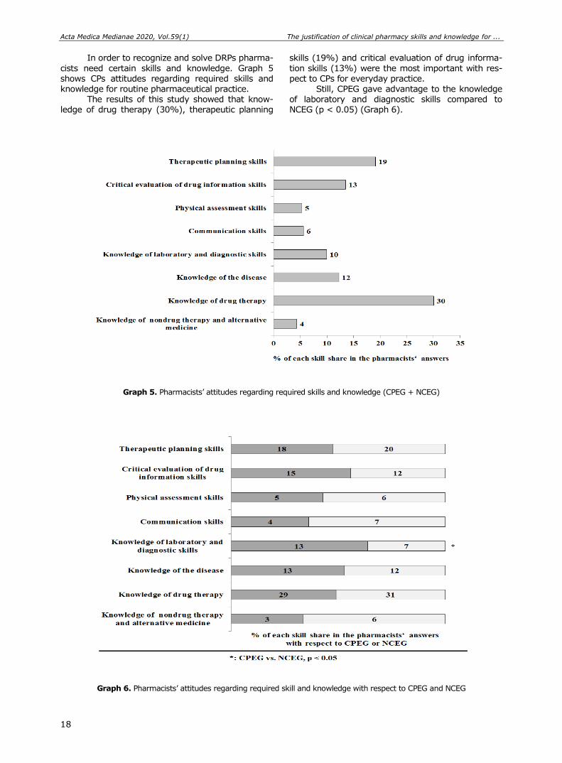
In order to recognize and solve DRPs pharma-
cists need certain skills and knowledge. Graph 5
shows CPs attitudes regarding required skills and knowledge for routine pharmaceutical practice.
The results of this study showed that know-ledge of drug therapy (30%), therapeutic planning
skills (19%) and critical evaluation of drug informa-
tion skills (13%) were the most important with res-
pect to CPs for everyday practice. Still, CPEG gave advantage to the knowledge
of laboratory and diagnostic skills compared to NCEG (p < 0.05) (Graph 6).
Graph 5. Pharmacists’ attitudes regarding required skills and knowledge (CPEG + NCEG)
Graph 6. Pharmacists’ attitudes regarding required skill and knowledge with respect to CPEG and NCEG
Тhe justification of clinical pharmacy skills and knowledge for ... Maja Koraćević et al.
19
Discussion
The modern pharmacists realize that com-
munity pharmacy is a place which offers exchange of the information and experience, and therefore contributes to the better relationship and commu-nication between patient and his pharmacist, which lead to patients’ adherence improvement (2, 12, 16). The ongoing globalization impacts the pharma-ceutical practice creating pharmacy competence framework (18). “How should pharmacists be edu-cated to increase the quality of their practice - by improving their knowledge of drugs’ pharmacology, or by improving their managing and communication skills?”, is important question nowadays (11). Com-munity pharmacy in Serbia is the place for drug dispensing, prescribing over the counter drugs, and counselling about rational pharmacotherapy and chronic noncommunicable disease as well. There was ultimate need to increase the number of pharmacists in the world (19, 20). In Serbia, CPs with a clinical knowledge and skills is even more needed healthcare profession, considering the fact that undergraduate courses of clinical pharmacy have existed less than a decade. According to the studies of Lakic at al., Serbia had less pharmacists per 100,000 inhabitants than Bulgaria, Croatia and Slovenia, countries from the same European region (19).
Besides the need for more pharmacists, the CPs require continuous improvement of knowledge and skill important for patient-focused approach in pharmaceutical practice. Recent researches confirm-ed the need for constant promotion of pharmaceu-tical knowledge and skills for problem solving inter-ventions in everyday practice (21, 22). Accordingly, the results of this study showed insufficient fre-quency of the recognition of DRP and consequently pharmaceutical interventions among study enrolled CPs (Graph 1). These findings can be partly explai-ned by the fact that clinical pharmacy course has existed less than a decade in undergraduate phar-macy studies in Serbia. Also, this indicates that ongoing reforms of pharmacy educational system should be more pronounced regarding knowledge and skills and most importantly these reforms should be more patient-oriented (23). The introduc-tion of electronic evidence of pharmaceutical inter-ventions will be a new tool for gathering information regarding pharmaceutical service in Serbia and may contribute to overall improvement of community pharmacy service.
The results of the conducted study showed that the most frequent DRPs among respondents were drug interactions, followed by suboptimal effi-cacy of the treatment and inappropriate dosage selection. The research, conducted in Ireland, showed that medication review and check of the physician’s decision can also be useful screening tools that determined prescribing errors and it may reduce unnecessary medication in prescriptions, and adverse event (24). In accordance to these findings, an association was found between increasing num-ber of drugs and supplements and the appearance of the drug interactions and adverse effects among the older population (25).
The obtained results indicated that whether pharmacist had clinical pharmacy education or not, DRPs, such as drug interactions, treatment efficiency and inappropriate dosage selection were noticed most frequently. Nevertheless, clinical pharmacy may transform CP to the more patient-focused health professional, who perceives a patient in its entirety. In our investigation CPEG recognized low adherence statistically more frequent than NCEG, while NCEG recognized more often inappropriate dosing interval and omitted medication as well. This (leads to the conclusion) explains that CPEG is more oriented towards patients’ problems and their be-haviour during prescribed treatment. Conversely, NCEG is more focused on medication review and medication itself, with less interpersonal communi-cation.
According to Westerlund et al., CPs noticed that patients’ incomprehension of the aim of the therapy might cause low adherence and suboptimal treatment efficacy (26). Considering this, commu-nity pharmacy should be the place, which can pro-vide additional explanation about patients health problems and treatment-disease association in order to achieve better compliance with the patients and therefore desirable therapeutical outcomes (27, 28).
The most frequent pharmaceutical interven-tions were done on medication, including its replace-ment and modification of dosage regimen. This find-ing is in accordance with the results of other au-thors, who investigated the knowledge and skills of clinical pharmacy required in everyday practice. Tasaka et al., noticed that implementation of clinical pharmacy skills in pharmaceutical practice led to positive therapeutic and financial outcomes, which had benefit both for community and the patient (6). Additionally, interprofessional communication and patient counselling were also highly rated in routine practice of CP.
The results of this study reported that both groups had the same attitudes regarding required knowledge and skills, emphasizing that knowledge of drug therapy (30%) and therapeutic planning skills (19%) were the most important. This medica-tion-focused approach was followed by critical eva-luation of drug information skills (13%). Elliott et al. demonstrated that managing the therapeutic plan is one of the methods for better adherence (29). Still, pharmacists with previously acquired clinical phar-macy education gave a slight advantage to the knowledge of laboratory and diagnostic skills. It indicates that education of clinical pharmacy may cause the change from a medication- to a patient-focused pharmacist. Previous researches showed that CPs had less knowledge about signs and symp-toms of the diseases, which was needed for appro-priate pharmaceutical intervention and care of the patients as well (21, 30). This supports our finding that clinical pharmacy education may contribute to rational and timely decision regarding pharmacy-led interventions. The ultimate benefit of the introduc-tion of these skills and knowledge can be demon-strated through improved patients’ quality of life and reduced healthcare costs (18, 31). In accordance with this, the efforts on the national level have been made to improve Serbian pharmaceutical practice.
Acta Medica Medianae 2020, Vol.59(1) Тhe justification of clinical pharmacy skills and knowledge for ...
20
The most important improvement will be electronic documentation of pharmaceutical intervention to-wards patients.
Considering these results, it was shown that clinical pharmacy education and courses had a sig-nificant influence on forming of a modern CP with a patient in its focus. Still, pharmacist, both CPEG and NCEG highlighted the importance of drug therapy skills, which indicated that they would not cross their initial obligations and try to be physicians. This can represent a real background for interprofessional co-operation between pharmacist and the other health-care professionals. The newly obtained knowledge and skills may help pharmacist to deal with his or her tasks in responsible manner and therefore to optimize pharmacotherapy and cooperate with phy-sician in order to provide a maximum safety and benefit for the patient.
Conclusion
In conclusion, CPs deal with different DRPs and provide pharmaceutical interventions within their daily activity. The results of this study indicated
that education of clinical pharmacy skills and know-
ledge can move the focus of the CPs, from medicine
towards patient, but still position the drug in the center of their activity. Modern pharmacist with changed skills in his or her portfolio gives a huge contribution to interprofessional approach with a view to achieve desired health outcomes. Pharma-
cist with clinical pharmacy education were more fa-miliar with the knowledge of laboratory and dia-gnostic skills, necessary for the risk therapy mana-gement, which confirms the ongoing change in phar-macist orientation. Therefore, it is desirable that cli-nical pharmacy course should be part of the under-graduate curriculum of pharmacy students or conti-
nuous medical education for experienced pharmacist as well.
Acknowledgement
This work was supported by a grant awarded
by the Faculty of Medicine, University of Niš (Grant INT-MFN 37 , Grant INT-MFN 25).
References
1. Anderson C, Bates I, Beck D, Brock TP, Futter B, Mercer H et al. The WHO UNESCO FIP Pharmacy Education Taskforce. Hum Resour Health 2009;7:45. [CrossRef] [PubMed]
2. Correr CJ, Melchiors AC, de Souza TT, Rotta I, Salgado TM, Fernandez-Llimos F. A tool to characterize the components of pharmacist interventions in clinical pharmacy services: the DEPICT project. Ann Pharma-cother 2013;47:946-52. [CrossRef] [PubMed]
3. Basak SC, van Mil JW, Sathyanarayana D. The changing roles of pharmacists in community pharma-cies: perception of reality in India. Pharm World Sci 2009;31:612-18. [CrossRef] [PubMed]
4. Salgado TM, Correr CJ, Moles R, Benrimoj SI, Fernandez-Llimos F. Assessing the implementability of clinical pharmacist interventions in patients with chronic kidney disease: an analysis of systematic re-views. Ann Pharmacother 2013;47:1498-506. [CrossRef] [PubMed]
5. Alaqeel S, Abanmy NO. Counselling practices in community pharmacies in Riyadh, Saudi Arabia: a
cross-sectional study. BMC Health Serv Res 2015; 15:557. [CrossRef] [PubMed]
6. Tasaka Y, Yasunaga D, Tanaka M, Tanaka A, Asakawa T, Horio I et al. Economic and safety benefits of pharmaceutical interventions by community and hospital pharmacists in Japan. Int J Clin Pharm 2016; 38:321-9. [CrossRef] [PubMed]
7. Shirwaikar A. Objective structured clinical examination (OSCE) in pharmacy education - a trend. Pharm Pract 2015;13:627. [CrossRef] [PubMed]
8. Barnett CW, Matthews HW. Teaching evaluation practices in colleges and schools of pharmacy. Am J Pharm Educ 2009;73:103. [CrossRef] [PubMed]
9. Tai BB, Hata M, Wu S, Frausto S, Law AV. Prediction of pharmacist intention to provide medication disposal education using the theory of planned behaviour. J Eval Clin Pract, 2016; [CrossRef] [PubMed]
10. Wallman A, Vaudan C, Sporrong SK. Communications training in pharmacy education, 1995-2010. Am J Pharm Educ 2013;77:36. [CrossRef] [PubMed]
Тhe justification of clinical pharmacy skills and knowledge for ... Maja Koraćević et al.
21
11. Puspitasari HP, Aslani P, Krass I. Pharmacists' and consumers' viewpoints on counselling on prescription
medicines in Australian community pharmacies. Int J Pharm Pract, 2010;18:202-8. [CrossRef] [PubMed]
12. Rathbun RC, Hester EK, Arnold LM, Chung AM, Dunn SP. Importance of direct patient care in advanced pharmacy practice experiences. Pharmacotherapy 2012;32:e88-97. [CrossRef] [PubMed]
13. Veiga P, Lapão LV, Cavaco AM, Guerreiro MP. Quality supply of nonprescription medicines in Portuguese community pharmacy: An exploratory case study. Res Social Adm Pharm 2015;11:880-890. [CrossRef] [PubMed]
14. Damnjanovic I, Kitic D, Stefanovic N, Zlatkovic-Guberinic S, Catic-Djordjevic A, Velickovic-Radovano-vic R. Herbal self-medication use in patients with diabetes mellitus type 2. Turk J Med Sci 2015; 45: 964-71. [CrossRef] [PubMed]
15. Katoue MG, Awad AI, Schwinghammer TL, Kombian SB. Pharmaceutical care education in Kuwait: pharma-cy students' perspectives. Pharm Pract 2014;12:411. [CrossRef] [PubMed]
16. Barnett MJ, Frank J, Wehring H, Newland B, Von Muenster S, Kumbera P et al. Analysis of pharmacist-provided medication therapy management (MTM) ser-vices in community pharmacies over 7 years. J Manag Care Pharm 2009;15:18-31. [CrossRef] [PubMed]
17. Pande S, Hiller JE, Nkansah N, Bero L. The effect of pharmacist-provided non-dispensing services on pa-tient outcomes, health service utilisation and costs in low- and middle-income countries. Cochrane Database Syst Rev 2013;2:CD010398 [CrossRef] [PubMed]
18. Anderson C, Bates I, Brock T, Brown AN, Bruno A, Futter B et al. Needs-based education in the context of globalization. Am J Pharm Educ 2012;76:56. [CrossRef] [PubMed]
19. Lakić D, Tasić L, Kos M, Petrova G, Stoimenova A, Krajnović D. Pharmacy network and access to me-dicines in selected eastern European countries: com-parative analysis. Croat Med J 2012;53:53-9. [CrossRef] [PubMed]
20. Kheir N, Zaidan M, Younes H, El Hajj M, Wilbur K, Jewesson PJ. Pharmacy education and practice in 13 Middle Eastern countries. Am J Pharm Educ 2008; 72:133. [CrossRef] [PubMed]
21. Kashour TS, Joury A, Alotaibi AM, Althagafi M, Almufleh AS, Hersi A et al. Quality of assessment and
counselling offered by community pharmacists and medication sale without prescription to patients pre-senting with acute cardiac symptoms: a simulated
client study. Eur J Clin Pharmacol 2016;72:321-8. [CrossRef] [PubMed]
22. Lin K, Travlos DV, Wadelin JW, Vlasses PH. Simulation and introductory pharmacy practice experiences. Am J Pharm Educ 2011;75:209. [CrossRef] [PubMed]
23. Anderson C, Brock T, Bates I, Rouse M, Marriott J, Manasse H et al. Transforming health professional education. Am J Pharm Educ 2011;75:22. [CrossRef] [PubMed]
24. Galvin R, Moriarty F, Cousins G, Cahir C, Motterlini N, Bradley M et al. Prevalence of potentially inappropriate prescribing and prescribing omissions in older Irish adults: findings from The Irish LongituDinal Study on Ageing study (TILDA). Eur J Clin Pharmacol 2014; 70:599-606. [CrossRef] [PubMed]
25. Peklar J, Henman MC, Kos M, Richardson K, Kenny RA. Concurrent use of drugs and supplements in a community-dwelling population aged 50 years or more: potential benefits and risks. Drugs Aging 2014; 31:527-40. [CrossRef] [PubMed]
26. Westerlund T, Gelin U, Pettersson E, Skärlund F, Wågström K, Ringbom C. A retrospective analysis of drug-related problems documented in a national data-base. Int J Clin Pharm 2013;35:202-9. [CrossRef] [PubMed]
27. Morecroft CW, Mackridge AJ, Stokes EC, Gray NJ, Wilson SE, Ashcroft DM et al. Emergency supply of prescription-only medicines to patients by community pharmacists: a mixed methods evaluation incorpo-rating patient, pharmacist and GP perspectives. BMJ Open 2015;5:e006934. [CrossRef] [PubMed]
28. Horvat N, Kos M. Contribution of Slovenian community pharmacist counseling to patients' knowledge about their prescription medicines: a cross-sectional study. Croat Med J 2015;56:41-49. [CrossRef] [PubMed]
29. Elliott RA, Boyd MJ, Salema NE, Davies J, Barber N, Mehta RL et al. Supporting adherence for people start-ing a new medication for a long-term condition through community pharmacies: a pragmatic rando-mised controlled trial of the New Medicine Service. BMJ Qual Saf 2015; [CrossRef] [PubMed]
30. Bajorek B, LeMay K, Gunn K, Armour C. The potential role for a pharmacist in a multidisciplinary general practitioner super clinic. Australas Med J 2015;8:52-63. [CrossRef] [PubMed]
31. Waszyk-Nowaczyk M, Nowaczyk P, Simon M. Physi-cians' and patients' valuation of pharmaceutical care
implementation in Poznan (Poland) community phar-macies. Saudi Pharm J 2014;22:537-44. [CrossRef] [PubMed]
Acta Medica Medianae 2020, Vol.59(1) Тhe justification of clinical pharmacy skills and knowledge for ...
22
Originalni rad UDC: 615.01/.03:615.851.4
doi:10.5633/amm.2020.0102
OPRAVDANOST STICANJA VEŠTINA I ZNANJA IZ KLINIČKE FARMACIJE TOKOM OBRAZOVANJA SAVREMENOG FARMACEUTA
Maja Koraćević1,2, Aleksandra Catić-Đorđević1, Nikola Stefanović1, Ivana Damnjanović1,
Ivana Stošić1, Radmila Veličković-Radovanović1,3
1Univerzitet u Nišu, Medicinski fakultet, Odsek za farmaciju, Niš, Srbija; 2Univerzitet u Nišu, Inovacioni centar, Niš, Srbija; 3Klinika za nefrologiju, Klinički centar Niš, Niš, Srbija;
Kontakt: Aleksandra Catić-Đorđević
Bulevar dr Zorana Đinđića 81, 18000 Niš, Srbija
E-mail: [email protected]
Savremeni farmaceut u apoteci suočava se sa brojnim zahtevima vezanim za pregled
terapije i uočavanje problema vezanih za terapiju (drug-related problems — DRP) bolesnika.
Pored poznavanja leka, farmaceut danas aktivno učestvuje u ukupnoj terapiji bolesnika, savetuje i pomaže rešavanju manjih zdravstvenih problema izborom leka u režimu izdavanja
bez lekarskog recepta. Sledeći nove obaveze farmaceuta u apoteci, akademsko obrazovanje farmaceuta transformisano je u nameri da se fokus pomeri sa leka na bolesnika. Primarni cilj ovog istraživanja bila je procena povezanosti obrazovanja iz kliničke farmacije i dnevnih
aktivnosti farmaceuta u apoteci (community pharmacist — CP), uključujući prepoznavanje i
rešavanje DRP. Osim toga, cilj je bio i izvršiti analizu stavova CP-a u pogledu znanja i veština koje mogu biti korisne u svakodnevnom radu. Istraživanje je sprovedeno pomoću jednostavnog upitnika formulisanog za procenu značaja uvođenja veština kliničke farmacije u akademske studije. U istraživanju je čestvovalo 234 CP-a, koji su podeljeni u dve grupe: grupa sa edukacijom iz kliničke farmacije (CPEG) i grupa bez edukacije iz kliničke farmacije (NCEG). Najčešći DRP, koji su prepoznali ispitanici (CPEG i NCEG), su interakcije lekova, praćene suboptimalnom efikasnošću lečenja i neodgovarajućim doziranjem. Dodatno, CPEG su značajno češće uočavali nisku adherencu (p < 0,05), a NCEG neprikladan interval doziranja (p < 0,05) i lekove koji nedostaju, a mogli bi biti korisni u terapiji (p < 0,05). Ispitanici su se složili da su poznavanje lekova, veštine planiranja i rukovođenja terapijom i kritička procena informacija o lekovima najvažnije veštine i znanja iz kliničke farmacije potrebne za svako-dnevni rad u apoteci. Ipak, CPEG su dali prednost poznavanju laboratorijskih i dijagnostičkih veština (p < 0,05). Uočena razlika u važnosti poznavanja laboratorijskih i dijagnostičkih parametara potvrđuje pomeranje fokusa CPEG. Sprovedeno istraživanje pokazalo je da obrazovanje iz kliničke farmacije može pomeriti fokus CP-a ka bolesniku, ali i dalje sa lekom u centru farmaceutske aktivnosti.
Acta Medica Medianae 2020;59(1):14-22.
Ključne reči: klinička farmacija, farmaceutska praksa, obrazovanje farmaceuta
23 www.medfak.ni.ac.rs/amm
Original article UDC: 543.42:546.3/.9]:582.99
doi:10.5633/amm.2020.0103
DETERMINING THE CONTENT OF Cd, Cu, Pb AND Zn IN THE LEAVES OF DANDELION (TARAXACUM OFFICINALE WEBB.) AND IN THE SOIL
BY ICP-OES
Dragan Velimirović1, Biljana Kaličanin1, Milan Stojković2, Snežana Tošić2
Dandelion (Taraxacum officinale Webb.) is a plant capable of accumulating a certain
quantity of metals. The aim of this study was to determine the content of Cd, Cu, Pb and Zn in dandelion leaves and soil that have been sampled from different locations. One group of samples has been influenced by pollution sources of these metals and the other one has not been exposed to the pollutants. The amount of metals in the tested samples has been determined by inductively coupled plasma optical emission spectrometry (ICP-OES).The content of detected metals was higher in leaves and soil samples that were exposed to the negative effect of environmental pollutants, compared to those samples that were not under the influence of contamination sources. The increased content of detected metals in the samples of dandelion leaves that were under the influence of the pollutant may be the result of a synergistic effect, soil, on which this plant species thrives, and the air, that is contaminated by the effects of motor traffic and other forms of pollutants. The results of this study have shown that dandelion can provide a data of environmental pollution by the content of detected metals in its tissue. As dandelion is used in human nutrition, and since heavy metals (Pb, Cd) with cumulative and toxic effects have been detected in it, it is necessary, in order to protect human health, to check the presence and content of these metals in the dandelion plant that is used in human nutrition.
Acta Medica Medianae 2020;59(1):23-30. Key words: dandelion, metals, soil, pollutants, ICP-OES
1University of Niš, Faculty of Medicine, Department of Pharmacy, Niš, Serbia 2University of Niš, Faculty of Sciences and Mathematics,
Department of Chemistry, Niš, Serbia
Contact: Dragan Velimirović
81 Dr. Zoran Djindjić Blvd., 18000 Niš, Serbia
E-mail: [email protected]
Introduction The development and advance of technology,
in all spheres of life, are processes that have been rapidly evolving since the industrial revolution in the 18th century. Today we are witnesses of a huge and
constant development of technological and industrial processes that leads to a significant improvement in the life of an individual in all social aspects. Un-fortunately, such an unstoppable advance, with all the benefits, also brings different negative effects, that can also cause very serious and unsolvable problems for all of the living beings on the planet.
Global and local pollution of the environment that harms all the living organisms is definitely one of the biggest problems, and a direct result of constant
industrial development of the human species. Environmental pollutants are of organic and
non-organic origin. They can have a bigger or smaller negative effect on the living world. In the big group of non-organic pollutants cadmium and lead must be pointed out. They are especially dangerous, as they are toxic, even in small quantities (1). An
increased amount of metals in the environment, taking into consideration the fact that they are eco-
toxicological danger, creates a need to find and de-velop economical chemical methods whose aim would be to detect the presence of metal in tested samples and discover the dangers that living orga-nisms are exposed to because of these pollutants.
One of the ways to detect these pollutants is to identify the organisms that, thanks to their ability to accumulate different metals, can give adequate in-formation about the assessment of environmental contamination with these metals. These organisms that give quantitative data that is connected to the
environment, for example, how much pollutants are present at the tested location. Every kind of orga-nism that is distributed over a wider geographical area, that is toxitolerant to a large number of metals
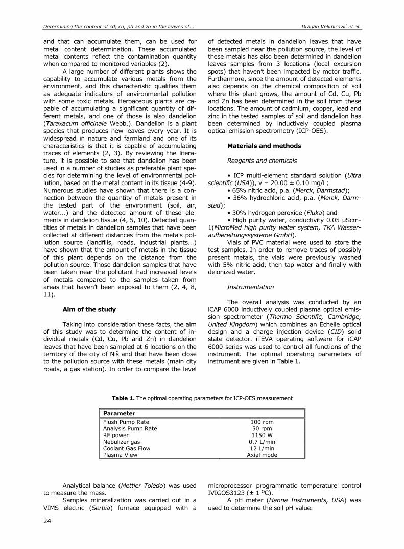
Determining the content of cd, cu, pb and zn in the leaves of... Dragan Velimirović et al.
24
and that can accumulate them, can be used for
metal content determination. These accumulated
metal contents reflect the contamination quantity when compared to monitored variables (2).
A large number of different plants shows the capability to accumulate various metals from the environment, and this characteristic qualifies them
as adequate indicators of environmental pollution with some toxic metals. Herbaceous plants are ca-pable of accumulating a significant quantity of dif-ferent metals, and one of those is also dandelion (Taraxacum officinale Webb.). Dandelion is a plant species that produces new leaves every year. It is widespread in nature and farmland and one of its
characteristics is that it is capable of accumulating traces of elements (2, 3). By reviewing the litera-ture, it is possible to see that dandelion has been
used in a number of studies as preferable plant spe-cies for determining the level of environmental pol-lution, based on the metal content in its tissue (4-9). Numerous studies have shown that there is a con-
nection between the quantity of metals present in the tested part of the environment (soil, air, water...) and the detected amount of these ele-ments in dandelion tissue (4, 5, 10). Detected quan-tities of metals in dandelion samples that have been collected at different distances from the metals pol-
lution source (landfills, roads, industrial plants...) have shown that the amount of metals in the tissue of this plant depends on the distance from the pollution source. Those dandelion samples that have been taken near the pollutant had increased levels of metals compared to the samples taken from
areas that haven’t been exposed to them (2, 4, 8,
11). Aim of the study Taking into consideration these facts, the aim
of this study was to determine the content of in-dividual metals (Cd, Cu, Pb and Zn) in dandelion
leaves that have been sampled at 6 locations on the territory of the city of Niš and that have been close to the pollution source with these metals (main city roads, a gas station). In order to compare the level
of detected metals in dandelion leaves that have
been sampled near the pollution source, the level of
these metals has also been determined in dandelion leaves samples from 3 locations (local excursion spots) that haven’t been impacted by motor traffic. Furthermore, since the amount of detected elements also depends on the chemical composition of soil
where this plant grows, the amount of Cd, Cu, Pb and Zn has been determined in the soil from these locations. The amount of cadmium, copper, lead and zinc in the tested samples of soil and dandelion has been determined by inductively coupled plasma optical emission spectrometry (ICP-OES).
Materials and methods Reagents and chemicals
• ICP multi-element standard solution (Ultra
scientific (USA)), γ = 20.00 ± 0.10 mg/L; • 65% nitric acid, p.a. (Merck, Darmstad);
• 36% hydrochloric acid, p.a. (Merck, Darm-stad);
• 30% hydrogen peroxide (Fluka) and • High purity water, conductivity 0.05 μScm-
1(MicroMed high purity water system, TKA Wasser-aufbereitungssysteme GmbH).
Vials of PVC material were used to store the test samples. In order to remove traces of possibly present metals, the vials were previously washed with 5% nitric acid, then tap water and finally with deionized water.
Instrumentation
The overall analysis was conducted by an
iCAP 6000 inductively coupled plasma optical emis-sion spectrometer (Thermo Scientific, Cambridge, United Kingdom) which combines an Echelle optical design and a charge injection device (CID) solid state detector. iTEVA operating software for iCAP
6000 series was used to control all functions of the instrument. The optimal operating parameters of instrument are given in Table 1.
Table 1. The optimal operating parameters for ICP-OES measurement
Parameter
Flush Pump Rate Analysis Pump Rate
100 rpm 50 rpm
RF power 1150 W Nebulizer gas 0.7 L/min
Coolant Gas Flow 12 L/min Plasma View Axial mode
Analytical balance (Mettler Toledo) was used to measure the mass.
Samples mineralization was carried out in a VIMS electric (Serbia) furnace equipped with a
microprocessor programmatic temperature control IVIGOS3123 (± 1 OC).
A pH meter (Hanna Instruments, USA) was used to determine the soil pH value.
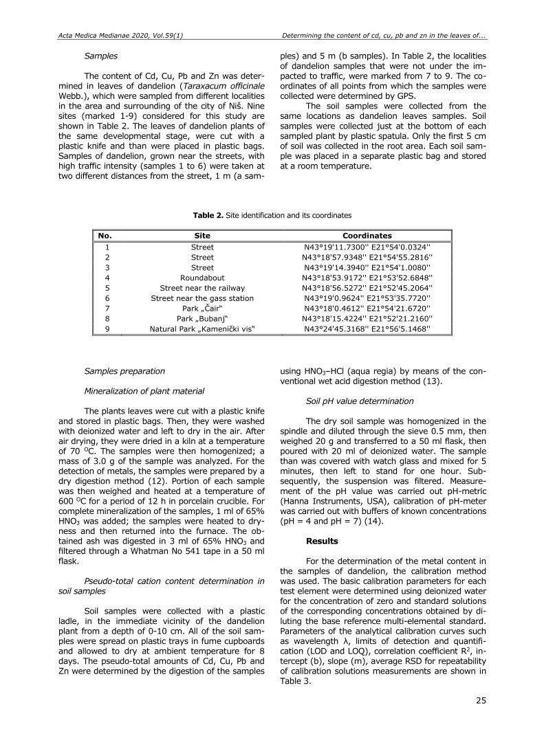
Acta Medica Medianae 2020, Vol.59(1) Determining the content of cd, cu, pb and zn in the leaves of...
25
Samples
The content of Cd, Cu, Pb and Zn was deter-mined in leaves of dandelion (Taraxacum officinale Webb.), which were sampled from different localities in the area and surrounding of the city of Niš. Nine sites (marked 1-9) considered for this study are
shown in Table 2. The leaves of dandelion plants of the same developmental stage, were cut with a plastic knife and than were placed in plastic bags. Samples of dandelion, grown near the streets, with high traffic intensity (samples 1 to 6) were taken at two different distances from the street, 1 m (a sam-
ples) and 5 m (b samples). In Table 2, the localities
of dandelion samples that were not under the im-
pacted to traffic, were marked from 7 to 9. The co-ordinates of all points from which the samples were collected were determined by GPS.
The soil samples were collected from the same locations as dandelion leaves samples. Soil
samples were collected just at the bottom of each sampled plant by plastic spatula. Only the first 5 cm of soil was collected in the root area. Each soil sam-ple was placed in a separate plastic bag and stored at a room temperature.
Table 2. Site identification and its coordinates
No. Site Coordinates
1 Street N43°19'11.7300'' E21°54'0.0324''
2 Street N43°18'57.9348'' E21°54'55.2816''
3 Street N43°19'14.3940'' E21°54'1.0080''
4 Roundabout N43°18'53.9172'' E21°53'52.6848''
5 Street near the railway N43°18'56.5272'' E21°52'45.2064''
6 Street near the gass station N43°19'0.9624'' E21°53'35.7720''
7 Park „Čair“ N43°18'0.4612'' E21°54'21.6720''
8 Park „Bubanj“ N43°18'15.4224'' E21°52'21.2160''
9 Natural Park „Kamenički vis“ N43°24'45.3168'' E21°56'5.1468''
Samples preparation
Mineralization of plant material The plants leaves were cut with a plastic knife
and stored in plastic bags. Then, they were washed with deionized water and left to dry in the air. After air drying, they were dried in a kiln at a temperature of 70 OC. The samples were then homogenized; a mass of 3.0 g of the sample was analyzed. For the detection of metals, the samples were prepared by a dry digestion method (12). Portion of each sample
was then weighed and heated at a temperature of 600 OC for a period of 12 h in porcelain crucible. For complete mineralization of the samples, 1 ml of 65% HNO3 was added; the samples were heated to dry-ness and then returned into the furnace. The ob-
tained ash was digested in 3 ml of 65% HNO3 and filtered through a Whatman No 541 tape in a 50 ml
flask. Pseudo-total cation content determination in
soil samples Soil samples were collected with a plastic
ladle, in the immediate vicinity of the dandelion plant from a depth of 0-10 cm. All of the soil sam-ples were spread on plastic trays in fume cupboards and allowed to dry at ambient temperature for 8 days. The pseudo-total amounts of Cd, Cu, Pb and Zn were determined by the digestion of the samples
using HNO3–HCl (aqua regia) by means of the con-ventional wet acid digestion method (13).
Soil pH value determination
The dry soil sample was homogenized in the spindle and diluted through the sieve 0.5 mm, then weighed 20 g and transferred to a 50 ml flask, then poured with 20 ml of deionized water. The sample than was covered with watch glass and mixed for 5 minutes, then left to stand for one hour. Sub-sequently, the suspension was filtered. Measure-
ment of the pH value was carried out pH-metric (Hanna Instruments, USA), calibration of pH-meter was carried out with buffers of known concentrations (pH = 4 and pH = 7) (14).
Results
For the determination of the metal content in the samples of dandelion, the calibration method was used. The basic calibration parameters for each test element were determined using deionized water for the concentration of zero and standard solutions of the corresponding concentrations obtained by di-
luting the base reference multi-elemental standard. Parameters of the analytical calibration curves such as wavelength λ, limits of detection and quantifi-cation (LOD and LOQ), correlation coefficient R2, in-tercept (b), slope (m), average RSD for repeatability of calibration solutions measurements are shown in Table 3.
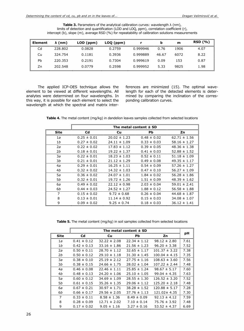
Determining the content of cd, cu, pb and zn in the leaves of... Dragan Velimirović et al.
26
Table 3. Parameters of the analytical calibration curves: wavelength λ (nm), limits of detection and quantification (LOD and LOQ, ppm), correlation coefficient (r),
intercept (b), slope (m), average RSD (%) for repeatability of calibration solutions measurements
Element λ (nm) LOD (ppm) LOQ (ppm) r b m RSD (%)
Cd 228.802 0.0828 0.2759 0.999946 0.76 1906 4.07
Cu 324.754 0.1181 0.3936 0.999889 46.67 6072 8.22
Pb 220.353 0.2191 0.7304 0.999619 0.09 153 0.87
Zn 202.548 0.0779 0.2598 0.999952 5.33 9825 1.98
The applied ICP-OES technique allows the
element to be viewed at different wavelengths. All analytes were determined on four wavelengths. In this way, it is possible for each element to select the
wavelength at which the spectral and matrix inter-
ferences are minimized (15). The optimal wave-length for each of the detected elements is deter-mined by comparing the inclination of the corres-ponding calibration curves.
Table 4. The metal content (mg/kg) in dandelion leaves samples collected from selected locations
The metal content ± SD
Site Cd Cu Pb Zn
1a 0.25 ± 0.01 20.02 ± 1.23 0.48 ± 0.02 62.71 ± 1.56
1b 0.27 ± 0.02 24.11 ± 1.09 0.33 ± 0.03 58.16 ± 1.27
2a 0.22 ± 0.02 17.83 ± 1.12 0.39 ± 0.05 48.36 ± 1.38
2b 0.18 ± 0.01 19.22 ± 1.37 0.41 ± 0.03 52.88 ± 1.52
3a 0.22 ± 0.01 18.23 ± 1.03 0.52 ± 0.11 51.18 ± 1.09
3b 0.21 ± 0.01 21.12 ± 1.29 0.49 ± 0.08 49.35 ± 1.17
4a 0.29 ± 0.01 16.25 ± 1.11 0.54 ± 0.09 57.26 ± 1.27
4b 0.32 ± 0.02 14.32 ± 1.03 0.47 ± 0.10 56.27 ± 1.09
5a 0.36 ± 0.02 24.07 ± 1.01 1.84 ± 0.02 56.28 ± 1.86
5b 0.32 ± 0.01 19.72 ± 1.26 1.51 ± 0.09 48.39 ± 1.62
6a 0.49 ± 0.02 22.12 ± 0.98 2.03 ± 0.04 59.01 ± 2.41
6b 0.44 ± 0.03 24.52 ± 1.27 1.88 ± 0.12 56.58 ± 1.88
7 0.15 ± 0.02 9.72 ± 0.68 0.26 ± 0.04 44.68 ± 1.87
8 0.13 ± 0.01 11.14 ± 0.92 0.15 ± 0.03 34.08 ± 1.07
9 0.09 ± 0.02 9.25 ± 0.74 0.18 ± 0.03 36.12 ± 1.41
Table 5. The metal content (mg/kg) in soil samples collected from selected locations
The metal content ± SD pH
Site Cd Cu Pb Zn
1a 0.41 ± 0.12 32.22 ± 2.08 22.34 ± 1.12 98.12 ± 2.80 7.61
1b 0.42 ± 0.13 33.16 ± 1.86 21.56 ± 1.23 96.20 ± 3.38 7.52
2a 0.50 ± 0.11 28.70 ± 1.12 32.65 ± 1.17 101.37 ± 3.22 7.38
2b 0.50 ± 0.12 29.10 ± 1.18 31.30 ± 1.45 100.04 ± 4.15 7.35
3a 0.38 ± 0.10 25.19 ± 2.12 27.75 ± 1.16 108.63 ± 3.60 7.56
3b 0.38 ± 0.15 24.66 ± 1.75 28.02 ± 1.04 107.22 ± 2.44 7.48
4a 0.46 ± 0.08 22.46 ± 1.11 25.85 ± 1.24 98.67 ± 5.17 7.60
4b 0.48 ± 0.13 24.20 ± 1.06 25.10 ± 1.05 99.04 ± 4.35 7.63
5a 0.60 ± 0.12 34.69 ± 1.09 28.55 ± 1.30 126.52 ± 3.20 7.52
5b 0.61 ± 0.15 35.26 ± 1.35 29.06 ± 1.12 125.20 ± 2.18 7.48
6a 0.67 ± 0.21 30.97 ± 1.71 38.28 ± 1.52 120.88 ± 5.17 7.28
6b 0.66 ± 0.17 29.56 ± 2.05 37.76 ± 1.13 121.02± 4.35 7.30
7 0.33 ± 0.11 8.58 ± 1.36 8.49 ± 0.09 92.13 ± 4.12 7.59
8 0.28 ± 0.09 12.71 ± 2.02 7.10 ± 0.14 75.76 ± 3.92 7.48
9 0.17 ± 0.02 9.05 ± 1.16 3.27 ± 0.16 53.52 ± 4.37 6.69
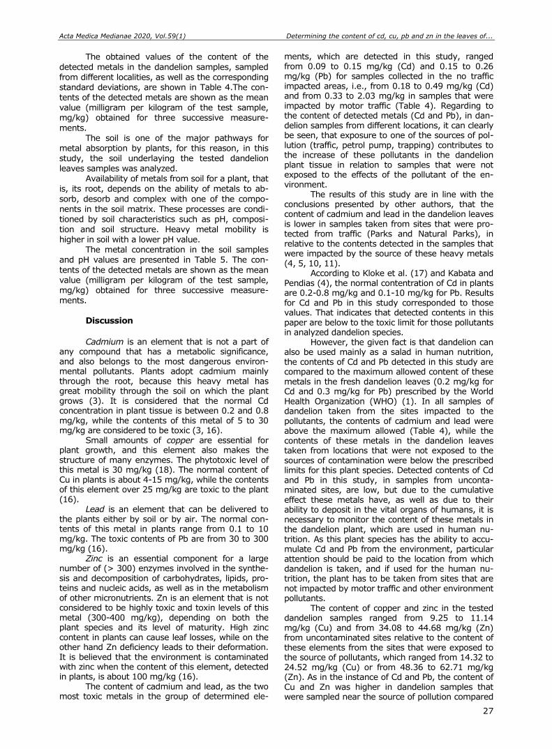
Acta Medica Medianae 2020, Vol.59(1) Determining the content of cd, cu, pb and zn in the leaves of...
27
The obtained values of the content of the
detected metals in the dandelion samples, sampled
from different localities, as well as the corresponding standard deviations, are shown in Table 4.The con-tents of the detected metals are shown as the mean value (milligram per kilogram of the test sample, mg/kg) obtained for three successive measure-
ments. The soil is one of the major pathways for
metal absorption by plants, for this reason, in this study, the soil underlaying the tested dandelion leaves samples was analyzed.
Availability of metals from soil for a plant, that is, its root, depends on the ability of metals to ab-
sorb, desorb and complex with one of the compo-nents in the soil matrix. These processes are condi-tioned by soil characteristics such as pH, composi-
tion and soil structure. Heavy metal mobility is higher in soil with a lower pH value.
The metal concentration in the soil samples and pH values are presented in Table 5. The con-
tents of the detected metals are shown as the mean value (milligram per kilogram of the test sample, mg/kg) obtained for three successive measure-ments.
Discussion
Cadmium is an element that is not a part of
any compound that has a metabolic significance, and also belongs to the most dangerous environ-mental pollutants. Plants adopt cadmium mainly through the root, because this heavy metal has great mobility through the soil on which the plant grows (3). It is considered that the normal Cd concentration in plant tissue is between 0.2 and 0.8 mg/kg, while the contents of this metal of 5 to 30 mg/kg are considered to be toxic (3, 16).
Small amounts of copper are essential for plant growth, and this element also makes the structure of many enzymes. The phytotoxic level of this metal is 30 mg/kg (18). The normal content of Cu in plants is about 4-15 mg/kg, while the contents of this element over 25 mg/kg are toxic to the plant (16).
Lead is an element that can be delivered to the plants either by soil or by air. The normal con-tents of this metal in plants range from 0.1 to 10 mg/kg. The toxic contents of Pb are from 30 to 300 mg/kg (16).
Zinc is an essential component for a large number of (> 300) enzymes involved in the synthe-sis and decomposition of carbohydrates, lipids, pro-teins and nucleic acids, as well as in the metabolism of other micronutrients. Zn is an element that is not considered to be highly toxic and toxin levels of this metal (300-400 mg/kg), depending on both the plant species and its level of maturity. High zinc content in plants can cause leaf losses, while on the other hand Zn deficiency leads to their deformation. It is believed that the environment is contaminated with zinc when the content of this element, detected in plants, is about 100 mg/kg (16).
The content of cadmium and lead, as the two most toxic metals in the group of determined ele-
ments, which are detected in this study, ranged from 0.09 to 0.15 mg/kg (Cd) and 0.15 to 0.26 mg/kg (Pb) for samples collected in the no traffic impacted areas, i.e., from 0.18 to 0.49 mg/kg (Cd) and from 0.33 to 2.03 mg/kg in samples that were impacted by motor traffic (Table 4). Regarding to the content of detected metals (Cd and Pb), in dan-delion samples from different locations, it can clearly be seen, that exposure to one of the sources of pol-lution (traffic, petrol pump, trapping) contributes to the increase of these pollutants in the dandelion plant tissue in relation to samples that were not exposed to the effects of the pollutant of the en-vironment.
The results of this study are in line with the conclusions presented by other authors, that the content of cadmium and lead in the dandelion leaves is lower in samples taken from sites that were pro-tected from traffic (Parks and Natural Parks), in relative to the contents detected in the samples that were impacted by the source of these heavy metals (4, 5, 10, 11).
According to Kloke et al. (17) and Kabata and Pendias (4), the normal contentration of Cd in plants are 0.2-0.8 mg/kg and 0.1-10 mg/kg for Pb. Results for Cd and Pb in this study corresponded to those values. That indicates that detected contents in this paper are below to the toxic limit for those pollutants in analyzed dandelion species.
However, the given fact is that dandelion can also be used mainly as a salad in human nutrition, the contents of Cd and Pb detected in this study are compared to the maximum allowed content of these metals in the fresh dandelion leaves (0.2 mg/kg for Cd and 0.3 mg/kg for Pb) prescribed by the World Health Organization (WHO) (1). In all samples of dandelion taken from the sites impacted to the pollutants, the contents of cadmium and lead were above the maximum allowed (Table 4), while the contents of these metals in the dandelion leaves taken from locations that were not exposed to the sources of contamination were below the prescribed limits for this plant species. Detected contents of Cd and Pb in this study, in samples from unconta-minated sites, are low, but due to the cumulative effect these metals have, as well as due to their ability to deposit in the vital organs of humans, it is necessary to monitor the content of these metals in the dandelion plant, which are used in human nu-trition. As this plant species has the ability to accu-mulate Cd and Pb from the environment, particular attention should be paid to the location from which dandelion is taken, and if used for the human nu-trition, the plant has to be taken from sites that are not impacted by motor traffic and other environment pollutants.
The content of copper and zinc in the tested dandelion samples ranged from 9.25 to 11.14 mg/kg (Cu) and from 34.08 to 44.68 mg/kg (Zn) from uncontaminated sites relative to the content of these elements from the sites that were exposed to the source of pollutants, which ranged from 14.32 to 24.52 mg/kg (Cu) or from 48.36 to 62.71 mg/kg (Zn). As in the instance of Cd and Pb, the content of Cu and Zn was higher in dandelion samples that were sampled near the source of pollution compared
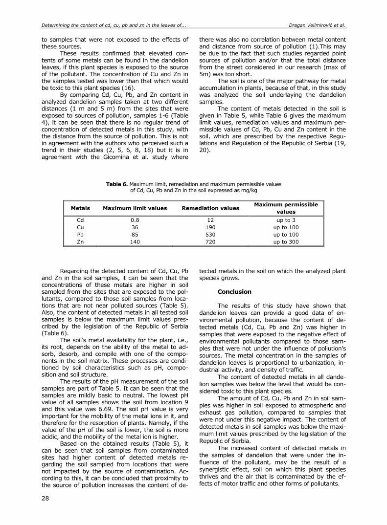
Determining the content of cd, cu, pb and zn in the leaves of... Dragan Velimirović et al.
28
to samples that were not exposed to the effects of these sources.
These results confirmed that elevated con-tents of some metals can be found in the dandelion leaves, if this plant species is exposed to the source of the pollutant. The concentration of Cu and Zn in the samples tested was lower than that which would be toxic to this plant species (16).
By comparing Cd, Cu, Pb, and Zn content in analyzed dandelion samples taken at two different distances (1 m and 5 m) from the sites that were exposed to sources of pollution, samples 1-6 (Table 4), it can be seen that there is no regular trend of concentration of detected metals in this study, with the distance from the source of pollution. This is not in agreement with the authors who perceived such a trend in their studies (2, 5, 6, 8, 18) but it is in agreement with the Gicomina et al. study where
there was also no correlation between metal content and distance from source of pollution (1).This may be due to the fact that such studies regarded point sources of pollution and/or that the total distance from the street considered in our research (max of 5m) was too short.
The soil is one of the major pathway for metal accumulation in plants, because of that, in this study was analyzed the soil underlaying the dandelion samples.
The content of metals detected in the soil is given in Table 5, while Table 6 gives the maximum limit values, remediation values and maximum per-missible values of Cd, Pb, Cu and Zn content in the soil, which are prescribed by the respective Regu-lations and Regulation of the Republic of Serbia (19, 20).
Table 6. Maximum limit, remediation and maximum permissible values of Cd, Cu, Pb and Zn in the soil expressed as mg/kg
Metals Maximum limit values Remediation values Maximum permissible
values
Cd 0.8 12 up to 3
Cu 36 190 up to 100
Pb 85 530 up to 100
Zn 140 720 up to 300
Regarding the detected content of Cd, Cu, Pb and Zn in the soil samples, it can be seen that the concentrations of these metals are higher in soil sampled from the sites that are exposed to the pol-lutants, compared to those soil samples from loca-tions that are not near polluted sources (Table 5). Also, the content of detected metals in all tested soil samples is below the maximum limit values pres-cribed by the legislation of the Republic of Serbia (Table 6).
The soil’s metal availability for the plant, i.e., its root, depends on the ability of the metal to ad-sorb, desorb, and compile with one of the compo-nents in the soil matrix. These processes are condi-tioned by soil characteristics such as pH, compo-sition and soil structure.
The results of the pH measurement of the soil samples are part of Table 5. It can be seen that the samples are mildly basic to neutral. The lowest pH value of all samples shows the soil from location 9 and this value was 6.69. The soil pH value is very important for the mobility of the metal ions in it, and therefore for the resorption of plants. Namely, if the value of the pH of the soil is lower, the soil is more acidic, and the mobility of the metal ion is higher.
Based on the obtained results (Table 5), it can be seen that soil samples from contaminated sites had higher content of detected metals re-garding the soil sampled from locations that were not impacted by the source of contamination. Ac-cording to this, it can be concluded that proximity to the source of pollution increases the content of de-
tected metals in the soil on which the analyzed plant species grows.
Conclusion The results of this study have shown that
dandelion leaves can provide a good data of en-vironmental pollution, because the content of de-
tected metals (Cd, Cu, Pb and Zn) was higher in samples that were exposed to the negative effect of environmental pollutants compared to those sam-ples that were not under the influence of pollution’s sources. The metal concentration in the samples of dandelion leaves is proportional to urbanization, in-dustrial activity, and density of traffic.
The content of detected metals in all dande-
lion samples was below the level that would be con-sidered toxic to this plant species.
The amount of Cd, Cu, Pb and Zn in soil sam-ples was higher in soil exposed to atmospheric and exhaust gas pollution, compared to samples that were not under this negative impact. The content of
detected metals in soil samples was below the maxi-mum limit values prescribed by the legislation of the Republic of Serbia.
The increased content of detected metals in the samples of dandelion that were under the in-fluence of the pollutant, may be the result of a
synergistic effect, soil on which this plant species thrives and the air that is contaminated by the ef-fects of motor traffic and other forms of pollutants.
Acta Medica Medianae 2020, Vol.59(1) Determining the content of cd, cu, pb and zn in the leaves of...
29
As dandelion is used in human nutrition, and
since heavy metals with cumulative and toxic effects
have been detected in it, Pb and Cd, it is necessary, in order to protect human health, to check the con-tent of these metals in the dandelion used in human nutrition. Also, due to the ability of this plant species to adsorb Pb and Cd, if it is additionally exposed to
them, it is necessary to take into account from which site this plant species is taken for nutrition.
The ICP-OES method proved to be a good
method for determining the content of cadmium,
copper, lead and zinc in this type of samples.
Acknowledgment
Results are part of the projects NO TR33034
financially supported by the Ministry of Education, Science and Technological Development of the
Republic of Serbia.
References
1. Giacomino A, Malandrino M, Colombo ML, Miaglia S, Maimone P, Blancato S, et al. Metal content in dan-delion (Taraxacum officinale) leaves: influence of
vehicular traffic and safety upon consumption as food. J Chem 2016; 1-9. [CrossRef]
2. Keane B, Collier MH, Shann JR, Rogstad SH. Metal content of dandelion (Taraxacum officinale) leaves in relation to soil contamination and airborne particulate matter. Sci of Tot Environ 2001;281(1-3):63-78. [CrossRef] [PubMed]
3. Kabata-Pendias A, Pendias H. Trace elements in soils and plants CRC Press Inc. Florida (FL): Boca Raton; 2001. [CrossRef]
4. Kuleff I, Djingova R. The dandelion (Taraxacum officinale)—amonitor for environmental pollution? Water Air Soil Pollut 1984; 21(1):77-85. [CrossRef]
5. Kabata-Pendias A., Dudka S. Trace metal contents of Taraxacum officinale (dandelion) as a convenient environmental indicator. Environ Geochem Health 1991;13(2):108-13. [CrossRef] [PubMed]
6. Kovac P. Heavy metal accumulation by ruderal and cultivated plants in a heavily polluted district of Budapest. Plants as Biomonitors 1993; 495-505.
7. Winter S. Biomonitoring in Stadten mit Taraxacum officinale Web.(Loewenzahn)-Eine Studie in der Stadt Zittau, Sachsen. Okotoxicologie/Okosystemare Ansatze und Methoden 1999; 238-47.
8. Marr K, Fyles H, Hendershot W. Trace metals in montreal urban soils and the leaves of Taraxacum officinale. Can J Soil Sci 1999;79(2):385-7. [CrossRef]
9. Djingova R, Kuleff I. Seasonal variations in the metal concentration of Taraxacum officinale, Plantago major and Plantago lanceolata. Chem Ecol 1999; 16(2):239-53. [CrossRef]
10. Krolak E. Accumulation of Zn, Cu, Pb and Cd by Dandelion (Taraxacum officinale Web.) in environ-ments with various degrees of metallic contamination. Pol J Environ Stud 2003; 12(6):713-21.
11. Djingova R, Kuleff I, Andreev N. Comparison of the ability of several vascular plants to reflect environ-mental pollution. Chemosphere 1993;27(8):1385-96.
[CrossRef] 12. Mitra S, editor. Sample preparation techniques in
analytical chemistry. John Wiley & Sons; 2004. [CrossRef]
13. Jamali MK, Kazi TG, Arain MB, Afridi HI, Jalbani N, Memon AR. Heavy metal contents of vegetables grown in soil, irrigated with mixtures of wastewater and sewage sludge in Pakistan, using ultra-sonic‐assisted pseudo‐digestion. J Agron Crop Sci
2007;193(3):218-28. [CrossRef] 14. EPA method 9045d: Soil and Waste pH. 15. BossCB, Kenneth JF. Concepts, instrumentation and
techniques in inductively coupled plasma optical emis-sion spectrometry. Norwalk: Perkin Elmer, 2004.
16. Gjorgieva D, Kadifkova-Panovska T, Bačeva K, Stafilov T. Assessment of heavy metal pollution in Republic of Macedonia using a plant assay. Arch Environ Contam Toxicol 2011;60(2):233-40. [CrossRef] [PubMed]
17. Kloke A, Sauerbeck DR, Vetter H. The contamination of plants and soils with heavy metals and the transport of metals in terrestrial food chains. In-Changing metal cycles and human health 1984;113-41. Berlin (GE): Springer. [CrossRef]
18. Czarnowska K, Milewska A. The content of heavy metals in an indicator plant (Taraxacum officinale) in Warsaw. Pol J Environ Stud 2000;9(2):125-8.
19. Službeni glasnik R. Srbije br. 30/2018. Uredba o gra-ničnim vrednostima zagađujućih, štetnih i opasnih materija u zemljištu.
20. Službeni glasnik R. Srbije br. 23/1994. Pravilnik o dozvoljenim količinama opasnih i štetnih materija u zemljištu i vodi za navodnјavanјe i metodama nјiho-vog ispitivanјa.
Determining the content of cd, cu, pb and zn in the leaves of... Dragan Velimirović et al.
30
Originalni rad UDC: 543.42:546.3/.9]:582.99
doi:10.5633/amm.2020.0103
ODREĐIVANJE SADRŽAJA KADMIJUMA, BAKRA, OLOVA I CINKA U LISTOVIMA MASLAČKA (TARAXACUM OFFICINALE WEBB.)
I ZEMLJIŠTU ICP-OES METODOM
Dragan Velimirović1, Biljana Kaličanin1, Milan Stojković2, Snežana Tošić2
1Univerzitet u Nišu, Medicinski fakultet, Odsek za farmaciju, Niš, Srbija 2Univerzitet u Nišu, Prirodno-matematički fakultet, Odsek za hemiju, Niš, Srbija
Kontakt: Dragan Velimirović
Bulevar dr Zoran Đinđić 81, 18000 Niš, Srbija
E-mail: [email protected]
Maslačak (Taraxacum officinale Webb.) je biljna vrsta koja može da akumulira
određenu količinu metala. Cilj ove studije bio je da se odredi sadržaj Cd, Cu, Pb i Zn u listovima maslačka i zemljištu, koji su uzorkovani sa različitih lokaliteta. Jedna grupa uzoraka bila je izložena uticaju izvora zagađenja ovih metala, dok druga grupa nije bila pod uticajem zagađivača. Količina metala u ispitivanim uzorcima određena je primenom tehnike indukovano kuplovane plazme optički emisione spektrometrije (ICP-OES). Sadržaj detektovanih metala bio je veći u uzorcima lista maslačka i zemljišta koji su bili izloženi negativnom uticaju zagađivača životne sredine, u odnosu na one uzorke koji nisu bili pod uticajem ovih izvora
zagađenja. Povećani sadržaj detektovanih metala u uzorcima listova maslačka, koji su bili pod uticajem štetnog dejstva zagađivača, može biti rezultat sinergističkog efekta zemljišta, na kome ova biljna vrsta uspeva i vazduha, koji je zagađen motornim saobraćajem i drugim vidovima zagađenja. Rezultati ove studije pokazali su da maslačak, na osnovu količine detektovanih metala u biljnom tkivu, može pružiti podatke o zagađenju životne sredine. Kako se maslačak koristi u ljudskoj ishrani, a kako su u listu ove biljne vrste detektovani teški metali (Pb i Cd), koji imaju kumulativno i toksično dejstvo, neophodno je, u cilju zaštite ljudskog zdravlja, proveravati prisustvo i sadržaj ovih metala u maslačku koji se koristi u ljudskoj ishrani.
Acta Medica Medianae 2020;59(1):23-30.
Ključne reči: maslačak, metali, zemljište, zagađivači, ICP-OES
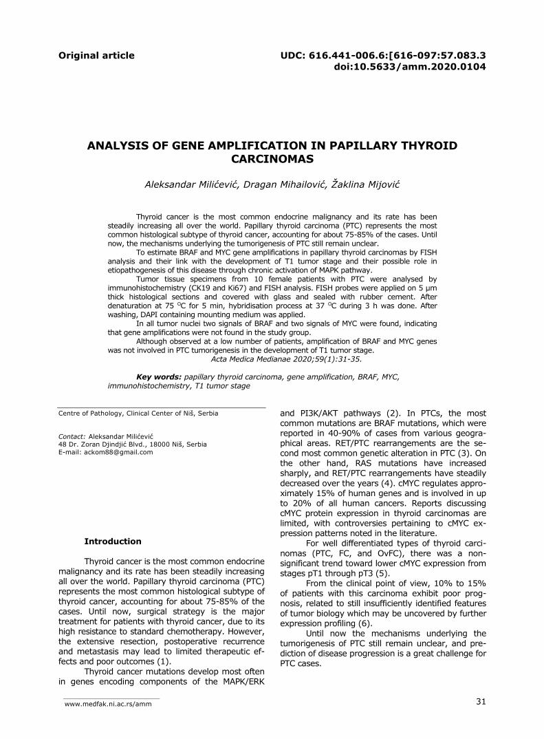
31 www.medfak.ni.ac.rs/amm
Original article UDC: 616.441-006.6:[616-097:57.083.3
doi:10.5633/amm.2020.0104
ANALYSIS OF GENE AMPLIFICATION IN PAPILLARY THYROID CARCINOMAS
Aleksandar Milićević, Dragan Mihailović, Žaklina Mijović
Thyroid cancer is the most common endocrine malignancy and its rate has been
steadily increasing all over the world. Papillary thyroid carcinoma (PTC) represents the most common histological subtype of thyroid cancer, accounting for about 75-85% of the cases. Until now, the mechanisms underlying the tumorigenesis of PTC still remain unclear.
To estimate BRAF and MYC gene amplifications in papillary thyroid carcinomas by FISH analysis and their link with the development of T1 tumor stage and their possible role in etiopathogenesis of this disease through chronic activation of MAPK pathway.
Tumor tissue specimens from 10 female patients with PTC were analysed by immunohistochemistry (CK19 and Ki67) and FISH analysis. FISH probes were applied on 5 μm thick histological sections and covered with glass and sealed with rubber cement. After denaturation at 75 OC for 5 min, hybridisation process at 37 OC during 3 h was done. After washing, DAPI containing mounting medium was applied.
In all tumor nuclei two signals of BRAF and two signals of MYC were found, indicating that gene amplifications were not found in the study group.
Although observed at a low number of patients, amplification of BRAF and MYC genes was not involved in PTC tumorigenesis in the development of T1 tumor stage.
Acta Medica Medianae 2020;59(1):31-35. Key words: papillary thyroid carcinoma, gene amplification, BRAF, MYC,
immunohistochemistry, T1 tumor stage
Centre of Pathology, Clinical Center of Niš, Serbia
Contact: Aleksandar Milićević
48 Dr. Zoran Djindjić Blvd., 18000 Niš, Serbia
E-mail: [email protected]
Introduction Thyroid cancer is the most common endocrine
malignancy and its rate has been steadily increasing all over the world. Papillary thyroid carcinoma (PTC) represents the most common histological subtype of
thyroid cancer, accounting for about 75-85% of the cases. Until now, surgical strategy is the major treatment for patients with thyroid cancer, due to its high resistance to standard chemotherapy. However, the extensive resection, postoperative recurrence and metastasis may lead to limited therapeutic ef-fects and poor outcomes (1).
Thyroid cancer mutations develop most often in genes encoding components of the MAPK/ERK
and PI3K/AKT pathways (2). In PTCs, the most common mutations are BRAF mutations, which were reported in 40-90% of cases from various geogra-phical areas. RET/PTC rearrangements are the se-cond most common genetic alteration in PTC (3). On the other hand, RAS mutations have increased sharply, and RET/PTC rearrangements have steadily
decreased over the years (4). cMYC regulates appro-ximately 15% of human genes and is involved in up to 20% of all human cancers. Reports discussing cMYC protein expression in thyroid carcinomas are limited, with controversies pertaining to cMYC ex-pression patterns noted in the literature.
For well differentiated types of thyroid carci-
nomas (PTC, FC, and OvFC), there was a non-significant trend toward lower cMYC expression from stages pT1 through pT3 (5).
From the clinical point of view, 10% to 15% of patients with this carcinoma exhibit poor prog-nosis, related to still insufficiently identified features
of tumor biology which may be uncovered by further expression profiling (6).
Until now the mechanisms underlying the tumorigenesis of PTC still remain unclear, and pre-diction of disease progression is a great challenge for PTC cases.
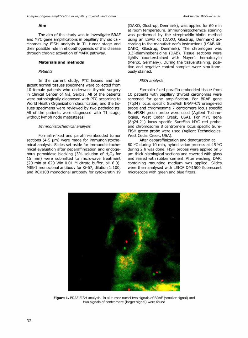
Analysis of gene amplification in papillary thyroid carcinomas Aleksandar Milićević et al.
32
Aim
The aim of this study was to investigate BRAF and MYC gene amplifications in papillary thyroid car-cinomas by FISH analysis in T1 tumor stage and their possible role in etiopathogenesis of this disease through chronic activation of MAPK pathway.
Materials and methods Patients In the current study, PTC tissues and ad-
jacent normal tissues specimens were collected from
10 female patients who underwent thyroid surgery in Clinical Center of Niš, Serbia. All of the patients were pathologically diagnosed with PTC according to
World Health Organization classification, and the tis-sues specimens were reviewed by two pathologists. All of the patients were diagnosed with T1 stage, without lymph node metastases.
Immnohistochemical analysis Formalin-fixed and paraffin-embedded tumor
sections (4-5 µm) were made for immunohistoche-mical analysis. Slides set aside for immunohistoche-
mical evaluation after deparaffinization and endoge-nous peroxidase blocking (3% solution of H2O2 for 15 min) were submitted to microwave treatment (20 min at 620 Win 0.01 M citrate buffer, pH 6.0). MIB-1 monoclonal antibody for Ki-67, dilution 1:100, and RCK108 monoclonal antibody for cytokeratin 19
(DAKO, Glostrup, Denmark), was applied for 60 min
at room temperature. Immunohistochemical staining
was performed by the streptavidin-biotin method using an LSAB kit (DAKO, Glostrup, Denmark) ac-cording to the manufacturer’s instructions (LSAB Kit, DAKO, Glostrup, Denmark). The chromogen was 3.3’-diaminobenzidine (DAB). Tissue sections were
lightly counterstained with Mayer’s hematoxylin (Merck, Germany). During the tissue staining, posi-tive and negative control samples were simultane-ously stained.
FISH analysis
Formalin fixed paraffin embedded tissue from 10 patients with papillary thyroid carcinomas were screened for gene amplification. For BRAF gene
(7q34) locus specific SureFish BRAF-CN orange-red probe and chromosome 7 centromere locus specific SureFISH green probe were used (Agilent Techno-logies, West Cedar Creek, USA). For MYC gene
(8q24.21) locus specific SureFish MYC red probe, and chromosome 8 centromere locus specific Sure-FISH green probe were used (Agilent Technologies, West Cedar Creek, USA).
After deparaffinization and denaturation at 80 OC during 10 min, hybridisation process at 45 OC
during 2 h was done. FISH probes were applied on 5 μm thick histological sections and covered with glass and sealed with rubber cement. After washing, DAPI containing mounting medium was applied. Slides were then analysed with LEICA DM1500 fluorescent microscope with green and blue filters.
Figure 1. BRAF FISH analysis. In all tumor nuclei two signals of BRAF (smaller signal) and two signals of centromere (larger signal) were found
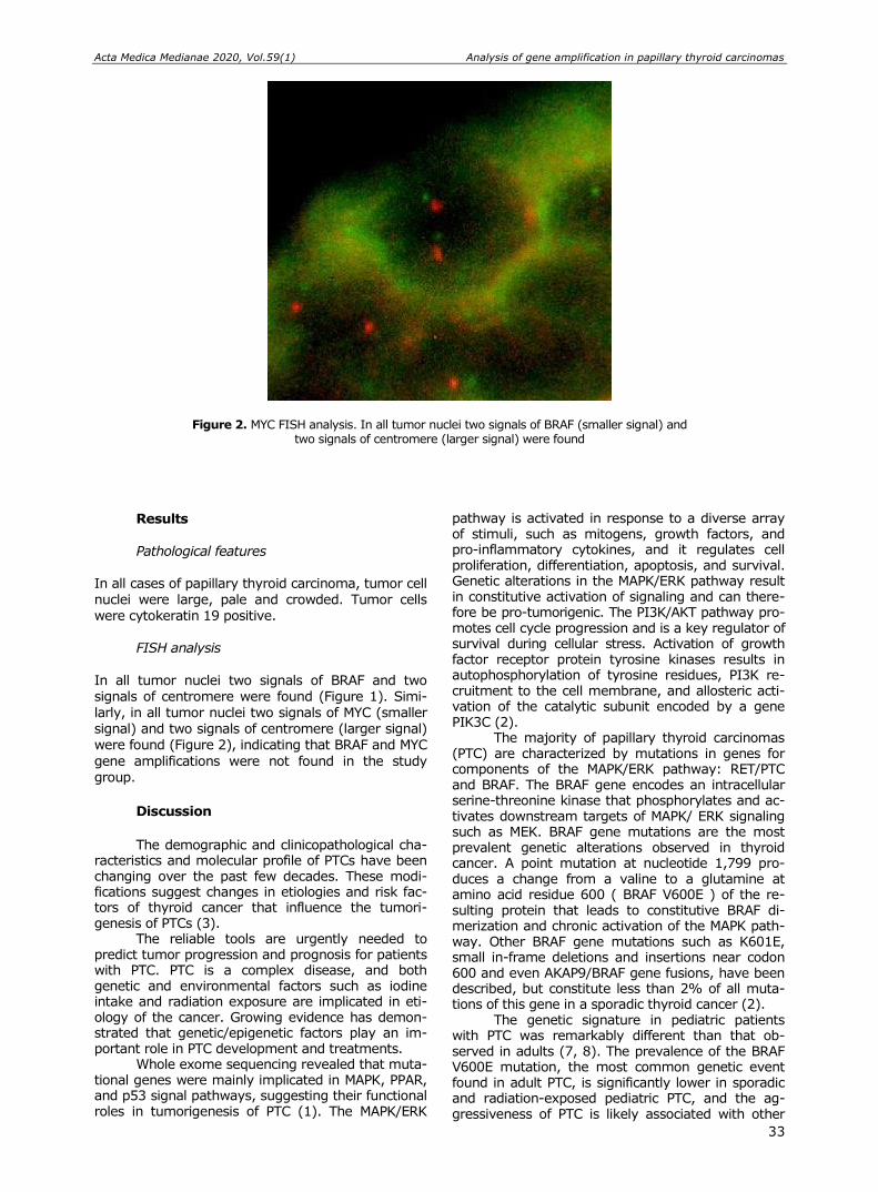
Acta Medica Medianae 2020, Vol.59(1) Analysis of gene amplification in papillary thyroid carcinomas
33
Figure 2. MYC FISH analysis. In all tumor nuclei two signals of BRAF (smaller signal) and two signals of centromere (larger signal) were found
Results
Pathological features
In all cases of papillary thyroid carcinoma, tumor cell nuclei were large, pale and crowded. Tumor cells were cytokeratin 19 positive.
FISH analysis
In all tumor nuclei two signals of BRAF and two signals of centromere were found (Figure 1). Simi-larly, in all tumor nuclei two signals of MYC (smaller signal) and two signals of centromere (larger signal) were found (Figure 2), indicating that BRAF and MYC
gene amplifications were not found in the study group.
Discussion
The demographic and clinicopathological cha-
racteristics and molecular profile of PTCs have been changing over the past few decades. These modi-fications suggest changes in etiologies and risk fac-tors of thyroid cancer that influence the tumori-genesis of PTCs (3).
The reliable tools are urgently needed to predict tumor progression and prognosis for patients with PTC. PTC is a complex disease, and both genetic and environmental factors such as iodine intake and radiation exposure are implicated in eti-ology of the cancer. Growing evidence has demon-strated that genetic/epigenetic factors play an im-portant role in PTC development and treatments.
Whole exome sequencing revealed that muta-tional genes were mainly implicated in MAPK, PPAR, and p53 signal pathways, suggesting their functional roles in tumorigenesis of PTC (1). The MAPK/ERK
pathway is activated in response to a diverse array of stimuli, such as mitogens, growth factors, and pro-inflammatory cytokines, and it regulates cell proliferation, differentiation, apoptosis, and survival. Genetic alterations in the MAPK/ERK pathway result in constitutive activation of signaling and can there-fore be pro-tumorigenic. The PI3K/AKT pathway pro-motes cell cycle progression and is a key regulator of survival during cellular stress. Activation of growth factor receptor protein tyrosine kinases results in autophosphorylation of tyrosine residues, PI3K re-cruitment to the cell membrane, and allosteric acti-vation of the catalytic subunit encoded by a gene PIK3C (2).
The majority of papillary thyroid carcinomas (PTC) are characterized by mutations in genes for components of the MAPK/ERK pathway: RET/PTC and BRAF. The BRAF gene encodes an intracellular serine-threonine kinase that phosphorylates and ac-tivates downstream targets of MAPK/ ERK signaling such as MEK. BRAF gene mutations are the most prevalent genetic alterations observed in thyroid cancer. A point mutation at nucleotide 1,799 pro-duces a change from a valine to a glutamine at amino acid residue 600 ( BRAF V600E ) of the re-sulting protein that leads to constitutive BRAF di-merization and chronic activation of the MAPK path-way. Other BRAF gene mutations such as K601E, small in-frame deletions and insertions near codon 600 and even AKAP9/BRAF gene fusions, have been described, but constitute less than 2% of all muta-tions of this gene in a sporadic thyroid cancer (2).
The genetic signature in pediatric patients with PTC was remarkably different than that ob-served in adults (7, 8). The prevalence of the BRAF V600E mutation, the most common genetic event found in adult PTC, is significantly lower in sporadic and radiation-exposed pediatric PTC, and the ag-gressiveness of PTC is likely associated with other
Analysis of gene amplification in papillary thyroid carcinomas Aleksandar Milićević et al.
34
genetic events. RET/PTC rearrangement, the second most common event in adult PTC, is the major genetic alteration found in sporadic and radiation-exposed pediatric PTC (9). Moreover, RAS mutations are a rare genetic event in pediatric PTC (8). These findings suggest that there are differences in tumor biology according to age.
The MYC protooncogene encodes a DNA-bind-ing factor that can activate and repress transcrip-tion. Via this mechanism, MYC regulates expression of numerous target genes that control key cellular functions, including cell growth and cell cycle pro-gression. MYC also has a critical role in DNA repli-cation. Deregulated MYC expression resulting from various types of genetic alterations leads to consti-tutive MYC activity in a variety of cancers and pro-motes oncogenesis (10).
The results of our study indicate that BRAF and MYC genes are not amplified in PTC.
There are some limitations to our study. First, our study is limited by the inherent biases of a retrospective analysis and, therefore, the lack of a proper follow-up with all of the patients. Second, it is limited by a small sample size.
Conclusion Although observed at a low number of pa-
tients, according to our results, amplification of BRAF and MYC genes was not involved in PTC tumori-genesis in T1 tumor stage.
Acknowledgments
The study was supported by research grants
from the Faculty of Medicine, University of Niš,
Serbia.
References
1. Fang Y, Ma X, Zeng J, Jin Y, Hu Y, Wang J, et al. The profile of genetic mutations in papillary thyroid cancer detected by whole exome sequencing. Cell Physiol Biochem 2018;50(1):169-78. [CrossRef] [PubMed]
2. Pozdeyev N, Lund G, McDermott MT. Molecular patho-genesis of thyroid cancer and oncogenes in thyroid cancer. In: Wartofsky L, Van Nostrand D, editors. Thyroid Cancer. New York: Springer; 2016.p.17-30. [CrossRef]
3. Vuong HG, Altibi AM, Abdelhamid AH, Ngoc PU, Quan VD, Tantawi MY, et al. The changing characteristics and molecular profiles of papillary thyroid carcinoma over time: a systematic review. Oncotarget 2017; 8(6):10637-49. [CrossRef] [PubMed]
4. Hong AR, Lim JA, Kim TH, Choi HS, Yoo WS, Min HS, et al. The frequency and clinical implications of the BRAF (V600E) mutation in papillary thyroid cancer patients in Korea over the past two decades. Endo-crinol Metab 2014; 29:505-13. [CrossRef] [PubMed]
5. Sakr H, Chute D, Nasr C, Sturgis C. cMYC expression in thyroid follicular cell-derived carcinomas: a role in thyroid tumorigenesis. Diagn Pathol 2017;12:71. [CrossRef] [PubMed]
6. Jarząb B, Wiench M, Fujarewicz K, Krzysztof S, Jarząb M, Oczko-Wojciechowska M, et al. Gene Expression Profile of Papillary Thyroid Cancer: Sources of Varia-bility and Diagnostic Implications. Cancer Res 2005; 65(4):1587-97. [CrossRef] [PubMed]
7. Cordioli MI, Moraes L, Carvalheira G, Sisdelli L, Alves MT, Delcelo R, et al. AGK-BRAF gene fusion is a recurrent event in sporadic pediatric thyroid carcino-ma. Cancer Med 2016;5(7):1535-41. [CrossRef] [PubMed]
8. Cordioli MI, Moraes L, Bastos AU, Besson P, Alves MT, Delcelo R, et al. Fusion oncogenes are the main genetic events found in sporadic papillary thyroid carcinomas from children. Thyroid 2017;27(2):182-8. [CrossRef] [PubMed]
9. Nikiforov YE, Rowland JM, Bove KE, Monforte-Munoz H, Fagin JA. Distinct pattern of RET oncogene rearr-angements in morphological variants of radiation-induced and sporadic thyroid papillary carcinomas in children. Cancer Res 1997;57:1690-4. [PubMed]
10. Dominguez-Sola D, Ying CY, Grandori C, Ruggiero L, Chen B, Li M, et al. Non-transcriptional control of DNA replication by c-Myc. Nature 2007;448:445-51. [CrossRef] [PubMed]
Acta Medica Medianae 2020, Vol.59(1) Analysis of gene amplification in papillary thyroid carcinomas
35
Originalni rad UDC: 616.441-006.6:[616-097:57.083.3
doi:10.5633/amm.2020.0104
ANALIZA GENSKIH AMPLIFIKACIJA U PAPILARNOM KARCINOMU ŠTITNE ŽLEZDE
Aleksandar Milićević, Dragan Mihailović, Žaklina Mijović
Centar za patologiju, Klinički centar Niš, Niš,Srbija
Kontakt: Aleksandar Milićević
Bulevar dr Zorana Đinđića 48, 18000 Niš, Srbija
E-mail: [email protected]
Rak štitne žlezde najčešći je endokrini malignitet i njegova stopa je u stalnom porastu u
celom svetu. Papilarni karcinom štitne žlezde (PTC) najčešći je histološki podtip karcinoma
štitne žlezde, koji čini oko 75% — 85% slučajeva. Do sada su mehanizmi na kojima se temelji
tumorigeneza PTC još uvek nejasni. Procena BRAF i MYC amplifikacije gena u papilarnim karcinomima štitne žlezde izvršena
je FISH analizom. Uzorci tumorskih tkiva 10 ženskih bolesnika sa PTC analizirani su imunohistohemijski
(CK19 i Ki67) i FISH analizom. FISH probe nanesene su na histološke preseke debljine 5 μm, prekrivene staklom i zapečaćene gumenim cementom. Nakon denaturacije na 75OC u tra-janju od 5 minuta, sproveden je postupak hibridizacije na 37OC tokom 3 sata. Nakon ispi-ranja, primenjen je DAPI, koji sadrži medijum za montiranje.
U svim tumorskim jedrima pronađena su dva signala BRAF i dva signala MYC, što ukazuje na to da amplifikacije gena nisu nađene u ispitivanoj grupi.
Iako je primećena kod malog broja bolesnika, amplifikacija BRAF i MYC gena nije bila uključena u PTC tumorigenezu T1 stadijuma tumora.
Acta Medica Medianae 2020;59(1):31-35.
Ključne reči: papilarni karcinom štitne žlezde, amplifikacija gena, BRAF, MYC,
imunohistohemija, T1 stadijum tumora
36 www.medfak.ni.ac.rs/amm
Original article UDC: 616-056.7:616.132-007.64-089
doi:10.5633/amm.2020.0105
EARLY POSTOPERATIVE OUTCOMES OF SURGICALLY TREATED AORTIC DISSECTION IN MARFAN SYNDROME PATIENTS
Ivan Nešić1, Jelena Dotlić2,3, Igor Živković1, Aleksandra Šljivić4, Petar Vuković1,2,
Slobodan Mićović1,2, Petar Milačić1,2, Miroslav Miličić1,2, Djordje Zdravković1
Milan Ćirković1, Miodrag Perić1,2
Acute aortic dissection remains the leading cause of morbidity and mortality of Marfan
syndrome patients. The study aimed to investigate the early postoperative outcomes of sur-gically treated aortic dissection in patients with Marfan syndrome. Study included all patients operated due to aortic dissection at the Cardiac Surgery Department, Dedinje Cardiovascular Institute in Belgrade during a six year period (2012–2017). Patients were divided regarding the diagnosis of Marfan syndrome. Preoperatively general data and detailed medical history were taken from every patient. Upon admission, patients underwent a thorough clinical and cardio-logic examination. Patients were surgically treated according to current protocols. Postopera-tively, patients were followed up for one month. During this follow-up period, we registered all complications and fatality. All pre and postoperatively collected data were compared and statistically analyzed. Study included 246 patients out of which 7.7% had Marfan syndrome. Marfan syndrome patients were significantly younger than control group patients (p = 0.001). There were no significant differences between patient groups regarding sex and smoking status. All patients with Marfan syndrome had dissection of type I. Marfan syndrome patients had fewer preoperative chronic illnesses and complications (p = 0.001). There were no significant differences in mortality (p = 0.702) and frequency of postoperative complications (p = 0.231) between patients with and without Marfan syndrome. In conclusion, it can be seen that prompt and adequate diagnosis and surgical treatment of aortic dissection can enable mostly good early postoperative outcomes in patients with Marfan syndrome.
Acta Medica Medianae 2020;59(1):36-43. Key words: Marfan syndrome, aortic dissection, surgical treatment, outcome
1Cardiac Surgery Department, Dedinje Cardiovascular Institute,
Belgrade, Serbia 2University of Belgrade, Medical Faculty, Belgrade, Serbia 3Clinic of Obstetrics and Gynecology, Clinical Center of Serbia,
Belgrade, Serbia 4Department of Cardiology, University Clinical Hospital Center
"Dr Dragiša Mišović-Dedinje", Belgrade, Serbia
Contact: Ivan Nešić
Heroja Milana Tepića 1, 11000 Belgrade, Serbia E-mail: [email protected]
Introduction
Marfan syndrome is an autosomal dominant
inherited disorder of the connective tissue in which cardiovascular, skeletal, ocular and other anomalies may be present to a variable degree (1). Its pre-valence is around 1 in 5000 individuals. The com-bination of structural microfibril matrix abnormali-
ties, deregulation of matrix homeostasis and inade-quate cell-matrix interactions present the basis for clinical characteristics of the Marfan syndrome (2,
3). The most common cardiovascular manifesta-
tions in patients with Marfan syndrome are progres-sive aortic dilatation and mitral valve prolapse. These anatomical abnormalities may cause mitral insufficiency as well as aortic insufficiency, aneu-
rysm, dissection and even rupture (4). Surgical re-
placement of the dilated aortic root and ascending aorta has significantly increased the life expectancy of patients with Marfan syndrome. Nevertheless, acute aortic dissection remains the most important complication and the leading cause of morbidity and sudden death in these patients (5, 6).
Therefore, the study aim was to investigate the early postoperative outcomes of surgically treated aortic dissection in patients with Marfan syndrome.
Materials and methods Study included all consecutive patients who
were operated due to aortic dissection at the Cardiac
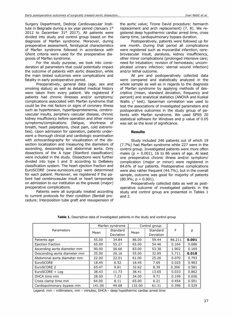
Early postoperative outcomes of surgically treated aortic dissection... Ivan Nešić et al.
37
Surgery Department, Dedinje Cardiovascular Insti-
tute in Belgrade during a six year period (January 1st
2012 to December 31st 2017). All patients were divided into study and control group based on the diagnosis of Marfan syndrome. Moreover, during preoperative assessment, fenotypical characteristics of Marfan syndrome followed in accordance with
Ghent criteria were used for the preoperative dia-gnosis of Marfan syndrome.
For the study purpose, we took into consi-deration all parameters that could potentially impact the outcome of patients with aortic dissection, while the main tested outcomes were complications and fatality in early postoperative period.
Preoperatively, general data (age, sex and smoking status) as well as detailed medical history were taken from every patient. We registered if
patients had chronic illnesses and/or symptoms/ complications associated with Marfan syndrome that could be the risk factors or signs of coronary illness such as hypertension, hyperlipoproteinemia, cerebro-
vascular insults, periphery vascular disease, chronic kidney insufficiency before operation and other minor symptoms/complications (fatigue, shortness of breath, heart palpitations, chest pain, cold extremi-ties). Upon admission for operation, patients under-went a thorough clinical and cardiologic examination
with echocardiography for visualization of the dis-section localization and measuring the diameters of ascending, descending and abdominal aorta. Only dissections of the A type (Stanford classification) were included in the study. Dissections were further divided into type I and II according to DeBakey
classification system. The heart ejection fraction and
EuroSCORE (www.euroscore.org) were determined for each patient. Moreover, we registered if the pa-tient had cerebrovasular insult or heart tamponade on admission to our institution as the gravest (major) preoperative complications.
Patients were all surgically treated according to current protocols for their condition (Bentall pro-
cedure; Interposition tube graft and resuspension of
the aortic valve; Tirone David procedure; hemiarch
replacement and arch replacement) (7, 8). We re-
gistered deep hypothermic cardiac arrest time, cross clamp time, cardiopulmonary bypass duration.
Postoperatively, patients were followed up for one month. During that period all complications were registered such as myocardial infarction, cere-
brovascular insult, paralysis, kidney insufficiency, other minor complications (prolonged intensive care; need for intubation; revision of hemostasis; uncom-plicated urinary infection; sternal wound infection) and/or lethal outcome.
All pre and postoperatively collected data were compared and statistically analyzed in the
whole sample as well as in regards to the diagnosis of Marfan syndrome by applying methods of des-criptive (mean, standard deviation, frequency and
percent) and analytical statistics (ANOVA and Kuskal
Wallis χ2 test). Spearman correlation was used to
test the associations of investigated parameters and
postoperative outcomes in the study group of pa-tients with Marfan syndrome. We used SPSS 20 statistical software for Windows and p value of 0.05 was set as the level of significance.
Results
Study included 246 patients out of which 19 (7.7%) had Marfan syndrome while 227 were in the control group. Investigated patients were more often males (p = 0.001), 16 to 86 years of age. At least one preoperative chronic illness and/or symptom/ complication (major or minor) were registered in
84.6% of our patients. Postoperative complications
were also rather frequent (44.7%), but in the overall sample, outcome was good for majority of patients (80.9%; p = 0.001).
Preoperatively collected data as well as post-operative outcome of investigated patients in the study and control group are presented in Tables 1
and 2.
Table 1. Descriptive data of investigated patients in the study and control group
Parameters
Marfan syndrome Control group
F p Mean
Standard
Deviation Mean
Standard
Deviation
Patients age 55.00 34.84 86.00 59.44 86.211 0.001
Ejection fraction 65.00 55.27 65.00 54.46 0.164 0.686
Ascending aorta diameter mm 90.00 56.66 83.00 53.38 1.902 0.169
Descending aorta diameter mm 35.00 26.16 55.00 32.85 5.711 0.018
Abdominal aorta diameter mm 22.00 22.01 61.00 25.26 0.070 0.793
EuroSCORE 18.45 6.52 18.45 7.69 0.025 0.983
EuroSCORE 2 65.47 9.81 32.62 8.39 0.306 0.581
EuroSCORE + Log 38.43 11.73 38.41 13.65 0.033 0.862
DHCA time min 28.00 7.23 34.00 9.71 0.199 0.656
Cross clamp time min 64.00 8.11 65.00 6.21 0.454 0.501
Cardiopulmonary bypass min 141.00 49.68 132.00 61.31 0.398 0.529
Legend: mm – millimeters; min – minutes; DHCA – deep hypothermic cardiac arrest time
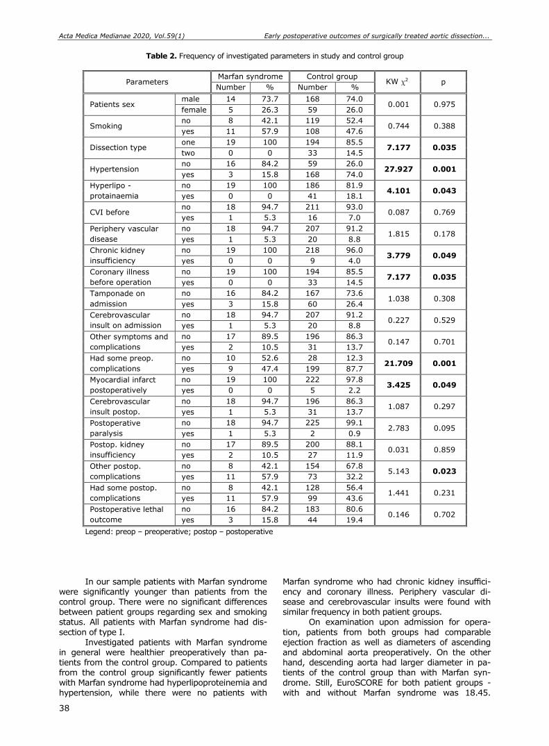
Acta Medica Medianae 2020, Vol.59(1) Early postoperative outcomes of surgically treated aortic dissection...
38
Table 2. Frequency of investigated parameters in study and control group
Parameters Marfan syndrome Control group
KW χ2 p Number % Number %
Patients sex male 14 73.7 168 74.0
0.001 0.975 female 5 26.3 59 26.0
Smoking no 8 42.1 119 52.4
0.744 0.388 yes 11 57.9 108 47.6
Dissection type one 19 100 194 85.5
7.177 0.035 two 0 0 33 14.5
Hypertension no 16 84.2 59 26.0
27.927 0.001 yes 3 15.8 168 74.0
Hyperlipo -
protainaemia
no 19 100 186 81.9 4.101 0.043
yes 0 0 41 18.1
CVI before no 18 94.7 211 93.0
0.087 0.769 yes 1 5.3 16 7.0
Periphery vascular
disease
no 18 94.7 207 91.2 1.815 0.178
yes 1 5.3 20 8.8
Chronic kidney
insufficiency
no 19 100 218 96.0 3.779 0.049
yes 0 0 9 4.0
Coronary illness
before operation
no 19 100 194 85.5 7.177 0.035
yes 0 0 33 14.5
Tamponade on
admission
no 16 84.2 167 73.6 1.038 0.308
yes 3 15.8 60 26.4
Cerebrovascular
insult on admission
no 18 94.7 207 91.2 0.227 0.529
yes 1 5.3 20 8.8
Other symptoms and
complications
no 17 89.5 196 86.3 0.147 0.701
yes 2 10.5 31 13.7
Had some preop.
complications
no 10 52.6 28 12.3 21.709 0.001
yes 9 47.4 199 87.7
Myocardial infarct
postoperatively
no 19 100 222 97.8 3.425 0.049
yes 0 0 5 2.2
Cerebrovascular
insult postop.
no 18 94.7 196 86.3 1.087 0.297
yes 1 5.3 31 13.7
Postoperative
paralysis
no 18 94.7 225 99.1 2.783 0.095
yes 1 5.3 2 0.9
Postop. kidney
insufficiency
no 17 89.5 200 88.1 0.031 0.859
yes 2 10.5 27 11.9
Other postop.
complications
no 8 42.1 154 67.8 5.143 0.023
yes 11 57.9 73 32.2
Had some postop.
complications
no 8 42.1 128 56.4 1.441 0.231
yes 11 57.9 99 43.6
Postoperative lethal
outcome
no 16 84.2 183 80.6 0.146 0.702
yes 3 15.8 44 19.4
Legend: preop – preoperative; postop – postoperative
In our sample patients with Marfan syndrome were significantly younger than patients from the control group. There were no significant differences between patient groups regarding sex and smoking status. All patients with Marfan syndrome had dis-
section of type I. Investigated patients with Marfan syndrome
in general were healthier preoperatively than pa-tients from the control group. Compared to patients from the control group significantly fewer patients with Marfan syndrome had hyperlipoproteinemia and hypertension, while there were no patients with
Marfan syndrome who had chronic kidney insuffici-ency and coronary illness. Periphery vascular di-sease and cerebrovascular insults were found with similar frequency in both patient groups.
On examination upon admission for opera-
tion, patients from both groups had comparable ejection fraction as well as diameters of ascending and abdominal aorta preoperatively. On the other hand, descending aorta had larger diameter in pa-tients of the control group than with Marfan syn-drome. Still, EuroSCORE for both patient groups - with and without Marfan syndrome was 18.45.
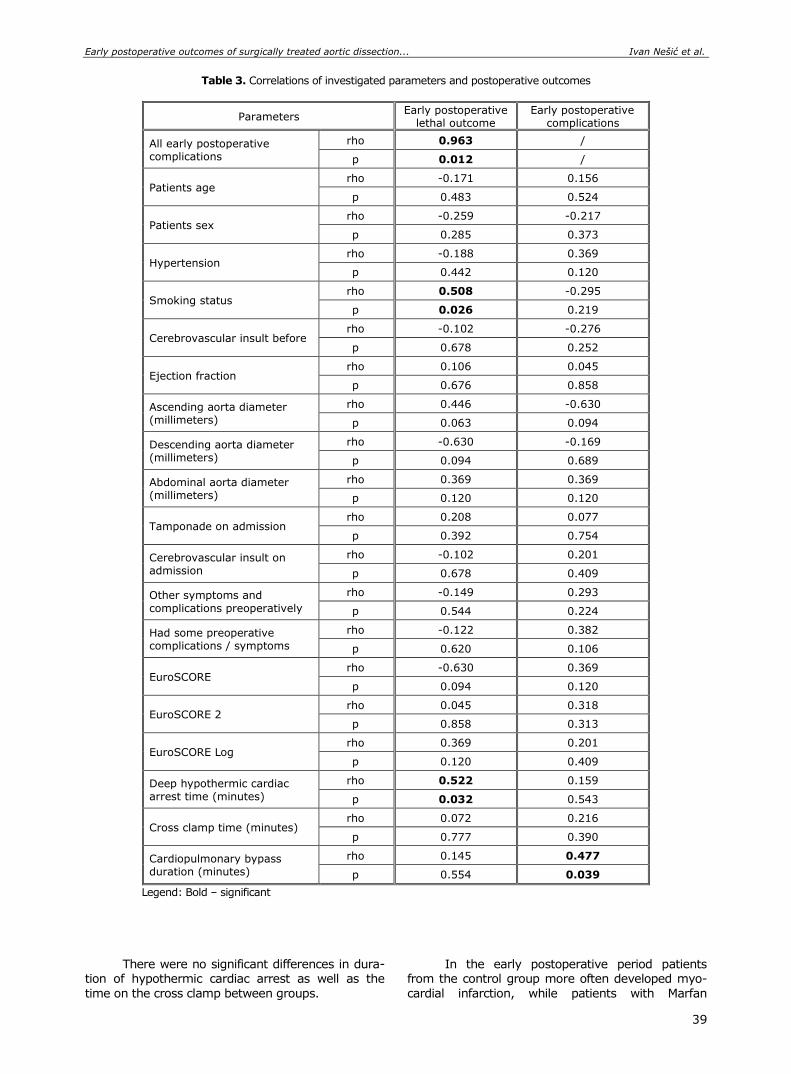
Early postoperative outcomes of surgically treated aortic dissection... Ivan Nešić et al.
39
Table 3. Correlations of investigated parameters and postoperative outcomes
Parameters Early postoperative
lethal outcome Early postoperative
complications
All early postoperative complications
rho 0.963 /
p 0.012 /
Patients age rho -0.171 0.156
p 0.483 0.524
Patients sex rho -0.259 -0.217
p 0.285 0.373
Hypertension rho -0.188 0.369
p 0.442 0.120
Smoking status rho 0.508 -0.295
p 0.026 0.219
Cerebrovascular insult before rho -0.102 -0.276
p 0.678 0.252
Ejection fraction rho 0.106 0.045
p 0.676 0.858
Ascending aorta diameter (millimeters)
rho 0.446 -0.630
p 0.063 0.094
Descending aorta diameter (millimeters)
rho -0.630 -0.169
p 0.094 0.689
Abdominal aorta diameter (millimeters)
rho 0.369 0.369
p 0.120 0.120
Tamponade on admission rho 0.208 0.077
p 0.392 0.754
Cerebrovascular insult on admission
rho -0.102 0.201
p 0.678 0.409
Other symptoms and
complications preoperatively
rho -0.149 0.293
p 0.544 0.224
Had some preoperative complications / symptoms
rho -0.122 0.382
p 0.620 0.106
EuroSCORE rho -0.630 0.369
p 0.094 0.120
EuroSCORE 2 rho 0.045 0.318
p 0.858 0.313
EuroSCORE Log rho 0.369 0.201
p 0.120 0.409
Deep hypothermic cardiac arrest time (minutes)
rho 0.522 0.159
p 0.032 0.543
Cross clamp time (minutes) rho 0.072 0.216
p 0.777 0.390
Cardiopulmonary bypass duration (minutes)
rho 0.145 0.477
p 0.554 0.039
Legend: Bold – significant
There were no significant differences in dura-tion of hypothermic cardiac arrest as well as the
time on the cross clamp between groups.
In the early postoperative period patients from the control group more often developed myo-
cardial infarction, while patients with Marfan
Acta Medica Medianae 2020, Vol.59(1) Early postoperative outcomes of surgically treated aortic dissection...
40
syndrome postoperatively more often had other
minor complications. Still, when all postoperative
complications were assessed together there were no significant differences in their frequency between groups.
Lethal outcome after the operation for aortic dissection was generally not very frequent in our
sample and occurred in similar percent of patients with and without Marfan syndrome.
Finally, in the study group of patients with Marfan syndrome postoperative lethal outcome sig-nificantly correlated with having early postoperative complications, being a smoker and longer duration of hypothermic cardiac arrest, while early postope-
rative complications were associated with longer duration of cardiopulmonary bypass (Table 3).
Discussion
Marfan syndrome is caused by more than a
thousand currently described mutations in the fi-brillin-1 (FBN1) gene (on chromosome 15) encoding a glycoprotein which is a principal component of the extracellular matrix microfibril (2, 3). Therefore, major clinical features of this syndrome result from weaker connective tissues caused by defects in fi-brillin-1, but the extent of signs and symptoms are highly variable because of varying genotype ex-pression (1). Moreover, some studies have shown that in Marfan syndrome, function of transforming growth factor beta (TGF-β), stimulator of inflam-mation, fibrosis and activator of matrix metallopro-teinase 2 and 9, is abnormal (9). Increased TGF-β activation in tissues can cause failure of lung sep-tation, development of a myxomatous mitral valve and aortic root dilation. In addition, as the des-truction of the elastic and collagen lamellae progres-ses over time, the loss of elasticity in the media causes increased aortic stiffness and decreased dis-tensibility. Therefore, due to the loss of appropriate medial layer support, aortic dissections are facili-tated (2, 3, 9). Therefore, it is considered that nearly all patients with Marfan syndrome will de-velop cardiovascular disease over their lifetimes, most frequently aortic root enlargement with asso-ciated aortic regurgitation, thoracic aortic aneurysm, aortic dissection or mitral valve prolapse (10).
Although nowadays there are well established clinical criteria and available genetic tests, Marfan syndrome patients are still diagnosed late. Conse-quently numerous patients do not get specialist care before they develop complications (6, 7). According to the data of a large study covering 20 years period conducted by The International Registry on Acute Aortic Dissections around 4% of patients with acute aortic dissection had Marfan syndrome (1). Studies on Marfan syndrome patient cohorts reported that around 36% of patients have an aortic event. The incidence of aortic complications at the age of 20 is less than 5% and it increases up to 50% by the age of 50 years (5, 10, 11). In our sample Marfan syn-drome was somewhat more frequent reaching 7.7%.
Different studies indicate that patients with Marfan syndrome have aortic dissection more often
at a significantly younger age compared to patients without Marfan syndrome. On the other hand, be-cause of their younger age, they were found to generally have less other comorbidities (12, 13). Patients with Marfan syndrome mostly have lower incidence of hypertension, atherosclerosis and dia-betes mellitus, acute renal failure and stroke, while they more frequently have aortic aneurysms, aortic insufficiency grade 3 or 4 and prior cardiac surgery (1, 6). The same was confirmed in our study as well.
Males were found to have a significantly in-creased risk of aortic events compared with women. In addition, aortic complications happened earlier in males than in females (8, 11). Our study also showed that male Marfan patients were significantly more often affected by acute dissection than women (73.7% vs. 26.3%). It is not clear why aortic dis-section in Marfan syndrome occurs more frequently in men, but there are several hypotheses, including a still unknown protective effect of the X chromo-some in women. This is suggested by the fact that women with a deficiency of the X chromosome (Turner syndrome) have a significantly increased risk of aortic disease, including dissection (2, 14).
Available data show that distribution of type A and B dissection seems to occur with a similar frequency in both patients with and without Marfan syndrome (11). In our sample, type II dissection was only registered in the control group patients. Moreover, the diameter of descending aorta was significantly larger in the control group patients than in patients with Marfan syndrome. In some in-vestigations, significantly larger diameters of the aortic annulus and root were registered in Marfan syndrome patients with aortic dissections, but not larger diameters more distally (1, 15). In our sam-ple, patients with and without Marfan syndrome had similar diameters of ascending and abdominal aorta. However, neither of the preoperatively measured diameters of the aorta was associated with early postoperative complications or outcomes in Marfan syndrome patients.
Another interesting observation from the literature is that the incidence of aortic rupture appears to be lower in Marfan syndrome, as sug-gested by the lower incidence of pericardial effusion and periaortic hematoma on preoperative imaging (1, 10). Contrary, in our study no significant dif-ferences in preoperative tamponade occurrence were noted between patients with and without Marfan syndrome.
Open aortic repair is the gold standard for treating patients with aortic dissection. The aortic root is commonly replaced while some patients may also need distal aortic repair (7, 8, 15, 16). Despite improvement in prevention and surgical techniques, aortic aneurysm repair is still a high-risk operation for patients with Marfan syndrome (17). Failure to extend the primary surgery to aortic root or arch repair was found to correlate with more postope-rative complications (13, 18). However, elective repair of aortic aneurysms in Marfan syndrome pa-tients usually has excellent survival and postope-rative quality of life. Mortality in urgent or emergent repair is somewhat higher, but still quite acceptable (18-20).
Early postoperative outcomes of surgically treated aortic dissection... Ivan Nešić et al.
41
The mortality rates in the first month after surgery for aortic dissection vary in literature data. Some centers managed to achieve very low early postoperative adverse events (2 to 6% of Marfan syndrome patients) which mostly occurred due to intraoperative ruptures (10, 11). In the USA authors reported 4.4% 30-day mortality in patients with Marfan syndrome who underwent urgent ascending aortic repair. Postoperative survival is usually above 60% even in case of reintervention (18). Other findings showed that the overall in-hospital mortality rate after surgery is 10.9% in patients with Marfan syndrome compared to 16.9% in patients without Marfan syndrome (1, 19). The early mortality in type A dissection was not significantly different in patients with or without Marfan syndrome, but in case of type B dissection mortality was lower in patients without Marfan syndrome (4, 13). In our sample, in-hospital mortality was not high and comparable with the mortality currently reported for the overall aortic dissection population, which ranges between 8% and 20% in high-volume centers (1, 20). In our study there were no significant differences in early postoperative mortality of patients with and without Marfan syndrome.
According to the literature data, risk factors for adverse postoperative outcome in Marfan syn-drome patients were younger age at surgery, post-operative use of ECMO, post-operative hemorrhage, neurologic complication and having additional coro-nary artery procedure (10, 16, 17). Sex and race/ ethnicity were not associated with mortality after surgery for aortic dissection regardless of having or not the Marfan syndrome (5, 10). We found that in Marfan syndrome patients, postoperative lethal out-come was associated with having early postopera-tive complications, being a smoker and longer dura-tion of hypothermic cardiac arrest, while early post-operative complications were associated with longer duration of cardiopulmonary bypass.
Postoperative complications after surgery for aortic dissection are mostly common in available studies reaching 45% (5, 20). The most frequently reported complications were arrhythmia, post-ope-
rative hemorrhage, pericardial complications and respiratory complications (10). The most common complications in Marfan syndrome patients are pul-monary complications. Postoperative neurological complications occurred only in patients with Marfan syndrome (1). On the other hand, permanent stroke and paraplegia, gastrointestinal ischemia as well as renal failure were found to occur rarely in Marfan syndrome patients (16, 19). In our study postope-rative complications were registered in around 50% of patients, but there were no significant differences in their frequency between patients with and without Marfan syndrome. Moreover, Marfan syndrome pa-tients postoperatively more often had only minor complications. These good results are probably re-lated to the relatively young age at the time of sur-gery and low prevalence of comorbidities in patients with Marfan syndrome.
Conclusion
Results of our study show that with prompt and adequate diagnosis and surgical treatment of aortic dissection early postoperative outcomes in patients with Marfan syndrome are mostly good. Mortality rates of Marfan syndrome patients are low
and postoperative complications are minor. More-over, the outcomes of surgically treated aortic dis-sections are generally comparable in patients with and without Marfan syndrome. The parameters that impact Marfan syndrome patients’ postoperative outcome are longer duration of cardiopulmonary bypass and hypothermic cardiac arrest, having early
postoperative complications and being a smoker.
Conflict of interest
Authors declare that they have no conflict of interest.
Funding: This research did not receive any
funding.
Acta Medica Medianae 2020, Vol.59(1) Early postoperative outcomes of surgically treated aortic dissection...
42
References
1. De Beaufort HW, De Beaufort HW, Trimarchi S, Korach A, Di Eusanio M, Gilon D, Montgomery DG, et al. Aortic dissection in patients with Marfan syndrome based on the IRAD data. Ann Cardiothorac Surg 2017; 6:633-41. [CrossRef] [PubMed]
2. Pyeritz RE. Etiology and pathogenesis of the Marfan syndrome: current understanding. Ann Cardiothorac Surg 2017;6:595-8. [CrossRef] [PubMed]
3. De Backer J. Cardiovascular characteristics in Marfan syndrome and their relation to the genotype. Verh K Acad Geneeskd Belg 2009;71:335-71. [PubMed]
4. Schoenhoff FS, Jungi S, Czerny M, Roost E, Reineke D, Matyas G, et al. Acute aortic dissection determines the fate of initially untreated aortic segments in Marfan syndrome. Circulation 2013;127:1569-75. [CrossRef] [PubMed]
5. Wagner AH, Zaradzki M, Arif R, Remes A, Muller OJ, Kallenbach K. Marfan syndrome: A therapeutic chal-lenge for long-term care. Biochem Pharmacol 2019; 164:53-63. [CrossRef] [PubMed]
6. Groth KA, Stochholm K, Hove H, Andersen NH,
Gravholt CH. Causes of Mortality in the Marfan Syn-drome (from a Nationwide Register Study). Am J Cardiol 2018;122:1231-5. [CrossRef] [PubMed]
7. Milewicz DM, Ramirez F. Therapies for Thoracic Aortic Aneurysms and Acute Aortic Dissections. Arterioscler Thromb Vasc Biol 2019;39:126-36. [CrossRef] [PubMed]
8. Zehr KJ, Matloobi A, Connolly HM, Orszulak TA, Puga FJ, Schaff HV. Surgical management of the aortic root in patients with Marfan syndrome. J Heart Valve Dis 2005;14:121-8. [PubMed]
9. Overwater E, Efrat R, Barge-Schaapveld DQCM, Lakeman P, Weiss MM, Maugeri A, et al. Autosomal dominant Marfan syndrome caused by a previously reported recessive FBN1 variant. Mol Genet Genomic Med 2019;7(2):e00518. [CrossRef] [PubMed]
10. Knadler JJ, LeMaire S, McKenzie ED, Moffett B, Morris SA. Thoracic Aortic, Aortic Valve and Mitral Valve Surgery in Pediatric and Young Adult Patients with Marfan Syndrome: Characteristics and Outcomes. Semin Thorac Cardiovasc Surg 2019;31(4):818-25. [CrossRef] [PubMed]
11. Groth KA, Stochholm K, Hove H, Kyhl K, Gregersen PA, Vejlstrup N, et al. Aortic events in a nationwide
Marfan syndrome cohort. Clin Res Cardiol 2017;106: 105-12. [CrossRef] [PubMed]
12. Zhu S, Zheng T, Qiao ZY, Chen L, Ou JF, Fang WG, et al. Acute Aortic Dissection in Young Adult Patients: Clinical Characteristics, Management, and Periopera-tive Outcomes. J Invest Surg 2019; [CrossRef] [PubMed]
13. Vanem TT, Geiran OR, Krohg-Sorensen K, Roe C, Paus B, Rand-Hendriksen S. Survival, causes of death, and cardiovascular events in patients with Marfan syn-drome. Mol Genet Genomic Med 2018;6:1114-23. [CrossRef] [PubMed]
14. Weinsaft JW, Devereux RB, Preiss LR, Feher A, Roman MJ, Basson CT, et al. Aortic Dissection in Patients With Genetically Mediated Aneurysms: Incidence and Predictors in the GenTAC Registry. J Am Coll Cardiol 2016;67:2744-54. [CrossRef] [PubMed]
15. Keane MG, Pyeritz RE. Medical management of Marfan syndrome. Circulation 2008;117:2802-13. [CrossRef] [PubMed]
16. Coselli JS, Green SY, Price MD, Hash JA, Ouyang Y,
Volguina IV, et al. Results of Open Surgical Repair in Patients With Marfan Syndrome and Distal Aortic Dissection. Ann Thorac Surg 2016;101:2193-201. [CrossRef] [PubMed]
17. Holscher CM, Dakour Aridi H, Locham SS, Hicks CW, Canner JK, Malas M, et al. Aortic Surgery Outcomes of Marfan Syndrome and Ehlers-Danlos Syndrome Pa-tients at Teaching and Nonteaching Hospitals. Ann Vasc Surg 2019;55:175-81.e3. [CrossRef] [PubMed]
18. Rylski B, Bavaria JE, Beyersdorf F, Branchetti E, Desai ND, Milewski RK, et al. Type A aortic dissection in Marfan syndrome: extent of initial surgery determines long-term outcome. Circulation 2014;129:1381-6. [CrossRef] [PubMed]
19. Attenhofer Jost CH, Connolly HM, Scott CG, Ammash NM, Bowen JM, Schaff HV. Aortic Root Surgery in Marfan Syndrome: Medium-Term Outcome in a Single-Center Experience. J Heart Valve Dis 2017; 26: 45-53. [PubMed]
20. Yetman AT, Bornemeier RA, McCrindle BW. Long-term outcome in patients with Marfan syndrome: is aortic dissection the only cause of sudden death? J Am Coll Cardiol 2003;41:329-32. [CrossRef] [PubMed]
Early postoperative outcomes of surgically treated aortic dissection... Ivan Nešić et al.
43
Originalni rad UDC: 616-056.7:616.132-007.64-089
doi:10.5633/amm.2020.0105
RANI POSTOPERATIVNI ISHODI HIRURŠKI TRETIRANE AORTNE DISEKCIJE KOD BOLESNIKA SA MARFANOVIM SINDROMOM
Ivan Nešić1, Jelena Dotlić2,3, Igor Živković1, Aleksandra Šljivić4, Petar Vuković1,2,
Slobodan Mićović1,2, Petar Milačić1,2, Miroslav Miličić1,2, Đorđe Zdravković1,
Milan Ćirković1, Miodrag Perić1,2
1Klinika za kardiohirurgiju, Institut za kardiovaskularne bolesti "Dedinje", Beograd, Srbija 2Univerzitet u Beogradu, Medicinski fakultet, Beograd, Srbija 3Klinika za ginekologiju i akušerstvo Kliničkog centra Srbije, Beograd, Srbija 4Odeljenje za kardiologiju, Kliničko-bolnički centar "Dr Dragiša Mišović — Dedinje", Beograd, Srbija
Kontakt: Ivan Nešić
Heroja Milana Tepića 1, 11000 Beograd, Srbija
E-mail: [email protected]
Akutna disekcija aorte ostaje vodeći uzrok morbiditeta i mortaliteta bolesnika sa
Marfanovim sindromom. Studija je imala za cilj da ispita rane postoperativne ishode hirurški tretirane disekcije aorte kod bolesnika sa Marfanovim sindromom. Studija je obuhvatila sve bolesnike operisane zbog disekcije aorte na Klinici za kardiohirurgiju Instituta za kardio-vaskularne bolesti ‶Dedinje″ u Beogradu, tokom perioda od šest godina (2012. – 2017.).
Bolesnici su bili podeljeni u odnosu na postojanje dijagnoze Marfanovog sindroma. Preo-perativno od svakog bolesnika uzeti su opšti podaci i detaljna medicinska istorija. Nakon prijema, bolesnici su bili podvrgnuti temeljnom kliničkom i kardiološkom pregledu. Bolesnici su hirurški tretirani prema aktuelnim protokolima. Postoperativno, bolesnici su praćeni mesec dana. Tokom ovog perioda praćenja registrovane su sve komplikacije i registrovana je smrtnost. Svi preoperativno i postoperativno prikupljeni podaci upoređeni su i statistički analizirani. Studija je obuhvatila 246 bolesnika od kojih je 7,7% imalo Marfanov sindrom. Bolesnici sa Marfanovim sindromom bili su značajno mlađi od bolesnika kontrolne grupe (p = 0,001). Nije bilo značajnih razlika između grupa bolesnika u pogledu pola i pušačkog statusa. Svi bolesnici sa Marfanovim sindromom imali su disekciju tipa I. Bolesnici sa Marfanovim sindromom imali su manje preoperativnih hroničnih bolesti i komplikacija (p = 0,001). Nije bilo značajnih razlika u mortalitetu (p = 0,702) i učestalosti postoperativnih komplikacija (p = 0,231) između bole-snika sa Marfanovim sindromom i bez Marfanovog sindroma. U zaključku se može videti da brza i adekvatna dijagnoza i hirurško lečenje disekcije aorte mogu omogućiti uglavnom dobre rane postoperativne ishode kod bolesnika sa Marfanovim sindromom.
Acta Medica Medianae 2020;59(1):36-43.
Ključne reči: Marfanov sindrom, disekcija aorte, hirurški tretman, ishod
44 www.medfak.ni.ac.rs/amm
Original article UDC: 613.79:616.89‑008
doi:10.5633/amm.2020.0106
RELATIONSHIPS BETWEEN QUALITY OF SLEEP AND INSOMNIA WITH DEPRESSION AND ANXIETY SYMPTOMS IN MEDICAL UNIVERSITY
STUDENTS IN SERBIA
Aleksandar Višnjić1,2, Snežana Miljković3, Dragan Nikolić2, Tamara Jovanović1,2,
Katarina Bulatović2, Marko Ristić4, Dragan Toskić5
Since sleep problems are very common among university students, this study explored
the interplay between symptoms of depression, anxiety, quality of sleep, and insomnia. The cross-sectional study was carried out at the University of Niš, Faculty of Medicine
(Serbia) in 2016 and included 600 students of both genders. Students completed the questionnaire, which was compiled and developed from the Depression Anxiety Stress Scale, the Pittsburgh Sleep Quality Index, and the Insomnia Severity Index.
Sleep problems are very frequent among university students: 432 (72%) and 258 (43%) students reported poor sleep quality or sub-threshold insomnia problems, respectively. Even 66 students (11%) reported moderate or severe insomnia. Above-threshold depression symptoms were reported by 168 students (28%) and anxiety symptoms by 180 of them
(30%). Depression was strongly associated with poor sleep quality (χ2 = 20.35; df = 1;
p < 0.001), and insomnia severity (χ2 = 13.05.; df = 1; p < 0.001). Above-threshold anxiety
was associated only with insomnia severity (χ2 = 16.42; df = 1; p < 0.001).
It has been found that an anxiety pathway was strongly associated with insomnia severity, while a depression was more relevant for worsening the quality of sleep.
Acta Medica Medianae 2020;59(1):44-50. Key words: quality of sleep, insomnia, depression, anxiety
1University of Niš, Faculty of Medicine, Niš, Serbia 2Institute of Public Health of Niš, Niš, Serbia 3Clinical Centre of Niš, Niš, Serbia 4University of Niš, Faculty of Medicine, PhD student, Niš, Serbia 5University of Priština, Faculty of sports and physical education,
Kosovska Mitrovica, Serbia
Contact: Aleksandar Višnjić
81 Dr. Zoran Djindjić Blvd., 18000 Niš, Serbia
E-mail: [email protected]
Introduction Sleep problems are very common among uni-
versity students. They are particularly pronounced in students of medical sciences, and among them, primarily in students at the state universities. In the
private faculties, which have become more and more frequent in recent times in Serbia as well as in other ex Yu territories, the students generally do not have problems that the students of the Faculties of Medicine of the Universities of Niš, Belgrade, Novi
Sad are facing. The regime of study in these Uni-versities is quite rigorous, and students often resort to various strategies that can influence their further
psychosocial development in order to achieve and demonstrate their maximum in exams.
Unfortunately, this reflects on their psycho-social status, in a negative sense (1, 2).
In addition to exhaustion and comorbid phy-sical or psychological problems, various psychoactive substances are often used by medical students for
these reasons. Students who suffer from insomnia often have
severe depressive symptoms and/or some anxiety disorders (3, 4).
Persistent insomnia can be a risk factor or an early symptom of bipolar, depressive, and anxiety disorders (1, 3). Accordingly, depression and anxiety
can be risk factors for insomnia (2, 5, 6). Insomnia often occurs at about the same time
or immediately after the onset of anxiety distress (3-5). However, the nature of the link between de-pression and developing insomnia is controversial. Loneliness is a disease of modern society! Since
sleep problems are very common among university students, this study explored the interplay between the symptoms of depression and anxiety, quality of sleep, and insomnia.
Relationships between quality of sleep and insomnia with depression... Aleksandar Višnjić et al.
45
Materials and methods
Participants and Ethics The participants were 600 undergraduate
students of the Faculty of Medicine of the University of Niš. Their mean age was 22.18 years (SD =
2.52). The survey was performed in the classrooms by trained assistants (interviewers) and was in-tended to last a maximum of 20 min, including the time needed for instructions. We have got verbal consent from all participants before data collection. The study procedures were carried out in accordance with the Declaration of Helsinki, and the approval of
the Ethical Committee of the Faculty of Medicine of the University of Niš (14-5785-3).
Measures The DASS device is a set of three self-re-
porting scales designed to measure the negative
emotional states of depression, anxiety, and stress. Each of the three scales contains 14 items, divided into the subscales of 2–5 items with similar content. DASS is designed to be used for further defining, understanding, and measuring all present and clini-cally significant emotional states in the examinees
(7, 8). The students were asked to mark from 0
(none) to 3 (mostly or almost always) the extent to which they have experienced each of the listed con-ditions during the previous week. The score results of depression, anxiety, and stress were calculated by
adding the points for each relevant scale. The result
was then calculated for every student and for each of the subscales, according to the score matrix, and then evaluated as per the severity-rating index.
The reliability scores of the scales in terms of Cronbach’s alpha scores rate the depression scale at 0.91, the anxiety scale at 0.84, and the stress scale at 0.90 in the normative sample. The means for
each scale are 6.34 (SD 6.97) for depression, 4.7 (SD 4.91) for anxiety, and 10.11 (SD 7.91) for stress.
Pittsburgh Sleep Quality Index measures re-trospective sleep quality and disturbances over one month (9). It comprises nineteen questions that
yield seven clinically derived component scores: sleep quality, sleep latency, sleep duration, habitual
sleep efficiency, sleep disturbances, use of sleeping medication, and daytime dysfunctions. All compo-nent scores range from 0 to 3, and the Global Score (α = .78 in the present study) ranges from 0 to 21. A conventional cutoff score of > 5 is used to se-
parate poor sleepers from good sleepers. The Pittsburgh Sleep Quality Index (PSQI) is
an effective instrument used to measure the quality and patterns of sleep in adults. It differentiates "poor" from "good" sleep quality by measuring seven areas (components): subjective sleep quality, sleep latency, sleep duration, habitual sleep efficiency,
sleep disturbances, use of sleeping medications, and daytime dysfunction over the last month.
Insomnia Severity Index consists of seven
items reflecting DSM-V criteria for an insomnia dis-
order (10). These are sleep onset, sleep main-tenance, early morning awakening problems, sleep dissatisfaction, the extent to which sleep problems interfere with daytime functioning, whether one’s sleep problems are noticeable to others, and the di-
stress caused by sleep difficulties. Respondents rate symptom severity during the past two weeks using a five-point scale (0 = affected me very little to 4 = affected me a lot). The ISI yields a total score ranging from 0 to 28 (α = .82 in this study). The guidelines for interpretation are as follows: 0–7 = no clinically significant insomnia; 8–14 = subthreshold
insomnia; 15–21 = clinical insomnia of moderate severity; 21–28 = severe clinical insomnia.
Data analysis All of the data were entered into Excel spread-
sheets (Microsoft Office 2003, Microsoft, Redmond,
DC, USA) by several teams each consisting of two people, whereby cross-checking was done for every given survey. The statistical analysis was performed using the SPSS 17.0 program (SPSS Inc., Chicago, IL, USA) in Windows 7 Ultimate. The research results were presented in tables.
The statistical analysis of the data included the application of descriptive tests and analytical parametric tests, as well as binary logistic regression tests and correlation tests. The descriptive statistics were performed to report the analysis of the data that were presented as mean and standard devia-
tions. The categorical variables were shown as fre-
quency and percentages. The independent t-test was used to compare the parametric variables be-tween the genders. Pearson and Spearman correla-tions were used to determine the strength of the relationships between the examined variables. Bi-nary logistic regression was used to estimate the odds ratios (ORs) and 95% confidence intervals
(CIs) of the independent and interactive relation-ships of several prediction variables with depression, anxiety, and stress. The statistical significance was set at p < 0.05.
Results
Descriptive analyses and correlations
More than two thirds of the surveyed students
(n = 432, 72%) and 258 (43%) of the students re-ported poor sleep quality or sub-threshold insomnia problems, respectively. Even 66 students (11%) re-
ported moderate or severe insomnia (Table 1). Above-threshold depression symptoms were
reported by 168 students (28%) and anxiety symp-toms by 180 of them (30%) (Table 2).
Depression was strongly associated with poor
sleep quality (χ2 = 20.35; df = 1; p < 0.001), and
insomnia severity (χ2 = 13.05.; df = 1; p < 0.001).
Above-threshold anxiety was associated only
with insomnia severity (χ2 = 16.42; df = 1;
p < 0.001) (Table 2).
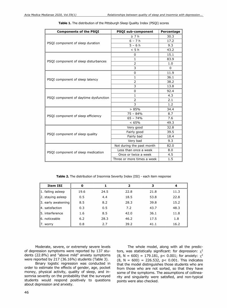
Acta Medica Medianae 2020, Vol.59(1) Relationships between quality of sleep and insomnia with depression...
46
Table 1. The distribution of the Pittsburgh Sleep Quality Index (PSQI) scores
Components of the PSQI PSQI sub-component Percentage
PSQI component of sleep duration
≥ 7 h 30.3
6 – 7 h 17.2
5 – 6 h 9.3
< 5 h 43.2
PSQI component of sleep disturbances
0 15.1
1 83.9
2 1.0
3 0
PSQI component of sleep latency
0 11.9
1 36.1
2 38.2
3 13.8
PSQI component of daytime dysfunction
0 92.4
1 4.3
2 2.1
3 1.2
PSQI component of sleep efficiency
> 85% 34.4
75 – 84% 8.7
65 – 74% 7.6
< 65% 49.3
PSQI component of sleep quality
Very good 32.8
Fairly good 39.5
Fairly bad 18.4
Very bad 9.3
PSQI component of sleep medication
Not during the past month 82.0
Less than once a week 8.0
Once or twice a week 4.5
Three or more times a week 1.5
Table 2. The distribution of Insomnia Severity Index (ISI) - each item response
Item ISI 0 1 2 3 4
1. falling asleep 19.6 24.5 22.8 21.8 11.3
2. staying asleep 0.5 4.4 18.5 53.8 22.8
3. early awakening 8.5 8.2 28.3 39.8 15.2
4. satisfaction 0.3 0.5 7.2 43.7 48.3
5. interference 1.6 8.5 42.0 36.1 11.8
6. noticeable 6.2 28.3 46.2 17.5 1.8
7. worry 0.8 2.7 39.2 41.1 16.2
Moderate, severe, or extremely severe levels
of depression symptoms were reported by 137 stu-dents (22.8%) and "above mild" anxiety symptoms were reported by 217 (36.16%) students (Table 3).
Binary logistic regression was conducted in order to estimate the effects of gender, age, pocket money, physical activity, quality of sleep, and in-somnia severity on the probability that the surveyed
students would respond positively to questions about depression and anxiety.
The whole model, along with all the predic-
tors, was statistically significant: for depression: χ2
(8, N = 600) = 179.181, p< 0.001; for anxiety: χ2
(8, N = 600) = 226.532, p< 0.001. This indicates that the model distinguishes those students who are from those who are not sorted, so that they have some of the symptoms. The assumptions of collinea-
rity and singularity were satisfied, and non-typical points were also checked.
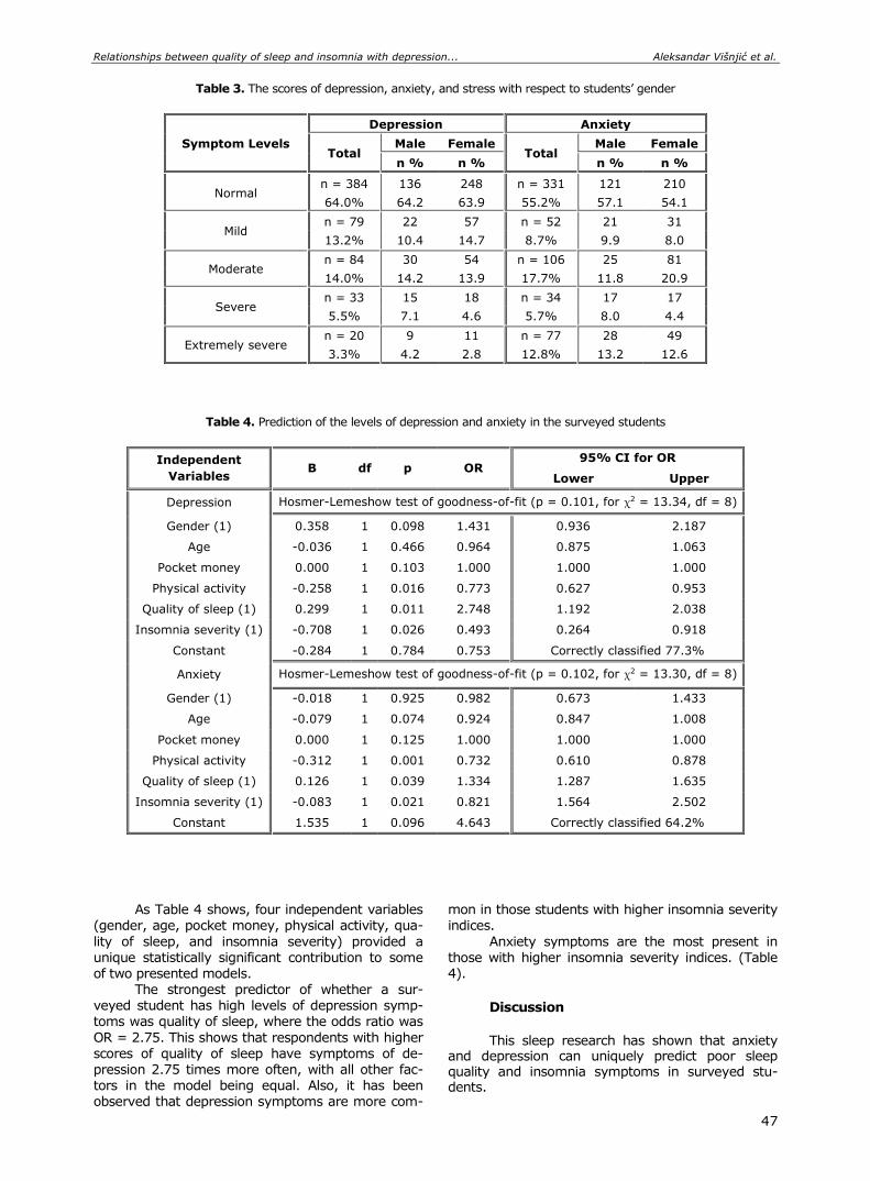
Relationships between quality of sleep and insomnia with depression... Aleksandar Višnjić et al.
47
Table 3. The scores of depression, anxiety, and stress with respect to students’ gender
Symptom Levels
Depression Anxiety
Total Male Female
Total Male Female
n % n % n % n %
Normal n = 384 136 248 n = 331 121 210
64.0% 64.2 63.9 55.2% 57.1 54.1
Mild n = 79 22 57 n = 52 21 31
13.2% 10.4 14.7 8.7% 9.9 8.0
Moderate n = 84 30 54 n = 106 25 81
14.0% 14.2 13.9 17.7% 11.8 20.9
Severe n = 33 15 18 n = 34 17 17
5.5% 7.1 4.6 5.7% 8.0 4.4
Extremely severe n = 20 9 11 n = 77 28 49
3.3% 4.2 2.8 12.8% 13.2 12.6
Table 4. Prediction of the levels of depression and anxiety in the surveyed students
Independent
Variables B df p OR
95% CI for OR
Lower Upper
Depression Hosmer-Lemeshow test of goodness-of-fit (p = 0.101, for χ2 = 13.34, df = 8)
Gender (1) 0.358 1 0.098 1.431 0.936 2.187
Age -0.036 1 0.466 0.964 0.875 1.063
Pocket money 0.000 1 0.103 1.000 1.000 1.000
Physical activity -0.258 1 0.016 0.773 0.627 0.953
Quality of sleep (1) 0.299 1 0.011 2.748 1.192 2.038
Insomnia severity (1) -0.708 1 0.026 0.493 0.264 0.918
Constant -0.284 1 0.784 0.753 Correctly classified 77.3%
Anxiety Hosmer-Lemeshow test of goodness-of-fit (p = 0.102, for χ2 = 13.30, df = 8)
Gender (1) -0.018 1 0.925 0.982 0.673 1.433
Age -0.079 1 0.074 0.924 0.847 1.008
Pocket money 0.000 1 0.125 1.000 1.000 1.000
Physical activity -0.312 1 0.001 0.732 0.610 0.878
Quality of sleep (1) 0.126 1 0.039 1.334 1.287 1.635
Insomnia severity (1) -0.083 1 0.021 0.821 1.564 2.502
Constant 1.535 1 0.096 4.643 Correctly classified 64.2%
As Table 4 shows, four independent variables (gender, age, pocket money, physical activity, qua-
lity of sleep, and insomnia severity) provided a unique statistically significant contribution to some of two presented models.
The strongest predictor of whether a sur-veyed student has high levels of depression symp-toms was quality of sleep, where the odds ratio was
OR = 2.75. This shows that respondents with higher scores of quality of sleep have symptoms of de-pression 2.75 times more often, with all other fac-tors in the model being equal. Also, it has been observed that depression symptoms are more com-
mon in those students with higher insomnia severity indices.
Anxiety symptoms are the most present in those with higher insomnia severity indices. (Table 4).
Discussion
This sleep research has shown that anxiety
and depression can uniquely predict poor sleep quality and insomnia symptoms in surveyed stu-dents.
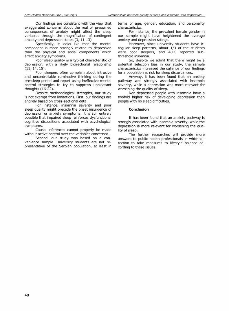
Acta Medica Medianae 2020, Vol.59(1) Relationships between quality of sleep and insomnia with depression...
48
Our findings are consistent with the view that exaggerated concerns about the real or presumed consequences of anxiety might affect the sleep variables through the magnification of contingent anxiety and depression states (3, 11-13).
Specifically, it looks like that the mental component is more strongly related to depression than the physical and social components which affect anxiety symptoms.
Poor sleep quality is a typical characteristic of depression, with a likely bidirectional relationship (11, 14, 15).
Poor sleepers often complain about intrusive and uncontrollable ruminative thinking during the pre-sleep period and report using ineffective mental control strategies to try to suppress unpleasant thoughts (16-22).
Despite methodological strengths, our study is not exempt from limitations. First, our findings are entirely based on cross-sectional data.
For instance, insomnia severity and poor sleep quality might precede the onset insurgence of depression or anxiety symptoms; it is still entirely possible that impaired sleep reinforces dysfunctional cognitive dispositions associated with psychological symptoms.
Causal inferences cannot properly be made without active control over the variables concerned.
Second, our study was based on a con-venience sample. University students are not re-presentative of the Serbian population, at least in
terms of age, gender, education, and personality characteristics.
For instance, the prevalent female gender in our sample might have heightened the average anxiety and depression ratings.
Moreover, since university students have ir-regular sleep patterns, about 1/3 of the students were poor sleepers, and 40% reported sub-threshold insomnia.
So, despite we admit that there might be a potential selection bias in our study, the sample characteristics increased the salience of our findings for a population at risk for sleep disturbances.
Anyway, it has been found that an anxiety pathway was strongly associated with insomnia severity, while a depression was more relevant for worsening the quality of sleep.
Non-depressed people with insomnia have a twofold higher risk of developing depression than people with no sleep difficulties.
Conclusion
It has been found that an anxiety pathway is strongly associated with insomnia severity, while the depression is more relevant for worsening the qua-lity of sleep.
The further researches will provide more
answers to public health professionals in which di-rection to take measures to lifestyle balance ac-cording to these issues.
Relationships between quality of sleep and insomnia with depression... Aleksandar Višnjić et al.
49
References
1. Schlarb AA, Kulessa D, Gulewitsch MD. Sleep charac-teristics, sleep problems, and associations of self-efficacy among German university students. Nat Sci Sleep 2012;4:1-7. [CrossRef] [PubMed]
2. Lund HG, Reider BD, Whiting AB, Prichard JR. Sleep Patterns and Predictors of Disturbed Sleep in a Large Population of College Students. J Adolesc Health 2010; 46:124-32. [CrossRef] [PubMed]
3. Seun-Fadipe CT, Mosaku KS. Sleep quality and psychological distress among undergraduate students of a Nigerian university. Sleep Health 2017;3:190-4. [CrossRef] [PubMed]
4. Kang JH, Chen SC. Effects of an irregular bedtime
schedule on sleep quality, daytime sleepiness, and fatigue among university students in Taiwan. BMC Public Health 2009;9:248. [CrossRef] [PubMed]
5. Harvey A. A Transdiagnostic Approach to Treating Sleep Disturbance in Psychiatric Disorders. Cogn Behav Ther 2009;38:35-42. [CrossRef] [PubMed]
6. Višnjić A, Veličković V, Stojanović M, Milošević Z, Rangelov T, Bulatović K, et al. The frequency of using screen-based media among children and adolescents and its impact on health-related behaviors. Acta Med Median 2015;54, 64-73. [CrossRef]
7. Crawford JR, Henry JD. The Depression Anxiety Stress Scales (DASS): Normative data and latent structure in a large non-clinical sample. Br J Clin Psychol 2003; 42:111-31. [CrossRef] [PubMed]
8. Lovibond SH. Manual for the Depression Anxiety Stress Scales. 2nd ed. Australia (Sydney): Psychology Foundation; 1995. [CrossRef]
9. Buysse DJ, Reynolds CF, Monk TH, Berman SR, Kupfer DJ. The Pittsburgh Sleep Quality Index (PSQI): A new instrument for psychiatric research and practice. Psy-chiatry Res 1989;28:193-213. [CrossRef] [PubMed]
10. Castronovo V, Galbiati A, Marelli S, Brombin C, Cugnata F, Giarolli, L, et al.Validation study of the Italian version of the Insomnia Severity Index (ISI). Neurol Sci 2016;37:1517-24. [CrossRef] [PubMed]
11. Taylor DJ, Lichstein KL, Durrence HH. Insomnia as a health risk factor. Behav Sleep Med 2003;1:227-47. [CrossRef] [PubMed]
12. Baglioni C, Battagliese G, Feige B, Spiegelhalder K, Nissen C, Voderholzer U, et al. Insomnia as a predictor of depression: A meta-analytic evaluation of longitu-
dinal epidemiological studies. J Affect Disord 2011; 135:10-9. [CrossRef] [PubMed]
13. Jansson M, Linton SJ. The role of anxiety and depression in the development of insomnia: Cross-sectional and prospective analyses. Psychol Health 2006;21:383-97. [CrossRef]
14. Morphy H, Dunn KM, Lewis M, Boardman HF, Croft PR. Epidemiology of insomnia: a longitudinal study in a UK population. Sleep 2007;30:274-80. [PubMed]
15. Johnson EO, Roth T, Breslau N. The association of insomnia with anxiety disorders and depression: Exploration of the direction of risk. J Psychiatr Res 2006;40:700-8. [CrossRef] [PubMed]
16. Ohayon MM, Roth T. Place of chronic insomnia in the course of depressive and anxiety disorders. PubMed Commons. J Psychiatr Res 2003;37:1-2. [CrossRef] [PubMed]
17. Alvaro PK, Roberts RM, Harris JK. A Systematic Review Assessing Bidirectionality between Sleep Disturbances, Anxiety, and Depression. Sleep 2013; 36:1059-68. [CrossRef] [PubMed]
18. Riemann D, Spiegelhalder K, Feige B, Voderholzer U, Berger M, Perlis M, et al. The hyperarousal model of insomnia: A review of the concept and its evidence. Sleep Med Rev 2010;14:19-524 31. [CrossRef] [PubMed]
19. Višnjić A, Veličković V, Sokolović D, Stanković M, Mijatović K, Stojanović M,et al. Relationship between the Manner of Mobile Phone Use and Depression, Anxiety, and Stress in University Students. Int J Environ Res Public Health 2018;15:697. [CrossRef] [PubMed]
20. Carleton RN. Fear of the unknown: One fear to rule them all? J Anxiety Disord 2016; 41:5-21. [CrossRef] [PubMed]
21. Hong RY, Cheung, MWL. The structure of cognitive vulnerabilities to depression and anxiety: Evidence for a common core etiologic process based on a meta-analytic review. Clin Psychol Sci 2015;530(3), 892-912. [CrossRef]
22. Alamir YA, Zullig KJ, Wen S, Montgomery-Downs H, Kristjansson AL, Misra R, et al. Association Between Nonmedical Use of Prescription Drugs and Sleep Quality in a Large College Student Sample. Behav Sleep Med 2017; 1-11. [CrossRef] [PubMed]
Acta Medica Medianae 2020, Vol.59(1) Relationships between quality of sleep and insomnia with depression...
50
Originalni rad UDC: 613.79:616.89‑008
doi:10.5633/amm.2020.0106
POVEZANOST KVALITETA SNA I NESANICE SA SIMPTOMIMA DEPRESIJE I ANKSIOZNOSTI KOD STUDENATA MEDICINE U SRBIJI
Aleksandar Višnjić1,2, Snežana Miljković3, Dragan Nikolić2, Tamara Jovanović1,2,
Katarina Bulatović2, Marko Ristić4, Dragan Toskić5
1Univerzitet u Nišu, Medicinski fakultet, Niš, Srbija 2Institut za javno zdravlje Niš, Niš, Srbija 3Klinički centar Niš, Niš, Srbija 4Univerzitet u Nišu, Medicinski fakultet, student postdiplomskih studija, Niš, Srbija 5Univerzitet u Prištini, Fakultet za sport i fizičko vaspitanje, Kosovska Mitrovica, Srbija
Kontakt: Aleksandar Višnjić
Bulevar dr Zoran Đinđić 81, 18000 Niš, Srbija
E-mail: [email protected]
Budući da su problemi sa spavanjem vrlo česti među studentima medicine, ova studija
je istraživala povezanost depresije i anksioznosti sa kvalitetom sna i nesanicom. Studija preseka izvedena je na Medicinskom fakultetu Univerziteta u Nišu 2016. godine
i obuhvatila je 600 studenata oba pola. Studenti su ispunili upitnik, koji je sastavljen i razvijen iz skala „Depression Anxiety Stress Scale“, „Pittsburgh Sleep Quality Index“ i „Insomnia Severity Index“.
Pokazalo se da su problemi sa spavanjem zaista česti među studentima: 432 (72%) studenta prijavila su loš kvalitet sna, dok 258 (43%) njih ima probleme sa nesanicom. Čak 66 studenata (11%) prijavilo je vrlo ozbiljnu ili ozbiljnu nesanicu. Simptome depresije iznad
praga prijavilo je 168 studenata (28%), a simptome anksioznosti njih 180 (30%). Depresija
je bila snažno povezana sa lošim kvalitetom sna (χ2 = 20,35; df = 1; p < 0,001) i stepenom
nesanice (χ2 = 13,05; df = 1; p < 0,001). Anksioznost iznad praga bila je povezana samo sa
ozbiljnošću nesanice (χ2 = 16,42; df = 1; p < 0,001).
Nađeno je da je anksioznost bila snažno povezana sa nesanicom, dok je depresija bila povezanija sa pogoršanjem kvaliteta sna.
Acta Medica Medianae 2020;59(1):44-50.
Ključne reči: kvalitet sna, nesanica, depresija, anksioznost
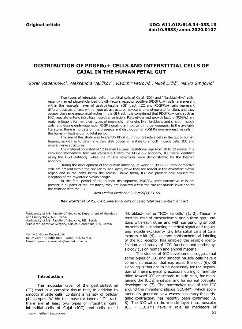
51 www.medfak.ni.ac.rs/amm
Original article UDC: 611.018:616.34-053.13
doi:10.5633/amm.2020.0107
DISTRIBUTION OF PDGFR+ CELLS AND INTERSTITIAL CELLS OF
CAJAL IN THE HUMAN FETAL GUT
Goran Radenković1, Aleksandra Veličkov1, Vladimir Petrović1, Miloš Dičić2, Marko Gmijović3
Two types of interstitial cells, interstitial cells of Cajal (ICC) and "fibroblast-like" cells,
recently named platelet-derived growth factors receptor positive (PDGFR+) cells, are present
within the muscular layer of gastrointestinal (GI) tract. ICC and PDGFR+ cells represent
different classes of cells with unique ultrastructure, molecular phenotype and function, and they occupy the same anatomical niches in the GI tract. It is considered that PDGFR+ cells such as
ICC, mediate enteric inhibitory neurotransmission. Platelet-derived growth factors (PDGFs) are
major mitogens for many cell types of mesenchymal origin, like fibroblasts and smooth muscle cells, and during embryogenesis, PDGF signaling is important in organogenesis. In the available literature, there is no data on the presence and distribution of PDGFR immunoreactive cells in
the human intestine during fetal period. The aim of this study was to identify PDGFR immunoreactive cells in the gut of human
fetuses, as well as to determine their distribution in relation to smooth muscle cells, ICC and enteric nerve structures.
The material consisted of 12 Human Fetuses, gestational age from 10 to 12 weeks. The immunohistochemical test was carried out with the PDGFR- antibody, ICC were identified
using the C-kit antibody, while the muscle structures were demonstrated by the Desmin antibody.
During the development of the human intestine, at week 11, PDGFR immunoreactive
cells are present within the circular muscle layer, while they are absent in the myenteric plexus region and in the parts below the serosa. Unlike them, ICC are present only around the inception of the myenteric plexus ganglia.
In the fetal period of the human development, PDGFR immunoreactive cells are
present in all parts of the intestines, they are localized within the circular muscle layer and do not coincide with the ICC.
Acta Medica Medianae 2020;59(1):51-59.
Key words: PDGFR, C-kit, interstitial cells of Cajal, fetal gastrointestinal tract
1University of Niš, Faculty of Medicine, Department of Histology
and Embryology, Niš, Serbia 2University of Niš, Faculty of Medicine, Niš, Serbia 3Clinic for Digestive Surgery, Clinical Center Niš, Niš, Serbia
Contact: Goran Radenković
81 Dr Zoran Djindjic Blvd., 18000 Niš, Serbia
E-mail: [email protected]
Introduction The muscular layer of the gastrointestinal
(GI) tract is a complex tissue that, in addition to smooth muscle cells, contains a variety of cellular phenotypes. Within the muscular layer of GI tract,
there are at least two types of interstitial cells, interstitial cells of Cajal (ICC) and cells called
"fibroblast-like" or "ICC-like cells" (1, 2). These in-terstitial cells of mesenchymal origin form gap junc-tions with each other and with surrounding smooth
muscles thus conducting electrical signal and regula-ting muscle excitability (3). Interstitial cells of Cajal express c-kit (4), so immunohistochemical labeling
of the Kit receptor has enabled the reliable identi-fication and study of ICC function and pathophy-siology (5) on human and animal material.
The studies of ICC development suggest that
some types of ICC and smooth muscle cells have a common precursor that expresses the c-kit (6). Kit signaling is thought to be necessary for the separa-tion of mesenchymal precursors during differentia-tition toward ICC or smooth muscle cells, for main-taining the ICC phenotype, and for normal postnatal
development (7). The pacemaker role of the ICC around the myenteric plexus (ICC-MY), which spon-taneously generate slow waves necessary for peris-taltic contraction, has recently been confirmed (3, 8). The ICC within the muscle layer (intramuscular ICC – ICC-IM) have a role as mediators of
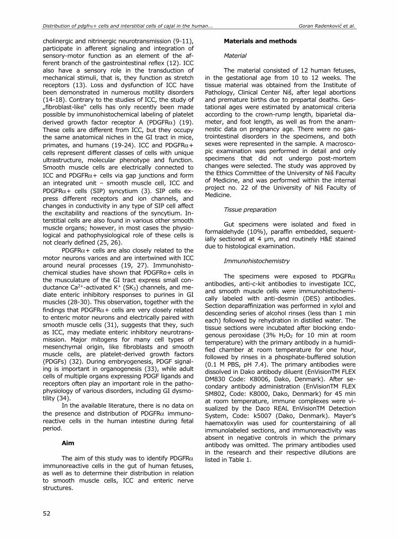
Distribution of pdgfr+ cells and interstitial cells of cajal in the human... Goran Radenković et al.
52
cholinergic and nitrinergic neurotransmission (9-11), participate in afferent signaling and integration of
sensory-motor function as an element of the af-ferent branch of the gastrointestinal reflex (12). ICC
also have a sensory role in the transduction of mechanical stimuli, that is, they function as stretch receptors (13). Loss and dysfunction of ICC have been demonstrated in numerous motility disorders (14-18). Contrary to the studies of ICC, the study of „fibroblast-like“ cells has only recently been made
possible by immunohistochemical labeling of platelet
derived growth factor receptor A (PDGFR) (19).
These cells are different from ICC, but they occupy the same anatomical niches in the GI tract in mice,
primates, and humans (19-24). ICC and PDGFR+
cells represent different classes of cells with unique ultrastructure, molecular phenotype and function.
Smooth muscle cells are electrically connected to
ICC and PDGFR+ cells via gap junctions and form
an integrated unit – smooth muscle cell, ICC and
PDGFR+ cells (SIP) syncytium (3). SIP cells ex-
press different receptors and ion channels, and changes in conductivity in any type of SIP cell affect the excitability and reactions of the syncytium. In-terstitial cells are also found in various other smooth
muscle organs; however, in most cases the physio-logical and pathophysiological role of these cells is not clearly defined (25, 26).
PDGFR+ cells are also closely related to the
motor neurons varices and are intertwined with ICC around neural processes (19, 27). Immunohisto-chemical studies have shown that PDGFRα+ cells in
the musculature of the GI tract express small con-
ductance Ca2+-activated K+ (SK3) channels, and me-diate enteric inhibitory responses to purines in GI muscles (28-30). This observation, together with the
findings that PDGFR+ cells are very closely related
to enteric motor neurons and electrically paired with smooth muscle cells (31), suggests that they, such
as ICC, may mediate enteric inhibitory neurotrans-mission. Major mitogens for many cell types of mesenchymal origin, like fibroblasts and smooth muscle cells, are platelet-derived growth factors (PDGFs) (32). During embryogenesis, PDGF signal-ing is important in organogenesis (33), while adult cells of multiple organs expressing PDGF ligands and
receptors often play an important role in the patho-physiology of various disorders, including GI dysmo-
tility (34). In the available literature, there is no data on
the presence and distribution of PDGFR immuno-
reactive cells in the human intestine during fetal period.
Aim
The aim of this study was to identify PDGFR
immunoreactive cells in the gut of human fetuses, as well as to determine their distribution in relation to smooth muscle cells, ICC and enteric nerve
structures.
Materials and methods
Material
The material consisted of 12 human fetuses, in the gestational age from 10 to 12 weeks. The tissue material was obtained from the Institute of Pathology, Clinical Center Niš, after legal abortions and premature births due to prepartal deaths. Ges-tational ages were estimated by anatomical criteria
according to the crown-rump length, biparietal dia-meter, and foot length, as well as from the anam-nestic data on pregnancy age. There were no gas-trointestinal disorders in the specimens, and both sexes were represented in the sample. A macrosco-pic examination was performed in detail and only specimens that did not undergo post-mortem
changes were selected. The study was approved by the Ethics Committee of the University of Niš Faculty of Medicine, and was performed within the internal project no. 22 of the University of Niš Faculty of Medicine.
Tissue preparation
Gut specimens were isolated and fixed in
formaldehyde (10%), paraffin embedded, sequent-ially sectioned at 4 μm, and routinely H&E stained due to histological examination.
Immunohistochemistry
The specimens were exposed to PDGFR
antibodies, anti-c-kit antibodies to investigate ICC, and smooth muscle cells were immunohistochemi-cally labeled with anti-desmin (DES) antibodies. Section deparaffinization was performed in xylol and
descending series of alcohol rinses (less than 1 min each) followed by rehydration in distilled water. The tissue sections were incubated after blocking endo-genous peroxidase (3% H2O2 for 10 min at room temperature) with the primary antibody in a humidi-fied chamber at room temperature for one hour, followed by rinses in a phosphate-buffered solution
(0.1 M PBS, pH 7.4). The primary antibodies were dissolved in Dako antibody diluent (EnVisionTM FLEX DM830 Code: К8006, Dako, Denmark). After se-condary antibody administration (EnVisionTM FLEX SМ802, Code: К8000, Dako, Denmark) for 45 min
at room temperature, immune complexes were vi-sualized by the Daco REAL EnVisionTM Detection
System, Code: k5007 (Dako, Denmark). Mayer’s haematoxylin was used for counterstaining of all immunolabeled sections, and immunoreactivity was absent in negative controls in which the primary antibody was omitted. The primary antibodies used in the research and their respective dilutions are
listed in Table 1.
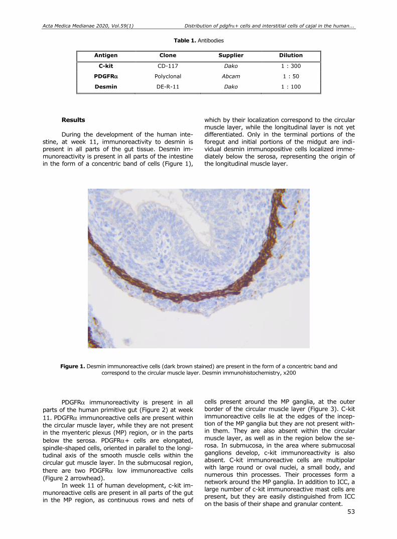
Acta Medica Medianae 2020, Vol.59(1) Distribution of pdgfr+ cells and interstitial cells of cajal in the human...
53
Table 1. Antibodies
Antigen Clone Supplier Dilution
C-kit CD-117 Dako 1 : 300
PDGFR Polyclonal Abcam 1 : 50
Desmin DE-R-11 Dako 1 : 100
Results During the development of the human inte-
stine, at week 11, immunoreactivity to desmin is present in all parts of the gut tissue. Desmin im-munoreactivity is present in all parts of the intestine in the form of a concentric band of cells (Figure 1),
which by their localization correspond to the circular muscle layer, while the longitudinal layer is not yet differentiated. Only in the terminal portions of the foregut and initial portions of the midgut are indi-vidual desmin immunopositive cells localized imme-diately below the serosa, representing the origin of the longitudinal muscle layer.
Figure 1. Desmin immunoreactive cells (dark brown stained) are present in the form of a concentric band and correspond to the circular muscle layer. Desmin immunohistochemistry, x200
PDGFR immunoreactivity is present in all
parts of the human primitive gut (Figure 2) at week
11. PDGFR immunoreactive cells are present within
the circular muscle layer, while they are not present in the myenteric plexus (MP) region, or in the parts
below the serosa. PDGFR+ cells are elongated,
spindle-shaped cells, oriented in parallel to the longi-tudinal axis of the smooth muscle cells within the
circular gut muscle layer. In the submucosal region,
there are two PDGFR low immunoreactive cells
(Figure 2 arrowhead). In week 11 of human development, c-kit im-
munoreactive cells are present in all parts of the gut in the MP region, as continuous rows and nets of
cells present around the MP ganglia, at the outer border of the circular muscle layer (Figure 3). C-kit immunoreactive cells lie at the edges of the incep-tion of the MP ganglia but they are not present with-in them. They are also absent within the circular muscle layer, as well as in the region below the se-rosa. In submucosa, in the area where submucosal
ganglions develop, c-kit immunoreactivity is also absent. C-kit immunoreactive cells are multipolar with large round or oval nuclei, a small body, and numerous thin processes. Their processes form a network around the MP ganglia. In addition to ICC, a large number of c-kit immunoreactive mast cells are present, but they are easily distinguished from ICC
on the basis of their shape and granular content.
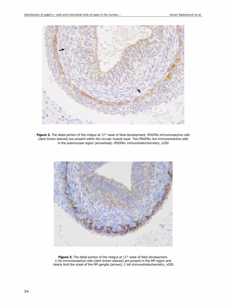
Distribution of pdgfr+ cells and interstitial cells of cajal in the human... Goran Radenković et al.
54
Figure 2. The distal portion of the midgut at 11th week of fetal development. PDGFRα immunoreactive cells
(dark brown stained) are present within the circular muscle layer. Two PDGFR low immunoreactive cells
in the submucosal region (arrowhead). PDGFR immunohistochemistry, x200
Figure 3. The distal portion of the midgut at 11th week of fetal development. C-kit immunoreactive cells (dark brown stained) are present in the MP region and
clearly limit the onset of the MP ganglia (arrows). C-kit immunohistochemistry, x200
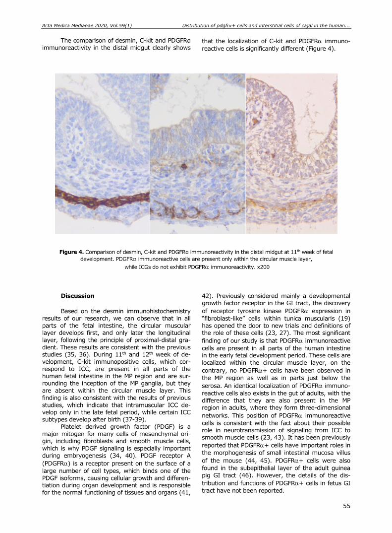
Acta Medica Medianae 2020, Vol.59(1) Distribution of pdgfr+ cells and interstitial cells of cajal in the human...
55
The comparison of desmin, C-kit and PDGFRα immunoreactivity in the distal midgut clearly shows
that the localization of C-kit and PDGFR immuno-
reactive cells is significantly different (Figure 4).
Figure 4. Comparison of desmin, C-kit and PDGFRα immunoreactivity in the distal midgut at 11th week of fetal
development. PDGFR immunoreactive cells are present only within the circular muscle layer,
while ICGs do not exhibit PDGFR immunoreactivity. x200
Discussion
Based on the desmin immunohistochemistry
results of our research, we can observe that in all parts of the fetal intestine, the circular muscular layer develops first, and only later the longitudinal layer, following the principle of proximal-distal gra-
dient. These results are consistent with the previous studies (35, 36). During 11th and 12th week of de-velopment, C-kit immunopositive cells, which cor-respond to ICC, are present in all parts of the human fetal intestine in the MP region and are sur-rounding the inception of the MP ganglia, but they
are absent within the circular muscle layer. This
finding is also consistent with the results of previous studies, which indicate that intramuscular ICC de-velop only in the late fetal period, while certain ICC subtypes develop after birth (37-39).
Platelet derived growth factor (PDGF) is a major mitogen for many cells of mesenchymal ori-gin, including fibroblasts and smooth muscle cells,
which is why PDGF signaling is especially important during embryogenesis (34, 40). PDGF receptor A
(PDGFR) is a receptor present on the surface of a
large number of cell types, which binds one of the PDGF isoforms, causing cellular growth and differen-tiation during organ development and is responsible for the normal functioning of tissues and organs (41,
42). Previously considered mainly a developmental growth factor receptor in the GI tract, the discovery
of receptor tyrosine kinase PDGFR expression in
“fibroblast-like” cells within tunica muscularis (19) has opened the door to new trials and definitions of the role of these cells (23, 27). The most significant
finding of our study is that PDGFR immunoreactive
cells are present in all parts of the human intestine in the early fetal development period. These cells are
localized within the circular muscle layer, on the
contrary, no PDGFR+ cells have been observed in
the MP region as well as in parts just below the
serosa. An identical localization of PDGFR immuno-
reactive cells also exists in the gut of adults, with the difference that they are also present in the MP region in adults, where they form three-dimensional
networks. This position of PDGFR immunoreactive
cells is consistent with the fact about their possible role in neurotransmission of signaling from ICC to smooth muscle cells (23, 43). It has been previously
reported that PDGFR+ cells have important roles in
the morphogenesis of small intestinal mucosa villus
of the mouse (44, 45). PDGFR+ cells were also
found in the subepithelial layer of the adult guinea
pig GI tract (46). However, the details of the dis-
tribution and functions of PDGFR+ cells in fetus GI
tract have not been reported.
Distribution of pdgfr+ cells and interstitial cells of cajal in the human... Goran Radenković et al.
56
Kurahashi et al. have described a specific type
of PDGFR+ cells in the lamina propria of the
human GI tract (47), and suggested that subepi-
thelial PDGFR+ cells have a role in sensory and
secretomotor signaling, proliferation, differentiation, and apoptosis of epithelial cells, and in epithelial cellular pathology, including inflammatory responses
and tumorigenesis. Subepithelial PDGFR+ cells in
adults form a sheath just beneath the epithelium and cover the crypts from their base to the luminal surface of the epithelium. It is suggested that these
cells may have modulatory functions in immune and sensory responses and in the maintenance of mu-cosal homeostasis, but the roles of these cells in physiological and pathophysiological processes are still unknown. In our research, there was a low
PDGFR submucosal immunoreactivity during the
early period of fetal development, and dominant
PDGFR were present within the circular muscle
layer. As already mentioned, due to their close con-tact with enteric nerve endings and smooth muscle cells within the circular muscle layer, it is assumed that these cells primarily play a role in the neuromo-dulation of peristalsis.
Another important result of our study is that
PDGFR cells differ from ICC and that they are
functionally close but still different cell types of in-
terstitial cells. In adults, PDGFR cells are widely
distributed within the MP region and in circular and longitudinal muscle layers throughout the human colon (23). Blair et al. (21) have shown relationships between enteric neurons and interstitial cells in pri-
mates. They have shown that PDGFR+ cells are
closely associated with ICC and occupy the same anatomical niches as ICC-MY and ICC-IM. However, in contrast to the distribution of intestinal cells in adults, we showed in our study that, in the fetal pe-
riod, PDGFR activity was not observed in the ICC-
MY domains around the ganglia. This finding indi-
cates that PDGFR cells develop later than the ICC
within GI tract. Further, our results show that unlike
the region of MP, PDGF cells are present in the circular muscle in the fetal period and there are still no differentiated ICC-IM.
Ultrastructural studies show that enteric neu-rons do not effectuate any direct contact with smooth muscle cells or synaptic specializations; on the other
hand, ICC make close contacts with both cholinergic
and nitrinergic neurons, forming synapse-like con-nections at one end and gap junctions with smooth muscle cells (3, 48). Calcium activated chloride channels (Ano1) are highly expressed and exclu-sively by ICC throughout the GI tract (21, 49), so the major excitatory neurotransmitter – acetylcho-
line induces depolarization by the activation of Ano1 currents (50). ICC-IM also respond to inhibitory neu-rotransmitters between neurons and smooth muscles
(51). PDGFR+ are very similar to ICC-IM in adults,
and also form gap junctions with surrounding smooth
muscle cells (3, 20, 21). PDGFR+ cells express
guanylate cyclase, purinergic P2Y1 receptors and small conductance Ca2+- activated K+ (SK3K+) chan-
nels (23, 27, 52) which indicates that PDGFR+ cells
mediate enteric inhibitory neurotransmision. It has
been confirmed that PDGFR+ cells, like ICC, gene-
rate inhibitory post junctional responses in GI muscles (27). Gap junctions provide electrical coupl-ing between cells, such that induction of a K+ cur-
rent in PDGFR+ cells results in hyperpolarization,
first of these cells and immediately after that in net
hyperpolarization of the SIP syncytium and reduce muscle contraction.
In contrast to our results, PDGFR+ cells in
adults form a network adjacent to ICC around the myenteric ganglia (23). ICC-MY have a pacemaker
role to generate spontaneous electrical activity,
while the function of myenteric PDGFR+ is still un-
known. Since they are interconnected with ICC-MP, they may have a role in the propagation and modu-lation of the electrical peristaltic waves.
At present, little is known about the involve-
ment of PDGFR+ cells in GI motor dysfunction. It is
certain that the changes in purinergic neural inputs could have effects on colonic motility. Enhanced
activation of PDGFR in mice, contribute to the de-
velopment of GI fibrosis and sarcoma (53). In a recent study (54), it has been concluded that colonic transit disorder may be due to the downregulation of the Kit and Ano1 channels and the upregulation of
SK3 channels in PDGFR+ cells, suggesting that the
imbalance between ICC and PDGFR distribution
might be a possible reason for gut dysmotility.
Furthermore, some stromal tumors (GIST) are po-
sitive for PDGFR (55, 56), so it is possible that
these cells, like ICC, can be malignantly trans-formed.
Conclusion
In the fetal period of human development,
PDGFR immunoreactive cells are present in all
parts of the intestine, localized within the circular muscle layer, and do not coincide with ICC.
Acknowledgements
This work was supported by the Ministry of
Science and Technological Development of the Republic of Serbia (project 43012) and by the Faculty of Medicine in Niš internal project number 22.
Acta Medica Medianae 2020, Vol.59(1) Distribution of pdgfr+ cells and interstitial cells of cajal in the human...
57
References
1. Komuro T, Seki K, Horiguchi K. Ultrastructural charac-terization of the interstitial cells of Cajal. Archives of histology and cytology 1999;62(4):295-316. [CrossRef] [PubMed]
2. Pieri L, Vannucchi MG, Faussone-Pellegrini MS. Histo-chemical and ultrastructural characteristics of an interstitial cell type different from ICC and resident in the muscle coat of human gut. Journal of cellular and molecular medicine 2008;12(5B):1944-55. [CrossRef] [PubMed]
3. Sanders KM, Ward SM, Koh SD. Interstitial cells: regulators of smooth muscle function. Physiological reviews 2014;94(3):859-907.[CrossRef] [PubMed]
4. Huizinga JD, Chen J-H, Mikkelsen HB, Wang X-Y, Parsons SP, Zhu YF. Interstitial cells of Cajal, from structure to function. Frontiers in Neuroscience 2013; 7:43.[CrossRef] [PubMed]
5. Ward SM, Burns AJ, Torihashi S, Sanders KM. Mutation of the proto-oncogene c-kit blocks develop-ment of interstitial cells and electrical rhythmicity in murine intestine. The Journal of physiology 1994;480 (Pt 1):91-7.[CrossRef] [PubMed]
6. Kluppel M, Huizinga JD, Malysz J, Bernstein A. Developmental origin and Kit-dependent development of the interstitial cells of cajal in the mammalian small intestine. Developmental dynamics : an official publi-cation of the American Association of Anatomists 1998;211(1):60-71.[CrossRef]
7. Beckett EA, Ro S, Bayguinov Y, Sanders KM, Ward SM. Kit signaling is essential for development and maintenance of interstitial cells of Cajal and electrical rhythmicity in the embryonic gastrointestinal tract. Developmental dynamics: an official publication of the American Association of Anatomists 2007;236(1):60-72.[CrossRef] [PubMed]
8. Takaki M. Gut pacemaker cells: the interstitial cells of Cajal (ICC). Journal of smooth muscle research = Nihon Heikatsukin Gakkai kikanshi 2003;39(5):137-61.[CrossRef] [PubMed]
9. Iino S, Ward SM, Sanders KM. Interstitial cells of Cajal are functionally innervated by excitatory motor neuro-
nes in the murine intestine. The Journal of physiology 2004;556(Pt 2):521-30.[CrossRef] [PubMed]
10. Ward SM, McLaren GJ, Sanders KM. Interstitial cells of Cajal in the deep muscular plexus mediate enteric motor neurotransmission in the mouse small intestine. The Journal of physiology 2006;573(Pt 1): 147-59. [CrossRef] [PubMed]
11. Kito Y. The functional role of intramuscular interstitial cells of Cajal in the stomach. Journal of smooth muscle research = Nihon Heikatsukin Gakkai kikanshi 2011;47(2):47-53.[CrossRef] [PubMed]
12. Huizinga JD, Reed DE, Berezin I, Wang XY, Valdez DT, Liu LW, et al. Survival dependency of intramuscular ICC on vagal afferent nerves in the cat esophagus. American journal of physiology Regulatory, integrative and comparative physiology 2008;294(2):R302-10. [CrossRef] [PubMed]
13. Powley TL, Wang XY, Fox EA, Phillips RJ, Liu LW, Huizinga JD. Ultrastructural evidence for communica-tion between intramuscular vagal mechanoreceptors and interstitial cells of Cajal in the rat fundus. Neuro-gastroenterology and motility: the official journal of the European Gastrointestinal Motility Society 2008;20 (1):69-79.[CrossRef] [PubMed]
14. Faussone-Pellegrini MS, Cortesini C. The muscle coat of the lower esophageal sphincter in patients with achalasia and hypertensive sphincter. An electron microscopic study. Journal of submicroscopic cytology 1985;17(4):673-85.[CrossRef] [PubMed]
15. Vanderwinden JM, Rumessen JJ. Interstitial cells of Cajal in human gut and gastrointestinal disease. Mi-croscopy research and technique 1999;47(5):344-60. [CrossRef] [PubMed]
16. Zarate N, Mearin F, Wang XY, Hewlett B, Huizinga JD, Malagelada JR. Severe idiopathic gastroparesis due to neuronal and interstitial cells of Cajal degeneration: pathological findings and management. Gut 2003;52 (7):966-70.[CrossRef] [PubMed]
17. Gockel I, Bohl JR, Eckardt VF, Junginger T. Reduction of interstitial cells of Cajal (ICC) associated with neuronal nitric oxide synthase (n-NOS) in patients with achalasia. The American journal of gastroentero-logy 2008;103(4):856-64.[CrossRef] [PubMed]
18. Velickov A, Mitrovic O, Djordjevic B, Sokolovic D, Zivkovic V, Velickov A, et al. The effect of bilberries on diabetes-related alterations of interstitial cells of Cajal in the lower oesophageal sphincter in rats. Histology and histopathology 2016:11829.[CrossRef] [PubMed]
19. Iino S, Horiguchi K, Horiguchi S, Nojyo Y. c-Kit-negative fibroblast-like cells express platelet-derived growth factor receptor alpha in the murine gastro-intestinal musculature. Histochemistry and cell biology 2009;131(6):691-702.[CrossRef] [PubMed]
20. Grover M, Bernard CE, Pasricha PJ, Parkman HP, Abell TL, Nguyen LA, et al. Platelet-derived growth factor receptor alpha (PDGFRalpha)-expressing "fibroblast-like cells" in diabetic and idiopathic gastroparesis of humans. Neurogastroenterology and motility: the official journal of the European Gastrointestinal Motility Society 2012;24(9):844-52.[CrossRef] [PubMed]
21. Blair PJ, Bayguinov Y, Sanders KM, Ward SM. Rela-tionship between enteric neurons and interstitial cells in the primate gastrointestinal tract. Neurogastro-enterology and motility: the official journal of the European Gastrointestinal Motility Society 2012;24(9):
e437-49.[CrossRef] [PubMed] 22. Koh BH, Roy R, Hollywood MA, Thornbury KD, McHale
NG, Sergeant GP, et al. Platelet-derived growth factor receptor-alpha cells in mouse urinary bladder: a new class of interstitial cells. Journal of cellular and mole-cular medicine 2012;16(4):691-700. [CrossRef] [PubMed]
23. Kurahashi M, Nakano Y, Hennig GW, Ward SM, Sanders KM. Platelet-derived growth factor receptor α-positive cells in the tunica muscularis of human colon. Journal of cellular and molecular medicine 2012;16 (7):1397-404.[CrossRef] [PubMed]
24. Monaghan KP, Johnston L, McCloskey KD. Identifica-tion of PDGFRalpha positive populations of interstitial cells in human and guinea pig bladders. The Journal of urology2012;188(2):639-47.[CrossRef] [PubMed]
25. Koh SD, Ward SM, Sanders KM. Ionic conductances regulating the excitability of colonic smooth muscles. Neurogastroenterology and motility: the official journal of the European Gastrointestinal Motility Society 2012;24(8):705-18.[CrossRef] [PubMed]
26. Sanders KM, Koh SD, Ro S, Ward SM. Regulation of gastrointestinal motility--insights from smooth muscle biology. Nature reviews Gastroenterology & hepato-logy 2012;9(11):633-45.[CrossRef] [PubMed]
Distribution of pdgfr+ cells and interstitial cells of cajal in the human... Goran Radenković et al.
58
27. Kurahashi M, Zheng H, Dwyer L, Ward SM, Koh SD, Sanders KM. A functional role for the 'fibroblast-like cells' in gastrointestinal smooth muscles. The Journal of physiology 2011;589(Pt 3):697-710. [CrossRef] [PubMed]
28. Vanderwinden JM, Rumessen JJ, de Kerchove d'Exaerde A, Jr, Gillard K, Panthier JJ, de Laet MH, et al. Kit-negative fibroblast-like cells expressing SK3, a Ca2+-activated K+ channel, in the gut musculature in health and disease. Cell and tissue research 2002;310 (3):349-58.[CrossRef] [PubMed]
29. Klemm MF, Lang RJ. Distribution of Ca2+-activated K+ channel (SK2 and SK3) immunoreactivity in intestinal smooth muscles of the guinea-pig. Clinical and experimental pharmacology & physiology 2002; 29(1-2):18-25.[CrossRef] [PubMed]
30. Fujita A, Takeuchi T, Jun H, Hata F. Localization of Ca2+-activated K+ channel, SK3, in fibroblast-like cells forming gap junctions with smooth muscle cells in the mouse small intestine. Journal of pharmacological sciences 2003;92(1):35-42.[CrossRef] [PubMed]
31. Horiguchi K, Komuro T. Ultrastructural observations of fibroblast-like cells forming gap junctions in the W/W(nu) mouse small intestine. Journal of the autonomic nervous system 2000;80(3):142-7. [CrossRef] [PubMed]
32. Fredriksson L, Li H, Eriksson U. The PDGF family: four gene products form five dimeric isoforms. Cytokine & growth factor reviews 2004;15(4):197-204. [CrossRef] [PubMed]
33. Hoch RV, Soriano P. Roles of PDGF in animal develop-ment. Development (Cambridge, England) 2003;130 (20):4769-84.[CrossRef] [PubMed]
34. Andrae J, Gallini R, Betsholtz C. Role of platelet-derived growth factors in physiology and medicine. Genes & development 2008;22(10):1276-312. [CrossRef] [PubMed]
35. Wallace AS, Burns AJ. Development of the enteric nervous system, smooth muscle and interstitial cells of Cajal in the human gastrointestinal tract. Cell and tissue research 2005;319(3):367-82. [CrossRef] [PubMed]
36. Chevalier NR, Fleury V, Dufour S, Proux-Gillardeaux V, Asnacios A. Emergence and development of gut motility in the chicken embryo. PloS one 2017;12 (2):e0172511.[CrossRef] [PubMed]
37. Abramovic M, Radenkovic G, Velickov A. Appearance of interstitial cells of Cajal in the human midgut. Cell and tissue research 2014;356(1):9-14. [CrossRef] [PubMed]
38. Radenkovic G. Two patterns of development of interstitial cells of Cajal in the human duodenum. Journal of cellular and molecular medicine 2012;16 (1):185-92.[CrossRef] [PubMed]
39. Radenkovic G, Radenkovic D, Velickov A. Develop-ment of interstitial cells of Cajal in the human diges-tive tract as the result of reciprocal induction of me-senchymal and neural crest cells. Journal of cellular and molecular medicine 2018;22(2):778-85. [CrossRef] [PubMed]
40. Kurahashi M, Niwa Y, Cheng J, Ohsaki Y, Fujita A, Goto H, et al. Platelet-derived growth factor signals play critical roles in differentiation of longitudinal smooth muscle cells in mouse embryonic gut. Neurogastroenterology and motility: the official journal of the European Gastrointestinal Motility Society
2008;20(5):521-31.[CrossRef] [PubMed] 41. Soriano P. The PDGF alpha receptor is required for
neural crest cell development and for normal pat-terning of the somites. Development 1997;124(14): 2691-700.[CrossRef] [PubMed]
42. Hamilton TG, Klinghoffer RA, Corrin PD, Soriano P. Evolutionary divergence of platelet-derived growth factor alpha receptor signaling mechanisms. Molecular and cellular biology 2003;23(11):4013-25. [CrossRef] [PubMed]
43. Kurahashi M, Mutafova-Yambolieva V, Koh SD, Sanders KM. Platelet-derived growth factor receptor-alpha-positive cells and not smooth muscle cells mediate purinergic hyperpolarization in murine colonic muscles. American journal of physiology Cell physiology 2014;307(6):C561-70. [CrossRef] [PubMed]
44. Karlsson L, Lindahl P, Heath JK, Betsholtz C. Abnormal gastrointestinal development in PDGF-A and PDGFR-(alpha) deficient mice implicates a novel mesenchymal structure with putative instructive properties in villus morphogenesis. Development 2000;127(16):3457-66. [CrossRef] [PubMed]
45. Walton KD, Kolterud A, Czerwinski MJ, Bell MJ, Prakash A, Kushwaha J, et al. Hedgehog-responsive mesenchymal clusters direct patterning and emer-
gence of intestinal villi. Proceedings of the National Academy of Sciences of the United States of America 2012;109(39):15817-22.[CrossRef] [PubMed]
46. Chan F, Liu Y, Sun H, Li X, Shang H, Fan D, et al. Distribution and possible role of PDGF-AA and PDGFR-alpha in the gastrointestinal tract of adult guinea pigs. Virchows Archiv : an international journal of pathology 2010;457(3):381-8.[CrossRef] [PubMed]
47. Kurahashi M, Nakano Y, Peri LE, Townsend JB, Ward SM, Sanders KM. A novel population of subepithelial platelet-derived growth factor receptor α-positive cells in the mouse and human colon. American journal of physiology Gastrointestinal and liver physiology 2013; 304(9):G823-G34.[CrossRef] [PubMed]
48. Faussone-Pellegrini MS, Cortesini C, Romagnoli P. The ultrastructure of the muscle coat of human gastro-oesophageal junction, with special reference to “inter-stitial cells of Cajal”. Frontiers in Neuroscience 2013; 7:49.[CrossRef] [PubMed]
49. Gomez-Pinilla PJ, Gibbons SJ, Bardsley MR, Lorincz A, Pozo MJ, Pasricha PJ, et al. Ano1 is a selective marker of interstitial cells of Cajal in the human and mouse gastrointestinal tract. American journal of physiology Gastrointestinal and liver physiology 2009;296(6): G1370-81.[CrossRef] [PubMed]
50. Hwang SJ, Blair PJ, Britton FC, O'Driscoll KE, Hennig G, Bayguinov YR, et al. Expression of anoctamin 1/TMEM16A by interstitial cells of Cajal is fundamental for slow wave activity in gastrointestinal muscles. The Journal of physiology 2009;587(Pt 20):4887-904. [CrossRef] [PubMed]
51. Sanders KM, Salter AK, Hennig GW, Koh SD, Perrino BA, Ward SM, et al. Responses to enteric motor neurons in the gastric fundus of mice with reduced intramuscular interstitial cells of cajal. Journal of neurogastroenterology and motility 2014;20(2):171-84.[CrossRef] [PubMed]
52. Cobine CA, Hennig GW, Kurahashi M, Sanders KM, Ward SM, Keef KD. Relationship between interstitial cells of Cajal, fibroblast-like cells and inhibitory motor nerves in the internal anal sphincter. Cell and tissue research 2011;344(1):17-30.[CrossRef] [PubMed]
53. Olson LE, Soriano P. Increased PDGFRalpha activation disrupts connective tissue development and drives systemic fibrosis. Developmental cell 2009;16(2):303-
13.[CrossRef] [PubMed] 54. Wang Q, Zang J, Huang X, Lu H, Xu W, Chen J.
Colonic Dysmotility in Murine Partial Colonic Ob-struction Due to Functional Changes in Interstitial Cells. Journal of neurogastroenterology and motility 2019;25(4):589-601.[CrossRef] [PubMed]
Acta Medica Medianae 2020, Vol.59(1) Distribution of pdgfr+ cells and interstitial cells of cajal in the human...
59
55. Joensuu H. Gastrointestinal stromal tumor (GIST). Annals of oncology: official journal of the European Society for Medical Oncology 2006; 17(10):x280-6. [CrossRef] [PubMed]
56. Hirota S, Ohashi A, Nishida T, Isozaki K, Kinoshita K, Shinomura Y, et al. Gain-of-function mutations of
platelet-derived growth factor receptor alpha gene in gastrointestinal stromal tumors. Gastroenterology 2003;125(3):660-7.[CrossRef] [PubMed]
Originalni rad UDC: 611.018:616.34-053.13
doi:10.5633/amm.2020.0107
DISTRIBUCIJA PDGFR+ ĆELIJA I INTERSTICIJALNIH ĆELIJA KAHALA
U CREVU FETUSA ČOVEKA
Goran Radenković1, Aleksandra Veličkov1, Vladimir Petrović1, Miloš Dičić2, Marko Gmijović3
1Univerzitet u Nišu, Medicinski fakultet, Katedra za histologiju i embriologiju, Niš, Srbija 2Univerzitet u Nišu, Medicinski fakultet, Niš, Srbija 3Klinika za digestivnu hirurgiju, Klinički centar Niš, Niš, Srbija
Kontakt: Goran Radenković
Bulevar dr Zoran Đinđić 81, 18000 Niš, Srbija
E-mail: [email protected]
Unutar mišićnog sloja gastrointestinalnog (GI) trakta prisutna su dva tipa intersticijalnih
ćelija: Intersticijalne ćelije Kahala (IĆK) i „fibroblastima-slične“ ćelije, nedavno nazvane receptor za trombocitni faktora rasta pozitivne (PDGFR+) ćelije. IĆK i PDGFR+ ćelije
predstavljaju različite ćelijske klase jedinstvene ultrastrukture i funkcije i zauzimaju iste anatomske niše u GI traktu. Smatra se da PDGFR+ ćelije, kao i IĆK, učestvuju u modulaciji
inhibitorne neurotransmisije. Trombocitni faktor rasta (PDGF) glavni je mitogen za mnoge ćelije mezenhimalnog porekla, kao što su fibroblasti i glatko mišićne ćelije, te je PDGF signalizacija neophodna tokom organogeneze.
Cilj rada je da identifikuje PDGFR imunoreaktivne ćelije u crevima fetusa čoveka, kao i
da odredi njihovu distribuciju u odnosu na glatko mišićne ćelije, IĆK i strukture enteričkog sistema.
Materijal je činilo 12 humanih fetusa, gestacione starosti od 10. do 12. nedelje. Imu-nohistohemijsko ispitivanje vršeno je PDGFR antitelom, IĆK identifikovane su pomoću C-kit
antitela, dok su mišićne strukture dokazane desmin antitelom. PDGFR imunoreaktivne ćelije prisutne su unutar kružnog mišićnog sloja, dok su
odsutne u regionu mijenteričnog pleksusa i u delovima ispod seroze. Za razliku od njih, IĆK prisutne su samo oko začetaka gangliona mijenteričnog pleksusa.
U fetusnom periodu razvića čoveka, PDGFR imunoreaktivne ćelije prisutne su u svim
delovima creva, lokalizovane su unutar kružnog mišićnog sloja i ne podudaraju se sa IĆK.
Acta Medica Medianae 2020;59(1):51-59.
Ključne reči: PDGFR, C-kit, intersticijalne ćelije Kahala, gastrointestinalni trakt
fetusa
60 www.medfak.ni.ac.rs/amm
Original article UDC: 343.976:616-074
doi:10.5633/amm.2020.0108
EARLY DETECTION OF POTENTIAL CHRONIC ALCOHOLISM BY DETERMINING THE LEVEL OF IGA, MCV AND TRANSFERRIN
Muhamed Sarvan1, Sonja Ketin2, Radmila Maksimović1, Rade Biočanin1
The level of IgA, transferrin, and the MCV and the ratio of IgA/transferrin were used for
the detection of parameters of chronic alcoholism and in parallel, the GGT activity was measured. Three groups of subjects were processed for this study: healthy subjects (N = 18), chronic alcoholics (N = 20) treated chronic alcoholics (N = 15), and finally, a group of workers who are employed at the plant spirits (N = 15). A nonparametric Mann-Whitney U test was used to process the obtained values. There were no significant differences between treated alcoholics and healthy subjects in all parameters tested (mean value with an AA: IgA = 2.9g/L, MCV = 93.8, transferrin = 3.1g/L, IgA/transferrin = 0.92, GGT = 11.4U/L; healthy subjects: IgA = 2.83g/L, MCV = 93.6, transferrin = 3.2g/L, IgA/transferrin = 0.9, GGT = 7.1U/L). In the group of untreated alcoholics, IgA values, MCV, and IgA/transferrin ratio were increased as well as the GGT activity (as expected) compared to healthy and treated alcoholics (alcoholics: IgA = 5.1g/L, MCV = 101, transferrin = 2.78g/L, IgA/transferrin = 1.74, GGT = 101.4U/L). Regarding the workers employed in the factory of alcoholic beverages, the level of MCV was significantly higher than in healthy individuals and that value was close to that in alcoholics (IgA = 1.95g/L, MCV = 99.6, transferrin = 2.8g/L, IgA/transferrin = 1.14, GGT = 18.7U/L). The concentration of transferrin was reduced in workers, so the ratio of IgA/transferrin is slightly higher than in healthy individuals, but without a significant difference. It can be seen that the results of the examined parameters may help not only to detect chronic alcoholics but also for monitoring the treatment, given the fact that, paradoxically, many alcoholics use alcohol while under treatment.
Acta Medica Medianae 2020;59(1):60-63. Key words: chronic disease, alcoholism, erythrocyte indices, MCV, transferrin, IgA,
early detection
1Faculty of Pharmacy and Health, Travnik, Bosnia and Herzegovina 2Faculty of Maritime Academic Studies Belgrade, Serbia
Contact: Sonja Ketin
7 Vojvoda Putnik Blvd., 11000 Belgrade, Serbia E-mail: [email protected]
Introduction
Alcoholism is one of the most important pro-blems of modern society because of its health and social consequences. Alcohol exerts harmful effects on the human body, causes psychopathological changes, neurological diseases, as well as organic changes in the cardiac muscle, especially in the
stomach and liver. The social consequences of al-cohol abuse are manifested in all aspects of life and work. One first starts in the family and the im-
mediate social environment of alcoholics, and then expands to a broader, particularly work environ-ment. Thus, alcoholism is not an isolated, personal problem or matter for the individual alcoholic, but a problem of the whole family and the social group in which the patient lives and works. Recent statistics
show that alcoholism is the third most common disease in men.
The rich vineyard of the parish of Aleksandro-vac is a significant producer and consumer of alco-
holic beverages. The increased number of cases of chronic alcoholism in the municipality of Kruševac has led to the inclusion of a Clinic for mental health
to provide treatment for people addicted to alcohol. The group family therapy was chosen as the most effective type of treatment, and the Club of Treated Alcoholics was formed, in the period from 1987 to 1990, and it included 142 patients. Of this number, 67 patients were cured without recurrence, 20 pa-tients with recurrence, and 45 patients left the treat-
ment. Since then, 48 other regular Club members have been cured. The success of the family group therapy was 45.53%, which encourages and pro-motes the further work of the Club, as a reputable
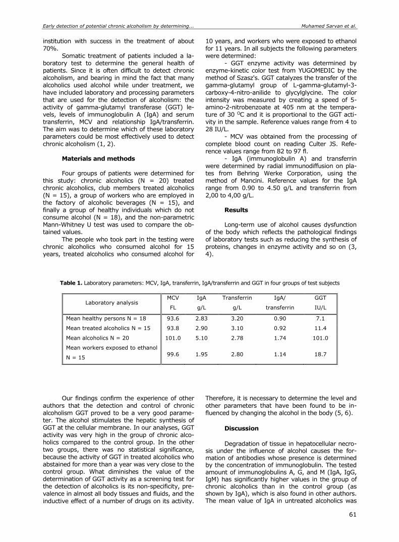
Early detection of potential chronic alcoholism by determining... Muhamed Sarvan et al.
61
institution with success in the treatment of about
70%.
Somatic treatment of patients included a la-boratory test to determine the general health of patients. Since it is often difficult to detect chronic alcoholism, and bearing in mind the fact that many alcoholics used alcohol while under treatment, we
have included laboratory and processing parameters that are used for the detection of alcoholism: the activity of gamma-glutamyl transferase (GGT) le-vels, levels of immunoglobulin A (IgA) and serum transferrin, MCV and relationship IgA/transferrin. The aim was to determine which of these laboratory parameters could be most effectively used to detect
chronic alcoholism (1, 2). Materials and methods
Four groups of patients were determined for
this study: chronic alcoholics (N = 20) treated chronic alcoholics, club members treated alcoholics
(N = 15), a group of workers who are employed in the factory of alcoholic beverages (N = 15), and finally a group of healthy individuals which do not consume alcohol (N = 18), and the non-parametric Mann-Whitney U test was used to compare the ob-tained values.
The people who took part in the testing were chronic alcoholics who consumed alcohol for 15 years, treated alcoholics who consumed alcohol for
10 years, and workers who were exposed to ethanol
for 11 years. In all subjects the following parameters
were determined: - GGT enzyme activity was determined by
enzyme-kinetic color test from YUGOMEDIC by the method of Szasz's. GGT catalyzes the transfer of the gamma-glutamyl group of L-gamma-glutamyl-3-
carboxy-4-nitro-anilide to glycylglycine. The color intensity was measured by creating a speed of 5-amino-2-nitrobenzoate at 405 nm at the tempera-ture of 30 OC and it is proportional to the GGT acti-vity in the sample. Reference values range from 4 to 28 IU/L.
- MCV was obtained from the processing of
complete blood count on reading Culter JS. Refe-rence values range from 82 to 97 fl.
- IgA (immunoglobulin A) and transferrin
were determined by radial immunodiffusion on pla-tes from Behring Werke Corporation, using the method of Mancini. Reference values for the IgA range from 0.90 to 4.50 g/L and transferrin from
2,00 to 4,00 g/L. Results Long-term use of alcohol causes dysfunction
of the body which reflects the pathological findings
of laboratory tests such as reducing the synthesis of proteins, changes in enzyme activity and so on (3, 4).
Table 1. Laboratory parameters: MCV, IgA, transferrin, IgA/transferrin and GGT in four groups of test subjects
Laboratory analysis MCV
FL
IgA
g/L
Transferrin
g/L
IgA/
transferrin
GGT
IU/L
Mean healthy persons N = 18 93.6 2.83 3.20 0.90 7.1
Mean treated alcoholics N = 15 93.8 2.90 3.10 0.92 11.4
Mean alcoholics N = 20 101.0 5.10 2.78 1.74 101.0
Mean workers exposed to ethanol
N = 15 99.6 1.95 2.80 1.14 18.7
Our findings confirm the experience of other
authors that the detection and control of chronic alcoholism GGT proved to be a very good parame-ter. The alcohol stimulates the hepatic synthesis of GGT at the cellular membrane. In our analyses, GGT activity was very high in the group of chronic alco-holics compared to the control group. In the other two groups, there was no statistical significance,
because the activity of GGT in treated alcoholics who abstained for more than a year was very close to the control group. What diminishes the value of the determination of GGT activity as a screening test for the detection of alcoholics is its non-specificity, pre-valence in almost all body tissues and fluids, and the
inductive effect of a number of drugs on its activity.
Therefore, it is necessary to determine the level and
other parameters that have been found to be in-fluenced by changing the alcohol in the body (5, 6).
Discussion
Degradation of tissue in hepatocellular necro-
sis under the influence of alcohol causes the for-mation of antibodies whose presence is determined by the concentration of immunoglobulin. The tested amount of immunoglobulins A, G, and M (IgA, IgG, IgM) has significantly higher values in the group of chronic alcoholics than in the control group (as shown by IgA), which is also found in other authors. The mean value of IgA in untreated alcoholics was
Acta Medica Medianae 2020, Vol.59(1) Early detection of potential chronic alcoholism by determining...
62
significantly increased as compared to the control group, while the other groups showed no statistical difference compared to the control group. Accor-dingly, the IgA is the second parameter that would be used in screening assays for the detection and control of alcoholics.
In the group of chronic alcoholics, those with a reduced amount of transferrin were found in com-parison with the control group, which is explained by alcohol-toxic liver damage which results in difficult protein synthesis and their transfer. The workers exposed to ethanol showed decreased transferrin concentration, while the mean transferrin in treated alcoholics was unchanged compared to the control group. Increased concentrations of IgA under the influence of alcohol and reduced levels of transferrin expressed by the quotient of IgA/transferrin result in an increase of the quotient which may be useful in the study of alcoholics are available as the third parameter. The ratio of IgA/transferrin in chronic alcoholics is increased with a statistical significance, as a group of workers in this ratio is slightly higher than in the control group, but without significant differences.
Finally, MCV in alcoholics showed an increase that reflects discrete anemia. MCV has increased in the group of untreated alcoholics compared to healthy persons and recovering alcoholics.
As for the workers employed in the factory of alcoholic beverages, MCV level was significantly in-creased compared to healthy subjects and reached a value close to that of an alcoholic.
Conclusion
The level of IgA, transferrin, and the MCV and the ratio of IgA/transferrin are parameters that can
be used for the detection of alcohol in parallel with
the determination of GGT activity.
There was no significant difference between the treated alcoholics and healthy controls in all tested parameters, which indicates a well-chosen way of treating alcoholics.
In the group of untreated alcoholics, the value
of the alcoholic IgA, MCV, and the ratio of IgA/ transferrin were increased as well as the activity of GGT (as expected) compared to the healthy indi-viduals and treated alcoholics.
As for the workers employed in the factory of alcoholic beverages, the MCV level was significantly higher than in healthy individuals, and this value is
close to that of alcoholics as a reflection of anemia. In this group, the transferrin concentration is re-duced, so that the ratio of IgA/ transferrin is slightly
higher than that in healthy persons, but without significant differences. Both parameters indicate chronic ethanol intoxication of workers (7, 8).
Looking at the results of the examined para-
meters, one can see that they can help in the de-tection of chronic alcoholics and to monitor the treatment given the fact that many alcoholics use alcohol while under treatment. In chronic alcoholic’s abstinence, the increase in IgA levels and the ratio of IgA/transferrin, as well as the increase in the
activity of the enzyme GGT provide evidence of even minor or sporadic use of alcohol, and normal levels of these parameters in the serum during the treatment of abstinence were confirmed.
References
1. Nikolic J, Nickovic V, Acimovic D. Contemporary aspects of the diagnostics of alcoholic liver disease: VSP 2012; 69(10):874-9. [CrossRef] [PubMed]
2. Nikolic J. Alcoholic intoxication. In: Ali Qureshi G, Parvez H, Caudy P, Parvez S, editors. Neurochemical markers of degenerative nervous diseases and drug addiction. Utrecht: VSP; 1998. p. 193-221.
3. Mathurin P, Abdelnour M, Ramond MJ, Carbonell N, Fartoux L, Serfaty L et al. Early change in bilirubin levels is an important prognostic factor in severe alco-holic hepatitis treated with prednisolone. Hepatology 2003; 38(6):1363-9. [CrossRef] [PubMed]
4. Sillanaukee P, Olsson U. Improved diagnostic classi-fication of alcohol abusers by combining carbohydrate-deficient transferrin and gamma-glutamyltransferase. Clin Chem 2001; 47(4):681-5. [CrossRef] [PubMed]
5. Nomura F, Itoga S. Transferrin (Tf), carbohydrate-deficient transferrin (CDT). Nihon Rinsho 2004; 62 Suppl 11: 240-3. [PubMed]
6. Everitt H. The effect of acute alcohol exposure on hepatic oxidative stress and function: prevention by micronutrients. [dissertation]. Westminster: University of Westminster, School of Electronics and Computer Science; 2010.
7. Bulmer RD, Pointers Towards. Early Detection of Alcoholism, Can Fam Physician 1980; (28):259-61. [PubMed]
8. Enoch MA, Godlberg D. Problem Drinking and Alcoho-lism: Diagnosis and Treatment. Am Fam Physician 2002; 65(3):441-9. [PubMed]
Early detection of potential chronic alcoholism by determining... Muhamed Sarvan et al.
63
Originalni rad UDC: 343.976:616-074
doi:10.5633/amm.2020.0108
RANO OTKRIVANJE POTENCIJALNOG HRONIČNOG ALKOHOLIZMA ODREĐIVANJEM NIVOA IGA, MCV I TRANSFERINA
Muhamed Sarvan1, Sonja Ketin2, Radmila Maksimović1, Rade Biočanin1
1Farmaceutsko zdravstveni fakultet, Travnik, Bosna i Hercegovina 2Visoka brodarska škola akademskih studija Beograd, Srbija
Kontakt: Sonja Ketin
Bulevar vojvode Putnika 7, 11000 Belgrade, Srbija
E-mail: [email protected]
Nivo IgA, MCV i transferina, kao i odnos IgA i transferina, korišćeni su parametri za
detekciju hroničnog alkoholizma. Paralelno sa time određivana je aktivnost GGT. Četiri grupe ispitanika obrađivano je za ovo istraživanje: zdrave osobe (N = 18), hronični alkoholičari ( N = 20), lečeni hronični alkoholičari (N = 15) i najzad grupa radnika zaposlenih u fabrici alkoholnih pića (N = 15). Neparametrijski Mann-Whitney U-test korišćen je za procesuiranje dobijenih vrednosti. Nije bilo značajnije razlike između lečenih alkoholičara i zdravih osoba u svim ispitivanim parametrima (srednja vrednost kod lečenih alkoholičara: IgA = 2,9 g/L; MCV = 93,8; transferin = 3,1 g/L; IgA/transferin = 0,92; GGT = 11,4U /L; zdrave osobe: IgA = 2,83
g/L; MCV = 93,6; transferin = 3,2 g/L; IgA/transferin = 0,9; GGT = 7,1 U/L).U grupi nele-čenih alkoholičara vrednosti IgA, MCV i odnos IgA i transferin povećane su kao što je pove-ćana i aktivnost GGT (što je uobičajeno) u odnosu na aktivnost GGT kod zdravih osoba i lečenih alkoholičara (alkoholičari: IgA = 5,1 g/L; MCV = 101; transferin = 2,78 g/L. IgA/transferin = 1,74; GGT = 101,4 U/L). Što se tiče radnika zaposlenih u fabrici alkoholnih pića, nivo MCV značajno je povećan u odnosu na nivo kod zdravih osoba i ta je vrednost bliska onoj kod alkoholičara (radnici: IgA = 1,95 g/L; MCV = 99,6; transferin = 2,8 g/L; IgA/transferin = 1,14; GGT = 18,7 U/L). Koncentracija transferina smanjena je kod radnika, pa je i odnos IgA/ transferin neznatno viši nego kod zdravih osoba, ali bez značajne razlike. Može se videti da dobijeni rezultati ispitivanih parametara mogu pomoći ne samo za otkrivanje hroničnog alkoholizma, već i za praćenje lečenja, imajući u vidu činjenicu da mnogi alkoholičari koriste alkohol dok su podvrgnuti tretmanu.
Acta Medica Medianae 2020;59(1):60-63.
Ključne reči: hronična bolest, alkoholizam, eritrocitni indeksi, MCV, transferin IgA,
rano otkrivanje
64 www.medfak.ni.ac.rs/amm
Original article UDC: 796.012.414.6‑056.1-053.6
doi:10.5633/amm.2020.0109
RELATIONSHIP BETWEEN BODY COMPOSITION AND VERTICAL JUMP PERFORMANCE AMONG ADOLESCENTS
Darko Stojanović1, Zoran Savić2, Hadži Miloš Vidaković2, Tijana Stojanović3,
Zoran Momčilović4, Toplica Stojanović2
With the aim to investigate the relationship between the body composition of
adolescents and their vertical jump performance, this research was carried out on a sample of seventh grade elementary school students (47 male students). The sample of measuring instruments for body composition assessment included: body height, body mass, sum of five skinfolds thicknesses (biceps, triceps, subscapularis, suprailiac, and calf), body fat percentage, and muscle mass percentage. The SJ and CMJ tests were used for the assessment of vertical jump performance. At the multivariate level the results showed that body composition, as a
predictor system, explained 44% (p = .000) of the variance of SJ and 41% (p = .000) of the variance of CMJ. At the univariate level of it was noted that the sum of five skinfolds had a high influence on the predictor system for SJ (t = -3.77; p = .001) and also a high influence on CMJ (t = -2.98; p = .005). The sum of five skinfolds had a negative impact on SJ and CMJ tests for vertical jump performance assessment. It could be concluded that the relationship between body composition components and vertical jump performance was clearly demonstrated in adolescents.
Acta Medica Medianae 2020;59(1):64-70. Key words: relationship, body composition, vertical jump, adolescents
1University of Niš, Faculty of Sport and Physical Education, Niš, Serbia 2University of Priština – Kosovska Mitrovica, Faculty of Sport
and Physical Education, Leposavić, Serbia 3Club for synchronized swimming "Niš", Niš, Serbia, 4University of Niš, Pedagogical Faculty, Vranje, Serbia
Contact: Darko Stojanović
66a/3 Nemanjića Blvd., 18000 Niš, Serbia
E-mail: [email protected]
Introduction Vertical jump performance is an important
factor in many sports, and the increasing vertical
displacement of an athlete can positively affect achi-evement in sports (1). Also, vertical jump perfor-mance is very often used for talent identification and prediction of future success in sports, especially among adolescents in sensitive periods, when growth and development occur very dynamically (2,
3). Beside physiological and biomechanical fac-
tors, vertical jump performance is affected by va-rious anthropometric characteristics (4, 5). Most of the studies report that well-developed muscle mass
and low percentage of subcutaneous adipose tissue are predictors of good sport performance (6-8).
Body composition has been described as a confounding factor in vertical jump performance, and several studies have attempted to categorize
those body composition variables which better ex-plain jump ability during childhood and adolescence (9), and tried to determine the nature of the rela-tionship between anthropometric factors and vertical jump performance (4, 5, 10-14).
Two studies have identified a significant ne-gative relationship between the sum of skinfold
thickness and vertical jump among karate athletes (12) and hockey and cycling athletes (5). Resear-chers have also suggested lower skinfold thickness is
preferable as it indicates lesser fat mass which ge-nerates no power but has to be carried vertically while jumping. Similar results were found among re-creational athletes (10), where body fat percentage
is negatively related to vertical jump height. Marković and Jarić (11) investigate the relationship between vertical jump height and body mass, and the results show that body mass is independent of vertical jump height. Previous studies found no sig-nificant relationship between body height relative to
vertical jump performance (10, 13, 15). Anthropometry is considered a simple, inex-
pensive and easy-to-use method in epidemiological studies. Skinfold measurements provide excess
Relationship between body composition and vertical jump... Darko Stojanović et al.
65
weight information, and the sum of skinfolds indi-
cates body fat distribution (16).
A reliable and valid approach for estimation of human body composition parameters is a technique known as bioelectrical impedance (BIA). This me-thod is noninvasive, safe, provides rapid measure-ments, requires little operator skills and subject co-
operation, and is portable (17). The most commonly used tests for assessing
explosive strength of the legs and jumping ability are the squad jump (SJ) and countermovement jump (CMJ). The SJ and CMJ tests are a simple, practical, valid, and very reliable measure of the lower body power compared to other jumping ability
tests. The SJ is commonly used to test the con-centric strength of the leg extensors, whereas the CMJ is used to measure the reactive strength of the
lower body (18). The aim
The aim of this study was to investigate the associations between body composition, the vertical jump performance of adolescents, and ability to predict SJ and CMJ from body composition variables.
Materials and methods
The sample of this study consisted of 47 7th
grade elementary school male students, aged 13 years ± 6 months (BH: 165.44 ± 8.69 cm; BM: 56.62 ± 10.29 kg). The sample included every student who volunteered to participate in the re-
search with the consent of their parents. An addi-
tional requirement was that students were clinically healthy during the testing.
All experimental procedures and possible risks and benefits were explained to each student. Signed consent was obtained from their parents prior to the onset of participation and approval to conduct the study was granted by school principals. The study
protects the children's privacy by allowing for ano-nymity and was designed in compliance with the re-commendations for clinical research of the Decla-ration of Helsinki (2013) of the World Medical Asso-ciation. This study was also reviewed and approved by the Ethics Committee of the Faculty of Sport and
Physical Education, University of Niš. Body height (BH) was measured using the
Martin anthropometer GPM 101 (GPM GmbH Switzer-land), following standard procedures (19). Values of BH were measured and recorded in millimeters (mm). Body mass (BM) was measured with an accuracy of 0.1 kg with an electronic body weight
scale Omron BF511 (Omron Healthcare Co, Kyoto, Japan). Skinfold thickness was measured using GPM 6100 (GPM GmbH Switzerland), with an accuracy of 0.2 mm at the biceps, triceps, subscapular, suprai-liac and calf sites, according to the methodology re-commended by the International Biological Program (20). A GPM caliper provides a constant pressure of
10g/mm2. The measurement results were evaluated 2 seconds after the grip was caught on the skin. All five sites of skinfold thicknesses were summed up to
provide the sum of skinfolds (SUM5). Body compo-
sition components, body fat percent (BF%), and
muscle mass percent (MM%) were assessed using the BIA electronic scale Omron BF511. The partici-pants were asked to avoid the following procedures before body composition measurement, as described by Rech et al. (21), not to perform any physical
exercises 12 hours before testing, not to eat or drink anything during the four hours before the evalua-tion, to urinate at least 30 minutes before the eva-luation, not to take any diuretics during the seven days prior to the test, and not to consume alcohol during 48 hours preceding the test.
Optojump (Microgate, Italy) is used for SJ
and CMJ assessment. The Optojump system consists of 2 transmitting and receiving bars equipped with 33 optical Light-Emitting Diods (LEDs) fitted in the
transmitting bar at 3,125 cm intervals. The LEDs on the transmitting bar communicate continuously with those on the receiving bar. The system detects any interruptions in communication between the bars
and calculates their duration. This makes it possible to measure flight and contact times during the per-formance of a series of jumps with an accuracy of 1/1,000 of a second.
For the Sqaut Jump (SJ), the participants started from an upright standing position with their
hands on their hips; they were then instructed to flex their knees and hold a predetermined knee position (~90O) for a count of 3 s. At that point, the participants were instructed to jump as high as pos-sible without performing any countermovement phase. For the Countermovement Jump (CMJ), the
participants started from an upright standing posi-
tion with their hands on their hips (i.e., without an arm swing); they were then instructed to flex their knees (~90O) as quickly as possible, and then jump as high as possible in the ensuing concentric phase (22).
Statistical analyses
Descriptive statistics, the Kolmogorov–Smirnov
(normality of the distribution), and Levene’s (homo-geneity of variance) tests were calculated for all experimental data before inferential testing. Pearson simple and partial correlation coefficients were used
to determine the correlation between the predictor variables (BH, BM, SUM5, BF%, MM%) and criterion
variables (SJ, CMJ). For the further investigation of the association and explanatory power of the pre-dictor variables for criterion variable, a multiple linear regression analysis was used. Statistical procedures were performed using STATISTICA 10 (StatSoft Inc,
Tulsa OK, USA) and the level of significance was set at p ≤ 0.05.
Results The Kolmogorov-Smirnov test showed that
the data were normally distributed and no violation
of homogeneity of variance was found using Levene’s test. With the normal distribution of the results of BC, SJ, and CMJ assessment, it is possible
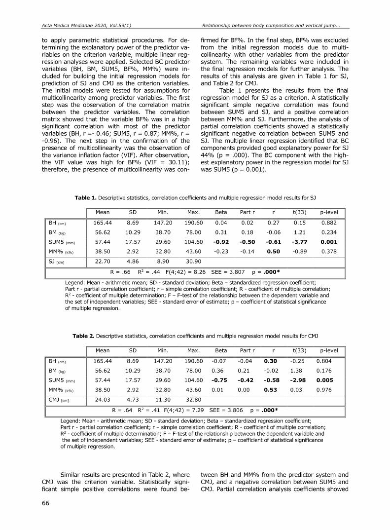
Acta Medica Medianae 2020, Vol.59(1) Relationship between body composition and vertical jump...
66
to apply parametric statistical procedures. For de-
termining the explanatory power of the predictor va-
riables on the criterion variable, multiple linear reg-ression analyses were applied. Selected BC predictor variables (BH, BM, SUM5, BF%, MM%) were in-cluded for building the initial regression models for prediction of SJ and CMJ as the criterion variables.
The initial models were tested for assumptions for multicollinearity among predictor variables. The first step was the observation of the correlation matrix between the predictor variables. The correlation matrix showed that the variable BF% was in a high significant correlation with most of the predictor variables (BH, r =- 0.46; SUM5, r = 0.87; MM%, r =
-0.96). The next step in the confirmation of the presence of multicollinearity was the observation of the variance inflation factor (VIF). After observation,
the VIF value was high for BF% (VIF = 30.11); therefore, the presence of multicollinearity was con-
firmed for BF%. In the final step, BF% was excluded
from the initial regression models due to multi-
collinearity with other variables from the predictor system. The remaining variables were included in the final regression models for further analysis. The results of this analysis are given in Table 1 for SJ, and Table 2 for CMJ.
Table 1 presents the results from the final regression model for SJ as a criterion. A statistically significant simple negative correlation was found between SUM5 and SJ, and a positive correlation between MM% and SJ. Furthermore, the analysis of partial correlation coefficients showed a statistically significant negative correlation between SUM5 and
SJ. The multiple linear regression identified that BC components provided good explanatory power for SJ 44% (p = .000). The BC component with the high-
est explanatory power in the regression model for SJ was SUM5 (p = 0.001).
Table 1. Descriptive statistics, correlation coefficients and multiple regression model results for SJ
Mean SD Min. Max. Beta Part r r t(33) p-level
BH (cm) 165.44 8.69 147.20 190.60 0.04 0.02 0.27 0.15 0.882
BM (kg) 56.62 10.29 38.70 78.00 0.31 0.18 -0.06 1.21 0.234
SUM5 (mm) 57.44 17.57 29.60 104.60 -0.92 -0.50 -0.61 -3.77 0.001
MM% (k%) 38.50 2.92 32.80 43.60 -0.23 -0.14 0.50 -0.89 0.378
SJ [cm] 22.70 4.86 8.90 30.90
R = .66 R2 = .44 F(4;42) = 8.26 SEE = 3.807 p = .000*
Legend: Mean - arithmetic mean; SD - standard deviation; Beta – standardized regression coefficient; Part r - partial correlation coefficient; r – simple correlation coefficient; R - coefficient of multiple correlation; R2 - coefficient of multiple determination; F – F-test of the relationship between the dependent variable and the set of independent variables; SEE - standard error of estimate; p – coefficient of statistical significance of multiple regression.
Table 2. Descriptive statistics, correlation coefficients and multiple regression model results for CMJ
Mean SD Min. Max. Beta Part r r t(33) p-level
BH (cm) 165.44 8.69 147.20 190.60 -0.07 -0.04 0.30 -0.25 0.804
BM (kg) 56.62 10.29 38.70 78.00 0.36 0.21 -0.02 1.38 0.176
SUM5 (mm) 57.44 17.57 29.60 104.60 -0.75 -0.42 -0.58 -2.98 0.005
MM% (k%) 38.50 2.92 32.80 43.60 0.01 0.00 0.53 0.03 0.976
CMJ [cm] 24.03 4.73 11.30 32.80
R = .64 R2 = .41 F(4;42) = 7.29 SEE = 3.806 p = .000*
Legend: Mean - arithmetic mean; SD - standard deviation; Beta – standardized regression coefficient; Part r - partial correlation coefficient; r – simple correlation coefficient; R - coefficient of multiple correlation;
R2 - coefficient of multiple determination; F – F-test of the relationship between the dependent variable and the set of independent variables; SEE - standard error of estimate; p – coefficient of statistical significance of multiple regression.
Similar results are presented in Table 2, where CMJ was the criterion variable. Statistically signi-ficant simple positive correlations were found be-
tween BH and MM% from the predictor system and CMJ, and a negative correlation between SUM5 and CMJ. Partial correlation analysis coefficients showed
Relationship between body composition and vertical jump... Darko Stojanović et al.
67
a statistically significant negative correlation only be-
tween SUM5 and CMJ. The multiple linear regression
identified that BC components provided good ex-planatory power for CMJ 41% (p = .000). The BC component with the highest explanatory power in the regression model for CMJ was SUM5 (p = 0.005).
Discussion The results of anthropometric measures ob-
tained from our sample showed that the average age, BH, and BM were similar to the values obtained in similar studies (3, 5, 9, 23).
The purpose of this study was to examine the relationship between body composition components and the vertical jump performance of 13-year-old boys. The SJ and CMJ have widely been accepted as a criterion for the assessment of vertical jump per-formance.
The results obtained from simple correlation coefficients for SJ indicate a significant negative cor-relation between SUM5 and SJ (r = -0.61), and a significant positive correlation between MM% and SJ (r = 0.50). The results from the simple correlation coefficients for CMJ are very similar with the pre-viously outlined results for SJ in Table 1. A signifi-cant negative correlation was found between SUM5 and CMJ (r = -0.58), and significant positive correla-tions were found between MM% and CMJ (r = 0.53), and BH and CMJ (r = 0.30). These results were expected and support the findings of many previous studies which found similar correlation coefficients between body fat, muscle mass, and vertical jump performance (3-5, 9, 10, 12, 15, 23-26).
After the exclusion of BF% due to multicolli-nearity, all other body composition components were analyzed in order to explain the SJ and CMJ variation of adolescents included in this study.
At the multivariate level, the results showed that body composition, as a predictor system, ex-plained 44% (p = .000) of the variance of SJ and 41% (p = .000) of the variance of CMJ. At the univariate level it was noticed that the sum of five skinfolds (biceps, triceps, subscapular, suprailiac and calf), with a significant negative partial correlation (SJ: Part. r = -0.50; CMJ: Part. r = -0.42) and a strong beta coefficient (SJ: Beta = -0.92; CMJ: Beta = -0.75), had a high influence on the predictor system for SJ (Table 1: t = -3.77; p = .001), and also a high influence on CMJ (Table 2: t = -2.98; p = .005). The sum of five skinfolds had a negative impact on SJ and CMJ tests for vertical jump per-formance assessment and represents a body com-position marker that had the highest predictive power for SJ and CMJ.
The best explanatory power of SUM5 for pre-dicting SJ and CMJ may indicate that using the sum of five sites of the skinfolds is more accurate for estimating the influence of total body fat on vertical jump performance, as fat distribution does not occur in a similar way for individuals. These results can be explained by the fact that body fat percentage is a good indicator of the total amount of adipose tissue in the body, but not the distribution of adipose tissue, while anthropometric measures of skinfolds are good indicators of body fat distribution in the
body (27). When using the sum of these skinfolds, it is possible to clearly see the trend of global accu-mulation of body fat (16). Our data indicate that the participants have much more subcutaneous adipose tissue on their abdominal region (suprailiac skinfold) than the others skinfold sites.
The participants with lower body fat demon-strated the highest vertical jump performance, es-pecially in SJ, in accordance with higher negative simple and partial correlation coefficients between SUM5 and SJ, as compared to SUM5 and CMJ. That can be explained by the difference in vertical jump techniques that are not biomechanically similar for SJ and CMJ (28). Hip extension velocity and trunk angular displacement are essential for jump height during the propulsive phase of the CMJ test (29). According to Piucco and Santos (25), excess body fat induces an increase in body mass which results in a loss of athletic performance in movements that involve speed and explosive power, such as jumps, since acceleration is equal to force divided by mass (30). It can be stated that lower subcutaneous adi-pose tissue, especially in the trunk region, is a de-sirable body composition component, since it means less ballast mass (fat) which generates no power. Thus it provides a lighter body mass to be carried and allows a higher vertical velocity to be achieved during jump performance (12).
The results of this study suggest that there is no significant relationship between body mass and vertical jump performance among adolescents. Our findings are similar to those of previous studies (4, 9, 10), which also showed that body mass has no significant bearing on the vertical jump. This is sup-ported by the findings of Marković and Jarić (11), who studied the relationship between vertical jump height and body mass, where the result showed that body mass is independent of vertical jump height.
Our results indicate no significant relationship between body height relative to vertical jump per-formance on the SJ and CMJ tests, in accordance with previous studies (10, 13, 15), except a weak but still significant simple correlation that was found between BH and CMJ. Based on the conclusions of Maćkala et al. (31), that the highest correlation exists between body height and trunk length (r = 0.93), our assumption leads us again to propulsive phase of the CMJ test where trunk angular dis-placement is crucial for jump height (29).
Conclusion
From the obtained results it could be con-
cluded that the relationship between body compo-sition components and vertical jump performance was clearly demonstrated among the adolescents. With the exception of BH, BM, and MM%, the remaining predictor, SUM5, was able to explain the
SJ and CMJ variation among the adolescents in this study. The sum of biceps, triceps, subscapularis, suprailiac, and calf skinfolds showed the best power to explain vertical jump performance with a negative impact on SJ and CMJ values among the adoles-cents. However, this study has some limitations which have to be pointed out. The sample included
Acta Medica Medianae 2020, Vol.59(1) Relationship between body composition and vertical jump...
68
only seventh grade students, and only field tests
were used to estimate body composition. Future
studies should be conducted including the same and other samples by using direct measures of body composition, such as computed tomography and magnetic resonance imaging, in order to develop more accurate regression models. Also, we must
consider that the participants in this study were
adolescents in sensitive periods, when growth and
motor skill development occur very dynamically, so
the obtained results are valid only for the same age groups. Future studies can be done to evaluate further influence of each skinfold site on vertical jump performance, to obtain more accurate in-formation.
References
1. Häkkinen K. Changes in physical fitness profile in fe-male basketball players during the competitive season including explosive type strength training. J Sports Med Phys Fitness 1993;33(1):19-26.[PubMed]
2. Vaeyens R, Lenoir M, Williams AM, Philippaerts RM. Talent identification and development programmes in sport: Current models and future directions. Sports Med 2008;38(9):703-14.[CrossRef][PubMed]
3. Nikolaidis PT, Gkoudas K, Afonso J, Clemente-Suarez VJ, Knechtle B, Kasabalis S, et al. Who jumps the highest? Anthropometric and physiological correlations of vertical jump in youth elite female volleyball players. J Sports Med Phys Fitness 2017;57(6):802-10. [CrossRef][PubMed]
4. Abidin NZ, Adam MB. Prediction of vertical jump height from anthropometric factors in male and fe-male martial arts athletes. Malays J Med Sci 2013; 20(1):39-45.[PubMed]
5. Sharma HB, Gandhi S, Meitei KK, Dvivedi J, Dvivedi S. Anthropometric basis of vertical jump performance: a study in young Indian national players. J Clin Diagn Res 2017;11(2):CC01-CC05.[CrossRef][PubMed]
6. Ostojić SM, Živanić S. Effects of training on anthro-
pometric and physiological characteristics of elite Serbian soccer players. Acta Biologiae et Medicinae Experimentalis 2001;27:76.
7. Popović S, Akpinar S, Jakšić D, Matić R, Bjelica D. Comparative study of anthropometric measurement and body composition between elite soccer and basketball players. Int J Morphology 2013;31(2):461-7. [CrossRef]
8. Gligoroska JP, Manchevska S, Efremova Lj, Todo-rovska L, Nikolić S. Body composition and maximal oxygen consumption in adult soccer players in the Republic of Macedonia. J Health Sciences 2015; 5(3): 85-92.[CrossRef]
9. Pérez-López A, Sinovas MC, Álvarez-Valverde I, Valades D. Relationship between body composition
and vertical jump performance in young spanish soccer players. J Sport and Human Performance 2015;3(3):1-12.[CrossRef]
10. Davis DS, Briscoe DA, Markowski CT, Saville SE, Taylor CJ. Physical characteristics that predict vertical jump performance in recreational male athletes. Physical Therapy in Sport 2003;4(4):167-74. [CrossRef]
11. Markovic G, Jaric S. Is vertical jump height a body size-independent measure of muscle power? J Sports Sci 2007;25(12):1355-63.[CrossRef][PubMed]
12. Roschel H, Batista M, Monteiro R, Bertuzzi RC, Barroso R, Loturco I, et al. Association between neuromuscular tests and kumite performance on the Brazilian Karate National Team. J Sports SciMed 2009;8(CSSI3):20-4. [PubMed]
13. Aslan CS, Koc H, Aslan M, Ozer U. The effect of height on the anaerobic power of sub-elite athletes. World Applied Science Journal 2011; 12(2):208-11.
14. Nikolaidis PT. Body mass index and body fat percent-age are associated with decreased physical fitness in adolescent and adult female volleyball players. J Res Med Sci 2013;18(1):22-6.[PubMed]
15. Aragón-Vargas LF, Gross MM. Kinesiological factors in vertical jump performance: differences among indivi-duals. J Applied Biomechanics 1997;13(1):24-44. [CrossRef]
16. de Andrade Goncalves EC, Nunes HEG, Silva DAS. Which body fat anthropometric indicators are most strongly associated with maximum oxygen uptake in adolescents? Asian J Sports Med 2017; 8(3):e13812. [CrossRef]
17. Lukaski HC, Johnson PE, Bolonchuk WW, Lykken GI. Assessment of fat-free mass using bioelectrical im-pedance measurements of the human body. Am J Clin Nutr 1985;41(4):810-7.[CrossRef][PubMed]
18. Young W. Laboratory strength assessment of athletes. New Studies in Athletics 1995; 10(1):89-96.
Relationship between body composition and vertical jump... Darko Stojanović et al.
69
19. Norton K, Marfell-Jones M, Whittingham N, Kerr D, Carter L, Saddington K, Gore CJ. Anthropometric
assessment protocols. In: CJ Gore (Ed.) Physiological tests for elite athletes: Australian Sports Commission. Champaign, IL: Human kinetics; 2000. p.66-85.
20. Weiner JS, Lourie JA. Human Biology. A Guide to Field Methods. Oxford, Edinburgh: Blackwell Scientific; 1969.
21. Rech CR, Cordeiro BA, Petroski EL, Vasconcelos FA. Validation of bioelectrical impedance for the prediction of fat-free mass in Brazilian elderly subjects. Arq Bras Endocrinol Metabol 2008;52(7):1163-71. [CrossRef][PubMed]
22. Attia A, Dhahbi W, Chaouachi A, Padulo J, Wong DP, Chamari K. Measurement errors when estimating the vertical jump height with flight time using photocell devices: the example of Optojump. Biol sport 2017; 34(1):63-70.[CrossRef][PubMed]
23. Nikolaidis PT, Asadi A, Santos EJAM, Calleja-González J, Padulo J, Chtourou H, et al. Relationship of body mass status with running and jumping performances in young basketball players. Muscles Ligaments Tendons J 2015;5(3):187-94.[CrossRef][PubMed]
24. MacDonald CJ, Israetel MA, Dabbs NC, Chander H, Allen CR, Lamont HS, et al. Influence of body com-position on selected jump performance measures in collegiate female athletes. Journal of Trainology 2013; 2(2):33-7.[CrossRef]
25. Piucco T, Santos SG. Association between body fat, vertical jump performance and impact in the inferior
limbs in volleyball athletes. Fit Perf J 2009; 8(1):9-15. 26. Ribeiro BG, Mota HR, Sampaio-Jorge F, Morales AP,
Leite TC. Correlation between body composition and the performance of vertical jumps in basketball players. J Exerc Physiol Online 2015;18(5):69-78.
27. Deurenberg P, Pieters JJ, Hautvast JG. The asses-sment of the body fat percentage by skinfold thickness measurements in childhood and young adolescence. Br J Nutr 1990;63(2):293-303.[CrossRef][PubMed]
28. Akl AR. A biomechanical comparison of different vertical jump techniques with and without arm swing. International Journal of Sports and Physical Education 2015;1(1):14-22.[CrossRef]
29. Hseih CT, Cheng L. Kinematic factors in countermove-ment jump for female volleyball players with different skill levels. International Journal of Sports Science 2016;6(1):6-10.[CrossRef]
30. Lorenzo MG, Chamorro RPG. Indice de masa corporal y composición corporal: Un estudio antropometrico de 2500 deportistas de alto nivel. Educación física y deportes 2004;76.
31. Maćkala K, Michalski R, Čoh M, Rausavljević N. The relationship between 200 m performance and selected anthropometric variables and motor abilities in male sprinters. Coll Antropol 2015;39(Suppl 1):69-76. [CrossRef][PubMed]
Acta Medica Medianae 2020, Vol.59(1) Relationship between body composition and vertical jump...
70
Originalni rad UDC: 796.012.414.6‑056.1-053.6
doi:10.5633/amm.2020.0109
RELACIJE IZMEĐU TELESNE KOMPOZICIJE I VISINE VERTIKALNOG SKOKA KOD ADOLESCENATA
Darko Stojanović1, Zoran Savić2, Hadži Miloš Vidaković2, Tijana Stojanović3,
Zoran Momčilović4, Toplica Stojanović2
1Univerzitet u Nišu, Fakultet sporta i fizičkog vaspitanja, Niš, Srbija 2Univerzitet u Prištini – Kosovska Mitrovica, Fakultet za sport i fizičko vaspitanje, Leposavić, Srbija 3Klub za sinhrono plivanje „Niš“, Niš, Srbija 4Univerzitet u Nišu, Pedagoški fakultet, Vranje, Srbija
Kontakt: Darko Stojanović
Bulevar Nemanjića 66a/3, 18000 Niš, Srbija
E-mail: [email protected]
Sa ciljem da se utvrde relacije između telesne kompozicije i visine vertikalnog skoka
kod adolescenata, sprovedeno je istraživanje na uzorku učenika sedmog razreda (47 dečaka). Uzorak mernih instrumenata za procenu telesne kompozicije je bio sačinjen od: telesne visine, telesne mase, sume pet kožnih nabora (tricepsa, bicepsa, leđa, trbuha i potkolenice), pro-
centa masnog tkiva i procenta mišićnog tkiva, kao sistem prediktorskih varijabli. Kao kriteri-jumske varijable, visina skoka iz čučnja (SJ) i visina skoka iz čučnja sa pripremom (CMJ) primenjeni su za procenu visine vertikalnog skoka. Na multivarijantnom nivou, rezultati su pokazali da komponente telesne kompozicije, kao prediktorski sistem, objašnjavaju 44% (p = ,000) varijanse SJ i 41% (p = ,000) varijanse CMJ, kao kriterijuma. Na univarijantnom nivou uočeno je da suma kožnih nabora ima najveći uticaj u prediktorskom sistemu za krite-rijum SJ (t = -3,77; p = ,001), kao i za kriterijum CMJ (t = -2,98; p = ,005). Suma kožnih nabora ima negativan uticaj na visinu vertikalnog skoka SJ i CMJ. Može se zaključiti da postoje značajne relacije između komponenti telesne kompozicije, kao prediktora, i visine vertikalnog skoka, kao kriterijuma, kod adolescenata.
Acta Medica Medianae 2020;59(1):64-70.
Ključne reči: relacije, telesna kompozicija, vertikalni skok, adolescenti
71 www.medfak.ni.ac.rs/amm
Original article UDC: 616‑002.44-036.1-053.9
doi:10.5633/amm.2020.0110
CHARACTERISTICS OF CHRONIC VENOUS ULCERATION RELATED TO AGE
Vesna Karanikolić, Maša Golubović, Nataša Djindjić, Nila Kučar
Venous ulceration occurs in 1% of the total human population, and occurs more often
in people older than 65 years. The aim of the study was to determine the characteristics of chronic venous ulceration (CVU) related to the patients age. The study included 102 ambulatory patients with CVU, treated at the Clinic of Skin Diseases of the Clinical Center Niš. The study group consisted of patients ≥ 65 years, while the control group consisted of patients < 65 years. The study used patients' data such as age, gender, history of deep vein thrombosis, previous episodes of ulceration, previous operation on the veins, body mass index, calf circumference (CC), number of ulcerations, ulceration localization, ulcer size, duration of the ulcer and ulcer locoregional characteristics. Patients aged ≥ 65 years had longer incidence of ulceration and a larger number of previous episodes of ulceration. In patients aged ≥ 65 years, calf circumference was statistically higher, which can significantly affect the speed and healing rate. Larger surface area were more commonly reported in patients aged < 65 years with statistically significant p < 0.05, while elderly patients had statistically (p < 0.01) prolonged ulceration. Concerning the locoregional characteristics of CVU, dermatitis was more commonly seen in patients aged ≥ 65 years, while lipodermatosclerosis was more prevalent in patients aged < 65 years. In relation to the wound infection, patients aged ≥ 65 years had a statistically higher prevalence of ulcer infections. Elderly patients with CVU represent a distinct group in terms of aetiology, natural history and prognosis.
Acta Medica Medianae 2020;59(1):71-75.
Key words: chronic leg ulceration, venous ulceration, the elderly
Clinic of Dermatology, Clinical Center Niš, Serbia
Contact: Vesna Karanikolić
48 Dr. Zoran Djindjić Blvd., 18000 Niš, Serbia
E-mail: [email protected]
Introduction
Venous ulceration is one of the most common
vascular diseases in the human population. It arises as a complication of chronic venous insufficiency. Chronic venous insufficiency is responsible for the occurrence of about 70% of chronic ulcerations of the lower extremities, especially those that do not
heal within 6 weeks (1). Venous ulceration occurs in 1% of the total
human population, and occurs more often in people older than 65 years of age. The ratio of presentation shows a significantly higher proportion of women in the ratio of 3:1 (women: men) (2-5).
Chronic venous ulceration (CVU) occurs in
0.6-3% of the population over 60 years and in patients over 80 years the percentage increases by
another 5%. Chronic venous ulceration is a common cause of morbidity and their prevalence in human population range from 1.9% -13.1% (6-10). The incidence of ulceration is thought to increase as a result of aging and increased factors for athero-sclerosis, such as smoking, obesity, and diabetes are also responsible. During the life, almost 10% of the
population can develop a chronic wound, with a mortality of 2.5% (10).
The aim of the study was to determine the characteristics of CVU related to the patient’s age.
Materials and methods
The study included 102 ambulatory patients
aged over 18 years with a CVU, treated at the Clinic for Skin Diseases of the Clinical Center Niš. The study lasted from April 2015 to February 2016. According to the age, patients were divided into two groups. The study group consisted of patients ≥65
years of age, while the control group consisted of patients < 65 years of age.
Before joining the study, all patients were subjected to Doppler ultrasound (DUS) examination in order to evaluate the venous and arterial leg system. The ankle-brachial pressure index (ABPI) was measured in all patients. Venous aetiology of
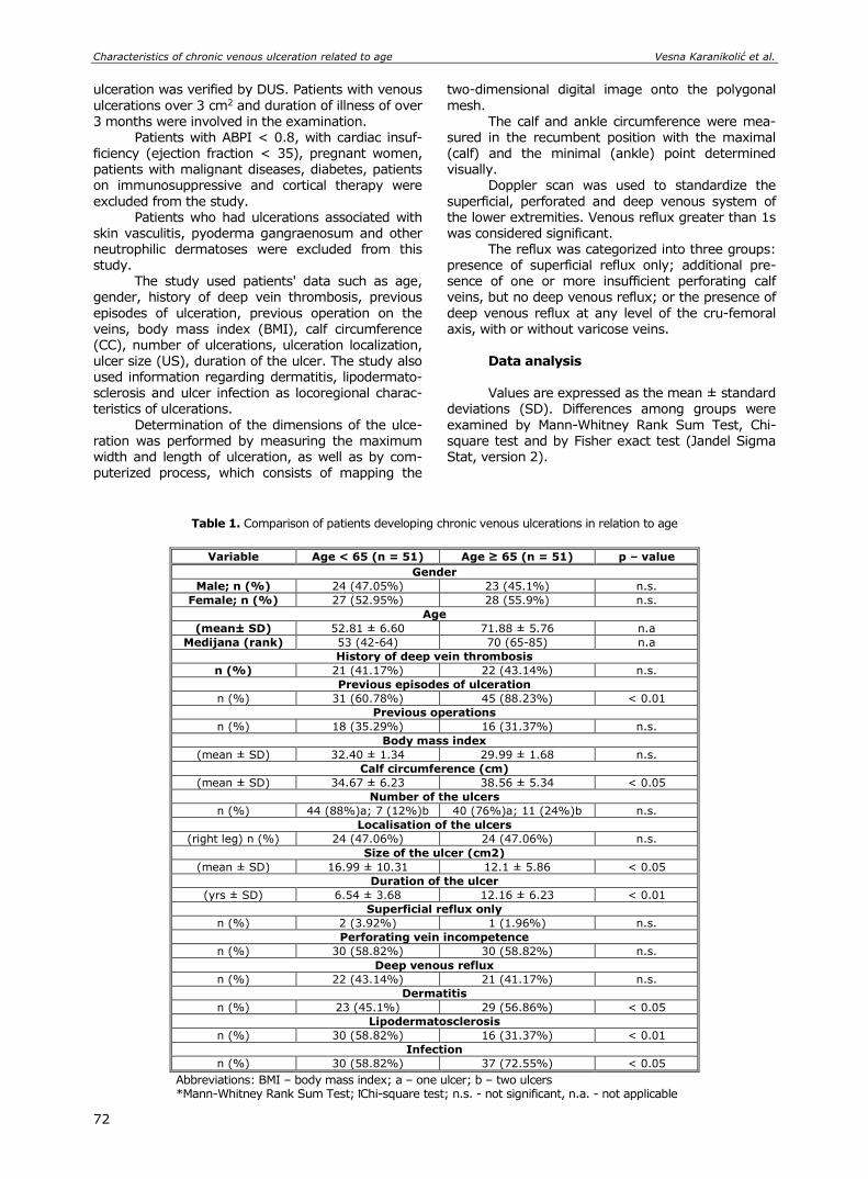
Characteristics of chronic venous ulceration related to age Vesna Karanikolić et al.
72
ulceration was verified by DUS. Patients with venous
ulcerations over 3 cm2 and duration of illness of over
3 months were involved in the examination. Patients with ABPI < 0.8, with cardiac insuf-
ficiency (ejection fraction < 35), pregnant women, patients with malignant diseases, diabetes, patients on immunosuppressive and cortical therapy were
excluded from the study. Patients who had ulcerations associated with
skin vasculitis, pyoderma gangraenosum and other neutrophilic dermatoses were excluded from this study.
The study used patients' data such as age, gender, history of deep vein thrombosis, previous
episodes of ulceration, previous operation on the veins, body mass index (BMI), calf circumference (CC), number of ulcerations, ulceration localization,
ulcer size (US), duration of the ulcer. The study also used information regarding dermatitis, lipodermato-sclerosis and ulcer infection as locoregional charac-teristics of ulcerations.
Determination of the dimensions of the ulce-ration was performed by measuring the maximum width and length of ulceration, as well as by com-puterized process, which consists of mapping the
two-dimensional digital image onto the polygonal
mesh.
The calf and ankle circumference were mea-sured in the recumbent position with the maximal (calf) and the minimal (ankle) point determined visually.
Doppler scan was used to standardize the
superficial, perforated and deep venous system of the lower extremities. Venous reflux greater than 1s was considered significant.
The reflux was categorized into three groups: presence of superficial reflux only; additional pre-sence of one or more insufficient perforating calf veins, but no deep venous reflux; or the presence of
deep venous reflux at any level of the cru-femoral axis, with or without varicose veins.
Data analysis Values are expressed as the mean ± standard
deviations (SD). Differences among groups were
examined by Mann-Whitney Rank Sum Test, Chi-square test and by Fisher exact test (Jandel Sigma Stat, version 2).
Table 1. Comparison of patients developing chronic venous ulcerations in relation to age
Variable Age < 65 (n = 51) Age ≥ 65 (n = 51) p – value
Gender
Male; n (%) 24 (47.05%) 23 (45.1%) n.s.
Female; n (%) 27 (52.95%) 28 (55.9%) n.s.
Age
(mean± SD) 52.81 ± 6.60 71.88 ± 5.76 n.a
Мedijana (rank) 53 (42-64) 70 (65-85) n.a
History of deep vein thrombosis
n (%) 21 (41.17%) 22 (43.14%) n.s.
Previous episodes of ulceration
n (%) 31 (60.78%) 45 (88.23%) < 0.01
Previous operations
n (%) 18 (35.29%) 16 (31.37%) n.s.
Body mass index
(mean ± SD) 32.40 ± 1.34 29.99 ± 1.68 n.s.
Calf circumference (cm)
(mean ± SD) 34.67 ± 6.23 38.56 ± 5.34 < 0.05
Number of the ulcers
n (%) 44 (88%)a; 7 (12%)b 40 (76%)a; 11 (24%)b n.s.
Localisation of the ulcers
(right leg) n (%) 24 (47.06%) 24 (47.06%) n.s.
Size of the ulcer (cm2)
(mean ± SD) 16.99 ± 10.31 12.1 ± 5.86 < 0.05
Duration of the ulcer
(yrs ± SD) 6.54 ± 3.68 12.16 ± 6.23 < 0.01
Superficial reflux only
n (%) 2 (3.92%) 1 (1.96%) n.s.
Perforating vein incompetence
n (%) 30 (58.82%) 30 (58.82%) n.s.
Deep venous reflux
n (%) 22 (43.14%) 21 (41.17%) n.s.
Dermatitis
n (%) 23 (45.1%) 29 (56.86%) < 0.05
Lipodermatosclerosis
n (%) 30 (58.82%) 16 (31.37%) < 0.01
Infection
n (%) 30 (58.82%) 37 (72.55%) < 0.05
Abbreviations: BMI – body mass index; a – one ulcer; b – two ulcers *Mann-Whitney Rank Sum Test; ꝉChi-square test; n.s. - not significant, n.a. - not applicable
Acta Medica Medianae 2020, Vol.59(1) Characteristics of chronic venous ulceration related to age
73
Results
Both the investigated group, age ≥ 65, and the control group, age < 65, had 51 patients. The average age of the age < 65 group was 53 years, while the average age of the age ≥ 65 group was 70 years. There was no statistically significant differ-
ence between sex and history of deep vein throm-bosis. Somewhat higher representation of CVU was in the female population. Previous episodes of ul-ceration were statistically more common in the age ≥ 65 group (p > 0.01). Concerning previous ope-rations on the venous system and body mass index values, the number of ulceration and localization of
ulceration did not have a statistically significant dif-ference between the examined and the control group of patients. Calf circumference was statis-
tically higher p < 0.05 in the age ≥ 65 group. Larger surface area were more commonly reported in pa-tients aged < 65 years with statistically significant p < 0.05, while elderly patients aged ≥ 65 years had
statistically (p < 0.01) longer prolonged ulcerations. There was no statistically significant difference in surface reflux only, perforating vein incompetence and deep venous reflux among examined groups. Concerning the locoregional characteristics of CVU, dermatitis was more prevalent in patients aged ≥ 65
years (p < 0.05), while lipodermatosclerosis was more prevalent in patients aged < 65 years (p < 0.01). In relation to the infection of ulcers, sta-tistically higher prevalence was found in the age ≥ 65 group (p < 0.05) (Table 1).
Discussion
Venous leg ulcers are one of the most com-
mon vascular diseases in human populations. Ve-nous ulcer represents the clinical expression of de-compensated chronic venous insufficiency. The aver-age age of patients with chronic venous ulcers is between 50 and 60 years, out of which 55% of pa-tients are female and 45% male (11). Venous ulce-ration occurs most commonly in patients older than 65 years, although the incidence and prevalence of chronic venous ulceration in the elderly population is not well established. The study of Margolis et al. (12) aimed to highlight the prevalence and incidence of chronic venous ulceration in the population of patients over 65 years of age. Data are obtained from the primary health care register. The annual prevalence of venous ulceration among the older population was 1.69. The overall incidence of venous ulceration was 0.76 for men and 1.42 for women. The study showed that chronic venous ulceration is a significant health problem in elderly patients.
In our study, the female gender was some-what more represented. The average age of patients in the age < 65 group was 53 years, while in the age > 65 group it was 70 years.
A study by MacKenzie et al. (13) showed that patients who developed CVU before their 50th birth-
day were more likely to be male, obese, had a worse disease that appeared less responsive to treatment and had a history of deep vein thrombosis (DVT) and/or long bone fracture suggesting a post-throm-botic aetiology. The proportion of CVUs that are post-thrombotic in aetiology remains controversial. However, recent data suggesting that up to 50% of all CVU patients have at least one thrombophilia suggests that previous, often unrecognized DVT pro-bably plays a significant role in the development of a larger proportion of ulcers than previously appre-ciated.
There was no statistically significant difference between sex groups and history of deep vein thrombosis. Higher representation of CVU was in the female population. Previous episodes of ulceration were statistically more common in the age ≥ 65 group (p > 0.01). Concerning previous operations on the venous system and body mass index values, the number of ulceration and localization of ulce-ration did not have a statistically significant differ-ence between the examined and the control group of patients. Calf circumference was statistically higher (p < 0.05) in the age ≥ 65 group of patients. Several studies (11, 14) showed the importance of calf circumference in healing CVU. Calf circumfer-ence < 33cm represents a positive predictor for suc-cessful healing of venous ulceration, and patients with a larger calf circumference represent a risk group and require more intensive and prolonged therapy.
A number of studies were concerned with the presence of chronic venous disorders, surgery on the venous system and their effect on the healing effect of chronic venous ulceration in patients with the use of a multiplex compressive bandage. Also, dermatological local characteristics of venous ulce-ration were monitored for their presence and influence on the effectiveness of healing chronic venous ulceration in patients with the applied multi-layer compressive bandage. (1, 11, 14)
Large-area ulceration was more common in age < 65 with statistically significant p < 0.05, while elderly patients age ≥ 65 had statistically (p < 0.01) prolonged ulceration.
In summary, patients aged ≥ 65 years had longer incidence of ulceration and a larger number of previous episodes of ulceration. In patients aged ≥ 65 years, calf circumference was statistically higher, which can significantly affect the speed and effectiveness of healing of CVU. Larger surface area were more commonly reported in patients aged < 65 years with statistically significant p < 0.05, while elderly patients had statistically (p < 0.01) pro-longed ulceration. Concerning the locoregional characteristics of CVU, dermatitis was more com-monly seen in patients aged ≥ 65 years, while lipodermatosclerosis was more prevalent in patients aged < 65 years. In relation to the wound infection, patients aged ≥ 65 years had a statistically higher prevalence of infections.
Characteristics of chronic venous ulceration related to age Vesna Karanikolić et al.
74
References
1. Karanikolic V, Karanikolic A, Petrovic D, Stanojevic M. Prognostic factors related to delayed healing of venous leg ulcer treated with compression therapy. Dermato-logica Sinica 2015;33(4):206-9.[CrossRef]
2. Grover G, Tanase A, Elstone A, Ashley S. Chronic venous leg ulcers: Effects of foam sclerotherapy on healing and recurrence.Phlebology 2014.31(1):34-41. [CrossRef][PubMed]
3. Bradbury AW, Evans CJ, Allan PL, Lee AJ, Ruckley CV, Fowkes FGR. What are the symptoms of varicose veins? Edinburgh vein study cross sectional population survey. Br Med J 1999;318(7180):353-6. [CrossRef][PubMed]
4. Evans CJ, Fowkes FGR, Ruckley CV, Lee AJ. Pre-valence of varicose veins in men and women in the general population: Edinburgh Vein Study. J Epidemiol Community Health 1999;53(3):149-53. [CrossRef][PubMed]
5. Allan PL, Bradbury AW, Evans CJ, Lee AJ, Ruckley CV, Fowkes FGR. Patterns of reflux and severity of varicose veins in the general population - Edinburgh
Vein Study. Eur J Vasc Endovasc Surg 2000; 20(5):470-7.[CrossRef][PubMed]
6. MacKenzie RK, Paisley A, Allan PL, Lee AJ, Ruckley CV, Bradbury AW. The effect of long saphenous vein stripping on quality of life. J Vasc Surg 2002;35(6): 1197-203.[CrossRef][PubMed]
7. Phillips TJ. Chronic cutaneous ulcers: etiology and epidemiology. J Invest Dermatol 1994;102(6):38S-41S. [PubMed]
8. Chatterjee SS. Venous ulcers of the lower limb: where do we stand? Indian J Plast Surg 2012;45(2):266-74. [PubMed]
9. Amir O, Liu A, Chang ALS. Stratification of highest-risk patients with chronic skin ulcers in a Stanford retro-spective cohort includes diabetes, need for systemic antibiotics, and albumin levels. Ulcers 2012; ID 767861. [CrossRef]
10. Rayner R, Carville K, Keaton J, Prentice J, Santamaria XN. Leg ulcers: atypical presentations and associated co-morbidities. Wounds 2009;17(4):168-85.
11. Karanikolic V, Binic I, Jovanovic D, Golubovic M, Golubovic I, Djindjic N, et al. The effect of age and compression strength on venous leg ulcer healing. Phlebology 2018;33(9):618-26.[CrossRef][PubMed]
12. Margolis D, Bilker W, Santanna J, Baumgarten M. Venous leg ulcer: Incidence and prevalence in the elderly. J Am AcadDermatol 2002;46(3):381-6. [CrossRef][PubMed]
13. MacKenzie RK, Brown DA, Allan PL, Bradbury AW, Ruckley CV. A comparison of patients who developed
venous leg ulceration before and after their 50th birthday. Eur J Vasc Endovasc Surg 2003;26(2):176-8. [CrossRef][PubMed]
14. Milic DJ, Zivic SS, Bogdanovic DC, Jovanovic MM, Jankovic RJ, Milosevic ZD, et al. The influence of different sub-bandage pressure values on venous leg ulcers healing when treated with compression therapy. J Vasc Surg 2010;51(3):655-61. [CrossRef][PubMed]
Acta Medica Medianae 2020, Vol.59(1) Characteristics of chronic venous ulceration related to age
75
Originalni rad UDC: 616‑002.44-036.1-053.9
doi:10.5633/amm.2020.0110
KARAKTERISTIKE HRONIČNE VENSKE ULCERACIJE POVEZANE SA STAROŠCU
Vesna Karanikolić, Maša Golubović, Nataša Đinđić, Nila Kučar
Klinika za dermatologiju, Klinički centar Niš, Niš, Srbija
Kontakt: Vesna Karanikolić
Bulevar dr Zoran Đinđić 48, 18000 Niš, Srbija
E-mail: [email protected]
Venska ulceracija javlja se kod 1% ukupne humane populacije, a češće se javlja kod
osoba starijih od 65 godina. Cilj studije bio je da se odrede karakteristike hroničnih venskih
ulceracija (CVU) u odnosu na starosnu dob bolesnika. Studijom su obuhvaćena 102 ambu-lantna bolesnika sa CVU, lečena na Klinici za kožne bolesti Kliničkog centra Niš. Studijska grupa sastojala se od bolesnika koji su imali 65 ili više godina, dok su kontrolnu grupu činili bolesnici mlađi od 65 godina. U studiji su korišćeni podaci bolesnika kao što su starost, pol, istorija duboke venske tromboze, prethodne epizode ulceracije, prethodna operacija na venama, indeks telesne mase, obim gležnja (CC), broj ulceracija, lokalizacija ulceracije, veličina ulceracija, trajanje ulceracija i lokalne karakteristike ulceracija. Bolesnici od 65 i više godina imali su dužu istoriju ulceracija, kao i veći broj prethodnih epizoda ulceracija.
Kod bolesnika starijih od 65 godina obim gležnja bio je statistički veći, što može znatno uticati na brzinu i efikasnost lečenja. Veća površina javlja se kod bolesnika starosne dobi manje od 65 godina sa statističkom značajnošću (p < 0,05), dok su stariji bolesnici imali dužu istoriju ulceracija (p < 0,01). Što se tiče lokalno-regionalnih karakteristika CVU, dermatitis se češće javlja kod bolesnika starijih od 65 godina, dok je lipodermatoskleroza bila preovlađujuća kod bolesnika starijih od 65 godina. U odnosu na infekciju rane, bolesnici koji su imali 65 ili više godina imali su statistički veću prevalenciju infekcija ulceracija. Stariji bolesnici sa CVU predstavljaju posebnu grupu u smislu etiologije, istorije i prognoze bolesti.
Acta Medica Medianae 2020;59(1):71-75.
Ključne reči: hronične ulceracije nogu, venska ulceracija, starije osobe
76 www.medfak.ni.ac.rs/amm
Review article UDC: 616-056.7-08:[575.224:577.15
doi:10.5633/amm.2020.0111
SIGNIFICANCE OF GENOTYPIC ALPHA GALACTOSIDASE A MUTATIONS IN FABRY DISEASE TREATMENT
Jelena Randjelović1,2, Mina Cvetković1,2, Tamara Vrećić1,2, Andriana Jovanović1,2,
Marina Randjelović1,3, Tatjana Cvetković1,2
Fabry disease (FD) is a rare inherited X-linked lysosomal storage disorder caused by a
deficiency in alfa-galactosidase A (α-GAL). It is resulting in the accumulation of glycosphingo-lipids that leads to multiple organ dysfunction and ultimately signs and symptoms of the disease. The aim of this study was to examine the significance of genotypic α-GAL mutations in the treatment of FD. The disease can be divided into a severe, classical phenotype, and a milder nonclassical phenotype. Numerous α-GAL mutations are described in gene mutation databases. Missense, nonsense, consensus splice site, cryptic splicing, and frameshift mutations are reported. Enzyme replacement therapy (ERT) can lead to a significant clinical improvement.
Depending on the α-GAL mutation, there are various recommendations for initiation of ERT in adult male and female patients with classic Fabry mutations, later-onset Fabry mutations or α-GAL variants of unknown significance. ERT with recombinant human agalsidase alfa and agalsidase beta is currently available therapy. Although there are no uniform guidelines, development of signs or symptoms related to FD should be an indication to start ERT. Treat-ment with ERT should be combined with adjuvant treatments for specific disease manifesta-tions. Migalastat is a new oral pharmacological molecule developed as an alternative treatment to intravenous ERT for patients with FD and amenable mutations. Migalastat and ERT have similar effects on renal function in patients with FD. Long-term treatment of adult Fabry patients should involve timely ERT, regular assessment of disease progression in all patients, use of appropriate adjunctive therapies and multidisciplinary team approach.
Acta Medica Medianae 2020;59(1):76-82. Key words: Fabry disease, alfa-galactosidase A mutations, treatment
1University of Niš, Faculty of Medicine, Niš, Serbia 2Clinical Center of Niš, Niš, Serbia 3Public Health Institute Niš, Serbia
Contact: Jelena Ranđelović
11/23 Velikotrnavska, 18000 Niš, Serbia
E-mail: [email protected]
Introduction Alfa-galactosidase A (α-GAL) is an enzyme
that cleaves the terminal α-galactosyl unit of the α-gal epitopes and converts this epitope into a disac-charide called N-acetyllactosamine. This enzyme predominantly hydrolyzes ceramide trihexoside, and it can catalyze the hydrolysis of melibiose into galac-tose and glucose. The enzyme is encoded by the α-GAL gene. Defects in human α-GAL result in Fabry disease (FD), because of failure to catabolize α-D-
galactosyl glycolipid moieties (1, 2). FD is a rare inherited X-linked lysosomal storage disorder caused by a deficiency in alfa-galactosidase A (α-GAL), which removes terminal galactose from various ga-lacto-glycolipids (3). It is resulting in accumulation of glycosphingolipids with terminal a-D-galactosyl residue, particularly globotriaosylceramide and glo-botriaosylsphingosine, in plasma, vascular endothe-lial cells, podocytes, cardiomyocytes, and arterial smooth muscle cells (4–6).
Although the exact pathophysiology of FD is still only partly understood, this accumulation of gly-cosphingolipids leads to multiple organ involvement and ultimately signs and symptoms of the disease (7). This multisystem disorder is manifested by de-veloping progressive proteinuric kidney disease, a fibrotic cardiac disease resulting in rhythm and con-duction disturbances, progressive hypertrophic car-diomyopathy, small-fiber neuropathy, and mostly is-chemic cerebrovascular stroke (4). Although FD is X–linked disorder, women often have less severe signs and symptoms of FD, compared with men (8). Cardiac involvement contributes to considerable morbidity and early death due to heart failure or ventricular arrhythmias. The incidence of FD has been estimated to be approximately 1 in 40,000 to 1
Significance of genotypic alpha galactosidase a mutations in fabry... Jelena Ranđelović et al.
77
in 117,000 male (9, 10). Prevalence of FD may be much higher than previously believed. High inci-dence of later-onset FD was revealed by newborn screening (1 in 1,250 to 1 in 3,100 male newborns). Data in the literature shows that 1.2% of young patients with the unexplained acute cerebrovascular disease have FD (11–13).
The disease can be divided into a severe, classical phenotype, and a milder nonclassical phe-notype. Patients with classical FD had a history of more events than patients with nonclassical disease and they are more likely to develop complications. Patients with classical FD most often have symp-toms such as neuropathic pain, cornea verticillata, and angiokeratoma. Clinical manifestations include hypertrophic cardiomyopathy, arrhythmias, progres-sive renal failure, and stroke. Furthermore, patients with classical FD have lower glomerular filtration rate (GFR), higher left ventricular mass and higher plasma globotriaosylsphingosine concentrations than patients with nonclassical FD. Nonclassical FD is characterized by a more variable disease course, pa-tients are generally moderately and mildly affected and disease manifestations may be limited to a single organ (4, 14, 15). There are described va-rious manifestations of FD, as a cardiac variant with manifestations limited to the heart (with left ven-tricular hypertrophy of unknown etiology) and a renal variant among hemodialysis patients with un-known etiology. Lung manifestations, as dyspnea, wheezing and a dry cough are frequent symptoms in many Fabry patients (16). Thus, the great import-ance for the treatment of FD is accurate and early diagnosis (13, 17, 18).
There is a wide phenotypic variability among patients with FD. Patients with missense α-GAL mu-tations and variable residual enzyme activity have milder disease manifestations. For example, the N215S mutation may have residual enzyme activity in plasma. In heterozygous females, random X-in-activation may result in the normal level of α-GAL activity in the plasma or white blood cells in up to 60% of women (19). FD in early adulthood is cha-racterized by more extensive angiokeratomas, high albuminuria (> 1g/24 hours), edema or lymphe-dema, fever, hypohidrosis or anhidrosis, lymphade-nopathy, heat sensitivity, diarrhea, abdominal pain, and cardiac problems. FD in later adulthood is ma-nifested by heart disease (usually fibrotic, left and right ventricular hypertrophy, heart valve abnorma-lities and dysrhythmias, angina, diastolic heart failure), chronic kidney disease including end-stage renal disease, stroke or transient ischemic attacks and deafness (19–22).
The aim of this study was to examine the significance of genotypic α-GAL mutations in the treatment of FD. The data used in the research are obtained from the books and relevant literature by means of PubMed browser.
Genetics The complete genomic sequences of the hu-
man α-GAL gene have been determined and to date, several disease-causing α-GAL mutations have been identified, including missense mutations, small dele-
tions, insertions, splice mutations, and large gene rearrangements (23).
More than 500 pathologic variants of α-GAL have already been described; most of them are family-specific. Numerous α-GAL mutations are de-scribed in gene mutation databases (24, 25). FD is caused by missense, nonsense, consensus splice site, cryptic splicing, and frameshift mutations (small and large deletions and insertions). Mentioned mu-tations are associated with the classic or later-onset FD phenotype, α-GAL variants of unclear signifi-cance, and benign variants. Patients with missense and nonsense mutations have nearly absent GLA activities and increased lyso- globotriaosylceramide levels (20, 26).
Patients with mutations within intronic or re-gulatory α-GAL regions have nonclassical FD pheno-types accompanied by late-onset organ manifesta-tions such as cardiomyopathy, kidney failure, stroke, or neuropathic pain. The clinical impact of the α-GAL p.A143T variation in adult Fabry patients was de-termined in the recent study and patients with this mutation have clinical symptoms and manifestations compared with other classical missense mutations. Most male p.A143T patients had only slightly de-creased residual α-GAL activities and normal lyso-Gb3 levels, while α-GAL activity in heterozygous fe-males was normal. Female and male p.A143T pa-tients had a less renal and cardiac involvement in comparison to FD patients with other missense mu-tations and these patients showed less severe FD-typical symptoms. Both gender p.A143T patients suffering from stroke or transient ischemic attack showed no further FD-typical organ manifestations. The risk for albuminuria and increased disease se-verity scores was significantly lower in p.A143T pa-tients in comparison to groups with classical FD mis-sense mutations. There was no accumulation of ne-urologic events in family members of p.A143T pa-tients with stroke or transient ischemic attack. Fe-male p.A143T patients with stroke or TRANSIENT ISCHEMIC ATTACK did not show skewed X chromo-some inactivation. This study suggested the p.A143T variation to be more likely a neutral variant or a genetic modifier than a disease causing mutation. p.A143T could be a genetic variant of unknown sig-nificance. Thus, recommendation is that p.A143T patients with stroke or TRANSIENT ISCHEMIC ATTACK of unknown etiology should be further eva-luated, since the diagnosis of FD is not probable and enzyme replacement therapy (ERT) or chaperone treatment should not be an unreflected option (27).
The multiplicity of mutations may contribute to variations in the residual enzyme activity and the different clinical presentations. Most of α-GAL gene variants is unique for each family. In study on FD screening in a predominantly hypertensive popula-tion with left ventricular hypertrophy (LVH) was found a mutation of unknown significance in α-GAL gene not previously described in the literature – GLA c.785G>T; p.W262L, whose significance could not be defined. This clinical investigation was able to establish the association between the mutation and the clinical presentation. It is also documented that the clinical management required defining the role of the mutation on the development of the clinical
Acta Medica Medianae 2020, Vol.59(1) Significance of genotypic alpha galactosidase a mutations in fabry...
78
presentation. This study allowed the definition of a novel causal mutation for Fabry disease – GLA c.785G > T; p.W262L (28).
Two new mutations were identified in Turkey. The female patient with M11V mutation had rhe-umatologic symptoms and microalbuminuria. The male patient with R190X mutation had a classical phenotype. R190X mutation causes premature ter-mination, and probably leads to degradation of the protein (29).
Benign and probably benign variants have a high frequency. Medical specialists should be aware that, due to this high frequency, such mutations may be seen in screening studies. This benign poly-morphism may not be related to actual Fabry-re-lated manifestations, as there is no published evi-dence of lysosomal substrate accumulation in the tissues expressing them (30). Most often, nonsense, consensus splice site, and most frameshift mutations are associated with the classic phenotype. They result in low or no α-GAL enzyme activity. On the other side, missense mutations and rare cryptic splicing mutations are associated with the later-onset phenotypes and they result in enzymes with residual α-GAL activity. Current clinical studies have not stratified Fabry patients by genotype (26, 31).
Women with heterozygous α-GAL mutations suffer from significant multisystemic disease and re-duced quality of life and must be monitored and treated accordingly. The asymptomatic female car-rier of FD is the exception, not the rule (32). In female heterozygous Fabry patients who express the normally functioning α-GAL allele, symptoms are mild and rarely occur, while female patients who express the mutant α-GAL allele have a disease course which may be similar to the male disease phenotype (either classic or later-onset). It depends on the underlying α-GAL mutation in their family. At least 43% of obligate carrier women have severe clinical manifestations (33). While the onset of first Fabry-associated symptoms in affected hemizygous males with low or absent enzymatic α-GAL activity starts in early childhood, females with FD present with a heterogeneous clinical picture and variable di-sease progression, independent of the presence of a nonsense or a missense mutation. Recent study showed that although females with missense muta-tions seem to have a lower risk to suffer from severe FD manifestations, affected females showed a si-milar disease burden compared to females with non-sense mutations. The variation and the missing genotype-phenotype correlation might be due to the controversially discussed X-chromosomal inactiva-tion.
Individuals with the c.196G > C nucleotide change which leads to the E66Q enzyme having low α-GAL activity and they have been suspected to have the later-onset Fabry disease phenotype lead-ing to renal and cardiac disease. Biochemical, patho-logical and structural studies strongly suggest that the c.196G > C is not a pathogenic mutation but is a functional polymorphism (34).
Analysis of genotype-phenotype correlations in FD is complicated by a number of factors, such as the high proportion of private mutations and the large phenotypic heterogeneity (35). Some geno-
type-phenotype correlations, as the missense muta-tion p.N215S is established in patients with cardiac manifestations (hypertrophic cardiomyopathy) (36). Determining genotype-phenotype relationships is important for the assessment of whether affected patients may benefit from ERT. The atypical or late-onset type phenotypes present a therapeutic di-lemma.
The genetic background of the patient, con-comitant diseases, environmental modifiers and the presence of additional deleterious α-GAL variants or variants of unknown significance may alter the im-pact of a given gene mutation (30).
Management recommendations Enzyme replacement therapy (ERT) can lead
to significant clinical improvement. ERT with re-combinant human agalsidase alfa and agalsidase beta is currently available therapy aimed at the etio-logy of FD. There is no scientific evidence when the optimal moment of ERT initiation is. Although there are no uniform guidelines, development of signs or symptoms related to FD should be an indication to start ERT. If ERT has not already been started earlier for nonrenal manifestations such as pain, the deve-lopment of chronic kidney disease (CKD), patholo-gical albuminuria, decreased GFR or progressive de-crease in GFR may be an indication to start (37). Research recommendations for initiation of therapy include:
1) determination of the beginning treatment in asymptomatic patients, females, patients with nonclassic disease;
2) obtaining expanded information on the natural history of FD in classic female patients and nonclassic FD patients, and the effects of ERT in these groups and
3) undertaking X-linked inactivation studies and early initiation of therapy in females (19).
Depending on the mutation there are various recommendations for initiation of ERT in adult male and female patients with classic Fabry mutations, later-onset Fabry mutations or α-GAL variants of un-clear significance. Depending on the approach to treatment, patients with classic Fabry mutation can be divided into three groups: symptomatic or asymptomatic male patients, symptomatic female patients, and asymptomatic female patients. In the first group, ERT should be considered and is appro-priate in all patients at any age of presentation. Treatment decisions may be influenced by the ad-vanced elderly age of the patient and severe co-morbidity. In the second group, initiation of ERT is warranted when signs and symptoms suggesting major organ involvement occur. In the third group, ERT should be considered if there is a laboratory, histological, or imaging evidence of kidney, heart, or the cerebral injury. ERT should also be considered if a skewed X chromosome inactivation pattern with predominant expression of the mutant α-GAL allele with or without very low α-GAL activity has been de-monstrated in the presence of signs and symptoms of the disease. Male and female patients with later-onset Fabry mutation or missense α-GAL variants of unclear significance should start ERT if there is a
Significance of genotypic alpha galactosidase a mutations in fabry... Jelena Ranđelović et al.
77
laboratory, histological, or imaging evidence of in-jury to the kidney, heart, or the central nervous system, even in the absence of typical Fabry symp-toms. The abnormalities should be attributable to FD; this may require histological assessment or biochemical evidence of globotriaosylceramide accu-mulation. The advice of an expert in genetics and management of FD should be sought for interpreta-tion of the pathogenicity of any α-GAL variants of unclear significance. Individuals with well-charac-terized benign α-GAL polymorphisms should not be treated with ERT. In the absence of demonstrable FD-related tissue pathology or clinical symptoms, ERT may not be appropriate, particularly in hetero-zygous female patients. These patients should be monitored regularly by a multidisciplinary care team (19, 37–39). Current FD guidelines and recommend-ations suggest ERT initiation in females with FD after the onset of first FD-typical renal, cardiac, and ce-rebral complications, or in rapidly progressive di-sease (39, 40). Fifty-seven percent of female FD pa-tients were under ERT, nearly all presented with different organ manifestations that justify ERT initia-tion according to current European guidelines. One third of females were untreated although indications (organ manifestations). Some of them refused ERT, or can’t receive ERT due to pregnancy or future family planning, or they were refusing ERT because of possible benefit from future chaperone treatment (41).
Periodic monitoring of anti-agalsidase antibo-dies in patients receiving ERT is also recommended, considering these natural defense mechanisms may block the effectiveness of ERT treatment and lead to dose adjustment. Early treatment of FD may im-prove clinical outcome, initiation of ERT at a younger age in patients with classical FD results in a better biochemical response (42). ERT should not be rest-ricted to hemizygous men but should be considered for both heterozygous females and children (20).
ERT should be combined with adjunctive the-rapies for adult patients with FD. Renal, cardiac, neurological, gastrointestinal, cerebrovascular and other complications of FD that cause chronic tissue injury should be treated with symptomatic specific therapy (43–45).
Migalastat is an oral pharmacological chape-rone developed as an alternative treatment to intra-venous ERT for patients with FD and amenable mu-tations. This new oral small-molecule stabilizes spe-cific mutant (amenable) forms of α-Gal to facilitate normal lysosomal trafficking. In this way, this drug may increase endogenous enzymatic activity in pa-tients with specific missense α-Gal mutations. Miga-lastat and ERT have similar effects on renal function. There is no significant change in left ventricular mass index in patients treated with Migalastat com-pared to patients treated by ERT. In randomized, active-controlled study renal, cardiac or cerebrovas-cular events occurred in 29% and 44% of patients in the migalastat and ERT groups, respectively. Plasma globotriaosylsphingosine was low and stable in migalastat treated patient with FD. Migalastat was generally safe and well tolerated (46–48).
Conclusion
FD affects very emotionally and physically on patients and their families. Long-term treatment of adult patients with FD should include ERT, regular assessment of disease progression, and the use of appropriate adjunctive therapies (38, 40).
Numerous α-Gal mutations complicate thera-peutic management of this rare chronic disease. FD requires multidisciplinary care in the treatment of organ-specific complications. Active participation of medical specialists experienced in treating this dis-order is necessary, as well as communication be-tween the neurologist, nephrologist, cardiologist,
medical geneticist, genetic counselor, psychologist,
and nurse. Significance of genetic polymorphisms in FD pathology remains unknown (49) and the res-ponse to therapy may depend on α-GAL gene muta-tions.
Acknowledgment
This study was supported by grant of Ministry
of Science and Technological Development – project number 41018.
79
Acta Medica Medianae 2020, Vol.59(1) Significance of genotypic alpha galactosidase a mutations in fabry...
80
References
1. Scriver CR, Beaudet AL, Sly WS, Valle D, Childs B, Kinzler KW, Vogelstein B, eds. The metabolic and molecular bases of inherited disease. 8th ed. New York:McGraw-Hill;2001.
2. Galili U. The natural anti-Gal antibody as foe turned friend in medicine. Philadelphia:Saunders (Elsevier); 2018.287-300 p. [CrossRef]
3. Swaiman KF, Ashwal S, Ferriero D, Schor N, eds. Swaiman’s pediatric neurology : principles and prac-tice. 5th ed. Philadelphia: Saunders (Elsevier); 2012. 107 p. [CrossRef]
4. Desnick R, Ioannou Y, Eng C. A-galactosidase a deficiency: Fabry disease. In: Valle D, Beaudet AL, Vogelstein B, Kinzler KW, Antonarakis SE, Ballabio A, et al., editors. The Online Metabolic and Molecular Bases of Inherited Disease. New York:McGraw-Hill Medical;2014. [CrossRef]
5. Germain DP. Fabry disease. Orphanet J Rare Dis 2010;5:30. [CrossRef] [PubMed]
6. Brady RO, Gal AE, Bradley RM, Martensson E, Warshaw AL, Laster L. Enzymatic defect in Fabry’s
disease. N Engl J Med 1967;276(21):1163-7. [CrossRef] [PubMed]
7. Schuller Y, Arends M, Körver S, Langeveld M, Hollak CEM. Adaptive pathway development for Fabry di-sease: a clinical approach. Drug Discov Today 2018; 23(6):1251-7. [CrossRef] [PubMed]
8. MacDermot KD, Holmes A, Miners AH. Anderson-Fabry disease: clinical manifestations and impact of disease in a cohort of 60 obligate carrier females. J Med Genet 2001;38(11):769-75. [CrossRef] [PubMed]
9. Ioannou YA, Zeidner KM, Gordon RE, Desnick RJ. Fabry disease: preclinical studies demonstrate the effectiveness of alpha-galactosidase A replacement in enzyme-deficient mice. Am J Hum Genet 2001;68 (1):14-25. [CrossRef] [PubMed]
10. Meikle PJ, Hopwood JJ, Clague AE, Carey WF. Prevalence of lysosomal storage disorders. JAMA 1999;281(3):249-54. [CrossRef] [PubMed]
11. Rolfs A, Böttcher T, Zschiesche M, Morris P, Winchester B, Bauer P, et al. Prevalence of Fabry di-sease in patients with cryptogenic stroke: a pros-pective study. Lancet 2005;366(9499):1794-6. [CrossRef] [PubMed]
12. Spada M, Pagliardini S, Yasuda M, Tukel T, Thiagarajan G, Sakuraba H, et al. High incidence of later-onset Fabry disease revealed by newborn screening. Am J Hum Genet 2006;79(1):3140. [CrossRef] [PubMed]
13. Yoshimitsu M, Higuchi K, Miyata M, Devine S, Mattman A, Sirrs S, et al. Identification of novel mutations in the α-galactosidase A gene in patients with Fabry disease: pitfalls of mutation analyses in patients with low α-galactosidase A activity. J Cardiol 2011;57(3):345-53. [CrossRef] [PubMed]
14. Smid BE, van der Tol L, Biegstraaten M, Linthorst GE, Hollak CEM, Poorthuis BJHM. Plasma globotria-osylsphingosine in relation to phenotypes of Fabry disease. J Med Genet 2015;52(4):262-8. [CrossRef] [PubMed]
15. Arends M, Wanner C, Hughes D, Mehta A, Oder D, Watkinson OT, et al. Characterization of classical and nonclassical Fabry disease: A multicenter study. J Am Soc Nephrol 2017;28(5):1631-41. [CrossRef] [PubMed]
16. Magage S, Lubanda JC, Susa Z, Bultas J, Karetová D, Dobrovolný R, et al. Natural history of the respiratory involvement in Anderson–Fabry disease. J Inherit Metab Dis 2007;30(5):790-9. [CrossRef] [PubMed]
17. Sachdev B, Takenaka T, Teraguchi H, Tei C, Lee P, McKenna WJ, et al. Prevalence of Anderson-Fabry disease in male patients with late onset hypertrophic cardiomyopathy. Circulation 2002;105(12):1407-11. [CrossRef] [PubMed]
18. Nakao S, Kodama C, Takenaka T, Tanaka A, Yasumoto Y, Yoshida A, et al. Fabry disease: detection of undiagnosed hemodialysis patients and identi-fication of a “renal variant” phenotype. Kidney Int 2003;64(3):801-7. [CrossRef] [PubMed]
19. Schiffmann R, Hughes DA, Linthorst GE, Ortiz A, Svarstad E, Warnock DG, et al. Screening, diagnosis, and management of patients with Fabry disease: conclusions from a "Kidney Disease: Improving Global Outcomes’’ (KDIGO) Controversies Conference. Kidney Int 2017;91(2):284-93. [CrossRef] [PubMed]
20. Mehta A, Ricci R, Widmer U, Dehout F, Garcia de
Lorenzo A, Kampmann C, et al. Fabry disease defined: baseline clinical manifestations of 366 patients in the Fabry Outcome Survey. Eur J Clin Invest 2004; 34(3):236-42. [CrossRef] [PubMed]
21. Ramaswami U, Whybra C, Parini R, Pintos-Morell G, Mehta A, Sunder-Plassmann G, et al. Clinical mani-festations of Fabry disease in children: data from the Fabry Outcome Survey. Acta Paediatr 2006; 95(1): 86-92. [CrossRef] [PubMed]
22. MacDermot KD, Holmes A, Miners AH. Anderson-Fabry disease: clinical manifestations and impact of disease in a cohort of 98 hemizygous males. J Med Genet 2001;38(11):750-60. [CrossRef] [PubMed]
23. Tuttolomondo A, Duro G, Miceli S, Di Raimondo D, Pecoraro R, Serio A, et al. Novel alpha-galactosidase A mutation in a female with recurrent strokes. Clin Biochem 2012;45(16-17):1525-30. [CrossRef] [PubMed]
24. International Fabry Disease Genotype-Phenotype Database (dbFGP). International Fabry Disease Geno-type-Phenotype Database (dbFGP) 2016 "cited 2019 Apr 4"; Available from: http://dbfgp.org/dbFgp/fabry/
25. The Fabry disease mutation database. "cited 2019 Apr 4";Available from: http://fabry-database.org/ mutants/
26. Niemann M, Rolfs A, Störk S, Bijnens B, Breunig F, Beer M, et al. Gene mutations versus clinically relevant phenotypes: lyso-Gb3 defines Fabry disease. Circ Cardiovasc Genet 2014;7(1):8-16. [CrossRef] [PubMed]
27. Lenders M, Weidemann F, Kurschat C, Canaan-Kühl S, Duning T, Stypmann J, et al. Alpha-Galactosidase A p.A143T, a non-Fabry disease-causing variant. Orpha-net J Rare Dis 2016;11:54. [CrossRef] [PubMed]
28. Baptista A, Magalhães P, Leão S, Carvalho S, Mateus P, Moreira I. Screening for Fabry disease in left ventricular hypertrophy: Documentation of a novel mutation. Arq Bras Cardiol 2015;105(2):139-44. [CrossRef] [PubMed]
29. Celtikci B, Topçu M, Ozkara A. Two novel alpha-galactosidase A mutations causing Fabry disease: A missense mutation M11V in a heterozygote woman and a nonsense mutation R190X in a hemizygote man. Clin Biochem 2011;44(10-11):809-12. [CrossRef] [PubMed]
Significance of genotypic alpha galactosidase a mutations in fabry... Jelena Ranđelović et al.
81
30. Desnick RJ, Doheny DO, Chen B, Yu C, Nazarenko I, Lee B, et al. Fabry disease: The α-galactosidase A
(GLA) c.427G>A (A143T) mutation, effect of the 5′-10C>T polymorphism. Mol Genet Metab 2015; 114 (2):S37. [CrossRef]
31. Ferreira S, Ortiz A, Germain DP, Viana-Baptista M, Caldeira-Gomes A, Camprecios M, et al. The alpha-galactosidase A p.Arg118Cys variant does not cause a Fabry disease phenotype: data from individual patients and family studies. Mol Genet Metab 2015; 114(2):248-58. [CrossRef] [PubMed]
32. Wang RY, Lelis A, Mirocha J, Wilcox WR. Heterozygous Fabry women are not just carriers, but have a significant burden of disease and impaired quality of life. Genet Med 2007;9(1):34-45. [CrossRef] [PubMed]
33. Wilcox WR, Oliveira JP, Hopkin RJ, Ortiz A, Banikazemi M, Feldt-Rasmussen U, et al. Females with Fabry disease frequently have major organ involvement: Lessons from the Fabry Registry. Mol Genet Metab 2008;93(2):112-28. [CrossRef] [PubMed]
34. Togawa T, Tsukimura T, Kodama T, Tanaka T, Kawashima I, Saito S, et al. Fabry disease: Bio-chemical, pathological and structural studies of the α-galactosidase A with E66Q amino acid substitution. Mol Genet Metab 2012;105(4):615-20. [CrossRef] [PubMed]
35. Ries M, Gal A. Genotype–phenotype correlation in Fabry disease. In: Mehta A, Beck M, Sunder-Plassmann G, editors. Fabry Disease: Perspectives from 5 Years of FOS. Oxford: PharmaGenesis; 2006. [CrossRef] [PubMed]
36. Patel V, O’Mahony C, Hughes D, Rahman MS, Coats C, Murphy E, et al. Clinical and genetic predictors of major cardiac events in patients with Anderson–Fabry Disease. Heart 2015;101(12):9616. [CrossRef] [PubMed]
37. Beck M, Hughes D, Kampmann C, Larroque S, Mehta A, Pintos-Morell G, et al. Long-term effectiveness of agalsidase alfa enzyme replacement in Fabry disease: A Fabry Outcome Survey analysis. Mol Genet Metab Rep 2015;3:21-7. [CrossRef] [PubMed]
38. Ortiz A, Germain DP, Desnick RJ, Politei J, Mauer M, Burlina A, et al. Fabry disease revisited: Management and treatment recommendations for adult patients. Mol Genet Metab 2018;123(4):416-27. [CrossRef] [PubMed]
39. Biegstraaten M, Arngrímsson R, Barbey F, Boks L, Cecchi F, Deegan PB, et al. Recommendations for initiation and cessation of enzyme replacement therapy in patients with Fabry disease: the European Fabry Working Group consensus document. Orphanet J Rare Dis 2015;10:36. [CrossRef] [PubMed]
40. Eng CM, Germain DP, Banikazemi M, Warnock DG, Wanner C, Hopkin RJ, et al. Fabry disease: Guidelines
for the evaluation and management of multi-organ system involvement. Genet Med 2006;8(9):539-48.
[CrossRef] [PubMed] 41. Lenders M, Hennermann JB, Kurschat C, Rolfs A,
Canaan-Kühl S, Sommer C, et al. Multicenter Female Fabry Study (MFFS) - Clinical survey on current treatment of females with Fabry disease. Orphanet J Rare Dis 2016;11:88. [CrossRef] [PubMed]
42. Arends M, Wijburg FA, Wanner C, Vaz FM, van Kuilenburg ABP, Hughes DA, et al. Favourable effect of early versus late start of enzyme replacement therapy on plasma globotriaosylsphingosine levels in men with classical Fabry disease. Mol Genet Metab 2017; 121(2):157-61. [CrossRef] [PubMed]
43. Politei JM, Bouhassira D, Germain DP, Goizet C, Guerrero-Sola A, Hilz MJ, et al. Pain in Fabry disease: Practical recommendations for diagnosis and treat-ment. CNS Neurosci Ther 2016; 22(7):568-76. [CrossRef] [PubMed]
44. Burlina AP, Sims KB, Politei JM, Bennett GJ, Baron R, Sommer C, et al. Early diagnosis of peripheral nervous system involvement in Fabry disease and treatment of neuropathic pain: the report of an expert panel. BMC Neurol 2011;11:61. [CrossRef] [PubMed]
45. Attal N, Cruccu G, Baron R, Haanpää M, Hansson P, Jensen TS, et al. EFNS guidelines on the pharma-cological treatment of neuropathic pain: 2010 revision. Eur J Neurol 2010;17(9):1113-e88. [CrossRef] [PubMed]
46. Hughes DA, Nicholls K, Shankar SP, Sunder-Plassmann G, Koeller D, Nedd K, et al. Oral pharmacological chaperone migalastat compared with enzyme replacement therapy in Fabry disease: 18-month results from the randomised phase III ATTRACT study. J Med Genet 2017;54(4):288-96. [CrossRef] [PubMed]
47. Germain DP, Hughes DA, Nicholls K, Bichet DG, Giugliani R, Wilcox WR, et al. Treatment of Fabry’s disease with the pharmacologic chaperone migalastat. N Engl J Med 2016;375(6):545-55. [CrossRef] [PubMed]
48. Benjamin ER, Della Valle MC, Wu X, Katz E, Pruthi F, Bond S, et al. The validation of pharmacogenetics for the identification of Fabry patients to be treated with migalastat. Genet Med 2017;19(4):430-8. [CrossRef] [PubMed]
49. Tuttolomondo A, Duro G, Pecoraro R, Simonetta I, Miceli S, Colomba P, et al. A family with various symptomatology suggestive of Anderson-Fabry di-sease and a genetic polymorphism of alpha galacto-sidase A gene. Clin Biochem 2015;48(1-2):55-62. [CrossRef] [PubMed]
Acta Medica Medianae 2020, Vol.59(1) Significance of genotypic alpha galactosidase a mutations in fabry...
82
Revijalni rad UDC: 616-056.7-08:[575.224:577.15
doi:10.5633/amm.2020.0111
ZNAČAJ GENOTIPSKIH MUTACIJA ALFA GALAKTOZIDAZE A U TERAPIJI FABRIJEVE BOLESTI
Jelena Ranđelović1,2, Mina Cvetković1,2, Tamara Vrećić1,2, Andriana Jovanović1,2,
Marina Ranđelović1,3, Tatjana Cvetković1,2
1Univerzitet u Nišu, Medicinski fakultet, Niš, Srbija 2Klinički centar Niš, Niš, Srbija 3Institut za javno zdravlje Niš, Niš, Srbija
Kontakt: Jelena Ranđelović
Velikotrnavska 11/23,18000 Niš, Srbija
E-mail: [email protected]
Fabrijeva bolest (FB) je retko nasledno „X vezano“ lizozomalno oboljenje izazvano
deficijencijom alfa galaktozidaze A (α-GAL). To dovodi do nagomilavanja glikosfingolipida, što dalje vodi do disfunkcije mnogih organa i na kraju do znakova i simptoma bolesti. Cilj ovog rada bio je da se ispita značaj genotipskih mutacija α-GAL u terapiji FB. Bolest se može
manifestovati u vidu teškog, klasičnog fenotipa i blagog, neklasičnog fenotipa. Opisane su brojne α-GAL mutacije u bazi podataka o genskim mutacijama, kao što su mutacije po-grešnog smisla (missense), besmislene mutacije (nonsense), mutacije obrade (splice site), kriptičke mutacije (cryptic splicing) i mutacije pomeranja okvira čitanja (frameshift). Terapija zamene enzima (TZE) može doprineti značajnom kliničkom poboljšanju. U zavisnosti od vrste mutacije α-GAL, postoje različite preporuke o započinjanju TZE kod odraslih muškaraca i žena sa klasičnim mutacijama, mutacijama koje dovode do kasnog početka Fabrijeve bolesti i varijantama α-GAL nepoznatog značaja. Trenutno dostupna TZE je primena rekombinantne agalzidaze alfa i agalzidaze beta. Iako nema jedinstvenih vodiča, razvoj znakova i simptoma bolesti trebalo bi da bude indikacija za početak lečenja TZE. Ovu terapiju treba kombinovati sa adjuvantnom terapijom za specifične manifestacije bolesti. Migalastat je novi farmakološki oralni molekul, razvijen kao alternativa intravenskoj TZE za bolesnike sa FB. Migalastat i TZE imaju slične efekte na bubrežnu funkciju kod bolesnika sa ovom bolešću. Dugoročni tretman odraslih bolesnika sa FB treba da podrazumeva pravovremenu TZE, redovno praćenje progresije bolesti svih obolelih, primenu odgovarajuće adjuvantne terapije i multidisciplinarni pristup bolesti.
Acta Medica Medianae 2020;59(1):76-82.
Ključne reči: Fabrijeva bolest, mutacije alfa galaktozidaze A, terapija
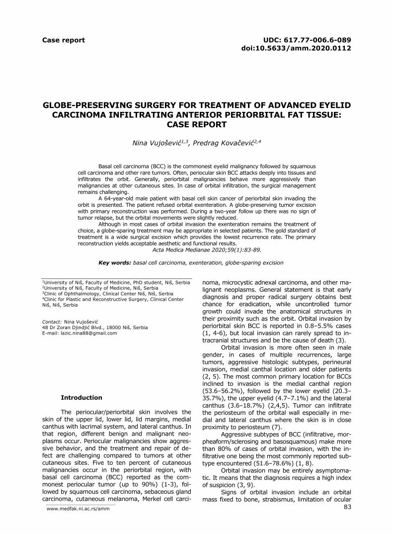
83 www.medfak.ni.ac.rs/amm
Case report UDC: 617.77-006.6-089
doi:10.5633/amm.2020.0112
GLOBE-PRESERVING SURGERY FOR TREATMENT OF ADVANCED EYELID CARCINOMA INFILTRATING ANTERIOR PERIORBITAL FAT TISSUE:
CASE REPORT
Nina Vujošević1,3, Predrag Kovačević2,4
Basal cell carcinoma (BCC) is the commonest eyelid malignancy followed by squamous
cell carcinoma and other rare tumors. Often, periocular skin BCC attacks deeply into tissues and infiltrates the orbit. Generally, periorbital malignancies behave more aggressively than malignancies at other cutaneous sites. In case of orbital infiltration, the surgical management remains challenging.
A 64-year-old male patient with basal cell skin cancer of periorbital skin invading the orbit is presented. The patient refused orbital exenteration. A globe-preserving tumor excision with primary reconstruction was performed. During a two-year follow up there was no sign of tumor relapse, but the orbital movements were slightly reduced.
Although in most cases of orbital invasion the exenteration remains the treatment of choice, a globe-sparing treatment may be appropriate in selected patients. The gold standard of treatment is a wide surgical excision which provides the lowest recurrence rate. The primary reconstruction yields acceptable aesthetic and functional results.
Acta Medica Medianae 2020;59(1):83-89. Key words: basal cell carcinoma, exenteration, globe-sparing excision
1University of Niš, Faculty of Medicine, PhD student, Niš, Serbia 2University of Niš, Faculty of Medicine, Niš, Serbia 3Clinic of Ophthalmology, Clinical Center Niš, Niš, Serbia 4Clinic for Plastic and Reconstructive Surgery, Clinical Center
Niš, Niš, Serbia
Contact: Nina Vujošević
48 Dr Zoran Djindjić Blvd., 18000 Niš, Serbia
E-mail: [email protected]
Introduction The periocular/periorbital skin involves the
skin of the upper lid, lower lid, lid margins, medial canthus with lacrimal system, and lateral canthus. In that region, different benign and malignant neo-
plasms occur. Periocular malignancies show aggres-sive behavior, and the treatment and repair of de-fect are challenging compared to tumors at other cutaneous sites. Five to ten percent of cutaneous malignancies occur in the periorbital region, with basal cell carcinoma (BCC) reported as the com-monest periocular tumor (up to 90%) (1-3), fol-
lowed by squamous cell carcinoma, sebaceous gland carcinoma, cutaneous melanoma, Merkel cell carci-
noma, microcystic adnexal carcinoma, and other ma-lignant neoplasms. General statement is that early diagnosis and proper radical surgery obtains best chance for eradication, while uncontrolled tumor
growth could invade the anatomical structures in their proximity such as the orbit. Orbital invasion by periorbital skin BCC is reported in 0.8–5.5% cases (1, 4-6), but local invasion can rarely spread to in-tracranial structures and be the cause of death (3).
Orbital invasion is more often seen in male gender, in cases of multiple recurrences, large
tumors, aggressive histologic subtypes, perineural invasion, medial canthal location and older patients (2, 5). The most common primary location for BCCs inclined to invasion is the medial canthal region
(53.6–56.2%), followed by the lower eyelid (20.3–35.7%), the upper eyelid (4.7–7.1%) and the lateral
canthus (3.6–18.7%) (2,4,5). Tumor can infiltrate the periosteum of the orbital wall especially in me-dial and lateral canthus where the skin is in close proximity to periosteum (7).
Aggressive subtypes of BCC (infiltrative, mor-pheaform/sclerosing and basosquamous) make more than 80% of cases of orbital invasion, with the in-
filtrative one being the most commonly reported sub-type encountered (51.6–78.6%) (1, 8).
Orbital invasion may be entirely asymptoma-tic. It means that the diagnosis requires a high index of suspicion (3, 9).
Signs of orbital invasion include an orbital mass fixed to bone, strabismus, limitation of ocular
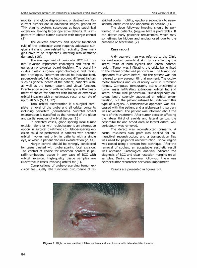
Globe-preserving surgery for treatment of advanced eyelid carcinoma... Nina Vujošević et al.
84
motility, and globe displacement or destruction. Re-
current tumors are in advanced stages, graded by
TMN staging system, suspicious at huge subclinical extension, leaving larger operative defects. It is im-portant to obtain tumor excision with margin control (2).
The delicate anatomy and specific functional
rule of the periocular zone requires adequate sur-gical skills and care related to radicality (free mar-gins have to be respected), meeting also aesthetic demands (10).
The management of periocular BCC with or-bital invasion represents challenges and often re-quires an oncological board decision. The board in-
cludes plastic surgeon, ophthalmologist and radia-tion oncologist. Treatment should be individualized, patient-related, taking into account different factors
such as general health of the patient, patient’s wish, as well as the tumor extent and visual function. Exenteration alone or with radiotherapy is the treat-ment of choice for patients with bulbar or extensive
orbital invasion with an estimated recurrence rate of up to 28.5% (5, 11, 12).
Total orbital exenteration is a surgical com-plete removal of the globe and all orbital contents including periorbita (periosteum). Subtotal orbital exenteration is classified as the removal of the globe
and partial removal of orbital tissues (13). In selected cases, globe-sparing local tumor
excision alone or with radiotherapy is an alternative option in surgical treatment (5). Globe-sparing ex-cision could be performed in patients with anterior orbital involvement only, in patients with a single
eye, or when a patient declines exenteration (2, 14).
Margin control should be strongly considered for cases treated with globe sparing local excision. The control of choice for resection borders is pa-raffin-embedded tissue in any case of BCC with orbital invasion. High-quality tissue samples are illustrative in cases involving orbital fat (1).
Complications of globe-preserving tumor ex-
cision are usually late functional disturbance of re-
stricted ocular motility, epiphora secondary to naso-
lacrimal obstruction and abnormal lid position (1).
The close follow-up imaging should be per-formed in all patients, (regular MRI is preferable). It can detect early posterior recurrences, which may sometimes be hidden and undiagnosed due to the presence of scar tissue (2).
Case report A 64‐year-old man was referred to the Clinic
for exulcerated periorbital skin tumor affecting the lateral third of both eyelids and lateral canthal region. Tumor was infiltrating the orbit, being fixed to the lateral orbital wall periosteum. The tumor had
appeared four years before, but the patient was not referred to any surgeon till that moment. The oculo-
motor functions and visual acuity were in reference ranges. Computed tomography scans presented a tumor mass infiltrating extraconal orbital fat and lateral orbital wall periosteum. Multidisciplinary on-
cology board strongly suggested an orbital exen-teration, but the patient refused to underwent this type of surgery. A conservative approach was dis-cussed with the patient and a globe-sparing surgery was advocated. The patient was informed about the risks of this treatment. After tumor excision affecting the lateral third of eyelids and lateral cantus, the
periorbital fat and broad area of lateral orbital wall periosteum was removed.
The defect was reconstructed primarily. A partial thickness skin graft was applied for co-njunctival reconstruction, and a transposition flap
was used for palpebral reconstruction. Donor region was closed using a tension free technique. After the
removal of stiches, an acceptable aesthetic result was obtained. Pathological analysis indicated the diagnosis of BCC and clear resection margins on all samples. During a two-year follow-up, there was neither tumor recurrence nor visual impairment.
Results are presented in figures 1-7.
Figure 1. Right lateral canthal infiltrative basal cell carcinoma with lateral orbital invasion
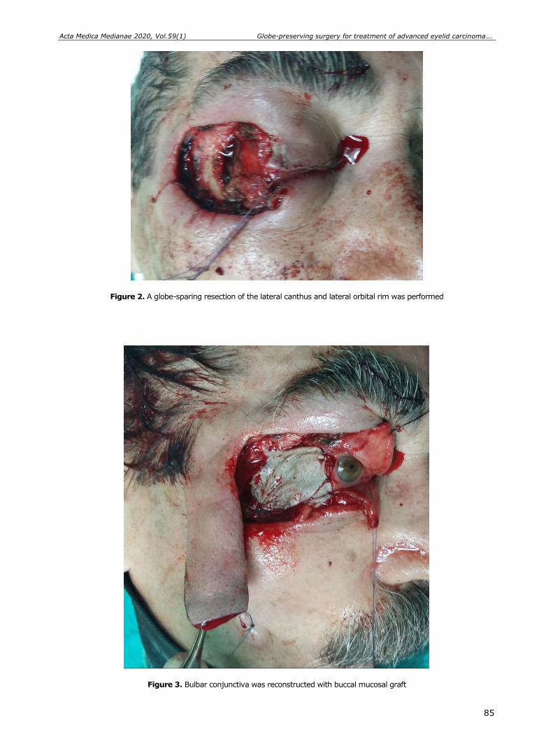
Acta Medica Medianae 2020, Vol.59(1) Globe-preserving surgery for treatment of advanced eyelid carcinoma...
85
Figure 2. A globe-sparing resection of the lateral canthus and lateral orbital rim was performed
Figure 3. Bulbar conjunctiva was reconstructed with buccal mucosal graft
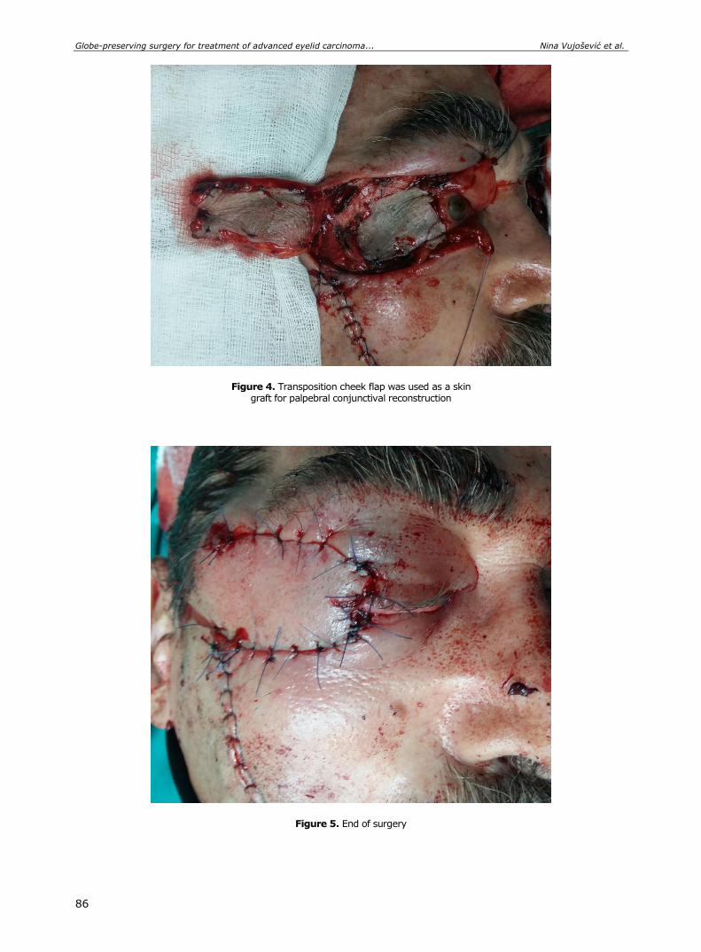
Globe-preserving surgery for treatment of advanced eyelid carcinoma... Nina Vujošević et al.
86
Figure 4. Transposition cheek flap was used as a skin graft for palpebral conjunctival reconstruction
Figure 5. End of surgery
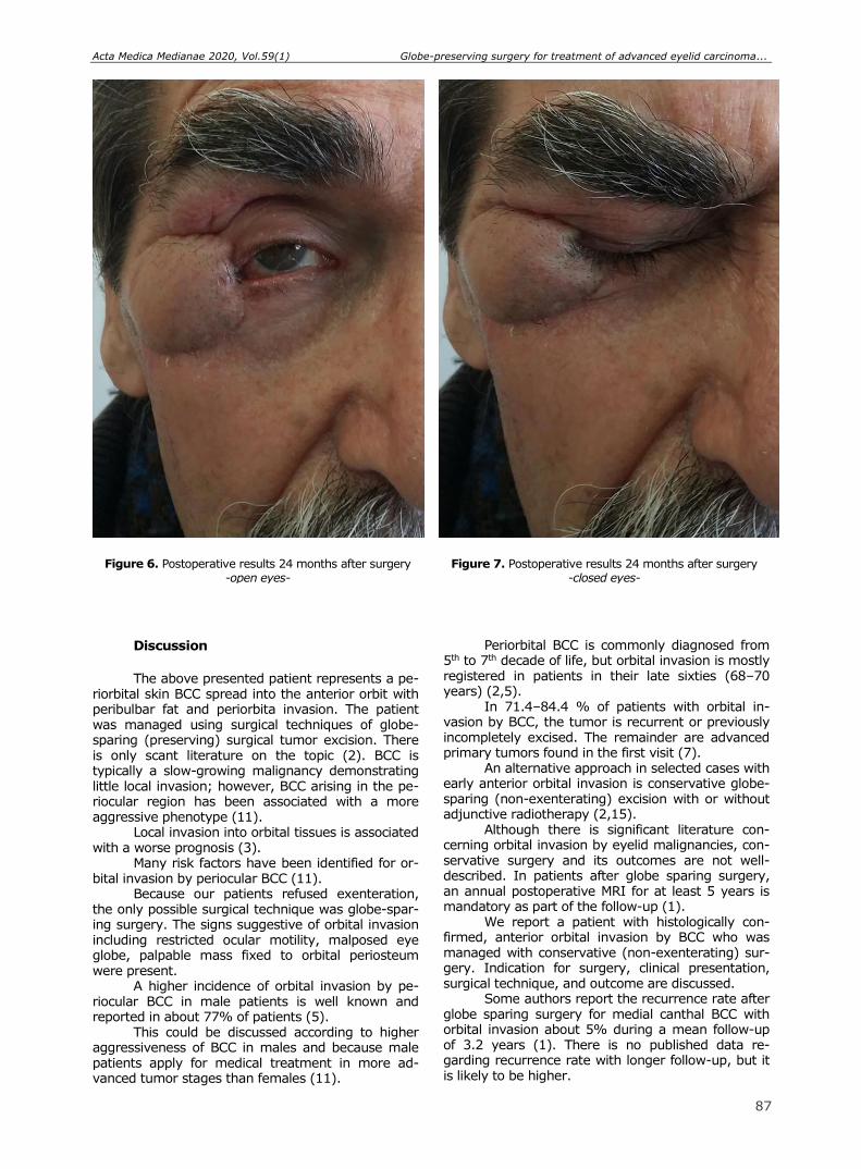
Acta Medica Medianae 2020, Vol.59(1) Globe-preserving surgery for treatment of advanced eyelid carcinoma...
85
Figure 6. Postoperative results 24 months after surgery -open eyes-
Figure 7. Postoperative results 24 months after surgery
-closed eyes-
Discussion
The above presented patient represents a pe-
riorbital skin BCC spread into the anterior orbit with peribulbar fat and periorbita invasion. The patient was managed using surgical techniques of globe-sparing (preserving) surgical tumor excision. There is only scant literature on the topic (2). BCC is typically a slow-growing malignancy demonstrating little local invasion; however, BCC arising in the pe-riocular region has been associated with a more aggressive phenotype (11).
Local invasion into orbital tissues is associated with a worse prognosis (3).
Many risk factors have been identified for or-bital invasion by periocular BCC (11).
Because our patients refused exenteration, the only possible surgical technique was globe-spar-ing surgery. The signs suggestive of orbital invasion including restricted ocular motility, malposed eye globe, palpable mass fixed to orbital periosteum were present.
A higher incidence of orbital invasion by pe-riocular BCC in male patients is well known and reported in about 77% of patients (5).
This could be discussed according to higher aggressiveness of BCC in males and because male patients apply for medical treatment in more ad-vanced tumor stages than females (11).
Periorbital BCC is commonly diagnosed from 5th to 7th decade of life, but orbital invasion is mostly registered in patients in their late sixties (68–70 years) (2,5).
In 71.4–84.4 % of patients with orbital in-vasion by BCC, the tumor is recurrent or previously incompletely excised. The remainder are advanced primary tumors found in the first visit (7).
An alternative approach in selected cases with early anterior orbital invasion is conservative globe-sparing (non-exenterating) excision with or without adjunctive radiotherapy (2,15).
Although there is significant literature con-cerning orbital invasion by eyelid malignancies, con-servative surgery and its outcomes are not well-described. In patients after globe sparing surgery, an annual postoperative MRI for at least 5 years is mandatory as part of the follow-up (1).
We report a patient with histologically con-firmed, anterior orbital invasion by BCC who was managed with conservative (non-exenterating) sur-gery. Indication for surgery, clinical presentation, surgical technique, and outcome are discussed.
Some authors report the recurrence rate after globe sparing surgery for medial canthal BCC with orbital invasion about 5% during a mean follow-up of 3.2 years (1). There is no published data re-garding recurrence rate with longer follow-up, but it is likely to be higher.
87
Globe-preserving surgery for treatment of advanced eyelid carcinoma... Nina Vujošević et al.
88
The decision of radical (exenteration) or con-servative- globe sparing surgery in case of orbital invasion in periorbital BCC must be made after de-tailed discussion with the patient. The same principle applies to lateral canthal BCC with limited anterior orbital invasion and the globe-sparing approach may be considered. This type of surgery brings the risk of postoperative complications and possible revision procedures. When the tumor is highly aggressive and has higher risk of recurrence, or perineural in-vasion, exenteration may be suggested as the preferred treatment choice (1,10).
Conclusion
Orbital invasion by periocular BCC is rare, but the treatment is difficult and the complication rate is
high. Radical tumor removal must be confirmed by
paraffin margin control. Besides orbital exenteration
in selected cases, conservative surgery with globe sparing excision is one of the recommended ap-proaches. Globe-sparing surgical excision with man-datory yearly performed MR imaging as a regular follow-up may be appropriate in case of anterior
orbital involvement only, in patients with a single eye or with poor vision in the opposite eye. Also, globe-sparing procedure could be performed in cases when patient refuses exenteration.
In case of orbital infiltration by periorbital skin BCC, the orbital extenteration remains the gold standard, but globe-sparing surgery can be consi-
dered in select cases.
References
1. Madge SN, Khine AA, Thaller VT, Davis G, Malhotra R, McNab A, et al. Globe-sparing surgery for medial canthal Basal cell carcinoma with anterior orbital invasion. Ophthalmology 2010; 117(11):2222-8. [CrossRef] [PubMed]
2. Leibovitch I, McNab A, Sullivan TJ, Davis G, Selva D. Orbital invasion by periocular basal cell carcinoma. Ophthalmology 2005; 112(4):717-23. [CrossRef] [PubMed]
3. Prabhakaran VC, Gupta A, Huilgol SC, Selva D. Basal cell carcinoma of the eyelids. Compr Ophthalmol Update 2007; 8(1):1-14. [PubMed]
4. Wong VA, Marshall JA, Whitehead KJ, Williamson RM, Sullivan TJ. Management of periocular basal cell carcinoma with modified en face frozen section con-trolled excision. Ophthalmic Plast Reconstr Surg 2002; 18(6), 430-5. [CrossRef] [PubMed]
5. Iuliano A, Strianese D, Uccello G, Diplomatico A, Tebaldi S, Bonavolonta G. Risk factors for orbital exenteration in periocular Basal cell carcinoma. Am J Ophthalmol 2012; 153(2):238-41. [CrossRef] [PubMed]
6. Malhotra R, Huilgol SC, Huynh NT, Selva D. The Australian Mohs database, part I: periocular basal cell carcinoma experience over 7 years. Ophthalmology 2004; 111(4):624-30. [CrossRef] [PubMed]
7. Walling HW, Fosko SW, Geraminejad PA, Whitaker DC, Arpey CJ. Aggressive basal cell carcinoma: presentation, pathogenesis, and management. Cancer Metastasis Rev 2004; 23(3–4), 389-402. [CrossRef] [PubMed]
8. Paavilainen V, Aaltonen M, Tuominen J, Saari KM. Histological characteristics of basal cell carcinoma of the eyelid. Ophthalmic Res 2007; 39(1):45-8. [CrossRef] [PubMed]
9. Selva D, Hale L, Bouskill K, Huilgol SC. Recurrent morphoeic basal cell carcinoma at the lateral canthus with orbitocranial invasion. Australas J Dermatol 2003; 44(2):126-8. [CrossRef] [PubMed]
10. Slutsky JB, Jones EC. Periocular cutaneous malig-nancies: a review of the literature. Dermatol Surg 2012; 38(4):552-69. [CrossRef] [PubMed]
11. Meads SB, Greenway HT. Basal cell carcinoma associated with orbital invasion: clinical features and treatment options. Dermatol Surg 2006; 32(3):442-6. [CrossRef] [PubMed]
12. Williams LS, Mancuso AA, Mendenhall WM. Perineural spread of cutaneous squamous and basal cell carcinoma: CT and MR detection and its impact on patient management and prognosis. Int J Radiat Oncol Biol Phys 2001; 49(4):1061-9. [CrossRef] [PubMed]
13. Ben Simon GJ, Schwarcz RM, Douglas R, Fiaschetti D, McCann JD, Goldberg RA. Orbital exenteration: one size does not fit all. Am J Ophthalmol 2005; 139 (1): 11-17. [CrossRef] [PubMed]
14. Satchi K, Mcnab AA. Orbital spread of eyelid and periocular cutaneous cancer. Int Ophthalmol Clin 2009; 49(4):223-36. [CrossRef] [PubMed]
15. Sun MT, Wu A, Figueirae E, Huilgol S, Selva D. Management of periorbital basal cell carcinoma with orbital invasion. Future Oncol 2015; 11(22):3003-10. [CrossRef] [PubMed]
Acta Medica Medianae 2020, Vol.59(1) Globe-preserving surgery for treatment of advanced eyelid carcinoma...
89
Prikaz bolesnika UDC: 617.77-006.6-089
doi:10.5633/amm.2020.0112
HIRURŠKO LEČENJE UZNAPREDOVALOG KARCINOMA KAPKA KOJI INFILTRIRA PREDNJE PERIBULBARNO MASNO TKIVO UZ OČUVANJE
OČNE JABUČICE — PRIKAZ SLUČAJA
Nina Vujošević1,3, Predrag Kovačević2,4
1Univerzitet u Nišu, Medicinski fakultet, student doktorskih studija, Niš, Srbija 2Univerzitet u Nišu, Medicinski fakultet, Niš, Srbija 3Klinika za oftalmologiju, Klinički centar Niš, Niš, Srbija 4Klinika za plastičnu i rekonstruktivnu hirurgiju, Klinički centar Niš, Niš, Srbija
Kontakt: Nina Vujošević
Bul. dr Zorana Đinđića 48, 18000 Niš, Srbija
E-mail: [email protected]
Karcinom bazalnih ćelija (KBĆ) najčešči je malignitet kapka, koji prate karcinom
skvamoznih ćelija i drugi retki tumori. KBĆ periokularne kože često se širi u dublje slojeve i infiltrira orbitu. Periorbitalni maligniteti uglavnom se ponašaju mnogo agresivnije nego mali-gniteti na ostalim mestima na koži. U slučaju infiltracije orbite, hiruško lečenje ostaje izazov.
Prikazan je šezdesetčetvorogodišnji bolesnik muškog pola sa karcinomom bazalnih ćelija kože periorbite koji invadira u orbitu. Bolesnik je odbio egzenteraciju orbite. Izvedena je ekscizija tumora sa primarnom rekonstrukcijom i očuvanjem očne jabučice. Za vreme dvogodišnjeg praćenja, nije bilo znakova recidiva tumora, ali su pokreti očne jabučice bili blago redukovani.
Iako u većini slučajeva orbitalne invazije egzenteracija ostaje izborna metoda lečenja, poštedna operacija očne jabučice može biti odgovarajuća kod određenih bolesnika. Zlatni standard u lečenju predstavlja ekstenzivna hirurgija, kod koje se očekuje najniža stopa reci-diva. Primarna rekonstrukcija omogućava zadovoljavajuće estetske i funkcionalne rezultate.
Acta Medica Medianae 2020;59(1):83-89.
Ključne reči: karcinom bazalnih ćelija, egzenteracija, poštedna ekscizija očne
jabučice
90 www.medfak.ni.ac.rs/amm
Case report UDC: 618.19-006.6::616.31-006
doi:10.5633/amm.2020.0113
INVASIVE DUCTAL BREAST CANCER METASTASIS INTO THE TEMPOROMANDIBULAR JOINT TWO YEARS AFTER THE INITIAL
TREATMENT: A CASE REPORT
Predrag Radović1,2, Milovan Papović1, Miloš Trajković1, Nikola Živković2,3, Andrija Ćosić1,2
A tumor metastatic to the oral cavity and jaws is a relatively rare finding. These types of
tumor represent a serious challenge for surgical oncologists, because of difficult diagnosis, un-specific symptomatology and atypical radiographic findings. The symptoms that occur with tu-mors metastatic to the temporomandibular joint and ramus of the lower jaw are usually un-specific, and usually include painful sensations, tumescence, difficulty opening mouth, which often mislead the clinician towards a temporomandibular joint disease, such as arthritis, joint subluxation, degenerative changes and infection.
In this case, the patient described is a 62-year-old woman, with a metastatic tumor in the temporomandi-bular joint and lower jaw region, and a very uncertain prognosis. In this pa-per, we will discuss surgical and oncological therapeutic options recommendable in this case, based on radiographic findings and histopathological cancer type.
Acta Medica Medianae 2020;59(1):90-95. Key words: breast cancer metastasis, temporomandibular joint, histopathological
type and subtype, surgical treatment
1Clinic of Dentistry, Department of Maxillofacial Surgery, Niš, Serbia 2University of Niš, Faculty of Medicine, Niš, Serbia 3Centar for Pathology and Pathological Anatomy, Clinical Center
Niš, Niš, Serbia
Contact: Milovan Papović
48 Dr. Zoran Djindjić Blvd., 18000 Niš, Serbia
E-mail: [email protected]
Introduction Metastatic tumors represent a rare pathologi-
cal entity in the head and neck region, constituting about 1% of the total number of all malignant tu-
mors affecting the region (HNR) (1). Lymphatic and hematogenous dissemination of
infraclavicular malignancies which metastasize to the HNR much more frequently occurs via the ductus thoracicus, compared to the spread via the Batson paraspinal venous plexus (2).
In the available literature, the occurrence of
mtastatic tumors is described in all of the anatomical sections of the NHR, i.e. in the lymph nodes, salivary
glands, and thyroid, while most of the studies report-
ed so far have presented the data about the oral ca-vity and jaw soft tissue involvement (3).
The most frequent site of the primary tumor
metastasizing to the NHR are lungs in men and bre-asts in women (4).
Clinical manifestations of the tumors metasta-tic to the region are rather diverse. These vary from the changes such as anamnestically longterm pain-less tumor formations involving the salivary gland, bulbous protrusions caused by retrobulbar process-
es, severe headaches, neck lymphadenopathy in the area covering the tumescences and pain in the re-gion of jaws, exophytic and ulcerous changes in the oral cavity, followed by paresthesias and progressive tooth loss.
The occurrence of metastases in distant or-
gans indicates the onset of the terminal disease sta-ge, with the average patient survival from 3.7 to 32 months after the histopathological verification of me-tastatic disease (5).
We will here present the case of a patient with completed diagnostic and therapeutic procedures conducted in order to treat breast cancer metastasis
to the lower jaw region. Case report A female patient aged 62 years was referred
to the Clinic of Dentistry, Department for Maxillofaci- al Surgery in Niš by her oncologist, in order to have
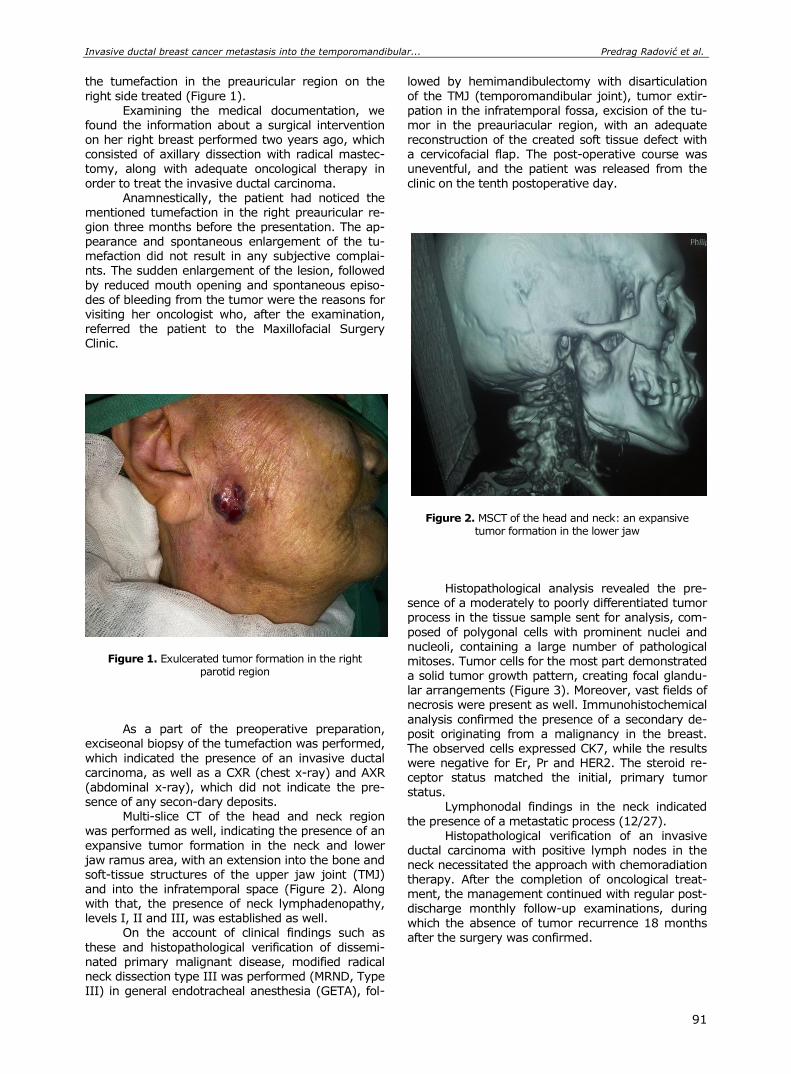
Invasive ductal breast cancer metastasis into the temporomandibular... Predrag Radović et al.
91
the tumefaction in the preauricular region on the
right side treated (Figure 1).
Examining the medical documentation, we found the information about a surgical intervention on her right breast performed two years ago, which consisted of axillary dissection with radical mastec-tomy, along with adequate oncological therapy in
order to treat the invasive ductal carcinoma. Anamnestically, the patient had noticed the
mentioned tumefaction in the right preauricular re-gion three months before the presentation. The ap-pearance and spontaneous enlargement of the tu-mefaction did not result in any subjective complai-nts. The sudden enlargement of the lesion, followed
by reduced mouth opening and spontaneous episo-des of bleeding from the tumor were the reasons for visiting her oncologist who, after the examination,
referred the patient to the Maxillofacial Surgery Clinic.
Figure 1. Exulcerated tumor formation in the right parotid region
As a part of the preoperative preparation, exciseonal biopsy of the tumefaction was performed,
which indicated the presence of an invasive ductal carcinoma, as well as a CXR (chest x-ray) and AXR
(abdominal x-ray), which did not indicate the pre-sence of any secon-dary deposits.
Multi-slice CT of the head and neck region was performed as well, indicating the presence of an
expansive tumor formation in the neck and lower jaw ramus area, with an extension into the bone and soft-tissue structures of the upper jaw joint (TMJ) and into the infratemporal space (Figure 2). Along with that, the presence of neck lymphadenopathy, levels I, II and III, was established as well.
On the account of clinical findings such as
these and histopathological verification of dissemi-nated primary malignant disease, modified radical neck dissection type III was performed (MRND, Type III) in general endotracheal anesthesia (GETA), fol-
lowed by hemimandibulectomy with disarticulation
of the TMJ (temporomandibular joint), tumor extir-
pation in the infratemporal fossa, excision of the tu-mor in the preauriacular region, with an adequate reconstruction of the created soft tissue defect with a cervicofacial flap. The post-operative course was uneventful, and the patient was released from the
clinic on the tenth postoperative day.
Figure 2. MSCT of the head and neck: an expansive tumor formation in the lower jaw
Histopathological analysis revealed the pre-sence of a moderately to poorly differentiated tumor process in the tissue sample sent for analysis, com-
posed of polygonal cells with prominent nuclei and nucleoli, containing a large number of pathological mitoses. Tumor cells for the most part demonstrated a solid tumor growth pattern, creating focal glandu-lar arrangements (Figure 3). Moreover, vast fields of necrosis were present as well. Immunohistochemical
analysis confirmed the presence of a secondary de-posit originating from a malignancy in the breast. The observed cells expressed CK7, while the results were negative for Er, Pr and HER2. The steroid re-ceptor status matched the initial, primary tumor
status. Lymphonodal findings in the neck indicated
the presence of a metastatic process (12/27). Histopathological verification of an invasive
ductal carcinoma with positive lymph nodes in the neck necessitated the approach with chemoradiation therapy. After the completion of oncological treat-ment, the management continued with regular post-discharge monthly follow-up examinations, during
which the absence of tumor recurrence 18 months after the surgery was confirmed.
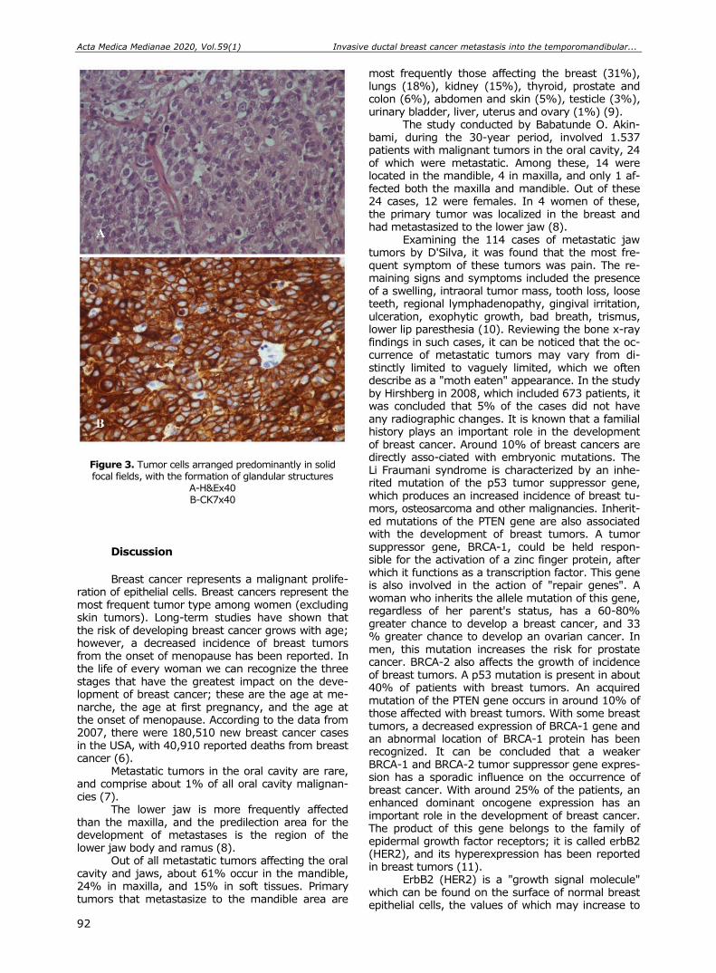
Acta Medica Medianae 2020, Vol.59(1) Invasive ductal breast cancer metastasis into the temporomandibular...
92
Figure 3. Tumor cells arranged predominantly in solid focal fields, with the formation of glandular structures
A-H&Ex40 B-CK7x40
Discussion
Breast cancer represents a malignant prolife-
ration of epithelial cells. Breast cancers represent the most frequent tumor type among women (excluding skin tumors). Long-term studies have shown that the risk of developing breast cancer grows with age; however, a decreased incidence of breast tumors from the onset of menopause has been reported. In the life of every woman we can recognize the three stages that have the greatest impact on the deve-lopment of breast cancer; these are the age at me-narche, the age at first pregnancy, and the age at the onset of menopause. According to the data from 2007, there were 180,510 new breast cancer cases in the USA, with 40,910 reported deaths from breast cancer (6).
Metastatic tumors in the oral cavity are rare, and comprise about 1% of all oral cavity malignan-cies (7).
The lower jaw is more frequently affected than the maxilla, and the predilection area for the development of metastases is the region of the lower jaw body and ramus (8).
Out of all metastatic tumors affecting the oral cavity and jaws, about 61% occur in the mandible, 24% in maxilla, and 15% in soft tissues. Primary tumors that metastasize to the mandible area are
most frequently those affecting the breast (31%), lungs (18%), kidney (15%), thyroid, prostate and colon (6%), abdomen and skin (5%), testicle (3%), urinary bladder, liver, uterus and ovary (1%) (9).
The study conducted by Babatunde O. Akin-bami, during the 30-year period, involved 1.537 patients with malignant tumors in the oral cavity, 24 of which were metastatic. Among these, 14 were located in the mandible, 4 in maxilla, and only 1 af-fected both the maxilla and mandible. Out of these 24 cases, 12 were females. In 4 women of these, the primary tumor was localized in the breast and had metastasized to the lower jaw (8).
Examining the 114 cases of metastatic jaw tumors by D'Silva, it was found that the most fre-quent symptom of these tumors was pain. The re-maining signs and symptoms included the presence of a swelling, intraoral tumor mass, tooth loss, loose teeth, regional lymphadenopathy, gingival irritation, ulceration, exophytic growth, bad breath, trismus, lower lip paresthesia (10). Reviewing the bone x-ray findings in such cases, it can be noticed that the oc-currence of metastatic tumors may vary from di-stinctly limited to vaguely limited, which we often describe as a "moth eaten" appearance. In the study by Hirshberg in 2008, which included 673 patients, it was concluded that 5% of the cases did not have any radiographic changes. It is known that a familial history plays an important role in the development of breast cancer. Around 10% of breast cancers are directly asso-ciated with embryonic mutations. The Li Fraumani syndrome is characterized by an inhe-rited mutation of the p53 tumor suppressor gene, which produces an increased incidence of breast tu-mors, osteosarcoma and other malignancies. Inherit-ed mutations of the PTEN gene are also associated with the development of breast tumors. A tumor suppressor gene, BRCA-1, could be held respon-sible for the activation of a zinc finger protein, after which it functions as a transcription factor. This gene is also involved in the action of "repair genes". A woman who inherits the allele mutation of this gene, regardless of her parent's status, has a 60-80% greater chance to develop a breast cancer, and 33 % greater chance to develop an ovarian cancer. In men, this mutation increases the risk for prostate cancer. BRCA-2 also affects the growth of incidence of breast tumors. A p53 mutation is present in about 40% of patients with breast tumors. An acquired mutation of the PTEN gene occurs in around 10% of those affected with breast tumors. With some breast tumors, a decreased expression of BRCA-1 gene and an abnormal location of BRCA-1 protein has been recognized. It can be concluded that a weaker BRCA-1 and BRCA-2 tumor suppressor gene expres-sion has a sporadic influence on the occurrence of breast cancer. With around 25% of the patients, an enhanced dominant oncogene expression has an important role in the development of breast cancer. The product of this gene belongs to the family of epidermal growth factor receptors; it is called erbB2 (HER2), and its hyperexpression has been reported in breast tumors (11).
ErbB2 (HER2) is a "growth signal molecule" which can be found on the surface of normal breast epithelial cells, the values of which may increase to
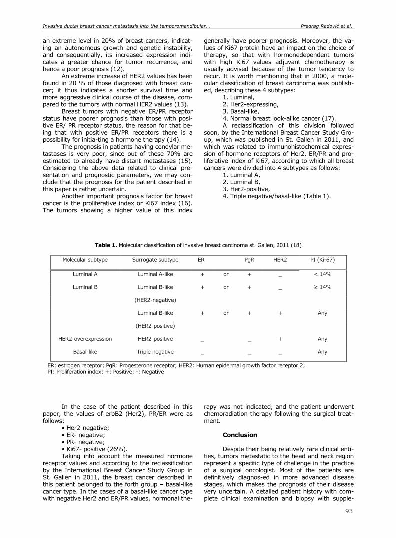
Invasive ductal breast cancer metastasis into the temporomandibular... Predrag Radović et al.
91
an extreme level in 20% of breast cancers, indicat-ing an autonomous growth and genetic instability, and consequentially, its increased expression indi-cates a greater chance for tumor recurrence, and hence a poor prognosis (12).
An extreme increase of HER2 values has been found in 20 % of those diagnosed with breast can-cer; it thus indicates a shorter survival time and more aggressive clinical course of the disease, com-pared to the tumors with normal HER2 values (13).
Breast tumors with negative ER/PR receptor status have poorer prognosis than those with posi-tive ER/ PR receptor status, the reason for that be-ing that with positive ER/PR receptors there is a possibility for initia-ting a hormone therapy (14).
The prognosis in patients having condylar me-tastases is very poor, since out of these 70% are estimated to already have distant metastases (15). Considering the above data related to clinical pre-sentation and prognostic parameters, we may con-clude that the prognosis for the patient described in this paper is rather uncertain.
Another important prognosis factor for breast cancer is the proliferative index or Ki67 index (16). The tumors showing a higher value of this index
generally have poorer prognosis. Moreover, the va-lues of Ki67 protein have an impact on the choice of therapy, so that with hormonedependent tumors with high Ki67 values adjuvant chemotherapy is usually advised because of the tumor tendency to recur. It is worth mentioning that in 2000, a mole-cular classification of breast carcinoma was publish-ed, describing these 4 subtypes:
1. Luminal, 2. Her2-expressing, 3. Basal-like, 4. Normal breast look-alike cancer (17). A reclassification of this division followed
soon, by the International Breast Cancer Study Gro-up, which was published in St. Gallen in 2011, and which was related to immunohistochemical expres-sion of hormone receptors of Her2, ER/PR and pro-liferative index of Ki67, according to which all breast cancers were divided into 4 subtypes as follows:
1. Luminal A, 2. Luminal B, 3. Her2-positive, 4. Triple negative/basal-like (Table 1).
Table 1. Molecular classification of invasive breast carcinoma st. Gallen, 2011 (18)
Molecular subtype Surrogate subtype ER PgR HER2 PI (Ki-67)
Luminal A Luminal A-like + or + _ < 14%
Luminal B Luminal B-like + or + _ ≥ 14%
(HER2-negative)
Luminal B-like + or + + Any
(HER2-positive)
HER2-overexpression HER2-positive _ _ + Any
Basal-like Triple negative _ _ _ Any
ER: estrogen receptor; PgR: Progesterone receptor; HER2: Human epidermal growth factor receptor 2; PI: Proliferation index; +: Positive; -: Negative
In the case of the patient described in this paper, the values of erbB2 (Her2), PR/ER were as follows:
• Her2-negative; • ER- negative; • PR- negative; • Ki67- positive (26%). Taking into account the measured hormone
receptor values and according to the reclassification by the International Breast Cancer Study Group in St. Gallen in 2011, the breast cancer described in this patient belonged to the forth group – basal-like cancer type. In the cases of a basal-like cancer type with negative Her2 and ER/PR values, hormonal the-
rapy was not indicated, and the patient underwent chemoradiation therapy following the surgical treat-ment.
Conclusion Despite their being relatively rare clinical enti-
ties, tumors metastatic to the head and neck region represent a specific type of challenge in the practice of a surgical oncologist. Most of the patients are definitively diagnos-ed in more advanced disease stages, which makes the prognosis of their disease very uncertain. A detailed patient history with com-plete clinical examination and biopsy with supple-
93
Acta Medica Medianae 2020, Vol.59(1) Invasive ductal breast cancer metastasis into the temporomandibular...
94
mental radiological findings, are vital for making the definitive diagnosis and for a therapy to be succes-sful. Based on the presented findings, it can be concluded that an adequate surgical therapy, with proper immunohistochemistry and determination of
the type and subtype of these carcinomas, repre-sents a promising, individualized oncological appro-ach in the treat-ment of affected patients, increasing the percentage of therapeutic success and healing rates.
References
1. Hirshberg A, Berger R, Allon I, Kaplan I. Metastatic tumors to the jaws and mouth. Head Neck Pathol 2014; 8(4):463-74. [CrossRef] [PubMed]
2. McClure SA, Movahed R, Salama A, Ord RA. Maxillofacial metastases: a retrospective review of one institution's 15-year experience. J Oral Maxillofac Surg 2013; 71(1):178-88. [CrossRef] [PubMed]
3. Owosho AA, Xu B, Kadempour A, Yom SK, Randazzo J, Ghossein RA, et al. Metastatic solid tumors to the jaw and oral soft tissue: A retrospective clinical analysis of 44 patients from a single institution. J Craniomaxillofac Surg 2016; 44(8):1047-53. [CrossRef] [PubMed]
4. Allon I, Pessing A, Kaplan I, Allon DM, Hirshberg A. Metastatic tumors to the gingiva and the presence of teeth as a contributing factor: a literature analysis. J Periodontol 2014; 85(1): 132-9. [CrossRef] [PubMed]
5. Thiele OC, Freier K, Bacon C, Flechtenmacher C, Scherfler S, Seeberger R. Craniofacial metastases: a 20-year survey. J Craniomaxillofac Surg 2011; 39 (2):135-7. [CrossRef] [PubMed]
6. Wiener C, Fauci AS, Braunwald E, Kasper DL, Hauser SL, Longo DL, Jameson JL, Loscalzo J. Harrison's principles of internal medicine. Self-Assessment & Board Review. 17th ed. New York: McGraw-Hill Companies; 2008.
7. Dib LL, Soares AL, Sandoval RL, Nannmark U. Breast metastasis around dental implants: a case report. Clin Implant Dent Relat Res 2007; 9(2):112-5. [CrossRef] [PubMed]
8. Akinbami BO. Metastatic carcinoma od jaws: a review of literature. Niger J Med 2009; 18(2):139-42. [CrossRef] [PubMed]
9. Rocha WC, Curado MP, Vencio EF, Caixeta WB. Endometrial carcinoma metastatic to the mandible: a case report. J Oral Maxillofac Surg 2000; 58(8):914-6. [CrossRef] [PubMed]
10. Menezes AV, Lima MP, Mendonca JE, Haiter-Neto F, Kurita LM. Breast adenocarcinoma mimicking tempo-romandibular disorders: a case report. J Contemp Dent Pract 2008; 9(5):100-6. [CrossRef] [PubMed]
11. Hirshberg A, Shnaiderman-Shapiro A, Kaplan I, Berger R. Metastatic tumours to the oral cavity- pathogenesis and analysis of 673 cases. Oral Oncol 2008; 44(8): 743-52. [CrossRef] [PubMed]
12. Abeloff MD, Weber BL, Wolff AC, McCormick B, Zaks TZ, Sacchini V. Cancer of the breast. In: Abeloff MD, Armitage JO, Niederhuter JE, Kastan MB, McKena WG, editors. Abeloff’s Clinical Oncology. 4th ed. London: Churchill Livingstone; 2008.
13. Wolff AC, Hammond ME, Schwartz JN, Hagerty KL, Allred DC, Cote RJ, et al. American Society of Clinical Oncology/College of American Pathologists guideline recommendations for human epidermal growth factor receptor 2 testing in breast cancer. Arch Pathol Lab Med 2007; 131(1):18-43. [PubMed]
14. Fisher B, Redmond C, Fisher ER, Caplan R. Relative worth of estrogen or progesterone receptor and pa-thologic characteristics of differentiation as indicators of prognosis in node negative breast cancer patients: findings from National Surgical Adjuvant Breast and Bowel Project Protocol B-06. J Clin Oncol 1988; 6(7):1076-87. [CrossRef] [PubMed]
15. Kaufmann MG, Perren A, Gratz KW, Eyrich GKH. Condylar metastasis. Review of the literature and report of a case. Mund Kiefer Gesichtschir 2005; 9(5): 336-40. [CrossRef] [PubMed]
16. Horimoto Y, Arakawa A, Tanabe M, Kuroda K, Matsuoka J, Igari F, et al. Menstrual cycle could affect Ki-67 expression in estrogen receptor-positive breast cancer patients. J Clin Pathol 2015; 68(10):825-9. [CrossRef] [PubMed]
17. Perou CM, Sorlie T, Eisen MB, Van de Rijn M, Jeffrey SS, Rees CA, et al. Molecular portraits of human breast tumours. Nature 2000; 406(6797):747-52. [CrossRef] [PubMed]
18. Goldhirsch A, Wood WC, Coates AS, Gelber RD, Thürlimann B, Senn HJ. Strategies for subtypes –
dealing with the diversity of breast cancer: highlights of the St. Gallen international expert consensus on the primary therapy of early breast cancer 2011. Ann Oncol 2011; 22(8):1736-47. [CrossRef] [PubMed]
Invasive ductal breast cancer metastasis into the temporomandibular... Predrag Radović et al.
95
Prikaz bolesnika UDC: 618.19-006.6::616.31-006
doi:10.5633/amm.2020.0113
METASTAZA INVAZIVNOG DUKTALNOG KARCINOMA DOJKE U PREDELU TEMPOROMANDIBULARNOG ZGLOBA DVE GODINE NAKON
INICIJALNOG TRETMANA: PRIKAZ SLUČAJA
Predrag Radović1,2, Milovan Papović1, Miloš Trajković1, Nikola Živković2,3, Andrija Ćosić1,2
1Klinika za stomatologiju, Odeljenje za maksilofacijalnu hirurgiju, Niš, Srbija 2Univerzitet u Nišu, Medicinski fakultet Niš, Srbija 3Institut za patologiju, Niš, Srbija
Kontakt: Milovan Papović Bulevar dr Zoran Djindjić 48, 18000 Niš, Serbia
E-mail: [email protected]
Metastaze tumora u predelu usne duplje i vilice predstavljaju redak patološki entitet.
Ovakvi tipovi tumora predstavljaju težak izazov za onkološke hirurge zbog otežane dija-gnostike, nespecifične simptomatologije i netipičnog radiografskog nalaza. Simptomi koji se javljaju kod metastatskih tumora temporomandibularnog zgloba i ramusa donje vilice naj-češće su nespecifični i manifestuju se u vidu bolnih senzacija, otoka, otežanog otvaranja usta, što kliničara često odvodi na oboljenja temporomandibularnog zgloba kao što su artritis, subluksacija zgloba, degenerativne promene i infekcije.
U našem slučaju prikazana je pacijentkinja stara 62 godine sa postojanjem meta-statskog tumora u predelu temporomandibularnog zgloba i donje vilice sa veoma neizvesnom prognozom. U daljem tekstu diskutovaće se o izobru hirurške i onkološke terapije na osnovu radiografskih nalaza i histopatološkog tipa karcinoma
Acta Medica Medianae 2020;59(1):90-95.
Ključne reči: metastaza karcinoma dojke, temporomandibularni zglob,
histopatološki tip i podtip, hirurški tretman
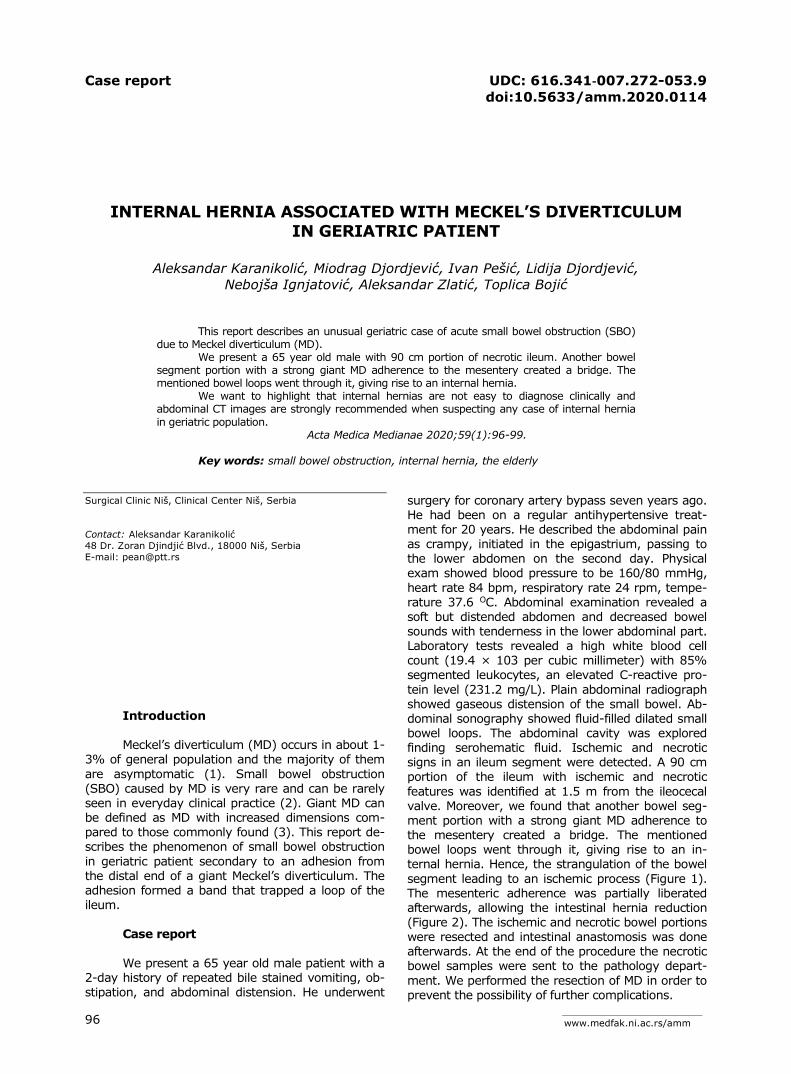
96 www.medfak.ni.ac.rs/amm
Case report UDC: 616.341‑007.272-053.9
doi:10.5633/amm.2020.0114
INTERNAL HERNIA ASSOCIATED WITH MECKEL’S DIVERTICULUM IN GERIATRIC PATIENT
Aleksandar Karanikolić, Miodrag Djordjević, Ivan Pešić, Lidija Djordjević,
Nebojša Ignjatović, Aleksandar Zlatić, Toplica Bojić
This report describes an unusual geriatric case of acute small bowel obstruction (SBO)
due to Meckel diverticulum (MD). We present a 65 year old male with 90 cm portion of necrotic ileum. Another bowel
segment portion with a strong giant MD adherence to the mesentery created a bridge. The mentioned bowel loops went through it, giving rise to an internal hernia.
We want to highlight that internal hernias are not easy to diagnose clinically and abdominal CT images are strongly recommended when suspecting any case of internal hernia
in geriatric population. Acta Medica Medianae 2020;59(1):96-99.
Key words: small bowel obstruction, internal hernia, the elderly
Surgical Clinic Niš, Clinical Center Niš, Serbia
Contact: Aleksandar Karanikolić
48 Dr. Zoran Djindjić Blvd., 18000 Niš, Serbia
E-mail: [email protected]
Introduction Meckel’s diverticulum (MD) occurs in about 1-
3% of general population and the majority of them
are asymptomatic (1). Small bowel obstruction
(SBO) caused by MD is very rare and can be rarely seen in everyday clinical practice (2). Giant MD can be defined as MD with increased dimensions com-pared to those commonly found (3). This report de-scribes the phenomenon of small bowel obstruction
in geriatric patient secondary to an adhesion from the distal end of a giant Meckel’s diverticulum. The adhesion formed a band that trapped a loop of the ileum.
Case report
We present a 65 year old male patient with a 2-day history of repeated bile stained vomiting, ob-stipation, and abdominal distension. He underwent
surgery for coronary artery bypass seven years ago. He had been on a regular antihypertensive treat-ment for 20 years. He described the abdominal pain as crampy, initiated in the epigastrium, passing to
the lower abdomen on the second day. Physical exam showed blood pressure to be 160/80 mmHg,
heart rate 84 bpm, respiratory rate 24 rpm, tempe-rature 37.6 OC. Abdominal examination revealed a soft but distended abdomen and decreased bowel sounds with tenderness in the lower abdominal part. Laboratory tests revealed a high white blood cell count (19.4 × 10³ per cubic millimeter) with 85% segmented leukocytes, an elevated C-reactive pro-
tein level (231.2 mg/L). Plain abdominal radiograph showed gaseous distension of the small bowel. Ab-dominal sonography showed fluid-filled dilated small bowel loops. The abdominal cavity was explored finding serohematic fluid. Ischemic and necrotic signs in an ileum segment were detected. A 90 cm portion of the ileum with ischemic and necrotic
features was identified at 1.5 m from the ileocecal valve. Moreover, we found that another bowel seg-ment portion with a strong giant MD adherence to the mesentery created a bridge. The mentioned bowel loops went through it, giving rise to an in-ternal hernia. Hence, the strangulation of the bowel
segment leading to an ischemic process (Figure 1). The mesenteric adherence was partially liberated afterwards, allowing the intestinal hernia reduction (Figure 2). The ischemic and necrotic bowel portions were resected and intestinal anastomosis was done afterwards. At the end of the procedure the necrotic bowel samples were sent to the pathology depart-
ment. We performed the resection of MD in order to prevent the possibility of further complications.
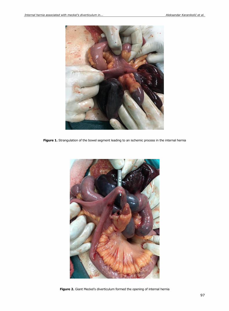
Internal hernia associated with meckel’s diverticulum in... Aleksandar Karanikolić et al.
97
Figure 1. Strangulation of the bowel segment leading to an ischemic process in the internal hernia
Figure 2. Giant Meckel’s diverticulum formed the opening of internal hernia
Acta Medica Medianae 2020, Vol.59(1) Internal hernia associated with meckel’s diverticulum in...
98
Discussion
Patients with Meckel’s diverticulum have the
lifetime risk of complications of around 4% (4). Complications due to Meckel’s diverticulum include hemorrhage, intestinal obstruction, diverticulitis,
Littre’s hernia, umbilicus sinus, and tumours (5). In-testinal obstruction is the most common compli-cation in adults and gastrointestinal haemorrhage is the most common complication in children. Intesti-nal obstruction caused by Meckel's diverticulum can occur for several reasons: by entangling a loop of small bowel around a fibrous cord or within a me-
sodiverticular band, intussusception, volvulus, incar-ceration within a hernia sac (Littre’s hernia), chronic Meckel’s diverticulitis, foreign body, or neoplasm (4).
Clinically, intestinal obstruction can be asymp-
tomatic or cause significant discomfort ranging from constant vague epigastric pain to intermittent colicky periumbilical pain. Additional symptoms include
nausea, vomiting and recurrent intestinal obstruc-tion. Symptom severity relates to the duration and reducibility of the internal hernia and the presence or absence of incarceration and strangulation (1).
Imaging studies often play an important role in the diagnosis of internal hernias caused by va-
rious etiological factors, because they are often dif-ficult to identify clinically. Native abdominal radio-graphy and ultrasound had no interest in setting up adequate diagnosis. These diagnostic procedures showed a picture of acute intestinal obstruction. However, abdominal CT has become the first-line imaging technique in these patients because of its
availability, speed, and multiplanar reformatting ca-pabilities. Unfortunately, in this case, we did not use the abdominal CT in the diagnosis, which proved to be a bad decision. However, one should take into account that in our hospital abdominal CT is not used as a standard tool in the diagnosis of intestinal obstruction.
Meckel's diverticulum is traditionally conside-red a pediatric disease that is associated with in-testinal hemorrhage or perforation. Meckel's diverti-culum complicated with internal hernia presenting as an intestinal obstruction is exceedingly rare (5).
Jain and Viswanath (2013) (6) reported case
of small bowel obstruction due to a Meckel's di-
verticulum complicated with internal hernia in a 60 year old male patient.
Fuentes-Diaz et al. (2015) (3) presented a 19 year old male with acute abdominal pain suggestive of appendicitis. During appendectomy, they dis-
covered ischemic and necrotic signs in a bowel seg-ment, leading them to perform a laparotomy that revealed a portion of ischemic and necrotic jejunum, and another bowel segment with a strong adherence to the mesentery root that created an internal her-nia. The internal hernia was reduced and the injured bowel portions were resected.
Devanaboyna et al. (2008) (7) presented MD causing mechanical small bowel obstruction in a 33 year old gentlman. The MD and adhesion were ex-
cised with translinear cutting stapling device and the no ischemic small bowel was decompressed.
Meckel's diverticulum as a cause of SBO in elderly patients is very rare. The specificity of this
case is that MD was not part of gangrenous or inflammatory affected small bowel loops. For this reason MD should be considered as the cause of SBO in the geriatric population. Whenever we have a dilemma regarding the etiology of SBO, CT exa-mination of the abdomen should be done.
A timely and accurate diagnosis of intestinal obstruction in elderly patients caused by giant MD in clinical practice is not simple. Abdominal CT images are strongly recommended when suspecting any case of internal hernia in geriatric population in order to prevent intestinal gangrene.
Conflict of Interest
The authors declare no conflicts of interest Statement of Ethics
The research was conducted ethically in
accordance with the World Medical Association
Declaration of Helsinki. Guardian of the subject gave the informed written consent to participate in this research.
Internal hernia associated with meckel’s diverticulum in... Aleksandar Karanikolić et al.
99
References
1. Al Jabri KA,El Sherbini A. Small bowel obstruction due to Meckel's diverticulum: acase report. Oman Med J 2012;27(1):1-3.[PubMed]
2. Elsayes KM, Menias CO, Harvin HJ, Francis IR. Imaging manifestations of Meckel's diverticulum. ARJ Am J Roentgenol 2007;189(1):81-8. [CrossRef] [PubMed]
3. Fuentes-Diaza JM, Trujillo-Vasquezc CA, Parra-Vargasd AM, Andrea Sofía Rovira-Chaves AS, Tinoco-Guzman LV, Garcia-Garcia JM. Strangulated internal hernia by giant Meckel diverticulum presented as acute appendicitis. Int J Surg Case Rep2015;13:61-3. [CrossRef] [PubMed]
4. Cartanese C, Pettiti T, Marinelli E, Pignatelli A, Marginetti D, Zuccarino M, et al. Intestinal obstruction
caused by torsed gangrenous meckel's diverticulum encircling terminal ileum. World J Gastrointest Surg 2011;3(7):106-9.[CrossRef] [PubMed]
5. Papaziogas B, Makris I, Tsiaousisp P, Chatzimavroudis G, Paraskevas G, Atmatzidis K. An unusual case of intestinal obstruction caused by a Meckel's diverti-culum. Aristotle University Medical Journal 2007; 34 (1):55-9.
6. Jain AKC, Viswanath S. Acute intestinal obstruction due to Meckel’s diverticulum: a case report. Int Med Case Rep J2013;2013:1-3.[CrossRef]
7. Devanaboyna G, Singh-Ranger D, Shami S. Meckel's diverticulum causing mechanical small bowel obstruct-tion. Can J Surg 2008;51(2):156.[PubMed]
Prikaz bolesnika UDC: 616.341‑007.272-053.9
doi:10.5633/amm.2020.0114
UNUTRAŠNJA INKARCERACIJA UZROKOVANA MECKELOVIM
DIVERTIKULUMOM KOD BOLESNIKA STARIJEG ŽIVOTNOG DOBA
Aleksandar Karanikolić, Miodrag Đorđević, Ivan Pešić, Lidija Đorđević,
Nebojša Ignjatović, Aleksandar Zlatić, Toplica Bojić
Klinika za opštu hirurgiju, Klinički centar Niš, Niš, Srbija
Kontakt: Aleksandar Karanikolić
Bulevar dr Zoran Đinđić 48, 18000 Niš, Srbija
E-mail: [email protected]
Predstavljamo redak slučaj akutne obstrukcije tankog creva uzrokovane Meckelovim
divertikulumom (MD) kod bolesnik starijeg životnog doba. Prikazan je bolesnik star 65 godina, kod kojeg je verifikovan nekrotični ileum u dužini
od 90 cm. Dugačak MD slepljen za mezenterijum tankog creva formirao je otvor kroz koji je prošao deo tankog creva dovodeći do unutrašnje inkarceracije, ishemije i nekroze.
Želimo istaći da unutrašnju inkarceraciju nije lako dijagnostifikovati, te je potrebno uraditi MSCT snimanje abdomena kada postoji sumnja na unutrašnju inkarceraciju, naročito kod bolesnika starijeg životnog doba.
Acta Medica Medianae 2020;59(1):96-99.
Ključne reči: obstrukcija tankog creva, unutrašnja kila, gerijatrijski bolesnici
100 www.medfak.ni.ac.rs/amm
Review article UDC: 616-092:[669.71:615.9
doi:10.5633/amm.2020.0115
PATHOPHYSIOLOGICAL MECHANISMS OF ALUMINIUM TOXICITY
Novica Bojanić1, Jelena Milenković2, Dijana Stojanović2, Maja Milojković2,
Nataša Djindjić1, Marko Gmijović3
Aluminium forms about 8% of the earth’s crust. It is most commonly found as bauxite
ore, which is used for extraction of this metal. Aluminium has a high reactivity and forms compounds such as aluminium oxide, aluminium hydroxide, and potassium aluminium sulfate. Exposure of these compounds to oxidants leads to the formation of a superficial coating of aluminium oxide, which is highly resistant to corrosion and insoluble in water. However, acid rains have allowed the dissolution of these compounds and the entry of aluminium into biological systems. It can enter in human body through water, food, drugs, and inhalation of polluted air.
Once when accumulates in the body aluminium exhibits toxic effects on different organ systems: central nervous, respiratory, skeletal, hematopoietic, reproductive, digestive (liver), and integumentary system. Toxic systemic effects of aluminium are first observed in patients with kidney failure treated with medicines containing aluminium compounds which manifest as: dialysis encephalopathy syndrome, osteomalacia with osteodystrophy and microcytic anaemia.
Aluminium is on the top of a surprisingly short list of neurotoxic inorganic elements and their compounds. It is linked with development of neurodegenerative diseases, including autism, attention deficit disorders, amyotrophic lateral sclerosis, Alzheimer's disease, dementia, Gulf war syndrome, and Parkinsonism. Clinical and experimental studies suggest several possible mechanisms of toxic aluminium action on cells. Those are: increased production of oxidative stress, alteration of membrane function, disruption of intracellular signaling, and alteration or inhibition of enzyme functions.
Acta Medica Medianae 2020;59(1):100-109. Key words: aluminium, toxicity, oxidative stress, neurodegenerative diseases,
pathogenesis
1University of Niš, Faculty of Medicine, Niš, Serbia 2University of Niš, Faculty of Medicine, Department of
Pathophysiology, Niš, Serbia 3Digestive Surgery Clinic, Clinical Center Niš, Niš, Serbia
Contact: Novica Bojanić
11/37 Pariske Komune St., 18000 Niš, Serbia
E-mail: [email protected]
Introduction Aluminium (Al) is a silvery-white, lightweight,
ductile, malleable and non-magnetic metal. It has atomic number 13 and belongs to boron group of the periodic table of the elements. Al forms about 8% of the earth’s crust. Native aluminium is ex-tremely rarely found in nature. It is most commonly found as bauxite ore, which is used for extraction of
this metal. Aluminium has a high reactivity and forms compounds such as aluminium oxide, alu-minium hydroxide, and potassium aluminium sul-fate. Exposure of Al to water, oxygen or other oxi-dants leads to the formation of a superficial coating of aluminium oxide, which is highly resistant to corrosion. Aluminium oxide is insoluble in water, but it is soluble in mineral acids and strong alkalis. The concentration of dissolved Al3+ is low in surface and subsoil waters, because Al minerals are insoluble at neutral pH. Rain-borne acidification and the use of acidifying fertilizers increase the concentration of soluble Al3+ in soil and waters (1-3). The result of soil pollution is an increase in concentration of alu-minium in cultivated plants. This is one of the ways how aluminium may enter in food chain. It was believed that aluminium is harmless to environment, but it is shown that it is toxic to plants and animals (1-3).
Aluminium is widely used in metal alloy pro-duction, as a construction material in automotive and aviation industry, electrical industry, as solid fuel rocket propellent, for manufacture of explosives and fireworks. It is also used for the production of cooking utensils and dishes, for food packaging (cans, containers, foils), and as food additives (4, 5).
Pathophysiological mechanisms of aluminium toxicity Novica Bojanić et al.
101
Mineral compounds of aluminium of natural origin (beonite and zeolite) are used to purify drinking water as coagulants in order to reduce the level of organic matter, color, cloudiness and microorga-nisms (4-6). Aluminium is also used in pharmacy, medicine, cosmetics and dentistry (antacids, astrin-gents, antiperspirants, dental crowns and dentures) (5).
Aluminium can enter in human body through water, food, drugs, and inhalation of polluted air (5-7). Aluminium is a non-essential element which is not found in large quantities in human body. The total human body content of aluminium may range from 50-150 mg, with an average of about 65 mg. Daily intake of aluminium may range from 10-110 mg (8). It is characterized by low intestinal ab-sorption, slow tissue uptake and rapid urinary ex-cretion. Absorption of Al in the digestive tract is estimated to be less than 1%. It is influenced by solubility of Al compounds and enhanced with in-creased gastric acidity, presence of organic acids (ascorbic, citric) and lack of Fe and Ca in the diet. The accumulation of Al is reported in patients treat-ed with medicines containing Al compounds (anta-cids, aluminium in dialysis fluid) in renal failure (8). About half of body content is found in the skeleton, one quarter in the lungs and the rest is in brain, kidneys, liver, spleen and thyroid. Aluminium may pass both blood-brain and placental barrier. It is not recognized that Al3+ has some function in living organisms and it is regarded as biologically inert (9, 10). Aluminium when accumulate in human body exhibits toxic effects on different organ systems: central nervous, respiratory, skeletal, hematopoietic, reproductive, digestive (liver), and integumentary system.
Effects of aluminium on central nervous
system Aluminium is on the top of a surprisingly short
list of neurotoxic inorganic elements and their com-pounds. It is linked with development of neurodege-nerative diseases, including autism, attention deficit disorder, amyotrophic lateral sclerosis (ALS), Alzheimer's disease (AD), dementia, Gulf war syn-drome and parkinsonism (7, 11-17).
Aluminium is accused to develop neurotoxicity through oxidative stress, cell mediated toxicity, apoptosis, inflammatory events in the brain, glu-tamate toxicity, effects on calcium homeostasis, gene expression, aluminium induced neurofibrillary tangle (NFT) formation and irreversible blockade of ion channels by beta-amyloid (11, 18). Neurotoxicity of aluminium salts has been reported in numerous animal studies. Exposition to excessive intake of aluminium causes learning and memory disorders in rats due to deposition of Aβ in the hippocampus and cortex. Al-induced neurophysical and neurobeha-vioral pathological changes similar to AD were re-gistered in animals (19, 20). Chronic exposition of rats to high-dose AlCl3 injections over a prolonged period can reduce locomotor and cognitive functions in rats as well as reduced body weight gain which may be a sign of systemic toxicity (21).
A number of clinical studies have shown that exposure to aluminium in dialysis fluid in patients with kidney failure resulted with encephalopathy. Aluminium induced encephalopathy due to bladder irrigation with 1% alum in patient with kidney failure is also reported (22). People in Camelford (south west of England) were exposed to the drinking water contaminated with 20 tonnes of aluminium sulphate in July 1988. They had considerable damages of cerebral function, which were not related to anxiety (23). A mild form of encephalopathy was registered in 64 former aluminium dust-exposed foundry workers in Italy. It was characterized by mild in-tellectual deficit, loss of muscle control, tremor, and spinocerebellar degeneration (24).
Aluminium levels were assessed in 118 pa-tients with neurodegenerative diseases: multiple sclerosis (MS), amyotrophic lateral sclerosis (ALS), Parkinson’s disease (PD), and Alzheimer’s disease (AD) as well as in 73 healthy subjects. MS was diag-nosed in 85.6% of total neurodegenerative diseases (ND). Al was present in 44.8% of cases compre-hensive of ND and healthy patients. Al level was sig-nificantly higher in patients with neurodegenerative diseases than in healthy subjects. Calcium disodium ethylene diaminetetra acetic acid (EDTA) chelation treatment reduced Al burden in patients with neuro-degenerative diseases and ameliorated their clinical conditions (25).
Macrophagic myofasciitis was registered after intramuscular injections of Al-containing vaccines in patients (5). It was characterized by persistence of aluminium hydroxide within macrophages at the site of immunization, muscle lesions, arthromyalgias, chronic fatigue, and cognitive impairment (both vi-sual and verbal memory deficit; including attention deficit) (26).
Aluminium salts (aluminium hydroxide, alumi-nium phosphate, and aluminium potassium sulfate) have been used in vaccines over eight decades. Even though it has been reported that these salts used as adjuvants can cause severe local reactions (erythema, subcutaneous nodules and contact hy-persensitivity) they are still in use, because they enhance antigenicity of some vaccines (as diphtheria and tetanus vaccines) (27).
It was recorded that vaccinated children were exposed more to aluminium (11-26%) than under-vaccinated children. “Power analyses demonstrated that safety studies of aluminium could detect rela-tive risks ranging from 1.1 to 5.8 for a range of adverse event incidence” (28). It has been hypothe-sized that aluminium hydroxide, which was used as adjuvant in multiple vaccines that soldiers under-went, is associated with Gulf War syndrome (29, 30). The Global Advisory Committee on Vaccine Safety of WHO does not find that there is basis at present to change recommendations for vaccination practices (31).
Though, aluminium neurotoxicity is docu-mented, there is common opinion that healthy adults may tolerate repeated oral exposures to alu-minium (up to 3500-7200 mg/day from antacids and buffered aspirin) without any toxic effect. It is explained by low absorption and rapid and primarily urinary excretion of Al compounds. But it is clear
Acta Medica Medianae 2020, Vol.59(1) Pathophysiological mechanisms of aluminium toxicity
102
that even low daily doses of aluminium can cause systemic intoxication in patients with kidney failure, preterm infants and young children (5).
Effects of aluminium on respiratory system
Aluminium dust has hazardous effect on res-
piratory system of aluminium production workers. It is shown that they suffer from respiratory symptoms such as cough, wheezing, dyspnea, and chest tight-ness. Potroom workers who breathe large amounts of aluminium dusts can get wide range of lung di-seases: chronic bronchitis, chronic obstructive pul-monary disease, potroom asthma, alveolitis, pneu-moconiosis, and even oncological respiratory di-seases. All these toxic effects cannot be attributed solely to aluminium. Aluminium potroom workers are exposed not only to alumina dust, but also to particles of fluorides and traces of different elements (vanadium, chromium, nickel), polycyclic aromatic hydrocarbon, mineral dusts (as silica and asbestos), coal tar pitch volatiles, fumes and gases (hydrogen fluoride, carbon oxides, sulfur dioxide, and oxides of nitrogen), extreme heat, and high static magnetic fields (5, 24, 32-34).
Effects of aluminium on bone and
hematopoietic tissue Most of the aluminium that accumulates in
body is deposited in the bones (8). Aluminium deposits are found also in the hydroxyapatite of the bone matrix in patients with coeliac disease (due to an increased intestinal permeability) and in the case of long-term use of aluminium anti-acid drugs (35, 36).
Patients with kidney failure due to the toxic effect of aluminium in dialysis fluid developed osteo-malacia, osteodystrophy and microcytic anemia and dialysis encephalopathy. Aluminium bone disease is characterized by low serum parathyroid hormone (PTH) levels. Removal of aluminium from the dialysate has resulted in disappearance of the bone disease and in an increase in plasma PTH levels (35). The administration of aluminium in rats with chronic renal failure has resulted in reduction of both synthesis and release of PTH (37).
Although very low doses of Al have mitogenic effect in bones of experimental animals, high doses of Al inhibit remodeling of bone by slowing osteo-blast and osteoclast activities (35, 36). In neonatal rat osteoblast tissue culture AlCl3 has been shown to destroy calcium homeostasis, which activates the Ca2+/calmodulin-dependent protein kinase II signal-ing pathway and thus promotes osteoblast apoptosis (38). Al occupies the unmineralized type I collagen, which is freshly laid down at the mineralization front of the bone surface instead of calcium. The result of impaired bone calcification is development of osteo-malacia associated with hypercalcemia, and hyper-calciuria (25).
Aluminium chronic intoxication causes a mi-crocytic hypochromic anemia in patients with com-promised kidney function (39-41). This anemia is not reversible by iron and it is characterized by de-creased red cell count as well as hematocrit and
hemoglobin concentration. Pathogenesis factors res-ponsible for Al induced anemia are: shortened ery-throcyte lifespan due to reduced erythrocyte mem-brane integrity; inhibition of δ-aminolevulinic acid dehydratase (42), reduced Fe uptake by transferrin due to competitive interaction between iron and alu-minium (43).
Downregulation of transferrin receptor ex-pression and impaired intracellular delivery of Fe from transferrin is also recognized as pathogenesis factor for this type of anemia (39, 40).
Similar values of affinity constant of alumi-nium and iron for the binding of transferrin receptors are recorded in cultured Human erythroleukemic K562 cells. Opposite to previous findings, it is re-ported that Al modified Fe uptake without affecting the expression of transferrin receptors. It is con-cluded that Al induced upregulation of nontransferrin bound iron uptake as an adaptation aimed to enable incorporation of iron which is essential for cellular metabolism (43).
Effects of aluminium on reproductive
system The decline in male fertility, observed in the
twentieth century, is a very current issue in con-temporary science. Male fertility has declined by 50% over several decades of the industrial re-volution. It was not known for a long time about harmful effects of aluminium on male fertility (44-47). Exposure to Al has been reported to affect testicular development and testosterone synthesis in experimental animals (48-51). Although it was shown that Al is capable of compromising male fertility by inducing a state of oxidative stress in the testes (48-52), other mechanisms such as inhibition of microtubule assembly could also be involved in Al-induced testicular damage. Aluminium showed negative impact on reproductive abilities of adult bank voles by causing morphologically abnormal de-velopment of the gonads and by decreasing the quality and quantity of sperm (53). Intraperitoneal administration of AlCl3 induced dose dependent de-crease of testosterone levels in the testes and plasma of mice (48). In male Swiss albino mice treated intraperitoneally with AlCl3 were observed “deformations of the Sertoli cells, epithelial slough-ing, tubular atrophy, and abnormal germ cells” (50). In Wistar rats treated with aluminium sulphate in drinking water is recorded significant decrease of sexual accessory glands: seminal vesicles, prostates, bulbourethral glands and of seminiferous tubules (54). Chronic oral exposure to aluminium at low levels is reported to have as negative impact as high levels on reproductive parameters in Wistar rats. These findings are suggesting adverse impact of aluminium on male fertility (55).
Decreased pregnancy rate was observed in untreated females mated with males treated intra-peritoneally with 100 or 200 mg/kg/day of alumi-nium nitrate for 4 weeks. Treated male mice showed significantly decreased body weight, as well as testi-cular and epididymal weights. Also, significant de-creases in testicular and spermatid counts and epi-didymal sperm counts were recorded. Sperm moti-
Pathophysiological mechanisms of aluminium toxicity Novica Bojanić et al.
101
lity and morphology were unaffected. Histological changes manifested as necrosis of spermatocy-tes/spermatids in the testes, whereas the tubular diameters were unaffected by aluminium admini-stration (56). A negative impact of aluminium on rabbit sperm cell motility and viability has been shown in vitro (51). Rabbits orally treated with AlCl3 for 16 weeks had significantly decreased libido, eja-culate volume, sperm concentration, total sperm output, sperm motility (%), total motile sperm per ejaculate, packed sperm volume and total functional sperm fraction. Relative weights of testes and epididymis were also significantly decreased (49). High concentrations of Al in human semen, seminal plasma, spermatozoa, blood, and urine have been linked to poor sperm quality and viability in men (47).
The effects of aluminium on the female re-productive system are not enough elucidated yet. Histopathological changes in the mice ovaries and decreased fertility, after 12 weeks of aluminium chloride administration (dose range 1000-1400 mg/kg) were showed. Both the number of pregnant females and the number of absorbed fetuses were decreased (53). Female mice were exposed by nasal drip during whole pregnancy to Alumina nanopar-ticles. Aluminium content in hippocampus of new-borns was significantly increased. Neurodevelop-mental toxicity was registered in the offspring at first month of age as significantly increased anxiety-like behavior with impaired learning and memory perfor-mance (57). Subchronic oral exposure to AlCl3 caused the damage of the ovarian structure in rats. Metabolism of Fe, Zn and Cu was disturbed. Acti-vities of Na+-K+-ATPase, Mg2+-ATPase and Ca2+-ATPase in ovaries decreased, and expression of follicle stimulating hormone (FSH) and luteinizing hormone (LH) receptors were suppressed (58, 59). The significant decrease of litter size, modification of sex ratio (increase of female pups number), and significant delay of vaginal opening compared to control group were registered in female Wistar rats exposed to aluminium sulphate by drinking water (60). Oral application of AlCl3 (200 mg daily) during 30 days resulted in a significant decline in uterine and ovarian protein levels and in 3β- and 17β-hydroxysteroid dehydrogenase activities. Estradiol levels also declined. Hypercholesterolemia and signi-ficant accumulation of cholesterol in the ovaries of treated mice as well as accumulation of glycogen in uterus were reported too. Toxic effect in female mice due administration of aluminium chloride has affect-ed steroidogenesis in ovary, and carbohydrate me-tabolism in uterus. These effects were reversible upon withdrawal of the treatments (61). In adult female Wistar rats treated with aluminium nitrate, the presence of electron-dense material in lysoso-mes of myometrium and endometrium cells as well as in the cells of the internal theca and granulosa cells was showed by transmission electron micro-scopy. Also, impaired endoplasmic reticulum, mito-chondria and vacuolation were registered. It was concluded that lysosomes of uterus and ovary cells had defense function and extract aluminium and deposit it in an insoluble form (62).
Effects of aluminium on liver Aluminium is accumulated in the liver, less
than in bones, but the manifestations of Al toxicity in liver have been described. Morphological and mor-phometric changes highly suggestive of toxic he-patitis were registered in Albino Wistar rats treated with aluminium chloride solved in distilled water intragastrically for 21 days. Architectural derange-ment was observed as well as degenerative changes at cellular level: nuclear variations such as karyorr-hexis and pyknosis (63). In of AlCl3-treated rats a significant rises in plasma levels of AST, ALT, ALP, and LDH in AlCl3 were recorded as well as a signi-ficant reduction in total protein level. A significant level of oxidative stress in liver tissue was also registered (64). High doses of Al induce toxic effects and damage the lysosomes in the liver and the extent of lysosomal damage depended of dose and duration of Al loading (65). Accumulation of bile acids in serum in rats and piglets (66), and in-creased transferrin excretion in the bile were also found (67). Reduction of some cytochrome P450 isoenzymes, nicotine adenine dinucleotide phos-phate (NADPH), cytochrome c reductase, and a 4-fold increase in glucuronyltransferase activity were registered in rats treated parenterally with Al. These findings indicate increased conjugating activity (68) and changes in cytochrome P450 isoenzymes may alter metabolism of drugs.
Effects of aluminium on skin Although absorbed through the skin, alumi-
nium exposure via intact skin is rather mild, con-firming this is an effective barrier (69). Aluminium salts, particularly aluminium chlorohydrate, are used in various antiperspirant cosmetics products, as they block secretion of sweat (70).
Following the dermal application of aluminium chlorohydrate penetration rate was very low (around 0.01%) (71). Only insignificant transdermal ab-sorption of Al was shown after application of three different antiperspirant formulations on intact skin. Also, there were low cutaneous quantities of Al ranging 0.5-1.8 μg/cm2. On the other hand, Al up-take after topical antiperspirant application, through the pre-damaged skin (stripped skin) was signi-ficantly higher (0.06% or 11.50 μg/cm2) (70). In this respect, there was a discussion of whether the breast cancer was associated with the use of Al containing antiperspirants, as a form of aluminium-related chronic diseases (69). However, no signifi-cant link between antiperspirants or deodorants use and increased risk for breast cancer was found. A population based case-control study involving 813 breast cancer patients revealed no significant asso-ciation of regular antiperspirant use or following hair removal 1h after shaving (72).
Intact skin is an effective barrier for Al ex-posure thus its effects on human keratinocytes are supposed minimal. However, the study of Al nano-particle interactions with human epidermal keratino-cytes showed the particles localization within the cytoplasmic vacuoles of the cells and there was indication of their interactions with cytokine assays
103
Acta Medica Medianae 2020, Vol.59(1) Pathophysiological mechanisms of aluminium toxicity
102
(73). Detrimental effects of Al were shown on human skin fibroblast cultures and were governed by lipid peroxidation as a pathway of Al cytotoxicity. The experiment showed significant malonodialde-hyde (MDA) production after 24 h incubation with Al (74). In the perspective, the epidemiological study on internal exposure after antiperspirants use should be performed, and the association with hair removal products or shaving.
Mechanisms of aluminium toxic effects Clinical and experimental studies suggest se-
veral mechanisms of toxic aluminium action on cells. Those are: increased production of oxidative stress, alteration of membrane function, disruption of intra-cellular signaling, and alteration or inhibition of en-zyme functions. All of them may eventually cause tissue damage.
1. Association of aluminium and increased oxidative stress
Large number of studies demonstrated there is a link between increased Al concentrations and oxidative stress. Generally, there are several mecha-nisms that produce imbalance between free radical production and antioxidant defense system. Alumi-nium was shown to take part in disruption of metal ion homeostasis and potential oxidative stress through the reactive oxygen species (ROS) and reactive nitrogen species (RNS) generation (75, 76).
Oxidative stress has multiple effects on mo-lecules structure and function, and leads to lipid pe-roxidation, protein modifications and DNA damage. Aluminium driven oxidative stress was shown to lead to the germ cell apoptosis and decrease in intra-cellular ATP level (hypoenergosis and motility) (77). Also, its redox state is implicated in a variety of neurological disorders. Another example is Al me-diated testicular damage through depletion of anti-oxidative enzymes protection, and an induction of NO byproducts and consequent inhibition of steroi-dogenesis (79). Although Al does not undergo redox change, it may enhance iron driven biological oxi-dation by formation of Al superoxide and by catalyzing H2O2 formation while reducing Fe3+ (78).
By creating a labile iron pool, a redox-active iron, Al interferes with cellular pathways of iron metabolism. This pool has a capacity to promote ROS generation. It is regulated by cytosolic iron regulatory proteins and dependent expression of iron import and storage machineries, or ferritin degradation or synthesis (80).This relationship was proven in animal model study where Al serum levels were found inversely correlated with Fe serum levels, implying on Al intoxication intervene in Fe metabolism (48).
Al can also displace other biological cations, such as calcium, zinc, copper and magnesium from their binding sites. Thus released ions can catalyze hydrogen peroxide transformation to the highly reactive hydroxyl radical, and further initiate lipid peroxidation (81).
Another mechanism of detrimental Al effect is impairment of mitochondrial bioenergetics, also associated with ROS generation (79). Dysfunction of mitochondrial bioenergetics progressively leads to
myocardial failure, because energy insufficiency plays a key role in systolic heart failure (82).
Aluminium was also linked to the production of RNS by induction of NO byproducts. Also, in-creased ROS, through other mechanisms, reduce the amount of bioactive NO by formation of toxic peroxynitrite. Al administration significantly in-creased NO production and decreased adenosine 3, 5-cyclic monophosphate (cAMP) and testosterone (83).
Several studies demonstrated that Al may cause changes in antioxidants activity, such as superoxide dismutase (SOD), catalase, and gluta-thione peroxidase/reductase (84). AlCl3 was sug-gested to inhibit the activity of superoxide dismutase (SOD). It is demonstrated that one month admini-stration of AlCl3 significantly decreased the activities of SOD, suggesting that Al have catalytic activity for ROS production. Reduced SOD and glutathione pe-roxidase (GPX) activities might be attributed to the elevated level of protein and lipioxidative products (85).
By decreasing the activity of glutathione syn-thetase, Al might slow the glutathione (GSH) syn-thesis, one of the most important antioxidants in cells. It is hypothesized that this process occurs through the depletion of ATP. Particularly, Al forms a strong complex with ATP, as it has high affinity toward phosphate ions, and lowers its availability in the cell (79). Another suggested mechanism of de-creased GSH is the insufficient supply of NADPH, due to the Al mediated inhibition of NADPH gene-ration. Al inhibits NADP-isocitrate dehydrogenase, the only enzyme supplying NADPH in mitochondria (86). Al, at doses above 120 mg/kg bw in Wistar rats (orally), produced significant reduction of GSH content and an increase of oxidized/reduced glu-tathione ratio, in the small intestine mucosa. Also, activities of both GSH synthase and GSSG reductase were significantly reduced. This change in epithelial cells redox state contributed to alteration in GSH-dependent absorptive functions (87).
Al induced oxidative unbalances are asso-ciated with lipid peroxidation process. Malondialde-hyde (MDA), a lipid peroxidation indicator, is found increased in testis and epididymis after Al exposure (88). Also, lipid peroxidation was found significantly increased in the small intestine when higher Al doses were used (>120 mg/kg) in animal model (89).
2. Aluminium caused alteration of membrane function
The plasma membrane seems to be the target for Al related toxicity, as these trivalent cations readily engaged in interactions with the membrane components thereby affecting associated processes. Al may form electrostatic bonds with oxygen donor ligands or interact with the membrane lipids (79). Also, it may directly alter electrical potential and membrane surface potential (89). Interestingly, binding of Al to the membrane lipids causes the membrane to become more rigid, which ultimately affects the cell motility and viability (90). Production of ROS and lipid peroxidation of membrane lipids has profound and progressive negative consequen-ces. It hinders membrane fluidity (to become rigid), increase permeability, alter r
104
Pathophysiological mechanisms of aluminium toxicity Novica Bojanić et al.
101
eceptor function, etcetera (79). All these changes further influence intracellular processes such as enzyme inactivation, DNA damage and cell death. Chronic aluminium-in-duced neurotoxicity has been related to lipid per-oxidation of the brain cells that probably arises from altered lipoprotein metabolism (91).
3. The channel hypothesis of Alzheimer’s disease (AD)
There are controversies about ion channel hypothesis for Alzheimer amyloid peptide neuro-toxicity. According to some reports beta-amyloid (Aβ) peptides which accumulate in plaques in the brain in Alzheimer’s disease can form ion channels in lipid bilayers. liposomes, neurons, oocyctes, and en-dothelial cells. These channels are heterogeneous in size, voltage-independent, and poorly selective for ions and they can allow influx of (Ca2+), Na+, K+, Cs+, Li+, and possibly Cl− (18). Overload with po-sitive ions may damage and/or kill neurons.
There is no doubt that Aβ is capable to induce transmembrane ion fluctuations in living cells. But in more recent report are presented data which sug-gest “that Aβ is capable of self-assembling into structures that either form a pore through mem-branes or generate transient defects in membra-nes”. It is concluded that Ca2+ influx through Aβ-induced pores or membrane defects and disruption of Ca2+ homeostasis could contribute to develop-ment of Alzheimer’s disease. These authors left open the possibility that Aβ activates intrinsic ion channels or ion pumps in cells (92).
4. Aluminium and disruption of intracellular signaling
Several intracellular signaling pathways are reported to be modified by Al ions. Al was found to disturb secondary messenger signaling, including cAMP, Ca2+, and phosphoinositide and inositol-1, 4, 5-triphosphate (IP3), all of which are involved in a variety of processes, ranging from cell growth, dif-ferentiation to apoptosis (93).
Aluminium alters cyclic AMP and cyclic GMP levels (less sensitive than AMP). Al has elevated cyclic AMP levels in rat cortex following oral admi-nistration for 4 weeks. Supposed mechanism is a G-protein stimulation of adenylate cyclase as Al may replace the Mg2+ that confers the structure to the triphosphate of GMP (94).
Al is reported as an effective voltage sensitive calcium channel blocker. It blocks Ca entry into the cell and inhibits Ca2+/Mg2+-ATPase (on endoplasmic reticulum) dependent sequestration of Ca2+ from the cytosol. Al action in this process is complex as it increases the activity of the Ca2+ ATPase, similarly to Mg2+, but also displaces these two molecules and thus disrupts calcium transport (95). When applied extracellularly, Al reduces voltage-activated calcium channel currents in a concentration and pH depen-dent manner and irreversibly (96).
Al interference with IP3 and its intracellular depletion is based on Al3+ much higher affinity for the phosphatidylcholine surface than Ca2+ (additio-nally disturbing Ca homeostasis) (97).
5. Aluminium caused alteration of enzyme functions Nikotinamide Adenine Dinucleotide Phosphate
Taking into account previously described me-chanisms of Al action it can be concluded that there is substantial influence on cellular protein function and metabolism. Its effects on proteins are varied, ranging from alteration in their expression due to the Al binding to deoxyribonucleic acid (DNA) and ribonucleic acid (RNA), to direct inhibition of several enzymes such as hexokinase, phosphatases, phos-phodiesterase and phosphooxydase, glucose-6-phosphate dehydrogenase and NADP isocitrate de-hydrogenase. For example, Al inhibits hexokinase because it changes Mg ion in Mg-ATPase, a hexo-kinase substrate (97). Or, by perturbation of intra-cellular Fe metabolism and Fe-S cluster Al promotes the inhibition of aconitase activity, enzyme involved in metabolism of citrate, in Pseudomonas fluores-cens (98).
Al exposure (AlCl3) induced significant change of intestinal enzymes, as well as expression of the multidrug resistance-associated protein 2 which was nearly 3-fold increased. Gamma-glutamyltranspep-tidase activity was also increased, while glutathione (GSH) synthase and glutathione disulfide (GSSG)-reductase were decreased (87).
By changing phosphoinositides metabolism Al causes cytoskeletal rearrangements and abnorma-lities, which alter cellular motility and viability (78). Already mentioned increased NO production by Al may react with superoxide or hydrogen peroxide to generate more reactive compounds which cause oxidation of thiol proteins (79).
105
Acta Medica Medianae 2020, Vol.59(1) Pathophysiological mechanisms of aluminium toxicity
106
References
1. Becaria A, Campbell A, Bondy SC. Aluminum as a toxicant. Toxicol Ind Health 2002; 18(7):309-20. [CrossRef] [PubMed]
2. Kucharczak E, Moryl A. [Contents of metals in cultivated plants in Zgorzelec-Bogatynia region. Part 1. Lead, cadmium, aluminium]. Ochr Sr Zasobów Nat 2010;42:52-61. Polish
3. Zołotajkin M, Ciba J, Kluczka J, Skwira M, Smoliński A. Exchangeable and bioavailable aluminium in the mountain forest soil of Barania Góra Range (Silesian Beskids, Poland). Water Air Soil Pollut 2011;216(1-4):571-80. [CrossRef] [PubMed]
4. World Health Organization. Division of operational support in environmental health. Guidelines for drink-ing-water quality. Vol. 2. Health criteria and other sup-porting information: addendum, 2nd ed. Geneva: World Health Organization; 1998.
5. Krewski D, Yokel RA, Nieboer E, Borchelt D, Cohen J, Harry J, et al. Human health risk assessment for
aluminum, aluminum oxide and aluminum hydroxide. J Toxicol Environ Health B Crit Rev 2007;10(Suppl 1): 1-269. [CrossRef] [PubMed]
6. Federal-Provincial-Territorial Committee on Drinking Water. Guidelines for Canadian Drinking Water Quality Summary Table. February 2017.
7. Rondeau V, Jacqmin-Gadda H, Commenges D, Helmer C, Dartigues JF. Aluminum and silica in drinking water and the risk of Alzheimer's disease or cognitive decline:findings from 15-year follow-up of the PAQUID cohort. Am J Epidemiol 2009;169(4):489-96. [CrossRef] [PubMed]
8. Długaszek M, Fiejka MA, Graczyk A, Aleksandrowicz JC, Słowikowska M. Effects of various aluminium compounds given orally to mice on Al tissue distri-bution and tissue concentrations of essential elements. Pharmacol Toxicol 2000;86(3):135-9. [CrossRef] [PubMed]
9. Ganrot PO. Metabolism and possible health effects of aluminum. Environ Health Perspect 1986; 65:363-441. [CrossRef] [PubMed]
10. World Health Organization. Guidelines for Drinking-water Quality. 3rd edition. 2010.
11. Maya S, Prakash T, Madhu KD, Goli D. Multifaceted effects of aluminium in neurodegenerative diseases: A review. Biomed Pharmacother 2016;83:746-54. [CrossRef] [PubMed]
12. Mold M, Umar D, King A, Exley C. Aluminium in brain tissue in autism. J Trace Elem Med Biol 2018;46:76-82. [CrossRef] [PubMed]
13. Walton JR. Aluminum in hippocampal neurons from humans with Alzheimer’s disease. Neurotoxicology 2006;27(3):385-94. [CrossRef] [PubMed]
14. Bhattacharjee S, Zhao Y, Hill JM, Culicchia F, Kruck TP, Percy ME, et al. Selective accumulation of alumi-num in cerebral arteries in Alzheimer’s disease. J Inorg Biochem 2013;126:35-7. [CrossRef] [PubMed]
15. Exley C, Vickers T. Elevated brain aluminum and early onset Alzheimer’s disease in an individual occupation-ally exposed to aluminum: a case report. J Med Case Rep 2014;8:41. [CrossRef] [PubMed]
16. Fujiwara T, Morisaki N, Honda Y, Sampei M, Tani, Y. Chemicals, nutrition, and autism spectrum disorder: a mini-review. Front Neurosci 2016;10:174. [CrossRef] [PubMed]
17. Sealey LA, Sealey LA, Hughes BW, Sriskanda AN, Guest JR, Gibson AD, Johnson-Williams L, et al. Environmental factors in the development of autism spectrum disorders. Environ Int 2016;88:288-98.
[CrossRef] [PubMed] 18. Kagan BL, Hirakura Y, Azimov R, Azimova R, Lin MC.
The channel hypothesis of Alzheimer's disease: current status. Peptides 2002;23(7):1311-5. [CrossRef] [PubMed]
19. Justin Thenmozhi A, Raja TR, Janakiraman U, Manivasagam T. Neuroprotective effect of hesperidin on aluminium chloride induced Alzheimer’s disease in wistar rats. Neurochem Res 2015;40(4):767-76. [CrossRef] [PubMed]
20. Justin Thenmozhi A, Dhivyabharathi M, William Raja TR, Manivasagam T, Essa MM. Tannoid principles of Emblica officinalis renovate cognitive deficits and attenuate amyloid pathologies against aluminum chlo-ride induced rat model of Alzheimer's disease. Nutr Neurosci 2016;19(6)269-78. [CrossRef] [PubMed]
21. Abdel-Aal RA, Assi AA, Kostandy BB. Rivastigmine reverses aluminum-induced behavioral changes in rats. Eur J Pharmacol 2011;659(2-3):169-76.
[CrossRef] [PubMed] 22. Phelps KR, Naylor K, Brien TP, Wilbur H, Haqqie SS.
Encephalopathy after bladder irrigation with alum: case report and literature review. Am J Med Sci 1999; 318(3):181-5. [CrossRef] [PubMed]
23. Altmann P, Cunningham J, Dhanesha U, Ballard M, Thompson J, Marsh F. Disturbance of cerebral function in people exposed to drinking water contaminated with aluminium sulphate: retrospective study of the Camelford water incident. BMJ 1999;319(7213):807-11. [CrossRef] [PubMed]
24. Polizzi S, Pira E, Ferrara M, Bugiani M, Papaleo A, Albera R, et al. Neurotoxic effects of aluminium among foundry workers and Alzheimer's disease. Neurotoxi-cology 2002;23(6):761-74. [CrossRef] [PubMed]
25. Fulgenzi A, Vietti D, Ferrero ME. Aluminium involve-ment in neurotoxicity. Biomed Res Int 2014;2014: 758323. [CrossRef] [PubMed]
26. Couette M, Boisse MF, Maison P, Brugieres P, Cesaro P, Chevalier X, et al. Long-term persistence of vaccine-derived aluminum hydroxide is associated with chronic cognitive dysfunction. J Inorg Biochem 2009;103(11):1571-8. [CrossRef] [PubMed]
27. Baylor NW, Egan W, Richman P. Aluminum salts in vaccines - US perspective. Vaccine 2002; 20(Suppl 3):S18-S23. [CrossRef] [PubMed]
28. Glanz JM, Newcomer SR, Daley MF, McClure DL, Baxter RP, Jackson ML, et al. Cumulative and episodic vaccine aluminum exposure in a population-based cohort of young children. Vaccine 2015; 33(48):6736-44. [CrossRef] [PubMed]
29. Petrik MS, Wong MC, Tabata RC, Garry RF, Shaw CA. Aluminum adjuvant linked to Gulf War illness induces motor neuron death in mice. Neuromolecular Med 2007;9(1):83-100. [CrossRef] [PubMed]
30. Israeli E. Gulf War syndrome as a part of the autoimmune (autoinflammatory) syndrome induced by adjuvant (ASIA). Lupus 2012;21(2):190-4. [CrossRef] [PubMed]
31. World Health Organization. Global Advisory Committee on Vaccine Safety (GACVS). Statement from the Global Advisory Committee on Vaccine Safety on aluminium-containing vaccines. 2008. “cited 2020 May 5” Avaialble from: https://www.who.int/vaccine_safety/committee/topics
/aluminium/statement_112002/en/ 32. Shaaban LH, Zayet HH, Aboufaddan HH, Elghazally
SA. Respiratory hazards: clinical and functional
Pathophysiological mechanisms of aluminium toxicity Novica Bojanić et al.
107
assessment in aluminum industry workers. Egypt J Chest Dis Tuberc 2016;65(2):537-43. [CrossRef]
33. Kongerud J, Søyseth V. Respiratory disorders in alu-
minium smelter workers. J Occup Environ Med 2014; 56(5 Suppl):S60-S70. [CrossRef] [PubMed]
34. Donoghue AM, Frisch N, Ison M, Walpole G, Capil R, Curl C, et al. Occupational asthma in the aluminum smelters of Australia and New Zealand: 1991–2006. Am J Ind Med 2010;54(3):224-31. [CrossRef] [PubMed]
35. Chappard D, Bizot P, Mabilleau G, Hubert L. Aluminum and bone: review of new clinical circumstances associated with Al(3+) deposition in the calcified matrix of bone. Morphologie 2016;100(329):95-105. [CrossRef] [PubMed]
36. Klein GL. Aluminum toxicity to bone: A multisystem effect? Osteoporos Sarcopenia 2019;5(1): 2-5. [CrossRef] [PubMed]
37. Díaz-Corte C, Fernández-Martín JL, Barreto S, Gómez C, Fernández-Coto T, Braga S, et al. Effect of alu-minium load on parathyroid hormone synthesis. Nephrol Dial Transplant 2001;16(4):742-5. [CrossRef] [PubMed]
38. Cao Z, Liu D, Zhang Q, Sun X, Li Y. Aluminum chloride induces osteoblasts apoptosis via disrupting calcium homeostasis and activating Ca(2+)/CaMKII Signal Pathway. Biol Trace Elem Res 2016;169(2):247-53. [CrossRef] [PubMed]
39. Mahieu S, del Carmen Contini M, Gonzalez M, Millen N, Elias MM. Aluminum toxicity. Hematological effects. Toxicol Lett 2000;111(3):235-42. [CrossRef] [PubMed]
40. Zhang L, Li X, Gu Q, Zhu Y, Zhao H, Li Y, et al. Effects of subchronic aluminum exposure on serum con-centrations of iron and iron- associated proteins in rats. Biol Trace Elem Res 2011;141(1-3):246-53. [CrossRef] [PubMed]
41. Jeffery EH, Abreo K, Burgess E, Cannata J, Greger JL. Systemic aluminum toxicity: effects on bone, hemato-poietic tissue, and kidney. J Toxicol Environ Health 1996;48(6):649-65. [CrossRef] [PubMed]
42. Schroeder TM, Schroeder TM, Caspers ML. Kinetics of aluminum-induced inhibition of delta-aminolevulinic acid dehydratase in vitro. Biochem Pharmacol 1996; 52(6):927-31. [CrossRef] [PubMed]
43. Pérez G, Pregi N, Vittori D, Di Risio C, Garbossa G, Nesse A. Aluminum exposure affects transferrin-de-pendent and -independent iron uptake by K562 cells. Biochim Biophys Acta 2005;1745(1):124-30. [CrossRef] [PubMed]
44. Carlsen E, Carlsen E, Giwercman A, Keiding N, Skakkebaek NE. Evidence for deacreasing quality of semen during past 50 years. BMJ 1992;305(6854): 609-13. [CrossRef] [PubMed]
45. Sengupta P, Borges E Jr, Dutta S, Krajewska-Kulak E. Decline in sperm count in European men during the past 50 years. Hum Exp Toxicol 2018;37(3):247-55. [CrossRef] [PubMed]
46. Merzenich H, Zeeb H, Blettner M. Decreasing sperm quality: a global problem? BMC Public Health 2010; 10(1):24. [CrossRef] [PubMed]
47. Hovatta O, Venalainen ER, Kuusimaki L, Heikkila J, Hirvi T, Reima I. Aluminum, lead and cadmium con-centrations in seminal plasma and spermatozoa, and semen quality in Finnish men. Hum Reprod 1998; 13(1):115-9. [CrossRef] [PubMed]
48. Guo C, Huang C, Chen S, Wang Hsu G. Serum and testicular testosterone and nitric oxide products in aluminum treated mice. Environ Toxicol Pharmacol 2001;10(1-2):53-60. [CrossRef] [PubMed]
49. Yousef MI, El-Morsy AM, Hassan MS. Aluminium-induced deterioration in reproductive performance and
seminal plasma biochemistry of male rabbits: protec-tive role of ascorbic acid. Toxicology 2005;215(1-2):
97-107. [CrossRef] [PubMed] 50. Abdel-Moneim AM. Effects of taurine against histomor-
phological and ultrastructural changes in the testes of mice exposed to aluminium chloride. Arh Hig Rada Toksikol 2013;64(3):405-14. [CrossRef] [PubMed]
51. Yousef MI, Kamel KI, El-Guendi MI, El-Demerdash FM. An in vitro study on reproductive toxicity of aluminium chloride on rabbit sperm: The protective role of some antioxidants. Toxicology 2007;239(3):213-23. [CrossRef] [PubMed]
52. El-Demerdash FM. Antioxidant effect of vitamin E and selenium on lipid peroxidation, enzyme activities and biochemical parameters in rats exposed to aluminium. J Trace Elem Med Biol 2004;18(1):113-21. [CrossRef] [PubMed]
53. Miska-Schramm A, Kapusta J, Kruczek M. The effect of aluminum exposure on reproductive ability in the bank vole (Myodes glareolus). Biol Trace Elem Res 2017;177(1):97-106. [CrossRef] [PubMed]
54. Trif A, MuselinF, Argherie D, Dumitrescu E, Macinic I. The consequences of chronic exposure to aluminium on some morphological biomarkers of reproductive function (body, genital organs, sexual accessory glands weight, seminiferous tubules diameter) in male rats. Luc Stin Med Vet 2007; 40:652-8.
55. Mouro VGS, Menezes TP, Lima GDA, Domingues RR, Souza ACF, Oliveira JA, et al. How bad is aluminum exposure to reproductive parameters in rats? Biol Trace Elem Res 2018;183(2):314-24. [CrossRef] [PubMed]
56. Llobet JM, Colomina MT, Sirvent JJ, Domingo JL, Corbella J. Reproductive toxicology of aluminum in male mice. Fund Appl Toxicol 1995;25(1):45-51. [CrossRef] [PubMed]
57. Zhang Q, Ding Y, He K, Li H, Gao F, Moehling TJ, et al. Exposure to alumina nanoparticles in female mice during pregnancy induces neurodevelopmental toxicity in the offspring. Front Pharmacol 2018;9:253. [CrossRef] [PubMed]
58. Mohammed A, Mayyas I, Elbetieha A, Shoter A, Khamas W, Elnasser Z. Toxicity evaluation of alu-minium chloride on adult female mice. J Anim Vet Adv 2008;7(5):552-6.
59. Fu Y, Jia FB, Wang J, Song M, Liu SM, Li YF, et al. Effects of sub-chronic aluminum chloride exposure on
rat ovaries. Life Sci 2014;100(1):61-6. [CrossRef] [PubMed]
60. Trif A, Dumitrescu E, Muselin F. Aluminium sulphate impact on some markers of female reproductive system performances (litter size, sex ratio) and of physical development (vaginal opening) in rats. Bull USAMV Vet Med 2008; 65(2):97-106.
61. Chinoy NJ, Patel TN. Effects of sodium fluoride and aluminium chloride on ovary and uterus of mice and their reversal by some antidotes. Fluoride 2001;34 (1):9-20.
62. Marwa M, Adrian F, Nedra B, Samira M, Horea M, Walid-Habib T, et al. The role of lysosomes in the phenomenon of concentration of aluminum and in-dium in the female reproductive system. An ultra-structural study. J Trace Elem Med Biol 2017;44:59-64. [CrossRef] [PubMed]
63. Agarwal DR, Gupta SB. An alarming hazard in the community using aluminium in day to day life on the basis of toxic effects on the liver of Albino rats by ingestion of aluminium. Nat J Comm Med 2010; 1(2):82-4.
64. Cheraghi E, Roshanaei K. The protective effect of curcumin against aluminum chloride induced oxidative
Acta Medica Medianae 2020, Vol.59(1) Pathophysiological mechanisms of aluminium toxicity
108
stress and hepatotoxicity in rats. Pharm Biomed Res 2019;5(1):6-13. [CrossRef]
65. Lewandowska-Szumiel M, Komender J. Aluminium release as a new factor in the estimation of alumina bioceramic implants. Clin Mater 1990;5(2-4):167-75. [CrossRef] [PubMed]
66. Klein GL, Sedman AB, Heyman MB, Marathe G, Battifora HA, Worrall JL. Hepatic abnormalities asso-ciated with aluminum loading in piglets. JPEN - J Parenter Enter Nutr 1987;11(3):293-7. [CrossRef] [PubMed]
67. Klein GL, Goldblum RM, Moslen MT, Pyron DL, Mann PA, Lee TC, et al. Increased biliary transferrin ex-cretion following parenteral aluminium administration to rats. Pharmacol Toxicol 1993;72(6):373-6. [CrossRef] [PubMed]
68. Bidlack WR, Bidlack WR, Brown RC, Meskin MS, Lee TC, Klein GL. Effect of aluminum on the hepatic mixed function oxidase and drug metabolism. Drug Nutr Interact 1987;5(1):33-42. [PubMed]
69. Klotz K, Weistenhöfer W, Neff F, Hartwig A, van Thriel C, Drexler H. The health effects of aluminum ex-posure. Dtsch Arztebl Int 2017;114(39):653-9. [CrossRef] [PubMed]
70. Pineau A, Guillard O, Favreau F, Marrauld A, Fauconneau B. In vitro study of percutaneous ab-sorption of aluminum from antiperspirants through human skin in the Franz diffusion cell. J Inorg Biochem 2012;110:21-6. [CrossRef] [PubMed]
71. Flarend R, Bin T, Elmore D, Hem SL. A preliminary study of the dermal absorption of aluminium from antiperspirants using aluminium-26. Food Chem Toxicol 2001;39(2):163-8. [CrossRef] [PubMed]
72. Mirick DK, Davis S, Thomas DB. Antiperspirant use and the risk of breast cancer. J Natl Cancer Inst 2002; 94(20):1578-80. [CrossRef] [PubMed]
73. Monteiro-Riviere NA, Oldenburg SJ, Inman AO. Inter-actions of aluminum nanoparticles with human epider-mal keratinocytes. J Appl Toxicol 2010;30(3):276-85. [CrossRef] [PubMed]
74. Anane R, Creppy EE. Lipid peroxidation as pathway of aluminium cytotoxicity in human skin fibroblast cult-ures: Prevention by superoxide dismutase+catalase and vitamins E and C. Hum Exp Toxicol 2001;20(9): 477-81. [CrossRef] [PubMed]
75. Flora SJ, Mittal M, Mehta A. Heavy metal induced oxidative stress & its possible reversal by chelation
therapy. Indian J Med Res 2008;128(4):501-23. [PubMed]
76. Jomova K, Valko M. Advances in metal-induced oxi-dative stress and human disease. Toxicology 2011; 283(2-3):65-87. [CrossRef] [PubMed]
77. Turner TT, Lysiak JJ. Oxidative stress: a common factor in testicular dysfunction. J Androl 2008;29(5): 488-98. [CrossRef] [PubMed]
78. Mahdi AA, Tripathi S, Neerja J, Hasan M. Aluminium mediated oxidative stress: possible relationship to cognitive impairment of Alzheimer's type. Ann Neurosci 2006;13(1):19-24. [CrossRef]
79. Pandey G, Jain GC. A review on toxic effects of alu-minium exposure on male reproductive system and probable mechanisms of toxicity. International Journal of Toxicology and Applied Pharmacology 2013;3(3): 48-57.
80. Kakhlon O, Cabantchik ZI. The labile iron pool: characterization, measurement, and participation in cellular processes (1). Free Radic Biol Med 2002; 33 (8):1037-46. [CrossRef] [PubMed]
81. Yokel RA. Brain uptake, retention, and efflux of aluminum and manganese. Environ Health Perspect 2002; 110(Suppl 5):699-704. [CrossRef] [PubMed]
82. Sheeran FL, Sheeran FL, Pepe S. Mitochondrial bio-energetics and dysfunction in failing heart. Adv Exp
Med Biol 2017;982:65-80. [CrossRef] [PubMed] 83. Guo CH, Lin CY, Yeh MS, Hsu GS. Aluminum-induced
suppression of testosterone through nitric oxide pro-duction in male mice, Environ Toxicol Pharmacol 2005; 19(1):33-40. [CrossRef] [PubMed]
84. Moumen R, Ait-Oukhatar N, Bureau F, Fleury C, Bouglé D, Arhan P, et al. Aluminum increases xanthine oxidase activity and disturbs antioxidant status in the rat. J Trace Elem Med Biol 2001;15(2-3):89-93. [CrossRef] [PubMed]
85. El-Gendy AM. Amelioration of Aluminium - intake oxidative stress by some antioxidants in male albino rats. Egypt J Hosp Med 2011; 45:536-46.
86. Murakami K, Yoshino M. Aluminum decreases the glutathione regeneration by the inhibition of NADP-isocitrate dehydrogenase in mitochondria. J Cell Bio-chem 2004;93(6):1267-71. [CrossRef] [PubMed]
87. Orihuela D, Meichtry V, Pregi N, Pizarro M. Short-term oral exposure to aluminium decreases glutathione in-testinal levels and changes enzyme activities involved in its metabolism. J Inorg Biochem 2005; 99(9):1871-8. [CrossRefCrossRef] [PubMed]
88. Hala AH, Khattab ZA, Abdallah G, Kamel M. Grape seed extract alleviate reproductive toxicity caused by aluminium chloride in male rats. J Am Sci 2010;6 (12):352-61.
89. Ahn S, Matsumoto H. The role of the plasma mem-brane in the response of plant roots to aluminum toxicity. Plant Signal Behav 2006;1(2):37-45. [CrossRef] [PubMed]
90. Deleers M, Servais JP, Wülfert E. Neurotoxic cations induce membrane rigidification and membrane fusion at micromolar concentrations. Biochim Biophys Acta 1986;855(2):271-6. [CrossRef] [PubMed]
91. Belaïd-Nouira Y, Bakhta H, Bouaziz M, Flehi-Slim I, Haouas Z, Ben Cheikh H. Study of lipid profile and parieto-temporal lipid peroxidation in AlCl3 mediated neurotoxicity. Modulatory effect of fenugreek seeds. Lipids Health Dis 2012;11:16. [CrossRef] [PubMed]
92. Capone R, Quiroz FG, Prangkio P, Saluja I, Sauer AM, Bautista MR, et al. Amyloid-β-induced ion flux in arti-ficial lipid bilayers and neuronal cells: resolving a con-troversy. Neurotox Res 2009;16(1):1-13. [CrossRef] [PubMed]
93. Haug A, Haug A, Shi B, Vitorello V. Aluminum inter-
action with phosphoinositide-associated signal trans-duction. Arch Toxicol 1994;68(1):1-7. [CrossRef] [PubMed]
94. Johnson GV, Jope RS. Aluminum alters cyclic AMP and cyclic GMP levels but not presynaptic cholinergic markers in rat brain in vivo. Brain Res 1987; 403(1): 1-6. [CrossRef] [PubMed]
95. Mundy WR, Kodavanti PR, Dulchinos VF, Tilson HA. Aluminum alters calcium transport in plasma mem-brane and endoplasmic reticulum from rat brain. J Biochem Toxicol 1994;9(1):17-23. [CrossRef] [PubMed]
96. Platt B, Büsselberg D. Actions of aluminum on voltage-activated calcium channel currents. Cell Mol Neurobiol 1994;14(6):819-29. [CrossRef] [PubMed]
97. Ochmanski W, Barabasz W. [Aluminium-occurrence and toxicity for organisms]. Przegl Lek 2000;57(11): 665-8. Polish [CrossRef] [PubMed]
98. Middaugh J, Middaugh J, Hamel R, Jean-Baptiste G, Beriault R, Chenier D, Appanna VD. Aluminum triggers decreased aconitase activity via Fe-S cluster disruption and the overexpression of isocitrate dehydrogenase and isocitrate lyase: a metabolic network mediating cellular survival. J Biol Chem 2005;280(5):3159-65. [CrossRef] [PubMed]
Pathophysiological mechanisms of aluminium toxicity Novica Bojanić et al.
109
Revijalni rad UDC: 616-092:[669.71:615.9
doi:10.5633/amm.2020.0115
PATOFIZIOLOŠKI MEHANIZMI ALUMINIJUMSKE TOKSIČNOSTI
Novica Bojanić1, Jelena Milenković2, Dijana Stojanović2, Maja Milojković2,
Nataša Đinđić1, Marko Gmijović3
1Univerzitet u Nišu, Medicinski fakultet, Niš, Srbija 2Univerzitet u Nišu, Medicinski fakultet, Katedra za patološku fiziologiju, Niš, Srbija 3Klinika za digestivnu hirurgiju, Klinički centar Niš, Niš, Srbija
Kontakt: Novica Bojanić
Pariske Komune 11/37, 18000 Niš, Srbija
E-mail: [email protected]
Aluminijum čini oko 8% minerala zemljine kore. Najčešće se nalazi u obliku boksita
rude koja se koristi za dobijanje tog metala. Aluminijum je visoko reaktivan i formira jedinjenja kao što su aluminijum-oksid, aluminijum-hidroksid i natrijum-aluminijum-sulfat. U kontaktu sa oksidansima, ta jedinjenja stvaraju površni pasivizirajući sloj aluminijum-oksida, koji sprečava koroziju i čini ga nerastvorljivim u vodi. Međutim, kisele kiše omogućavaju rastvaranje tih jedinjenja i ulazak aluminijuma u biološke sisteme. Aluminijum može ući u ljudski organizam preko vode, hrane, lekova i udisanjem zagađenog vazduha. Nakon što se akumulira u telu, on ispoljava toksične efekte na: centralni nervni, respiratorni, hemato-poetski, reproduktivni, digestivni (jetru) i koštani sistem. Toksični sistemski efekti aluminijuma najpre su uočeni kod bolesnika sa bubrežnom insuficijencijom, lečenih lekovima koji sadrže aluminijumska jedinjenja (dijalizna encefalopatija, osteomalacija sa osteodistrofijom i mikrocitna anemija).
Aluminijum je u vrhu kratke liste neurotoksičkih neorganskih elemenata i njihovih jedinjenja. Povezuje se sa razvojem neurodegenerativnih bolesti, uključujući autizam, pore-mećaje pažnje, amiotrofičnu lateralnu sklerozu, Alchajmerovu bolest, demenciju, sindrom Zalivskog rata i parkinsonizam. Kliničke i eksperimentalne studije ukazale su na više mogućih mehanizama kojima aluminijum toksično utiče na ćelije. Tu spadaju: povećana produkcija oksidativnog stresa, promena funkcije membrana, poremećaj intracelularne signalizacije i promena ili inhibicija funkcije enzima.
Acta Medica Medianae 2020;59(1):100-109.
Ključne reči: aluminijum, toksičnost, oksidativni stres, neurodegenerativne bolesti,
patogeneza
110 www.medfak.ni.ac.rs/amm
Review article UDC: 615.8:616.831-009-053.2
doi:10.5633/amm.2020.0116
USE OF BOTULINUM TOXIN A AND SUBSEQUENT REHABILITATION IN AMBULATORY CHILDREN WITH SPASTIC CEREBRAL PALSY – EFFECTS
AND DILEMMAS
Hristina Čolović1,2, Vesna Živković1,2, Dragan Zlatanović1,2, Nadica Milošević-Milenović3,
Dragana Janošević-Radović4,5
The definition of cerebral palsy (CP) includes a group of permanent developmental
disorders of movement and posture that cause activity limitation due to non-progressive lesions in the developing fetal or newborn infant brain. The most common type of CP is spastic cerebral palsy (SCP). One of the primary aims in treating children with SCP is to enable them to perform functional activities, to stimulate effective movement, to prevent deformities of bone and joint system, and to reduce pain. Limb deformities are the most prominent manifestation in children with SCP, greatly preventing them from performing activities of daily living. The reduction of spasticity and prevention of contractures, the development of performing functional activities in the full potential, as well as delays in performing surgical intervention, have dramatically increased the use of botulinum toxin type A (BTA) in treating children with CP lately. It is known as the most potent neurotoxin found in nature that reduces spasticity after application and results in irreversible denervation at the neuromuscular junction, while functional recovery is time limited. In that time period it is necessary to evaluate the functional motor status of a child, clearly and realistically define aims in cooperation with parents, and apply adequate rehabilitation protocol. The international consensus statement in 2010 defined the protocol, dosage, the site of application, and rehabilitation protocol. After being used for two and a half decades, certain dilemmas arose, particularly those regarding rehabilitation effects after BTA application.
Acta Medica Medianae 2020;59(1):110-115. Key words: cerebral palsy, children, botulinum toxin type A, spasticity,
rehabilitation
1University of Niš, Faculty of Medicine, Department of Physical
Medicine and Rehabilitation, Niš, Serbia 2Physical Medicine and Rehabilitation Clinic, Clinical Center Niš,
Serbia 3Health Center "Hipokrat Group" Niš, Serbia 4University of Niš, Faculty of Medicine, Department of
Obstetrics and Gynecology, Niš, Serbia 5Clinic of Obstetrics and Gynecology, Clinical center Niš, Serbia
Contact: Hristina Čolović
17/34 Branka Krsmanovića St., 18000 Niš, Serbia
E-mail: [email protected],
Introduction Cerebral palsy (CP) is a movement disorder
produced by an injury to the immature brain. CP may be regarded as a static brain lesion causing a
permanent motor impairment with evolving mus-culoskeletal manifestations. A broad spectrum of etiologies mediates neonatal brain injury in preterm and term infants. The most common cause can be cerebrovascular injury. Other etiologies includes: trauma, infections, metabolic dysfunction, etc. CP is the most common movement disorder in children. It occurs in about 1-3 per 1,000 live births (1). Signs and symptoms appear during infancy or preschool years and can vary greatly. In general, CP causes impaired movement associated with abnormal ref-lexes, floppiness or rigidity of the limbs and trunk, abnormal posture, involuntary movements, unsteady walking, or some combination of these. Children with CP also may suffer reduced range of motion at various joints of their bodies due to muscle stiffness (2). Spastic CP (SCP) is the most common type of CP. Up to 80% of all individuals with CP suffer from some degree of spasticity. The degree of spasticity can vary from mild muscle stiffness to severe, pain-ful, and uncontrollable muscle spasms (3, 4). In a person with CP there is the damage in the brain which is usually in the area of the brain that controls muscle tone and movement of limbs.
Use of botulinum toxin a and subsequent rehabilitation in ambulatory... Hristina Čolović et al.
111
The neural control of movement The only action a human can bring about is
muscular contraction. This muscular contraction re-sults in movements that may in turn produce walk-ing, writing or speech. But, the first step in per-forming any action is a thought which emerges in certain areas of the brain, which stimulate cortical motor centers via their connections. So, before an action is made it has been recorded as an electrical potential known as the “readiness potential“, and it is located in the supplementary motor area, just anterior to the motor strip. This readiness potential occurs up to one second prior to a voluntary move-ment, whether the movement occurs in the hand, toe, mouth, tongue or eye, and irrespective of whether the movements are complex and program-med, or simple. Large, fast-firing neurons in the cortex, which are known as Betz cells, contribute axons to motor tracts which descend from the cortex to the brainstem (corticobulbar fibres) and spinal cord (corticospinal tracts) to connect with motor nerves that innervate muscles. The muscles, when stimulated by these motor tracts, then con-tract to bring about the desired purposeful move-ment and action (5).
Spasicity and musculoskeletal problems Spasticity is one of the major problems in
patients who have an upper motor lesion in the brain or spinal cord. The clinical feature of spasticity is increased muscle tone evidenced by a velocity-dependent increase in resistance to passive move-ment. Due to spasticity, the muscle growth in a child with CP will be abnormal for the following reasons: A spastic muscle will not allow stretch to the same degree as one with normal tone, as a result a muscle that initially has dynamic contracture, will soon develop static contracture. Bone growth in CP child is likely to be abnormal. The best example is femoral anteversion. A child with CP typically stands and walks with hips and knees in some flexion. De-formities of the feet are the most common muscu-loskeletal problem in children with CP. The most common deformity is equinus, which is present in the large majority of children with CP at the begin-ning of standing and walking. Children with SCP and equinus gait have longer-than-normal Achilles tend-ons and shorter-than-normal muscle bellies (6). Spasticity can affect the entire body, but it is ge-nerally worse in the lower limbs of children with bilateral involvement and in the upper limbs of children with unilateral involvement (7). Spasticity of the trunk muscles can cause postural problems while spasticity of bulbar origin can result in difficulty in feeding and communication (8). The most com-monly affected lower limb muscles in children with SCP are gastroc-soleus, hamstrings, rectus femoris, adductors, and psoas. In the upper limb, spasticity is most frequently found in the shoulder external ro-tators, elbow, wrist and finger flexors, and the elbow pronators (9). Spasticity is thought to interfere with voluntary control and to increase energy consump-tion during movement (10). A child who is trying to walk with impaired motor control as well as dynamic
and structural musculoskeletal deformities does not have an easy task, and he or she must learn to cope with the resultant problems. Pathological gait is a mixture of many abnormalities.
Management of spasticity in cp The treatment includes functional therapies
(physiotherapy, occupational therapy, speech thera-py, constraint-induced movement therapy, robotic-assisted therapy, etc.); injections of botulinum toxin A (BTA); orthoses, casting and splinting; pharmaco-therapies; intrathecal baclofen; selective dorsal rhi-zotomy (SDR); and single-event multi-level ortho-paedic surgery, including the minimal invasive and other surgical reconstructive techniques (11). CP re-habilitation programs for children utilize a multidisci-plinary approach where members of a team are selected with respect to a child's age, developmental level, severity of impairment, and availability of services. Management of spasticity in CP involves multidisciplinary intervention intended to increase functionality, sustain health, and improve quality of life for children and their carers. Several methods have been developed to assess the degree of spa-sticity and success of the treatment. The most com-monly used tests in clinical practice are the MAS-Modified Ashworth scale and Modified Tardieu scale (12).
A priority in selecting rehabilitation protocol should be arranged as individual therapy approach. By analyzing up-to-date literature data it can be concluded that the most common and compre-hensive parts of each treatment are physiotherapy and occupational therapy, with greatly expanding BTA application aiming at avoiding or delaying sur-gical intervention.
Botulinum Toxin Type A (BTA) and
Rehabilitation BTA is a neurotoxic protein produced by the
bacterium Clostridium botulinum and related species. It prevents the release of the neurotransmitter ace-tylcholine from axon endings at the neuromuscular junction and thus causes flaccid paralysis. There are eight types of botulinum toxin, named type A–H. Injections of BTA are recommended for isolated (focal) spasticity. The effects of BTA last for approxi-mately three-four months as the muscle will recover via proximal axonal sprouting, the formation of new neuromuscular junctions, and the regeneration of the original neuromuscular junctions. BTA is consi-dered a safe and effective therapy for children with CP, especially in the hands of experienced injectors and for the majority of children. Over the past two decades BTA has been established as an important treatment modality for spastic movement disorders in children with CP. In most countries worldwide, it is licensed for children older than two years. Recom-mendations for treatment with BTA have been pu-blished since 1993, with continuous optimization and development of new treatment concepts. This leads to modifications in the clinical decision making pro-cess, indications, injection techniques, assessments, and evaluations. BTA is an important part of
Acta Medica Medianae 2020, Vol.59(1) Use of botulinum toxin a and subsequent rehabilitation in ambulatory...
112
multimodal management, to support motor develop-ment and improve function when the targeted ma-nagement of spasticity in specific muscle groups is clinically indicated. Individualized assessment and treatment are essential, and should be part of an integrated approach chosen to support the achieve-ment of motor milestones. To this end, goals should be set for both the long term and for each injection cycle. The correct choice of target muscles is also important; not all spastic muscles need to be in-jected. A more focused approach needs to be esta-blished to improve function and motor development, and to prevent adverse compensations and contrac-tures. The conclusion is that there is no uniform BTA treatment strategy in SCP (11). The main reason is primarily the diversity of clinical manifestations in children with SCP.
Extensive usage and long-term application of BTA in treating spasticity in children with CP have caused a lot of dilemmas, revised certain attitudes and opened a lot of questions. Some very important current dilemmas are those regarding the import-ance of rehabilitation after BTA application to the lower extremity spastic muscle in ambulatory children, as well as measurements and comparison of individual contribution of these two treatments. Functional tests have an important role in monitor-ing and evaluating the success of treatment out-comes. Most commonly used ones include Gross Motor Function Classification System (GMFCS) for the estimation of achieved functional motoric level of evaluated patients–the classification system for the estimation of adopted rough motoric functions in children with CP (13), and Gross Motor Function Measure (GMFM) score to measure the change in gross motor function during the follow-up of eva-luated patients with CP we used (14).
BTA is licensed for use in the management of spasticity in children aged 2 years or over, so the majority of studies lack outcome effects in younger children (15). In ambulant children with SFCP, gait disturbances are evident at the age of two. During the period of abrupt growth period fixed contrac-tures develop rapidly, as well as the resulting defor-mities. Besides the aforementioned, the recom-mendation of the Surveillance of Cerebral Palsy in Europe (SCPE) is that definitive diagnosis of CP should not be established until the age of 3 years, when motor impairment is evident (2).
Currently, application of BTA treatment in children under the age of two is being debated. By analyzing the results from 3 randomized trials, with the youngest participant aged 11 months, it has been demonstrated that BTA application resulted in reducing spasticity, preventing contractures, and postponing surgery interventions. But, there is no evidence regarding the improvement in general motor development. Further studies are needed to further the knowledge, as well as the development of reliable assessment tools for such young infants. It is important to point out that two studies included a rehabilitation modality as a part of compulsory therapeutic treatment after application of BTA, such as stretching program and occupational therapy (16).
The safety profile of the recommended doses of BTA is the same for children under two years as for older children (17).
According to the last international consensus from 2010, it is recommended to apply individua-lized rehabilitation approach encompassing physio-therapy (especially stretching and strengthening) and occupational therapy, after BTA treatment of lower limbs in children with SCP. The recommenda-tions are based on the result of papers published up to 2010, stating that the aforementioned therapy combined with BTA therapy is more beneficial than occupational and physiotherapy alone (18), and is recommended in patients receiving BTA therapy (19). The authors recommend precise measure-ments of qualitative and quantitative data in order to obtain high quality assessment of the outcomes. Considering the fact that there is no measurement unit encompassing both values, it is recommended that outcome measures should include at least one objective measurement on limb mobility related to local response to BTA application, as well as at least one measurement of functional outcome and treat-ment satisfaction. There are numerous tests pre-pared for children, but the first recommendation states that in ambulatory children with CP, PRS (Physician Rating Scale for Gait Analysis) is used to describe the quality of gate. In clinical practice, des-cription and function of gait are relevant not only in monitoring therapy effects, but also in selecting target muscles and possible orthotic applications. The gold standard for the comprehensive assess-ment of gait function in ambulant children with CP is 3-dimensional gait analysis (3DGA), but it is not available at all levels of the health-care system. Besides the aforementioned, it is also not applicable in many children with best results achieved by BTA treatment, aged from one to four years, children with restricted walking ability (GMFMC levels I-III), due to difficulties in cooperation with children and inadequate physical measurements for obtaining a complete 3DGA analysis. 3DGA is complex, expen-sive, not always available, thus being impractical for routine practice. Because of that, simplified methods of gait analysis have been developed for spastic cerebral palsy, using standardized scoring system from video recordings. In the absence of 3DGA, utilization of modified PRS is recommended for gait analysis by the International Committee for BTA treatment (19).
Kinesiotherapy and occupational therapy are not the only modalities of rehabilitation protocol after BTA application; they may include hydro- and thermotherapy along with orthotic applications, based on physician’s assessment. The authors of this paper investigated the effects of BTA injection and the effects of rehabilitation on spastic equinus correction. The youngest participant was 2 years and 9 months old, and the oldest was 6 years old. Rehabilitation protocol included: physical therapy individually designed, including thermotherapy; ki-nesiotherapy (exercises included movement range increase, ankle dorsiflexion facilitation, muscle stretching, antagonists strengthening, gait training, coordination, and correction of acquired improper
Use of botulinum toxin a and subsequent rehabilitation in ambulatory... Hristina Čolović et al.
111
motor functions); occupational and functional thera-py, and prescription of adequate orthoses for defor-mity correction. The program was initiated 5 days after BTA-ABO application, with the plan of the stan-dardized physical therapy to be performed 3 times weekly in duration of 1 h per child. In the literature, such a plan implies intensive physical therapy (IPT). For the purpose of this study the physical therapy program lasted for 16 weeks. The values of passive foot dorsiflexion after 6 months, even though they were close to the initial values, were still highly sig-nificantly different (p < 0.05). After 6 months there was non-significant change in the proportion of pa-tients regarding spasticity levels, even though a dis-tribution trend of lower spasticity levels was noticed. Mean values of GMFM-D score (motor functions related to abilities of standing up and standing) after 3, 8, 16 weeks, as well as after 6 months, sta-tistically significantly differ (higher values) versus the initial value before treatment (20).
In children with SFCP gait disorders, BTA ap-plication in lower limbs was followed by iPT (3 times weekly, 45-60 min duration per child, during a 16-week period). By the protocol, after BTA application (5-7 days), the rehabilitation programme included paraffin therapy, kinesiotherapy (exercises to in-crease movement range, elongation of muscles with reduced length, strengthening of the antagonists, balance and coordination exercises, exercises for the correction of improperly developed motoric func-tions: sitting, crawling, walking and for the stimula-tion of non-developed motor functions), occupational and functional therapy and application of adequate orthoses for the correction of foot equinus deformity. The effects of the therapy were assessed by moni-toring functional motor status, using the motor func-tion standardization system in children with CP (GMFCS), by quantification of adopted motor func-tions (GMFM-88), and by spasticity measurements as well. It has been noticed that children with CP achieve a higher level of motor development through BTA treatment and IPT, they benefit from this development for longer than they would solely from the pharmacological effect of BTA (21).
The analysis of the protocol stating that it is necessary to conduct rehabilitation treatment after BTA application into the spastic musculature of lower limbs gives rise to the question on the contribution of each separate treatment. Although it is well known from scientific literature that such combinations are more effective than sporadic physical therapy, the contribution of expensive BTA injections in overall treatment effect is unknown. Schasfoort et al. (2018) (22) published a study in 2018 to determine the effectiveness of BTA treatment prior to intensive physiotherapy (iPT) in comparison to iPT alone in a
group of 65 children with SCP, aged between 4 and 12 years. The effectiveness of the treatment was monitored by the measurements of the following pa-rameters: leg muscle strength, muscle length and spasticity of several leg muscles, CP-related pain, walking speed, several gait parameters, the degree of achieved individually tailored therapeutic goals and general functioning reported by parents. The results showed no differences between the groups. This suggests that extensive prescribing and utili-zation of BTA in ambulatory children with SFCP in this age group requires critical reconsideration. The authors of the study do not question the working mechanism or efficacy of BTA, but they demonstrate the possibility of false indications for BTA treatment, although the medical specialists included in the study had enough experience with BTA. A possible answer is that clinical assessment of spasticity is a subjective method representing a response to pas-sive muscle stretching. The assessment is compli-cated because it is difficult to make a reliable dis-tinction between non-neural (tissue-related) and neural (central nervous system related) contribu-tions to hyper-resistance (23). That is why future studies should employ instrumented/quantitative assessment of different components of hyper-resist-ance aiming at precisely defining indications and categories of patients who require BTA treatment. Unlike BTA treatment, the results revealed un-questionable effect of iPT in both groups. Of course, a revised international consensus is needed to de-termine time periods for intensive rehabilitation.
Conclusion
The treatment of lower limbs spasticity in ambulatory children with SFCP requires a multidisci-plinary approach, primarily employing rehabilitation and BTA application.
However, despite widespread use of BTA, its role prior to period of iPT with orthoses utilization
remains unclear. Critical evaluation of literature data on effectiveness of BTA shows that the conclusions have been made on implicated assumption that BTA is the most effective component of the combined modalities for spasticity treatment. For this reason, positive clinical experience in combining BTA and other therapies may unjustly be attributed to BTA
injections. In current papers that compared contri-bution of BTA and iPT, iPT is undoubtedly the domi-
nant component for effectiveness. Of course, the effectiveness of BTA in spasticity treatment is un-questionable; however, it is necessary to define the subgroup of children with gait impairment that is
most likely to benefit from BTA treatment.
113
Acta Medica Medianae 2020, Vol.59(1) Use of botulinum toxin a and subsequent rehabilitation in ambulatory...
114
References
1. Rosenbaum P, Rosenbloom L. Cerebral palsy: from diagnosis to adult life. Dorchester: Mac Keith Press; 2012.
2. Prevalence and characteristics of children with cerebral palsy in Europe. Dev Med Child Neurol 2002;44(9): 633-40. [CrossRef] [PubMed]
3. Ramstad K, Jahnsen R, Skjeldal OH, Diseth TH. Characteristics of recurrent musculoskeletal pain in children with cerebral palsy aged 8 to 18 years. Dev Med Child Neurol 2011;53(11):1013-8. [CrossRef] [PubMed]
4. Parkinson KN, Dickinson HO, Arnaud C, Lyons A, Colver A; SPARCLE group. Pain in young people aged 13 to 17 years with cerebral palsy: cross-sectional, multicentre European study. Arch Dis Child 2013;98 (6):434-40. [CrossRef] [PubMed]
5. Crossman AR, Neary D. Neuroanatomy: an illustrated colour text. 3rd edition. Edinburgh, New York: Elsevier; 2005.
6. Bar-On L, Molenaers G, Aertbelien E, Van Campen-hout A, Feys H, Nuttin B, et al. Spasticity and its
contribution to hypertonia in cerebral palsy. Biomed Res Int 2015; 2015:317047. [CrossRef] [PubMed]
7. Sakzewski L, Ziviani J, Boyd R. Systematic review and meta-analysis of therapeutic management of upper-limb dysfunction in children with congenital hemi-plegia. Pediatrics 2009;123(6):e1111-22. [CrossRef] [PubMed]
8. Barnes MP, Johnson GR, editors. Upper Motor Neurone Syndrome and Spasticity. Clinical Management and Neurophysiology. 2nd edition. Cambridge, UK: Camb-ridge University Press; 2008. [CrossRef]
9. Klingels K, Demeyere I, Jaspers E, De Cock P, Molenaers G, Boyd R, et al. Upper limb impairments and their impact on activity measures in children with unilateral cerebral palsy. Eur J Paediatr Neurol 2012; 16(5):475-84. [CrossRef] [PubMed]
10. Gage JR, Schwartz MH, Koop SE, Novacheck TF. The identification and treatment of gait problems in cerebral palsy. Dorchester: Mac Keith Press; 2009.
11. Strobl W, Theologis T, Brunner R, Kocer S, Viehweger E, Pascual-Pascual I, et al. Best clinical practice in botulinum toxin treatment for children with cerebral palsy. Toxins (Basel) 2015;7(5):1629-48. [CrossRef] [PubMed]
12. Alhusaini AA, Dean CM, Crosbie J, Shepherd RB, Lewis J. Evaluation of spasticity in children with cerebral palsy using Ashworth and Tardieu scales compared with laboratory measures. J Child Neurol 2010;25 (10):1242-7. [CrossRef] [PubMed]
13. Rosenbaum PL, Palisano RJ, Bartlett DJ, Galuppi BE, Russell DJ. Development of the gross motor function classification system for cerebral palsy. Dev Med Child Neurol 2008; 50(4):249-53. [CrossRef] [PubMed]
14. Lundkvist Josenby A, Jarnlo GB, Gummesson C, Nordmark E. Longitudinal construct validity of the
GMFM-88 total score and goal total score and the GMFM-66 score in a 5-year follow-up study. Phys Ther 2009;89(4):342-50. [CrossRef] [PubMed]
15. Allergan. Global in Reach. Specialed in Focus. Available from: URL: http://www.allergan.com Internet article in electronic format: Morse SS. Factors in the emergence of infectious diseases. Emerg Infect Dis (serial online) 1995 Jan-Mar “cited 1996 Jun 5”; 1(1)(24 screens). Available from: URL: http://www.cdc.gov/ncidod/EID/eid.htm
16. Druschel C, Althuizes HC, Funk JF, Placzek R. Off label use of botulinum toxin in children under two years of age: a systematic review. Toxins (Basel) 2013; 5(1): 60-72. [CrossRef] [PubMed]
17. Pascual-Pascual SI, Pascual-Castroviejo I. Safety of botulinum toxin type A in children younger than 2 years. Eur J Paediatr Neurol 2009; 13(6):511-5. [CrossRef] [PubMed]
18. Kanovsky P, Bares M, Severa S, Richardson A. Long-term efficacy and tolerability of 4-monthly versus yearly botulinum toxin type A treatment for lower-limb
spasticity in children with cerebral palsy. Dev Med Child Neurol 2009; 51(6):436-45. [CrossRef] [PubMed]
19. Love SC, Novak I, Kentish M, Desloovere K, Heinen F, Molenaers G, et al. Botulinum toxin assessment, intervention and after-care for lower limb spasticity in children with cerebral palsy: International consensus statement. Eur J Neurol 2010;17(Suppl 2):9-37. [CrossRef] [PubMed]
20. Čolović H, Dimitrijević L, Stanković I, Nikolić D, Radović-Janošević D, Živanović D. The effects of botulinum toxin type a on improvement and dynamic spastic equinus correction in children with cerebral palsy- preliminary results. Arch Med Sci 2014; 10(5): 979-84. [CrossRef] [PubMed]
21. Čolović H, Dimitrijević L, Stanković I, Nikolić D, Radović-Janošević D. Estimation of botulinum toxin type A efficacy on spasticity and functional outcome in children with spastic cerebral palsy. Biomed Pap Med Fac Univ Palacky Olomouc Czech Repub 2012; 156 (1):41-7. [CrossRef] [PubMed]
22. Schasfoort F, Pangalila R, Sneekes E, Catsman C, Becher J, Horemans H, et al. Intramuscular botulinum toxin prior to comprehensive rehabilitation has no added value for improving motor impairments, gait kinematics and goal attainment in walking children with spastic cerebral palsy. J Rehabil Med 2018;50(8): 732-42. [CrossRef] [PubMed]
23. van den Noort JC, Bar-On L, Aertbeliën E, Bonikowski M, Braendvik SM, Broström EW, et al. European consensus on the concepts and measurement of the pathophysiological neuromuscular responses to pas-sive muscle stretch. Eur J Neurol 2017; 24(7):981-e38. [CrossRef] [PubMed]
Use of botulinum toxin a and subsequent rehabilitation in ambulatory... Hristina Čolović et al.
115
Revijalni rad UDC: 615.8:616.831-009-053.2
doi:10.5633/amm.2020.0116
UPOTREBA BOTULINUM TOKSINA A I NAREDNA REHABILITACIJA AMBULANTNA KOD DECE SA SPASTIČNOM CEREBRALNOM PARALIZOM
– EFEKTI I DILEME
Hristina Čolović1,2, Vesna Živković1,2, Dragan Zlatanović1,2, Nadica Milošević-Milenović3,
Dragana Janošević-Radović4,5
1 Univerzitet u Nišu, Medicinski fakultet, Katedra za fizikalnu medicinu i rehabilitaciju, Niš, Srbija 2Klinika za fizikalnu medicinu i rehabilitaciju, Klinički centar Niš, Niš, Srbija 3Dom zdravlja "Hipokrat Group" Niš, Niš, Srbija 4 Univerzitet u Nišu, Medicinski fakultet, Katedra za ginekologiju sa akušerstvom, Niš, Srbija 5Klinika za ginekologiju i akušerstvo, Klinički centar Niš, Niš, Srbija
Kontakt: Hristina Čolović Branka Krsmanovića 17/34, 18000 Niš, Srbija
E-mail: [email protected],
Cerebralna paraliza (CP) po definiciji obuhvata grupu trajnih razvojnih poremećaja
pokreta i držanja, koji uzrokuju ograničenje aktivnosti, a uslovljeni su neprogresivnom lezijom mozga u razvoju fetusa ili novorođenčeta. Najzastupljeniji tip je spastična cerebralna paraliza (SCP). Jedan od primarnih ciljeva lečenja dece sa SCP je omogućavanje aktivacija, promovi-sanje efikasnog kretanja, sprečavanje nastanka deformiteta koštano-zglobnog sistema i sma-njenje bola. Deformiteti ekstremiteta predstavljaju najuočljiviju karakteristiku dece obolele od SCP, koja ih u velikoj meri ograničava u aktivnostima svakodnevnog života. Smanjenje spasti-citeta i prevencija kontraktura, uz mogućnost pune aktivacije i odlaganje hiruške intervencije, uslovila je pravu ekspanziju primene botulinskog toksina tipa A (BTA) u terapiji dece sa CP poslednjih godina. Poznat kao najjači neurotoksin u prirodi, BTA nakon aplikacije smanjuje spazam ireverzibilnom denervacijom u nivou neuromišićne spojnice, dok je funkcionalni efekat vremenski ograničen. U tom vremenskom periodu neophodno je proceniti funkcijski motorički status deteta, jasno i realno definisati ciljeve u saradnji sa roditeljima i primeniti adekvatan rehabilitacioni protokol. Internacionalni konsenzus 2010 god. definisao je prokolol, doze, mesto aplikacije, rehabilitacioni protokol. Nakon dvoipodecenijske primene, pored ostalih, javile su se i dileme u pogledu efekta rehabilitacije nakon primene BTA.
Acta Medica Medianae 2020;59(1):110-115.
Ključne reči: cerebralna paraliza, deca, botulinum toksin tip A, spasticitet,
rehabilitacija
116 www.medfak.ni.ac.rs/amm
Review article UDC: 616.71‑018.44-073
doi:10.5633/amm.2020.0117
RADIOLOGICAL CHARACTERISTICS OF PERIOSTEAL REACTIONS
Nemanja Rančić1, Aleksandar Jovanovski1, Jelena Stevanović1, Ratko Stamatović1,
Dejan Kostić1, Igor Sekulić1, Berislav Vekić2,3, Jasenka Vasić-Vilić1
The periosteum is a thin fibrous tissue in a form of fibro-vascular membrane which
covers the outer surface of bones. On conventional radiography normal periosteum is com-pletely invisible. The periosteum can be visualized when it is elevated by underlying pathology. Periosteal reaction can be unilateral or bilateral, or localized and generalized. Depends on how fast it takes periosteum to react, it can be divided into aggressive and non-aggressive. The role of computed tomography as superior method vs. nuclear magnetic resonance, ultrasonography or scintigraphy, has been noticed a long time ago.
Acta Medica Medianae 2020;59(1):116-124. Key words: periosteal reaction, radiology, differential diagnose, causes
1Institute of Radiology, Military Medical Academy, Belgrade, Serbia 2Department of Surgery, "Dr Dragiša Mišović" Clinical Center
Belgrade, Serbia 3University of Kragujevac, Faculty of Medical Science,
Kragujevac, Serbia
Contact: Nemanja Rancic
MMA, Crnotravska 17, 11000 Belgrade, Serbia
E-mail: [email protected]
Introduction
The periosteum is a thin specialized fibrous
tissue in a form of fibro-vascular membrane which covers the outer surface of all bones, except at the joints of bones (which are protected by articular cartilage) (1, 2). Histologically, it contains the inner
layer which is made of both osteoblasts and fibro-blasts (so-called "cambium layer") and the outer layer which contains only fibroblasts (so-called "fib-rous layer"). It is a membrane that has the ability for bone metaplasia. On conventional radiography normal periosteum is completely invisible. Today it is well known that the outer layer contains fibroblast,
collagen, elastin, nervous and microvascular struc-tures which all together provide stability to the pe-riosteum. The inner layer is highly cellular (inner cellular layer- cambium), and these cells, such as mesenchymal progenitor cells, osteoblasts, fibroblast and symphatic nervous system, are responsible for bone growth and reparation. All of these charac-
teristics allow the periosteum high bone regenera-
tive potential. With aging there is a decrease in the number of the osteoblasts which leads to senile atrophy and thinning of the inner layer. The outer
layer is vascularized by many branches of muscle blood vessels from attaching muscles. This way blood system nourishes the exterior of the cortex and anastomoses with bone blood vessels through
Haversian system and Volkmann’s canals. The perio-steum (cambium) is responsible for normal lamellar bone apposition on cortical bone that grows in width
(periosteal ossification) (3). Inside of the periosteum there is a large
number of endothelial pericytes (4). Perycites are cells that are in close contact with endothelial cells. In the certain circumstances, these cells may dif-ferentiate into osteoblasts; these are why these cells are considered to be important source of osteopro-
genitor cells (5). Periosteum can be visualized when it is ele-
vated by underlying pathology (1). It takes up to ten days to three weeks for it to be visualized after the specific pathological process has begun. Through
whole lifetime, periosteum can produce new bone
layers and that is being used in orthopedic and maxillofacial surgery (6-8).
Inner layer of the periosteum and bones are bound together by collagen fibres called Sharpey’s fibres that penetrate into bone. This location is sub-ject to the traumatic separation followed with sub-periosteal hemorrhage and hematoma (9).
Periosteal reaction is caused by anything that can irritate the bone. How it looks depends on the age of the patient, the cause, the intensity of the lesion, etc. Periosteal reaction is much more intense in childhood, because in this age periosteum is attached more loosely than in the adult age and there is less number of Sharpey’s fibres which are
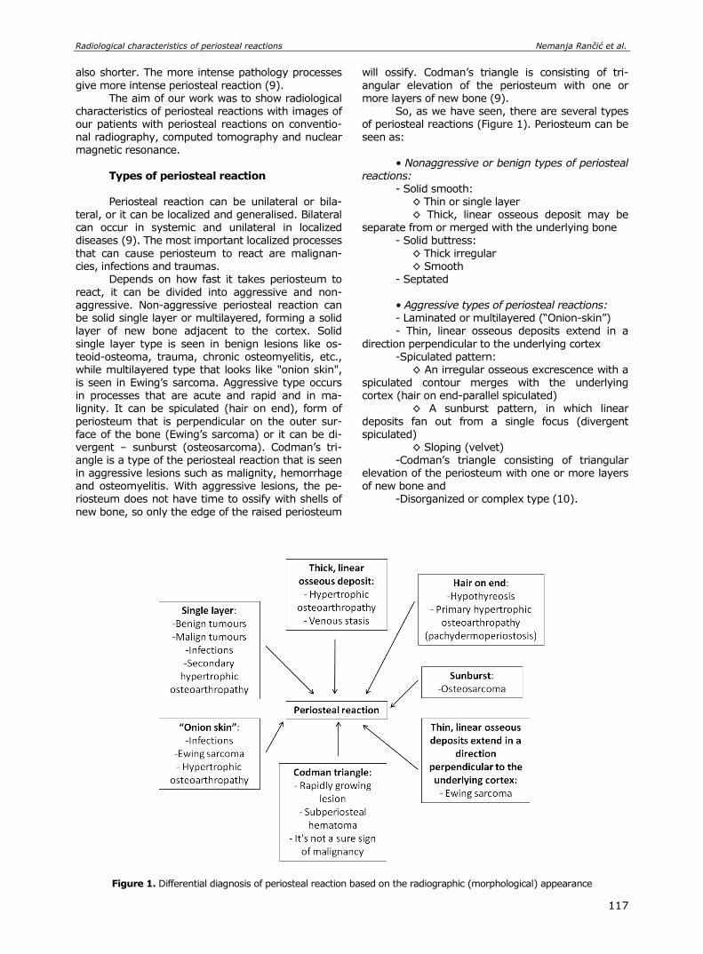
Radiological characteristics of periosteal reactions Nemanja Rančić et al.
117
also shorter. The more intense pathology processes
give more intense periosteal reaction (9).
The aim of our work was to show radiological characteristics of periosteal reactions with images of our patients with periosteal reactions on conventio-nal radiography, computed tomography and nuclear magnetic resonance.
Types of periosteal reaction Periosteal reaction can be unilateral or bila-
teral, or it can be localized and generalised. Bilateral can occur in systemic and unilateral in localized diseases (9). The most important localized processes
that can cause periosteum to react are malignan-cies, infections and traumas.
Depends on how fast it takes periosteum to
react, it can be divided into aggressive and non-aggressive. Non-aggressive periosteal reaction can be solid single layer or multilayered, forming a solid layer of new bone adjacent to the cortex. Solid
single layer type is seen in benign lesions like os-teoid-osteoma, trauma, chronic osteomyelitis, etc., while multilayered type that looks like "onion skin", is seen in Ewing’s sarcoma. Aggressive type occurs in processes that are acute and rapid and in ma-lignity. It can be spiculated (hair on end), form of
periosteum that is perpendicular on the outer sur-face of the bone (Ewing’s sarcoma) or it can be di-vergent – sunburst (osteosarcoma). Codman’s tri-angle is a type of the periosteal reaction that is seen in aggressive lesions such as malignity, hemorrhage and osteomyelitis. With aggressive lesions, the pe-
riosteum does not have time to ossify with shells of
new bone, so only the edge of the raised periosteum
will ossify. Codman’s triangle is consisting of tri-
angular elevation of the periosteum with one or
more layers of new bone (9). So, as we have seen, there are several types
of periosteal reactions (Figure 1). Periosteum can be seen as:
• Nonaggressive or benign types of periosteal reactions:
- Solid smooth: ◊ Thin or single layer ◊ Thick, linear osseous deposit may be
separate from or merged with the underlying bone - Solid buttress:
◊ Thick irregular ◊ Smooth
- Septated
• Aggressive types of periosteal reactions: - Laminated or multilayered (“Onion-skin”) - Thin, linear osseous deposits extend in a
direction perpendicular to the underlying cortex -Spiculated pattern:
◊ An irregular osseous excrescence with a spiculated contour merges with the underlying cortex (hair on end-parallel spiculated)
◊ A sunburst pattern, in which linear
deposits fan out from a single focus (divergent spiculated)
◊ Sloping (velvet) -Codman’s triangle consisting of triangular
elevation of the periosteum with one or more layers of new bone and
-Disorganized or complex type (10).
Figure 1. Differential diagnosis of periosteal reaction based on the radiographic (morphological) appearance
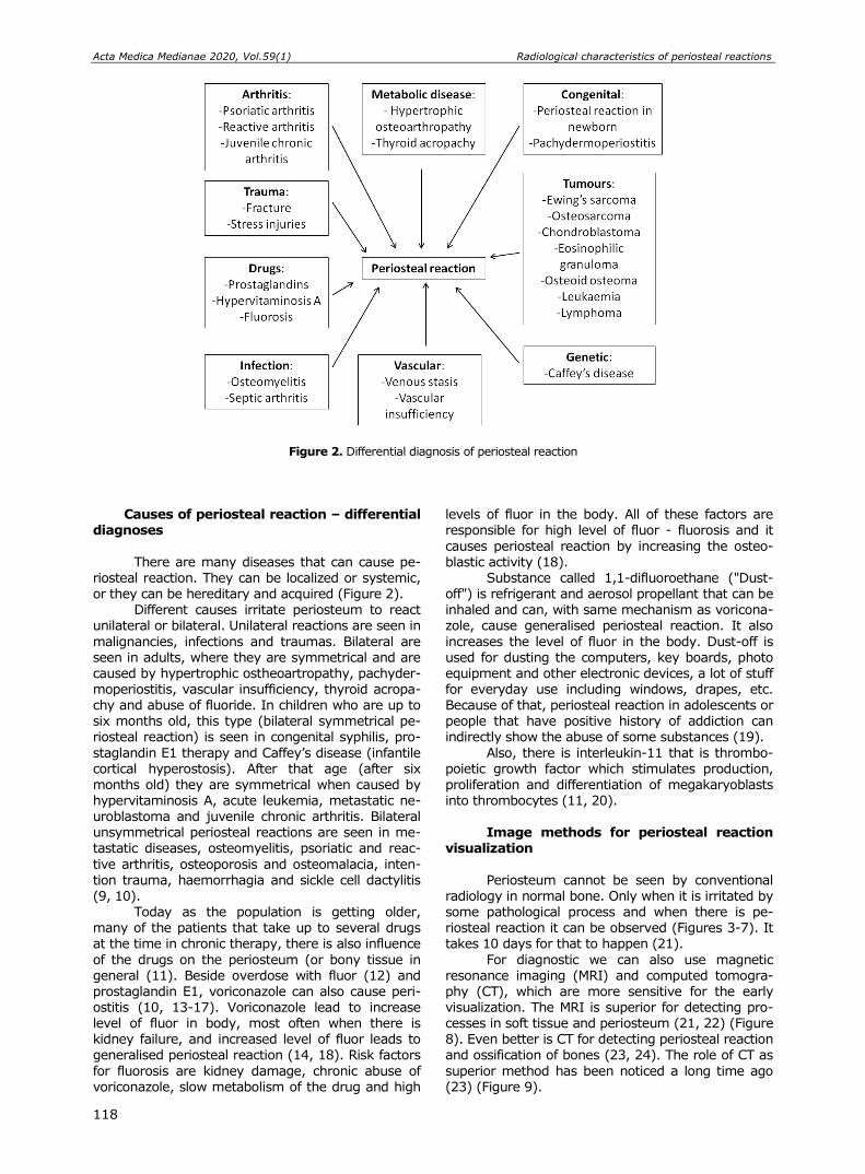
Acta Medica Medianae 2020, Vol.59(1) Radiological characteristics of periosteal reactions
118
Figure 2. Differential diagnosis of periosteal reaction
Causes of periosteal reaction – differential diagnoses
There are many diseases that can cause pe-
riosteal reaction. They can be localized or systemic, or they can be hereditary and acquired (Figure 2).
Different causes irritate periosteum to react unilateral or bilateral. Unilateral reactions are seen in
malignancies, infections and traumas. Bilateral are seen in adults, where they are symmetrical and are caused by hypertrophic ostheoartropathy, pachyder-moperiostitis, vascular insufficiency, thyroid acropa-chy and abuse of fluoride. In children who are up to six months old, this type (bilateral symmetrical pe-riosteal reaction) is seen in congenital syphilis, pro-
staglandin E1 therapy and Caffey’s disease (infantile cortical hyperostosis). After that age (after six months old) they are symmetrical when caused by hypervitaminosis A, acute leukemia, metastatic ne-uroblastoma and juvenile chronic arthritis. Bilateral unsymmetrical periosteal reactions are seen in me-tastatic diseases, osteomyelitis, psoriatic and reac-
tive arthritis, osteoporosis and osteomalacia, inten-tion trauma, haemorrhagia and sickle cell dactylitis (9, 10).
Today as the population is getting older, many of the patients that take up to several drugs at the time in chronic therapy, there is also influence
of the drugs on the periosteum (or bony tissue in general (11). Beside overdose with fluor (12) and prostaglandin E1, voriconazole can also cause peri-ostitis (10, 13-17). Voriconazole lead to increase level of fluor in body, most often when there is kidney failure, and increased level of fluor leads to generalised periosteal reaction (14, 18). Risk factors
for fluorosis are kidney damage, chronic abuse of voriconazole, slow metabolism of the drug and high
levels of fluor in the body. All of these factors are responsible for high level of fluor - fluorosis and it
causes periosteal reaction by increasing the osteo-blastic activity (18).
Substance called 1,1-difluoroethane ("Dust-off") is refrigerant and aerosol propellant that can be
inhaled and can, with same mechanism as voricona-zole, cause generalised periosteal reaction. It also
increases the level of fluor in the body. Dust-off is used for dusting the computers, key boards, photo equipment and other electronic devices, a lot of stuff for everyday use including windows, drapes, etc. Because of that, periosteal reaction in adolescents or people that have positive history of addiction can indirectly show the abuse of some substances (19).
Also, there is interleukin-11 that is thrombo-poietic growth factor which stimulates production, proliferation and differentiation of megakaryoblasts into thrombocytes (11, 20).
Image methods for periosteal reaction
visualization
Periosteum cannot be seen by conventional
radiology in normal bone. Only when it is irritated by some pathological process and when there is pe-riosteal reaction it can be observed (Figures 3-7). It takes 10 days for that to happen (21).
For diagnostic we can also use magnetic resonance imaging (MRI) and computed tomogra-phy (CT), which are more sensitive for the early visualization. The MRI is superior for detecting pro-cesses in soft tissue and periosteum (21, 22) (Figure 8). Even better is CT for detecting periosteal reaction and ossification of bones (23, 24). The role of CT as
superior method has been noticed a long time ago (23) (Figure 9).
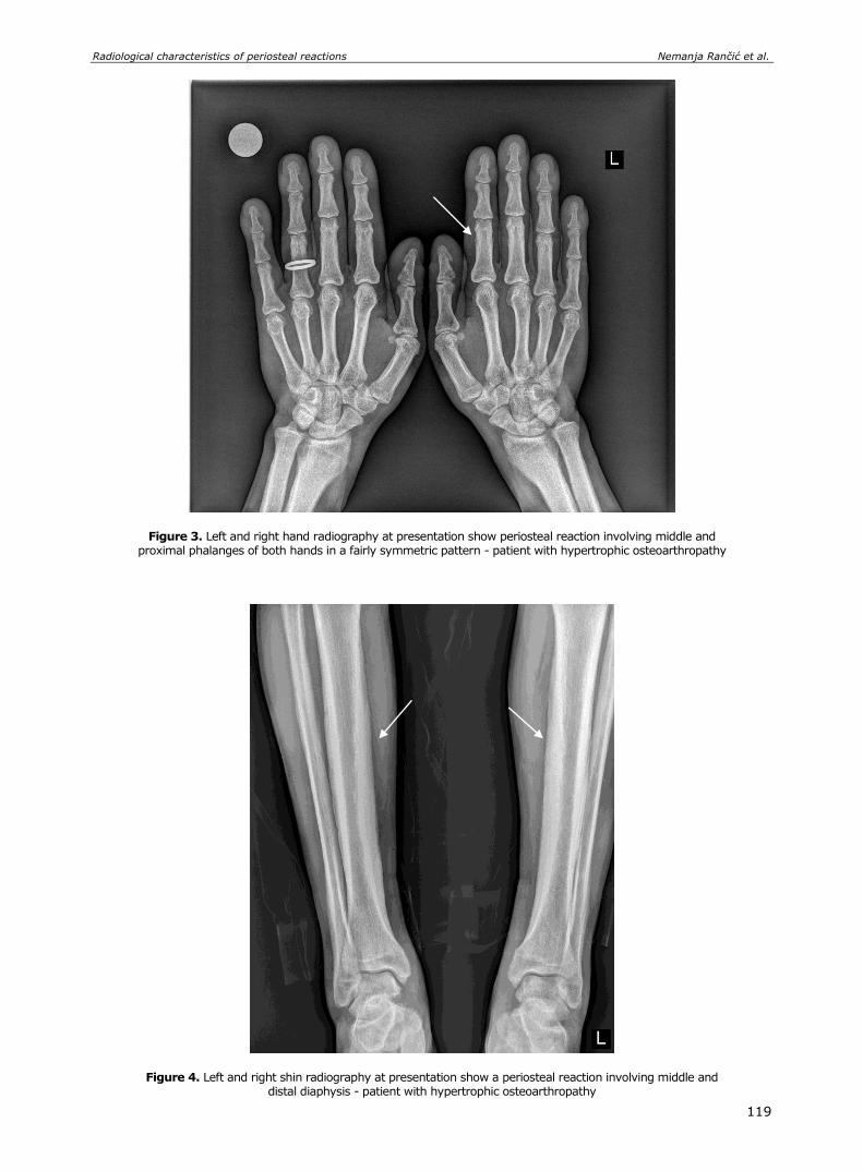
Radiological characteristics of periosteal reactions Nemanja Rančić et al.
119
Figure 3. Left and right hand radiography at presentation show periosteal reaction involving middle and proximal phalanges of both hands in a fairly symmetric pattern - patient with hypertrophic osteoarthropathy
Figure 4. Left and right shin radiography at presentation show a periosteal reaction involving middle and distal diaphysis - patient with hypertrophic osteoarthropathy
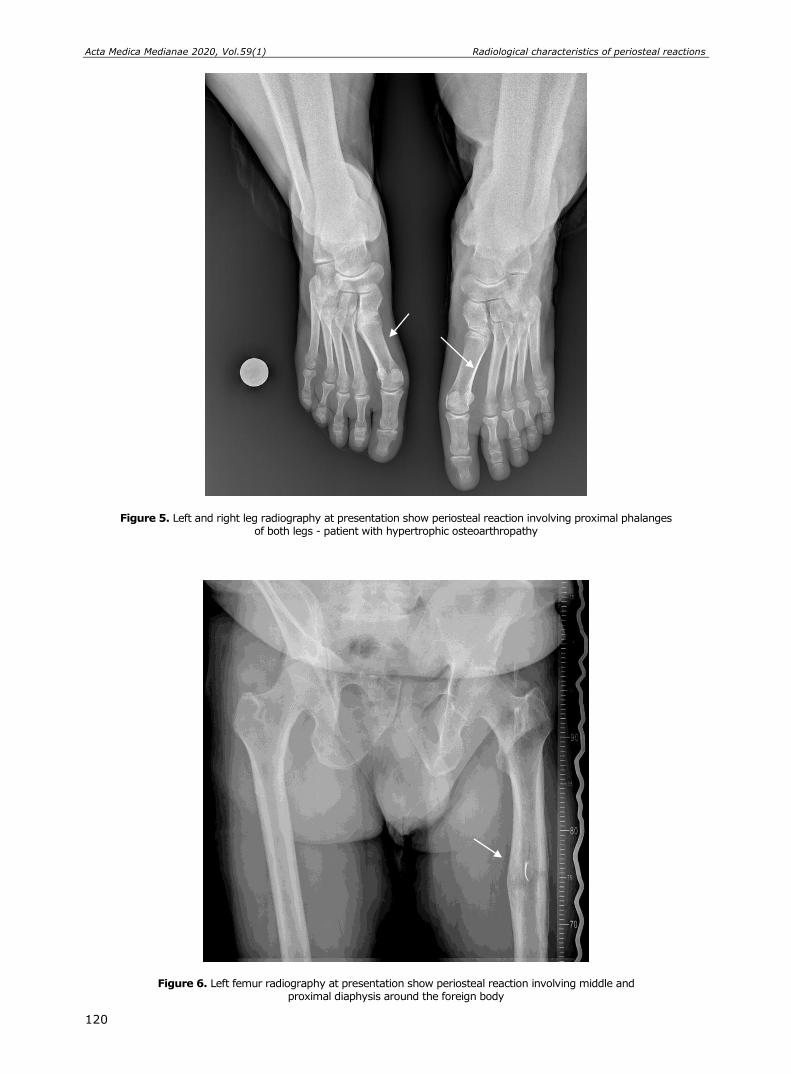
Acta Medica Medianae 2020, Vol.59(1) Radiological characteristics of periosteal reactions
120
Figure 5. Left and right leg radiography at presentation show periosteal reaction involving proximal phalanges of both legs - patient with hypertrophic osteoarthropathy
Figure 6. Left femur radiography at presentation show periosteal reaction involving middle and proximal diaphysis around the foreign body
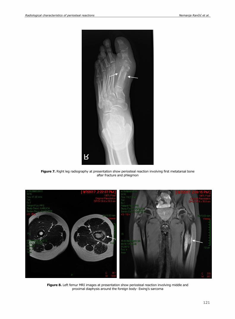
Radiological characteristics of periosteal reactions Nemanja Rančić et al.
119
Figure 7. Right leg radiography at presentation show periosteal reaction involving first metatarsal bone after fracture and phlegmon
Figure 8. Left femur MRI images at presentation show periosteal reaction involving middle and proximal diaphysis around the foreign body- Ewing’s sarcoma
121
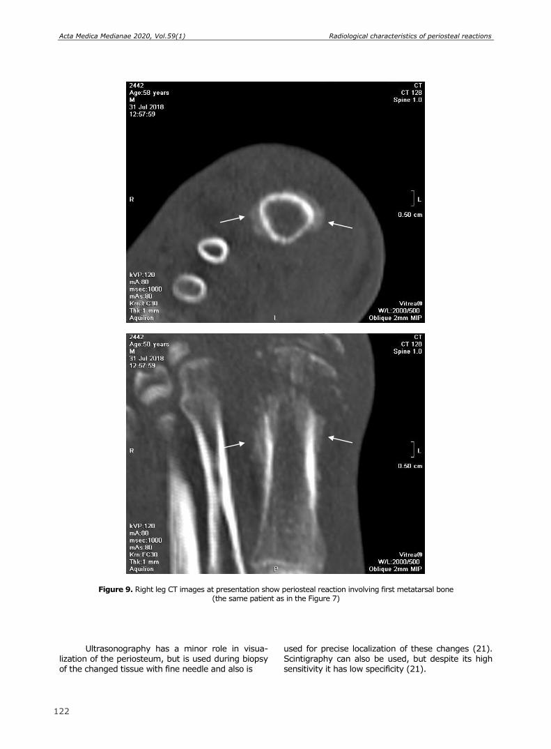
Acta Medica Medianae 2020, Vol.59(1) Radiological characteristics of periosteal reactions
120
Figure 9. Right leg CT images at presentation show periosteal reaction involving first metatarsal bone (the same patient as in the Figure 7)
Ultrasonography has a minor role in visua-lization of the periosteum, but is used during biopsy of the changed tissue with fine needle and also is
used for precise localization of these changes (21). Scintigraphy can also be used, but despite its high sensitivity it has low specificity (21).
122
Radiological characteristics of periosteal reactions Nemanja Rančić et al.
123
Conclusion
Periosteal reactions are very important in ra-diology diagnosis. Many localized or systemic di-seases can cause periosteum to react. Because of that, general knowledge of the periosteal reactions is
necessary in radiology diagnostic of the skeletal
system.
Conflict of Interest
The authors declare that they have no conflict
of interest.
References
1. Negovanović D. [Clinical radiology of the disease skeleton in childhood]. 2nd edition. Belgrade: Savre-mena administracija; 1990. Serbian
2. Dwek JR. The periosteum: what is it, where is it, and what mimics it in its absence? Skeletal Radiol 2010; 39(4):319-23. [CrossRef] [PubMed]
3. Allen MR, Hock JM, Burr DB. Periosteum: biology, regulation, and response to osteoporosis therapies. Bone 2004;35(5):1003-12. [CrossRef] [PubMed]
4. Diaz-Flores L, Gutierrez R, Lopez-Alonso A, Gonzalez R, Varela H. Pericytes as a supplementary source of osteoblasts in periosteal osteogenesis. Clin Orthop Relat Res 1992;(275):280-6. [CrossRef] [PubMed]
5. Reilly TM, Seldes R, Luchetti W, Brighton CT. Simi-larities in the phenotypic expression of pericytes and bone cells. Clin Orthop Relat Res 1998;(346):95-103.
[CrossRef] [PubMed] 6. Jovičić B, Lazić Z, Nedić M. [Therapeutic efficacy of
guided tissue regeneration and connective tissue auto-transplants with periosteum in the management of gingival recession]. Vojnosanit pregl 2008;65(10): 758-62. Serbian [CrossRef] [PubMed]
7. Lin Z, Fateh A, Salem DM, Intini G. Periosteum: biology and applications in craniofacial bone regenera-tion. J Dent Res 2014;93(2):109-16. [CrossRef] [PubMed]
8. de Oliveira HAAB, de Moraes RPF, Limirio PHJO, Dechichi P. Repair of a perforated sinus membrane with an autogenous periosteal graft: a study in 24 patients. Br J Oral Maxillofac Surg 2018;56(4):299-303. [CrossRef] [PubMed]
9. Barlas NB, Narkhede PN, Wilkinson M, Bilal A. Periosteal reaction: a radiographic guide to disease process. European Congress of Radiology 2013; 2013 March 7-11; Vienna, Austria;2013. p. 1-21.
10. Resnick D, Kransdorf MJ. Bone and joint imaging. 3rd edition. Philadelphia: Saunders; 2004.
11. Chen L, Mulligan M. Medication induced periostitis resembling hypertrophic osteoarthropathy in lung transplant patients. European Congress of Radiology 2010; 2010 March 4-8; Vienna, Austria; 2010. p. 1-14.
12. Jamshidi K, Givehchian B, Mirzaei A. Florid reactive periostitis of the long bone: A case series of seven patients. J Orthop Sci 2017;22(3):560-5. [CrossRef] [PubMed]
13. Adwan MH. Voriconazole-induced periostitis: a new rheumatic disorder. Clin Rheumatol 2017;36(3):609-15. [CrossRef] [PubMed]
14. Glushko T, Colmegna I. Voriconazole-induced perio-stitis. CMAJ 2015;187(14):1075. [CrossRef] [PubMed]
15. Letts M, Pang E, Simons J. Prostaglandin-induced neonatal periostitis. J Pediatr Orthop 1994; 14(6): 809-13. [CrossRef] [PubMed]
16. Badheka A, Bangalore Prakash P, Allareddy V. Pro-staglandin E1-induced periostitis and reversibility with discontinuation. J Pediatr 2017;189:237-237.e1. [CrossRef] [PubMed]
17. Oliveira-Filho IL, Rocha CT, de Menezes AKA, Bezerra MF, Neves BG. Proliferative periostitis in a pediatric patient. J Dent Child (Chic) 2018;85(1):23-7. [PubMed]
18. Gerber B, Guggenberger R, Fasler D, Nair G, Manz MG, Stussi G, et al. Reversible skeletal disease and high fluoride serum levels in hematologic patients receiving voriconazole. Blood 2012;120(12):2390-4. [CrossRef] [PubMed]
19. Hock LE, Honkanen I, Fiordellisi W, Bettendorf B. Reactive periostitis from inhalant abuse. Arthritis Rheumatol 2018;70(9):1391. [CrossRef] [PubMed]
20. Milman E, Berdon WE, Garvin JH, Cairo MS, Bess-mertny O, Ruzal-Shapiro C. Periostitis secondary to interleukin-11 (Oprelvekin, Neumega). Treatment for thrombocytopenia in pediatric patients. Pediatr Radiol 2003;33(7):450-2. [CrossRef] [PubMed]
21. Wenaden AE, Szyszko TA, Saifuddin A. Imaging of periosteal reactions associated with focal lesions of bone. Clin Radiol 2005;60(4):439-56. [CrossRef] [PubMed]
22. Davis DL. Voriconazole-related periostitis presenting on magnetic resonance imaging. Clin Cases Miner Bone Metab 2015;12(1):78-81. [CrossRef] [PubMed]
23. Ida M, Tetsumura A, Kurabayashi T, Sasaki T. Perio-steal new bone formation in the jaws. A computed tomographic study. Dentomaxillofac Radiol 1997; 26 (3):169-76. [CrossRef] [PubMed]
24. Kopys-Wiszniewska I. [Evaluation of images of perio-steum on computed tomography in children with ma-lignant bone tumours before and after chemotherapy]. Med Wieku Rozwoj 2008;12(1):463-76. Polish [PubMed]
Acta Medica Medianae 2020, Vol.59(1) Radiological characteristics of periosteal reactions
124
Revijalni rad UDC: 616.71‑018.44-073
doi:10.5633/amm.2020.0117
RADIOGRAFSKE KARAKTERISTIKE PERIOSTNIH REAKCIJA
Nemanja Rančić1, Aleksandar Jovanovski1, Jelena Stevanović1, Ratko Stamatović1,
Dejan Kostić1, Igor Sekulić1, Berislav Vekić2,3, Jasenka Vasić-Vilić1
1Institut za radiologiju, Vojnomedicinska akademija, Beograd, Srbija 2Odeljenje hirurgije, Klinički centar "Dr Dragiša Mišović", Beograd, Srbija 3Univerzitet u Kragujevcu, Fakultet medicinskih nauka, Kragujevac, Srbija
Kontakt: Nemanja Rančić
VMA, Crnotravska 17, 11000 Beograd, Srbija
E-mail: [email protected]
Periost je tanko fibrozno tkivo (fibro-vaskularna membrana) koje prekriva spoljašnju
površinu kosti. Na klasičnoj radiografiji normalan periost potpuno je nevidljiv. Može se vizualizovati kada je nadražen nekim patološkim procesom. Periostna reakcija može biti unilateralna ili bilateralna, odnosno lokalizovana ili generalizovana. Periostne reakcije dele se, u odnosu na brzinu kojom se javljaju, na agresivne i neagresivne. Uloga kompjuterizovane tomografije, kao superiorne metode, u odnosu na nuklearnu magnetnu rezonancu, ultrasono-grafiju ili scintigrafiju, pokazana je ranije.
Acta Medica Medianae 2020;59(1):116-124.
Ključne reči: periostna reakcija, radiologija, diferencijalna dijagnoza, uzrok
125 www.medfak.ni.ac.rs/amm
Review article UDC: 616.378-008.64:575.113
doi:10.5633/amm.2020.0118
THE ASSOCIATION OF GENETIC POLYMORPHISMS WITH DIABETES MELLITUS TYPE 1
Maja Jović1, Vesna Cvetković3, Milena Despotović2, Tatjana Jevtović-Stoimenov1,2
One of the most common endocrine disorders in children and adolescents, Diabetes
mellitus (DM), is a chronic, polygenic and complex illness. Based on etiology, DM can be divided into two basic groups: Diabetes mellitus type 1 (DMT1) and Diabetes mellitus type 2 (DMT2). DMT1 is a disease mediated by immune mechanisms based on the autoimmune destruction of insulocyte by immunoreactive T cells and antibodies which leads to insulin resistance and hyperglycemia syndrome.
Several different molecular mechanisms contribute to the destruction of β-cells of Langerhans islets, such as: the production of autoantibodies directed against antigens on insulocytes, lysis of Langerhans islets mediated by cytotoxic CD8+ T lymphocytes, local production of cytokines (TNF and IL-1) by macrophages that damage insulocytes and delayed hypersensitivity reactions mediated by CD4+ Th1 lymphocytes.
The most important genes that show a high degree of association with DMT1 are found in the region which encodes MHC (major histocompatibility complex) of molecules class I and II. However, there is very strong evidence of association of other genes (outside of the MHC gene domain) with the expression of DMT1. These are: insulin gene (INS), Cytotoxic T-Lymphocyte Associated Protein 4 (CTLA4), Protein Tyrosine Phosphatase Non-Receptor Type 22 (PTPN22), Protein Tyrosine Phosphatase Non-Receptor Type 2 (PTPN2), C-type lecithin gene (CLEC16A, KIAA0350), interleukin 2 receptor α gene (IL2RA/CD25), interferon-induced helicase gen domain (IFIH1), Potassium Voltage-Gated Channel Subfamily J Member 11 (KCNJ11), Platelet and T Cell Activation Antigen 1 (CD226), vitamin D receptor gene (VDR), tumor
necrosis factor gene (TNF) and lymphotoxin-α (LTA). In addition to these genes, researchers are still searching for other genes that are associated with the appearance and expression of DMT1.
Acta Medica Medianae 2020;59(1):125-132. Key words: Diabetes mellitus type 1, autoimmune endocrinopathy, non-HLA gene
polymorphisms
1University of Niš, Faculty of Medicine, Research Center for Biomedicine, Niš, Serbia 2University of Niš, Faculty of Medicine, Department of
Biochemistry, Niš, Serbia 3Pediatric Internal Diseases Clinic, Clinical Center Niš, Serbia
Contact: Maja Jović
81 Dr. Zoran Djindjić Blvd., 18000 Niš, Serbia
E-mail: [email protected]
Diabetes mellitus type 1
Diabetes mellitus type 1 (DMT1) is an im-mune-mediated disease based on the autoimmune destruction of Langernahs pancreatic insulocytes by immunoreactive T cells and antibodies, resulting in a
progressive decrease in the number of insulocytes and their ability of insulin secretion leading to insulin resistance and hyperglycemia syndrome. This chro-nically progressive autoimmune disease occurs in a person with a genetic predisposition to its develop-ment. The first symptoms of this disorder most
commonly occur early in childhood while the disease
appears in puberty and progresses with age. According to the classifications given by the World Health Organization (WHO) and ADA, there are two types of DMT1. The more common type (1A) be-longs to autoimmune diseases of complex etiology,
while the rare type (1B) is characterized by idiopathic etiology. Both types are characterized by the destruction of β-cells Langerhans’s isles in the pancreas in genetically predisposed individuals. This occurs under the influence of infectious or toxic environmental factors which consequently leads to a lack of insulin. Based on the appearance of typical
symptoms of the disease (polydipsia, polyuria, poly-phagia), and the presence of elevated blood glucose levels, the diagnosis of DMT1 is established.
The association of genetic polymorphisms with diabetes mellitus type 1. Maja Jović et al.
126
Complications of DMT1 have been common including
macroangiopathies, such as progressive atheroscle-
rosis of the arteries leading to ischemic sclerosis of the extremities and internal organs and microangio-pathy, i.e., microvascular obstructions leading to damage of the retina, renal glomeruli, and peri-pheral nerves. All mentioned complications of DMT1
can lead to renal and cardiovascular disease, blind-ness, and early death of adults, the insulin therapy is necessary both for disease control and for pro-longing the life age. Most people with DMT1 depend on external insulin compensation and without in-sulin, they develop severe metabolic complications such as acute ketoacidosis and coma (1, 2).
Etiopathogenesis of DMT1
The mutual action of certain genetic, immu-nological and environmental factors leads to the ini-tiation of the process that will affect the destruction of type B insulocytes. Environmental factors include
viral infections (cytomegalovirus, mumps, rubeola, coxsackievirus), early infant feeding with cow’s milk, additives (nitrosamines) and toxins in food. External factors include climate change, stress and vaccina-tion. These factors can represent a trigger for the occurrence of DMT1. Viruses, for example, promote
the development of DMT1 mainly by molecular mimicry (GAD – the glutamic acid decarboxylase antigen on type B insulocytes and Coxsackie B4 virus have similar molecular properties) or non-spe-cific cytokine-mediated reactions. The onset of dia-betes is also affected by the secretion of certain
hormones within physiological limits. The highest
incidence of this type of diabetes is from 11 to 13 years of age when the growth hormone is most secreted as well as sex hormones that have the effect opposite to insulin (3–5).
There are several molecular mechanisms that contribute to the destruction of the β-cells of the Langerhans islets. The most important of these are:
a) production of autoantibodies directed against antigens on insulocytes,
b) lysis of Langerhans’s islets mediated by cytotoxic CD8+ T lymphocytes,
c) local production of cytokines (TNF or IL-1) by macrophages that damage the insulocytes,
d) delayed-hypersensitivity mediated by CD4+ Th1 lymphocytes that are reactive to insulocyte
antigen (including insulin) (6, 7).
Genetic factors that affect the appearance of DMT1
DMT1 is a complex, genetically determined disease whose development and manifestation also depends on environmental factors. Genes that can lead to this disease are found in 60 chromosomal regions. The most important genes that show a high degree of association with DMT1 are found in the region that encodes the MHC (major histocompa-
tibility complex) class II molecule located on the 6p21 (HLA-D) chromosome, as well as the genes for MHC class I molecule. MHC genes are designated in
humans as HLA (human leukocyte antigen) genes.
Besides these, there are other genes that are
associated with the manifestation of DMT1, but the evidence of their association is still investigated. These are: 5' end of insulin gene (INS), gene for protein 4 linked to cytotoxic T lymphocyte (CTLA4), gene for protein tyrosine phosphatase - 22
(PTPN22), gene for protein tyrosine phosphatase - 2 (PTPN2), gene for protein lecithin type C (CLEC16A, known as KIAA0350), gene for receptor of α in-terleukin 2 (IL2RA/CD25), gene region of interferon-induced helixase (IFIH1), KCNJ11, CD226, gene for vitamin D receptor (VDR), genes for tumor necrosis factor (TNF) and lymphotoxin-α (LTA) and gene
regions on chromosomes 12q24, 12q13, 16p13 and 18p11. In addition to these genes, researchers are still searching for other genes that are associated
with the appearance and expression DMT1 (2, 7, 8). Human leukocyte antigen
The HLA system plays an important role in human immune responses. There are three classes of HLA systems with their corresponding functions: HLA I (HLA-A, HLA-B and HLA-C), HLA II (HLA-DR, HLA-DQ and HLA-DP) and HLA III. The genes HLA class I and II encode molecules that have a major
role in the treatment and presentation of antigen by T lymphocytes (9, 10).
The HLA region of chromosome 6 is res-ponsible for nearly 50% of the genetic predisposition to the development of DMT1. The subregions of DR4 and DR3 HLA class II are strongly related to the
occurrence of DMT1 and the combination of DR3 /
DR4 allele multiplies the risk of illness. It is believed that haplotype combinations of class II of the HLA gene DR4-DQB1-0302 and DR3-DQB1*0201 are the main markers of the tendency towards this disease. About 90% of children with DMT1 are carriers of one or both of these haplotypes of the gene. It is known that about 95% of Caucasians with DMT1 are
wearing HLA-DR3, DR4 or both antigens. In carriers of DR3 and DR4 antigens, the risk of developing this disease is increased tenfold (2, 3, 9).
Gene for insulin (INS)
Insulin has a major role in the clinical, genetic and immunological aspects of DMT1. Gene for
insulin is localized to 11p15 chromosome. This gene encodes a protein, pre-proinsulin, consisting of 110 amino acids, which is synthesized by beta cells of the Langerhans’s islets. By reaction of proteases that eliminate the signal peptide pre-proinsulin is modi-
fied to proinsulin which is transformed into biolo-gically active insulin after splitting of the C-peptide (11, 12).
The insulin gene locus is the second most important locus after the HLA locus which indicates a predisposition to the development of DMT1. The insulin gene comprises 3 exons and 2 introns, sepa-
rated from each other by several polymorphisms in linkage disequilibrium (LD). The insulin locus repre-sents a variable number of tandem repeats (VNTR)
Acta Medica Medianae 2020, Vol.59(1) The association of genetic polymorphisms with diabetes mellitus type 1.
127
in the INS gene promoter. The alleles of this region
are divided into three classes, I, II and III. Alleles
class I contain about 570 base pairs (bp) (26-63 repeats), allele class II have 1200 base pairs (around 80 repeats) and allele class III have 2200 base pairs (140-210 repeats). Studies have shown that allele class I are associated with a higher risk of
developing DMT1 than allele class III that are con-sidered to have a protective role (13–15).
The genetic susceptibility mechanism is explained in such a way that the own antigens for allele class I are less expressed in thymus compared to own antigens for allele class III that are two to three times more expressed in the thymus. This will
affect the positive and negative selection of T lymphocytes in the thymus. There is the inverse pattern of expression in the pancreas (16, 17).
Gene for protein 4 associated with
cytotoxic T-lymphocyte antigen-4 (CTLA4)
CTLA-4 gene is located on chromosome 2q33. It encodes the homonymous molecule localized on the surface of activated T-lymphocytes which is responsible for suppressing the immune response. Inactive T-lymphocytes do not express CTLA-4 on their surface while when activating T-lymphocytes
this molecule binds to the receptor on antigen pre-senting cells (APC), which stops activation of T-lym-phocyte thereby interrupting the immune response. The CTLA-4 binding to the APC receptor creates an inhibitory signal for the production of IL-2 and IL-2 receptors, as well as the continuation of the cell
cycle. In this way, CTLA-4 plays an important role in
the regulation of lymphocytic expansion which leads to a gradual reduction of the immune response. Changes in the expression of the gene for CTLA4 can lead to an increase in the autoreactivity or reactivity of T-lymphocytes, which may affect the onset of autoimmune diseases such as DMT1 (18, 19).
The onset of many other autoimmune disea-ses is associated with the mutation of the CTLA-4 gene. Mutation of this gene can cause asthma, Addison's disease, myasthenia gravis, Sjogren’s syn-drome, systemic lupus erythematosus, systemic sclerosis, ulcerative colitis and thyroiditis (20–23).
The following polymorphisms of the CTLA-4 gene have shown the association with DMT1: 49A/G
in exon 1, CT60 in 3’ UTR (untranslated region), -318C/T, -1661A/G i -1722C/T in the promoter region. Several studies in which the polymorphism of CTLA-4 gene was showed that the greatest association with DMT1 shows allele G polymorphism
49A/G of CTLA-4 gene. The SNP CTLA-4 gene is associated with the symptoms of DMT1 in several human populations (19, 24, 25).
Gene for protein tyrosine phosphatase-
22 (PTPN22)
Gene for PTPN22 is localized to chromosome 1p13.3-13.1. This gene encodes lymphoid tyrosine phosphatase (LYP) which inhibits the proliferation
and activation of T-lymphocytes (26). LYP in in-
teraction with Csk, C-terminal tyrosine kinase, re-
duces signal transduction via TCR (T-cell receptor), thus regulating various cellular activities including cell growth, differentiation, division and apoptosis. The polymorphism at position 1858 within this gene, when cysteine is replaced by thymine leads to a
mutation at the codon 620 for LYP. This mutation leads to the loss of interaction between LYP and Csk which creates conditions for inadequate prolonged activation of T-lymphocytes leading to autoimmunity and the possibility of DMT1 (8, 19).
The variant of PTPN22 R620W is associated with multiple autoimmune diseases such as lupus
erythematosus, rheumatoid arthritis, juvenile idio-pathic arthritis, multiple sclerosis, autoimmune thy-roiditis, psoriasis and Addison's disease, as con-
firmed by more studies in Europe and the United States (27, 28).
Gene for the α interleukin 2 receptor
(IL2RA/CD25) The CD25 gene is located on chromosome
10p15. This gene encodes the α-chain IL-2 receptor which is expressed for several hours after antigen activation on CD4+ and CD8+, as well as on regu-
latory T-lymphocytes. Only after the formation of the third subunit of the IL-2 receptor (α-chain) this receptor can bind IL-2. IL-2 generates the same cells that this interleukin acts through its receptor. IL-2 is also referred to as T-lymphocyte growth factor, because its basic activity is stimulation of T-
lymphocyte proliferation, that is, the stimulation of
the entry of T-lymphocytes into the cell cycle and their consequent division. This leads to an increase in the number of antigen-specific T-lymphocytes. Expression of CD25 on regulatory T-lymphocytes is an important factor in suppressing the T-cell im-mune response and consequently also on the pro-tection against the onset of autoimmune diseases.
However, mutations on this gene in humans lead to severe forms of autoimmune diseases, including DMT1 (8, 29).
The genetic region for interferon-induced
helicase (IFIH1)
The genetic region for interferon-induced he-
licase (IFIH1) is located on chromosome 2q24. IFIH1 encodes cytoplasmic viral sensor melanoma differentiation associated protein 5 (MDA5). After viral infection, MDA5 binds to viral RNA chains ac-tivating transcriptional factors (IRF7, IRF3, IRF1 and
NFκB) which induce the formation and secretion of several proinflammatory factors, interferons and in-terleukins: INF-α, INF- β, INF- γ IL-1, IL-6, IL-8. Proinflammatory cytokines activate NK and T-lym-phocytes, then attract dendritic cells, macrophages, and granulocytes to the site of infection leading to the destruction of beta cells of the pancreas. In this
way, the presentation of the own antigens of the pancreatic beta cells to the immune system is in-creased resulting in an autoimmune reaction, which
The association of genetic polymorphisms with diabetes mellitus type 1. Maja Jović et al.
126
is a trigger for the development of DTM1. This con-
firms the fact that viruses can contribute to the
initiation of DMT1 (27, 30). Several polymorphisms of IFIH1 gene locus,
rs1990760 or Thr946Ala, rs2111485, rs3747517, and rs13422767, are associated with the emergence of DMT1 (31).
Gene for vitamin D receptor (VDR) DMT1 is a common autoimmune endocrino-
pathy in which the deficiency of Vitamin D can be one of the causes of its initiation. It has been shown that the application of 1,25-dihydroxy vitamin D3, in
the experimental model of animals suffering from T-cell-dependent autoimmune diabetes reduces the incidence of this disease. The way in which disease
progression is stopped is not yet fully clarified but there is evidence of cytokine modulation of T helper (Th1/Th2) lymphocyte balance in local pancreatic lesions. Recent studies show that a significant pa-
thophysiological role in the onset of diabetes mellitus has a specific vitamin D (VDR) nuclear receptor, which is particularly expressed on monocytes and activated T-lymphocytes. By binding the ligand to VDR receptor there is a decrease in secretion of cytokines (IL-1, IL-2, IL-6, IL-12, TNF, and inter-
feron-γ) (32, 33) involved in the pathogenesis of autoimmune diseases. In this regard, it has been shown that VDR receptor polymorphism has an effect on the occurrence of DMT1 (34, 35).
The VDR gene is localized to a long arm of chromosomes 12 and 14. Although there are many
polymorphisms of this gene only four SNPs have
been tested so far: - Bsm1 (rs1544410), - Fok1 (rs10735810), - Apa1 (rs7975232) and - Taq1 (rs731236). All of the aforementioned polymorphisms are
associated with some of the autoimmune disorders
in humans, such as celiac disease, multiple sclerosis, systemic lupus erythematosus, rheumatoid arthritis and Hashimoto thyroiditis (36, 37).
Genes for tumor necrosis factor (TNF)
and lymphotoxin-α (LTA)
The tumor necrosis factor (TNF) is a proin-
flammatory cytokine. It belongs to TNF superfamily of cytokines. It is also marked as TNF-α, to differ from TNF-β, which is also called lymphotoxin (LTA). Cells which secrete TNF are activated phagocytes, antigen-stimulated T-lymphocytes, NK cells and ma-
stocytes (35). There are two types of TNF molecules,
membrane, and secretion. Membrane TNF (mTNF) is a non-glycose type II membrane protein with the intracellular amino-terminal end and a large extra-cellular carboxy-terminal end. Secretory TNF (sTNF) has a triangular pyramidal shape where each side of
the pyramid forms on a subunit of this molecule. Biologically active forms are trimer forms of protein, homotrimer (23, 38).
Biological functions of TNF are carried out
over two types of external membrane receptors
TNFR1 and TNFR2. By binding the ligand to the receptors, signal pathways for apoptosis are acti-vated, nuclear kappa factor B (NF-kappa B), ERK-kinase, p38 protein kinase, and c-Jun-N-terminal kinase. The biological effects are stimulation of
migration and activation of leukocytes at the site of infection, induction of expression of adhesion mole-cules (selectins and integrins) on endothelial cells, inhibition or induction of apoptosis. TNF also works on macrophages, stimulating them to produce IL-1, which has a similar role to TNF (29, 35, 38, 39)
The TNF encoding genes are located on a
short arm of chromosome 6, in class III of the MHC locus, between the HLA-B and the HLA-DR gene. It has been proven that there is a strong bond
between THF allele with DR haplotypes, such as HLA-A1-B8-DR3 and Bw62-DR4. Both alleles are expressed in patients with DMT1. The most re-searched TNF polymorphisms associated with DMT1
are rs1800629 (at position 308) and rs361525 (at position 238) (40).
Gene CLEC16A and gene for protein
tyrosine phosphatase-2 (PTPN2)
Gene CLEC16A (C-type lectin domain family 16, also known as KIAA0350) is localized on chromosome 16p13 and encodes a protein with Ca+ dependent or type C lectin binding structure. This protein plays important roles in binding carbo-hydrates, cell adhesion, and pathogen recognition.
KIAA0350 is almost exclusively expressed in cells of
the immune system (dendritic cells, NK cells, B lymphocytes). These cells are crucial in the pa-thogenesis of DMT1 which indicates that modulation of immunity contributes to the onset of disease. Several polymorphisms of the KIAA0350 gene (rs2903692, rs725613, rs17673553 and 12708716) are associated with the occurrence of multiple scle-
rosis and DMT1 (29, 41). PTPN2 (also known as TC-PTP or PTP-S2)
gene, which is localized on chromosome 18, encodes tyrosine phosphatase 2 which is a negative regulator of inflammatory processes and is particularly present in immune system cells (CD4+ T-lymphocytes and
B-lymphocytes), but also in pancreatic β-cells and intestinal enterocytes. This molecule inhibits the
STAT1 transcription factor in the IFN signaling pathway, whereby the downregulation type I (IFNα and IFNβ) and type II (IFNγ) IFN receptors are activated. The inhibition of PTPN2 expression in pan-creatic insulocytes and intestinal enterocytes leads
to the activation of STAT and the subsequent formation of apoptosis through Bim proteins and the mitochondrial pathway of activation. This suggests that polymorphism of the PTPN2 gene can cause β-cell apoptosis and loss of the intestinal epithelial barrier followed by the appearance of symptoms of DMT1. Too early start of intake of cow's milk or
gliadin from wheat in children, as well as the viral infections, are the most common triggers that can cause the onset of DMT1 and β-cell autoimmunity,
128
Acta Medica Medianae 2020, Vol.59(1) The association of genetic polymorphisms with diabetes mellitus type 1.
127
caused by polymorphism of the PTPN2 gene (42,
43). Polymorphisms of this gene (rs478582,
rs1893217, and rs2542151) were also identified in persons suffering from inflammatory intestinal di-sease, celiac disease and rheumatoid arthritis (28).
Potassium voltage-gated channel subfamily
J member 11 (KCNJ11) The KCNJ11 gene (gene for the potassium
voltage channel, subfamily J, member 11) is localized on a short arm of chromosome 11 at po-sition 15.1. This gene encodes the formation of four subunits of the hetero-octamer complex of the ATP-
sensitive potassium channel (KATP), while the other four subunits are encoded by the ABCC8 gene. Namely, the KCNJ11 gene encodes Kir6.2 subunit
(inward-rectifier potassium channel subunit) which participate in the formation of KATP pores, while the ABCC8 gene encodes the SUR1 (receptor sulpho-nylurea) regulatory subunits (44, 45).
KATP channels are transmembrane channels that are most commonly found in the plasma mem-brane of the β-cells of the Langer-Hans pancreatic islet. In addition to these cells, KATP channels can also be found on muscle and nerve cells. They open or close depending on the amount of blood glucose.
Increasing intracellular metabolic activity leads to a change in the relationship between ATP and ADP in the cell and conditions the closure of the KATP channel. By closing these channels, depolarization of the pancreatic β-cells occurs, which is also a trigger for the secretion of insulin (44, 46).
Mutations of KCNJ11 and ABCC8 genes are
present in about 50% of patients with DMT1. These mutations prevent the closure of the KATP channel and hence the insulin secretion in response to hyperglycemia. Also, in children with this disorder, neurological disorders are observed as well. One of the most serious disorders that can occur is DEND syndrome, which is characterized by neonatal dia-
betes, a developmental disorder, epilepsy and later onset of speech. To date, several polymorphisms of the KCNJ11 genes which are associated with the occurrence of DMT1 are found. The most prevalent in the population are E23K, A190A, L267L, L270V, I337V, K381K and S385C (45, 47).
Platelet and T cell activation antigen 1
(CD226) CD226 or DNAM-1 (DNAX auxiliary molecule-
1) is an immunoglobulin-like transmembrane glyco-protein found on plasma membranes of monocytes,
macrophages, megakaryocytes, thrombocytes, NK and T-lymphocytes, as well as in some subpopu-lations of B-lymphocytes. This receptor participates
in numerous immunological functions, such as cell
signaling and adhesion, cytokine secretion, differen-
tiation of naive T-cells, as well as proliferation and activation of cytotoxic T-lymphocytes, then migra-tion of leukocytes to the site of infection, mastocyte degranulation, and thrombocyte aggregation. Li-gands for the CD226 receptor are CD112 and CD155
(poliovirus receptor). Their binding to the receptor leads to cytotoxicity reaction (NK and T-lympho-cytes) and cytokine secretion (48, 49).
In recent years, the CD226 gene polymor-phism found at 18q22.3 is increasingly associated with the development of multiple autoimmune di-seases including DMT1, rheumatoid arthritis, syste-
mic lupus erythematosus, Grave's disease, and pso-riasis. The polymorphism of this gene, with DMT1, occurs in exon 7 when cysteine is replaced by thy-
mine (rs763361 C>T), which leads to the replace-ment of a glycine with serine at the site 307 (Gly307Ser). Lazano et al. (2013) have shown that by activating the CD226 receptor on T-lymphocytes,
the balance between Th1, Th2 and Th17 immune responses changes. Activation of this receptor affects the increased production of proinflammatory cytokine and T-cell proliferation leading to a pro-inflammatory response, while its blockage affects the reduction of IL-17 secretion, indicating the po-
tential for the use of a CD226 receptor blocker for the treatment of Th1 and Th17 autoimmune di-seases (50, 51).
Conclusion
Although DMT1 is a disease known for more
than a century, scientists still intensively research determination of its more precise etiology and genetic basis of the disease. For this purpose, the monitoring of the polymorphisms of genes for which there are already evidence of their association to DMT1, as well as the initiation of more research for testing of the new polymorphisms of the genes
would provide a great contribution. Such discoveries which we could name “preventive genetics” would greatly contribute to the early detection of patients and the individualization of the therapy, which can also encourage the development of vaccine usage in patients with DMT1.
Acknowledgments
The present study was financially supported
through grant from the Ministry of Education, Science and Technological Development of the Republic of Serbia (project number III 41018). The authors do not have any conflicts of interest in the
present study.
129
The association of genetic polymorphisms with diabetes mellitus type 1. Maja Jović et al.
130
References
1. Kaufman FR. Medical management of type 1 diabetes. 6th ed. Virginia: American Diabetes Association; 2012. p 5-50.
2. Bugarin A. HLA rizični genotipovi za dijabetes tipa 1 u oboljelih u Istočnoj Hrvatskoj [diplomski rad]. Sveu-čilište Josipa Jurja Strossmayera u Osijeku: Medicinski fakultet; 2015.
3. Russo L, Iafusco D, Brescianini S, Nocerino V, Bizzarri C, Toni S, et al. Permanent diabetes during the first year of life: Multiple gene screening in 54 patients. Diabetologia 2011;54(7):1693-701. [CrossRef][PubMed]
4. Søvik O, Irgens HU, Molnes J, Sagen J V, Bjørkhaug L, Ræder H, et al. Monogenic diabetes mellitus in Norway. Nor Epidemiol 2013;23(1):55-60. [CrossRef]
5. Ilić S. Interna medicina. 1st ed. Niš: Prosveta Niš; 2004. p 352-386.
6. Kumar V, Abbas A, Fausto N, Mitchell R. Robinsonove osnove patologije. 8th ed. Beograd: Datastatus; 2007. p 755-787.
7. Abbas A, Lichtman A, Pillai S. Cellular and molecular
immunology. 9th ed. Philadelphia: ELSEVIER; 2018. p 425-434.
8. Smyth DJ, Plagnol V, Walker NM, Cooper JD, Downes K, Yang JHM, et al. Shared and Distinct Genetic Variants in Type 1 Diabetes and Celiac Disease. N Engl J Med 2008; 359(26):2767-77. [CrossRef][PubMed]
9. Noble JA, Valdes AM. Genetics of the HLA region in the prediction of type 1 diabetes. Curr Diab Rep 2011; 11(6):533-42. [CrossRef][PubMed]
10. Bertrams J. The HLA association of insulin-dependent (type I) diabetes mellitus. Behring Inst Mitt 1984; (75):89-99. [CrossRef][PubMed]
11. Boitard C. Pancreatic islet autoimmunity. Presse Med 2012; 41(12 P 2): e636-50. [CrossRef][PubMed]
12. Nakayama M, Abiru N, Moriyama H, Babaya N, Liu E, Miao D, et al. Prime role for an insulin epitope in the development of type 1 diabetes in NOD mice. Nature 2005; 435(7039):220-3. [CrossRef][PubMed]
13. Barratt BJ, Payne F, Lowe CE, Hermann R, Healy BC, Harold D, et al. Remapping the insulin gene/IDDM2 locus in type 1 diabetes. Diabetes 2004;53(7):1884-9. [CrossRef][PubMed]
14. Kim MS, Polychronakos C. Immunogenetics of type 1 diabetes. Horm Res 2005;64(4): 180-8. [CrossRef][PubMed]
15. So M, Elso CM, Tresoldi E, Pakusch M, Pathiraja V, Wentworth JM, et al. Proinsulin C-peptide is an autoantigen in people with type 1 diabetes. Proc Natl Acad Sci U S A 2018; 115(42):10732-7. [CrossRef][PubMed]
16. Yoon JW, Jun HS. Cellular and molecular pathogenic mechanisms of insulin-dependent diabetes mellitus. Ann N Y Acad Sci 2001;928:200-11. [CrossRef][PubMed]
17. Durinovic-Bello I. Autoimmune diabetes: the role of T cells, MHC molecules and autoantigens. Autoimmunity 1998;27(3):159-77. [CrossRef][PubMed]
18. Heward J, Gordon C, Allahabadia A, Barnett AH, Franklyn JA, Gough SC. The A-G polymorphism in exon 1 of the CTLA-4 gene is not associated with systemic lupus erythematosus. Ann Rheum Dis 1999;58(3):193-5. [CrossRef][PubMed]
19. Putarek NR. Povezanost biljega polimorfizma gena HLA razreda II, CTLA-4 i PTPN22 te specifičnih autoantitijela protiv beta stanica Langerhansovih
otočića s nastankom dijabetesa melitusa tipa 1 u bolesnika s autoimunom bolešću štitnjače [diserta-cija]. Sveučilište u Zagrebu: Medicinski fakultet; 2015.
20. Chang W-W, Zhang L, Yao Y-S, Su H. Association between CTLA-4 exon-1 +49A/G polymorphism and systemic lupus erythematosus: an updated analysis. Mol Biol Rep 2012;39(9):9159-65. [CrossRef][PubMed]
21. Jiang Y, Xia B, Jiang L, Lv M, Guo Q, Chen M, et al. Association of CTLA-4 gene microsatellite polymor-phism with ulcerative colitis in Chinese patients. Inflamm Bowel Dis 2006;12(5):369-73. [CrossRef][PubMed]
22. Almasi S, Erfani N, Mojtahedi Z, Rajaee A, Ghaderi A. Association of CTLA-4 gene promoter polymorphisms with systemic sclerosis in Iranian population. Genes Immun 2006;7(5):401-6. [CrossRef][PubMed]
23. Dilas LT, Icin T, Paro JN, Bajkin I. [Autoimmune thyroid disease and other non-endocrine autoimmune diseases]. Med Pregl 2011;64(3–4):183-7. [CrossRef][PubMed]
24. Anjos S, Polychronakos C. Mechanisms of genetic susceptibility to type I diabetes: beyond HLA. Mol Genet Metab 2004;81(3):187-95. [CrossRef][PubMed]
25. Baniasadi V, Narain N, Goswami R, Das SN. Promoter region -318 C/ T and -1661 A/G CTLA-4 single nucleotide polymorphisms and type 1 diabetes in North Indians. Tissue Antigens 2006;67(5):383-9. [CrossRef][PubMed]
26. Gloria-Bottini F, Saccucci P, Manca-Bitti ML, Rapini N, Verrotti A, Neri A, et al. Type 1 diabetes mellitus. Comparison between the association with PTPN22 genotype and the association with ACP1-ADA1 joint genotype. Diabetes Res Clin Pract 2014;106(1):e7-9. [CrossRef][PubMed]
27. Rodríguez A, Alfaro JM, Balthazar V, Pineda Trujillo N. Association analysis of PTPN22, CTLA4 and IFIH1 genes with type 1 diabetes in Colombian families. J Diabetes 2015; 7(3):402-10. [CrossRef][PubMed]
28. Sharp RC, Abdulrahim M, Naser ES, Naser SA. Genetic Variations of PTPN2 and PTPN22: Role in the Pathogenesis of Type 1 Diabetes and Crohn’s Disease. Front Cell Infect Microbiol 2015;5:95. [CrossRef][PubMed]
29. Bakay M, Pandey R, Hakonarson H. Genes involved in type 1 diabetes: An update. Genes (Basel) 2013; 4(3):499-521. [CrossRef][PubMed]
30. Jermendy Á, Szatmári I, Körner A, Szabó AJ, Tóth-Heyn P, Hermann R. Association between interferon-induced helicase (IFIH1) rs1990760 polymorphism and seasonal variation in the onset of type 1 diabetes mellitus. Pediatr Diabetes 2018;19(2):300-4. [CrossRef][PubMed]
31. Winkler C, Lauber C, Adler K, Grallert H, Illig T, Ziegler AG, et al. An interferon-induced helicase (IFIH1) gene polymorphism associates with different rates of progression from autoimmunity to type 1 diabetes. Diabetes 2011;60(2):685-90. [CrossRef][PubMed]
32. Hadzija MP, Korolija M, Jemin N, Pavkovic I, Pavkovic P, Medvidovic EP, et al. Polymorphisms in the IL-18 and IL-12B genes and their association with the clinical outcome in Croatian patients with Type 1 diabetes. Gene 2013;512(2):477-81. [CrossRef][PubMed]
Acta Medica Medianae 2020, Vol.59(1) The association of genetic polymorphisms with diabetes mellitus type 1.
131
33. Bonakdaran S, Abbaszadegan MR, Dadkhah E, Khajeh-Dalouie M. Vitamin D receptor gene
polymorphisms in type 1 diabetes mellitus: a new pattern from Khorasan Province, Islamic Republic of Iran. East Mediterr Health J 2012;18(6):614-9. [CrossRef][PubMed]
34. Despotovic M, Jevtovic Stoimenov T, Stankovic I, Basic J, Pavlovic D. Vitamin D Receptor Gene Polymorphisms in Serbian Patients With Bronchial Asthma: A Case-Control Study. J Cell Biochem 2017;118(11):3986-92. [CrossRef][PubMed]
35. Despotović MR. Polimorfizmi gena uključenih u infla-matorne, antioksidativne i imunoregulatorne procese kod obolelih od bronhijalne astme [disertacija]. Univerzitet u Nišu: Medicinski fakultet; 2015.
36. Eltahir Khalid K. Vitamin D receptor gene poly-morphisms in Sudanese children with type 1 diabetes. AIMS Genet 2016;3(3):167-76. [CrossRef]
37. Motohashi Y, Yamada S, Yanagawa T, Maruyama T, Suzuki R, Niino M, et al. Vitamin D receptor gene polymorphism affects onset pattern of type 1 dia-betes. J Clin Endocrinol Metab 2003;88(7):3137-40. [CrossRef][PubMed]
38. Jevtovic-Stoimenov T, Cvetkovic T, Despotovic M, Basic J, Cvetkovic J, Marjanovic G, et al. The influence of TNF alpha -308 G/A polymorphism on oxidative stress in patients with chronic lymphocytic leukemia. Leuk Res 2017;54: 66-72. [CrossRef][PubMed]
39. Jevtovic-Stoimenov T, Kocic G, Pavlovic D, Macu-kanovic-Golubovic L, Marjanovic G, Djordjevic V, et al. Polymorphisms of tumor-necrosis factor-alpha - 308 and lymphotoxin-alpha + 250: possible modulation of susceptibility to apoptosis in chronic lymphocytic leukemia and non-Hodgkin lymphoma mononuclear cells. Leuk Lymphoma 2008;49(11):2163-9. [CrossRef][PubMed]
40. Santin I, Eizirik DL. Candidate genes for type 1 diabetes modulate pancreatic islet inflammation and β-cell apoptosis. Diabetes Obes Metab 2013;15(S3): 71-81. [CrossRef][PubMed]
41. Hakonarson H, Grant SFA, Bradfield JP, Marchand L, Kim CE, Glessner JT, et al. A genome-wide association study identifies KIAA0350 as a type 1 diabetes gene. Nature 2007;448(7153):591-4. [CrossRef][PubMed]
42. Rheinheimer J, Oliveira F dos S de, Canani LH, Crispim D. The rs1893217 (T/C) polymorphism in PTPN2 gene is not associated with type 1 diabetes mellitus in
subjects from Southern Brazil. Arq Bras Endocrinol Metabol 2014;58(4):382-8. [CrossRef][PubMed]
43. Santin I, Moore F, Colli ML, Gurzov EN, Marselli L, Marchetti P, et al. PTPN2, a candidate gene for type 1 diabetes, modulates pancreatic β-cell apoptosis via regulation of the BH3-only protein bim. Diabetes 2011;60(12):3279-88. [CrossRef][PubMed]
44. Haghvirdizadeh P, Mohamed Z, Abdullah NA, Haghvirdizadeh P, Haerian MS, Haerian BS. KCNJ11: Genetic Polymorphisms and Risk of Diabetes Mellitus. J Diabetes Res 2015; 2015: 908152. [CrossRef][PubMed]
45. Holstein A, Plaschke A, Stumvoll M, Kovacs P. The Glu23Lys polymorphism in KCNJ11 and impaired hypoglycaemia awareness in patients with type 1 diabetes. J Hum Genet 2005; 50(10): 530-3. [CrossRef][PubMed]
46. Edghill EL, Gloyn AL, Gillespie KM, Lambert AP, Raymond NT, Swift PG, et al. Activating Mutations in the KCNJ11 Gene Encoding theATP-Sensitive Kⴙ
Channel Subunit Kir6.2 Are Rare in Clinically Defined Type 1 Diabetes Diagnosed Before 2 Years. Diabetes 2004; 53(11):2998-3001. [CrossRef][PubMed]
47. Cho JH, Kang E, Lee BH, Kim GH, Choi JH, Yoo HW. DEND Syndrome with Heterozygous KCNJ11 Mutation Successfully Treated with Sulfonylurea. J Korean Med Sci 2017;32(6): 1042-5. [CrossRef][PubMed]
48. Abu El-Ella SS, Khattab ESAEH, El-Mekkawy MS, El-Shamy AA. CD226 gene polymorphism (rs763361 C>T) is associated with susceptibility to type 1 diabetes mellitus among Egyptian children. Arch Pediatr 2018;25(6):378-82. [CrossRef][PubMed]
49. Mattana TCC, Santos AS, Fukui RT, Mainardi-Novo DTO, Costa VS, Santos RF, et al. CD226 rs763361 is associated with the susceptibility to type 1 diabetes and greater frequency of GAD65 autoantibody in a Brazilian cohort. Mediators Inflamm 2014;2014: 694948. [CrossRef][PubMed]
50. Xi JS, Nie CL, Wang J, Ma Y, Ma AH. Association of CD226 polymorphisms with the susceptibility to type 1 diabetes in Chinese children. Genet Mol Res 2015; 14(4):15249-55. [CrossRef][PubMed]
51. Zeng H, Liu Q, Guan Z, Zhao F, Wang H, Chen L. Correlation between CD226 polymorphism ( rs763361 ) and the susceptibility of lung cancer in nonsmoking Chinese population. Int J Clin Exp Pathol 2016; 9(3):3905-11. [CrossRef]
The association of genetic polymorphisms with diabetes mellitus type 1. Maja Jović et al.
132
Revijalni rad UDC: 616.378-008.64:575.113
doi:10.5633/amm.2020.0118
UDRUŽENOST GENSKIH POLIMORFIZAMA SA POJAVOM DIABETES MELITUSA TIP 1
Maja Jović1, Vesna Cvetković3, Milena Despotović2, Tatjana Jevtović-Stoimenov1,2
1Univerzitet u Nišu, Medicinski fakultet, Naučnoistraživački centar za biomedicinu, Niš, Srbija 2Univerzitet u Nišu, Medicinski fakultet, Katedra za biohemiju, Niš, Srbija 3Klinika za dečje interne bolesti, Klinički centar Niš, Srbija
Kontakt: Maja Jović
Bul. dr Zorana Đinđića 81, 18000 Niš, Srbija
E-mail: [email protected]
Jedan od najčešćih endokrinih poremećaja kod dece i adolescenata, Diabetes mellitus
(DM), predstavlja hroničnu, poligensku i kompleksnu bolest. Na osnovu etiologije, DM se može podeliti na dve osnovne grupe: Diabetes mellitus tip 1 (DMT1) i Diabetes mellitus tip 2 (DMT2). DMT1 predstavlja oboljenje posredovano imunološkim mehanizmima, u čijoj osnovi je autoimunska destrukcija insulocita od strane imunoreaktivnih T-ćelija i antitela, što dovodi do insulinske rezistencije i sindroma hiperglikemije.
Nekoliko različitih molekularnih mehanizama doprinose uništavaju β-ćelija Langerha-nsovih ostrvaca, kao što su: produkcija autoantitela usmerenih protiv antigena na insulo-citima, liza Langerhansovih ostrvaca posredovana citotoksičnim CD8+ T limfocotitima, lokalna produkcija citokina (TNF i IL-1) od strane makrofaga, koji oštećuju insulocite, i reakcije kasne preosetljivosti posredovane CD4+ Th1 limfocitima.
Najvažniji geni, koji pokazuju visok stepen povezanosti sa DMT1 nalaze se u regionu
koji kodira MHC (major histocompatibility complex) molekule I i II klase. Međutim, postoje veoma čvrsti dokazi o povezanosti i drugih gena (van MHC genskog područja) sa ispo-ljavanjem DMT1. To su: gen za insulin (INS), gen za protein 4 vezan na citotoksični T limfocit
(CTLA4), gen za protein tirozin fosfatazu — 22 (PTPN22), gen za protein tirozin fosfatazu — 2
(PTPN2), gen za protein lecitin tipa C (CLEC16A, KIAA0350), gen za receptor α interleukina 2 (IL2RA/CD25), genska regija za interferon indukovanu helikazu (IFIH1), KCNJ11, CD226, gen za receptor vitamina D (VDR), geni za faktor tumorske nekroze (TNF) i limfotoksin-α (LTA). Pored ovih gena, istraživači i dalje tragaju za drugim genima koji su povezani sa pojavom i ekspresijom DMT1.
Acta Medica Medianae 2020;59(1):125-132.
Ključne reči: Diabetes mellitus tip 1, autoimunska endokrinopatija, polimorfizmi
non-HLA gena
133 www.medfak.ni.ac.rs/amm
Case report UDC: 617.735-053.6/.81
doi:10.5633/amm.2020.0119
RETINAL PIGMENT EPITHELIUM TEARS IN YOUNGER PATIENTS: CAUSES AND CONSEQUENCES
Sonja Cekić1,2, Jasmina Djordjević-Jocić1,2, Branislav Tomašević1, Ivan Jovanović3,
Predrag Jovanović1,2, Milena Vujanović1
We present three different cases of patients with retinal pigment epithelium (RPE) tear:
one case with choroidal osteoma, one with central serous chorioretinopathy (CSHR) and one with idiopathic polypoidal vasculopathy (IPCV). Fotodocumentation, fluorescein angiography, optical coherence tomography (OCT) and OCT angiography showed typical choroidal neovascularization in patient with osteoma and IPCV. In patient with CSHR, large PED without choroidal neovascularization was present. PED tear was detected in all patients.
Various eye disorders, such as tear of retinal pigment layer, can complicate clinical picture and be associated with PED. Large height and diameter of PED, association with CNV,
small ratio of CNV size to PED size, lines in infrared images, and duration of PED may predict the course of RPE tear. Therefore, it should be monitored for this complication. Treatment and outcome depends on cause of tear.
Acta Medica Medianae 2020;59(1):133-138. Key words: retinal pigment epithelium, tears, causes
1Clinic for Eye Diseases, Clinic Center Niš, Serbia 2University of Niš, Faculty of Medicine, Department of
Ophthalmology, Serbia 3University of Niš, Faculty of Medicine, Department of Anatomy
and Clinical Anatomy, Serbia
Contact: Sonja Cekić
48 Dr. Zoran Djindjić Blvd., 18000 Niš, Serbia
E-mail: [email protected]
Introduction
Retinal pigment epithelium (RPE) is a mono-layer of pigmented cells in outer part of retina. The apical membrane of retinal pigment cells faces the light sensitive outer segments of the photoreceptors. The basolateral membranes of pigment epithelium cells are in contact with the choroid and the blood-
stream. The cells of RPE have an important role in the function and survival of photoreceptors (1). Tear of RPE can be caused by different disorders and is complication involved with devastating visual loss (2, 3, 4, 5, 6). The mechanism and potential risk factors for RPE tear in patients with age related macular degeneration are defined while they still
have been unclear in rare retinal disorders. (7, 8, 9, 10, 11).
The aim of our study was to present three different cases with the tear of RPE. The first patient
was with central serous chorioretinopathy (CSHR), the second was with choroidal osteoma and the third with idiopathic polypoidal vasculopathy (IPCV). In such way, we want to emphasize that different etio-
logy, then pathophysiological bases can underpin tear of RPE, and to estimate possible risk factors for its development.
First case A 33 year old male presented with metamor-
phosia, blurred central vision and small decrease of visual acuity, a disturbance of color vision in the left eye presented as a dark spot in the central vision. Visual acuity tested by Snellen chart was 1.0 in the
right eye and 0.5 in the left eye. Intraocular pres-sure was 14 mmHg in the right eye and 16 mm Hg
in the left eye. A slit lamp biomicroscopy showed a normal appearance. Family and life history were negative. The patient suffered from sleep disorders.
A posterior segment examination showed edema of posterior pole between vascular arcade of superior and inferior temporal retinal arterial bran-ches in the left eye (Figure 1a). In the left eye, small
area of grey color was noted in the edematous zone close to the superior temporal artery (Figure 1a). The appearance of posterior pole of the right eye was normal. During fluorescein angiography, in the left eye, leaking as point in early phase and in form of umbrella in late phase was present (Figure 1b). OCT of macula on the left eye showed detachment
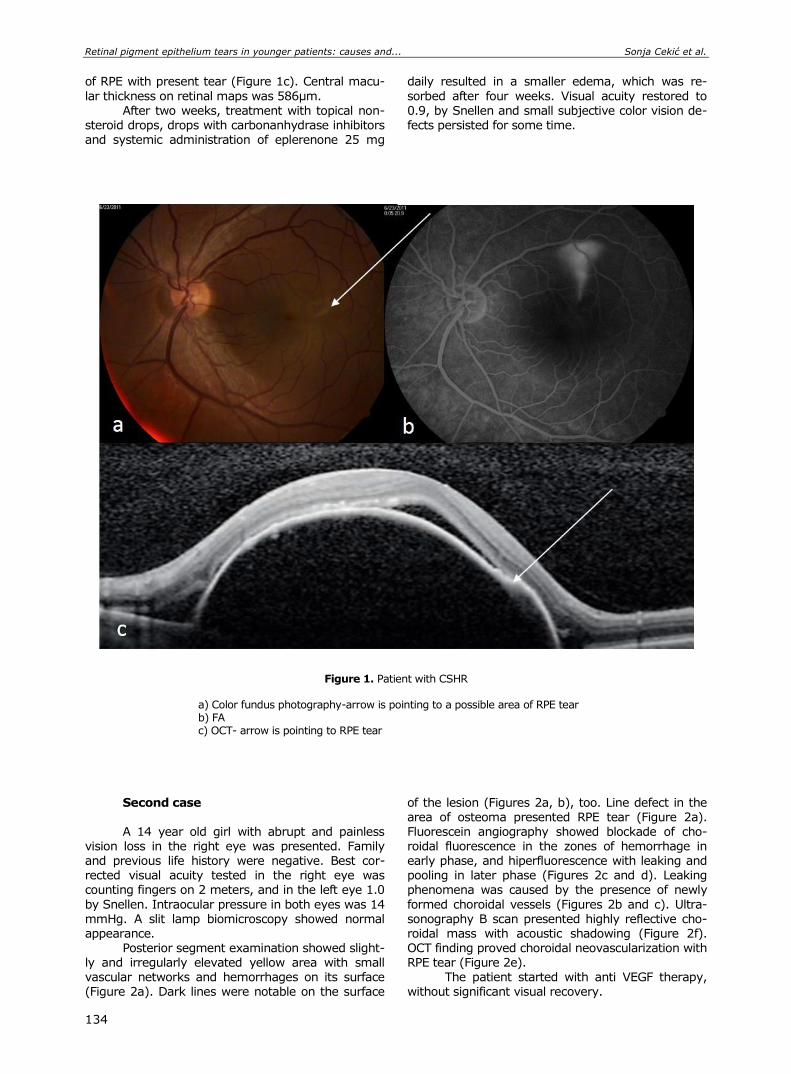
Retinal pigment epithelium tears in younger patients: causes and... Sonja Cekić et al.
134
of RPE with present tear (Figure 1c). Central macu-
lar thickness on retinal maps was 586µm.
After two weeks, treatment with topical non-steroid drops, drops with carbonanhydrase inhibitors and systemic administration of eplerenone 25 mg
daily resulted in a smaller edema, which was re-
sorbed after four weeks. Visual acuity restored to
0.9, by Snellen and small subjective color vision de-fects persisted for some time.
Figure 1. Patient with CSHR
a) Color fundus photography-arrow is pointing to a possible area of RPE tear b) FA c) OCT- arrow is pointing to RPE tear
Second case A 14 year old girl with abrupt and painless
vision loss in the right eye was presented. Family and previous life history were negative. Best cor-rected visual acuity tested in the right eye was counting fingers on 2 meters, and in the left eye 1.0
by Snellen. Intraocular pressure in both eyes was 14 mmHg. A slit lamp biomicroscopy showed normal appearance.
Posterior segment examination showed slight-ly and irregularly elevated yellow area with small vascular networks and hemorrhages on its surface (Figure 2a). Dark lines were notable on the surface
of the lesion (Figures 2a, b), too. Line defect in the area of osteoma presented RPE tear (Figure 2a). Fluorescein angiography showed blockade of cho-roidal fluorescence in the zones of hemorrhage in early phase, and hiperfluorescence with leaking and pooling in later phase (Figures 2c and d). Leaking phenomena was caused by the presence of newly
formed choroidal vessels (Figures 2b and c). Ultra-sonography B scan presented highly reflective cho-roidal mass with acoustic shadowing (Figure 2f). OCT finding proved choroidal neovascularization with RPE tear (Figure 2e).
The patient started with anti VEGF therapy, without significant visual recovery.
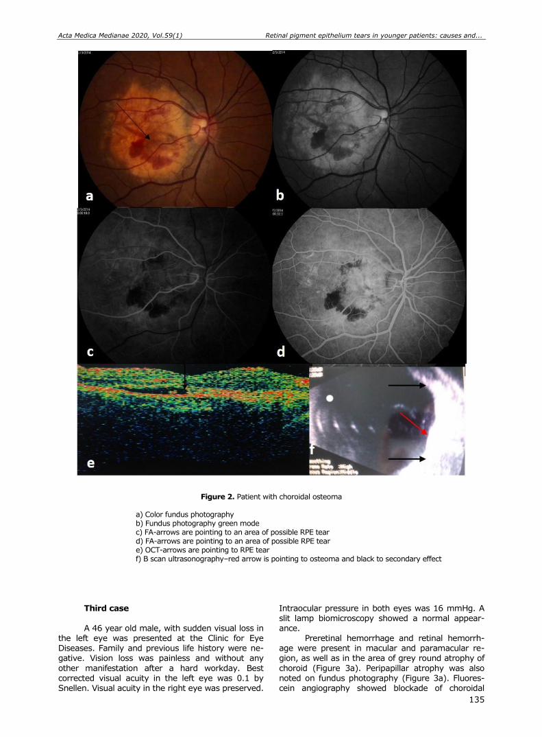
Acta Medica Medianae 2020, Vol.59(1) Retinal pigment epithelium tears in younger patients: causes and...
135
Figure 2. Patient with choroidal osteoma
a) Color fundus photography b) Fundus photography green mode c) FA-arrows are pointing to an area of possible RPE tear d) FA-arrows are pointing to an area of possible RPE tear e) OCT-arrows are pointing to RPE tear f) B scan ultrasonography–red arrow is pointing to osteoma and black to secondary effect
Third case A 46 year old male, with sudden visual loss in
the left eye was presented at the Clinic for Eye Diseases. Family and previous life history were ne-gative. Vision loss was painless and without any
other manifestation after a hard workday. Best corrected visual acuity in the left eye was 0.1 by Snellen. Visual acuity in the right eye was preserved.
Intraocular pressure in both eyes was 16 mmHg. A slit lamp biomicroscopy showed a normal appear-ance.
Preretinal hemorrhage and retinal hemorrh-age were present in macular and paramacular re-gion, as well as in the area of grey round atrophy of
choroid (Figure 3a). Peripapillar atrophy was also noted on fundus photography (Figure 3a). Fluores-cein angiography showed blockade of choroidal
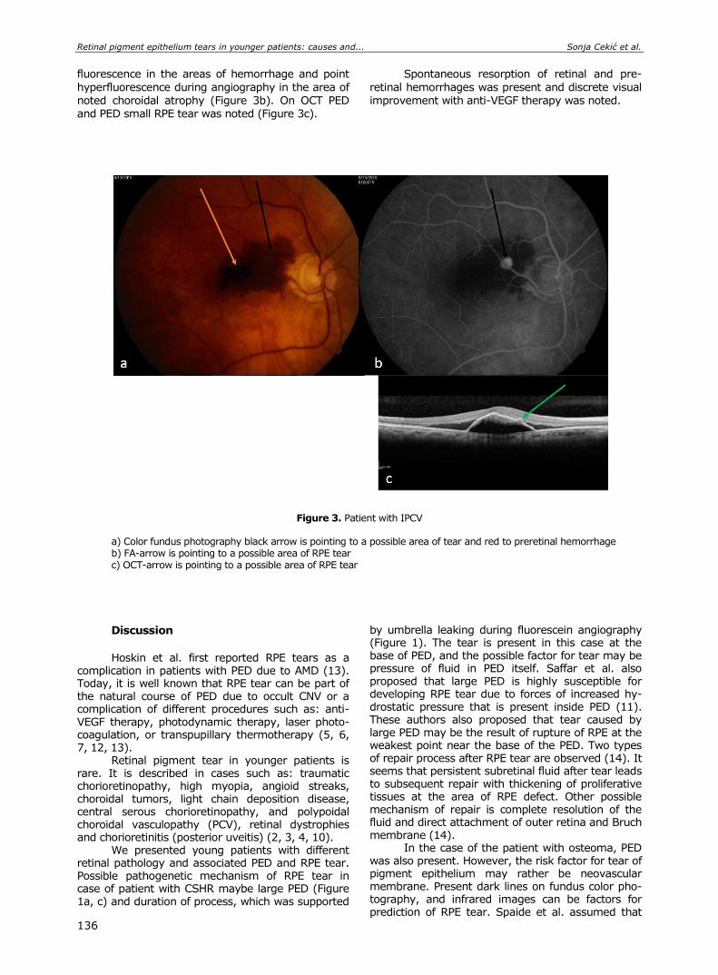
Retinal pigment epithelium tears in younger patients: causes and... Sonja Cekić et al.
136
fluorescence in the areas of hemorrhage and point
hyperfluorescence during angiography in the area of
noted choroidal atrophy (Figure 3b). On OCT PED and PED small RPE tear was noted (Figure 3c).
Spontaneous resorption of retinal and pre-
retinal hemorrhages was present and discrete visual
improvement with anti-VEGF therapy was noted.
Figure 3. Patient with IPCV
a) Color fundus photography black arrow is pointing to a possible area of tear and red to preretinal hemorrhage b) FA-arrow is pointing to a possible area of RPE tear c) OCT-arrow is pointing to a possible area of RPE tear
Discussion
Hoskin et al. first reported RPE tears as a
complication in patients with PED due to AMD (13). Today, it is well known that RPE tear can be part of the natural course of PED due to occult CNV or a complication of different procedures such as: anti-VEGF therapy, photodynamic therapy, laser photo-coagulation, or transpupillary thermotherapy (5, 6, 7, 12, 13).
Retinal pigment tear in younger patients is rare. It is described in cases such as: traumatic chorioretinopathy, high myopia, angioid streaks, choroidal tumors, light chain deposition disease, central serous chorioretinopathy, and polypoidal choroidal vasculopathy (PCV), retinal dystrophies and chorioretinitis (posterior uveitis) (2, 3, 4, 10).
We presented young patients with different retinal pathology and associated PED and RPE tear. Possible pathogenetic mechanism of RPE tear in case of patient with CSHR maybe large PED (Figure 1a, c) and duration of process, which was supported
by umbrella leaking during fluorescein angiography (Figure 1). The tear is present in this case at the base of PED, and the possible factor for tear may be pressure of fluid in PED itself. Saffar et al. also proposed that large PED is highly susceptible for developing RPE tear due to forces of increased hy-drostatic pressure that is present inside PED (11). These authors also proposed that tear caused by large PED may be the result of rupture of RPE at the weakest point near the base of the PED. Two types of repair process after RPE tear are observed (14). It seems that persistent subretinal fluid after tear leads to subsequent repair with thickening of proliferative tissues at the area of RPE defect. Other possible mechanism of repair is complete resolution of the fluid and direct attachment of outer retina and Bruch membrane (14).
In the case of the patient with osteoma, PED was also present. However, the risk factor for tear of pigment epithelium may rather be neovascular membrane. Present dark lines on fundus color pho-tography, and infrared images can be factors for prediction of RPE tear. Spaide et al. assumed that
Acta Medica Medianae 2020, Vol.59(1) Retinal pigment epithelium tears in younger patients: causes and...
137
contracture of CNV adherent to the undersurface of the RPE applies traction at the junction of the attached and detached RPE which represents the weakest point (15). The force of CNV is acting per-pendicular on RPE (Figure 2).
In the case of the patient with IPCV, PED is not large, but RPE tear was located in foveal and parafoveal zone, which caused low central visual acuity (Figure 3). Sudden visual loss may be ex-plained by newly developed CNV. It has been reported that inverse relationship between the du-ration of the PED and a risk for RPE tear is caused by vascularized PED (9, 11, 16). A short duration of PED means that the neovascular process is new with immature vessels. Immature vessels are more vul-nerable and susceptible to anti-VEGF therapy (16).
Conclusion
So, to conclude, different eye disorders can be complicated with tear of retinal pigment epithe-
lium. Treatment and outcome depend on the cause
of tear. In the large, PED, specially associated with
CNV, may predict development of RPE tear as well as an atrophy of RPE. Duration of PED is important for preservation of visual acuity. Therefore, it should be counseled and monitored for this complication, which may limit visual prognosis.
Acknowledgement
Authors declare no financial support. Conflict of Interests
No conflict of interests Declaration of Funding Sources
We declare no funding sources.
References
1. Strauβ O. Pharmacology of the retinal pigment epithelium, interface between retina and body system. Eur J Pharmacol 2016;787:84-93. [CrossRef] [PubMed]
2. Lim JI, Lam S. A retinal pigment epithelium tear in a patient with angioid streaks. Arch Ophthalmol 1990; 109:1672-4. [CrossRef] [PubMed]
3. Levin LA, Seddon JM, Topping T. Retinal pigment epithelial tears associated with trauma. Am J Ophthalmol 1991;112:396-400. [CrossRef] [PubMed]
4. Gass JDM. Retinal pigment epithelial rip during krypton red laser photocoagulation. A Ophthalmol 1984;98:700-6. [CrossRef] [PubMed]
5. Gelisken F, Indhofen W, Partsch M, Schneider U, Kreissig I. Retinal pigment epithelial tear after photodynamic therapy for choroidal neovasculariza-tion. Am J Ophthalmol 2001; 131:518-20. [CrossRef] [PubMed]
6. Pece A, Introini U, Bottoni F, Brancato R. Acute retinal pigment epithelial tear after photodynamic therapy. Retina 2001;21:661-5. [CrossRef] [PubMed]
7. Goldstein M, Heilweil G, Barak A, Loewenstein A. Retinal pigment epithelial tear following photodynamic therapy for choroidal neovascularization secondary to AMD. Eye 2005; 19(12):1315-24. [CrossRef] [PubMed]
8. Coscas G, Koenig F, Soubrane G. The pretear characteristics of pigment epithelial detachments. Arch Ophthalmol 1990;108:1687-93. [CrossRef] [PubMed]
9. Christoph R, Eter CE. Retinal Pigment Epithelium Tears: Risk Factors, Mechanism and Therapeutic Monitoring. Ophthalmologica 2016;235:1-9. [CrossRef] [PubMed]
10. Doguizi S, Ozdek S. Pigment epithelial tears asso-ciated with anti-VEGF therapy: incidence, long-term visual outcome, and relationship with pigment epithet-lial detachment in age-related macular degeneration. Retina 2014;34:1156-62. [CrossRef] [PubMed]
11. Sarraf D, Joseph A, Rahimy E. Retinal pigment epithelial tears in the area of intravitreal pharma-cotherapy: risk factors, pathogenesis, prognosis and treatment (an American Ophthamology Society thesis). Trans Am Ophthalmol Soc 2014;112:142-59. [PubMed]
12. Guber J, Praveen A, Saeed MU. Higher incidence of retinal pigment epithelium tears after ranibizumab in neovascular age-related macular degeneration with increasing pigment epithelium detachment height. Br J Ophthalmol 2013;97:1486-7. [CrossRef] [PubMed]
13. Chan A, Duker JS, Ko TH, Schuman J, Fujimoto J. Ultra-high resolution optical coherence tomography of retinal pigment epithelial tear following blunt trauma. Arch Ophthalmol 2006;124:281-2. [CrossRef] [PubMed]
14. Mukai R, Sato T, Kishi S. Repair mechanism of retinal pigment epithelial tears in age-related macular dege-neration. Retina 2015; 35: 473-80. [CrossRef] [PubMed]
Retinal pigment epithelium tears in younger patients: causes and... Sonja Cekić et al.
138
15. Spaide RF. Enhanced depth imaging optical coherence tomography of retinal pigment epithelial detachment
in age-related macular degeneration. Am J Ophthal-mol 2009; 147:644-52. [CrossRef] [PubMed]
16. Clemens CR, Bastian N, Alten F, Milojcic C, Heiduschka P, Eter N. Prediction of retinal pigment epithelial tear
in serous vascularized pigment epithelium detach-ment. Acta Ophthalmol 2014; 92: 50-6. [CrossRef] [PubMed]
Prikaz bolesnika UDC: 617.735-053.6/.81
doi:10.5633/amm.2020.0119
RASCEP PIGMENTNOG EPITELA RETINE KOD MLADIH
PACIJENTA — UZROCI I POSLEDICE
Sonja Cekić1,2, Jasmina Đorđević-Jocić1,2, Branislav Tomašević1, Ivan Jovanović3,
Predrag Jovanović1,2, Milena Vujanović1
1Klinika za očne bolesti, Klinički centar Niš, Srbija 2Univerzitet u Nišu, Medicinski fakultet, Katedra za oftalmologiju, Niš, Srbija 3Univerzitet u Nišu, Medicinski fakultet, Katedra za anatomiju, Niš, Srbija
Kontakt: Sonja Cekić
Bulevar dr Zoran Đinđić 48, 18000 Niš, Srbija
E-mail: [email protected]
Cilj rada je prikaz tri različita slučaja bolesnika sa rascepom pigmentnog epitela:
bolesnika sa dijagnozom osteoma, bolesnika sa centralnom seroznom retinopatijom (CSHR) i
bolesnika sa idiopatskom polipoidnom vaskulopatijom (IPCV). Urađena fotodokumentacija, fluoresceinska angiografija optička koherentna tomografija (OCT) i optička koherentna angiografija (OCTA) dokazala su horoidalnu neovaskularizaciju (CNV) kod bolesnika sa osteomom i bolesnika sa dijagnozom IPCV. Kod bolesnika sa CSHR istim dijagnostičkim metodama dokazana je i potvrđena velika ablacija RPE (PED) bez CNV. Rascep retinalnog pigmentnog epitela bio je prisutan kod svih prikazanih bolesnika.
Rascep RPE može se sresti kao komplikacija kod različitih oboljenja oka, nakon ablacije RPE. Velika ablacija RPE, po visini i dijametru, zatim udruženost sa CNV, mali odnos CNV i PED, kao i dužina trajanja PED, mogu ukazivati na razvoj rascepa RPE. Zato je neophodno stalno i obazrivo praćenje nastanka ove komplikacije. Lečenje kao i ishod zavise od uzroka.
Acta Medica Medianae 2020;59(1):133-138.
Ključne reči: retinalni pigmentni epitel, rascep, uzroci
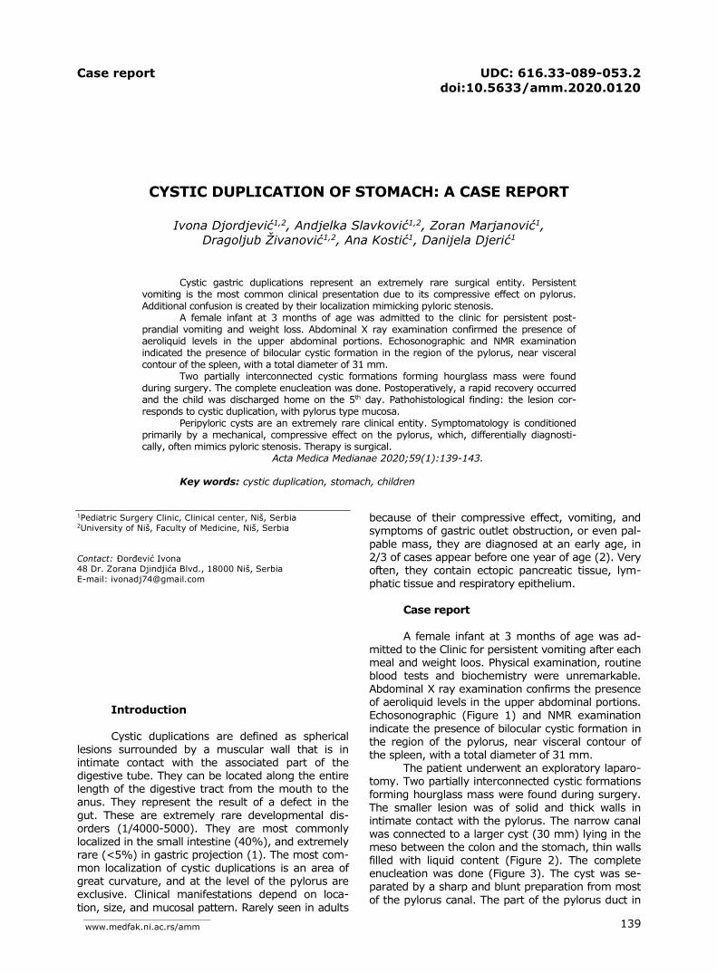
139 www.medfak.ni.ac.rs/amm
Case report UDC: 616.33-089-053.2
doi:10.5633/amm.2020.0120
CYSTIC DUPLICATION OF STOMACH: A CASE REPORT
Ivona Djordjević1,2, Andjelka Slavković1,2, Zoran Marjanović1,
Dragoljub Živanović1,2, Ana Kostić1, Danijela Djerić1
Cystic gastric duplications represent an extremely rare surgical entity. Persistent
vomiting is the most common clinical presentation due to its compressive effect on pylorus. Additional confusion is created by their localization mimicking pyloric stenosis.
A female infant at 3 months of age was admitted to the clinic for persistent post-prandial vomiting and weight loss. Abdominal X ray examination confirmed the presence of aeroliquid levels in the upper abdominal portions. Echosonographic and NMR examination indicated the presence of bilocular cystic formation in the region of the pylorus, near visceral contour of the spleen, with a total diameter of 31 mm.
Two partially interconnected cystic formations forming hourglass mass were found during surgery. The complete enucleation was done. Postoperatively, a rapid recovery occurred and the child was discharged home on the 5th day. Pathohistological finding: the lesion cor-responds to cystic duplication, with pylorus type mucosa.
Peripyloric cysts are an extremely rare clinical entity. Symptomatology is conditioned primarily by a mechanical, compressive effect on the pylorus, which, differentially diagnosti-cally, often mimics pyloric stenosis. Therapy is surgical.
Acta Medica Medianae 2020;59(1):139-143. Key words: cystic duplication, stomach, children
1Pediatric Surgery Clinic, Clinical center, Niš, Serbia 2University of Niš, Faculty of Medicine, Niš, Serbia
Contact: Đorđević Ivona
48 Dr. Zorana Djindjića Blvd., 18000 Niš, Serbia
E-mail: [email protected]
Introduction
Cystic duplications are defined as spherical
lesions surrounded by a muscular wall that is in intimate contact with the associated part of the digestive tube. They can be located along the entire length of the digestive tract from the mouth to the anus. They represent the result of a defect in the
gut. These are extremely rare developmental dis-orders (1/4000-5000). They are most commonly localized in the small intestine (40%), and extremely rare (<5%) in gastric projection (1). The most com-mon localization of cystic duplications is an area of great curvature, and at the level of the pylorus are exclusive. Clinical manifestations depend on loca-
tion, size, and mucosal pattern. Rarely seen in adults
because of their compressive effect, vomiting, and symptoms of gastric outlet obstruction, or even pal-
pable mass, they are diagnosed at an early age, in 2/3 of cases appear before one year of age (2). Very often, they contain ectopic pancreatic tissue, lym-phatic tissue and respiratory epithelium.
Case report
A female infant at 3 months of age was ad-mitted to the Clinic for persistent vomiting after each meal and weight loos. Physical examination, routine blood tests and biochemistry were unremarkable. Abdominal X ray examination confirms the presence of aeroliquid levels in the upper abdominal portions.
Echosonographic (Figure 1) and NMR examination
indicate the presence of bilocular cystic formation in the region of the pylorus, near visceral contour of the spleen, with a total diameter of 31 mm.
The patient underwent an exploratory laparo-tomy. Two partially interconnected cystic formations forming hourglass mass were found during surgery.
The smaller lesion was of solid and thick walls in intimate contact with the pylorus. The narrow canal was connected to a larger cyst (30 mm) lying in the meso between the colon and the stomach, thin walls filled with liquid content (Figure 2). The complete enucleation was done (Figure 3). The cyst was se-parated by a sharp and blunt preparation from most
of the pylorus canal. The part of the pylorus duct in
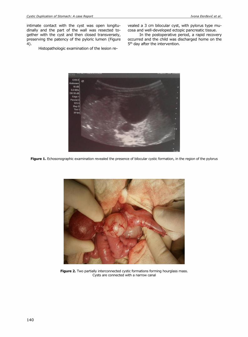
Cystic Duplication of Stomach: A case Report Ivona Đorđević et al.
140
intimate contact with the cyst was open longitu-
dinally and the part of the wall was resected to-
gether with the cyst and then closed transversely, preserving the patency of the pyloric lumen (Figure 4).
Histopathologic examination of the lesion re-
vealed a 3 cm bilocular cyst, with pylorus type mu-
cosa and well-developed ectopic pancreatic tissue.
In the postoperative period, a rapid recovery occurred and the child was discharged home on the 5th day after the intervention.
Figure 1. Echosonographic examination revealed the presence of bilocular cystic formation, in the region of the pylorus
Figure 2. Two partially interconnected cystic formations forming hourglass mass. Cysts are connected with a narrow canal
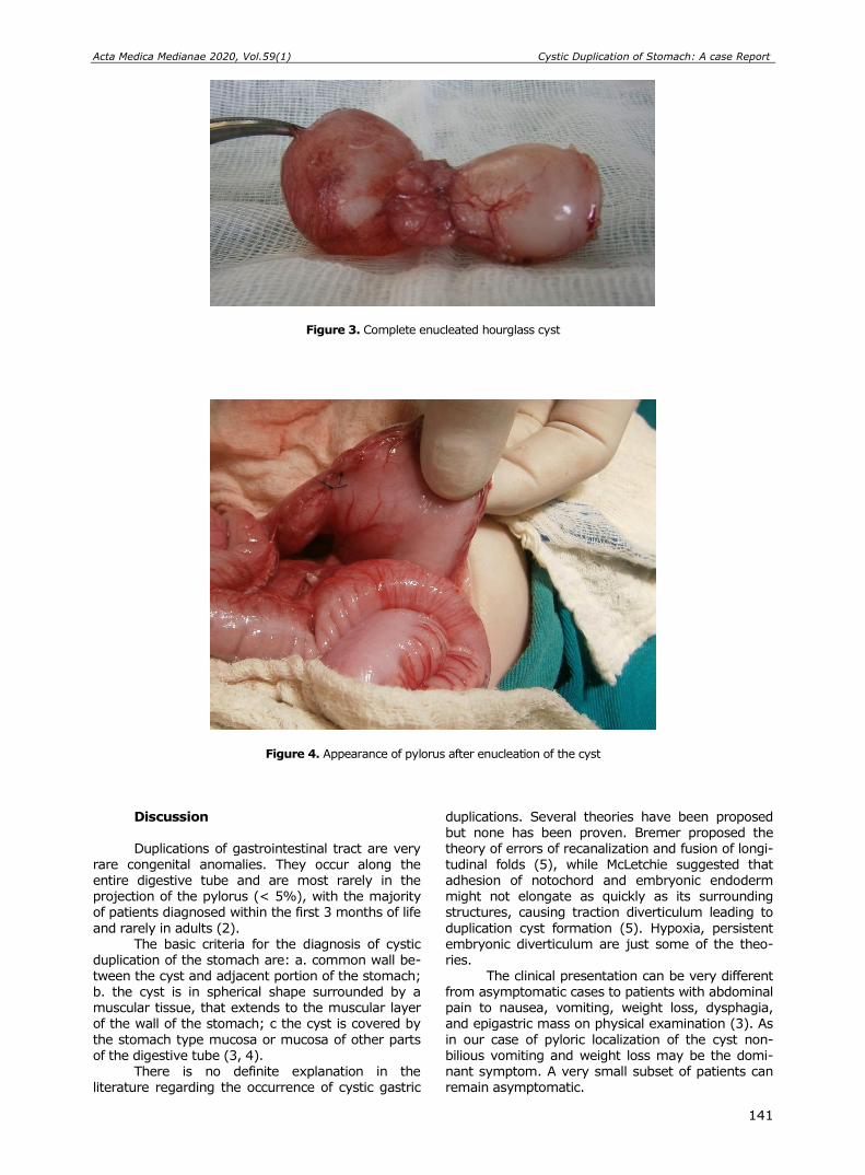
Acta Medica Medianae 2020, Vol.59(1) Cystic Duplication of Stomach: A case Report
141
Figure 3. Complete enucleated hourglass cyst
Figure 4. Appearance of pylorus after enucleation of the cyst
Discussion Duplications of gastrointestinal tract are very
rare congenital anomalies. They occur along the entire digestive tube and are most rarely in the projection of the pylorus (< 5%), with the majority of patients diagnosed within the first 3 months of life
and rarely in adults (2). The basic criteria for the diagnosis of cystic
duplication of the stomach are: a. common wall be-tween the cyst and adjacent portion of the stomach; b. the cyst is in spherical shape surrounded by a muscular tissue, that extends to the muscular layer of the wall of the stomach; c the cyst is covered by
the stomach type mucosa or mucosa of other parts of the digestive tube (3, 4).
There is no definite explanation in the literature regarding the occurrence of cystic gastric
duplications. Several theories have been proposed but none has been proven. Bremer proposed the theory of errors of recanalization and fusion of longi-
tudinal folds (5), while McLetchie suggested that adhesion of notochord and embryonic endoderm might not elongate as quickly as its surrounding structures, causing traction diverticulum leading to
duplication cyst formation (5). Hypoxia, persistent embryonic diverticulum are just some of the theo-ries.
The clinical presentation can be very different from asymptomatic cases to patients with abdominal pain to nausea, vomiting, weight loss, dysphagia, and epigastric mass on physical examination (3). As
in our case of pyloric localization of the cyst non-bilious vomiting and weight loss may be the domi-nant symptom. A very small subset of patients can remain asymptomatic.
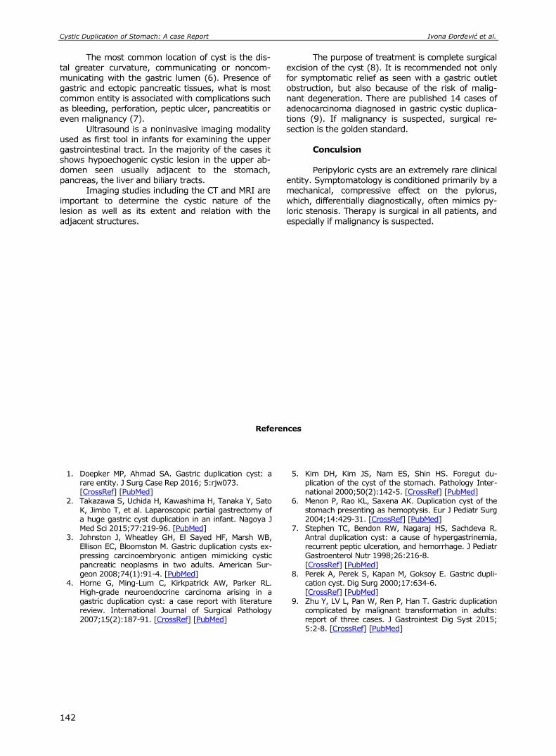
Cystic Duplication of Stomach: A case Report Ivona Đorđević et al.
142
The most common location of cyst is the dis-
tal greater curvature, communicating or noncom-
municating with the gastric lumen (6). Presence of gastric and ectopic pancreatic tissues, what is most common entity is associated with complications such as bleeding, perforation, peptic ulcer, pancreatitis or even malignancy (7).
Ultrasound is a noninvasive imaging modality used as first tool in infants for examining the upper gastrointestinal tract. In the majority of the cases it shows hypoechogenic cystic lesion in the upper ab-domen seen usually adjacent to the stomach, pancreas, the liver and biliary tracts.
Imaging studies including the CT and MRI are
important to determine the cystic nature of the lesion as well as its extent and relation with the adjacent structures.
The purpose of treatment is complete surgical
excision of the cyst (8). It is recommended not only
for symptomatic relief as seen with a gastric outlet obstruction, but also because of the risk of malig-nant degeneration. There are published 14 cases of adenocarcinoma diagnosed in gastric cystic duplica-tions (9). If malignancy is suspected, surgical re-
section is the golden standard. Conculsion Peripyloric cysts are an extremely rare clinical
entity. Symptomatology is conditioned primarily by a mechanical, compressive effect on the pylorus,
which, differentially diagnostically, often mimics py-loric stenosis. Therapy is surgical in all patients, and especially if malignancy is suspected.
References
1. Doepker MP, Ahmad SA. Gastric duplication cyst: a rare entity. J Surg Case Rep 2016; 5:rjw073. [CrossRef] [PubMed]
2. Takazawa S, Uchida H, Kawashima H, Tanaka Y, Sato K, Jimbo T, et al. Laparoscopic partial gastrectomy of a huge gastric cyst duplication in an infant. Nagoya J Med Sci 2015;77:219-96. [PubMed]
3. Johnston J, Wheatley GH, El Sayed HF, Marsh WB, Ellison EC, Bloomston M. Gastric duplication cysts ex-pressing carcinoembryonic antigen mimicking cystic
pancreatic neoplasms in two adults. American Sur-geon 2008;74(1):91-4. [PubMed]
4. Horne G, Ming-Lum C, Kirkpatrick AW, Parker RL. High-grade neuroendocrine carcinoma arising in a gastric duplication cyst: a case report with literature review. International Journal of Surgical Pathology 2007;15(2):187-91. [CrossRef] [PubMed]
5. Kim DH, Kim JS, Nam ES, Shin HS. Foregut du-plication of the cyst of the stomach. Pathology Inter-national 2000;50(2):142-5. [CrossRef] [PubMed]
6. Menon P, Rao KL, Saxena AK. Duplication cyst of the stomach presenting as hemoptysis. Eur J Pediatr Surg 2004;14:429-31. [CrossRef] [PubMed]
7. Stephen TC, Bendon RW, Nagaraj HS, Sachdeva R. Antral duplication cyst: a cause of hypergastrinemia, recurrent peptic ulceration, and hemorrhage. J Pediatr Gastroenterol Nutr 1998;26:216-8.
[CrossRef] [PubMed] 8. Perek A, Perek S, Kapan M, Goksoy E. Gastric dupli-
cation cyst. Dig Surg 2000;17:634-6. [CrossRef] [PubMed]
9. Zhu Y, LV L, Pan W, Ren P, Han T. Gastric duplication complicated by malignant transformation in adults: report of three cases. J Gastrointest Dig Syst 2015; 5:2-8. [CrossRef] [PubMed]
Acta Medica Medianae 2020, Vol.59(1) Cystic Duplication of Stomach: A case Report
143
Prikaz bolesnika UDC: 616.33-089-053.2
doi:10.5633/amm.2020.0120
CISTIČNA DUPLIKACIJA ŽELUCA — PRIKAZ SLUČAJA
Ivona Đorđević1,2, Anđelka Slavković1,2, Zoran Marjanović1,
Dragoljub Živanović1,2, Ana Kostić1, Danijela Đerić1
1Klinika za pedijatrijsku hirurgiju, Klinički centar Niš, Niš, Srbija 2Univerzitet u Nišu, Medicinski fakultet, Niš, Srbija
Kontakt: Ivona Đorđević
Bulevar dr Zoran Đinđić 48, 18000 Niš, Srbija
E-mail: [email protected]
Cistične duplikacije želuca predstavljaju izuzetno redak hirurški entitet. Zbog kom-
presivnog delovanja na pilorus, uporno povraćanje predstavlja najčešću kliničku prezentaciju.
Dodatnu konfuziju stvara njihova lokalizacija, te često diferencijalno-dijagnostički upućuju na stenozu pilorusa.
Žensko novorođenče u 3. mesecu života primljeno je u kliniku zbog upornog po-vraćanja, posle svakog obroka, i nenapredovanja. Nativna grafija trbuha potvrđuje prisustvo hidrogasnih nivoa u gornjim partijama trbuha. Ehosonografski i NMR pregledi ukazuju na prisustvo bilokularne cistične formacije, u regiji pilorusa i uz viscelarnu konturu slezine, ukupnog promera 31 mm.
Intraoperativno je evidentirana promena izgleda peščanog sata, koju čine dve delom međusobno povezane cistične formacije. Učinjena je kompletna enukleacija cisti. Post-operativno, dolazi do brzog oporavka i dete je 5. dana otpušteno kući. Patohistološki nalaz: lezija odgovara cističnoj duplikaciji sa mukozom pilorusnog tipa.
Peripilorične ciste predstavljaju izuzetno redak klinički entitet. Simptomatologija je uslovljena, pre svega, mehaničkim, kompresivnim efektom na pilorus, što diferencijalno-dija-gnostički često imitira stenozu pilorusa. Terapija je hirurška.
Acta Medica Medianae 2020;59(1):139-143.
Ključne reči: cistična duplikacija, želudac, deca
144 www.medfak.ni.ac.rs/amm
Case report UDC: 617.741-004.1-089
doi:10.5633/amm.2020.0121
Nd: YAG LASER ANTERIOR CAPSULOTOMY IN CAPSULAR PHIMOSIS IN THE EYE WITH PEX AND ZONULAR LESION
Aleksandar Veselinović, Marija Cvetanović, Dragan Veselinović
Secondary cataract formation is significantly present after cataract surgery and
develops at different time intervals after the intervention. Blepharophimosis or anterior capsule contraction may result in numerous problems in the operated eye. In patients with cataract associated with pseudoexfoliation syndrome the risk of anterior capsule contraction is greater, especially if small capsulorhexis had previously been performed.
The paper presents blepharophimosis of the anterior capsule with phacodonesis and secondary cataract eight months after phacoemulsification with intraocular lens implantation. Due to anterior capsule opacification and formation of capsular phimosis, visual acuity decreased and monocular diplopia developed in the operated eye. After sectoral YAG laser capsulotomy of adjustable intensity was performed, visual acuity improved and the patient’s subjective complaints disappeared.
YAG laser capsulotomy is a significant method in managing secondary cataract. Management of secondary cataract due to anterior lens capsule opacification and the presence of anterior capsule phimosis associated with expressed pseuoexfoliations and zonular lesion poses a particular challenge to ophthalmologists and requires a certain amount of experience in the management of these conditions.
Acta Medica Medianae 2020;59(1):144-148. Key words: YAG laser capsulotomy, capsular phimosis, pseudoexfoliation
Ophthalmology Clinic, Clinical Center Niš, Niš, Serbia
Contact: Aleksandar Veselinović
48 Dr. Zoran Djindjić Blvd., 18000 Niš, Serbia E-mail: [email protected]
Introduction
Capsular phimosis or anterior capsule con-traction syndrome has been described as one of the potential complications in patients with cataract. Capsular phimosis occurs as a postoperative com-plication resulting from a small diameter capsulor-hexis and postoperative inflammatory reactions in
the eye (1, 2, 3, 4). After cataract surgery residual lens epithelial cells may proliferate over the posterior lens capsule and on the intraocular lens surface as well, causing myofibroblast transdifferentiation (4, 5). A small capsulorhexis diameter most commonly presents a risk for capsular phimosis either as a
consequence of inexperience of the surgeon or due to a small pupil size in cataract patients. Most com-monly registered associated condition is pseudo-
exfoliation syndrome, but it has also been described in patients with retinitis pigmentosa, diabetes, uvei-tis and Behcet’s syndrome, high myopia, uveitis, pars planitis, and myotonic dystrophy (5, 6, 7, 8, 9). A small anterior capsule opening makes surgery more difficult and may cause numerous intraope-rative complications. Capsular contraction syndrome
may also occur after combined cataract and glau-coma surgery and due to intraoperative complica-tions that may result in prolonged postoperative inflammation. According to some authors’ expe-rience, intraocular lens material, especially haptic lenses, may be contributing factors for capsular phi-mosis (10, 11, 12, 13).
Postoperatively, subsequent constriction of the anterior capsular opening may occur, capsular phimosis, as a consequence of proliferative and in-flammatory processes. Severe anterior lens constric-tion, especially in patients with pseudoexfoliation syndrome, may result in subluxation of artificial
lenses due to zonular lesions. As a consequence of severe anterior lens constriction, tractional ciliary body detachment may also occur accompanied with choroidal effusion (14, 15).
Case report
Patient Z.M. aged 72, presented to her doctor with the complaints of monocular diplopia and de-
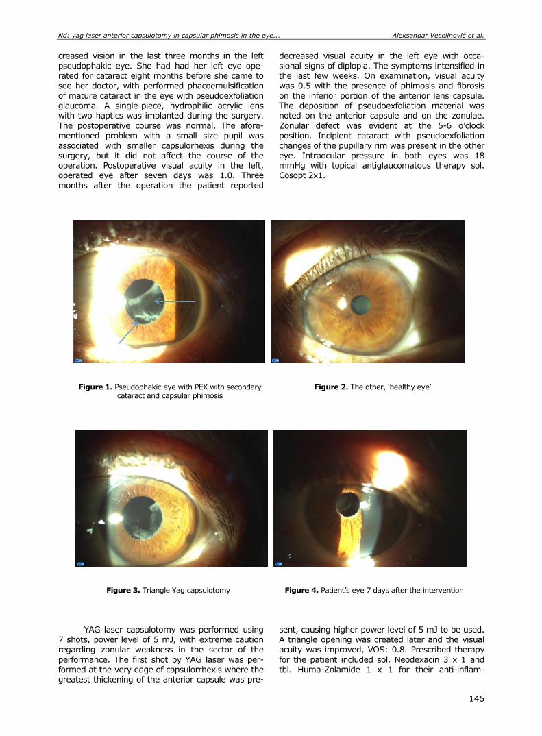
Nd: yag laser anterior capsulotomy in capsular phimosis in the eye... Aleksandar Veselinović et al.
145
creased vision in the last three months in the left
pseudophakic eye. She had had her left eye ope-
rated for cataract eight months before she came to see her doctor, with performed phacoemulsification of mature cataract in the eye with pseudoexfoliation glaucoma. A single-piece, hydrophilic acrylic lens with two haptics was implanted during the surgery.
The postoperative course was normal. The afore-mentioned problem with a small size pupil was associated with smaller capsulorhexis during the surgery, but it did not affect the course of the operation. Postoperative visual acuity in the left, operated eye after seven days was 1.0. Three months after the operation the patient reported
decreased visual acuity in the left eye with occa-
sional signs of diplopia. The symptoms intensified in
the last few weeks. On examination, visual acuity was 0.5 with the presence of phimosis and fibrosis on the inferior portion of the anterior lens capsule. The deposition of pseudoexfoliation material was noted on the anterior capsule and on the zonulae.
Zonular defect was evident at the 5-6 o’clock position. Incipient cataract with pseudoexfoliation changes of the pupillary rim was present in the other eye. Intraocular pressure in both eyes was 18 mmHg with topical antiglaucomatous therapy sol. Cosopt 2x1.
Figure 1. Pseudophakic eye with PEX with secondary
cataract and capsular phimosis Figure 2. The other, ‘healthy eye’
Figure 3. Triangle Yag capsulotomy
Figure 4. Patient’s eye 7 days after the intervention
YAG laser capsulotomy was performed using 7 shots, power level of 5 mJ, with extreme caution regarding zonular weakness in the sector of the
performance. The first shot by YAG laser was per-formed at the very edge of capsulorrhexis where the greatest thickening of the anterior capsule was pre-
sent, causing higher power level of 5 mJ to be used. A triangle opening was created later and the visual acuity was improved, VOS: 0.8. Prescribed therapy
for the patient included sol. Neodexacin 3 x 1 and tbl. Huma-Zolamide 1 x 1 for their anti-inflam-
Acta Medica Medianae 2020, Vol.59(1) Nd: yag laser anterior capsulotomy in capsular phimosis in the eye...
146
matory properties and because of possible transient
increase of intraocular pressure.
Follow-up examination revealed that visual acuity in the left eye was 1.0, IOP: 18 mmHg with regular antiglaucomatous treatment. The patient had no more complaints of diplopia and was satisfied with the effects of the intervention.
Discussion Anterior capsular contraction syndrome has
multiple causes and its occurrence can greatly di-minish primary effect of cataract surgery. The most common causes of capsular phimosis include a small
diameter capsulorhexis in patients with chronic infla-mmatory processes, uveitis, diabetes and pseudoex-foliation syndrome (2, 3, 4). Good postoperative
acuity in these patients may greatly be decreased and result in patient’s condition similar to pre-operative one. If a tendency for capsular phimosis is noted in early postoperative period, relaxing radial
incisions along the rim may be performed using the YAG laser to prevent further constriction (16). Due to strong contractions and intraocular lens de-centration, monocular diplopia, glare and binocular vision problems are very common.
Application of the YAG laser is of extreme
importance in secondary cataract treatment and in patients with postoperative anterior capsular con-traction. A special problem in performing the YAG laser anterior capsulotomy in blepharophimosis is a condition accompanied with zonular lesion, as well as phacodonesis and intraocular lens decentration.
In patients with blepharophimosis it is necessary to
perform anterior capsulotomy requiring adjustment
of the laser shock waves intensity in order not to
damage anterior surface of the artificial lens. In some cases, it is necessary to use YAG laser with higher intensity due to anterior capsular thickening and fibrosis, which makes correct dosage and the surgeon’s experience very important. Additional YAG
laser treatment, due to exerted stress on the zonu-lae, may in some cases produce more zonular lesions and lens decentration, thus increasing mono-cular diplopia intensity and the patient’s subjective visual disturbances. In severe contraction syndrome dislocation of artificial lens towards the vitreous body is often present, resulting in refractive changes and
hypermetropia. The YAG laser anterior capsulotomy results in capsular bag relaxation and a possible re-fractive change regarding the reduction of hyper-
metropia (17). There were no refractive anomaly changes in our patient.
Conclusion
Before anterior laser capsulotomy is perfor-
med, the evaluation of anterior capsular blurriness and fibrosis degree should be evaluated to deter-mine the intensity of the laser beam. Anterior cap-sular extension must not be performed in the direc-
tion of zonular defects and the first wave shot should be directed to capsulorhexis edge. A triangular an-terior capsule opening in the eye with dilated pupil should be big enough to avoid glare that may be caused by peripheral portion of anterior capsular remnants after normal pupil size is achieved.
Nd: yag laser anterior capsulotomy in capsular phimosis in the eye... Aleksandar Veselinović et al.
147
References
1. Joo CK, Shin JA, Kim JH. Capsular opening contraction after continuous curvilinear capsulorhexis and intra-ocular lens implantation. J Cataract Refract Surg 1996; 22:585-90. [CrossRef] [PubMed]
2. Davison JA. Capsule contraction syndrome. J Cataract Refract Surg 1993; 19:582-9. [CrossRef] [PubMed]
3. Kurosaka D, Ando I, Kato K, Oshima T, Kurosaka H, Yoshino M, et al. Fibrous membrane formation at the capsular margin in capsule contraction syndrome. J Cataract Refract Surg 1999; 25:930-5. [CrossRef] [PubMed]
4. Gills JP. Anterior capsule contraction. J Cataract Refract Surg 2003; 29:1652. [CrossRef] [PubMed]
5. Hayashi H, Hayashi K, Nakao F, Hayashi F. Anterior capsule contraction and intraocular lens dislocation in eyes with pseudoexfoliation syndrome. Br J Ophthal-mol 1998; 82:1429-32. [CrossRef] [PubMed]
6. Hayashi H, Hayashi K, Nakao F, Hayashi F. Area reduction in the anterior capsule opening in eyes of diabetes mellitus patients. J Cataract Refract Surg 1998; 24:1105-10. [CrossRef] [PubMed]
7. Jin-Poi T, Shatriah I, Khairy-Shamel ST, Zunaina E. Rapid anterior capsular contraction after phacoemul-sification surgery in a patient with retinitis pigment-osa. Clin Ophthalmol 2013; 7:839-42. [CrossRef] [PubMed]
8. Najjar DM, Igbre AO, Tsai FF. Late capsular bag contraction and intraocular lens subluxation in retinitis pigmentosa: a case report. J Med Case Rep 2011; 5:65. [CrossRef] [PubMed]
9. Alintas AGK, Dal D. Capsule contraction syndrome in Behcet’s disease. Int J Ophthalmol 2010; 3:358-60. [CrossRef] [PubMed]
10. Cochener B, Jacq PL, Colin J. Capsule contraction after continuous curvilinear capsulorhexis: poly (methyl methacrylate) versus silicone intraocular lenses. J Cataract Refract Surg 1999; 25:1362-9. [CrossRef] [PubMed]
11. Gonvers M, Sickenberg M, Van Melle G. Change in capsulorhexis size after implantation of three types of intraocular lenses. J Cataract Refract Surg 1997; 23:231-8. [CrossRef] [PubMed]
12. Toldos JJM, Roig AA, Benabent EC. Total anterior capsule closure after silicone intraocular lens implanta-tion. J Cataract Refract Surg 1996; 22:269-71. [CrossRef] [PubMed]
13. Apple DJ. Influence of intraocular lens material and design on postoperative intracapsular cellular reacti-vity. Trans Am Ophthalmol Soc 2000; 98: 257-83. [PubMed]
14. Srinivasan S, van der Hoek J, Green F, Atta HR. Tractional ciliary body detachment, choroidal effusion, and hypotony caused by severe anterior lens capsule contraction following cataract surgery. Br J Ophthal-
mol 2001; 85:1261-2. [CrossRef] [PubMed] 15. Salzmann J, Khaw PT, Laidlaw A. Choroidal effusions
and hypotony caused by severe anterior lens capsule contraction after cataract surgery. Am J Ophthalmol 2000; 129:253-4. [CrossRef] [PubMed]
16. Hayashi K, Yoshida M, Hirata A, Hayashi H. Anterior capsule relaxing incisions with neodymium: YAG laser for patients at high-risk for anterior capsule con-traction. J Cataract Refract Surg 2001; 37:97-103. [CrossRef] [PubMed]
17. Abd Elmagid M, Tag Eldin. Refractive change after YAG laser anterior capsulotomy in capsular phimosis syndrome. Journal of American Science 2014; 10(8).
Acta Medica Medianae 2020, Vol.59(1) Nd: yag laser anterior capsulotomy in capsular phimosis in the eye...
148
Prikaz bolesnika UDC: 617.741-004.1-089
doi:10.5633/amm.2020.0121
Nd: YAG LASER KAPSULOTOMIJA PREDNJE KAPSULE KOD KAPSULOFIMOZE U OKU SA PEX-OM I LEZIJOM ZONULA
Aleksandar Veselinović, Marija Cvetanović, Dragan Veselinović
Klinika za očne bolesti, Klinički centar Niš, Niš, Srbija
Kontakt: Aleksandar Veselinović
Bulevar dr Zoran Đinđić 48, 18000 Niš, Srbija
E-mail: [email protected]
Pojava sekundarne katarakte nakon operacije katarakte prisutna je u značajnom broju
i nastaje u različitom vremenskom periodu od intervencije. Blefarofimoza ili konstrikcija prednje kapsule može prouzrokovati brojne probleme na operisanom oku. Kod bolesnika kod
kojih je katarakta udružena sa pseudoeksfolijacionim sindromom postoji veći rizik za nastanak kon-strikcije prednje kapsule, posebno ukoliko je predhodno urađena mala kapsuloreksa.
U radu je dat prikaz pojave blefarofimoze prednje kapsule, sa fakodonezom i se-kundarnom kataraktom, osam meseci nakon urađene fakoemulzifikacije sa ugradnjom intra-okularnog sočiva. Usled zamućenja prednje kapsule sočiva i pojave kapsulofimoze, došlo je do smanjenja oštrine vida i pojave monokularnih diplopija na operisanom oku. Nakon izvođenja sektoraste YAG laser kapsulotomije dozirane snage, došlo je do poboljšanja oštrine vida i gubitka subjektivnih tegoba bolesnice.
Yag laser kapsulotomija prestavlja značajnu metodu u rešavanju pojave sekundarne katarakte. Rešavanje problema sekundarne katarakte usled prisustva zamućenja prednje kapsule sočiva i prisustva fimoze prednje kapsule, udružene sa izraženim pseudoeksfoli-jacijama i lezijom zonula, predstavlja poseban izazov za oftalmologa i zahteva iskustvo u rešavanju ovakvih stanja.
Acta Medica Medianae 2020;59(1):144-148.
Ključne reči: Yag laser kapsulotomija, kapsulofimoza, pseudoexfoliatio
149 www.medfak.ni.ac.rs/amm
Case report UDC: 617.764.1:616.988.55
doi:10.5633/amm.2020.0122
ACUTE DACRYOADENITIS ASSOCIATED WITH INFECTIOUS MONONUCLEOSIS
Marija Cvetanović1, Aleksandar Veselinović1, Marija Trenkić-Božinović1,
Kristina Stojanović2, Ivona Trajković2, Dragan Veselinović1
The paper presents a case report of a patient with unilateral acute dacryoadenitis
caused by infectious mononucleosis. The aim of the paper was to point out therapeutic and diagnostic possibilities in patients with dacryoadenitis.
Hyposecretion of tears was determined by Schirmer’s test. MRI revealed lacrimal gland enlargement with signs of inflammation in the surrounding orbital tissue. Laboratory tests for Epstein Barr virus infection were positive.
Clinical manifestation of infectious mononucleosis was proved by a pediatrician. The application of local and general therapy resulted in withdrawal of the lacrimal gland
enlargement, stabilization of inflammation symptoms and normalization of a grey eye in our patient.
Acute dacryoadenitis is a relatively rare pathological condition requiring appropriate diagnostics for proper treatment, as it was the case with our patient in whom infectious mononucleosis caused the disease.
Acta Medica Medianae 2020;59(1):149-152. Key words: dacryoadenitis, infectious mononucleosis
1Ophthalmology Clinic, Clinical Center Niš, Serbia 2Ophthalmology Surgery Veselinović Zone of Vision Niš, Serbia
Contact: Marija Cvetanović
48 Dr. Zoran Djindjić Blvd., 18000 Niš, Serbia
E-mail: [email protected]
Introduction The frequency of dacryoadenitis is low; it
occurs in only one case per 10,000 ophthalmic pa-tients. There is no predilection for race, gender or age for the onset of the disease. Dacryoadenitis can be acute or chronic (1, 2, 3).
Acute dacryoadenitis is usually unilateral and characterized by painful swelling of the eyelid with the signs of chemosis, conjunctival hyperemia, ery-thema of the lids and lymphadenopathy (subman-dibular). Its characteristic features include swelling of the outer third of the lid, proptosis, occasionally ocular motility restriction and painful globe move-
ment (1, 2). It is commonly an independent disease (viral,
bacterial infections) with good prognosis. Factors that may cause acute dacryoadenitis are mostly due
to infections with viral etiologies (mumps, Epstein-Barr virus, herpes zoster virus), or bacterial etiolo-gies caused by staphylococcus, streptococcus, hae-mophilus influenza, gonorrhea or chlamydia. Infec-tion may spread to the lacrimal gland hematoge-nously, transneuronally, from the inflamed conjun-
ctiva or via traumatic injuries (4, 5, 6, 7). Chronic dacryoadenitis is usually bilateral and
painless with minimal ocular signs. Chronic condition is accompanied by enlargement of the lacrimal gland present for months (8, 9, 10).
It is usually associated with a systemic di-
sease (sarcoidosis, tuberculosis, Mikulicz’s syndro-me, Sjogren’s syndrome, syphilis, leukemia). Prog-nosis depends on the underlying cause of the sys-temic disease. Symptoms of mild to severe dry eyes
are also present (11, 12, 13). Case study
A girl aged 14 presented to her doctor with
the complaints of pain and swelling of her right outer upper eyelid.
The local finding revealed the absence of tarsal and bulbar conjunctival inflammation, en-larged right lacrimal gland with the signs of in-
flammation and edema in that region. Mild proptosis and upper lid swelling were present in the outer part with characteristic “comma-like” appearance (Figure 1). The outer upper portion of the eyelid was painful on palpation, and the patient reported the feeling of
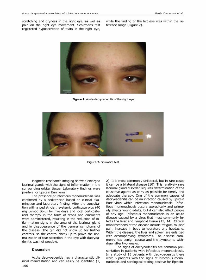
Acute dacryoadenitis associated with infectious mononucleosis Marija Cvetanović et al.
150
scratching and dryness in the right eye, as well as
pain on the right eye movement. Schirmer’s test
registered hyposecretion of tears in the right eye,
while the finding of the left eye was within the re-
ference range (Figure 2).
Figure 1. Acute dacryoadenitis of the right eye
Figure 2. Shirmer’s test
Magnetic resonance imaging showed enlarged lacrimal glands with the signs of inflammation in the surrounding orbital tissue. Laboratory findings were positive for Epstein Barr virus.
The presence of infectious mononucleosis was confirmed by a pediatrician based on clinical exa-mination and laboratory finding. After the consulta-
tion with a pediatrician, systemic corticosteroids (40 mg Lemod Solu) for five days and local corticoste-roid therapy in the form of drops and ointments
were administered, resulting in the reduction of in-flammation signs in the area of the lacrimal gland and in disappearance of the general symptoms of the disease. The girl did not show up for further controls, so the control check-up to prove the nor-malization of tear secretion in the eye with dacryoa-denitis was not possible.
Discussion
Acute dacryoadenitis has a characteristic cli-
nical manifestation and can easily be identified (1,
2). It is most commonly unilateral, but in rare cases it can be a bilateral disease (10). This relatively rare lacrimal gland disorder requires determination of the causative agents as early as possible for timely and adequate therapy. One of the common causes of dacryoadenitis can be an infection caused by Epstein Barr virus within infectious mononucleosis. Infec-tious mononucleosis occurs sporadically and prima-rily affects young adults, but it can also affect people of any age. Infectious mononucleosis is an acute disease caused by a virus that most commonly in-fects the liver and lymphoid tissue (13, 14). Clinical manifestations of the disease include fatigue, muscle pain, increase in body temperature and headache. Within the disease, the liver and spleen are enlarged with accompanying symptoms. The disease com-monly has benign course and the symptoms with-draw after two weeks.
The signs of dacryoadenitis are common pre-sentation in patients with infectious mononucleosis. In a study of 16 patients with dacryoadenitis there were 6 patients with the signs of infectious mono-nucleosis and serological testing positive for Epstein-
Acta Medica Medianae 2020, Vol.59(1) Acute dacryoadenitis associated with infectious mononucleosis
151
Barr virus infection (3). Acute dacryoadenitis caused by Epstein-Barr virus most commonly occurs in younger population, rarely in elderly people (1, 2). In our patient characteristic symptoms were mani-fested unilaterally. Hyposecretion of tears in the affected eye was associated with other symptoms of the disease.
Conclusion Acute dacryoadenitis can often be associated
with infectious mononucleosis and it can be mani-fested in children and young adults. Upon the con-
firmation of the presence of infectious mononucleo-
sis a pediatrician and an infectologist should be
consulted for appropriate therapy. Despite the fact that some authors support conservative treatment, administration of general corticosteroid therapy speeds up the recovery of general symptoms in these patients and local symptoms of dacryoadenitis
as well. Hyposecretion of tears in dacryoadenitis may be a characteristic symptom of the disease. It disappears with the treatment and the loss of inflammatory signs.
References
1. Aburn NS, Sullivan TJ. Infectious mononucleosis presenting with dacryoadenitis. Ophthalmology 1996; 103(5):776-8. [CrossRef] [PubMed]
2. Rai P, Shah SA, Kirshan H. Acute Dacryadenitis-analysis of 23 cases. Med Channel 2009;15:71-6.
3. Rhem MN, Wilhelmus KR, Jones DB. Epstein-Barr virus dacryoadenitis. Am J Ophthalmol 2000;129(3):372-5. [CrossRef] [PubMed]
4. Srivastava VK. Acute Suppurative Dacryoadenitis. Medical Journal Armed Forces India 2000;56:151-2. [CrossRef] [PubMed]
5. Goold LA, Madge SN, Au A, Leibovitch I, McNab A, Tumuluri K, et al. Acute suppurative bacterial dacryoadenitis: a case series. Br J Ophthalmol 2013;97(6):735-8. [CrossRef] [PubMed]
6. Hartikainen J, Lehtonen OP, Saari M. Bacteriology of
lacrimal duct obstruction in adults. Br J Ophthalmol 1997;81:37-40. [CrossRef] [PubMed]
7. Kubal A, Garibaldi DC. Dacryoadenitis caused by methicillin-resistant Staphylococcus aureus. Ophthal Plast Reconstr Surg 2008;24(1):50-1. [CrossRef] [PubMed]
8. Derr C, Shah A. Bilateral dacryoadenitis. J Emerg Trauma Shock 2012;5(1):92-4. [CrossRef] [PubMed]
9. Baoke H, Shihui W, Maonian Z, Zhaohui L, Zhitong Z, Zhigang S, et al. Bilateral dacryoadenitis complicated by lymphocytic hypophysitis. J Neuroophthalmol 2009; 29:214-8. [CrossRef] [PubMed]
10. Moscovici BK, Romero IL, Vital Filho J, Bison SH. Epstein-Barr virus bilateral dacryoadenitis: case report. Arq Bras Oftalmol 2009;72(6):826-8. [CrossRef] [PubMed]
11. Dutt S, Cartwright MJ, Nelson CC. Acute dacryoa-denitis and Crohn's disease: findings and manage-ment. Ophthal Plast Reconstr Surg 1992;8(4):295-9. [CrossRef] [PubMed]
12. Nieto JC, Kim N, Lucarelli MJ. Dacryoadenitis and orbital myositis associated with lyme disease. Arch Ophthalmol 2008;126(8):1165-6. [CrossRef] [PubMed]
13. Bailey RE. Diagnosis and treatment of infectious mononucleosis. Am Fam Physician 1994;49:879-88. [PubMed]
14. Mark H. Ebell. Epstein-Barr Virus Infectious Mono-nucleosis. Am Fam Physician 2004;70(7):1279-87. [PubMed]
Acute dacryoadenitis associated with infectious mononucleosis Marija Cvetanović et al.
152
Prikaz bolesnika UDC: 617.764.1:616.988.55
doi:10.5633/amm.2020.0122
AKUTNI DAKRIOADENITIS UDRUŽEN SA INFEKTIVNOM MONONUKLEOZOM
Marija Cvetanović1, Aleksandar Veselinović1, Marija Trenkić-Božinović1,
Kristina Stojanović2, Ivona Trajković2, Dragan Veselinović1
1Klinika za očne bolesti, Klinički centar Niš, Niš, Srbija 2Ordinacija za očne bolesti „Zona vida Veselinović“, Niš, Srbija
Kontakt: Marija Cvetanović
Bulevar dr Zoran Đinđić 48, 18000 Niš, Srbija
E-mail: [email protected]
U radu je dat prikaz bolesnika sa jednostranim akutnim dakrioadenitisom prouzro-
kovanim infektivnom mononukleozom. Cilj rada je da se ukaže na terapijske i dijagnostičke mogućnosti kod bolesnika sa dakrioadenitisom.
Hiposekrecija suza dokazana je Schirmerovim testom. MR pokazala je uvećanje suzne žlezde sa znacima inflamacije okolnog orbitalnog tkiva. Registrovani su pozitivni laboratorijski nalazi na Epstein-Barr virus.
Od strane pedijatra, potvrđena je klinička slika infektivne momonukleoze. Na primenu lokalne i opšte terapije došlo je do povlačenja otoka suzne žlezde, smiri-
vanja simptoma inflamacije i normalizovanja simptoma sivog oka kod bolesnika. Akutni dakrioadenitis je relativno retko patološko stanje i zahteva odgovarajuću dija-
gnostiku, nakon koje je olakšana terapija, što se pokazalo i u slučaju našeg bolesnika, kod kojeg je bolest bila prouzrokovana infektivnom mononukleozom.
Acta Medica Medianae 2020;59(1):149-152.
Ključne reči: dakrioadenitis, infektivna mononukleoza
153 www.medfak.ni.ac.rs/amm
Case report UDC: 616.11-006.03-089
doi:10.5633/amm.2020.0123
SURGICAL TREATMENT OF GIANT PERICARDIAL CYST THROUGH THE LATERAL THORACOTOMY
Stefan Timčić1, Igor Živković1, Staša Krasić2, Marko Andjelković1, Ivan Nešić1,
Milan Ćirković1, Miodrag Perić1,3
Pericardial cysts are uncommon benign tumors with the prevalence about 7% of all
mediastinal tumors. Patients are mostly asymptomatic unless when cysts compress major anatomic structures in the chest cavity. We represented a patient with a pericardial cyst near to the apex of the heart. Magnetic resonance examination revealed 9 x 4 cm cystic formation. Surgical treatment was performed through left side lateral thoracotomy without cardio-pulmonary bypass support. Tumorous formation was completely resected and sent for the pathohistological examination. Surgical or percutaneous treatment for pericardial cysts might be occasionally necessary, depending on the location of the cyst and its relationship with the
adjacent structures. Morbidity and mortality are low. Surgery has been demonstrated as the only definitive curative treatment.
Acta Medica Medianae 2020;59(1):153-157. Key words: pericardial cyst, lateral thoracotomy
1Cardiac Surgery Department, Dedinje Cardiovascular Institute, Belgrade, Serbia 2Pediatric Department, Mother and child health care institute of
Serbia “Dr Vukan Čupić”, Belgrade, Serbia 3Faculty of Medicine, University of Belgrade, Belgrade, Serbia
Contact: Igor Živković
Heroja Milana Tepića 1, 11000 Belgrade, Serbia
E-mail: [email protected]
Introduction-Background
Pericardial cysts are uncommon benign tumors
with prevalence about 7% of all mediastinal tumors (1, 2). They usually arise from failure of fusion of one of the mesenchymal lacunae that form the pericardial sac (3). Etiology of pericardial cyst could
be congenital, inflammatory (rheumatic pericarditis, bacterial infection particularly tuberculosis, echinoco-ccosis), traumatic and after cardiac surgery (4). The size of the cysts varies from 2 to 28 cm (5). They are usually found in the third or fourth decade of life, male and the females are affected equally.
Patients are mostly asymptomatic unless
major anatomic structures compressed with tumor or rupture of the cyst occurs. The most common
symptoms are dyspnea, chest pain, or persistent cough. Hemoptysis, fever, and pneumothorax are unusual presentations (6).
In 70% of the cases, these tumors are lo-cated in right cardiophrenic angle, 22% in the left cardiophrenic angle and in 8% cases are located in
the posterior or the anterior-superior part of the mediastinum.
However, rarely, this pathology is associated with serious complications such as cardiac tampon-ade, cyst rupture or even sudden death (7).
Case presentation
A 69 year old male with a history of cardiac
weakness and arrhythmia was admitted to the hospital. The chest roentgenography presented su-pradiaphragmatic oval homogenous shading on the
right side and enlarged cardiac vessel silhouette. The
transesophageal examination showed large non-homogeneous mass (dimensions 4.5 x 9 cm), near to the lateral wall of the left ventricle without com-pressive effect on the left heart. Magnetic resonance scan of the heart revealed tumorous formation dimension 90 x 50 mm (Figure 1). The pericardial cyst was near to the left ventricle without com-
munication with chamber. Surgical treatment was performed through
the lateral thoracotomy, without cardio – pulmonary bypass and pericardial sac dissection. On the right side and outside the pericardium single, soft and filled with liquidity pericardial cyst was found (Figure 2).
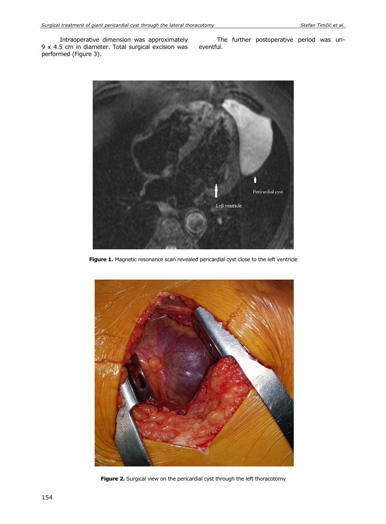
Surgical treatment of giant pericardial cyst through the lateral thoracotomy Stefan Timčić et al.
154
Intraoperative dimension was approximately
9 x 4.5 cm in diameter. Total surgical excision was
performed (Figure 3).
The further postoperative period was un-
eventful.
Figure 1. Magnetic resonance scan revealed pericardial cyst close to the left ventricle
Figure 2. Surgical view on the pericardial cyst through the left thoracotomy
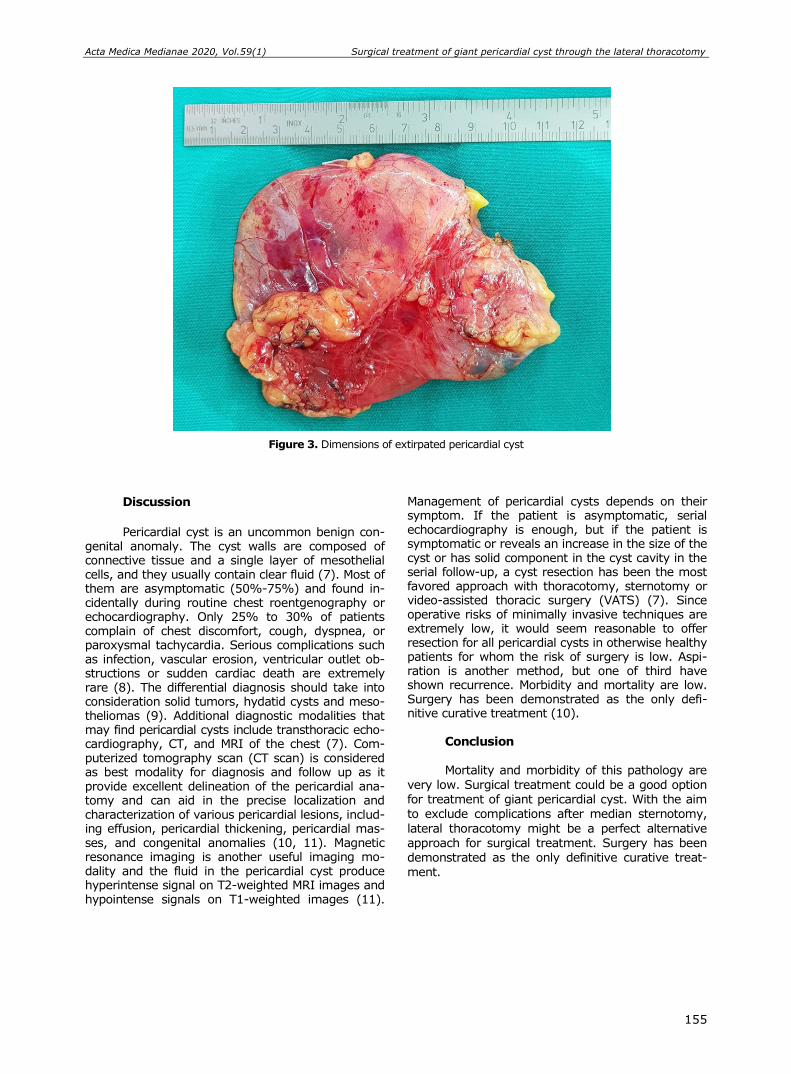
Acta Medica Medianae 2020, Vol.59(1) Surgical treatment of giant pericardial cyst through the lateral thoracotomy
155
Figure 3. Dimensions of extirpated pericardial cyst
Discussion
Pericardial cyst is an uncommon benign con-
genital anomaly. The cyst walls are composed of connective tissue and a single layer of mesothelial cells, and they usually contain clear fluid (7). Most of them are asymptomatic (50%-75%) and found in-cidentally during routine chest roentgenography or echocardiography. Only 25% to 30% of patients complain of chest discomfort, cough, dyspnea, or paroxysmal tachycardia. Serious complications such as infection, vascular erosion, ventricular outlet ob-structions or sudden cardiac death are extremely rare (8). The differential diagnosis should take into consideration solid tumors, hydatid cysts and meso-theliomas (9). Additional diagnostic modalities that may find pericardial cysts include transthoracic echo-cardiography, CT, and MRI of the chest (7). Com-puterized tomography scan (CT scan) is considered as best modality for diagnosis and follow up as it provide excellent delineation of the pericardial ana-tomy and can aid in the precise localization and characterization of various pericardial lesions, includ-ing effusion, pericardial thickening, pericardial mas-ses, and congenital anomalies (10, 11). Magnetic resonance imaging is another useful imaging mo-dality and the fluid in the pericardial cyst produce hyperintense signal on T2-weighted MRI images and hypointense signals on T1-weighted images (11).
Management of pericardial cysts depends on their symptom. If the patient is asymptomatic, serial echocardiography is enough, but if the patient is symptomatic or reveals an increase in the size of the cyst or has solid component in the cyst cavity in the serial follow-up, a cyst resection has been the most favored approach with thoracotomy, sternotomy or video-assisted thoracic surgery (VATS) (7). Since operative risks of minimally invasive techniques are extremely low, it would seem reasonable to offer resection for all pericardial cysts in otherwise healthy patients for whom the risk of surgery is low. Aspi-ration is another method, but one of third have shown recurrence. Morbidity and mortality are low. Surgery has been demonstrated as the only defi-nitive curative treatment (10).
Conclusion Mortality and morbidity of this pathology are
very low. Surgical treatment could be a good option for treatment of giant pericardial cyst. With the aim to exclude complications after median sternotomy,
lateral thoracotomy might be a perfect alternative approach for surgical treatment. Surgery has been demonstrated as the only definitive curative treat-ment.
Surgical treatment of giant pericardial cyst through the lateral thoracotomy Stefan Timčić et al.
156
References
1. Edwards MH, Ahmad A. Epicardial cyst–a case report. Thorax 1972; 27(4):503-6. [CrossRef] [PubMed]
2. Debus V, Krasemann T, Semik M. An epicardial cyst in a child. Cardiol Young 2001; 11:660-2. [CrossRef] [PubMed]
3. Elamin WF, Hannan K. Pericardial cyst: an unusual cause of pneumonia. Cases J 2008; 10;1(1):26. [CrossRef] [PubMed]
4. Maisch B, Seferovic PM, Ristic AD, Erbel R, Rienmaller R, et al. Guidelines on the diagnosis and management of pericardial diseases executive summary; The Task force on the diagnosis and management of pericardial diseases of the European society of cardiology. Eur Heart J 2004; 25:587-610. [CrossRef] [PubMed]
5. Patel J, Park C, Michaels J, Rosen S, Kort S. Pericardial cyst: casereports and a literature review. Echocardio-graphy 2004; 21(3):269-72. [CrossRef] [PubMed]
6. Najib MQ, Chaliki HP, Raizada A, Ganji JL, Panse PM, Click RL. Symptomatic pericardial cyst: a case series. Eur J Echocardiogr 2011; 12(11):E43.
[CrossRef] [PubMed] 7. Comoglio C, Sansone F, Delsedime L, Campanella A,
Ceresa F, Rinaldi M. Mesothelial cyst of the peri-cardium, absent on earlier computed tomography. Tex Heart Inst J 2010; 37:354-7. [PubMed]
8. Restrepo CS, Vargas D, Ocazionez D, Martínez-Jiménez S, Betancourt Cuellar SL, Gutierrez FR. Primary pericardial tumors. Radiographics 2013; 33: 1613-30. [CrossRef] [PubMed]
9. Alar T, Bayram AS, Gebitekin C. Pericardial cysts: an analysis of 12 cases. J Laparoendosc Adv Surg Tech A 2011; 21:595-8. [CrossRef] [PubMed]
10. Patel J, Park C, Michaels J, Rosen S, Kort S. Pericardial cyst: case reports and a literature review. Echocardio-graphy 2004; 21(3):269-72. [CrossRef] [PubMed]
11. Wang ZJ, Reddy GP, Gotway MB, Yeh BM, Hetts SW, Higgins CB. CT and MR imaging of pericardial disease. Radiographics 2003; 23:S167-80. [CrossRef] [PubMed]
Acta Medica Medianae 2020, Vol.59(1) Surgical treatment of giant pericardial cyst through the lateral thoracotomy
157
Prikaz bolesnika UDC: 616.11-006.03-089
doi:10.5633/amm.2020.0123
HIRURŠKO LEČENJE GIGANTSKE PERIKARDNE CISTE KROZ LATERALNU TORAKOTOMIJU
Stefan Timčić1, Igor Živković1, Staša Krasić2, Marko Anđelković1, Ivan Nešić1,
Milan Ćirković1, Miodrag Perić1,3
1Klinika za kardiohirurgiju, Institut za kardiovaskularne bolesti “Dedinje”, Beograd, Srbija 2Klinika za pedijatriju, Institut za zdravstvenu zaštitut majke i deteta Srbije “Dr Vukan Čupić”, Beograd, Srbija 3Medicinski fakultet u Beogradu, Beograd, Srbija
Kontakt: Igor Živković
Heroja Milana Tepića 1, 11000 Beograd, Srbija
E-mail: [email protected]
Perikardne ciste predstavljaju retke benigne tumore sa ukupnom prevalencijom oko
7% svih medijastinalnih tumora. Obično su bolesnici bez simptomatologije, izuzev kada cista komprimuje anatomske strukture unutar grudnog koša. Mi smo predstavili veliku perikardnu cistu lokalizovanu bluzu srčanog vrha. Magnetna rezonanca je kod bolesnika detektovala cističnu formaciju dimenzija 9 cm x 4 cm. Hirurška procedura izvedena je kroz levu lateralnu torakotomiju bez primene kardiopulmonalnog bajpasa kao podrške. Čitava tumorska forma-cija izvađena je i poslata na dalja patohistološka ispitivanja. Hirurška ili perkutana procedura neophodna je za lečenje perikardnih cisti, zavisno od lokalizacije i odnosa sa ostalim strukturama. Stope morbiditeta i mortalita su niske. Hirurška intervencija predstavlja jedini definitivni način lečenja.
Acta Medica Medianae 2020;59(1):153-157.
Ključne reči: perikarda cista, lateralna torakotomija
158 www.medfak.ni.ac.rs/amm
Review article UDC: 615.851
doi:10.5633/amm.2020.0124
GESTALT PSYCHOTHERAPY: SCIENCE OR QUASI-SCIENCE?
Jana Milić
The term Gestalt appears for the first time in Gestalt psychology, created by
Wertheimer, Keller, Koffka and Rubin, to mark an entity, good form. Elements do not determine an entity, and an entity is not a mere set of elements. It should be specified that a particular part of an entity is not the same when alone or included in some other entity because that part absorbs the characteristics coming from its place and function in various entities (for example, a scream in an empty street is not the same as a scream made by children playing).
The proof for gestalt therapy’s success is an introspective report on subjective perception of a person’s improvement, or more precisely "maintenance and development of balance and good condition of the whole organism."
To scientifically approach gestalt psychotherapy, it is necessary to operationalize positive effects or changes that this therapy brings. All terms used to validate the effects of psychotherapy are subjective constructs. An introspective perception of improvement is an indicator of a positive effect of psychotherapy. Intersubjective consent, that clients give in psychotherapy, after some time of applying gestalt therapeutic techniques results in improvement, is a guarantee that the achieved therapeutic effect is objective. Gestalt psychotherapy is a branch of psychotherapy that bases its theory on a so called "empty chair". This psychotherapeutic school’s basic goals are solving the problem of the now and here, and as the other psychotherapeutic schools, it bases usually its research methods on a case study. This is clearly seen in journals and articles which discuss this psychotherapeutic school. As a therapy, it is suitable for treating mild mental disabilities and anxiety. Its therapists are constantly improving themselves and students, or psychotherapists to be, consider it to be challenging.
Acta Medica Medianae 2020;59(1):158-163. Key words: psychotherapy, individual growth and development, work on oneself,
time and man
University of Niš, Faculty of Philosophy, Psychology
Department, Niš, Serbia
Contact: Jana Milić
21/20 Pariske komune St., 18000 Niš, Serbia
Email: [email protected]
Introduction Gestalt psychotherapy is a branch of psycho-
therapy which bases its theory on gestalt theory,
especially on Kurt Lewin’s theory, and it bases its therapeutic techniques on a so called "empty chair". This psychotherapeutic school’s basic goals are solv-ing the problems of the here and now, and as other schools of psychotherapy, it usually bases its fun-damental methods on the analysis of a case. That is clearly seen in journals and articles which discuss
this psychotherapy school.
Historically speaking, gestalt therapy was created by Fritz Perls in 1942. The practitioners of this branch of psychotherapy are Laura Perls, Yontef,
Zinker, as well as the Parisian’s gestalt school prac-titioners, such as Gonzague Masquelier, Brigitte Martel, Giles Delisle from Montreal and Edoardo Giusti from Rome. Considering that this branch of psychotherapy has its main support in the gestalt theory, we will mention only the key concepts and principles of this theory.
The Basic Scientific Principles in Gestalt
Theory and Psychotherapy Beginning with Descartes, it was thought if
we would want to objectively comprehend and analyze a phenomenon or behavior, then we would need to find their causes which are usually found in the past. Causal determination is based on connect-ing the past events and their current consequences. Once we see the cause of a phenomenon, then it becomes predictable for us and we can control it.
However, contemporary physicians want us to understand there is no such thing as an "objective" analysis because a mere observation of a phenomenon changes it, and predict the most of the
Gestalt psychotherapy: science or quasi-science Jana Milić
159
causes is found in the future! (For instance: I went to bed earlier last night so that I would be well rested today as I could teach an important class, so, the cause is in the future), says Ginger (1). This only proves that theological determination and explana-tion by comprehension have always been present in psychology, since the ancient division of psychology to a natural science and a spiritual science. Where do gestalt theory and psychotherapy belong to? What kind of determination it deals with?
In the beginning of the development of this psychological movement, the object of analysis was the observation or the laws under which observation was constructed. The laws of organizing stimuli into observation have been experimentally proven and verified. Hereby the disciples of this school in psy-chology have chosen an original approach to access perception, as a phenomenon which is dynamic, not static, organized, not chaotic, predictable, but not unexpected. The term Gestalt is a term which appears for the first time in Gestalt psychology founded by Wertheimer, Keller, Koffka and Rubin, to signify a whole, good form. Elements do not deter-mine a whole, and a whole is not a mere set of elements. It should be said that a particular part of a whole is not the same when alone or included in some other whole, because that part gets the cha-racteristics coming from its placement and its role in various wholes (for example, a scream in an empty street is not the same scream of children who play together).
Disciples of gestalt school in psychology started off with a radically new approach to pheno-menology of observation, which are then experi-mentally tested and confirmed, thus making a sig-nificant step in comparison to theories which had dealt with perception until then. These principles (the significance of restructuration of the observa-tional field-figures and backgrounds, significance of nearness, similarity, good form and common faith) found while explaining and understanding the pro-cesses of observation were generalized as other cognitive processes, whereas their followers to psy-chotherapy.
When we speak about "Gestalt psychothe-rapy", we emphasize that gestalt psychotherapists consider that a man’s behavior cannot be under-stood outside of the context it is developed in, the context of the field.
a) Field is a scientific construct which the disciples of the gestalt principle in psychology and psychotherapy rely on. It refers to the notion that life is an open system in which there is interaction between organisms and environment. An organism is an active participant in the creation of its own reality and can be the basic force in shaping our experience. Perls thought that a man and his be-havior cannot be understood out of context in which the behavior is being developed (2, 3). We are the people in context!
b) Dialogue-Me: You conversation, the now and here.
c) Phenomenology-stretches towards the understanding based on what is obvious or what is discovered by the situation itself, but not on inter-pretation of an observer (3).
Apart from these terms, according to Ginger’s systematization there are 20 more constructs of gestalt therapy and they are:
1) The now and how, 2) Process, 3) Awareness, 4) The limits of contact, 5) The field and system, 6) Creative adaptation, 7) The experience cycle, 8) Unfinished businesses/ Unfinished gestalt, 9) Resistance, 10) Homeostasis, 11) The Potentiation of responsibility, 12) Experimentation, 13) The right on diversity-unrepeatability of
every man, 14) The Attitude for sympathy, 15) The Holistic approach to a man, 16) The Polarities which complete each other, 17) The Involvement of emotions and body, 18) Aggression, 19) Creativity and imagination, 20) Individual inside a group. The effects of gestalt psychotherapy and
diagnostics Proving the success of gestalt therapy is an
introspective statement on impressions of the sub-jective better in a person, or more precisely "main-taining and developing balance and good condition of the whole body. Health is not just the absence of a disease or defect, but the condition of a complete physical, mental and social wellness" (1).
This principle, as Ginger (1) puts it in his book, states that Gestalt therapy’s task is not treat-ment but maintaining balance and wellness of the whole organism, leads to the "rejection" of discus-sing normality and disorder which opposes Van Baalen’s statement, the author of the article "Gestalt diagnosis" from 1999, which is about finding the appropriate diagnostic criteria for evaluation of the severity and gravity of a patient’s psychopathology (4).
In his article, Van Baalen (4) gave detailed instructions on the criteria to use before diagnosing. He accurately explained the theory of Gestalt psy-chotherapy, trying to be very specific so that the reader truly understands the interaction among parts within the field, in the way that the sense of the figure and background is that every figure has its own background, and that sometimes it can happen for the background to be primary and stand out, or that these two (the figure and background) change in accordance with the focus. He also de-scribed the role of a therapist as the part which engages into interaction within a field with a client. The accent is on the change (behavioral or emo-tional) in a client, which would be closely related to Fajgelj (5), a methodologist, whose theory is about wrong patterns in behavior that change through relations, which would be closely related to psycho-logy’s task classified as a behavioral science. The scientific value of the article is high, given the fact it gives directions to Gestalt therapists in understand-
Acta Medica Medianae 2020, Vol.59(1) Gestalt psychotherapy: science or quasi-science
160
ing the process of diagnosing while being closely connected to the clinical practice of psychology.
Baalen’s theory is very similar to Mann Dave’s (6) theory, which states that a good Gestalt is clear, or the relation between the figure and background is clear, that is, between a primary need and the back-ground, hence the energy flow which every person should have in a certain moment depends on the dominant need.
The founding source of pathology, according to Gestalt, is the incomplete emotional Gestalt or the "unfinished business" (2).
To scientifically approach gestalt psychothe-rapy, it is needed to operationalize positive effects or changes which this therapy provides. All the terms which validate the effect of psychotherapy are subjective constructs. The introspective perception of improvement is the indicator of the positive effect in psychotherapy. Intersubjective consent, that clients give in psychotherapy, after some time of applying gestalt therapeutic techniques results in improvement, is a guarantee that the achieved therapeutic effect is objective. The generality of these findings, their systematic record and evalua-tion, as well as the development of application and methods, confirm Ginger’s and Van Baalam’s scienti-fic approach.
Gestalt psychotherapy in scientific journals and articles
According to the data in KoBSON database,
there is a journal that publishes articles about Gestalt psychotherapy. It is the journal called South African Journal of Psychology. The journal is not strictly related to the field of Gestalt psychotherapy, as British Gestalt journal, but it publishes numerous articles about Gestalt psychotherapy. It is found by using the key words gestalt psychotherapy.
Susanne Jacobs often publishes her articles in this journal. In her articles she always refers to the founders of Gestalt psychotherapy, Yontef and Zinker, which would imply she doesn’t digress from the primary theory and the methods of Gestalt psy-chotherapy, and is consistent in maintaining the tra-dition (7). Her article which deals with humor in Gestalt therapy consistently follows the theory of Gestalt therapy. The method of the research is the case study, which belongs to the clinical method but the author has precisely explained the science be-hind this method, referring to various authors who use it in their research. Susanne states that the facts will be collected before and after a treatment, gestalt theory will be consistently applied in practice and that the method of observation will be used. The author defined a theory which will be applied, illu-strated to the details the method which will be used and systematically showed the results of the obser-vation, that is, the case study. The article is consis-tent in following scientific methodological norms by applying the Gestalt theoretical approach, describes detailed observations so that the process could be checked. Her work is quite close to a scientific approach, which separates her greatly from many other researchers in the field hence many authors
refer to and quote her. This is an extraordinary example how a serious scientific methodology could be applied to a field of psychotherapy, which by its nature belongs on the border between science and pseudoscience.
The reference Susanne uses is the book Skills in Gestalt Counselling and Psychotherapy by authors Joyce i Sills (8). The book describes how to lead the Gestalt session and operate with the terms related to religion. One part of the book is about the dif-ference between a normal condition and a transcen-dental experience and esoteric, describes how a man regresses to the level of an infant, has psycho-tic reactions, and even can be autistic. Religion does not have an empiric proof. Various religions are founded on a religious experience of an individual, and are characterized by the feeling of admiration for God. The main controversy between various re-presentatives of materialism and religion lies beyond the borders of experimental, verifiable, and refers to the issue of the genesis of the universe. Materialists claim that the universe is endless in space and time, and numerous religions consider the universe has its own Maker, whom not even the greatest minds, such as Einstein, renounced. He explains that his religion is based on the belief of a Higher reason’s existence, who reveals itself in the world available for cognition (9). However, if the meanings of nume-rous mentioned terms (esotery, transcendental ex-perience, religion, deity) are understood from the point of view we have nowadays, Gestalt therapy in this case, approached by Joyce and Sills, is not a science.
Valerie Aiach Dominitz following the theory of Gestalt psychotherapy applied in the practice of the case study (10). The terms are defined and ex-plained as well as the work with a client. Therefore, this journal publishes articles about Gestalt psy-chotherapy. Among its authors, we can encounter the founders of Gestalt therapy and Zinker, Yontef, Perls.
In his article published in Gestalt Journal, Edwin S. Hariss, describes the attitude towards God and disease (11). The article is not consistent in following the norms of Gestalt psychotherapy. The method is based on an experiment in which one speaks to God and gives the answer to oneself from God’s perspective. The therapy is conducted with abused clients and those angry with God. A man’s world is spiritual, immaterial; spiritual being here defined as recognizing the reality greater than the one which is a convulsion of all visible things. Edwin S. Hariss says that Gestalt psychotherapy is more and more in collaboration with psychoanalysis, which Karl Poper does not consider to be a science because it does not provide the refutation, that is, from the beginning it was untrusting and in a my-thical way approaches to behaviour (12).
Yontef in British Gestalt Journal points out that Gestalt therapy has founded many useful and creative innovations related to theory and practice in psychotherapy (13). Kohler (1959 cited by Yontef, 1996) confirms that. He states that enthusiasm and ventures of the early Gestalt’s psychologists were virtues because they created new observations and discoveries. Moreover, Yontef says that many of
Gestalt psychotherapy: science or quasi-science Jana Milić
159
these discoveries and techniques are integrated into the general practice, many times with no credit. However, Gestalt techniques and methods represent a good model of psychotherapy and should be ap-plied and developed forwardly.
Some articles are about the connection be-tween Gestalt psychotherapy, existentialism and Zen Buddhism. Existentialist narrowness results in noogenic neuroses (Frankl, 1994) which imply over-accented existential frustration, which disrupts free action and development. It can lead to alienation ("My mind contradicts my feelings", a quote from the novel "The Stranger" by Albert Camus). This narrowness is called "the existential vacuum" which refers to the lack of sense or the state of boredom. Since Frankl’s logotherapy represents therapy in-volving sense, then the constant frustration disrupts sense. The greatest sense of a man’s existence is love.
Cannon Bard studied philosophy while study-ing Gestalt therapy (14). She thought that the Sartre’s existential philosophy could be connected to Gestalt therapy. Her opinion is that Gestalt therapy can learn a great deal from existentialists about anxiety. Existentialist psychotherapy still did not find its Freud. She relied on and supported the work of Frederick Fritz Perls and having compared existen-tialist and gestalt psychotherapy, said that both deny the Freudian unconscious, whereas unconsci-ous in their sense is a phenomenological relation be-tween consciousness and its objects. The fundamen-tal thread which connects them is the sense of emptiness (there is nothing there). That would be the idea of Zen Buddhism, and Perls was a student of Zen. "There is nothing but happenings, that is, things happen". Neurotic people cannot see, be-cause their eyes are always given to others so to reflect themselves as objects (Cannon, 2009). Free-dom is nothing, emptiness, detachment from things, the sense of emptiness, but Kant says there where the sense of emptiness is, is God. The scientific value of the article is funded on pointing out the clear similarities between Sartre’s existential philoso-phy and Gestalt therapy. The similarity is the sense of emptiness- unreality.
In her article, Gestalt Therapy in Psychological Practice, Palmer A. Kendra discusses "phenomenolo-gical" or "experienced" approach, which relates to a patients’ experience-the way he or she experiences things (15). Phenomenology is a discipline that helps patients digress from their usual way of thinking so they could make a difference between what they are really experiencing at the moment and what is a consequence of the past events (Yontef, 1993, cited by Palmer, 2011). Therapy, phenomenological and existential, focuses on people’s existence-relation-ships with others, joys and sufferings, and for exam-ple, immediate experience. Patients have freedom, but they are given the responsibility for their actions and thoughts. The challenge is to take responsibility for themselves in the present (Corey, 2009: cited by Palmer, 2011). Patients’ specific reality is under research, while the diagnosis and prognosis are
mainly ignored (Corey, 2009, cited by Palmer, 2011).
Gestalt therapy is efficient (16). This is a positive, humanistic therapy. It helps practitioners (both clients and therapists) become responsible for their own experiences and experiment with new ways of behaviour and thinking. It helps them prac-tice and completely experience key moments and emotions in their lives (17). Awareness about im-portant topics or patterns urges practitioners and clients to become more authentic and open for transition or change.
Many articles talk about the significance of the connection between Gestalt psychotherapy and me-ditation. Meditation (of the eastern religion) is equal to praying in a Christian religion. One who medita-tes, thinks about a particular content, goes deep in thoughts or strives to achieve relaxation using a breathing method. To hear and accept a prayer can-not be considered scientific.
Conclusion
Since its inception, Gestalt therapy, until to-day, has evolved into psychotherapy which field of interest is open to religion, meditation, play therapy, psychoanalysis, but primarily its concepts were based on understanding an individual’s existence
and it used the phenomenological method. So, the description of experience of the here and now. It could be said it is not rigid in relation to other psy-choterapies’ points of view and allows combining and eclecticism. What I have noticed is that the authors of the articles are always relying on the
founders of Gestalt psychotherapy, Perls, Yontef,
Zinker, which would imply that theoretical basis of psychotherapy should not be changed. The metho-dology of Gestalt psychotherapy is based on con-structs of the gestalt theory; the principles of causa-bility are manifested in therapeutic situation or field, whereas change in one part of the field causes changes in other parts of the field. The truth it
strives for is the patient’s truth. Gestalt psychothe-rapy confirms itself through the subjective better in a patient, that is, in a way a kind of a measurable subjective experience. Nowadays it establishes con-nection to religion for what there are no facts for proof. That would be the step backwards in science.
Gestalt psychotherapy, in general, does not
belong to a scientific field exactly because of the narrower connection to religion and meditation, but of course, it is possible to advance the methodology of verification and measure the effects of psycho-therapy. In that sense, the gap, scientifically and pseudonically does not have to be that deep, at least
when it is about gestalt psychotherapy, given that the theory which is based on experimental research of perception and possibility to explicitly define at least some, if not all, parameters of improvement which therapy can give to a person, that is, to people.
161
Acta Medica Medianae 2020, Vol.59(1) Gestalt psychotherapy: science or quasi-science
162
References
1. Ginger S. Gestalt Psychotherapy: The Art of Contact. Novi Sad: Psihopolis institute; 2010.
2. Perls F, Hefferline R, Goodman R. Gestalt therapy integrated: Excitement and growth in the human personality. New York: Dell; 1951.
3. Erić LJ. Psychotherapy. Belgrade: University of Medicine; 2002.
4. Van Baalen D. Gestalt Diagnosis. Oslo: Norsk Gestaltinstitutt; 1999.
5. Fajgelj S. The Observational Methods of Behavior. Belgrade: The Centre of Applied Psychology; 2004.
6. Mann D. Gestalt Therapy-100 Key Point and Techni-ques. Abingdon USA: Taylor & Francis Group; 2010. [CrossRef]
7. Jacobs S. Humour in Gestalt Therapy — Curative Force and Catalyst for Change: A Case Study. S Afr J Psychol 2009;39(4):498-506. [CrossRef]
8. Joyce P, Sills C. Skills in Gestalt Counselling and Psychotherapy. London: Sage; 2001. [CrossRef]
9. Menj A. The History of Religion. Belgrade: Plato; 2005. 10. Dominitz AV. Gestalt Therapy Applied: A case study
with an Inpatient diagnosed with Substance use and
bipolar disorders. Clin Psychol Psychother 2017;24(1): 36-47. [CrossRef][PubMed]
11. Harris ES. God, Buber, and the Practice of Gestalt Therapy. The Gestalt Journal 2000;23(1):39-62. [CrossRef]
12. Milas G. Research Methods in Psychology and Other Social Sciences. Zagreb Jastrebarsko: Naklada slap; 2005.
13. Yontef G. Supervision from a Gestalt therapy perspective. BGJ 1996;5(2):92-102. [CrossRef]
14. Cannon B. Nothingness as the Ground for Change Gestalt Therapy and Existential Psychoanalysis. Existential Analysis 2009; 20(2):192-210.
15. Palmer KA. Gestalt Therapy in Psychological Practice. Inquiries Journal/Student Pulse 2011;3(11). [CrossRef]
16. Benjafield GJ. Revisiting Wittgenstein Köhler and Gestalt psychology. J Hist Behav Sci 2008;44(2):99-118. [CrossRef][PubMed]
17. Frankl Viktor E. Why do not You Commit Suicide? Belgrade: Prosveta; 1994.
Gestalt psychotherapy: science or quasi-science Jana Milić
163
Revijalni rad UDC: 615.851
doi:10.5633/amm.2020.0124
GEŠTALT PSIHOTERAPIJA: NAUKA ILI KVAZINAUKA?
Jana Milić
Univerzitet u Nišu, Filozofski fakultet, Departman za psihologiju, Niš, Srbija
Kontakt: Jana Milić Pariske komune 21/20, 18000 Niš, Srbija
E-mail: [email protected]
Pojam geštalt je pojam koji se prvi put pojavljuje u geštalt psihologiji, čiji su stvaraoci
Verthajmenr, Keler, Kofka i Rubin, kako bi označio celinu, dobru formu. Elementi ne determinišu celinu, a celina nije puki zbir elemenata. Treba naglasiti i to da određeni deo unutar neke celine nije isti deo kada je izdvojen ili kada je uključen u neku drugu celinu, jer taj deo poprima osobine koje proizilaze iz njegovog mesta i njegove uloge u različitim celinama (npr. vriska u pustoj ulici nije isto što i vriska dece koja se igraju).
Dokaz o uspešnosti geštalt terapije je introspektivni izveštaj o doživljaju subjektivog boljitka kod osobe ili preciznije rečeno "održavanje i razvijanje ravnoteže i dobrog stanja celog organizma". Za naučni pristup geštalt psihoterapiji potrebno je operacionalizovati pozitivne efekte ili promene koje ova terapija donosi. Svi termini kojima se validiraju efekti psihote-rapije subjektivni su konstrukti. Introspektivni doživljaj boljitka pokazatelj je pozitivnog efekta psihoterapije. Intersubjektivna usaglašenost klijenata, koji su na psihoterapiji, da posle izvesnog vremena, uz primenu geštalt terapijskih tehnika osećaju poboljšanje, jeste garancija da je postignuti terapijski efekat objektivan. Geštalt psihoterapija je grana psihoterapije koja svoju teoriju bazira na geštalt teoriji i to pre svega na teoriji polja, a svoje terapijske tehnike zasniva na tzv. praznoj stolici. U okviru ovog psihoterapisjkog pravca, bazični ciljevi su usme-reni na rešavanje problema sada i ovde, a kao i ostale psihoterapijske škole, svoje istraži-vačke metode bazira uglavnom na studiji slučaja. To se jasno vidi u časopisima i člancima u kojima se razmatra ova psihoterapijska škola. Kao terapija, pogodna je za lečenje blagih mentalnih poremećaja i anksioznosti. Njeni terapeuti konstantno napreduju, a učenici ili budući psihoterapueti smatraju je izazovnom.
Acta Medica Medianae 2020;59(1):158-163.
Ključne reči: psihoterapija, individualni rast i razvoj, rad na sebi, vreme i čovek
164 www.medfak.ni.ac.rs/amm
Review article UDC: 004:616-083
doi:10.5633/amm.2020.0125
THE APPLICATION OF INFORMATION TECHNOLOGIES IN THE PROCESS OF NURSING CARE
Мilena Cvetković-Jovanović1, Sunčica Ivanović2, Sanja Trgovčević2, Tatjana Kilibarda2,
Maja Stanković1, Suzana Milutinović1
Healthcare professionals are witnessing numerous innovations and technologies that
have made it possible to perform more complex diagnostic procedures, treatments and numerous multidisciplinary researches.
This paper presents the importance of information technology from the aspect of its applicability in the process of nursing.
A systematic review of quantitative studies published in the Serbian Library Consortium for Coordinated Acquisition (KOBSON).
The use of information technologies significantly improves the work of healthcare
institutions. In addition to the application in the nursing process, proper design and im-plementation of information technology should contribute to significant improvement in the education and research work of nurses.
The use of information technologies is essential in the daily work of nurses in the health care process.
Acta Medica Medianae 2020;59(1):164-169. Key words: health care, information technologies, education, scientific research
1University of Novi Sad, Faculty of Medicine, PhD Student, Novi Sad, Serbia 2The Academy of Applied Preschool Teaching and Health
Studies Kruševac – Department in Ćuprija, Serbia
Contact: Sunčica Ivanović 26. Maj 69, 18000 Niš, Serbia
E-mail: [email protected]
Introduction
Independently or in a team work, nurses are
constantly working to promote and improve health, to treat and rehabilitate patients, provide health care, as well as organize and provide continuing edu-cation. Therefore, the success of a nurse's work
depends not only on the application of nursing care, but also on the ability to apply modern technology, which helps the advancement of the nursing pro-fession through continuous learning and improve-ment.
Nursing care process
In the professional literature, Nursing Health
Care is cited as a basic discipline in nursing theory and practice within which a system of specific nurs-ing knowledge and skills is studied (1). It is also implemented in all forms of organization of health, social and child care, including private practice (2). Medical and nursing records can be
defined as do-cuments created in healthcare institutions (3). A broader concept of documentation is found in the law of the Republic of Serbia, according to which health documentation is defined as:
• patient and population health monitoring document;
• fulfilling the obligations of all health care entities;
• environmental risk factor and assessment of their impact on population health;
• health care resource;
• continuous improvement of the quality of health care;
• financing health care; • health care planning and programming; • monitoring and evaluating the implementa-
tion of health care curriculum; • conducting statistical and scientific research;
• informing the public; • fulfillment of international obligations in the
field of health care and for the development of health care and health insurance systems.
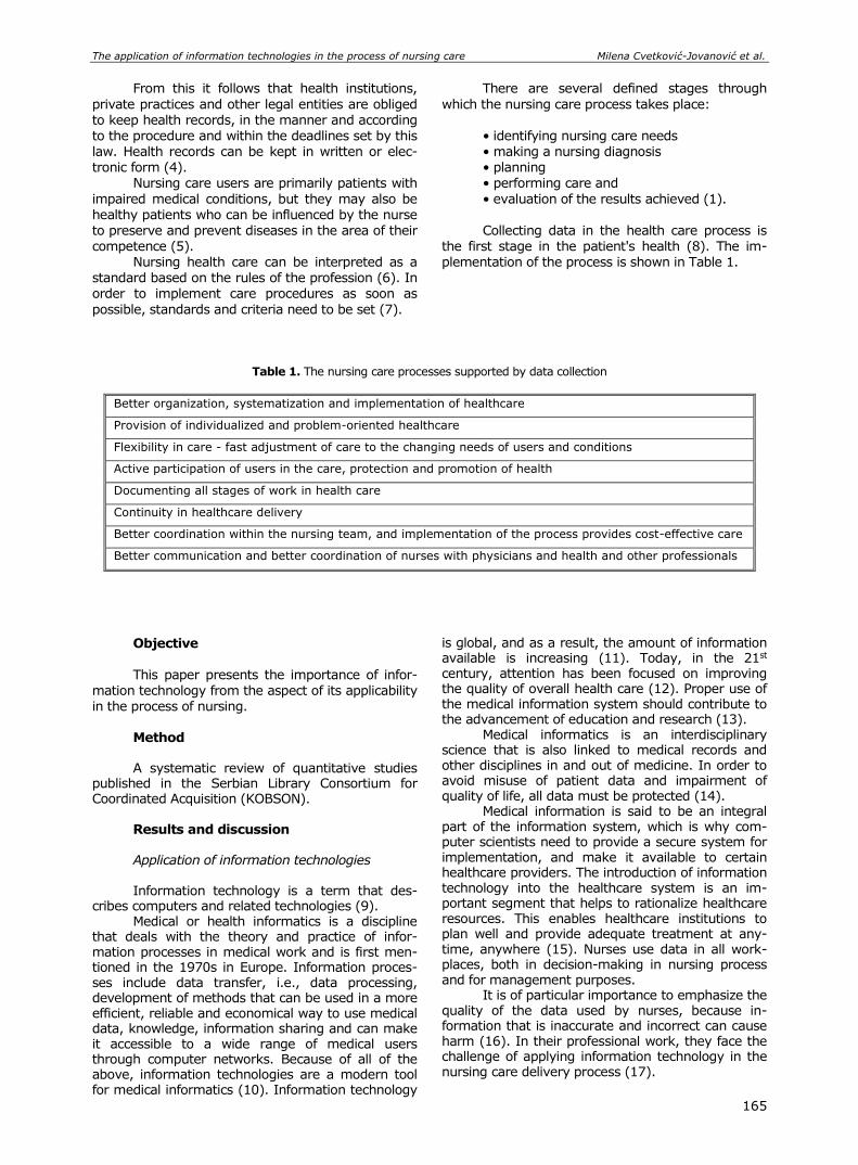
The application of information technologies in the process of nursing care Milena Cvetković-Jovanović et al.
165
From this it follows that health institutions,
private practices and other legal entities are obliged
to keep health records, in the manner and according to the procedure and within the deadlines set by this law. Health records can be kept in written or elec-tronic form (4).
Nursing care users are primarily patients with
impaired medical conditions, but they may also be healthy patients who can be influenced by the nurse to preserve and prevent diseases in the area of their competence (5).
Nursing health care can be interpreted as a standard based on the rules of the profession (6). In order to implement care procedures as soon as
possible, standards and criteria need to be set (7).
There are several defined stages through
which the nursing care process takes place:
• identifying nursing care needs • making a nursing diagnosis • planning • performing care and
• evaluation of the results achieved (1). Collecting data in the health care process is
the first stage in the patient's health (8). The im-plementation of the process is shown in Table 1.
Table 1. The nursing care processes supported by data collection
Better organization, systematization and implementation of healthcare
Provision of individualized and problem-oriented healthcare
Flexibility in care - fast adjustment of care to the changing needs of users and conditions
Active participation of users in the care, protection and promotion of health
Documenting all stages of work in health care
Continuity in healthcare delivery
Better coordination within the nursing team, and implementation of the process provides cost-effective care
Better communication and better coordination of nurses with physicians and health and other professionals
Objective This paper presents the importance of infor-
mation technology from the aspect of its applicability in the process of nursing.
Method A systematic review of quantitative studies
published in the Serbian Library Consortium for Coordinated Acquisition (KOBSON).
Results and discussion Application of information technologies Information technology is a term that des-
cribes computers and related technologies (9). Medical or health informatics is a discipline
that deals with the theory and practice of infor-mation processes in medical work and is first men-tioned in the 1970s in Europe. Information proces-ses include data transfer, i.e., data processing, development of methods that can be used in a more efficient, reliable and economical way to use medical data, knowledge, information sharing and can make it accessible to a wide range of medical users through computer networks. Because of all of the above, information technologies are a modern tool for medical informatics (10). Information technology
is global, and as a result, the amount of information available is increasing (11). Today, in the 21st century, attention has been focused on improving the quality of overall health care (12). Proper use of the medical information system should contribute to the advancement of education and research (13).
Medical informatics is an interdisciplinary science that is also linked to medical records and other disciplines in and out of medicine. In order to avoid misuse of patient data and impairment of quality of life, all data must be protected (14).
Medical information is said to be an integral part of the information system, which is why com-puter scientists need to provide a secure system for implementation, and make it available to certain healthcare providers. The introduction of information technology into the healthcare system is an im-portant segment that helps to rationalize healthcare resources. This enables healthcare institutions to plan well and provide adequate treatment at any-time, anywhere (15). Nurses use data in all work-places, both in decision-making in nursing process and for management purposes.
It is of particular importance to emphasize the quality of the data used by nurses, because in-formation that is inaccurate and incorrect can cause harm (16). In their professional work, they face the challenge of applying information technology in the nursing care delivery process (17).
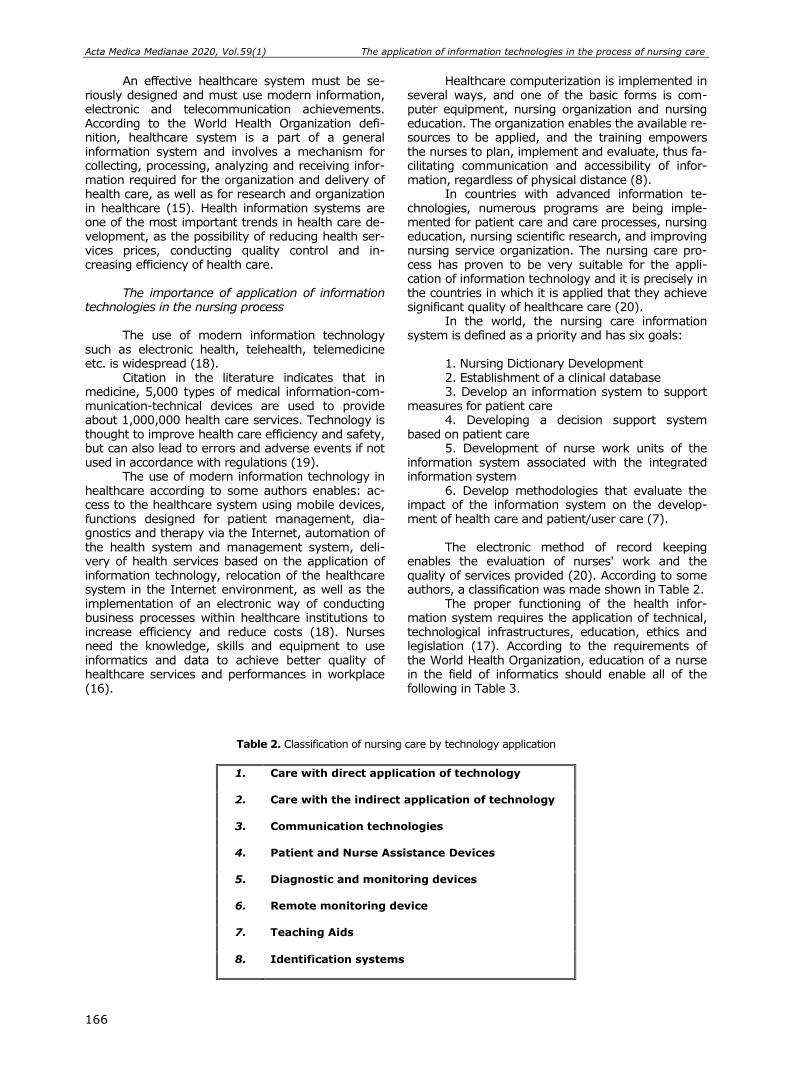
Acta Medica Medianae 2020, Vol.59(1) The application of information technologies in the process of nursing care
166
An effective healthcare system must be se-riously designed and must use modern information, electronic and telecommunication achievements. According to the World Health Organization defi-nition, healthcare system is a part of a general information system and involves a mechanism for collecting, processing, analyzing and receiving infor-mation required for the organization and delivery of health care, as well as for research and organization in healthcare (15). Health information systems are one of the most important trends in health care de-velopment, as the possibility of reducing health ser-vices prices, conducting quality control and in-creasing efficiency of health care.
The importance of application of information
technologies in the nursing process The use of modern information technology
such as electronic health, telehealth, telemedicine etc. is widespread (18).
Citation in the literature indicates that in medicine, 5,000 types of medical information-com-munication-technical devices are used to provide about 1,000,000 health care services. Technology is thought to improve health care efficiency and safety, but can also lead to errors and adverse events if not used in accordance with regulations (19).
The use of modern information technology in healthcare according to some authors enables: ac-cess to the healthcare system using mobile devices, functions designed for patient management, dia-gnostics and therapy via the Internet, automation of the health system and management system, deli-very of health services based on the application of information technology, relocation of the healthcare system in the Internet environment, as well as the implementation of an electronic way of conducting business processes within healthcare institutions to increase efficiency and reduce costs (18). Nurses need the knowledge, skills and equipment to use informatics and data to achieve better quality of healthcare services and performances in workplace (16).
Healthcare computerization is implemented in several ways, and one of the basic forms is com-puter equipment, nursing organization and nursing education. The organization enables the available re-sources to be applied, and the training empowers the nurses to plan, implement and evaluate, thus fa-cilitating communication and accessibility of infor-mation, regardless of physical distance (8).
In countries with advanced information te-chnologies, numerous programs are being imple-mented for patient care and care processes, nursing education, nursing scientific research, and improving nursing service organization. The nursing care pro-cess has proven to be very suitable for the appli-cation of information technology and it is precisely in the countries in which it is applied that they achieve significant quality of healthcare care (20).
In the world, the nursing care information system is defined as a priority and has six goals:
1. Nursing Dictionary Development 2. Establishment of a clinical database 3. Develop an information system to support
measures for patient care 4. Developing a decision support system
based on patient care 5. Development of nurse work units of the
information system associated with the integrated information system
6. Develop methodologies that evaluate the impact of the information system on the develop-ment of health care and patient/user care (7).
The electronic method of record keeping
enables the evaluation of nurses' work and the quality of services provided (20). According to some authors, a classification was made shown in Table 2.
The proper functioning of the health infor-mation system requires the application of technical, technological infrastructures, education, ethics and legislation (17). According to the requirements of the World Health Organization, education of a nurse in the field of informatics should enable all of the following in Table 3.
Table 2. Classification of nursing care by technology application
1. Care with direct application of technology
2. Care with the indirect application of technology
3. Communication technologies
4. Patient and Nurse Assistance Devices
5. Diagnostic and monitoring devices
6. Remote monitoring device
7. Teaching Aids
8. Identification systems
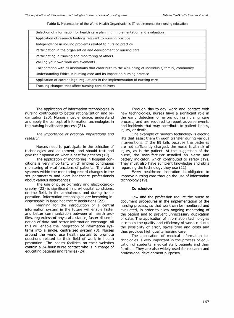
The application of information technologies in the process of nursing care Milena Cvetković-Jovanović et al.
167
Table 3. Presentation of the World Health Organization's IT requirements for nursing education
Selection of information for health care planning, implementation and evaluation
Application of research findings relevant to nursing practice
Independence in solving problems related to nursing practice
Participation in the organization and development of nursing care
Participating in training and monitoring of others
Valuing your own work achievements
Collaboration with all institutions that contribute to the well-being of individuals, family, community
Understanding Ethics in nursing care and its impact on nursing practice
Application of current legal regulations in the implementation of nursing care
Tracking changes that affect nursing care delivery
The application of information technologies in nursing contributes to better rationalization and or-ganization (20). Nurses must embrace, understand and apply the concept of information technologies in the nursing healthcare process (21).
The importance of practical implications and
research Nurses need to participate in the selection of
technologies and equipment, and should test and give their opinion on what is best for patients (19).
The application of monitoring in hospital con-ditions is very important, which implies continuous monitoring of vital functions of patients. The alarm systems within the monitoring record changes in the set parameters and alert healthcare professionals about various disturbances.
The use of pulse oximetry and electrocardio-graphy (23) is significant in pre-hospital conditions, on the field, in the ambulance, and during trans-portation. Information technologies are becoming in-dispensable in large healthcare institutions (22).
Planning for the introduction of a central information system in the future will enable faster and better communication between all health pro-files, regardless of physical distance, faster dissemi-nation of data and better information exchange. All this will enable the integration of information sys-tems into a single, centralized system (8). Nurses around the world use health portals to promote questions related to their field of work in health promotion. The health facilities on their websites contain a 24-hour nurse contact who is in charge of educating patients and families (24).
Through day-to-day work and contact with new technologies, nurses have a significant role in the early detection of errors during nursing care process, and are required to report adverse events and incidents that may contribute to patient illness, injury, or death.
One example of modern technology is electric lifts that assist them through transfer during various interventions. If the lift fails because the batteries are not sufficiently charged, the nurse is at risk of injury, as is the patient. At the suggestion of the nurse, the manufacturer installed an alarm and battery indicator, which contributed to safety (19). They must also have sufficient knowledge and skills regarding the technology they use (22).
Every healthcare institution is obligated to improve nursing care through the use of information technology (19).
Conclusion Law and the profession require the nurse to
document procedures in the implementation of the nursing process, so that work can be monitored and
evaluated, in order to allow ongoing monitoring of the patient and to prevent unnecessary duplication of data. The application of information technologies increases the quality and efficiency of work, reduces the possibility of error, saves time and costs and thus provides high quality nursing care.
The application of medical information te-
chnologies is very important in the process of edu-cation of students, medical staff, patients and their families. They are also widely used for research and professional development purposes.
Acta Medica Medianae 2020, Vol.59(1) The application of information technologies in the process of nursing care
168
References
1. Tijanić M, Tijanić M, Đuranović D, Rudić R, Milović Lj. Zdravstvena nega i savremeno sestrinstvo. Beograd: Naučna KMD;2010.
2. Komora medicinskih sestara i zdravstvenih radnika R. Srbije. Nacrt Zakona, uredbe, pravilnika o sestrinstvu. [citirano 2018 decembar 28] Dostupno: http://www.kmszts.org.rs/akti/nacrt.pdf
3. Vižintin K. Uloga bolničkog povjerenstva u stvaranju pravila za upravljanje elektroničkom medicinskom i sestrinskom dokumentacijom. Acta Med Croatica 2014;68:61-4.[CrossRef][PubMed]
4. Republika Srbija. Zakon o zdravstvenoj dokumentaciji i evidenciji u oblasti zdravstva.[citirano 2018 decembar 28] Dostupno:https://www.paragraf.rs/propisi_download/zakon-o-zdravstvenoj-dokumentaciji-i-evidencijama-u-oblasti-zdravstva.pdf
5. Vlaisavljević T. Sestrinstvo odraz u ogledalu. Inspi-rium2015; 14: 51-53. [CrossRef]
6. Babić D, Vico M. Sestrinska dokumentacija kao indi-kator zdravstvene njege. U: Pavić J, Turuk V. Zbornik
radova konferencije " Hrvatsko sestrinstvo ususret Europskoj uniji ", Opatija,2011;31-9.[CrossRef]
7. Bošković S, Milovanović V, Polak S. Primena dobre prakse zdravstvene nege – preduslov za povećanje kvaliteta u zdravstvenoj nezi. Tehnika - Kvalitet, standardizacija i metrologija2008;8(4):9-15. [CrossRef]
8. Kurtović B, Bile-Grgas C, Kundrata D. Informatizacija zdravstvene nege. Acta Med Croatica2014;68:55-9. [CrossRef][PubMed]
9. Dačić M. Biomedicinska naučna i stručna informatika. Beograd: Medicinska knjiga; 1988.
10. Deželić G. Medicinska informatika-nastanak naziva i njegovo znacenje. Medix 2004;10:54-6. [CrossRef]
11. Cvekan P. Informatičke tehnologije ikomuniciranje. Polit Misao 1998;35(2): 235-43. [CrossRef]
12. Aleksijević A. Stavovi medicinskih sestara prema uporabi računala u zdravstvenoj njezi. Sestrinski glasnik2015;20(2):110-14 .[CrossRef]
13. Janković D, Rajković P, Stanković T, Milenkovic A, Kocić I. Primjena medicinskih informacionih
tehnologija u edukaciji i istraživanjima u medicini. Acta Med Mediana2012;51(1):73-80. [CrossRef]
14. Ivanešić D. Mišljenja medicinskih sestara/tehničara o čuvanju profesionalne sestrinske tajne s obzirom na razvoj informatičke tehnologije (Diplomski rad). Osijek; 2016. [CrossRef]
15. Anđelić S, Kostić L. Virtuelni timovi u telemedicini. Infotehn-Jahorina 2012; 11:709-14. [CrossRef]
16. Režek B, Benceković Ž. Informatički aspekti primjene sestrinske dokumentacije u praksi. U: Pavić J, Turuk V. Zbornik radova konferencije " Hrvatsko sestrinstvo ususret Europskoj uniji ", Opatija,2011;31-9. [CrossRef]
17. Kern J. Informacijske i komunikacijske tehnologije u sestrinstvu. Acta Med Croatica 2014;68:3-5. [CrossRef][PubMed]
18. Milović B. Informaciono komunikacione tehnologije u zdravstvu i u razvoju E- Health-a. [CrossRef]
19. Rauche. Osnove informatike i tehnologije u sestrin- stvu. [citirano 2018 decembar 28] Dostupno:http://www.rauche.net/izdanja/broj-
1/osnove-informatike-i-tehnologije-u-sestrinstvu/ 20. Informatika u sestrinstvu - program MONEZIS.
[citirano 2018 decembar 28] Dostupno:https://wordpress72416.files.wordpress.com/2016/11/modul-nege-monezis.pdf
21. Marinković FM. Kontinuirana odgovornost za pro-fesionalnost i informacijsku pismenost medicinskih sestara. Acta Med Croatica 2014;68:33-6. [CrossRef][PubMed]
22. Sivić S. Osnovi zdravstvene informatike. Zenica: Zdravstveni fakultet Univerziteta u Zenici;2014. [CrossRef]
23. Janković R, Stamenković D, Budić I, Bradić Ž, Bojičić- Petrov I, Kalezić N. Monitoring vitalnih funkcija u urgentnom zbrinjavanju. U: Kalezić N, editor. Inicijalni tretman urgentnih stanja u medicini. 1st. Beograd: Medicinski fakultet Beograd; 2013.p.221-36. [CrossRef]
24. Kičić M. E-zdravlje – Savjetodavna uloga medicinskih sestara. Acta Med Croatica2014;68:65-9. [CrossRef][PubMed]
The application of information technologies in the process of nursing care Milena Cvetković-Jovanović et al.
169
Revijalni rad UDC: 004:616-083
doi:10.5633/amm.2020.0125
PRIMENA INFORMACIONIH TEHNOLOGIJA U PROCESU ZDRAVSTVENE NEGE
Мilena Cvetković-Jovanović1, Sunčica Ivanović2, Sanja Trgovčević2, Tatjana Kilibarda2,
Maja Stanković1, Suzana Milutinović1
1Univerzitet u Novom Sadu, Medicinski fakultet, student postdiplomskih studija, Novi Sad, Srbija 2Akademija predškolske nastave i zdravstvene studije Kruševac — odsek u Ćupriji, Ćuprija, Srbija
Kontakt: Sunčica Ivanović
25. Maj 69, 18000 Niš, Srbija
E-mail: [email protected]
Zdravstveni radnici svedoci su brojnih inovacija i tehnologija koje su omogućile
izvođenje složenijih dijagnostičkih postupaka, lečenja i brojnih multidisciplinarnih istraživanja. Ovaj rad predstavlja značaj informacione tehnologije, sa aspekta njene primenljivosti u
procesu zdravstvene nege.
Dat je sistematski pregled kvantitativnih studija, objavljenih u Konzorcijumu biblioteke za objedinjenu nabavku (KOBSON).
Upotreba informacionih tehnologija značajno poboljšava rad zdravstvenih ustanova. Pored primene u sestrinskom procesu, pravilno oblikovanje i primena informacionih tehno-logija trebalo bi da doprinesu značajnom poboljšanju obrazovnog i istraživačkog rada medicinskih sestara.
Upotreba informacionih tehnologija od suštinskog je značaja u svakodnevnom radu medicinskih sestara u procesu zdravstvene zaštite.
Acta Medica Medianae 2020;59(1):164-169.
Ključne reči: zdravstvena zaštita, informacione tehnologije, obrazovanje, naučna
istraživanja
www.medfak.ni.ac.rs/amm
170
UREDNIŠTVO
JEDINSTVENI KRITERIJUMI ZA OBJAVLJIVANJE NAUČNIH RADOVA U
BIOMEDICINSKIM ČASOPISIMA
Ideja o postavljanju jedinstvenih kriterijuma za
objavljivanje radova u časopisima za biomedicinske nauke iskristalisana je 1978. godine u Vankuveru. Ovi kriterijumi za rukopise, uključujući pravila za pisanje bibliografije, prvi put su objavljeni 1979. godine. Vankuverska grupa je vremenom prerasla u Međunarodni komitet urednika medicinskih časopisa — International Committee of Medical Journal Editors (ICMJE). Trenutno je na snazi peta revizija kriterijuma za objavljivanje radova u biomedicinskim časopisima, doneta 1997. godine.
Kriterijumi za citiranje i navođenje referenci Reference se obeležavaju arapskim brojevima u
zagradama, pri čemu se reference obele-žavaju brojevima onim redosledom kojim se pojavljuju u tekstu. Reference citirane jedino u tabelama ili legen-di moraju se obeležiti brojem u skladu sa redosledom pojavljivanja u tekstu.
Naslove medicinskih časopisa treba pisati u skraćenom obliku onako kako su navedeni u poglavlju List of Journals Indexed in Index Medicus. Lista skraćenih naziva medicinskih časopisa objavljuje se svake godine u januarskom broju Index Medicusa. Ova lista se takođe može naći na adresi www.nlm.nih.gov
Izbegavati upotrebu apstrakata kao referenci, već koristiti samo izvorne tekstove (in extenso članci). Reference koje se odnose na radove koji su prihvaćeni, ali još nisu odštampani, treba označiti sa “u štampi”, pri čemu autor mora imati pismeno odobrenje da citira takve radove i da priloži pismeni dokaz da je citirani rad prihvaćen za štampu. Informacije iz rukopisa koji nisu prihvaćeni za štampanje mogu se citirati u tekstu kao “neobjavljeni rezultati”, ali sa pismenom dozvolom autora.
Izbegavati citiranje prethodnih saopštenja (personal communication) ukoliko ona ne obezbeđuju esencijalne rezultate koji još nigde nisu objavljeni. U ovom slučaju, neophodno je u zagradi navesti ime osobe i datum usmenog saopštenja rezultata. Za objavljivanje ovih podataka neophodno je pismeno odobrenje autora.
Kriterijumi za pisanje referenci korišćenih u radu U ovom pregledu biće obrađena pravila za pi-
sanje literaturnih referenci samo za najčešće korišćene tipove publikacija.
Članci u časopisima 1. Standardni članak u časopisu
Navesti prvih šest autora, ukoliko ih je više iza šestog dodati et al. ukoliko je referenca na engleskom jeziku ili i sar. ukoliko je referenca na srpskom jeziku.
Vega KJ, Pina I, Krevsky B. Heart transplantation is associated with an increased risk for pancreatobiliary disease. Ann Intern Med 1996; 124(11):980-3.
Parkin DM, Clayton D, Black RJ, Masuyer E, Friedl HP, Ivanov E, et al. Childhood–leukaemia in Europe after Chernobyl: 5 year follow-up. Br J Cancer 1996;73:1006-12.
2. Organizacija kao autor The Cardiac Society of Australia and New
Zealand. Clinical exercise stress testing. Safety and performance guidelines. Med J Aust 1996;164:282-4.
3. Članak bez poznatih autora Cancer in South Africa (editorial). S Afr Med J
1994;84:15.
4. Volumen sa suplementom Shen HM, Zhang QF. Risk assessment of nickel
carcinogenicity and occupational lung cancer. Environ
Health Perspect 1994; 102 Suppl 1:275-82.
5. Broj sa suplementom
Payne DK, Sullivan MD, Massie MJ. Women's
psychological reactions to breast cancer. Semin Oncol 1996;23(1 Suppl 2):89-97.
6. Volumen sa više delova Ozben T, Nacitarhan S, Tuncer N. Plasma and
urine sialic acid in non-insulin dependent diabetes
mellitus. Ann Clin Biochem 1995;32(Pt 3):303-6.
7. Broj sa više delova
Poole GH, Mills SM. One hundred consecutive cases of flap lacerations of the leg in ageing patients. N
Z Med J 1994;107(986 Pt 1):377-8.
8. Časopisi sa brojem bez volumena
Turan I, Wredmark T, Fellander-Tsai L. Arthros-
copic ankle arthrodesis in rheumatoid arthritis. Clin Orthop 1995;(320):110-4.
9. Časopisi bez volumena i broja
Jedinstveni kriterijumi za objavljivanje naučnih radova u biomedicinskim časopisima
171
Browell DA, Lennard TW. Immunologic status of the cancer patient and the effects of blood transfusion on antitumor responses. Curr Opin Gen Surg 1993:325-33.
10. Reference u obliku apstrakta ili
prethodnih saopštenja Enzensberger W, Fischer PA. Metronome in
Parkinson's disease (letter) Lancet 1996;347:1337. Clement J, De Bock R. Hematological complica–
tions of hantavirus nephropathy (HVN) (abstract). Kidney Int 1992; 42:1285.
Udžbenici i monografije 11. Monografija Ringsven MK, Bond D. Gerontology and
leadership skills for nurses. 2nd ed. Albany (NY): Delmar Publishers; 1996.
12. Autori kao urednici Norman IJ, Redfern SJ, editors. Mental health
care for elderly people. New York: Churchill Livingstone; 1996.
13. Organizacija kao autor i izdavač Institute of Medicine (US). Looking at the future
of the Medicaid program. Washington: The Institute; 1992.
14. Poglavlje u knjizi Phillips SJ, Whisnant JP. Hypertension and
stroke. In: Laragh JH, Brenner BM, editors. Hypertension: pathophysiology, diagnosis, and management. 2nd ed. New York: Raven Press; 1995. p. 465-78.
15. Conference proceedings Kimura J, Shibasaki H, editors. Recent advances
in clinical neurophysiology. Proceedings of the 10th International Congress of EMG and Clinical Neurophysiology; 1995 Oct 15-19; Kyoto, Japan. Amsterdam: Elsevier; 1996.
16. Conference paper Bengtsson S, Solheim BG. Enforcement of data
protection, privacy and security in medical informatics. In: Lun KC, Degoulet P, Piemme TE, Rienhoff O, editors. MEDINFO 92. Proceedings of the 7th World Congress on Medical Informatics; 1992 Sep 6-10; Geneva, Switzerland. Amsterdam: North-Holland; 1992. p. 1561-5.
17. Istraživački ili tehnički izveštaji
Službeni izveštaji (Issued by funding / sponsoring agency): Smith P, Golladay K. Payment for durable medical
equipment billed during skilled nursing facility stays. Final report. Dallas (TX): Dept. of Health and Human Services (US), Office of Evaluation and Inspections; 1994 Oct. Report No.: HHSIGOEI69200860.
Sponzorisani izveštaji (Issued by performing agency) Field MJ, Tranquada RE, Feasley JC, editors.
Health services research: work force and educational issues. Washington: National Academy Press; 1995. Contract No.: AHCPR282942008. Sponsored by the Agency for Health Care Policy and Research.
18. Magistarske i doktorske disertacije Kaplan SJ. Post-hospital home health care: the
elderly's access and utilization [dissertation]. St. Louis (MO): Washington Univ.; 1995.
Druge vrste publikovanog materijala
Neobjavljeni materijal
19. U štampi (In press) Leshner AI. Molecular mechanisms of cocaine
addiction. N Engl J Med. In press 1996.
Elektronski zapisi 20. Internet članak u elektronskom formatu Morse SS. Factors in the emergence of infectious
diseases. Emerg Infect Dis (serial online) 1995 Jan-Mar “cited 1996 Jun 5”; 1(1)(24 screens). Available from: URL: http://www.cdc.gov/ncidod/EID/eid.htm
21. Monografija u elektronskom formatu CDI, clinical dermatology illustrated (monograph
on CD-ROM). Reeves JRT, Maibach H. CMEA Multimedia Group, producers. 2nd ed. Version 2.0. San Diego: CMEA; 1995.
22. Kompjuterski podaci Hemodynamics III: the ups and downs of
hemodynamics (computer program). Version 2.2. Orlando (FL): Computerized Educational Systems; 1993.
172
PROPOZICIJE ZA PISANJE RADOVA U ACTA MEDICA MEDIANAE
Acta Medica Medianae (AMM) je tematski časopis
iz oblasti medicinskih nauka. Časopis objavljuje origi-nalne radove koji nisu prethodno publikovani.
U AMM se objavljuju: uvodnici, naučni i stručni članci, prethodna ili kratka saopštenja, revijski radovi tipa opšteg pregleda, aktuelne teme, meta-analize, prikazi slučajeva, prikazi knjiga i drugi prilozi. Radovi se štampaju na srpskom ili engleskom jeziku sa apstrak-tom na srpskom i engleskom jeziku. Radovi na engles-kom jeziku se prezentuju u elektronskom formatu na sajtu Medicinskog fakulteta u Nišu, kao i na među-narodnim sajtovima iz oblasti medicinskih nauka. Acta Medica Medianae izlazi četiri puta godišnje, od 1962 godine.
Svi radovi koji se objavljuju u AMM podležu
anonimnoj recenziji, a Uređivački odbor određuje redosled njihovog štampanja. Primedbe i sugestije urednika i recenzenata dostavljaju se autoru radi konačnog oblikovanja. Radovi se predaju u pisanom ili elektronskom obliku na srpskom i engleskom jeziku. Rukopisi radova prihvaćenih za štampu ne vraćaju se autoru.
Rukopis treba predati sa jednostrukim proredom, formata A4, sa levom marginom od 3 cm.
Prva strana rada treba da sadrži: a) naslov rada b) puna imena i prezimena autora c) puni nazivi ustanova i organizacijskih jedinica u kojima je rad realizovan i mesta u kojima se ustanove nalaze, d) znacima *, **, ***, #, ##, ###,…označavaju se redom autori i njihove institucije e) puna adresa, broj telefona i e-mail osobe zadužene za korespondenciju u vezi predatog rukopisa.
Druga strana treba da sadrži samo naslov rada, rezime i ključne reči, bez imena autora i institucija. Veličina rezimea za naučne i stručne članke, revijske radove tipa opšteg pregleda i meta–analize može da bude do 250 reči. Ispod rezimea sa podnaslovom “Klučne reči” navesti 3-5 ključnih reči ili izraza. Poželjno je da autori za ključne reči koriste odgovarajuće deskriptore, tj. definisane termine iz Medical Subject Heading (MeSH) liste Index Medicus-a. Prva i druga strana se predaju na srpskom i engleskom jeziku i ne obeležavaju se brojevima.
Tekst članka: Naučni i stručni članci, kao i opšti pregledi i meta-analize ne smeju prelaziti 11 stranica sa prilozima; aktuelne teme– 6 stranica; kazuistika 6– stranica; prethodna saopštenja– 5 stranica, a izveštaji sa skupova i prikazi knjiga 2 stranice. Naučni i stručni članci obavezno treba da sadrže poglavlja: uvod, cilj, materijal i metode, rezultati, diskusija i zaključak. Zahvalnost ili komentar povodom sponzorstva rada dati na kraju teksta članka iza poglavlja “zaključak”.
U tekstu naznačiti mesta priloga i obeležiti ih onako kako su obeleženi u prilogu.
Literatura se daje u posebnom poglavlju, pri čemu se navodi onim redosledom kojim se citati pojav-ljuju u tekstu. Broj literaturne reference se u tekstu označava arapskim brojem u zagradi. Za navođenje li-terature koristiti pravila Vankuverske konvencije. Strane se numerišu arapskim brojevima u donjem des-nom uglu
Priloge u vidu teksta, tabela i ilustracija (gra-fikoni, crteži i dr.) ne unositi u tekst članka, već na kraju teksta, na posebnim stranicama obeleženim u gor-njem levom uglu sa “Tabela, Grafikon, Ilustracija” i arapskim brojem redosledom pojavljivanja u tekstu (npr. Tabela 1, Grafikon 1 i dr.) i svakoj se daje kratak
naslov. Kratka objašnjenja i skraćenice daju se u fusnoti. Za fusnotu koristiti sledeće simbole: *, **, ***, #, ##, ###, …itd. Tabele, grafikone i ilustracije treba praviti korišćenjem nekog od programa iz Microsoft Office paketa. Izbegavati upotrebu boja kod izrade grafika.
Za izradu grafičkih priloga može se koristiti bilo koji grafički program, pri čemu slike moraju biti snimljene u jpg formatu rezolucije 300 dpi (u originalnoj veličini). Grafički prilozi se ne unose u Word dokument već se predaju kao posebni JPG fajlovi.
Ukoliko je tabela ili ilustracija već negde objavljena, citirati izvor i priložiti pismeno odo-brenje, ukoliko se radi o zaštićenom materijalu. Ukoliko je na fotografiji prikazan bolesnik tako da se može prepoznati, potrebno je njegovo pismeno odobrenje, u suprotnom, delovi fotografije se moraju izbrisati da bolesnik ne može biti identifikovan.
Uz rad, na posebnom listu, treba dostaviti: a) izjavu da rad do sada nije objavljivan, b) potpise svih autora, c) ime, prezime, tačnu adresu i broj telefona prvog autora.
Rad je preporučljivo predati u elektronskom obliku na e-mail adresu uredništva: [email protected] ili poslati poštom na CD ili DVD disku sa materijalom u celini na srpskom i engleskom jeziku.
Rad treba otkucati u programu ms office Word verzija 2003. ili novija. Za verziju na engleskom jeziku koristiti font Verdana, veličine 9 pt, kodna stranica (English). Za verziju na srpskom jeziku koristiti font Verdana, veličine 9 pt, kodna stranica (Serbian lat ili Croatian).
U radu je obavezno korišćenje međunarodnog sistema mera (SI) i standardnih međunarodno prihva-ćenih termina.
AMM zadržava pravo dalje distribucije i štam-panja radova.
173
SECRETARIAT
GUIDELINES FOR PAPER SUBMISSION TO ACTA MEDICA MEDIANAE
Acta Medica Medianae (AMM) is a thematic journal for medical sciences. The Journal publishes original reasearch articles that have not been published before.
AMM also publishes editorials, observational and experimental articles, procedings or short communicati-ons, review articles, meta-analyses, case reports, current topics, articles from the history of medicine as well as other contributions related to medical sciences. All articles are printed in Serbian or English with short abstracts in both Serbian and English. Articles in English are published in electronic form on the University of Nis Medical Faculty website as well as international sites related to medical sciences. Acta Medica Medianae is published four times a year. The first issue appeared as early as 1962.
General Guidelines
Paper Submission All research articles published in this journal
undergo rigorous peer review, based on initial editor screening and anonymized refereeing by at least two anonymous referees. Remarks and suggestions made by the editors and reviewers are sent to the author for final revision. The papers in English are to be submitted by e-mail: [email protected]. Manuscripts accepted for publication are not returned to authors.
The first page of a research article must contain: a) article title b) full name of author(s) c) full name(s) of institutions and/or address(es) of department(s) where either reasearch was conducted or research article written d) following signs *, **, ***, #, ##, ### signifying author(s) and institutions e) full address, phone number and e-mail of a corresponding author.
The second page should contain only research article title, abstract and key words without names of author(s) and institution(s). Abstract for research and professional articles, review articles and meta-analyses should have up to 350 words while abstract for all other types of publications should consist of 250 words. Key Words section should have up to 5 key words or phrases related to a submitted article. It is desirable
that authors use corresponding descriptors from Medical Subject Heading (MeSH) that can be found on Index Medicus list for key words. The first and the second page should not be numbered.
Body of a Research Article – Research and professional articles, as well as general surveys and meta-analyses should not exceed 11 pages altogether; current topics - 6 pages, casuistics - 6 pages and proceding statements - 5 pages, history of medicine articles - 3 pages while conference reports and book reviews - 2 pages. Research and professional articles should comprise the following mandatory chapters: introduction, aim(s), material and method(s), result(s), discussion and conclusion(s). Result(s) and discussion
can be comprized into one chapter. Acknowledgments of any kind in a submitted article should be written at the end of the paper after "Conclusion(s)". It is necessary to clearly mark a place for additions in the text.
References should be written in a separate chapter in the same order of appearance as in a research article. Reference numbers that appear in the text should be written in Arabic numerals and put in brackets. All authors should be listed. If there are more than six authors this should be indicated with et al. Use the rules of the Vancouver convention when quoting literature. Pages should be enumerated in Arabic numerals in the bottom right corner.
Additions in form of texts, tables and
illustrations (photos, drawings, diagrams, etc) should
not be inserted in the reasearch article body but at the
end of the text on separate pages which should be
marked in the upper left corner as "Table(s),
Graphic(s), Illustration(s) etc) with Arabic numeral in
the same order of appearance as in the text (for
instance, Table 1, Graph 1, etc) with a short title. Short
explanations and abbreviations should be stated in
footnotes where the following symbols should be used:
*, **, ***, #, ##, ###, etc. Table(s), graph(s) and
ilustration(s) should be drawn in a Microsoft Office
Program. Color should be avoided. Any graphic program can be used for making
graphic addition(s) while picture(s) should be saved in
.jpg format with 300 dpi resolution (original size).
Graphic addition(s) should be sent as separate jpg
file(s), and not inserted in the body of a research or
any other article submitted to AMM.
If some additions, included in a submitted
reasearch article, have already been published, source
of publication should be clearly stated, alongside
written approval in case the material is copyright
protected. Patients on photographs have privacy rights
that should not be infringed without their consent.
Namely, if a photograph shows a patient who can be
recognized, his/her written approval should also be
submitted; otherwise, visible and recognisable facial or
bodily parts should be blackened so that the patient
cannot be identified by readership.
On a separate sheet the author should also
enclose: a) his/her statement that a submitted article
has not been published before, b) signatures of all
authors, c) full name, address, e-mail and phone
number of the first author.
Submitted article should be typed in Word Version 2003 for Windows (or more recent ones), font Verdana 9 pt size; code page (English) should be used.
The authors are required to use international measurement standards (SI) and internationally accepted
standard terms. Acta Medica Medianae reserves the right for further
distribution and printing of published reasearch articles.


