Embed Size (px)
Citation preview

CIinicu Chimica Acre, 186 (1989) 211-224
Elsevier 211
CCA 04560
Physiologic Release of Tissue Alkaline Phosphatases
Secretion of hepatic and intestinal alkaline phosphatases: similarities and differences
D.H. Alpers ‘, R. Eliakim’ and K. DeSchryver-Kecskemeti *
Departments of t Medicine and 2 Pathology, Washington University, St. Louis, MO (USA)
(Received 14 June 1989; accepted 19 June 1989)
Key words: Alkaline phosphatase; Intestinal alkaline phosphatase
Intmduction
The presence of plasma membrane-bound proteins in the serum of mammals has been noted for many years. Examples of such proteins include alkaline phosphatase (liver, bone, intestinal, and placental) [l], 5’-nucleotidase [2], and gamma-glutamyl transferase [3]. The mechanism of attachment of these proteins to the membrane is now fairly clear. Some proteins, e.g. gamma-glutamyl transferase, are attached to the plasma membrane by a hydrophobic transmembrane sequence [4]. Other pro- teins, including the alkaline phosphatases (AP), are bound by the fatty acid portion of a phosphatidylinositol-glycan anchor [5]. In spite of this information the mechanism for release of these enzymes from the membrane is not known. More- over, because secretion is stimulated by factors unique to each organ, the mecha- nism(s) may differ from organ to organ, e.g. liver and intestine.
A variety of suggestions have been offered to explain enzyme release, including detergent action, proteolysis, membrane fragmentation [6], or lipolysis [5]. Detergent action has been suggested most often for hepatocyte membrane proteins, because high concentrations of bile salts are present [7]. Normally the sinusoidal and canalicular alkaline phosphatases are secreted into the blood and bile respectively [S]. The enzyme in bile appears to be particulate [9], whereas that in the blood is largely soluble. The original interpretation of these data were that the sinusoidal enzyme was released by detergent action. However, recent work has shown that solublization with butanol was due not to detergent-like action, but to the activation of tissue phospholipase C that released fatty acids from the AP [lo].
Proteins attached to the membrane by a transmembrane segment could be released by the action of proteases in the membrane or in the surrounding fluid. Cell
Abbreviations: AP, alkaline phosphatase; PI-PLC, phosphatidylinositol-specific phospholipase C; PL-D,
phosphatidylinositol-specific phospholipase D; IAP, intestinal alkaline phosphatase. Correspondence to: David H. Alpers, M.D., Gastroenterology Division, Washington University School of Medicine, 660 S Euclid Avenue, St. Louis, MO 63110, USA.
0009-8981/89/$03.50 0 1989 Elsevier Science Publishers B.V. (Biomedical Division)

212
surface shedding has been suggested ever since the rapid turnover of membrane protein was appreciated [ll]. In many instances the proteins released into serum, e.g. immunoglobulin M, are the same as those that can be released from the cell surface by trypsin treatment [12]. Membrane proteases that could perform such a function have been described [13]. The potential roles of proteases and detergents acting in concert have been suggested for removal of intestinal apical brush border enzymes by pancreatic proteases and lumenal bile salts [14]. The presence of proteases or detergents in the proper anatomic location does not, however, ensure their role in removal of membrane proteins. As will be discussed below, there is considerable evidence suggesting the release of membrane fragments into the blood or intestinal lumen. These membrane fragments, released by cell death, membrane vesiculation, or some other mechanism, would then be substrates for releasing factors (proteases, phospholipases, or bile salts) in the lumenal fluid or the blood. Thus, in vitro demonstration of release of enzymes from the cell surface or isolated membrane by various factors does not ensure a role for that factor in the process of release from the cell itself.
This same dilemma is especially germane to the problem of AP secretion. AP can be released from membranes by the action of either phosphatidylinositol (PI)-specific phospholipase C (PI-PLC) [5] or PI-specific phospholipase D (PL-D) [15]. PI-PLC is present in liver [16] and in other insulin-sensitive tissues, at least [17], and PL-D is abundant in serum [15]. Thus, either enzyme is probably available to release AP from liver or intestine. The major source of AP in these organs, however, is the apical brush border membrane, a surface not in contact with the serum. Release from these membranes would deliver the AP into the bile or the intestinal lumen, and secretion into the blood would not occur without passage through the tight junction region between cells. For these reasons hypotheses and supporting data have been generated suggesting that membrane shedding or microvesiculation of membrane fragments occurs and delivers basolateral membrane fragments into the blood. Release of AP can occur during the process of membrane fragmentation or afterwards. We will discuss the data supporting such a scheme first in liver, and then in intestine, providing our own recent studies for the latter organ. These discussions may have relevance to enzymes other than AP, because many brush border proteins are attached by either peptide sequences or fatty acids. Because of the importance of serum AP as a diagnostic test, most of the data have been derived from studies of AP secretion.
Secretion of AP from liver
The hepatocyte has an apical or lumenal membrane facing the bile canaliculus, a basal membrane surface facing the sinusoid, and a lateral membrane connecting the other two domains. Recent studies using both apical and basolateral membrane proteins (AP was not studied) demonstrated that newly synthesized mature proteins reached a basolateral plasma membrane fraction at the same rate [18]. These data suggested that both the apical and basolateral proteins travel first to the basolateral portion of the membrane, from where the apical proteins are transferred to their
213
final destination at separate rates. The kinetics of transfer for hepatocyte proteins was different from observations made in polar kidney (MDCK) or intestinal (Caco-2) cells [19,20]. The hypothesis advanced for hepatocyte membrane proteins provides a good framework for understanding the data on AP secretion. Most of the studies have examined serum AP during cholestasis, because in that condition the liver AP is much increased in serum and tissue. In humans with cholestasis there is evidence for two forms of AP in serum, one derived from sinusoidal (basolateral) and the other from canalicular (apical) membranes [21]. The canalicular AP may appear in serum because of regurgitation from the bile, a fluid in which bile salts cause release of enzymes without cell lysis [22].
In addition to AP release by detergent action, AP is found in large particles in serum. These forms separate on density gradients at densities equal to that of the cell basolateral membrane (1.18-1.20, ref. [13]). The particles isolated from gradi- ents contained only plasma membrane enzymes, and were much enriched for AP compared with leucine aminopeptidase. These structures are not likely to be derived from canalicular domains, because no high molecular weight AP was found in cholestatic bile. The absence of AP-containing particles in bile makes the sinusoidal (basal) membrane domain the likely site for membrane shedding into serum, particularly in view of the hypothesis that directs newly synthesized apical proteins (including presumably AP) to that domain initially. On the other hand, there may be increased permeability of hepatocyte tight junctions that could occur in cholesta- sis [24]. Hepatocyte tight junctions have been shown to respond to peptide and other hormones by allowing increased transport of the protein horseradish per- oxidase [25].
Later, during cholestasis, sufficient distortion of the canalicular tight junctions may occur so that bile lipoproteins can enter the circulation. During cholestasis, the lipoprotein-X particle that appears is composed of phospholipid and serum pro- teins, particularly albumin [26]. Evidence for both tight junction passage [27] and transcellular regurgitation [28] have been reported to explain the presence of lipoprotein-X in serum. Both the circulating particulate AP and lipoprotein-X were detected in 25% of patients with cholestasis [29]. Soluble AP is associated with lipoprotein-X in many of these patients [30]. It is not clear whether this AP arises from the sinusoidal or canalicular membrane. In addition, it is not certain whether it is released by the action of a tissue PI-PLC, serum PL-D, bile salt detergent action, or a combination of these factors.
During cholestasis particulate (membrane associated) AP appears frequently (about 50% of patients) as a component of serum AP. The relative enrichment of the sinusoidal membrane fragments for AP suggests that only a portion of the mem- brane may be released. Similar results have been reported in cultures from various normal and neoplastic cell lines, using S’nucleotidase and ATPase as markers. The vesicles released into the medium were enriched for 5’nucleotidase, suggesting that membrane fragmentation was selective, and that the microvesicles might represent specific membranous domains [31]. Because of the abundance of PL-D in serum, it is surprising that much of the microvesicular AP remains bound to the membrane after release into either blood or culture medium containing serum. In cholestatic

214
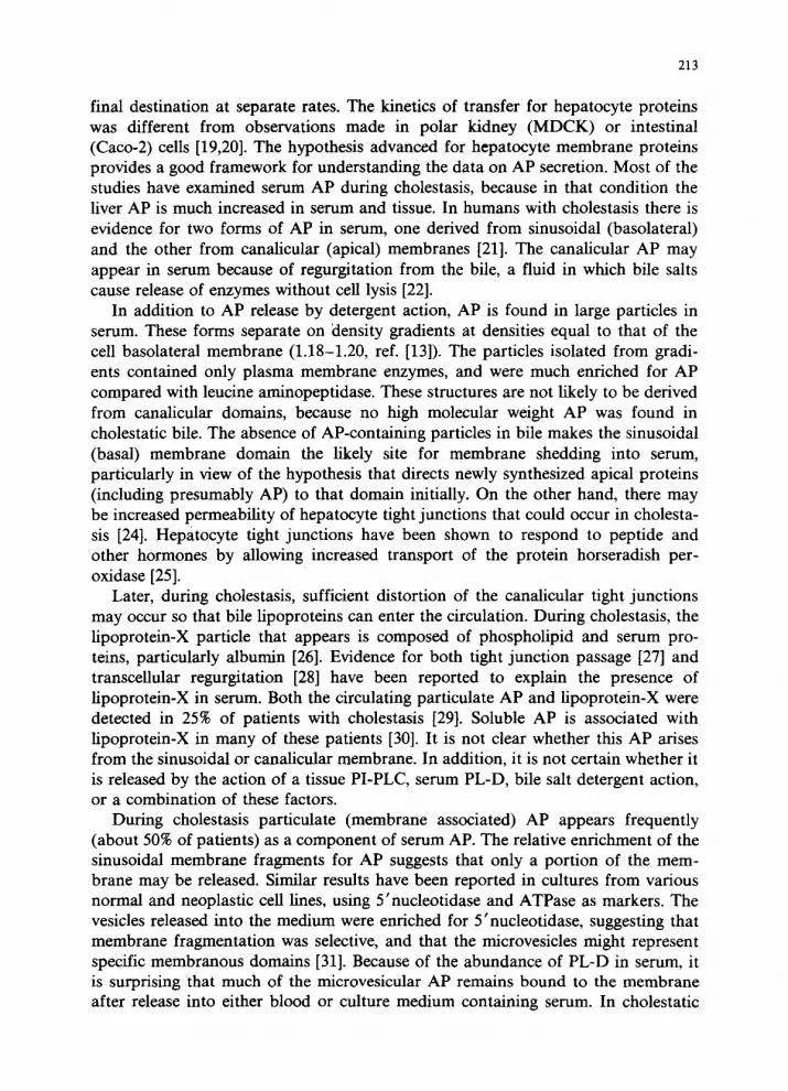
Hepatic AP Secretion During Cholestasis
Canaliculus
di
O- Microvesicle
Fig. 1. Hepatic alkaline phosphatase (AP) secretion during cholestasis. The processes demonstrated from left to right are: direct membrane release via Pi-specific phospholipase C or D, membrane vesiculation, transcellular or intercellular movement of AP, either attached to lipoprotein-X or released from
canalicular membranes.
serum this result is even more remarkable, because of the long half life of hepatic AP [32]. These findings suggest that, although PL-D and PI-PLC can readily release much of the membranous AP in vitro, they may not play a primary role in the release of particulate AP from the cell.
During cholestasis the ~~bution of AP that is normaIly mostly apical becomes equally spread over the sinusoidal membrane [33]. This shift in distribution may account for the enzymatic heterogeneity of the membrane and the AP enrichment, leading to the interpretation that microvesiculation has occurred. This shift would also expose more AP to the action of serum PL-D, thereby accounting for release of the soluble AP into blood without the necessity of regur~tation from the bile. In any event, the r~s~bution of AP during cholestasis is consistent with the finding that apical membranes first travel to the sinusoidal membrane. During cholestasis AP would be prevented from further movement to the apical (canalicuIar) mem- brane by increased bile duct pressure. This explanation seems more reasonable than one based on release of AP from the apical membrane, or on movement of apical AP to smusoidal membranes by intramembr~ous movement, which would necessi- tate movement of AP through the region of the tight junction_
The various hypotheses concerning the secretion of hepatic AP during cholestasis are smnrnarized in Fig. 1. AP appears in serum in various combinations of forms - free (or soluble), particulate on a plasma membrane-like fragment, or particulate on lipoprotein-X. It can have reached the serum either by direct release from the sinusoidal membrane, by treetops, or by regur~tation from the bile through tight junctions. The soluble form may result from detergent action, enzymatic

215
release, or a combination of the two. At the present time it is not possible to postulate a single mechanism for the secretion of hepatic AP.
Secretion of intestinal AP (IAP)
The mechanisms to consider in the secretion of IAP are similar to those discussed in the case of hepatic AP. The enterocyte is a polarized cell with an apical brush border membrane containing most of the cellular AP, and a basolateral domain. The apical (but not basolateral) domain is exposed to high concentration of bile salts and phospholipases from the lumen, and the basolateral domain is exposed to plasma ultrafiltrate, presumably containing PL-D. Membrane shedding has been observed from the apical membrane, and the tight junctions have been observed to respond to physiological stimuli. The specific details, however, are somewhat different from those in the hepatocyte, and the hypotheses regarding AP secretion need to be modified to fit the requirements of the data derived in the enterocyte.
Soluble IAP
Unlike the hepatic AP a soluble form of IAP has been reported with characteris- tics differing from the membranous IAP [34,35]. The presence of an intracellular pool of brush border enzymes has never been adequately explained, but would be consistent with a precursor pool for secreted AP. The enzymes most implicated in this soluble pool are IAP [34], glucoamylase [35], and sucrase [36], all apical membrane components. This soluble intracellular fraction is very prominent in neonatal rat intestine, but only IAP has been found in rat serum in large amounts. Neither glucoamylase nor sucrase would be easy to detect enzymatically in serum because of the high serum glucose content, and a search for immunoreactive enzymes in serum has not been made in the suckling animal. Some brush border enzymes are readily solubilized by pancreatic proteases [14], which remove the hydrophobic anchor peptide. IAP release in vivo might remain bound to the membrane by ionic forces, and be present in the tissue after homogenization. Alternatively, release of brush border glucoamylase or sucrase by contaminating proteases, or of IAP by phospholipases, could occur after homogenization of the tissue. This phenomenon might explain the presence of a soluble pool of enzyme, particularly in the case of IAP, because intracellular PI-PLC or contaminating serum PL-D could release IAP from the membrane. Such release could not entirely explain the soluble pool in the enterocyte, however, because the characteristics of soluble IAP differ from the membranous enzyme (e.g. size and carbohydrate composition).
The soluble enzyme is smaller (62 kDa), has a slightly more alkaline different ~1, and different carbohydrate composition than the membranous IAP [34]. The abun- dance of the soluble pool is greatest in the adult ileum, whereas the p1 of ileal AP is different from that of the duodenum and the serum [37]. Thus, the different ~1s of tissue AP in the various organs is not just the result of PI-PLC action, which produces consistent and slightly more acidic isomers because of an exposed phos-

216
phate moiety. The carbohydrate composition of soluble AP reveals an enrichment in fucose [34]. In addition, when rats are given 14Cfucose in vivo, there is more rapid transfer of highly fucose-labelled IAP into the serum than into the membrane of the cell [38]. It has been suggested that the transfer of IAP into the blood is closely related to blood groups with fucose as the recognition sugar [39]. It is interesting that IAP is cleared from the blood rapidly via the mannose/fucose receptor in the liver [40]. Thus, if soluble AP represents a pool for secreted AP, the additional fucose residues may be functionally significant.
The two intestinal (soluble and membranous) APs differ not only in carbohydrate composition, but also in peptide structure. Total RNA from rat intestine translated in a cell free system yields two peptides specific for IAP, 62 and 65 kDa [41]. Two separate mRNAs have been detected by Northern blot analysis, using a human placental AP cDNA as a probe [42]. Thus, one possible explanation for two peptides is that the two mRNAs encode proteins of different size. It is not certain whether these different mRNA and proteins are related to the process of secretion, but there are some data that hint at such a connection, There is very limited secretion of IAP into the serum in man 1431, and there is also very little soluble IAP in human intestine. Moreover, there are three mRNAs encoding TAP in human intestine, but differential use of polyadenylation sites can explain their presence entirely [44]. Thus, the rat may be relatively unique in having two separate IAPs. Fat feeding increases the serum ~ncentration of IAP in the rat, and leads to a different response in the two mRNAs 1421. These data suggest that the presence of two IAPs may be related to a mechanism of IAP secretion in the rat, but not in other species. The much reduced IAP secretion into blood in most other species may be related to the presence of a single IAP.
The presence of different rat IAP proteins and carbohydrate compositions suggests that there are at least two pools of ~~acellul~ IAP, with alternate intracellular pathways. Although it is presumed that brush border enzymes travel to that membrane directly from the interior of the cell, such a pathway has never been identified with certainty. Using a histochemical stain, IAP has been found along intercellular (lateral) membranes, as well as in apical membranes [45]. Following a~stration of labelled sugars in vivo in the rat, a pathway was suggested for brush border proteins that took them first to the basolateral membrane 1381, the same hypothesis that has been for the hepatocyte [lS]. Because of all these data, we sought a pathway by which IAP might be secreted via the basolateral membrane.
Particuiate extracellular LAP
Unlike the small fraction of hepatic AP that is associated with particles in serum, none of the IAP in serum has been found on membranes or particles. On the other hand, particles containing IAP have been reported frequently in the intestinal lumen, whether examined in vivo or in cultured explants either by morphologic or biochemical methods. Cholecystokinin (CCK) has been used to stimulate the secretion of IAP. Fifteen per cent of the tissue IAP was secreted into the lumen in response to CCK, twice that of the sucrase percent secreted 1461. Bile salt action was

217
felt to account for all the IAP secretion in that study. Two other studies, however, reported that the IAP secreted into the lumen in response to CCK was associated with a particle, whereas enterokinase, another brush border protein, was secreted in free form [47,48]. Bile salts have been shown to solubilize enterokinase from the brush border into the lumen [49].
The response to CCK seems unlikely to have been due to desquamation of cells, because the response was seen within 20 min, and was seen in the absence of bile and pancreatic enzymes [48]. It is interesting that in the presence of bile but without CCK, most of the lumenal IAP was soluble [48], suggesting that basal IAP lumenal secretion might occur by a different mechanism than CCK stimulated secretion. The effect of CCK was originally interpreted as stimulating pancreatic and biliary flow, thereby activating the secreted IAP. The presence of a basolateral secretory pathway (see below) may allow another interpretation of the CCK effect. The balance between soluble and particulate IAP in the lumen may be a function of residence time in the lumen or the species studied. In the lumen of the proximal calf intestine 80% of IAP is particulate, whereas in the distal gut, only 40% of the IAP was particulate [50]. In this same study only 27% of the total tissue IAP in the rat was found in the lumen, whereas 80% of the bovine IAP was lumenal.
In support of these data, particles have been seen near the lumenal surface of the brush border in hamsters [51], rats [52], and mice [53]. These particles are never larger than the microvillus, and appear to have the very regular ordered structure of microvilli [51]. They appear in larger numbers after fat feeding, at the same time as the microvilli are 25% longer than in fasted animals [51]. The suggestion has been made, therefore, that the process of renewal of the brush border membrane is associated with membrane shedding. Misch et al. report that careful inspection of studies on small intestinal structure reveals similar membrane bodies, so that they must be a characteristic feature of the tissue [51].
In mice and humans a particle has been isolated in either medium from explant culture [53] or from the lumen [54]. These particles were characterized biochemically as resembling the brush border membrane. Their hydrolytic enzyme content was similar, and particle density was 1.19, similar to plasma membranes [54]. These results support the hypothesis that membrane shedding leads to secretion of IAP into the lumen, but do not account for its presence in serum. Is this release of plasma membrane fragments a physiological process, or does it represent degenera- tion of the microvilli? In the chick embryonic intestine grown in culture this membrane vesiculation seemed to be part of the physiological growth process [55].
Characterization of particulate intra- and extracellular IAP
We were interested in defining a pathway whereby IAP could reach the serum, and lumenal membrane vesiculation seemed insufficient as an explanation. We chose to examine IAP secretion first in an ileal-like cell, Caco-2, derived from a human colonic carcinoma. With this continuous cell line, a monolayer of cells could be grown on a permeable membrane, allowing sampling of fluid bathing both the apical and basolateral surfaces [56]. IAP was secreted from differentiated 14-day

218
post-confluent cells, and the secretion occurred both into the apical (25%) as well as into the basolateral (75%) serum-free medium. The pattern of newly synthesized IAP followed exactly that of the secreted activity. In addition, a portion of the secreted IAP was still bound to membrane, as assessed by its sedimentation at 105 000 X g and its sequestration in the detergent rich phase of Triton X-114. Thus, the appearance of IAP in the culture medium was not due simply to removal from the cell surface, either by the PL-D activity in serum or by another mechanism. In contrast, carcinoembryonic antigen was released entirely in hydrophilic from in the presence of serum-free medium [57]. The basolateral preference for IAP secretion was consistent with a pathway for secretion into the serum. Caco-2 cells show a marked preference for secretion of other proteins, e.g. apolipoproteins, into the basolateral medium [20], suggesting that this directional secretion might reflect the biology of this cell line and not that of the generic enterocyte. Some apoproteins did not appear at all in the apical medium [20]. IAP secretion follows a different distribution, and one that is consistent with its known bidirectional movement. We interpreted these cell culture studies to mean that there was a secretory pathway via the basolateral membrane, with subsequent movement to the apical lumen via the tight junctions, and to the serum via the lamina propria. We sought morphological evidence for such a pathway, using the in vivo fat-fed rat as a model, because of its high rate of IAP secretion.
In searching for particles we first looked outside the cell, in the intercellular spaces and near the brush border where particles had been seen in earlier studies. Particles were indeed found, but they were not the small rigid structures appearing like microvilli, but rather whorled bodies in various stages of unfolding [58]. These bodies stained positively for phospholipid, and had a periodicity of about 4 rmr for the positive and negatively stained portions of the whorls. Similar structures were seen inside the cells, in the portion between the Golgi and the endosomal/lysosomal network. This distribution was found for rat sucrase-isomaltase [59], but was interpreted as diversion into a degradative compartment, probably multivesicular bodies. Although such a route is possible, our findings suggest that these subapical vesicles may represent the beginning of a secretory pathway. These whorled struc- tures had an appearance similar to that of lamellar bodies of surfactant found within Type II pneumocytes [60]. Surfactant-like material had been reported earlier in the rat intestine lining the lumen [61], but its source was not clear, and a pulmonary origin had even been suggested [62]. In order to verify that these structures were really surfactant-like, we isolated membranes from the lumenal washings and found that they lowered surface tension in a pulsating bubble assay [58]. Thus, it appears that the rat enterocyte produces a surfactant-like lamellar body.
These structures were ideal candidates to serve as a vehicle for the secretion of IAP. Similar particles are established to be a secretory product of type II pneumo- cytes. In the enterocyte itself, they were seen in the intercellular space and tight junctions, and appeared over the lumenal surface. Immunogold labelling of osmi- cated and non-osmicated sections using antiserum against rat IAP [34] showed that IAP was localized in the cell to these bodies [63]. Moreover, only some of the bodies

219
IAP Secretion From the Enterocyte
PI@ ?
PI-PLC A@ li) -----M-m
Lamina Propria Blood
Vessel
l - Fat Droplet
Fig. 2. Intestinal alkaline phosphatase (IAP) secretion from the enterocyte. (Reproduced by permission, ref. [63].) The lamellar bodies originate from the subapical endosomal compartment. Entry to this
compartment is from either the Golgi or by endocytosis from the brush border membrane. Fat droplets
enter blood vessels (mostly lymphatics) along with L4P. Release of LAP in lymphatics may occur in the
interstitium or in the vessel itself. Release of LAP from lame&u bodies or the lumenal surface may be by PI-PLC, although PGD activity cannot be ruled out.
outside the cell were positively stained, consistent with the observation in Caco-2 cells that some of the IAP was removed from the particles after secretion. The particles were found to unfold around fat droplets, and to accompany the droplets through the cell. Particles were again isolated from the lumenal washings of rat intestine, and were found to contain membrane-bound IAP that could be released by PI-PLC treatment.
Further purification of these particles revealed that they are rich in phosphati- dylcholine, mostly containing saturated fatty acids [64]. They are present in the intestinal lumen after fat feeding with the same kinetics as the appearance of IAP in the blood [40]. They have a density identical with that of rat pulmonary surfactant and are much lighter than plasma membrane vesicles. The particles contain a variety of digestive enzymes, all derived from the brush border, but in very different amounts from that apical membrane. In fact, IAP is markedly enriched in these particles.
Figure 2 summarizes our present understanding of the pathways of IAP secretion from the enterocyte. In the presence and absence of fat feeding lame&u bodies are seen in the upper half of the cell, and seem to be delivered to the basolateral space

220
between the cells. The fact that these particles accompany fat droplets explains the association between fat feeding and IAP secretion, but does not provide a reason for IAP enrichment of the particles. A special role for IAP in fat absorption is still uncertain. After release from the cell, the particles move in both directions, to enter the lumen or the blood. In the lumen they unwind and seem to cover the surface of the cell, where they may act as a lubricant or protective layer. They may also deliver digestive enzymes to a position where they begin to function on lumenal contents before substrates in the lumen actually reach the brush border. Some, but not all, of the IAP seems to be released from the membranes in the lumen. It is not clear whether IAP is an important functional apoprotein for the intestinal surfactant-like particles, or whether its appearance in the particle is fortuitous. As the particle moves in the other direction to the blood vessels, IAP is removed, presumably by the action of PL-D, because no acidic isomers (with an exposed phosphate} are seen in the serum. The exact location of this removal and its kinetics are at the moment obscure.
One can only speculate about the significance of the two cellular forms of IAP. The 65 kDa IAP is found attached to the membrane, and the 62 kDa form is in the 105 000 X g supemat~t [34]. It is not known if the membr~ous IAP is released directly into the lumen by bile salt and PI-PLC action, or even from the basolateral membrane into the blood by PL-D action. Within the cell IAP may be released by intracellular PI-PLC action, and converted further by proteolysis to the smaller form. The release of IAP into the cellular supematant fraction may even represent a preparation artefact, so that after homogenization of the tissue, either PI-PLC from the lumen or cell, or PL-D from the blood may release IAP. Lumenal or cellular proteases may further degrade the molecule after homogenization. However, there are many examples now of proteins linked to membranes via phosphatidylinositol, for which cDNA clones have been isolated that differ by RNA splicing in the COOH-terminal domain. This splicing leads to a soluble and membrane bound form. Examples of such proteins include decay-accelerating factor, neural cellular adhesion molecule, and a~tylcho~nest~ase 1651. It is possible that the two mRNAs for rat IAP result from a similar mRNA splicing event. These questions can be resolved by obtaining structural information on the two rat IAP forms, and by using antibodies to the COOH-terminal portions of the molecules.
Secretion of IAP from the enterocyte has always seemed an extraneous or unnecessary event. Study of this phenomenon, however, had led us to discover the presence of a unique surfactant-like particle in the intestine. The future of IAP studies in the intestine will be linked to a better ~d~st~~g of the biology and function of this secretory particle. It is possible that such a particle may explain secretion of AP from other organs such as the kidney and placenta. However, the biology and anatomy of the hepatocyte suggest that the existence of such a particle need not be postulated to explain secretion of hepatic AP. The possibility that each

221
organ may have some unique features of the mechanisms outlined in this paper is indeed intriguing.
References
5
6
I
8
9
10
11
12
13 14
15
16
17
18
19
20
21
22
Saini PK, Posen S. The origin of serum alkaline phosphatase in the rat. Biochim Biophys Acta 1969;177:42-49. Eshchar J, Radzi C, Zimmerman HJ. Serum levels of S’nucleotidase in disease. Am J Clin Path01 1967;47:598-606. Goldberg DM. Structural, functional and clinical aspects of gammaglutamyl transferase. CRC Crit Rev Clin Lab Sci 1980;12:1-58. Spies M, Hunxiker W, Lodish HF, Semenza G. Molecular cell biology of brush border hydrolases: sucrase-isomaltase and y-glutamyl transpeptidase. In: Kenny J, cd. Mammalian ectoenzymes. Amsterdam: Elsevier 1987;87-110. Low MG. Biochemistry of the glycosyl-phosphatidylinositol membrane protein anchors. Biochem J 1987;244:1-13. Moss DW. Post-translationally modified forms of enzymes of diagnostic importance. In: Werner M, Goldberg DM, eds. Selected topics in clinical enzymology. Berlin: Walter de Gruyter, 1984;2:517-533. Hatoff DE, Hardison WGM. Bile acid-dependent secretion of alkaline phosphatase in rat bile. Hepatology 1982;2:433-439. Hagerstrand L, Lindholm K, Lindroth Y. Endothelial and bile canalicular alkaline phosphatase in human liver and serum. Stand J Clin Lab Invest 1976;36:131-135. Price CP, Sammons HG. The nature of the serum alkaline phosphatases in liver diseases. J Clin Path01 1974;27:392-398. Miki A, Kominani T, Ikehara Y. pH-dependent conversion of liver-membranous alkaline phosphatase to a serum-soluble form by n-butanol extraction. Biochem Biophys Res Commun 1985;126:89-95. Chu F-F, Doyle D. Turnover of plasma membrane proteins in rat hepatoma cells and primary cultures of rat hepatocytes. J Biol Chem 1985;260:3097-3107. Doljanski F, Kapeller M. Cell surface shedding-the phenomenon and its possible significance. J Theoret Biol 1976;62:253-270. Bond JS, Butler PE. Intracelhtlar proteases. Ann Rev Biochem 1987;56:333-364. Alpers DH, Tedesco FJ. The possible role of pancreatic proteases in the turnover of intestinal brush border proteins. Biocbim Biophys Acta 1975;401:28-40. Low MG, Prasad ARS. A phospholipase D specific for the phosphatidylinositol anchor of cell-surface proteins is abundant in plasma. Proc Natl Acad Sci USA 1988;85:980-984. Nakamishi 0, Homme Y, Kanasaki H, Enori Y, Suzuki K, Takenama T. Purification of two distinct types of phosphoinositide-specific phospholipase C from rat liver. B&hem J 1988;256:453-459. Chan BL, Lisanti MP, Rodriguez-Boulan E, Saltiel AR. Insulin-stimulated release of lipoprotein lipase by metabolism of its phosphatidylinositol anchor. Science 1988;24:1670-1672. Bartles JR Feracci HM, Stieger B, Hubbard AL. Biogenesis of the rat hepatocyte plasma membrane in vivo: comparison of the pathway taken by apical and basolateral proteins using subcellular fractionation. J Cell Biol 1987;105:1241-1251. Gottlieb TA, Beaudry G, Rizzolo L, Coleman A, Adesnik M, Sabatini DD. Secretion of endogenous and exogenous proteins from polarized MDCK cell monolayer. Proc Nat1 Acad Sci USA 1986;83:2100-2104. Traber MG, Kayden I-H, Rindler MJ. Polarized secretion of newly synthesized lipoproteins by the Caco-2 human intestinal cell line. J Lipid Res 1987;28:1350-1363. Chuang N-N, Newby AC, Luzio JP. Characterization of different molecular forms of 5’nucleotidase in normal serum and in serum from cholestatic patients and bile-duct ligated rats. Biochem J 1984;224:689-695. Banwell SG, Godfrey PP, Lowe PJ, Coleman R. Biliary protein output by isolated perfused rat livers. Effects of bile salts. Biochem J 1983:210:549-557.

222
23
24
25
26
21
28
29
30
31
32
33
34
35
36
37
38
39
40
41
42
43
44
45
De Broe ME, Roels F, Nouwen EJ, Claeys L, Wieme RJ. Liver plasma membrane: the source of high molecular weight alkaline phosphatase in human serum. Hepatology 1985;5:118-128. Boyer JL. Tight junctions in normal and cholestatic liver: does the paracellular pathway have functional significance? Hepatology 1983;3:614-617. Lowe PJ, Miyai K, Steinback JH, Hardison WGM. Hormonal regulation of hepatocyte tight junction permeability. Am J Physiol 1988;255:G454-G461. Hamilton RL, Have1 RJ, Kane JL, Blauroche AE, Sata T. Cholestasis: lamellar structure of the abnormal human serum lipoprotein. Science 1971;172:415-478. Hinton RH, Mullock BM. Bile proteins in the serum of jaundiced rats. Clin Chim Acta 1977;78:159-162. Tanikawa K. Ultrastructural aspects of the liver and its disorders. Tokyo: Igaku-Shoin, 1968; 110-111. Brocklehurst D, Wilde CE, Doar JWH. The incidence and likely origins of serum particulate alkaline phosphatase and lipoprotein-X in liver disease. Clin Chim Acta 1978;88:509-515. Brocklehurst D, Lathe GH, Aparicio SR. Serum alkaline phosphatase, nucleotide pyrophosphatase, 5’ nucleotidase, and lipoprotein-X in cholestasis. Clin Chim Acta 1976;67:269-279. Trams EG, Lauter CJ, Salem N, Heine U. Exfoliation of membrane ecto-enzymes in the form of micro-vesicles. Biochim Biophys Acta 1981;645:63-70. Clubb JS, Neale FC, Posen S. The behavior of infused human placental alkaline phosphatase in human subjects. J Lab Clin Med 1965;66:493-505. Komoda T, Kumegawa M, Yajima T, Tamura G, Alpers DH. Induction of rat hepatic and intestinal alkaline phosphatase activity by bile duct ligation. Am J Physiol 1984;246:G293-G400. Ye&n ST, Young GP, Seetharam B, Seetharam S, Alpers DH. Characterization and comparison of soluble and membranous forms of intestinal alkaline phosphatase from the suckling rat. J Biol Chem 1987;256:5620-5626. Galand G, Forstner GG. Soluble neutral and acid maltase in the suckling rat intestine. The effect of cortisol and development. Biochem J 1974;144:281-292. Cexard J-P, Conklin KA, Das BC, Gray GM. Incomplete intracellular forms of intestinal surface membrane sucrase-isomaltase. J Biol Chem 1979;254:8969-8975. Koyama I, Arai K, Sakagishi Y, Ikexawa H, Komoda T. Blood appearance of rat alkaline phos- phatase originating from the duodenum in vitro. J Chromatogr 1987;420:275-286. Komoda T, Koyama I, Nagata A, Sakagishi Y, Kurata M, Kumegawa M. A possible mechanism of induction and translocation into blood stream of rat alkaline phosphatase activity by bile duct ligation. Arch Biochem Biophys 1986;251:323-335. Shreffler DC. Relationship of alkaline phosphatase levels in intestinal mucosa to ABO and secretory blood groups. Proc Sot Exp Biol Med 1966;123:423-427. Young GP, Friedman S, Yedlin ST, Alpers DH. Effect of fat feeding on intestinal alkaline phosphatase activity in tissue and serum. Am J Physiol 1981;241:G461-G468. Sussman NL, Seetharam S, Blaufuss ME, Alpers DH. Translation of rat intestinal RNA yields two alkaline phosphatases. Biochem J 1986;234:563-568. Seetharam S, Gvitt C, Strauss AW, Rubin D, Alpers DH. Fat feeding stimulates only one of the two mRNAs encoding rat intestinal membranes and secreted alkaline phosphatase. Biochem Biophys Res Commun 1987;145:363-368. KIeerekoper M, Howe M, Comish CJ, Posen S. Serum alkaline phosphatase after fat ingestion: an immunological study. Clin Sci 1970;38:339-345. Henthom PS, Raducha M, Kadesch, Weiss MJ, Harris H. Sequence and characterization of the human intestinal alkaline phosphatase gene. J Biol Chem 1988;263:12011-12019. Hugon J, Borgers M. Ultrastructural localization of alkaline phosphatam activity in the absorbing cells of the duodenum of mouse. J Histochem Cytochem 1966;14:629-640.
46 Nordstrom C. Release of enteropeptidase and other brush-border enzymes from the small intestinal wall in the rat. Biochim Biophys Acta 1972;289:367-377.
47 Dyck WP, Hall FF, Ratliff CR. Hormonal control of intestinal alkaline phosphatase secretion in the dog. Gastroenterology 1973;65:445-450.

223
48 Gotze H, Adelson JW, Hadom HB, Portmann R, Troesch V. Hormone-elicited enzyme release by the
small intestinal wall. Gut 1972;13:471-476.
49 Hadom B, Steiner N, Siemide C, Peters TJ, Intestinal enterokinase, mechanism of its ‘secretion’ into
the lumen of the small intestine. Lancet 1971;1:165-166.
50 Ehle H, Bublitz R, Horn A. Intraluminal alkaline phosphatase of the calf intestine. Biomed Biochim
Acta 1985;44:223-233.
51 Misch DW, Giebel PE, Faust RG. Intestinal microvilli: responses to feeding and fasting. Eur J Cell
Biol 1980;21:269-279.
52 Jacobs LR. Biochemical and ultrastructural characterization of the molecular topography of the rat
intestinal microvillus membrane. Distribution of hydrophilic groups and anionic binding sites.
Gastroenterology 1983;85:46-54.
53 Berteloot A, Chabot J-G, Hugon JS. Organ culture of the adult mouse intestine. V-Vesiculation of the
brush border membrane during the culture. Biol Cell 1981;42:109-114.
54 De Broe ME, Wieme RI. Logghe GN, Roels F. Spontaneous shedding of plasma membrane
fragments by human cells in vivo and in vitro. Clin Chim Acta 1977;81:237-245.
55 Black BL, Yoneyama Y, Moog F. Microvillus membrane vesicle accumulation in media during
culture of intestine of chick embryo. Biochim Biophys Acta 1980;601:343-348.
56 Sussman NL, Eliakim R, Rubin D, Perhnutter DH, DeSchryver-Kecskemeti K, Alpers DH. Intestinal
alkaline phosphatase is secreted bidirectionally from villous enterocytes. Am J Physiol 1989; 257:
G14-G23.
57 Sack TL, Gum JR, Lowe MG, Kim YS. Release of carcinoembryonic antigen from human colon
cancer cells by phosphatidylinositol-specific phospholipase C. J Clin Invest 1988;82:586-593.
58 DeSchryver-Kecskemeti K. Eliakim R, Carroll S, Stenson WF, Mokley MA, Alpers DH. Intestinal
surfactant-like material: a novel secretory product of the enterocyte. J Clin Invest 1989; in press. 59 Lorenzsonn V, Korsmo H, Olsen WA. Localization of sucrase-isomaltase in the rat enterocyte.
Gastroenterology 1987;92:98-105.
60 Gil J. Histological preservation and ultrastructure of alveolar surfactant. AM Rev Physiol
1985;47:753-763.
61 Butler BD, Lichtenberger LM, Mills BA. Distribution of surfactants in the canine gastrointestinal
tract and their ability to lubricate. Am J Physiol 1983;244:G645-G651.
62 Hills BA, Bryan-Brown CW. Role of surfactant in the lung and other organs. Crit Care Med
1983;11:951-956.
63 DeSchryver-Kecskemeti K, Eliakim R, Alpers DH. Membranous bodies carrying digestive enzymes: a
novel mechanism of secretion by the enterocyte. J Cell Biol 1988;107:117a.
64 Eliakim R, DeSchryver-Kecskemeti K, Stenson WF, Alpers DH. Isolation and characterization of a
small intestinal surfactant-like particle containing alkaline phosphatase and other digestive enzymes. J
Biol Chem 1990; in press.
65 Schumacher M, Maulet Y, Camp S, Taylor P. Multiple messenger RNA species give rise to the
structural diversity in acetylcholinesterase. J Biol Chem 1988;263:18979-18987.


