Embed Size (px)
Citation preview

Selection of Orphan Rhs Toxin Expression in EvolvedSalmonella enterica Serovar TyphimuriumSanna Koskiniemi1, Fernando Garza-Sanchez1, Linus Sandegren2, Julia S. Webb1, Bruce A. Braaten1,
Stephen J. Poole1, Dan I. Andersson2, Christopher S. Hayes1,3, David A. Low1,3*
1 Department of Molecular, Cellular and Developmental Biology, University of California, Santa Barbara, Santa Barbara, California, United States of America, 2 Department
of Medical Biochemistry and Microbiology, Uppsala University, Uppsala, Sweden, 3 Biomolecular Science and Engineering Program, University of California, Santa Barbara,
Santa Barbara, California, United States of America
Abstract
Clonally derived bacterial populations exhibit significant genotypic and phenotypic diversity that contribute to fitness inrapidly changing environments. Here, we show that serial passage of Salmonella enterica serovar Typhimurium LT2 (StLT2) inbroth, or within a mouse host, results in selection of an evolved population that inhibits the growth of ancestral cells bydirect contact. Cells within each evolved population gain the ability to express and deploy a cryptic ‘‘orphan’’ toxin encodedwithin the rearrangement hotspot (rhs) locus. The Rhs orphan toxin is encoded by a gene fragment located downstream ofthe ‘‘main’’ rhs gene in the ancestral strain StLT2. The Rhs orphan coding sequence is linked to an immunity gene, whichencodes an immunity protein that specifically blocks Rhs orphan toxin activity. Expression of the Rhs orphan immunityprotein protects ancestral cells from the evolved lineages, indicating that orphan toxin activity is responsible for theobserved growth inhibition. Because the Rhs orphan toxin is encoded by a fragmented reading frame, it lacks translationinitiation and protein export signals. We provide evidence that evolved cells undergo recombination between the main rhsgene and the rhs orphan toxin gene fragment, yielding a fusion that enables expression and delivery of the orphan toxin. Inthis manner, rhs locus rearrangement provides a selective advantage to a subpopulation of cells. These observationssuggest that rhs genes play important roles in intra-species competition and bacterial evolution.
Citation: Koskiniemi S, Garza-Sanchez F, Sandegren L, Webb JS, Braaten BA, et al. (2014) Selection of Orphan Rhs Toxin Expression in Evolved Salmonella entericaSerovar Typhimurium. PLoS Genet 10(3): e1004255. doi:10.1371/journal.pgen.1004255
Editor: Lotte Søgaard-Andersen, Max Planck Institute for Terrestrial Microbiology, Germany
Received October 2, 2013; Accepted February 5, 2014; Published March 27, 2014
Copyright: � 2014 Koskiniemi et al. This is an open-access article distributed under the terms of the Creative Commons Attribution License, which permitsunrestricted use, distribution, and reproduction in any medium, provided the original author and source are credited.
Funding: This study was supported by grants from the National Institutes of Health (U54 AI065359 to DAL, and R01 GM078634 to CSH), the National ScienceFoundation (0642052 to DAL) and the Swedish Research Council (to DIA). SK was supported by grants from Santa Barbara Cottage Hospital, and the Carl TryggersStiftelse and Wenner-Gren foundations. The funders had no role in study design, data collection and analysis, decision to publish, or preparation of themanuscript.
Competing Interests: The authors have declared that no competing interests exist.
* E-mail: [email protected]
Introduction
Bacteria often reside in complex communities such as biofilms
in which cells from multiple species touch one another in a three-
dimensional network [1]. These environments provide opportuni-
ties for cellular interactions, yet the mechanisms underlying
contact-dependent competition and cooperation have been largely
unexplored until recently. A diverse family of YD-peptide repeat
proteins mediates at least two distinct forms of contact-dependent
competition in Gram-negative and -positive bacteria [2]. The Rhs
(rearrangement hotspot) proteins of Gram-negative enterobacteria
[3,4] are large (,1,400–1,700 residues) toxic effectors that appear
to be exported through the type VI secretion machinery. Related
WapA (wall-associated protein A) proteins from Gram-positive
bacteria are somewhat larger (,2,200–3,600 residues) [5] and are
likely exported through the general secretory pathway [2]. Rhs
and WapA proteins are both characterized by sequence-diverse C-
terminal regions (Rhs-CT and WapA-CT) that vary considerably
between different strains of the same species. Analysis of several
Rhs-CTs and WapA-CTs from Dickeya dadantii 3937 and Bacillus
subtilis subspecies revealed that these domains contain the toxin
activities responsible for intercellular growth inhibition. All rhs and
wapA genes are closely linked to small downstream open reading
frames that encode RhsI and WapI immunity proteins, respec-
tively. These immunity proteins are also sequence-diverse and only
protect against their cognate Rhs-CT (or WapA-CT) toxins. Thus,
Rhs and WapA represent related, yet distinct, delivery platforms
for polymorphic toxin domains [2]. Because different strains
typically express unique rhs-CT/rhsI (wapA-CT/wapI) alleles, these
systems collectively form a complex network of toxin/immunity
pairs that are thought to mediate inter-strain competition for
environmental resources [2].
The rhs loci of Enterobacteriacae often contain one or more
additional rhs-CT/rhsI gene pairs located downstream of the main
rhs/rhsI pair. These modules have been termed ‘‘orphan’’ toxin/
immunity pairs, because the rhs-CT coding sequences resemble
displaced fragments from full-length rhs genes [6]. Orphan rhs-CT
genes often contain some coding sequence for portions of the
conserved N-terminal regions, but orphan fragments are much
smaller than full rhs genes and usually lack translation initiation
signals. Therefore, it is unclear whether orphan rhs-CT genes are
expressed, raising the question of whether these auxiliary elements
are functional. Here, we show that repeated passage of Salmonella
enterica serovar Typhimurium LT2 (StLT2) produces ‘‘evolved’’
lineages that deploy the orphan Rhs-CT toxin to inhibit the
growth of ancestral cells. We provide evidence that the rhs locus
PLOS Genetics | www.plosgenetics.org 1 March 2014 | Volume 10 | Issue 3 | e1004255

undergoes rearrangement to fuse the rhsmain and rhs-CTorphan genes,
thereby providing a mechanism to express and export the Rhs-
CTorphan toxin domain. These results indicate that rhs rearrange-
ment provides a selective advantage to a subpopulation of cells,
suggesting that rhs plays an important role in clonal selection and
bacterial evolution.
Results
In an effort to isolate StLT2 strains with increased fitness, we
serially passaged cells for ,1,000 generations in LB medium [7].
Analysis of six independently evolved cultures revealed that each
lineage outcompeted ancestral StLT2 cells in co-culture experi-
ments (Figures 1A & S1A). Remarkably, we observed the same
competitive advantage in four of eight StLT2 lineages that were
obtained by passage through multiple mouse hosts [8] (Figures 1A
& S1B). This competitive advantage was not due to faster growth
rate, because four of the evolved lineages grew more slowly than
the ancestral strain (Figure S2). To further explore this phenotype,
we tested whether evolved lineages inhibit ancestral cells in a
contact-dependent manner. We co-cultured evolved and ancestral
cells using trans-well culture dishes, in which the two populations
are separated by membranes of different porosities [9]. The
growth of ancestral cells was inhibited when the populations were
separated by a cell-permeable 8.0 mm filter, but not when cell
contact was prevented with a 0.4 mm filter (Figure 1B). These
results indicate that evolved cells must be in close proximity to
target cells in order to inhibit growth. This phenomenon is
reminiscent of Rhs-mediated growth inhibition, which we recently
characterized for D. dadantii 3937 [2]. StLT2 contains a single rhs
locus, which contains a full-length ‘‘main’’ rhs gene (STM0291)
and an ‘‘orphan’’ rhs gene fragment (STM0292) (Figure 2). Both
rhs genes are closely linked to small open reading frames
representing potential rhsI immunity genes (Figure 2), although
the predicted rhsImain immunity gene found downstream of rhsmain is
not annotated in the genome sequence NC_003197. To determine
if the rhs region is responsible for the observed growth inhibition,
we tested whether over-expression of either rhsImain or rhsIorphan
immunity genes provided protection against evolved StLT2
lineages. Parental StLT2 cells overexpressing rhsImain were still
inhibited by the evolved lineages, but overexpression of the
rhsIorphan gene fully protected targets from growth inhibition
(Figure 1A). These data strongly suggest that evolved StLT2 cells
gained the ability to deliver Rhs-CTorphan toxin into neighboring
cells.
We next tested each rhs/rhsI gene pair to confirm that they
encode functional toxin and immunity proteins. Nucleotides
3608 to 4095 of rhsmain and nucleotides 269 to 741 of rhs-CTorphan
were cloned under the control of the arabinose-inducible PBAD
promoter. The predicted rhsI immunity genes were cloned using
a compatible plasmid under control of the IPTG-inducible Ptrc
promoter. These plasmids were then introduced into StLT2
cells to evaluate toxin and immunity functions. Induction of
either rhs-CTmain or rhs-CTorphan in StLT2 resulted in rapid
growth arrest (Figure 3A). In each instance, growth inhibition
was neutralized by expression of the cognate rhsI immunity
gene. However, co-expression of non-cognate immunity genes
did not alleviate growth arrest (Figure 3A), demonstrating that
RhsImain and RhsIorphan immunity proteins are specific for their
cognate toxins. We obtained essentially identical results upon
expressing the rhs main toxin and immunity genes in E. coli cells
(Figure 3B). These results indicate that Rhs-CTorphan is capable
of inhibiting bacterial growth and support a model in which
evolved StLT2 lineages deploy the orphan toxin to inhibit the
ancestral strain.
The rhs-CTorphan sequence does not encode a full-length Rhs
protein, raising the question of how this toxin is synthesized and
exported from evolved cells. The rhsmain and rhs-CTorphan coding
regions share 95% sequence identity over 522 base-pairs (Figure 2),
raising the possibility that homologous recombination in the
evolved lines generates a new full-length rhs gene that encodes the
Rhs-CTorphan toxin domain [10]. Bacteria expressing this Rhs
chimera would have a growth advantage if rhsIorphan expression is
low in ancestral cells. However, the proposed recombination event
would also delete the rhsImain gene, rendering the evolved cells
sensitive to inhibition by siblings expressing the main Rhs-CT
toxin. Therefore, we hypothesized that rhs recombination occurs
subsequent to duplication of the locus such that evolved cells retain
the rhsImain immunity gene (Figure 2). To test this hypothesis, we
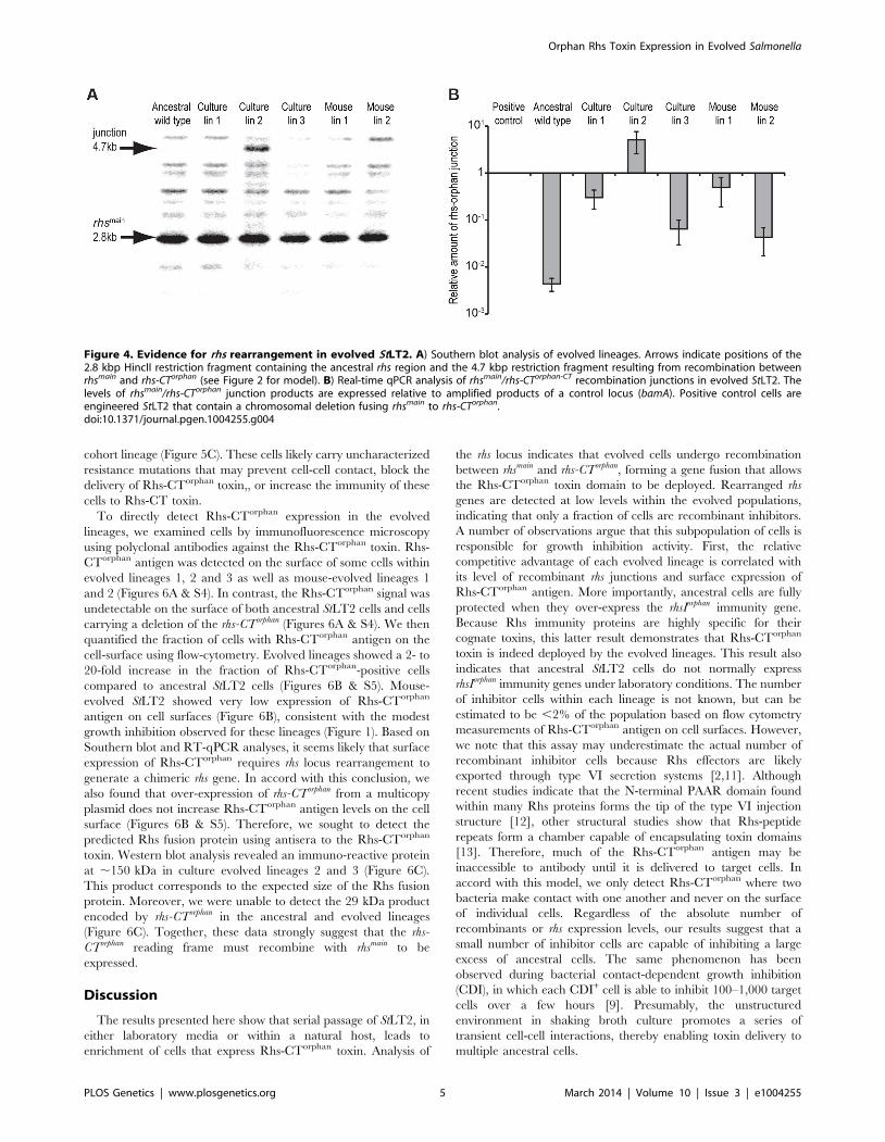
analyzed chromosomal DNA from evolved and ancestral lineages
by Southern blot. DNA was digested with HincII, which cleaves
between the rhsImain and rhs-CTorphan coding sequences, and probed
with a labeled DNA fragment that specifically hybridizes to rhsmain
(Figure 2). We detected a unique junction fragment representing
fusion of rhs-CTorphan to the upstream rhsmain gene in StLT2 lineage
2, which displayed the highest level of growth inhibition of all
lineages (Figures 1 & 4A). The wild-type rhs locus was also detected
in lineage 2 (Figure 4A), which is consistent with rhs region
amplification, but may also indicate distinct populations of
recombinant and non-recombinant cells. Orphan rhs recombi-
nants were not detected in the other evolved lineages by Southern
blot analysis (Figure 4A). Because the growth inhibition phenotype
varied in magnitude between the different evolved strains, it is
possible that only a fraction of the evolved StLT2 cells are rhs
recombinants. If so, then the proportion of recombined rhs loci in
the DNA sample may be below the detection limit of Southern
analysis. Therefore we analyzed each evolved lineage with
quantitative real-time PCR (qPCR) to measure the relative levels
of rhsmain-rhsorphan junction sequences. All five of the evolved lineages
contained 10- to 1,000-fold more rhsmain-rhsorphan junction than
ancestral StLT2 (Figure 4B), consistent with the ability of these
strains to deploy Rhs-CTorphan toxin.
Author Summary
Salmonella Typhimurium is a bacterium that causesintestinal diseases in a number of animals includinghumans. In mice, this pathogen invades tissues, causingsymptoms similar to typhoid fever. In an effort tounderstand the evolution of this pathogen, we grew S.Typhimurium in either liquid broth or in mice for manygenerations and examined the resulting ‘‘evolved’’ strainsto determine if they were different from the original‘‘parent’’ culture. We found that many of these evolvedstrains inhibited the growth of the parent after they weremixed together, and that this growth inhibition requiresthat the evolved and parental cells are in close contact.Genetic analysis showed that this contact-dependentgrowth inhibition requires Rhs protein, which has a toxictip. Salmonella is normally resistant to its Rhs toxinbecause it also produces an immunity protein that blockstoxin activity. However, evolved cells have undergone aDNA rearrangement that allows them to express adifferent Rhs toxic tip that inhibits growth of the parentalcells, which lack immunity to it. This allows the evolvedcells to outgrow the original parental cells. Our workindicates that populations of Salmonella are dynamic, withindividuals battling with each other for dominance.
Orphan Rhs Toxin Expression in Evolved Salmonella
PLOS Genetics | www.plosgenetics.org 2 March 2014 | Volume 10 | Issue 3 | e1004255

Figure 2. Proposed mechanism of rhs locus rearrangement in evolved inhibitor cells. The StLT2 rhs locus contains a full-length rhsmain
gene (STM0291) and an rhs-CTorphan gene fragment (STM0292) together with associated rhsI immunity genes. Duplication of the rhs locus wouldprovide the opportunity for subsequent homologous recombination between the 522 bp region of near sequence identity (95%) between rhsmain
and rhs-CTorphan (depicted as diagonal hatched regions). The binding site of the Southern blot hybridization probe is indicated by the double ochrebars. Primer binding sites for PCR amplification of the rhsmain/rhs-CTorphan junction are indicated by convergent horizontal arrows.doi:10.1371/journal.pgen.1004255.g002
Figure 1. Evolved StLT2 cells inhibit the growth of ancestral cells. A) The indicated evolved StLT2 lineages were co-cultured with theancestral strain for 24 h in broth. Viable cell counts for each population were determined as colony forming units and these data were used tocalculate the competitive index as described in Methods. Each evolved lineage was competed against ancestral cells (black bars), ancestral cellsoverexpressing rhsImain (light grey bars) and ancestral cells overexpressing rhsIorphan (white bars). Reported values represent the mean 6 SEM for atleast three independent experiments. B) The growth inhibition activity of evolved StLT2 requires cell-cell contact. Evolved cells were co-cultured withancestral cells in adjacent wells of a trans-well incubation chamber. Culture chambers were separated by membranes containing 0.4 mm or 8 mmpores as indicated. Reported competitive indices represent the mean 6 SEM for three independent experiments.doi:10.1371/journal.pgen.1004255.g001
Orphan Rhs Toxin Expression in Evolved Salmonella
PLOS Genetics | www.plosgenetics.org 3 March 2014 | Volume 10 | Issue 3 | e1004255

Because only a fraction of the passaged cells appeared to display
growth inhibitory activity, we asked whether inhibitor-cell clones
could be isolated from each population. As a control, we first
isolated colonies from an overnight culture of the ancestral strain
and tested these clones for growth inhibition activity. None of the
ten ancestral clones tested were inhibitory, suggesting that the
proposed rhs rearrangements occur at low frequency. By contrast,
approximately 30–90% of the clones isolated from the culture-
evolved lineages and ,20% of the clones from mouse-evolved
lineage 1 showed inhibition activity against ancestral cells
(Figure 5A). However, no inhibitor clones were isolated from
mouse-evolved lineage 2 (Figure 5A). Strikingly, the inhibition
activity of these clones varied considerably. For example,
competitive index values ranged from 1021 to 1025 for
competitions between ancestral cells and inhibitory clones isolated
from evolved lineage 2 (Figure S3). Although their potencies
varied, it appears that each inhibitor-cell clone deployed the Rhs-
CTorphan toxin because ancestral cells could be protected through
over-expression of rhsIorphan, but not rhsImain (Figure 5B). The
presence of DNA fragments corresponding to both ancestral and
recombinant rhs loci in lineage 2 (Figure 4A) suggests that either
the rhs region was duplicated or there are distinct populations of
recombinant and non-recombinant cells. In the latter case, single
colonies isolated from the inhibitory lineages would contain only
the rhs- rhs-CTorphan junction and not the rhs-CTmain sequence.
However, PCR analysis of the single colonies with inhibitory
activity in Figure 5B showed that each contained both ancestral
and recombinant rhs loci. In addition, sequence analysis of the
recombinant PCR product verified that recombination occurred
between the regions of homology shared by rhsmain and rhs-CTorphan.
Together, these data demonstrate that the evolved populations are
heterogeneous with respect to Rhs-CTorphan mediated inhibition
activity. Furthermore, these results suggest that the inhibition
phenotype of a given culture may be due entirely to a minor
subpopulation of potent inhibitor cells.
Because inhibitor cells represent a subpopulation in the evolved
cultures, the other non-recombinant cells in the cohort are
presumably resistant to the Rhs-CTorphan toxin. To test this
hypothesis, we isolated non-inhibitory clones from each of the
evolved cultures and tested them in competition co-cultures
against their respective evolved lineages. As predicted, each of the
non-inhibitory clones was either fully- or partially-resistant to its
Figure 3. The StLT2 rhs locus encodes cognate toxin/immunity pairs. A) Expression of rhs-CTmain or rhs-CTorphan inhibits the growth of StLT2cells. Expression the plasmid-borne rhs-CT genes was induced by addition of L-arabinose at the indicated time, and cell growth was monitored bymeasuring the optical density at 600 nm (OD600). The cells also co-expressed either rhsImain (dark squares) or rhsIorphan immunity genes (light greydiamonds) from IPTG-inducible promoters. Growth is compared to control cells that carry the empty vector plasmids (triangles). B) Expression of rhs-CTmain or rhs-CTorphan inhibits the growth of E.coli cells. The rhs-CT and rhsI genes were expressed in E. coli cells from the same plasmids described inpanel A, and growth monitored by measuring the OD600 of the cultures.doi:10.1371/journal.pgen.1004255.g003
Orphan Rhs Toxin Expression in Evolved Salmonella
PLOS Genetics | www.plosgenetics.org 4 March 2014 | Volume 10 | Issue 3 | e1004255
cohort lineage (Figure 5C). These cells likely carry uncharacterized
resistance mutations that may prevent cell-cell contact, block the
delivery of Rhs-CTorphan toxin,, or increase the immunity of these
cells to Rhs-CT toxin.
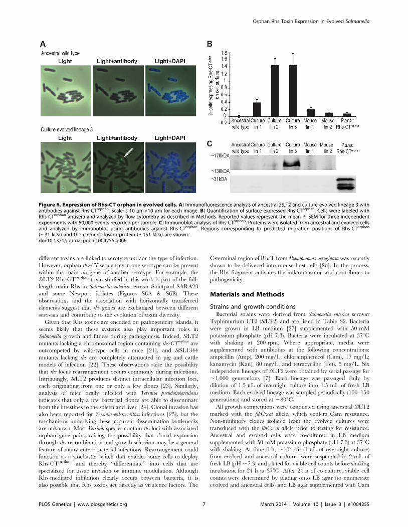
To directly detect Rhs-CTorphan expression in the evolved
lineages, we examined cells by immunofluorescence microscopy
using polyclonal antibodies against the Rhs-CTorphan toxin. Rhs-
CTorphan antigen was detected on the surface of some cells within
evolved lineages 1, 2 and 3 as well as mouse-evolved lineages 1
and 2 (Figures 6A & S4). In contrast, the Rhs-CTorphan signal was
undetectable on the surface of both ancestral StLT2 cells and cells
carrying a deletion of the rhs-CTorphan (Figures 6A & S4). We then
quantified the fraction of cells with Rhs-CTorphan antigen on the
cell-surface using flow-cytometry. Evolved lineages showed a 2- to
20-fold increase in the fraction of Rhs-CTorphan-positive cells
compared to ancestral StLT2 cells (Figures 6B & S5). Mouse-
evolved StLT2 showed very low expression of Rhs-CTorphan
antigen on cell surfaces (Figure 6B), consistent with the modest
growth inhibition observed for these lineages (Figure 1). Based on
Southern blot and RT-qPCR analyses, it seems likely that surface
expression of Rhs-CTorphan requires rhs locus rearrangement to
generate a chimeric rhs gene. In accord with this conclusion, we
also found that over-expression of rhs-CTorphan from a multicopy
plasmid does not increase Rhs-CTorphan antigen levels on the cell
surface (Figures 6B & S5). Therefore, we sought to detect the
predicted Rhs fusion protein using antisera to the Rhs-CTorphan
toxin. Western blot analysis revealed an immuno-reactive protein
at ,150 kDa in culture evolved lineages 2 and 3 (Figure 6C).
This product corresponds to the expected size of the Rhs fusion
protein. Moreover, we were unable to detect the 29 kDa product
encoded by rhs-CTorphan in the ancestral and evolved lineages
(Figure 6C). Together, these data strongly suggest that the rhs-
CTorphan reading frame must recombine with rhsmain to be
expressed.
Discussion
The results presented here show that serial passage of StLT2, in
either laboratory media or within a natural host, leads to
enrichment of cells that express Rhs-CTorphan toxin. Analysis of
the rhs locus indicates that evolved cells undergo recombination
between rhsmain and rhs-CTorphan, forming a gene fusion that allows
the Rhs-CTorphan toxin domain to be deployed. Rearranged rhs
genes are detected at low levels within the evolved populations,
indicating that only a fraction of cells are recombinant inhibitors.
A number of observations argue that this subpopulation of cells is
responsible for growth inhibition activity. First, the relative
competitive advantage of each evolved lineage is correlated with
its level of recombinant rhs junctions and surface expression of
Rhs-CTorphan antigen. More importantly, ancestral cells are fully
protected when they over-express the rhsIorphan immunity gene.
Because Rhs immunity proteins are highly specific for their
cognate toxins, this latter result demonstrates that Rhs-CTorphan
toxin is indeed deployed by the evolved lineages. This result also
indicates that ancestral StLT2 cells do not normally express
rhsIorphan immunity genes under laboratory conditions. The number
of inhibitor cells within each lineage is not known, but can be
estimated to be ,2% of the population based on flow cytometry
measurements of Rhs-CTorphan antigen on cell surfaces. However,
we note that this assay may underestimate the actual number of
recombinant inhibitor cells because Rhs effectors are likely
exported through type VI secretion systems [2,11]. Although
recent studies indicate that the N-terminal PAAR domain found
within many Rhs proteins forms the tip of the type VI injection
structure [12], other structural studies show that Rhs-peptide
repeats form a chamber capable of encapsulating toxin domains
[13]. Therefore, much of the Rhs-CTorphan antigen may be
inaccessible to antibody until it is delivered to target cells. In
accord with this model, we only detect Rhs-CTorphan where two
bacteria make contact with one another and never on the surface
of individual cells. Regardless of the absolute number of
recombinants or rhs expression levels, our results suggest that a
small number of inhibitor cells are capable of inhibiting a large
excess of ancestral cells. The same phenomenon has been
observed during bacterial contact-dependent growth inhibition
(CDI), in which each CDI+ cell is able to inhibit 100–1,000 target
cells over a few hours [9]. Presumably, the unstructured
environment in shaking broth culture promotes a series of
transient cell-cell interactions, thereby enabling toxin delivery to
multiple ancestral cells.
Figure 4. Evidence for rhs rearrangement in evolved StLT2. A) Southern blot analysis of evolved lineages. Arrows indicate positions of the2.8 kbp HincII restriction fragment containing the ancestral rhs region and the 4.7 kbp restriction fragment resulting from recombination betweenrhsmain and rhs-CTorphan (see Figure 2 for model). B) Real-time qPCR analysis of rhsmain/rhs-CTorphan-CT recombination junctions in evolved StLT2. Thelevels of rhsmain/rhs-CTorphan junction products are expressed relative to amplified products of a control locus (bamA). Positive control cells areengineered StLT2 that contain a chromosomal deletion fusing rhsmain to rhs-CTorphan.doi:10.1371/journal.pgen.1004255.g004
Orphan Rhs Toxin Expression in Evolved Salmonella
PLOS Genetics | www.plosgenetics.org 5 March 2014 | Volume 10 | Issue 3 | e1004255

Chromosomal duplications and amplifications occur frequently
in bacteria, typically at rates of about 0.1% per generation for any
given locus [14,15]. However, there is a cost to maintaining
amplified regions, and gene duplications are lost during segrega-
tion at frequencies up to 10% per generation [16,17]. Therefore,
positive selection is required to retain multiple gene copies. If the
amplified region can be stabilized, then the additional gene copy
can diverge towards a new function, thus providing a mechanism
for evolution [16,18]. Rearrangement of rhs loci represents a
previously unrecognized mechanism for bacteria to exploit
chromosomal amplifications for adaptation. We propose that,
subsequent to duplication, homologous recombination occurs
between rhsmain and rhs-CTorphan to generate a novel chimeric rhs
element. This recombination would necessarily delete one copy of
the rhsImain, but the other copy would remain and ensure that
recombinant cells retain immunity to the Rhsmain toxin should it
be deployed by neighboring non-recombinant siblings. This model
also predicts that evolved recombinant cells could undergo
homologous recombination to restore the original rhs locus (see
Figure 2, reverse of the duplication step). Thus, rhs rearrangement
could be exploited transiently under conditions where it confers a
selective advantage, but rapidly revert back to the ancestral
genotype as environmental circumstances dictate.
Analysis of over 150 Salmonella genomes shows that rhs-CT toxin
sequences are diverse with at least 57 distinct sequence types
(Figure S6A & Table S1). This is a common feature of rhs genes in
other bacteria as well and suggests that Rhs mediates inter-strain
competition. All Salmonella serovars contain at least one rhs gene,
located on pathogenicity islands SPI-6 or SPI-19 [19,20].
Approximately 50% of these serovars contain at least one
predicted rhs orphan sequence, with some strains containing as
many as eleven modules. There is generally high conservation of
Rhs-CTmain and Rhs-CTorphan. sequences within a given serotype.
For example, all sequenced Typhi isolates contain the same Rhs-
CTmain and Rhs-CTorphan. sequences, whereas these CT sequence
types are only found in one other serotype, thus suggesting that
Figure 5. A subpopulation of evolved cells has growth inhibition activity. A) Two sets of independent clones were isolated twice from theevolved lineages and tested for growth inhibition activity against ancestral cells. The percentage of evolved clones with inhibition activity is shown.Reported values represent the mean 6 SEM for at least two independent experiments. B) Growth inhibition activity of isolated evolved clones. Clonesfrom each evolved lineage were competed against ancestral cells (black bars), ancestral cells overexpressing rhsImain (light grey bars) and ancestralcells overexpressing rhsIorphan (white bars). Reported values represent the mean 6 SEM for at least three independent experiments. C) Growthinhibition activity of evolved lineages towards non-inhibitory clones. Evolved lineages were co-cultured with ancestral cells (black bars) and two non-inhibitory clones isolated from the evolved cultures (light grey and white bars). Competitive indices represent the mean 6 SEM for at least threeindependent experiments.doi:10.1371/journal.pgen.1004255.g005
Orphan Rhs Toxin Expression in Evolved Salmonella
PLOS Genetics | www.plosgenetics.org 6 March 2014 | Volume 10 | Issue 3 | e1004255
different toxins are linked to serotype and/or the type of infection.
However, orphan rhs-CT sequences in one serotype can be present
within the main rhs gene of another serotype. For example, the
StLT2 Rhs-CTorphan toxin studied in this work is part of the full-
length main Rhs in Salmonella enterica serovar Saintpaul SARA23
and some Newport isolates (Figures S6A & S6B). These
observations and the association with horizontally transferred
elements suggest that rhs genes are exchanged between different
serovars and contribute to the evolution of toxin diversity.
Given that Rhs toxins are encoded on pathogenicity islands, it
seems likely that these systems also play important roles in
Salmonella growth and fitness during pathogenesis. Indeed, StLT2
mutants lacking a chromosomal region containing rhs-CTorphan are
outcompeted by wild-type cells in mice [21], and StSL1344
mutants lacking rhs are completely attenuated in pig and cattle
models of infection [22]. These observations raise the possibility
that rhs locus rearrangement occurs commonly during infections.
Intriguingly, StLT2 produces distinct intracellular infection foci,
each originating from one or only a few clones [23]. Similarly,
analysis of mice orally infected with Yersinia pseudotuberculosis
indicates that only a few bacterial clones are able to disseminate
from the intestines to the spleen and liver [24]. Clonal invasion has
also been reported for Yersinia enterocolitica infections [25], but the
mechanisms underlying these apparent dissemination bottlenecks
are unknown. Most Yersinia species contain rhs loci with associated
orphan gene pairs, raising the possibility that clonal expansion
through rhs recombination and growth selection may be a general
feature of many enterobacterial infections. Rearrangement could
function as a stochastic switch that enables some cells to deploy
Rhs-CTorphan and thereby ‘‘differentiate’’ into cells that are
specialized for tissue invasion or immune modulation. Although
Rhs-mediated inhibition clearly occurs between bacteria, it is
also possible that Rhs toxins act directly as virulence factors. The
C-terminal region of RhsT from Pseudomonas aeruginosa was recently
shown to be delivered into mouse host cells [26]. In the process,
the Rhs fragment activates the inflammasome and contributes to
pathogenicity.
Materials and Methods
Strains and growth conditionsBacterial strains were derived from Salmonella enterica serovar
Typhimurium LT2 (StLT2) and are listed in Table S2. Bacteria
were grown in LB medium [27] supplemented with 50 mM
potassium phosphate (pH 7.3). Bacteria were incubated at 37uCwith shaking at 200 rpm. Where appropriate, media were
supplemented with antibiotics at the following concentrations:
ampicillin (Amp), 200 mg/L; chloramphenicol (Cam), 17 mg/L;
kanamycin (Kan), 80 mg/L; and tetracycline (Tet), 5 mg/L. Six
independent lineages of StLT2 were obtained by serial passage for
,1,000 generations [7]. Each lineage was passaged daily by
dilution of 1.5 mL of overnight culture into 1.5 mL of fresh LB
medium. Each evolved lineage was sampled periodically (100–150
generations) and stored at 280uC.
All growth competitions were conducted using ancestral StLT2
marked with the flhC::cat allele, which confers Cam resistance.
Non-inhibitory clones isolated from the evolved cultures were
transduced with the flhC::cat allele prior to testing for resistance.
Ancestral and evolved cells were co-cultured in LB medium
supplemented with 50 mM potassium phosphate (pH 7.3) at 37uCwith shaking. At time 0 h, ,106 cfu (1 mL of overnight culture)
from evolved and ancestral cultures were suspended in 2 mL of
fresh LB (pH ,7.3) and plated for viable cell counts before shaking
incubation for 24 h at 37uC. After 24 h of co-culture, viable cell
counts were determined by plating onto LB agar (to enumerate
evolved and ancestral cells) and LB agar supplemented with Cam
Figure 6. Expression of Rhs-CT orphan in evolved cells. A) Immunofluorescence analysis of ancestral StLT2 and culture-evolved lineage 3 withantibodies against Rhs-CTorphan. Scale is 10 mm610 mm for each image. B) Quantification of surface-expressed Rhs-CTorphan. Cells were labeled withRhs-CTorphan antisera and analyzed by flow cytometry as described in Methods. Reported values represent the mean 6 SEM for three independentexperiments with 50,000 events recorded per sample. C) Immunoblot analysis of Rhs-CTorphan. Proteins were isolated from ancestral and evolved cellsand analyzed by immunoblot using antibodies against Rhs-CTorphan. Regions corresponding to predicted migration positions of Rhs-CTorphan
(,31 kDa) and the chimeric fusion protein (,151 kDa) are shown.doi:10.1371/journal.pgen.1004255.g006
Orphan Rhs Toxin Expression in Evolved Salmonella
PLOS Genetics | www.plosgenetics.org 7 March 2014 | Volume 10 | Issue 3 | e1004255

(to enumerate ancestral cells). The competitive index was
calculated as the ratio of ancestral:evolved cells at time 24 h
divided by the cell ratio at 0 h. Ancestral StLT2 flhC::cat cells were
also supplemented with either rhsImain or rhsIorphan on the
chromosome under control of the lac promoter and on plasmid
pBR322 under the tet promoter. Chromosomal rhsIorphan and
plasmid-borne rhsIorphan individually provided partial protection
against the evolved lineages (data not shown), but both copies were
required for full immunity. Proximity-dependence of growth
inhibition was determined as described previously [9]. Cells were
grown to OD600 ,0.3, then transferred to a trans-well culture plate
(BD diagnostics) that separates the two populations with filter
containing 0.4 mm (no-contact) or 8.0 mm (contact) pores. Trans-
well culture plates were seeded at an evolved:ancestral cell ratio of
1:1 and incubated at 37uC with shaking for 24 h. Cultures were
then plated onto selective media to determine viable cell counts
and to calculate competitive indices.
Construction of plasmids and chromosomal insertsAll oligonucleotides used in this study are presented in Table S3.
The rhsImain and rhsIorphan genes were amplified from ancestral
StLT2 chromosomal DNA using oligonucleotides 2337/2338 and
2340/2544 (respectively) and ligated to plasmid pBR322 using
EcoRV and SalI restriction sites. The immunity genes were also
placed under the lac promoter at the glmS locus using bacterio-
phage l Red-mediated recombination [28]. Integration constructs
containing rhsI genes flanked by a Kan-resistance cassette and
glmS-derived homology regions were constructed by overlapping
end-PCR as described previously [29]. The following primer pairs
were used to amplify: upstream glmS homology (2666/2676), lac
promoter (2677/2678 for rhsImain and 2677/2682 for rhsIorphan),
rhsImain (2679/2680) or rhsIorphan (2683/2684), Kan-resistance
cassette (2618/2619) and downstream glmS homology (2681/
2667). The final PCR product was electroporated into StLT2 cells
that express Red recombinase proteins, and transformants were
selected on LB supplemented with Kan. Integrated immunity
genes were verified by PCR analysis using primers 2666/2667 and
subsequent DNA sequencing. The flhC::cat and STM0292::kan
alleles were generated by PCR using primers 2436/2437 and
2410/2490 to amplify the cat/kan cassettes of plasmids pKD3 and
pKD4, respectively. Each PCR product was integrated into the
StLT2 chromosome by Red-mediated recombination.
To evaluate toxin activity and the specificity of immunity,
individual rhs-CT and rhsI sequences were cloned under the control
of inducible promoters on compatible plasmids. The rhs-CTmain
and rhs-CTorphan coding sequences were amplified with primers Sty-
rhs(E1203)-Nco/Sty-rhs-Xho and Sty-rhs(E1203)-Nco/Sty-orph-
rhs-Xho (respectively) and ligated to plasmid pCH450 [30] using
NcoI and XhoI restriction sites. The rhsImain and rhsIorphan genes
were amplified and ligated to a derivative of plasmid pTrc99A
using KpnI and XhoI restriction sites. Rhs-CTorphan was expressed
and purified as a non-toxic variant fused to His6-tagged
thioredoxin. The his6-trxA sequence was amplified from plasmid
pSH21P::trxA [31] using primers pET-Sph and trxA-Bam-TEV-
Kpn. The product was digested with SphI/BamHI and ligated to
plasmid pET21b to generate plasmid pSH21P::trxA-TEV. The
coding sequences for Rhs-CTorphan residues 112–247 and RhsIor-
phan were amplified using primers Sty-rhs(D1225)-Kpn/Sty-orph-
rhsI-Xho) and the His208Ala mutation made by mega-primer PCR
using oligonucleotide Sty-CTo1-H208A. The final product was
digested with KpnI/XhoI and ligated to plasmid pSH21P::trxA-
TEV to generate plasmid pCH10068. The resulting construct was
used to overproduce His6-TrxA-Rhs-CT(H208A)orphan fusion
protein.
Chromosomal DNA analysisChromosomal DNAs were isolated using the Sigma genomic
DNA kit and digested with HincII restriction endonuclease.
Digested DNAs were resolved by electrophoresis on 0.7% agarose
gels at 34V for 15 h and blotted onto nylon membranes by
capillary transfer. A probe to nucleotides 2969–3128 of rhsmain was
generated by PCR using oligos 2226/2227 and labeled with [32P]-
labeled using the Prime-It Random Primer Labeling Kit (Agilent
Technologies). Southern blots were visualized by phosphor
imaging. Fragment sizes were calculated using a standard curve
based on HindIII digested l ladder (New England Biolabs, USA)
run on the same gel. The proportion of rhs recombination
junctions was determined by quantitative real-time PCR (qPCR)
using oligonucleotides 2226/2231 using the cycle threshold Ct-
value method according to the manufacturer (Bio-Rad). Fluores-
cence was monitored on-line using the MyIQ iCycler real-time
PCR system (Bio-Rad). The rhs-rhsorphan junction DNA levels were
calculated relative to bamA DNA (oligos 1981/1990) in each
sample and normalized to the level of junction DNA in ancestral
cells.
Antiserum preparation and immunoblot analysisHis6-TrxA-Rhs-CT(H208A)orphan fusion protein was overpro-
duced in E. coli CH2016 and purified by Ni2+-affinity chroma-
tography as described [32]. The Rhs-CT(H208A)orphan domain
was released by TEV protease digestion and used for antiserum
production in rabbits (CoCalico Biologicals). Non-specific anti-
bodies were removed by incubation with carbonyl-diimidazole-
activated agarose beads linked to soluble protein from E. coli
strain CH2016 [33]. Briefly, protein-linked beads were resus-
pended in 0.5 mL of antiserum (1:5 dilution) and mixed by
rotation for 1 h at room temperature followed by additional
incubation for 3 h at 4uC. This process was repeated at least four
times with fresh beads.
Evolved lineages were grown to mid-log phase in LB medium
supplemented with 50 mM potassium phosphate (pH 7.3) and
cells were collected by centrifugation and frozen at 280uC. Cell
pellets were resuspended in NuPage LDS-sample buffer (Invitro-
gen) at 70uC and treated with benzonase to degrade nucleic acids.
Cell lysates were run on 3–7% NuPage Tris-acetate gradient gels
(Novex) for the detection of the Rhsmain-Rhs-CTorphan chimera, or
on 4–10% Precise Tris-glycine gradient gels (Thermo Scientific) to
detect Rhs-CTorphan. Gels were electrotransferred to nitrocellulose
membranes and the blots incubated with polyclonal antisera
against Rhs-CTorphan (1:1,000 dilution) and secondary anti-rabbit
800CW antiserum (1:10,000 dilution). Immunoblots were visual-
ized using an Odyssey CLx Infrared Imaging System (LI-COR).
Immunofluorescence microscopy and flow cytometryCells were incubated overnight with 4% formaldehyde in
0.15 M phosphate buffered saline (PBS, pH = 7.2). Cells were
washed three times with PBS and incubated with polyclonal
antibodies to Rhs-CTorphan (1:50 dilution) for 30 min. Cells were
washed with PBS before incubation with secondary anti-rabbit
Alexa-Fluor480 antibodies (1:500 dilution) (Invitrogen) for 30 min
on ice. After washing with PBS, cells were applied to poly-D-lysine
coated slides, treated with Fluoro-gel II/DAPI (Electron Micros-
copy Sciences) and visualized by fluorescence microscopy. The
fraction of evolved cells expressing Rhs-CTorphan on the surface
was determined by flow cytometry. Antibody-labeled cells were
analyzed (50,000 events for each sample) with an Accuri C6 flow
cytometer with gates set to include bacteria-sized particles. StLT2
Drhs-CTorphan cells were used to assess non-specific binding of
the Rhs-CTorphan antisera. The fraction of cells with surface
Orphan Rhs Toxin Expression in Evolved Salmonella
PLOS Genetics | www.plosgenetics.org 8 March 2014 | Volume 10 | Issue 3 | e1004255

Rhs-CTorphan antigen was calculated as the ratio of green
fluorescent particles in the population after subtracting back-
ground fluorescence observed with StLT2 Drhs-CTorphan cells.
Supporting Information
Figure S1 Evolved StLT2 outcompete ancestral cells. The
indicated evolved StLT2 lineages were co-cultured with the
ancestral strain for 24 h in broth. Viable cell counts for each
population were determined as colony forming units and these
data were used to calculate the competitive index as described in
Methods. A) Culture-evolved lineages after 1000-generations of
growth in LB were competed against ancestral wild type cells. B)
Mouse evolved lineages after 150-generations of growth in mice
were competed against ancestral cells. Reported values represent
the mean 6 SEM for at least three independent experiments.
(PDF)
Figure S2 Growth rates of ancestral and evolved StLT2 strains.
The growth rates of evolved lineages are expressed relative to the
growth rate of ancestral cells, which was set to 1. Reported values
represent the mean 6 SEM for at least three independent
experiments.
(PDF)
Figure S3 Variability of growth inhibitory activities of evolved
inhibitor clones. Culture-evolved lineages were streaked on LB
agar plates to obtain individual colonies. Ten colonies from each
lineage were competed against the ancestral StLT2 strain as
described for Figure 1. Reported values represent the mean 6
SEM for at least two independent experiments. The hatched lines
in each panel indicate an arbitrary cut-off (C.I. = 1021) for
whether a clone was considered to express growth inhibitory
activity or not. Clones with C.I. error bars that cross the hatched
line were considered to express growth inhibitory activity.
(PDF)
Figure S4 Evolved cells express Rhsorphan-CT on the cell surface.
Immunofluorescence analysis of ancestral cells and evolved
lineages using antibodies against Rhs-CTorphan. Non-permeabi-
lized cells were fluorescently labeled with antibodies to Rhs-
CTorphan as described in Methods. The Drhs-CTorphan cells carry a
deletion of the rhs-CTorphan gene. Para::Rhs-CTorphan carry a
plasmid encoded Rhs-CTorphan under an arabinose inducible
promoter. Scale is 10 mm610 mm for each image. Cells were
grown under inducing conditions as described in Methods.
(PDF)
Figure S5 Representative flow cytometry data for quantitation
of the fraction of cells expressing cell surface Rhsorphan-CT. Non-
permeabilized cells were fluorescently labeled using antibodies
against Rhs-CTorphan protein as described in Methods. The Drhs-
CTorphan cells carry a deletion of the rhs-CTorphan gene. Para::Rhs-
CTorphan carry a plasmid encoded Rhs-CTorphan under an
arabinose inducible promoter. Cells were grown under inducing
conditions as described in Methods. Cells (50,000 events per
sample) were analyzed using an Accuri C6 flow cytometer as
described in Methods.
(PDF)
Figure S6 Rhs-CT sequence types from Salmonella isolates. A)
222 Salmonella rhs gene sequence from over 150 Salmonella isolates
encode 57 different predicted Rhs-CT toxin sequences. Sequences
are grouped together according to sequence homology, with
Taylor coloring for amino acids. Sequence starts at the conserved
DPxGL (boxed) demarking the beginning of the Rhs-CT. Orphan
toxins are indicted by lowercase ‘‘o’’ in the sequence identifier.
Numbers following the ‘‘o’’ indicate the position of the
corresponding gene in the orphan cluster. B) Orphan Rhs-CT
toxins are found on full-length Rhs proteins. The StLT2 rhs-
CTorphan coding sequence is fused to rhsmain in Salmonella serovar
Saintpaul str. SARA23 as well as several serovar Newport strains
(see panel A).
(PDF)
Table S1 Salmonella enterica strains used for rhs-CT analysis.
(XLSX)
Table S2 Strains and plasmids used in this study.
(DOCX)
Table S3 Oligonucleotides used in this study.
(DOCX)
Author Contributions
Conceived and designed the experiments: SK DIA CSH DAL. Performed
the experiments: SK FGS LS JSW BAB SJP. Analyzed the data: SK FGS
LS JSW BAB SJP DIA DAL CSH. Contributed reagents/materials/
analysis tools: SK FGS LS JSW BAB SJP. Wrote the paper: SK CSH DAL.
References
1. Lopez D, Vlamakis H, Kolter R (2010) Biofilms. Cold Spring Harb Perspect Biol
2: a000398.
2. Koskiniemi S, Lamoureux JG, Nikolakakis KC, t’Kint de Roodenbeke C,
Kaplan MD, et al. (2013) Rhs proteins from diverse bacteria mediate
intercellular competition. Proc Natl Acad Sci U S A 110: 7032–7037.
3. Hill CW (1999) Large genomic sequence repetitions in bacteria: lessons from
rRNA operons and Rhs elements. Res Microbiol 150: 665–674.
4. Lin RJ, Capage M, Hill CW (1984) A repetitive DNA sequence, rhs, responsible
for duplications within the Escherichia coli K-12 chromosome. J Mol Biol 177: 1–
18.
5. Foster SJ (1993) Molecular analysis of three major wall-associated proteins of
Bacillus subtilis 168: evidence for processing of the product of a gene encoding a
258 kDa precursor two-domain ligand-binding protein. Mol Microbiol 8: 299–
310.
6. Poole SJ, Diner EJ, Aoki SK, Braaten BA, t’Kint de Roodenbeke C, et al. (2011)
Identification of functional toxin/immunity genes linked to contact-dependent
growth inhibition (CDI) and rearrangement hotspot (Rhs) systems. PLoS Genet
7: e1002217.
7. Koskiniemi S, Sun S, Berg OG, Andersson DI (2012) Selection-driven gene loss
in bacteria. PLoS Genet 8: e1002787.
8. Nilsson AI, Kugelberg E, Berg OG, Andersson DI (2004) Experimental
adaptation of Salmonella typhimurium to mice. Genetics 168: 1119–1130.
9. Aoki SK, Pamma R, Hernday AD, Bickham JE, Braaten BA, et al. (2005)
Contact-dependent inhibition of growth in Escherichia coli. Science 309: 1245–
1248.
10. Lovett ST, Hurley RL, Sutera VA, Jr., Aubuchon RH, Lebedeva MA (2002)
Crossing over between regions of limited homology in Escherichia coli. RecA-
dependent and RecA-independent pathways. Genetics 160: 851–859.
11. Silverman JM, Brunet YR, Cascales E, Mougous JD (2012) Structure and
regulation of the type VI secretion system. Annu Rev Microbiol 66: 453–472.
12. Shneider MM, Buth SA, Ho BT, Basler M, Mekalanos JJ, et al. (2013) PAAR-
repeat proteins sharpen and diversify the type VI secretion system spike. Nature
500: 350–353.
13. Busby JN, Panjikar S, Landsberg MJ, Hurst MR, Lott JS (2013) The BC
component of ABC toxins is an RHS-repeat-containing protein encapsulation
device. Nature 501: 547–550.
14. Reams AB, Kofoid E, Savageau M, Roth JR (2010) Duplication frequency in a
population of Salmonella enterica rapidly approaches steady state with or without
recombination. Genetics 184: 1077–1094.
15. Anderson P, Roth J (1981) Spontaneous tandem genetic duplications in
Salmonella typhimurium arise by unequal recombination between rRNA (rrn)
cistrons. Proc Natl Acad Sci U S A 78: 3113–3117.
16. Bergthorsson U, Andersson DI, Roth JR (2007) Ohno’s dilemma: evolution of
new genes under continuous selection. Proc Natl Acad Sci U S A 104: 17004–
17009.
17. Pettersson ME, Sun S, Andersson DI, Berg OG (2009) Evolution of new gene
functions: simulation and analysis of the amplification model. Genetica 135:
309–324.
18. Nasvall J, Sun L, Roth JR, Andersson DI (2012) Real-time evolution of new
genes by innovation, amplification, and divergence. Science 338: 384–387.
Orphan Rhs Toxin Expression in Evolved Salmonella
PLOS Genetics | www.plosgenetics.org 9 March 2014 | Volume 10 | Issue 3 | e1004255

19. Folkesson A, Lofdahl S, Normark S (2002) The Salmonella enterica subspecies I
specific centisome 7 genomic island encodes novel protein families present inbacteria living in close contact with eukaryotic cells. Res Microbiol 153: 537–545.
20. Blondel CJ, Jimenez JC, Contreras I, Santiviago CA (2009) Comparative
genomic analysis uncovers 3 novel loci encoding type six secretion systemsdifferentially distributed in Salmonella serotypes. BMC Genomics 10: 354.
21. Mulder DT, Cooper CA, Coombes BK (2012) Type VI secretion system-associated gene clusters contribute to pathogenesis of Salmonella enterica serovar
Typhimurium. Infect Immun 80: 1996–2007.
22. Chaudhuri RR, Morgan E, Peters SE, Pleasance SJ, Hudson DL, et al. (2013)Comprehensive assignment of roles for Salmonella typhimurium genes in intestinal
colonization of food-producing animals. PLoS Genet 9: e1003456.23. Sheppard M, Webb C, Heath F, Mallows V, Emilianus R, et al. (2003)
Dynamics of bacterial growth and distribution within the liver during Salmonella
infection. Cell Microbiol 5: 593–600.
24. Barnes PD, Bergman MA, Mecsas J, Isberg RR (2006) Yersinia pseudotuberculosis
disseminates directly from a replicating bacterial pool in the intestine. J Exp Med203: 1591–1601.
25. Oellerich MF, Jacobi CA, Freund S, Niedung K, Bach A, et al. (2007) Yersinia
enterocolitica infection of mice reveals clonal invasion and abscess formation. Infect
Immun 75: 3802–3811.
26. Kung VL, Khare S, Stehlik C, Bacon EM, Hughes AJ, et al. (2012) An rhs gene
of Pseudomonas aeruginosa encodes a virulence protein that activates theinflammasome. Proc Natl Acad Sci U S A 109: 1275–1280.
27. Scott JR (1968) Genetic studies on bacteriophage P1. Virology 36: 564–
574.28. Datsenko KA, Wanner BL (2000) One-step inactivation of chromosomal genes
in Escherichia coli K-12 using PCR products. Proc Natl Acad Sci U S A 97: 6640–6645.
29. Aiyar A, Xiang Y, Leis J (1996) Site-directed mutagenesis using overlap
extension PCR. Methods Mol Biol 57: 177–191.30. Hayes CS, Sauer RT (2003) Cleavage of the A site mRNA codon during
ribosome pausing provides a mechanism for translational quality control. MolCell 12: 903–911.
31. Ruhe ZC, Hayes CS (2010) The N-terminus of GalE induces tmRNA activity inEscherichia coli. PLoS One 5: e15207.
32. Diner EJ, Beck CM, Webb JS, Low DA, Hayes CS (2012) Identification of a
target cell permissive factor required for contact-dependent growth inhibition(CDI). Genes Dev 26: 515–525.
33. Garza-Sanchez F, Gin JG, Hayes CS (2008) Amino acid starvation and colicin Dtreatment induce A-site mRNA cleavage in Escherichia coli. J Mol Biol 378: 505–
519.
Orphan Rhs Toxin Expression in Evolved Salmonella
PLOS Genetics | www.plosgenetics.org 10 March 2014 | Volume 10 | Issue 3 | e1004255


