Embed Size (px)
Citation preview

Sb
NAa
b
c
a
ARRAA
K1cPSLPI
1
umml“tbl
ts“tct
iT
1h
Journal of Photochemistry and Photobiology A: Chemistry 277 (2014) 62– 74
Contents lists available at ScienceDirect
Journal of Photochemistry and Photobiology A:Chemistry
journa l h om epa ge: www.elsev ier .com/ locate / jphotochem
elective ratiometric pH-sensing PAMAM light-harvesting dendrimerased on Rhodamine 6G and 1,8-naphthalimide
ikolai I. Georgieva, Abdullah M. Asirib,c, Khalid A. Alamryb,bdullah Y. Obaidb, Vladimir B. Bojinova,b,∗
Department of Organic Synthesis, University of Chemical Technology and Metallurgy, 8 Kliment Ohridsky Street, 1756 Sofia, BulgariaChemistry Department, Faculty of Sciences, King Abdulaziz University, P.O. Box 80203, Jeddah 21589, Saudi ArabiaCenter of Excellence for Advanced Materials Research (CEAMR), King Abdulaziz University, P.O. Box 80203, Jeddah 21589, Saudi Arabia
r t i c l e i n f o
rticle history:eceived 6 October 2013eceived in revised form 2 December 2013ccepted 7 December 2013vailable online 25 December 2013
eywords:
a b s t r a c t
The paper reports on the divergent synthesis and fluorescence characteristics of a polyamidoamine(PAMAM) dendron of second generation. Novel compound is configured as a light harvesting antennawhere the system surface is labeled with yellow-green emitting 4-(N-piperazinyl)-1,8-naphthalimide“donor” units capable of absorbing light and efficiently transferring the energy to a focal Rhodamine 6G“acceptor”. The energy transfer is calculated to 82%. Furthermore, the 1,8-naphthalimide periphery ofthe system is designed on the “fluorophore–spacer–receptor” format and is able to act as a molecularfluorescence photoinduced electron transfer based probe. Due to the both effects, photoinduced electron
,8-Naphthalimide/Rhodamine 6Gonjugateolyamidoamine (PAMAM) dendrimerelective ratiometric fluorescence pH probeight harvesting FRET-based systemhotoinduced electron transfer (PET)
transfer in the periphery of the system and pH dependent rhodamine core absorption, novel antenna isable to act as a highly selective ratiometric pH fluorescence probe. Thus, the distinguishing features oflight-harvesting systems (fluorescence resonance energy transfer) were successfully combined with theproperties of classical ring-opening sensor systems.
© 2013 Elsevier B.V. All rights reserved.
nternal charge transfer (ICT). Introduction
There is currently great interest within the field of supramolec-lar chemistry in developing miniaturized molecular devices thatimic or mirror the action of macroscopic devices such as switches,otors and other machinery [1]. Supramolecular devices that show
arge changes in their so called “off” and “on” states, where theirstates” refer to their luminescence, magnetic, or electronic proper-ies, are of particular interest as these can be modulated, or tuned,y employing external sources, or “inputs”, such as ions, molecules,
ight, etc. [2].The widespread use of fluorescent sensors was prompted by
he increasing need of fast and reliable sensing of chemicalpecies in many areas of human activity. With their intensivenaked eye detectable” fluorescent signal and high sensitivity,
hey allow immediate detection of protons [3,4], anions [5] andations [6,7] in vivo or in the environment [8], as well as detec-ion of potentially dangerous substances like alkylation agents∗ Corresponding author at: Department of Organic Synthesis, University of Chem-cal Technology and Metallurgy, 8 Kliment Ohridsky Street, 1756 Sofia, Bulgaria.el.: +359 2 8163206.
E-mail address: [email protected] (V.B. Bojinov).
010-6030/$ – see front matter © 2013 Elsevier B.V. All rights reserved.ttp://dx.doi.org/10.1016/j.jphotochem.2013.12.005
[9], organophosphorous compounds [10], chemical warfare [11],etc. Different design strategies are being employed in the devel-opment of fluorescent sensors and variety of sensor systemswhich differ in their operation principle exist, for instance PET(photoinduced electron transfer) based sensors [12], CT (chargetransfer) sensor [13], ET (energy transfer) sensors [14], ring-opening sensors [15]. The photoinduced electron transfer (PET)using the “fluorophore–spacer–receptor” format is the most com-monly exploited approach for the design of the fluorescent sensorsand switches.
Fluorescence resonance energy transfer (FRET) is a distance-dependent interaction between the electronic excited states of twodifferent dye molecules in which excitation is transferred from adonor molecule to an acceptor molecule without emission of a pho-ton [16,17]. The pH-dependent fluorescence properties of the FRETbased multifluorophoric systems are very promising because of thelong communication wavelengths exhibited by these molecules.Long-wavelength excitation reduces problems of autofluorescenceand scattering during fluorescent sensing within many biological
and industrial matrices [18]. Also it would be possible to fabricatea ratiometric probe based on the FRET mechanism, in which theratio of the fluorescent intensities at two different wavelengths pro-vides a built-in correction for environmental effects, and stability
y and Photobiology A: Chemistry 277 (2014) 62– 74 63
ut
tdtboodstmt
iwardc(rsh
sflrwmtRndnR
plp(dua“lt“b
tpp1(
2
2
Nua[
NOO
N
NH
N N
O
O N NH
N
N N
N
O
HN
NH
N
O
ONH
N
OO
NH
O
NH
FRETFRET
e-
e-e-
e-
XX
ORANGE-RED FLUORESCENCE
N NH
ONN N =
9
Scheme 1. PAMAM light-harvesting antenna of second generation 9, core andperipherally functionalized with Rhodamine 6G and 4-(N-methylpiperazinyl)-1,8-
NH
NH
O
ON
N
N
O
O
O
O
N
NH
NH
N
FRET
FRET
e-
e-
FLUORESCENCE
FLUORESCENCE
XX
X
X
O NHHN
HN
O
10
N.I. Georgiev et al. / Journal of Photochemistr
nder illumination [19]. This method allows precise and quantita-ive analysis and imaging even in complicated systems.
Molecular systems capable of light-harvesting and efficientlyransferring absorbed radiation unidirectionally over nanometeristances are currently of great interest [20]. The most attrac-ive artificial light-harvesting systems are the dendritic assembliesecause of their unique structures, reminiscent of the architecturef natural light-harvesting complexes [21–25]. The globular shapef dendritic architectures provides a large surface area that can beecorated with chromophores, resulting a large absorption crossection and efficient capture of photons. Furthermore, because ofheir proximity, the various functional groups of dendritic systems
ay easily interact with one another to give high efficiency energyransfer [26].
Among the different fluorescent probes, we were interestedn developing a new wavelength-shifting bichromophoric system
ith fluorescence sensing properties, based on Rhodamine 6Gnd 1,8-naphthalimide [27,28]. Because of their excellent fluo-escence properties and good photostability, 1,8-naphthalimideyes were used extensively in a number of areas, includinghemosensing materials [29–43]. On the basis of the spirolactamnon-fluorescent) to ring-open amide (fluorescent) equilibrium ofhodamine, series of rhodamine-based dyes with excellent “off–on”witching of fluorescence upon encountering the correct targetave been synthesized [44–47].
Recently, our group has synthesized FRET based wavelength-hifting bichromophoric systems using a 1,8-naphthalimide donoruorophore and a Rhodamine 6G acceptor dye [17,48–50]. Theesults obtained showed the high potential of the synthesizedavelength-shifting chromophores as efficient pH chemosensingaterials. However these systems exhibit lower ability to cap-
uring photons by donor units in comparison with the acceptorhodamine 6G due to the lower extinction coefficient of the 1,8-aphthalimide donor. This fact encouraged our efforts toward theesign and synthesis of light-harvesting systems containing higherumber of 1,8-naphthalimide donor fluorophores around a singlehodamine 6G unit.
In this paper, we report on the design, synthesis and photo-hysical properties of a novel ratiometric fluorescence “off–on”
ight-harvesting antenna of second generation based on a core anderipherally functionalized polyamidoamine (PAMAM) dendronScheme 1). The periphery of a novel light-harvesting den-ron is decorated with yellow-green emitting 1,8-naphthalimidenits as a “donor” surface that is capable of absorbing lightnd efficiently transferring the energy to a focal Rhodamine 6Gacceptor”. In order to impart “off–on” properties of the novelight harvesting dendron, unlike the previously described sys-em [17], the peripheral 1,8-naphthalimides were designed on afluorophore–spacer–receptor” format to act as fluorescence PETased probes.
In order to receive a more complete comparative picture forhe influence of the number of peripheral fluorophores on thehotophysical properties of the novel light harvesting antenna, areviously synthesized light harvesting dendron of first generation0 [50] was involved in the present study as a reference compoundScheme 2).
. Experimental
.1. Materials
Commercially available Rhodamine 6G 1, ethylenediamine,-methylpiperazine and methyl acrylate (Aldrich, Merck) were
sed without purification. The starting 4-nitro-1,8-naphthalicnhydride 7 was prepared according to the reported procedure33]. All solvents (Fluka, Merck) were pure for analysis or ofnaphthalimides.
spectroscopy grade. NaOH and HCl were supplied by Merck(Germany). Ammonia buffer solution (ammonia/ammonium chlo-ride, pH 10, Aldrich), metal stock solutions (1 × 10⁻⁴ M) ofZn(NO3)2, Cu(NO3)2, Ni(NO3)2, Co(NO3)2, Pb(NO3)2, Fe(NO3)3,Cd(NO3)2, AgNO3 and Hg(NO3)2 in DMF (all Aldrich salts at p.a.grade) and working dye solutions (1 × 10⁻⁵ M) were prepared daily.
Scheme 2. PAMAM light-harvesting dendron of first generation 10, core andperipherally functionalized with Rhodamine 6G and 4-(N-methylpiperazinyl)-1,8-naphthalimides.

6 y and
2
sFa5ssmoiwbisscmwms
tsidotn(a
2
aassta(H6aC1f7
2R
aowtso
2
at
4 N.I. Georgiev et al. / Journal of Photochemistr
.2. Methods
Absorption spectra were recorded on a Hewlett Packard 8452Apectrophotometer. Fluorescent spectra were recorded on a ScincoS-2 fluorescence spectrophotometer. The excitation source was
150 W Xenon lamp. Excitation and emission slits width were nm. Fluorescence measurement was carried out in right angleample geometry. A 1 cm × 1 cm quartz cuvette was used for thepectroscopic analysis. The fluorescence quantum yields (˚F) wereeasured relatively to Rhodamine 6G (˚F = 0.95 in ethanol [51])
r Coumarin 6 (˚F = 0.78 in ethanol [52]) as standards. All exper-ments were performed at room temperature. The spectral data
ere collected using FluoroMaster Plus 1.3 and further processedy OrginPro 6.1 software. FT-IR spectra were recorded on a Var-
an Scimitar 1000 spectrometer. The 1H NMR spectra (chemicalhifts are given as ı in ppm) were recorded on a Bruker DRX-250pectrometer, operating at 250.13 MHz. A pH meter Metrohm 704oupled with combined pH electrode was used for pH measure-ents. The commercial standard buffers for pH 2, 7 and 10 (Aldrich)ere used for calibration. The melting points were determined byeans of a Kofler melting point microscope. TLC was performed on
ilica gel, Fluka F60 254, 20 × 20, 0.2 mm.The effect of pH on the absorption and fluorescence proper-
ies was studied by multiple additions of NaOH and HCl aqueousolutions to 400 mL 1 × 10⁻⁵ M solution of examined compoundsn water/DMF (4:1, v/v). The addition was limited to 1 mL so thatilution remains insignificant. The solution pH, absorption and flu-rescence spectra were recorded at each addition. The effect ofhe metal cations was examined by addition of 0.2 mL of ammo-ia/ammonium chloride buffer (pH 10) solution to 2 mL water/DMF4:1, v/v) solution of the corresponding fluorophore (1 × 10⁻⁵ M)nd 20 �L of the metal stock solution (1 × 10⁻³ M).
.3. Synthesis of amino-functional Rhodamine 6G core (2)
To a solution of Rhodamine 6G 1 (2.3 g, 4.6 mM) in 90 mL ofbsolute ethyl alcohol, 1.8 mL of ethylenediamine (28 mM) wasdded dropwise at room temperature. The resulting solution wastirred at reflux for 5 h. After cooling to room temperature theolid precipitated was filtered off, washed with water and driedo give 1.9 g of 2 as pale pink crystals. FT-IR (KBr) cm−1: 3220 (�NHnd �NH2); 2942, 2848 (�CH); 1678 (�C O); 1634, 1528 and 1484�Ar CH). 1H NMR (CDCl3-d, 250.13 MHz) ppm: 7.93 (m, 1H, 9-Ph-3); 7.47 (m, 2H, 9-Ph H-4 and 9-Ph H-5); 7.06 (m, 1H, 9-Ph H-6);.34 (s, 2H, Rhodamine H-4 and H-5); 6.22 (s, 2H, Rhodamine H-1nd H-8); 3.51 (br.s, 2H, 2 × ArNH); 3.21 (m, 6H, 2 × CH2CH3 andH2NCO); 2.35 (t, 2H, J = 6.8 Hz, CH2NH2); 1.90 (s, 6H, 2 × ArCH3);.32 (m, 8H, 2 × CH2CH3 and NH2). Elemental analysis: calculatedor C28H32N4O2 (MW 456.58) C 73.66, H 7.06, N 12.27%; found C4.19, H 7.08, N 12.36%.
.4. General preparation procedure for ester-functionalizedhodamines 6G (3) and (5)
To a suspension of amino-functional Rhodamine 6G core 2 ormino-terminated dendron 4 in cooled to 0 ◦C methanol, a solutionf methyl acrylate (10 equiv. per reactive amine group) in methanolas added dropwise over a period of 30 min. The reaction mix-
ure was allowed to warm slowly to room temperature and thentirred for 3 days. The final product was obtained after evaporationf methyl acrylate and methanol under vacuum.
.4.1. Ester-functionalized Rhodamine 6G (3)General preparation procedure described above was used. To
suspension of 2 (1.82 g, 4 mM) in 40 mL of methanol, a solu-ion of methyl acrylate (3.6 mL, 40 mM) in 4 mL of methanol was
Photobiology A: Chemistry 277 (2014) 62– 74
added. The final product was obtained as white crystals (2.46 g).FT-IR (KBr) cm−1: 3324 (�NH); 2942, 2896 (�CH); 1728 (�COOMe);1670 (�C O); 1622, 1518 and 1450 (�ArCH). 1H NMR (CDCl3-d, 250.13 MHz) ppm: 7.89 (m, 1H, 9-Ph H-3); 7.44 (m, 2H, 9-PhH-4 and 9-Ph H-5); 7.05 (m, 1H, 9-Ph H-6); 6.35 (s, 2H, Rho-damine H-4 and H-5); 6.20 (s, 2H, Rhodamine H-1 and H-8);3.59 (s, 6H, 2 × OCH3); 3.48 (br.s, 2H, 2 × ArNH); 3.21 (q, 4H,J = 7.1 Hz, 2 × ArNHCH2); 2.58 (t, 4H, J = 7.2 Hz, 2 × CH2COOCH3);2.22 (t, 2H, J = 7.2 Hz, CH2NCOAr); 2.19 (m, 6H, N(CH2)3); 1.89 (s,6H, 2 × ArCH3); 1.32 (t, 6H, J = 7.1 Hz, 2 × CH2CH3). Elemental anal-ysis: calculated for C36H44N4O6 (MW 628.76) C 68.77, H 7.05, N8.91%; found C 68.96, H 6.99, N 8.96%.
2.4.2. Ester-functionalized Rhodamine 6G (5)General preparation procedure described above was used. To
a suspension of 4 (1.98 g, 2.9 mM) in 20 mL of methanol, a solu-tion of methyl acrylate (5 mL, 58 mM) in 6 mL of methanol wasadded. The final product was obtained as yellow-brown oil (2.89 g).FT-IR (oil) cm−1: 3328 (�NH); 2938 (�CH); 1724 (�COOMe);1670 (�C O); 1622, 1518 and 1450 (�Ar CH). 1H NMR (CDCl3-d, 250.13 MHz) ppm: 7.88 (m, 1H, 9-Ph H-3); 7.42 (m, 2H, 9-PhH-4 and 9-Ph H-5); 7.10 (t, 2H, J = 5.5, 2 × NHCO); 7.05 (m, 1H,9-Ph H-6); 6.35 (s, 2H, Rhodamine H-4 and H-5); 6.18 (s, 2H,Rhodamine H-1 and H-8); 3.64 (s, 12H, 4 × OCH3); 3.21 (m, 6H,2 × ArNHCH2 and 2 × ArNH); 3.04 (m, 4H, 2 × CONHCH2CH2NH2);2.75 (t, 8H, J = 6.9 Hz, 4 × CH2COOCH3); 2.58 (t, 4H, J = 6.1 Hz,2 × CH2CH2CONH); 2.42 (m, 18H, 3 × N(CH2)3); 2.16 (t, 2H,J = 6.7 Hz, CH2NCOAr); 1.89 (s, 6H, 2 × ArCH3); 1.32 (t, 6H, J = 7.2 Hz,2 × CH2CH3). Elemental analysis: calculated for C54H76N8O12 (MW1029.23) C 63.02, H 7.44, N 10.89%; found C 63.39, H 7.32, N 10.96%.
2.5. General preparation procedure for amino-functionalRhodamine 6G dendrons (4) and (6)
A suspension of ester-terminated compound (3 or 5) inmethanol was added portion wise to a cooled (0 ◦C) solution ofethylenediamine (30 equiv. per reactive ester group) in methanolover a period of 30 min. The reaction mixture was stirred for 7days at room temperature. Then the solvent and the ethylene-diamine excess were distilled under vacuum. Final traces ofexcess ethylenediamine were removed azeotropically using a 9:1toluene/methanol (v/v) solution.
2.5.1. Rhodamine 6G dendron (4)General preparation procedure described above was used.
To a solution of ethylenediamine (12.5 mL, 0.18 M) in 10 mLof methanol, a suspension of ester-functionalized rhodamine 3(1.88 g, 3 mM) in 15 mL of methanol was added. The final com-pound was obtained as white crystals (1.98 g). FT-IR (KBr) cm−1:3314 and 3204 (�NH and �NH2); 2922, 2878 (�CH); 1644 (�C O);1636 and 1490 (�Ar CH). 1H NMR (CDCl3-d, 250.13 MHz) ppm:7.88 (m, 1H, 9-Ph H-3); 7.47 (m, 2H, 9-Ph H-4 and 9-Ph H-5); 7.34(t, 2H, J = 5.7, 2 × NHCO); 7.06 (m, 1H, 9-Ph H-6); 6.36 (s, 2H, Rho-damine H-4 and H-5); 6.16 (s, 2H, Rhodamine H-1 and H-8); 3.64(m, 4H, 2 × CONHCH2CH2NH2); 3.55 (br.s, 2H, 2 × ArNH); 3.21 (m,8H, 2 × NH2 and 2 × ArNHCH2); 3.11 (m, 2H, CH2NCOAr); 2.73 (m,4H, 2 × CH2NH2); 2.55 (t, 4H, J = 6.1 Hz, 2 × CH2CH2CONH); 2.15(m, 6H, N(CH2)3); 1.90 (s, 6H, 2 × ArCH3); 0.93 (t, 6H, J = 7.2 Hz,2 × CH2CH3). Elemental analysis: calculated for C38H52N8O4 (MW684.87) C 66.64, H 7.65, N 16.36%; found C 66.96, H 7.49, N 16.58%.
2.5.2. Rhodamine 6G dendron (6)
General preparation procedure described above was used.To a solution of ethylenediamine (16.5 mL, 0.24 M) in 15 mLof methanol, a suspension of ester-functionalized rhodamine 5(2.06 g, 2 mM) in 20 mL of methanol was added. The final product
y and
w3147253641t8
2
dRwroaoNwpo(h225J(HPR(2221J(1
3
3
bwcPfl(rctsfat
N.I. Georgiev et al. / Journal of Photochemistr
as obtained as yellow-brown oil (2.1 g). FT-IR (oil) cm−1: 3300 and200 (�NH and �NH2); 2932, 2884 (�CH); 1648 (�C O); 1638 and510 (�Ar CH). 1H NMR (DMSO-d6, 250.13 MHz) ppm: 7.87 (br.s,H, 4 × NHCO); 7.79 (m, 1H, 9-Ph H-3); 7.70 (br.s, 2H, 2 × NHCO);.52 (m, 2H, 9-Ph H-4 and 9-Ph H-5); 7.00 (m, 1H, 9-Ph H-6); 6.28 (s,H, Rhodamine H-4 and H-5); 6.06 (s, 2H, Rhodamine H-1 and H-8);.05 (br.s, 2H, 2 × ArNH); 3.16 (br.s, 10H, 4 × NH2 and ArCONCH2);.06 (m, 16H, 6 × CONHCH2CH2NH2 and 2 × ArNHCH2); 2.64 (m, 8H,
× CH2CH2CONH); 2.56 (m, 4H, 2 × CH2CH2CONH); 2.42 (m, 8H, × CH2NH2); 2.19 (m, 12H, 2 × N(CH2)3); 2.01 (m, 6H, N(CH2)3);.87 (s, 6H, 2 × ArCH3); 1.22 (t, 6H, J = 7.1 Hz, 2 × CH2CH3). Elemen-al analysis: calculated for C58H92N16O8 (MW 1141.45) C 61.03, H.12, N 19.63%; found C 61.39, H 8.03, N 19.76%.
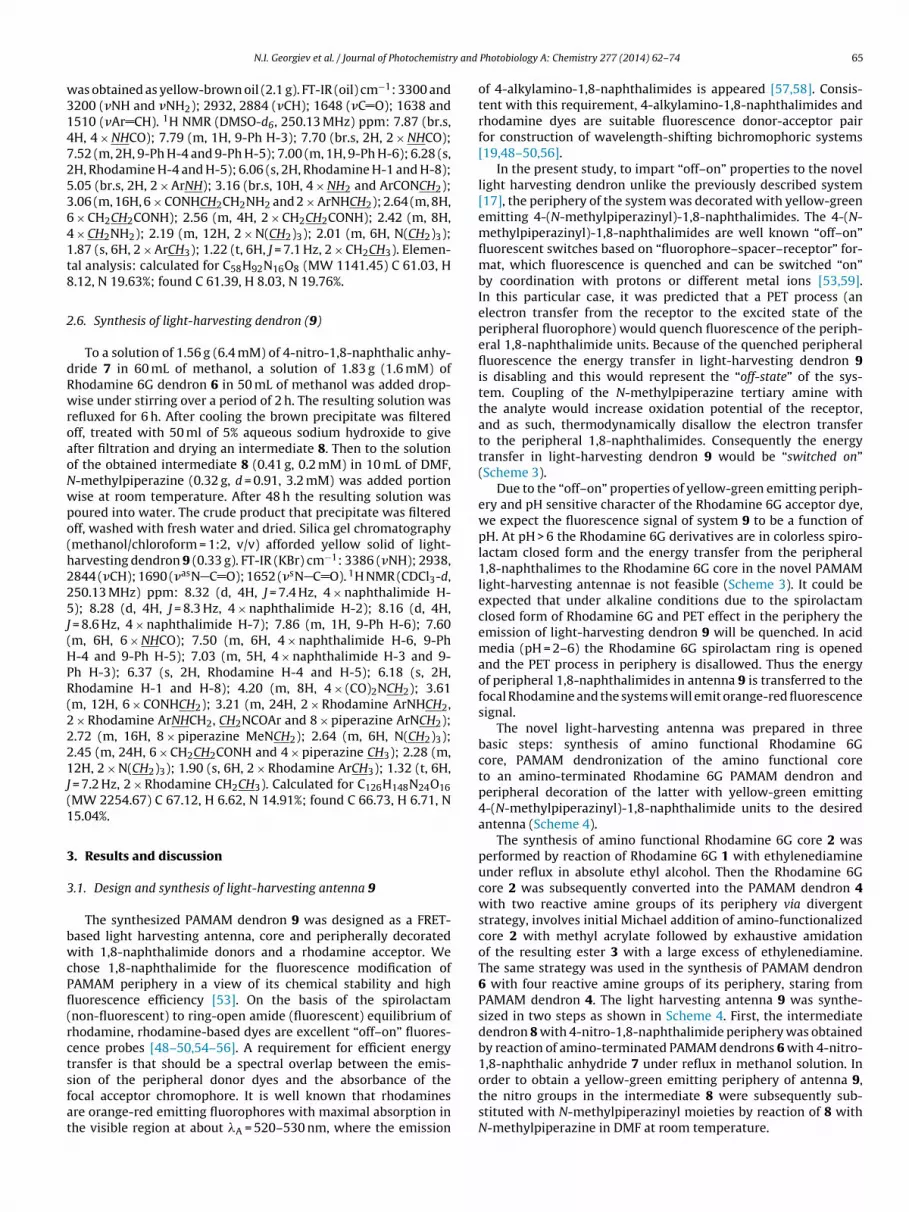
.6. Synthesis of light-harvesting dendron (9)
To a solution of 1.56 g (6.4 mM) of 4-nitro-1,8-naphthalic anhy-ride 7 in 60 mL of methanol, a solution of 1.83 g (1.6 mM) ofhodamine 6G dendron 6 in 50 mL of methanol was added drop-ise under stirring over a period of 2 h. The resulting solution was
efluxed for 6 h. After cooling the brown precipitate was filteredff, treated with 50 ml of 5% aqueous sodium hydroxide to givefter filtration and drying an intermediate 8. Then to the solutionf the obtained intermediate 8 (0.41 g, 0.2 mM) in 10 mL of DMF,-methylpiperazine (0.32 g, d = 0.91, 3.2 mM) was added portionise at room temperature. After 48 h the resulting solution wasoured into water. The crude product that precipitate was filteredff, washed with fresh water and dried. Silica gel chromatographymethanol/chloroform = 1:2, v/v) afforded yellow solid of light-arvesting dendron 9 (0.33 g). FT-IR (KBr) cm−1: 3386 (�NH); 2938,844 (�CH); 1690 (�asN C O); 1652 (�sN C O). 1H NMR (CDCl3-d,50.13 MHz) ppm: 8.32 (d, 4H, J = 7.4 Hz, 4 × naphthalimide H-); 8.28 (d, 4H, J = 8.3 Hz, 4 × naphthalimide H-2); 8.16 (d, 4H,
= 8.6 Hz, 4 × naphthalimide H-7); 7.86 (m, 1H, 9-Ph H-6); 7.60m, 6H, 6 × NHCO); 7.50 (m, 6H, 4 × naphthalimide H-6, 9-Ph-4 and 9-Ph H-5); 7.03 (m, 5H, 4 × naphthalimide H-3 and 9-h H-3); 6.37 (s, 2H, Rhodamine H-4 and H-5); 6.18 (s, 2H,hodamine H-1 and H-8); 4.20 (m, 8H, 4 × (CO)2NCH2); 3.61m, 12H, 6 × CONHCH2); 3.21 (m, 24H, 2 × Rhodamine ArNHCH2,
× Rhodamine ArNHCH2, CH2NCOAr and 8 × piperazine ArNCH2);.72 (m, 16H, 8 × piperazine MeNCH2); 2.64 (m, 6H, N(CH2)3);.45 (m, 24H, 6 × CH2CH2CONH and 4 × piperazine CH3); 2.28 (m,2H, 2 × N(CH2)3); 1.90 (s, 6H, 2 × Rhodamine ArCH3); 1.32 (t, 6H,
= 7.2 Hz, 2 × Rhodamine CH2CH3). Calculated for C126H148N24O16MW 2254.67) C 67.12, H 6.62, N 14.91%; found C 66.73, H 6.71, N5.04%.
. Results and discussion
.1. Design and synthesis of light-harvesting antenna 9
The synthesized PAMAM dendron 9 was designed as a FRET-ased light harvesting antenna, core and peripherally decoratedith 1,8-naphthalimide donors and a rhodamine acceptor. We
hose 1,8-naphthalimide for the fluorescence modification ofAMAM periphery in a view of its chemical stability and highuorescence efficiency [53]. On the basis of the spirolactamnon-fluorescent) to ring-open amide (fluorescent) equilibrium ofhodamine, rhodamine-based dyes are excellent “off–on” fluores-ence probes [48–50,54–56]. A requirement for efficient energyransfer is that should be a spectral overlap between the emis-
ion of the peripheral donor dyes and the absorbance of theocal acceptor chromophore. It is well known that rhodaminesre orange-red emitting fluorophores with maximal absorption inhe visible region at about �A = 520–530 nm, where the emissionPhotobiology A: Chemistry 277 (2014) 62– 74 65
of 4-alkylamino-1,8-naphthalimides is appeared [57,58]. Consis-tent with this requirement, 4-alkylamino-1,8-naphthalimides andrhodamine dyes are suitable fluorescence donor-acceptor pairfor construction of wavelength-shifting bichromophoric systems[19,48–50,56].
In the present study, to impart “off–on” properties to the novellight harvesting dendron unlike the previously described system[17], the periphery of the system was decorated with yellow-greenemitting 4-(N-methylpiperazinyl)-1,8-naphthalimides. The 4-(N-methylpiperazinyl)-1,8-naphthalimides are well known “off–on”fluorescent switches based on “fluorophore–spacer–receptor” for-mat, which fluorescence is quenched and can be switched “on”by coordination with protons or different metal ions [53,59].In this particular case, it was predicted that a PET process (anelectron transfer from the receptor to the excited state of theperipheral fluorophore) would quench fluorescence of the periph-eral 1,8-naphthalimide units. Because of the quenched peripheralfluorescence the energy transfer in light-harvesting dendron 9is disabling and this would represent the “off-state” of the sys-tem. Coupling of the N-methylpiperazine tertiary amine withthe analyte would increase oxidation potential of the receptor,and as such, thermodynamically disallow the electron transferto the peripheral 1,8-naphthalimides. Consequently the energytransfer in light-harvesting dendron 9 would be “switched on”(Scheme 3).
Due to the “off–on” properties of yellow-green emitting periph-ery and pH sensitive character of the Rhodamine 6G acceptor dye,we expect the fluorescence signal of system 9 to be a function ofpH. At pH > 6 the Rhodamine 6G derivatives are in colorless spiro-lactam closed form and the energy transfer from the peripheral1,8-naphthalimes to the Rhodamine 6G core in the novel PAMAMlight-harvesting antennae is not feasible (Scheme 3). It could beexpected that under alkaline conditions due to the spirolactamclosed form of Rhodamine 6G and PET effect in the periphery theemission of light-harvesting dendron 9 will be quenched. In acidmedia (pH = 2–6) the Rhodamine 6G spirolactam ring is openedand the PET process in periphery is disallowed. Thus the energyof peripheral 1,8-naphthalimides in antenna 9 is transferred to thefocal Rhodamine and the systems will emit orange-red fluorescencesignal.
The novel light-harvesting antenna was prepared in threebasic steps: synthesis of amino functional Rhodamine 6Gcore, PAMAM dendronization of the amino functional coreto an amino-terminated Rhodamine 6G PAMAM dendron andperipheral decoration of the latter with yellow-green emitting4-(N-methylpiperazinyl)-1,8-naphthalimide units to the desiredantenna (Scheme 4).
The synthesis of amino functional Rhodamine 6G core 2 wasperformed by reaction of Rhodamine 6G 1 with ethylenediamineunder reflux in absolute ethyl alcohol. Then the Rhodamine 6Gcore 2 was subsequently converted into the PAMAM dendron 4with two reactive amine groups of its periphery via divergentstrategy, involves initial Michael addition of amino-functionalizedcore 2 with methyl acrylate followed by exhaustive amidationof the resulting ester 3 with a large excess of ethylenediamine.The same strategy was used in the synthesis of PAMAM dendron6 with four reactive amine groups of its periphery, staring fromPAMAM dendron 4. The light harvesting antenna 9 was synthe-sized in two steps as shown in Scheme 4. First, the intermediatedendron 8 with 4-nitro-1,8-naphthalimide periphery was obtainedby reaction of amino-terminated PAMAM dendrons 6 with 4-nitro-1,8-naphthalic anhydride 7 under reflux in methanol solution. In
order to obtain a yellow-green emitting periphery of antenna 9,the nitro groups in the intermediate 8 were subsequently sub-stituted with N-methylpiperazinyl moieties by reaction of 8 withN-methylpiperazine in DMF at room temperature.
66 N.I. Georgiev et al. / Journal of Photochemistry and Photobiology A: Chemistry 277 (2014) 62– 74
N
O
O
N
N
N
N
O
O
N
N
N
N
NN
O
NH
NH
N O
N O
O
N
N
OO
N
e-
e-
XX
XX
e-
e-
X
NON-FLUORESCEN T
X
XFRET
H
OH
(pH 6)N
O
O
N
NH
N
N
O
O
N
NH
N
N
NN
O
NH
NH
N O
N O
O
NH
N
OO
NH
e-
e-
e-
e-
X
YELLOW-GREEN FLUORESCENCE
XX
FRET
FRET
X
X
X
X
X
X
X
FRET
(pH 2-6)H
OH
N
O
O
N
NH
N
N
O
O
N
NH
N
N
NN
O
NH
NH
N O
N O
O
NH
N
OO
NH
e-
e-
e-
e-
FRET
FRET
X
X
X
X
RES
X
XX
X
H
X
S withi
mfifl
a
TYn
ORANGE-RED FLUO
cheme 3. Photophysical behavior of antenna 9 as a function of pH after excitation
All of the synthesized compounds were characterized by theirelting points and TLC retention values Rf (Table 1) and identi-
ed by conventional techniques - elemental analysis data, UV-Vis,
uorescence, FT-IR and 1H NMR spectroscopy.For instance, in the 1H NMR (CDCl3-d, 250.13 MHz) spectrum ofntenna 9 (see Section 2) a resonance at 4.20 ppm was observed.
able 1ields, melting points and retention factors for intermediates 2–6 and target anten-ae 9.
Compound Yield (%) M.p. (◦C) Rf
2 88 >250 0.45a
3 98 134–136 0.66b
4 96 88–90 0.27c
5 97 Oil 0.39b
6 92 Oil 0.11c
9 74 164–166 0.32c
a TLC: chloroform/ethylacetate/ethanol = (1:1:1).b TLC: toluene/ethanol = (2:1).c TLC: n-propanol/ammonium hydroxide = (1:1).
CENCE
n a spectral region of maximal absorption of the peripheral fluorophores.
This is characteristic for the methylene protons coupled to N-position of 1,8-naphthalimide moiety, which proves the presenceof 1,8-naphthalimide units in the dendron’s periphery. Also the 1HNMR spectrum of antenna 9 contains two resonances at 6.18 ppmand 6.37 ppm that are typical for the core Rhodamine 6G protonsat position C-1, C-4, C-5 and C-8. The resonances at 4.20 ppm,6.18 ppm and 6.37 ppm are in ratio 4:1:1 suggesting that in thenovel dendron one Rhodamine 6G core is surrounded by four1,8-naphthalimide units. Furthermore, a resonance at 7.03 ppm ischaracteristic for the proton at position C-3 of the yellow-greenemitting 1,8-naphthalimide, substituted in position C-4 withan electron-donating piperazine group. This resonance is ratherdifferent from the corresponding value for a non-substituted 4-nitro-1,8-naphthalimide moieties (8.35–8.70 ppm) [17,60], whichis a solid evidence that the peripheral 4-nitro-1,8-naphthalimideunits in the intermediate dendron 8 were completely converted
in yellow-green emitting donor periphery. Moreover the 1HNMR spectrum contains all requisite peaks for rhodamine and 1,8-naphthalimide moieties as well as peaks in range of 2.40–3.30 ppm,attributed to the protons in the peripheral N-methylpiperazines.
N.I. Georgiev et al. / Journal of Photochemistry and Photobiology A: Chemistry 277 (2014) 62– 74 67
EtOH, Reflux
H2NNH2
MeOH, 20 oC
OMeO
MeOH, 20 oC
H2NNH2
O NHHN
4
N
ON
NH
NH
O
ONH2
NH2
O NHHN
1
O
O
Cl O NHHN
2
N
ONH2
O NHHN
3
N
ON
OMe
OMe
O
O
6
N
HN
HN
O
ON
NH
O
OHN
NO
NH
HNOH2N
NH2
H2N
NH2
OHN NH
N
O
MeOH, 20 oC
OMeO
5
N
NH
NH
O
ON
OCH3
O
OOCH3
N O
OCH3
OCH3O
O NHHN
N
O
MeOH, 20 oC
H2NNH2
8
N
NH
NH
O
ON
HN
O
ONH
N O
HN
NHO N
N
O2N
O
OO
ONO2
N
O
ONO2
NO
O
O2N
O NHHN
N
O
7
MeOH, Reflux
OO
O
O2N
N
NH HN
O O
N
NHO
O
HNN
OHN
HN
O
N
N
N
O
O
O O
N
N OO
N
N
O
O
N
N N
NN
O
NH
NH
N O
9
DMF, 20oC
NHN
Scheme 4. Synthesis of PAMAM light-harvesting antenna 9.

68 N.I. Georgiev et al. / Journal of Photochemistry and Photobiology A: Chemistry 277 (2014) 62– 74
Table 2Absorption and fluorescence characteristics of compounds 2–6 (�ex = 510 nm) and 9–10 (�ex = 400 nm) in water/DMF (4:1, v/v).
Compound �A (nm) ε (l mol−1 cm−1) �F (nm) �A − �F (cm−1) ФF
2 530a 56 300a 558a 947a 0.92a
3 532a 46 740a 558a 876a 0.72a
4 532a 42 990a 560a 940a 0.55a
5 532a 42 760a 560a 940a 0.52a
6 534a 42 490a 560a 820a 0.48a
9 394b 36 840b 527b 6400b 0.32b
536a 44 370a 558a 700a 0.39a
10 394b 17 560b 522b 6220b 0.28b
534a 48 740a 561a 900a 0.43a
a Photophysical data recorded at ca. pH 2 to a avoid rhodamine spirolactam closed (non-fluorescent) form and �ex = 510 nm.b
3
a1tflrbTftRa
ta(ip
9iimst(ef�
1eta1c2w1r
2c5d
pfl
The quantum yields of fluorescence were calculated using Rho-damine 6G (˚F = 0.95 in ethanol [51]) or Coumarin 6 (˚F = 0.78 inethanol [52]) as standards according to Eq. (2), where Aref, Sref, nrefand Asample, Ssample, nsample represent the absorbance at the exited
Photophysical data recorded at pH 6.5 and �ex = 400 nm.
.2. Photophysical characterization of antenna 9
Photophysical properties of the synthesized compounds 2–6nd 9 as well as a previously synthesized reference dendron0 were determined in water/DMF (4:1, v/v) solution. Underhese conditions the rhodamine moieties adopts a closed, non-uorescent spirolactam form. At ca. pH 2 the spirolactam ring ofhodamine is opened, which results in new absorption (rhodamine)ands between 450 and 575 nm with maximum at 530–534 nm.he listed Table 2 absorption data for compounds 2–6 are commonor Rhodamine 6G derivatives [61,62]. The presented data showhat the different alkylamino substituents in the 9-phenyl amide ofhodamine 6G (compounds 2–6) have a small effect on the energynd the shape of the dyes’ absorption bands.
In alkaline solution antennae 9 and 10 do not emit light dueo the PET quenching process in peripheral 1,8-naphthalimidesnd non-fluorescent spirolactam form of the focal rhodamineScheme 3). This was the reason to investigate their photophys-cal properties at pH 6.5 where the PET is not feasible and also atH 2 where the rhodamine is in its ring-opened form.
In water/DMF (4:1, v/v) at pH 6.5 light-harvesting dendrons and 10 showed only one longest-wavelength absorption band
n a range of about 340–520 nm, which is attributed to annternal charge transfer process in the 1,8-naphthalimide chro-
ophores. After acidification to ca. pH 2 where the rhodaminepirolactam form is opened, as expected the absorption spec-rum of light harvesting systems 9 and 10 showed two bandsTable 2) corresponding to the absorption location of the periph-ral 1,8-naphthalimide donor chromophores (�A = 394 nm) and theocal rhodamine acceptor unit (�A = 536 nm for antenna 9 andA = 534 nm for antenna 10).
The fluorescence spectra of light-harvesting antennae 9 and0 in water/DMF (4:1, v/v) solution at pH 6.5, recorded afterxcitation within the spectral region of maximal absorption ofhe donor fluorophore (�ex = 400 nm), showed emission band atbout 525 nm, corresponding to the emission band of the donor,8-naphthalimide fragments in the donor–acceptor systems. Inontrast, when the fluorescence spectra were recorded at ca. pH, the observed emission was shifted to about 560 nm (Table 2),hich can be attributed to the energy transfer from the donor
,8-naphthalimide to the ring-opened (fluorescence) form of thehodamine 6G acceptor under these conditions.
After direct excitation in the rhodamine core (510 nm) at ca. pH in water/DMF (4:1, v/v) compounds 2–6, 9 and 10 showed typi-al for Rhodamine 6G fluorescence spectra with maxima at about60 nm [61,62], suggesting that the substituents at 9-phenyl amideo not affect the energy of the dyes’ fluorescence maximum.
It is well know, that the spectral overlap is the main factor that
redetermines the efficiency of FRET. Peripheral 1,8-naphthalimideuorophores in the synthesized antenna 9 showed a broademission band in the visible region (450–650 nm) that practicallycovers completely a rhodamine absorption band (Fig. 1).
The efficiency of the energy transfer ET was calculated to 82%for antenna 9 and 91% for antenna 10 (Table 2) by applying Eq.(1) [16,63], where FDA and FD are the normalized to the opticaldensity fluorescence intensities of the donor in the presence ofacceptor (1,8-naphthalimide part in the examined antennae at pH2) and of the free donor without acceptor (1,8-naphthalimide partin examine antennae at pH 6.5 – FRET process to the acceptor is notfaceable), respectively. The reason for the weaker energy transferin dendron 9 is probably due to the increased distance betweenthe donor and acceptor in the antenna of the second generation.The calculated energy transfer data clearly show that the novellight-harvesting antenna 9 would be able to act as an efficientwavelength-shifting chromophore.
ET = 1 − FDA
FD(1)
The Stoke’s shift (�A − �F) values for the individual fluorophoresin compounds under study are typical for the rhodamines and 1,8-naphthalimides. The observed values, in range of 6220–6400 cm−1
for the peripheral 1,8-naphthalimides and 740–900 cm−1 for thefocal rhodamine, do not indicate remarkable changes in the fluo-rophore excited state due to their incorporation in the dendriticsystems.
Fig. 1. Normalized absorption spectrum of dendron 6 at pH 2 and emission spectrumof antenna 9 at pH 6.5 (�ex = 400 nm) in water/DMF (4:1, v/v).

N.I. Georgiev et al. / Journal of Photochemistry and Photobiology A: Chemistry 277 (2014) 62– 74 69
F −5 −1
i8
wr
˚
ptoi1idyirn[tl
3
osic1
icttopo
vpb
ig. 2. Absorption changes of 3 as a function of pH at concentration 1 × 10 mol Ln water/DMF (4:1, v/v). Inset: titration plots of compounds 2–6 in a pH range of ca.–2.
avelength, the integrated emission band area and the solventefractive index of the standard and the sample, respectively.
F = ˚ref
(Ssample
Sref
) (Aref
Asample
)(n2
sample
n2ref
)(2)
The fluorescence quantum yield of the 1,8-naphthalimideeripheries is approximately the same in the both antennae. Also,he molar extinction coefficient value of the peripheral absorptionf light-harvesting antenna 9, containing four donor fragments,s about two times higher than that of light-harvesting antenna0 with two donor units. This fact clearly illustrates the lack of
nteraction between the peripheral 1,8-naphthalimide units in theendritic bone. As can be seen from the data in Table 2, the quantumield of fluorescence of the rhodamine core is decreasing with thencrease of the molecular weight of the system. A similar effect waseported before for the core functional PAMAM dendrons using 1,8-aphthalimide and perylene-3,4,9,10-tetracarboxylic diimide units64,65]. This is probably due to the more flexible PAMAM scaffold inhe larger dendron’s generations, which are able to induce energyoses reducing the quantum yield of fluorescence.
.3. Influence of pH on the photophysical properties of antenna 9
The light-harvesting system 9 was designed as a molecular flu-rescence probe for determination of pH changes over a wider pHcale. This was the reason to investigate its photophysical behav-or in water/DMF (4:1, v/v) solution at different pH values and toompare obtained characteristics with those of reference dendron0.
In order to receive a more complete comparative picture for thenfluence of the dendron bone to the focal rhodamine, intermediateompounds 2–6 were involved in the present study. Fig. 2 presentshe changes of absorption spectra of 3 at different pH values as aypical example for the influence of pH on the absorption spectraf the examined compounds 2–6. As can be seen, the decrease ofH results in increase of the absorbance at 532 nm due to the ringpening reaction of rhodamine core.
Taking the part of the graphs located between pH 2 and 6, the pKa
alues of 2–6 have been calculated by Eq. (3) [18]. The calculatedKa value for amino-functional rhodamine 2 was 4.0, 3.9 for theranching compounds 3 and 4 and 3.8 for 5 and 6. Probably the
Fig. 3. Absorption changes of antenna 9 as a function of pH at concentration1 × 10−5 mol L−1 in water/DMF (4:1, v/v). Inset: titration plots of antennae 9 and10 in a pH range of ca. 10–2.
decrease of pKa values is a result of the core protective role of thePAMAM scaffold.
log[
Amax − A
A − Amin
]= pH − pKa (3)
The absorption spectra of light-harvesting antenna 9 do notshow significant pH-dependent changes in a pH window 8–10,since the 1,8-naphthalimide fluorophores do not affect their ICTexcited states. In contrast, upon acidification from pH 8 to ca. pH 2the absorption maxima of peripheral 1,8-naphthalimides are blueshifted of about 20 nm (Fig. 3). One of the reasons for this effectis that the protonation of the amine receptor exerts some weakcharge repulsion on the 4-amino moiety of the fluorophores. Onthe other hand in very acidic conditions the push–pull characterof the ICT state is partially reduced due to the protonation of thearomatic 4-amino moiety.
Furthermore, the decrease of pH from 6 to ca. 2 results in appear-ance of a novel band corresponding to the absorption of focalrhodamine due to the ring opening reaction. From the absorptionchanges at 534 nm and 536 nm the titration curves of antennae 9and 10 were obtained (inset in Fig. 3). Analysis of these absorbancechanges according to Eq. (3) gives two pKa values: 3.7 and 2.5for antenna 9 and 3.8 and 2.5 for antenna 10. The pKa values of3.7–3.8 are typical for the rhodamine core, while the pKa values of2.5 suggest reversible aggregation processes in alkaline medium.The decrease of a pKa value for antenna 9 could be related to thestronger core protective role of the PAMAM scaffold in the antennaof second generation.
The fluorescence spectra of compounds 2–6 and 9 were alsorecorded in water/DMF (4:1, v/v) solution at different pH values. Inalkaline solution rhodamines 2–6 are in spirolactam closed formand do not emit light. However, upon acidification an emissionsignal at 560 nm was gradually increased (Fig. 4).
Analysis of the fluorescence changes at 560 nm according to Eq.(4) [9] gives the pKa values 4.0, 3.9 for the branching compounds3–4 and 3.8 for 5–6. These pKa values are the same as these calcu-lated according to the absorption changes and should be attributedto the spirolactam opening reaction.
log[
IFmax − IFIF − IFmin
]= pH − pKa (4)
Family of fluorescent spectra (�ex = 400 nm) of antenna 9 at pHwindow 6.5–10 in water/DMF (4:1, v/v) are presented in Fig. 5.As expected in alkaline media the novel antennae showed veryweak fluorescence in range of 450–650 nm with maximum at

70 N.I. Georgiev et al. / Journal of Photochemistry and Photobiology A: Chemistry 277 (2014) 62– 74
Fac
5tTtctUtpr
o4flrglstsmi
F1p
HNO
O
N
O
ON N
N
Xe
e
δ+δ−
AttractiveField
RepulsionField
hν1 hν2 (fluorescence)
fluorophore receptorsp.
X
ig. 4. Fluorescent changes of 3 in water/DMF (4:1, v/v) as a function of pH (ca. 7–2)t concentration 1 × 10⁻⁵ mol L−1 in water/DMF (4:1, v/v). Inset: titration plots ofompounds 2–6 in a pH range of ca. 7–2.
27 nm (1,8-naphthalimide emission). However, upon acidifica-ion an emission signal was gradually increased (inset of Fig. 5).he increase in fluorescence intensity on transition from alkalineo slightly acidic medium indicates that the peripheral fluores-ence of examined antenna is being quenched by PET process fromhe tertiary amine receptor to the 1,8-naphthalimide fluorophore.pon recognition of the analyte (proton) the oxidation potential of
he receptor is increased thermodynamically thus disallowing PETrocess and as such, the peripheral fluorescence of the system isecovered.
Moreover, as demonstrated experimentally Liu and de Silva,nly the receptor that is directly attached to the 1,8-naphthalimide-amino moiety (the “lower” moiety) is capable of quenching theuorophore excited state [66]. The 4-aminonaphthalimide fluo-ophore is a “push–pull” � electron system with the C-4 amineroup as a donor and the 1,8-naphthalimide as an acceptor. Thiseads to strong internal charge transfer (ICT) in the lowest excitedinglet state and considerable dipole character (positive pole at
he 4-amino terminus). A large dipole moment in an excitedtate gives rise to a strong photogenerated electric field. Such aolecular electric field can, depending on its sign and magnitude,nhibit or accelerate a transiting electron in the 1,8-naphthalimide
ig. 5. Fluorescence changes (�ex = 400 nm) of antenna 9 as a function of pH (ca.0–6.5) at concentration 1 × 10⁻⁵ mol L−1 in water/DMF (4:1, v/v). Inset: titrationlots of antennae 9 and 10 in a pH range of ca. 11–2.
Scheme 5. Feasible photoinduced electron transfer in the 1,8-naphthalimideperiphery of antenna 9.
compounds. Thus the fluorescence quenching PET process isobserved only if the electron leaving the unprotonated amine donorcan enter the space of the 4-aminonaphthalimide fluorophoreacross C-4 position with its attractive electric field (Scheme 5). Thecorresponding PET path from the unprotonated amino receptor inN-position is just as feasible thermodynamically but requires theelectron to enter the fluorophore across the imide moiety with itsrepulsive electric field and is not observed.
Fluorescence intensity enhancement (FE) was used as a quali-tative parameter for the evaluation of the sensing potential of thesynthesized antenna 9 and reference compound 10. The FE = I/I0was determined as ratio between the maximum of fluorescenceintensity I at pH ca. 6.5 and the minimum fluorescence intensity I0at about pH 10. Upon acidification the fluorescence intensity of theyellow-green emitting periphery had enhanced by over an order ofmagnitude (FE = 20 for antenna 9 and FE = 11 for antenna 10). Thesechanges are of such magnitude that they can be considered as rep-resenting two different “states”, where the fluorescence emissionis “switched off” in alkaline solution and “switched on” in neutral toslightly acidic media. The higher FE value of antenna 9 in respectto that of the antenna of first generation 10 is obviously due tothe greater absorptivity of the system, containing four peripheralfluorophores.
As can be seen from the inset of Fig. 5, peripheral 1,8-naphthalimide fluorophores switch their “off” and “on” states in pHinterval of ca. 8.5–6.5. The calculated using Eq. (4) pKa values of 7.27
Fig. 6. Fluorescence changes of antenna 9 (�ex = 400 nm) as a function of pH (6.5–2)at concentration 1 × 10⁻⁵ mol L−1 in water/DMF (4:1, v/v). Inset: titration plots ofantennae 9 and 10 (�ex = 400 nm) in a pH range of ca. 6–2.

N.I. Georgiev et al. / Journal of Photochemistry and Photobiology A: Chemistry 277 (2014) 62– 74 71
Fig. 7. Fluorescence changes of antenna 9 (�ex = 510 nm) as a function of pH (6–2)aa
fdFstqcaatoi(di
tcestitArdh
ev15rpadsb
ceitf
t concentration 1 × 10⁻⁵ mol L−1 in water/DMF (4:1, v/v). Inset: titration plots ofntennae 9 and 10 (�ex = 510 nm) in a pH range of ca. 6–2.
or antenna 9 and 7.15 for antenna 10 are in agreement with theata for 4-piperazinyl-1,8-naphthalimide derivatives [4,67–69].urther acidification from pH 6.5 to ca. pH 2 opens the rhodaminepirolactam form, which allows the energy transfer from peripheryo the acceptor moiety (�ex = 400 nm). This results in fluorescenceuenching of the peripheral fluorescence and remarkable fluores-ence intensity enhancement in the rhodamine emission regiont 560 nm (Fig. 6). The FE was more than 30 times (FE = 31) forntenna 10 and FE = 16 for antenna 9. The different behavior ofhe both systems could be twofold: (i) the lower quantum yieldf focal rhodamine in the dendron of second generation in compar-son to the dendron of first generation (see Section 3.2, Table 2);ii) obviously the energy transfer efficiency in antenna 9 is lowerue to the greater distance between the donor and acceptor units
n the light-harvesting system of second generation.The comparison of the photophysical properties of novel sys-
em 9 with those of the previously synthesized similar antenna, notomprising piperazine moieties in the 1,8-naphthalimide periph-ry [17], shows that the core fluorescence enhancement of the newystem (9) is significantly higher. This could be due to the qua-ernization of the tertiary amines in the periphery of the systemn an acidic medium, which greatly enhances the hydrophilicity ofhe molecule and as such, reduces the possibility of aggregation.s a consequence, the self quenching effect in the novel system iseduces as well. This is well consistent with the fact that the rho-amine core quantum yield of fluorescence of 9 is about 2.5 timesigher than that of the previously synthesized similar antenna [17].
Analysis of the fluorescence changes at 525 nm and 560 nm afterxcitation at 400 nm (inset of Fig. 6) according to Eq. (4) gives pKa
alues of 3.6 and 2.5 for antenna 9 and 3.8 and 2.5 for antenna0. The same values were obtained after direct core excitation at10 nm (Fig. 7), indicating that only the spirolactam reaction ofhodamine cores in 9 and 10 are responsible for the pH sensingroperties of the systems. These results suggest that the novelntenna 9 is able to act as a ratiometric fluorescent probe for pHetermination, therefore it could be expected that the method willerve as a practical tool for analysis of environmental samples andiological studies.
Based on the presented study, it can be concluded that thehanges in the fluorescence intensity and ratiometric sensing prop-rties of the novel wavelength-shifting bichromophoric system 9
n the presence of protons are due to the simultaneous opera-ion of PET process in the periphery of the system, energy transferrom periphery to the core (FRET) and of the ICT spirolactamFig. 8. Fluorescence changes of antenna 9 (1 × 10⁻⁵ mol L−1) in the presence of metalcations (1 × 10⁻⁴ mol L−1) in DMF.
(non-fluorescent) to ring-open amide (fluorescent) equilibrium ofrhodamine core.
3.4. Influence of metal cations on the fluorescence intensity of theantennae
The signaling properties of novel antenna 9 in the presence oftransition metal ions were investigated spectrophotometrically inDMF and in water/DMF (4:1, v/v) with regard to their potentialapplication as a fluorescent probe for cations recognition. DMF hasbeen chosen since it is able as a polar solvent to stabilize the dyecharge separated state thus favoring the fluorescence switching byPET process. Also DMF guarantees a good solubility of the dye lig-ands, used metal salts and the respective complexes. Experimentshave been performed in the presence of different metal cations:Cd2+, Co2+, Cu2+, Fe3+, Ni2+, Pb2+, Zn2+, Hg2+ and Ag+.
The addition of metal ion solution to the antenna of second gen-eration 9 in DMF caused negligible changes only on the emissionproperties of the 1,8-naphthalimide periphery at 527 nm (Fig. 8).
For the reference antenna 10, considerable photophysicalchanges in DMF solution were observed only upon addition of Cu2+
and Pb2+, while the other metal ions produced a negligible effect(Fig. 9). The presence of Cu2+ and Pb2+ induces a hypsochromic shiftof the 1,8-naphthalimide absorption maximum of ��A = 10 nmfor Cu2+ and 12 nm for Pb2+ (Fig. 9A). This means that the bothnitrogen atoms comprised in the N-methylpiperazine substituentat C-4 of the peripheral 1,8-naphthalimides are subjected to thecoordination with metal cations [69]. Addition of Cu2+ and Pb2+
results in enhancement of the peripheral fluorescence intensity(1,8-naphthalimides) of antenna 10. The enhancement of the flu-orescence emission was FE = 16.0 for Cu2+ and FE = 16.7 for Pb2+
(Fig. 9B).The results obtained indicate that the PAMAM bone in the
light-harvesting antenna of second generation (antenna 9) is morereactive toward coordination with metal ions due to the largernumber of back bone tertiary amines and probably the couplingoccurs mainly in the PAMAM scaffold.
Furthermore, the examined metal ions did not induce anychanges in the photophysical properties of antennae core even ina high cation concentration (1 × 10⁻³ M), suggesting that only pro-tons are able to open the spirolactam cycle (see Section 3.3). All
these show that during the experiments the metal salts do not gen-erate protons from partial hydrolysis of the salts and as such, do notprovoke unusual fluorescent enhancement of the rhodamine core.
72 N.I. Georgiev et al. / Journal of Photochemistry and
Fi
ansiatbac
Fao
ig. 9. Absorption (A) and fluorescence (B) changes of antenna 10 (1 × 10⁻⁵ mol L−1)n the presence of metal cations (1 × 10⁻⁴ mol L−1) in DMF.
Coordination of metal ions with the receptor fragments inqueous solutions competes with a number of other anions anducleophiles. Therefore, it was of interest to investigate the emis-ion behavior of antennae 9 and 10 in the presence of metal ionsn aqueous medium with regard to their potential for practicalpplication. To avoid the effect of proton generation due to the par-ial hydrolysis of metal salts the experiments were conducted in
uffered solution (ammonia/ammonium chloride buffer – pH 10)s in alkaline medium the peripheral fluorophores are in their soalled “off” state (see Section 3.3). The performed tests showed thatig. 10. Effect of metal ions (1 × 10⁻³ mol L−1) on the fluorescence intensity ofntenna 9 (1 × 10⁻⁵ mol L−1) in water/DMF (4:1, v/v) in the presence of 100 mMf ammonia/ammonium chloride buffer (pH 10).
Photobiology A: Chemistry 277 (2014) 62– 74
the interaction of metal ions with the piperazine moieties is weak inthe antenna of first generation 10 and does not occur in the antennaof second generation 9.
The influence of various metal ions on the fluorescence behaviorof antenna 9 in buffered water/DMF (4:1, v/v) solution is presentedin Fig. 10. As can be seen, the addition of metal ion solution (up to1 × 10−3 mol L−1) to the antenna solution (1 × 10⁻⁵ mol L−1) doesnot caused any changes, suggesting that in water/DMF (4:1, v/v)the metal cations are not able to interact with the receptors of theperipheral PET based naphthalimides.
The experiments performed clearly show that the metal ions areable to coordinate neither in the peripheral receptors of the antennanor in the core spirolactam. This defines excellent selectivity ofantenna 9 toward protons over representative metal ions. The highselectivity and ratiometric pH sensitivity of the novel wavelength-shifting bichromophoric system may be beneficially for monitoringpH variations in complex samples.
4. Conclusions
Novel PAMAM light-harvesting dendron of second generation,core and peripherally functionalized with Rhodamine 6G and 1,8-naphthalimide fluorophores, respectively, was synthesized basedon a divergent approach. The system was designed as a wavelength-shifting bichromophore capable of absorbing light by its peripheryand efficiently transferring the energy to a single acceptor dyein the focal point of the molecule. The calculated energy trans-fer of 82% indicates that the selected fluorophores are suitabledonor–acceptor pair for constructing light harvesting materials.Moreover, the yellow-green emitting periphery of the antennawas designed on the “fluorophore–spacer–receptor” format, mak-ing it able to act as a fluorescence PET-based pH probe. Afterexcitation within the spectral region of maximal absorption ofperipheral 1,8-naphthalimides the synthesized antenna enhancedits peripheral fluorescence intensity by over an order of magni-tude (FE = 20) in a pH range ca. 6.5–10, while upon acidification topH 2 the fluorescence intensity of the core was enhanced by overan order of magnitude (FE = 16) with the simultaneous decreaseof the peripheral emission. Novel antenna also showed excellentselectivity toward protons over representative metal ions. Thusthe distinguishing features of the light-harvesting dendrons weresuccessfully combined with the properties of classical PET and ring-opening molecular sensing systems. These results suggest that thenovel antenna is able to act as a highly selective and pH sensitiveratiometric fluorescence probe, indicating that the system could beuseful for analysis of environmental samples and biological stud-ies.
Acknowledgements
This work was supported by the National Science Foundationof Bulgaria (project DDVU-02/97). Authors also acknowledge theScience Foundation at the University of Chemical Technology andMetallurgy (Sofia, Bulgaria).
References
[1] V. Balzani, Photochemical molecular devices, Photochem. Photobiol. Sci. 2(2003) 459–476.
[2] J. Callan, A.P. de Silva, D. Magri, Luminescent sensors and switches in the early21st century, Tetrahedron 61 (2005) 8551–8588.
[3] J. Jiang, B. Leng, X. Xiao, P. Zhao, H. Tian, “Off–on–off” fluorescent proton switchsynthesized by RAFT polymerization, Polymer 50 (2009) 5681–5684.
[4] N. Marinova, V. Bojinov, N. Georgiev, Design, synthesis and pH sensing prop-erties of novel 1,8-naphtalimide-based bichromophoric system, J. Photochem.Photobiol. A: Chem. 222 (2011) 132–140.
[5] M. Jun, B. Roy, K. Ahn, Turn-on fluorescent sensing with “reactive” probes,Chem. Commun. 47 (2011) 7583–7601.

y and
[
[
[
[
[
[
[
[
[
[
[
[
[
[
[
[
[
[
[
[
[
[
[
[
[
[
[
[
[
[
[
[
[
[
[
[
[
[
[
[
[
[
[
[
[
[
[
[
[
[
[
N.I. Georgiev et al. / Journal of Photochemistr
[6] H. Dai, H. Xu, A water-soluble 1,8-naphthalimide-based “turn on” fluorescentchemosensor for selective and sensitive recognition of mercury ion in water,Bioorg. Med. Chem. Lett. 21 (2011) 5141–5144.
[7] H. Kim, Z. Guo, W. Zhu, J. Yoon, H. Tian, Recent progress on polymer-based fluorescent and colorimetric chemosensors, Chem. Soc. Rev. 40 (2011)79–93.
[8] R. Duke, T. Gunnlaugsson, Selective fluorescent PET sensing of fluoride (F−)using naphthalimide-thiourea and -urea conjugates, Tetrahedron Lett. 48(2007) 8043–8047.
[9] S. Tal, H. Salman, Y. Abraham, M. Botoshansky, Y. Eichen, Sensitive and selectivephotoinduced-electron-transfer-based sensing of alkylating agents, Chem. Eur.J. 12 (2006) 4858–4864.
10] J. Dale, J. Rebek, Fluorescent sensors for organophosphorus nerve agent mimics,J. Am. Chem. Soc. 128 (2006) 4500–4501.
11] S. Zhang, T. Swager, Fluorescent detection of chemical warfare agents: func-tional group specific ratiometric chemosensors, J. Am. Chem. Soc. 125 (2003)3420–3421.
12] A.P. de Silva, S. Uchiyama, Molecular logic gates and luminescent sen-sors based on photoinduced electron transfer, Top. Curr. Chem. 300 (2011)1–28.
13] P. Kaur, S. Kaur, K. Singh, A fluoride selective dipyrromethane-TCNQ colorimet-ric sensor based on charge-transfer, Talanta 84 (2011) 947–951.
14] L. Fabbrizzi, M. Licchelli, P. Pallavicini, A. Perotti, A. Taglietti, D. Sacchi,Fluorescent sensors for transition metals based on electron-transfer andenergy-transfer mechanisms, Chem. Eur. J. 2 (1996) 75–82.
15] X. Chen, T. Pradhan, F. Wang, J. Kim, J. Yoon, Fluorescent chemosensors based onspiroring-opening of xanthenes and related derivatives, Chem. Rev. 112 (2012)1910–1956.
16] W.-S. Li, M.-J. Teng, X.-R. Jia, B.-B. Wang, J.-M. Yeh, Y. Wei, Synthesis and energy-transfer properties of poly(amidoamine) dendrons modified with naphthyl anddansyl groups, Tetrahedron Lett. 49 (2008) 1988–1992.
17] N. Georgiev, V. Bojinov, A. Venkova, Design, synthesis and pH sensing propertiesof novel PAMAM light-harvesting dendrons based on rhodamine 6G and 1,8-naphthalimide, J. Fluoresc. 23 (2013) 459–471.
18] L. Daffy, A.P. de Silva, H. Gunaratne, C. Huber, P. Lynch, T. Werner, O. Wolfbeis,Arenedicarboximide building blocks for fluorescent photoinduced electrontransfer pH sensors applicable with different media and communication wave-lengths, Chem. Eur. J. 4 (1998) 1810–1815.
19] Y. Liu, X. Lv, Y. Zhao, M. Chen, J. Liu, P. Wang, W. Guo, Anaphthalimide–rhodamine ratiometric fluorescent probe for Hg2+ basedon fluorescence resonance energy transfer, Dyes Pigments 92 (2012) 909–915.
20] V. Balzani, A. Credi, M. Venturi, Photochemical conversion of solar energy,ChemSusChem 1 (2008) 26–58.
21] J. Serin, D. Brousmiche, J. Frèchet, Cascade energy transfer in a conformationallymobile multichromophoric dendrimer, Chem. Commun. (2002) 2605–2607.
22] W.R. Dichtel, S. Hecht, J. Frèchet, Functionally layered dendrimers: a newbuilding block and its application to the synthesis of multichromophoric light-harvesting systems, Org. Lett. 7 (2005) 4451–4454.
23] P. Du, W.-H. Zhu, Y.-Q. Xie, F. Zhao, C.-F. Ku, Y. Cao, C.-P. Chang, H. Tian,Dendron-functionalized macromolecules: enhancing core luminescence andtuning carrier injection, Macromolecules 37 (2004) 4387–4398.
24] R. Mètivier, F. Kulzer, T. Weil, K. Müllen, T. Basch, Energy transfer ratesand pathways of single donor chromophores in a multichromophoric den-drimer built around a central acceptor core, J. Am. Chem. Soc. 126 (2004)14364–14365.
25] S. Li, W. Zhu, Z. Xu, J. Pan, H. Tian, Antenna-functionalized dendritic �-diketonates and europium complexes: synthetic approaches to generationgrowth, Tetrahedron 62 (2006) 5035–5048.
26] A. Bar-Haim, J. Klafter, Dendrimers as light harvesting antennae, J. Lumin. 76–77(1998) 197–200.
27] V. Bojinov, N. Georgiev, P. Nikolov, Synthesis and photophysical prop-erties of fluorescence sensing ester- and amidoamine-functionalized1,8-naphthalimides, J. Photochem. Photobiol. A: Chem. 193 (2008)129–138.
28] N. Georgiev, V. Bojinov, N. Marinova, Novel PAMAM light-harvesting antennaebased on 1,8-naphthalimide: synthesis, energy transfer, photophysical and pHsensing properties, Sens. Actuators B: Chem. 150 (2010) 655–666.
29] I. Ott, Y. Xu, X. Qian, Fluorescence properties and antiproliferative effects ofmono-, bis-, and tris thiophenylnaphthalimides: results of a comparative pilotstudy, J. Photochem. Photobiol. B: Biol. 105 (2011) 75–80.
30] W. Zhu, L. Song, Y. Yang, H. Tian, Novel bisthienylethene containing ferrocenyl-substituted naphthalimide: a photo- and redox multi-addressable molecularswitch, Chem. Eur. J. 18 (2012) 13388–13394.
31] V. Bojinov, D. Simeonov, N. Georgiev, A novel blue fluorescent 4-(1,2,2,6,6-pentamethylpiperidin-4-yloxy)-1,8-naphthalimide pH chemosensor based onphotoinduced electron transfer, Dyes Pigments 76 (2008) 41–46.
32] H. Wang, L. Yang, W. Zhang, Y. Zhou, B. Zhao, X. Li, A colorimetric probe for cop-per(II) ion based on 4-amino-1,8-naphthalimide, Inorg. Chim. Acta 381 (2012)111–116.
33] V. Bojinov, T. Konstantinova, Fluorescent 4-(2,2,6,6-tetramethylpiperidin-4-ylamino)-1,8-naphthalimide pH chemosensor based on photoinduced electron
transfer, Sens. Actuators B: Chem. 123 (2007) 869–876.34] V. Bojinov, I. Panova, J.-M. Chovelon, Novel blue emitting tetra- andpentamethylpiperidin-4-yloxy-1,8-naphthalimides as photoinduced electrontransfer based sensors for transition metal ions and protons, Sens. Actuators B:Chem. 135 (2008) 172–180.
[
Photobiology A: Chemistry 277 (2014) 62– 74 73
35] V. Bojinov, I. Panova, D. Simeonov, N. Georgiev, Synthesis and sensor activ-ity of photostable blue emitting 1,8-naphthalimides containing s-triazine UVabsorber and HALS fragments, J. Photochem. Photobiol. A: Chem. 210 (2010)89–99.
36] V. Bojinov, I. Panova, Novel 4-(2,2,6,6-tetramethylpiperidin-4-ylamino)-1,8-naphthalimide based yellow-green emitting fluorescence sensors for transitionmetal ions and protons, Dyes Pigments 80 (2009) 61–66.
37] N. Georgiev, I. Yaneva, A. Surleva, A. Asiri, V. Bojinov, Synthesis, sensor activityand logic behavior of a highly water-soluble naphthalimide derivative, Sens.Actuators B: Chem. 184 (2013) 54–63.
38] N. Georgiev, V. Bojinov, Design, synthesis and sensor activity of ahighly photostable blue emitting 1,8-naphthalimide, J. Lumin. 132 (2012)2235–2241.
39] N. Georgiev, V. Bojinov, P. Nikolov, Design, synthesis and photophysical prop-erties of two novel 1,8-naphthalimide fluorescent pH sensors based on PET andICT, Dyes Pigments 88 (2011) 350–357.
40] N. Georgiev, M. Lyulev, V. Bojinov, Sensor activity and logic behavior of PETbased dihydroimidazonaphthalimide diester, Spectrochim. Acta Part A 97(2012) 512–520.
41] V. Bojinov, N. Georgiev, Molecular sensors and molecular logic gates, J. Univ.Chem. Technol. Met. (Sofia) 46 (2011) 3–26.
42] N. Marinova, N. Georgiev, V. Bojinov, Facile synthesis, sensor activity and logicbehaviour of 4-aryloxy substituted 1,8-naphthalimide, J. Photochem. Photo-biol. A: Chem. 254 (2013) 54–61.
43] N. Georgiev, A. Asiri, A. Qusti, K. Alamry, V. Bojinov, Design andsynthesis of pH-selective fluorescence sensing PAMAM light-harvesting den-drons based on 1,8-naphthalimides, Sens. Actuators B: Chem. 190 (2014)185–198.
44] Y. Lei, Y. Su, J. Huo, Photophysical property of rhodamine-coredpoly(amidoamine) dendrimers: simultaneous effect of spirolactam ring-opening and PET process on sensing trivalent chromium ion, J. Lumin. 131(2011) 2521–2527.
45] H.-S. Lv, S.-Y. Huang, B.-X. Zhao, J.-Y. Miao, A new rhodamine B-based lysosomal pH fluorescent indicator, Anal. Chim. Acta 788 (2013)177–182.
46] Y. Lei, C. Zhang, H. Lei, J. Huo, Visible light photocatalytic activity of aromaticpolyamide dendrimer/TiO2 composites functionalized with spirolactam-basedmolecular switch, J. Colloid Interface Sci. 406 (2013) 178–185.
47] Y. Lei, Y. Su, J. Huo, A novel fluorescent sensor for Cr3+ based on rhodamine-cored poly(amidoamine) dendrimer, Spectrochim. Acta Part A 81 (2011)149–154.
48] N. Georgiev, R. Bryaskova, R. Tzoneva, I. Ugrinova, C. Detrembleur, S. Miloshev,A. Asiri, A. Qusti, V. Bojinov, A novel pH sensitive water soluble fluorescentnanomicellar sensor for potential biomedical applications, Bioorg. Med. Chem.21 (2013) 6292–6302.
49] V. Bojinov, A. Venkova, N. Georgiev, Synthesis and energy-transferproperties of fluorescence sensing bichromophoric system based on Rho-damine 6G and 1,8-naphthalimide, Sens. Actuators B: Chem. 143 (2009)42–49.
50] N. Georgiev, A. Asiri, A. Qusti, K. Alamry, V. Bojinov, A pH sensitive and selec-tive ratiometric PAMAM wavelength-shifting bichromophoric system based onPET, FRET and ICT, Dyes Pigments 102 (2014) 35–45.
51] R. Kubin, A. Fletcher, Fluorescence quantum yields of some rhodamine dyes, J.Lumin. 27 (1982) 455–462.
52] G. Reynolds, K. Drexhage, New coumarin dyes with rigidized struc-ture for flashlamp-pumped dye lasers, Opt. Commun. 13 (1975)222–225.
53] C. Niu, G. Zeng, L. Chen, G. Shena, R. Yu, Proton “off–on” behaviour ofmethylpiperazinyl derivative of naphthalimide: a pH sensor based on fluores-cence enhancement, Analyst 129 (2004) 20–24.
54] M. Dong, T.-H. Ma, A.-J. Zhang, Y.-M. Dong, Y.-W. Wang, Y. Peng, A series ofhighly sensitive and selective fluorescent and colorimetric “off–on” chemosen-sors for Cu(II) based on rhodamine derivatives, Dyes Pigments 87 (2010)164–172.
55] Q.-J. Mab, X.-B. Zhang, X.-H. Zhao, Z. Jin, G.-J. Mao, G.-L. Shen, R.-Q. Yu, Ahighly selective fluorescent probe for Hg2+ based on a rhodamine–coumarinconjugate, Anal. Chim. Acta 663 (2010) 85–90.
56] B. Ahamed, P. Ghosh, An integrated system of pyrene and Rhodamine-6G forselective colorimetric and fluorometric sensing of mercury(II), Inorg. Chim.Acta 372 (2011) 100–107.
57] N. Georgiev, V. Bojinov, P. Nikolov, Design and synthesis of a novel pH sensi-tive core and peripherally 1,8-naphthalimide-labeled PAMAM dendron as lightharvesting antenna, Dyes Pigments 81 (2009) 18–26.
58] N. Georgiev, V. Bojinov, Design, synthesis and photostability of novel 1,8-naphthalimide PAMAM light-harvesting dendrons, J. Fluoresc. 21 (2011)51–63.
59] J. Gan, K. Chen, C. Chang, H. Tian, Luminescent properties and photo-inducedelectron transfer of naphthalimides with piperazine substituent, Dyes Pig-ments 57 (2003) 21–28.
60] V. Bojinov, I. Panova, Synthesis and absorption properties of new yellow-green emitting benzo[de]isoquinoline-1,3-diones containing hindered amine
and 2-hydroxyphenylbenzotriazole fragments, Dyes Pigments 74 (2007)551–560.61] S. Bakkialakshmi, T. Menaka, A study of the interaction between rhodamine 6Gand hydroxy propyl �-cyclodextrin by steady state fluorescence, Spectrochim.Acta Part A 81 (2011) 8–13.

7 y and
[
[
[
[
[
[
[
triazine UV absorber and HALS units, Sens. Actuators B: Chem. 148 (2010)
4 N.I. Georgiev et al. / Journal of Photochemistr
62] M. Zakerhamidi, M. Moghadam, A. Ghanadzadeh, S. Hosseini, Anisotropic andisotropic solvent effects on the dipole moment and photophysical propertiesof rhodamine dyes, J. Lumin. 132 (2012) 931–937.
63] J. Fortier, P. Even-Hernandez, F. Baros, S. Poulain, N. Martinet, M. Donner, C.Gouyette, M. Carre, The synthesis, photophysical properties and energy trans-fer of a coumarin-based bichromophoric compound, Dyes Pigments 80 (2009)115–120.
64] V. Bojinov, N. Georgiev, P. Nikolov, Design and synthesis of core and peripher-ally functionalized with 1,8-naphthalimide units fluorescent PAMAM dendron
as light harvesting antenna, J. Photochem. Photobiol. A: Chem. 197 (2008)281–289.65] N. Georgiev, A. Sakr, V. Bojinov, Design and synthesis of novel fluorescencesensing perylene diimides based on photoinduced electron transfer, Dyes Pig-ments 91 (2011) 332–339.
[
Photobiology A: Chemistry 277 (2014) 62– 74
66] J. Liu, A.P. de Silva, Path-selective photoinduced electron transfer (PET) in amembrane-associated system studied by pH-dependent fluorescence, Inorg.Chim. Acta 381 (2012) 243–246.
67] N. Georgiev, V. Bojinov, The design and synthesis of a novel 1,8-naphthalimidePAMAM light-harvesting dendron with fluorescence “off–on” switching core,Dyes Pigments 84 (2010) 249–256.
68] V. Bojinov, N. Georgiev, N. Marinova, Design and synthesis of highlyphotostable fluorescence sensing 1,8-naphthalimide-based dyes containing s-
6–16.69] V. Bojinov, N. Georgiev, P. Bosch, Design and synthesis of highly photostable
yellow-green emitting 1,8-naphthalimides as fluorescent sensors for metalcations and protons, J. Fluoresc. 19 (2009) 127–139.


