Embed Size (px)
Citation preview

Citation: Morán-Serradilla, C.;
Angulo-Elizari, E.;
Henriquez-Figuereo, A.; Sanmartín,
C.; Sharma, A.K.; Plano, D.
Seleno-Metabolites and Their
Precursors: A New Dawn for Several
Illnesses? Metabolites 2022, 12, 874.
https://doi.org/10.3390/
metabo12090874
Academic Editor: Helen G. Gika
Received: 30 August 2022
Accepted: 13 September 2022
Published: 16 September 2022
Publisher’s Note: MDPI stays neutral
with regard to jurisdictional claims in
published maps and institutional affil-
iations.
Copyright: © 2022 by the authors.
Licensee MDPI, Basel, Switzerland.
This article is an open access article
distributed under the terms and
conditions of the Creative Commons
Attribution (CC BY) license (https://
creativecommons.org/licenses/by/
4.0/).
metabolites
H
OH
OH
Review
Seleno-Metabolites and Their Precursors: A New Dawn forSeveral Illnesses?Cristina Morán-Serradilla 1 , Eduardo Angulo-Elizari 1 , Andreina Henriquez-Figuereo 1,Carmen Sanmartín 1,* , Arun K. Sharma 2,3 and Daniel Plano 1,*
1 Department of Pharmaceutical Technology and Chemistry, University of Navarra, Irunlarrea 1,E-31008 Pamplona, Spain
2 Department of Pharmacology, Penn State College of Medicine, 500 University Drive, Hershey, PA 17033, USA3 Penn State Cancer Institute, 500 University Drive, Hershey, PA 17033, USA* Correspondence: [email protected] (C.S.); [email protected] (D.P.); Tel.: +34-948425600 (ext. 806388) (C.S.);
+34-948425600 (ext. 806358) (D.P.)
Abstract: Selenium (Se) is an essential element for human health as it is involved in differentphysiological functions. Moreover, a great number of Se compounds can be considered potentialagents in the prevention and treatment of some diseases. It is widely recognized that Se activity isrelated to multiple factors, such as its chemical form, dose, and its metabolism. The understandingof its complex biochemistry is necessary as it has been demonstrated that the metabolites of the Semolecules used to be the ones that exert the biological activity. Therefore, the aim of this reviewis to summarize the recent information about its most remarkable metabolites of acknowledgedbiological effects: hydrogen selenide (HSe−/H2Se) and methylselenol (CH3SeH). In addition, specialattention is paid to the main seleno-containing precursors of these derivatives and their role indifferent pathologies.
Keywords: selenium; seleno-metabolites; H2Se-precursors; CH3SeH-precursors; bioactive agents;cancer; cardiovascular diseases; neurodegenerative diseases
1. Introduction
Although selenium (Se) was discovered in 1817 by the Swedish chemist and doctorJacob Berzelius, the importance of this multifunctional trace element was not recognizeduntil many years after [1]. This was a consequence of the prejudices related to the fact thatSe was considered a harmful toxic substance. Thus, the research in the field devoted toobtaining deeper insight into the therapeutic potential use of this element was carried outby a small group of scientists worldwide [2]. In 1957, Klaus Schwartz and Calvin Foltzdemonstrated its benefits, as it could prevent liver necrosis in vitamin-E deficient rats [3].Since then, an enormous effort has been directed to ascertain the crucial role of this elementin the human body. Nevertheless, it should be emphasized that either deficient or elevatedlevels of Se can result in harmful outcomes [4,5]. Given its narrow therapeutic window, aconcentration limit has been established for Se to promote its beneficial effects and preventits toxicity. Currently, the daily allowance of Se intake recommended by the World HealthOrganization (WHO) is set at 55 µg for adults, and the main dietary sources of this elementinclude grains, seafood, meat, vegetables, and nuts [1,3,6–9]. Furthermore, it has beenwell-documented that Se is a pivotal component of selenoproteins that are involved in alarge number of bioactivities exerted by essential enzymes (Figure 1). Therefore, it can playa key role in the prevention and treatment of several pathologies [10–21].
Notably, it has been reported that the metabolism of selenocompounds is complexand closely regulated (Figure 2). Hydrogen selenide (HSe−/H2Se) and methylselenol(CH3SeH) are the main crucial metabolites involved in the biological properties of these
Metabolites 2022, 12, 874. https://doi.org/10.3390/metabo12090874 https://www.mdpi.com/journal/metabolites

Metabolites 2022, 12, 874 2 of 28
molecules [22]. Hydrogen selenide can be generated from organic and inorganic selenocom-pounds (i.e., selenite, element Se and L-selenocysteine). It is involved in several reactionsin cells and can be reversely methylated to give CH3SeH and later to dimethylselenide((CH3)2Se) [23–25]. Additionally, it is required for the synthesis of selenocysteine (SeCys),which is incorporated into selenoproteins. This metabolite has also a significant role inmany biological functions, such as Se homeostasis, the correct function of the thyroid, andseveral endogenous antioxidant systems [26–33]. It can produce reactive oxygen species(ROS) which can lead to cell cycle arrest by blocking the S/G2 phase, single-strand DNAlesions or nicks, and apoptosis, as reviewed in [34]. Noteworthy, hydrogen selenide sharessome features with the gasotransmitters (NO, CO, and H2S) and has been recently pro-posed as the fourth member of the endogenous gasotransmitters [25,26,28,29]. On theother hand, CH3SeH plays an important role in the innate immune response as it canactivate natural killer (NK) cells and raise the expression of natural killer group 2 memberD (NKG2D) ligands and interferon (IFN) [22,24,35]. Moreover, a large body of existingliterature indicates that CH3SeH can exert anticancer activity [34,36–39]. Over the lasttwo decades, convincing evidence has been accumulated indicating that its underlyingmechanism may be attributed to increasing ROS formation, triggering apoptosis, inducingDNA damage, and inhibiting angiogenesis [40]. In addition, CH3SeH can behave as aprotein redox modulator by targeting the cysteine residues [38]. Nonetheless, regardingits elevated reactivity and volatility, antitumoral therapy would require its production insitu or the use of precursors, such as methylseleninic acid, Se-methylselenocysteine, andselenomethionine [38,39].
In this regard, the main purpose of this review is to present a survey of the literaturethat demonstrates the therapeutical application of some of the most remarkable H2Se andCH3SeH precursors. These insights may aid future investigations aimed at discovering andimproving treatments against a wide range of pathologies.
Metabolites 2022, 12, x FOR PEER REVIEW 8 of 34
Figure 1. Schematic representation of the presence of several selenoproteins in different organs of the human body.
Notably, it has been reported that the metabolism of selenocompounds is complex and closely regulated (Figure 2). Hydrogen selenide (HSe−/H2Se) and methylselenol (CH3SeH) are the main crucial metabolites involved in the biological properties of these molecules [22]. Hydrogen selenide can be generated from organic and inorganic seleno-compounds (i.e., selenite, element Se and L-selenocysteine). It is involved in several re-actions in cells and can be reversely methylated to give CH3SeH and later to dime-thylselenide ((CH3)2Se) [23–25]. Additionally, it is required for the synthesis of seleno-cysteine (SeCys), which is incorporated into selenoproteins. This metabolite has also a significant role in many biological functions, such as Se homeostasis, the correct function of the thyroid, and several endogenous antioxidant systems [26–33]. It can produce reac-tive oxygen species (ROS) which can lead to cell cycle arrest by blocking the S/G2 phase, single-strand DNA lesions or nicks, and apoptosis, as reviewed in [34]. Noteworthy, hy-drogen selenide shares some features with the gasotransmitters (NO, CO, and H2S) and has been recently proposed as the fourth member of the endogenous gasotransmitters [25,26,28,29]. On the other hand, CH3SeH plays an important role in the innate immune response as it can activate natural killer (NK) cells and raise the expression of natural killer group 2 member D (NKG2D) ligands and interferon (IFN) [22,24,35]. Moreover, a large body of existing literature indicates that CH3SeH can exert anticancer activity [34,36–39]. Over the last two decades, convincing evidence has been accumulated indi-cating that its underlying mechanism may be attributed to increasing ROS formation, triggering apoptosis, inducing DNA damage, and inhibiting angiogenesis [40]. In addi-tion, CH3SeH can behave as a protein redox modulator by targeting the cysteine residues [38]. Nonetheless, regarding its elevated reactivity and volatility, antitumoral therapy would require its production in situ or the use of precursors, such as methylseleninic acid, Se-methylselenocysteine, and selenomethionine [38,39].
Figure 1. Schematic representation of the presence of several selenoproteins in different organs of thehuman body.

Metabolites 2022, 12, 874 3 of 28Metabolites 2022, 12, x FOR PEER REVIEW 8 of 34
Figure 2. Metabolic pathways of some Se-containing compounds that are the precursors of H2Se and CH3SeH.
In this regard, the main purpose of this review is to present a survey of the literature that demonstrates the therapeutical application of some of the most remarkable H2Se and CH3SeH precursors. These insights may aid future investigations aimed at discovering and improving treatments against a wide range of pathologies.
2. Hydrogen Selenide Precursors 2.1. Sodium Selenite
Emerging evidence suggests that selenium-containing molecules play an important role in a great number of pathologies. A plethora of research has been dedicated towards selenites (Se4+) as they ascertain a crucial role in medicine. They can undergo oxidation and reduction reactions as they can be reduced to their divalent cation (Se2+) that can act as an oxidant [5,41]. Amidst these inorganic Se compounds, sodium selenite (Na2SeO3, SS) has attracted considerable attention in the scientific community, and it is present in many dietary supplements. Its short-term toxicity can be considered minimal and has been well-described [42]. The metabolism of this compound to form hydrogen selenide (H2Se, the key metabolite of Se in cells) has been thoroughly studied over the past dec-ades [24]. In the following subsections, the role of this compound in different diseases will be discussed.
2.1.1. Antibacterial Activity of Sodium Selenite It is a well-known fact that bacterial multidrug resistance is a major ongoing clinical
issue as there is a growing number of strains resistant to the available antimicrobials [43–45]. Recent reports have shown that Na2SeO3 can enhance the sensitivity of different human pathogenic bacteria against traditional antibiotics: Staphylococcus aureus (S. aureus)
Figure 2. Metabolic pathways of some Se-containing compounds that are the precursors of H2Seand CH3SeH.
2. Hydrogen Selenide Precursors2.1. Sodium Selenite
Emerging evidence suggests that selenium-containing molecules play an importantrole in a great number of pathologies. A plethora of research has been dedicated towardsselenites (Se4+) as they ascertain a crucial role in medicine. They can undergo oxidationand reduction reactions as they can be reduced to their divalent cation (Se2+) that can actas an oxidant [5,41]. Amidst these inorganic Se compounds, sodium selenite (Na2SeO3,SS) has attracted considerable attention in the scientific community, and it is present inmany dietary supplements. Its short-term toxicity can be considered minimal and has beenwell-described [42]. The metabolism of this compound to form hydrogen selenide (H2Se,the key metabolite of Se in cells) has been thoroughly studied over the past decades [24]. Inthe following subsections, the role of this compound in different diseases will be discussed.
2.1.1. Antibacterial Activity of Sodium Selenite
It is a well-known fact that bacterial multidrug resistance is a major ongoing clinical is-sue as there is a growing number of strains resistant to the available antimicrobials [43–45].Recent reports have shown that Na2SeO3 can enhance the sensitivity of different humanpathogenic bacteria against traditional antibiotics: Staphylococcus aureus (S. aureus) ATCC25923 to oxacillin, cloxacillin and ampicillin/sulbactam and methicillin-resistant S. aureus(MRSA) to neomycin [46–48]. Furthermore, it has been proposed as an alternative therapeu-tic option for Clostridium difficile (C. difficile) infection (CDI) as it can cause a depletion of thevirulence and toxicity of this pathogen by reducing the exotoxin production and the sporeoutgrowth. Furthermore, significantly increased sensitivity to ciprofloxacin was observedwhen the samples were treated with the combination of Na2SeO3 and this antibiotic. Inview of the results, Na2SeO3 can be considered as a potential adjuvant in the treatment

Metabolites 2022, 12, 874 4 of 28
of CDI, albeit further studies are required [46]. Furthermore, in vitro and in vivo studieshave unveiled that Na2SeO3 has a bactericidal effect on the gastric pathogen Helicobacterpylori (H. pylori) and possesses ulcer healing properties. This bacterium has drawn greatattention owing to its capacity to cause gastric cancer, peptic ulcer disease, and chronicgastritis [46,48,49]. Keeping in mind the aforesaid antecedents, Na2SeO3 can be considereda highly favorable candidate for the treatment against a wide spectrum of bacteria.
2.1.2. Sodium Selenite and Asthma
Asthma is one of the most common chronic respiratory diseases and it is characterizedby airway hyper-reactivity, lung inflammation, and airway obstruction [50]. Studies havesuggested that oxidative stress plays a key role in the etiology of asthma [51–53]. Therefore,it has been proposed that dietary antioxidants might limit this phenomenon in the lungsand reduce the symptoms related to this respiratory disorder [51]. For this reason, somestudies have been carried out to determine if a higher selenium intake could result ina plausible depletion of the inflammation triggered by asthma [54]. A mouse model ofasthma has demonstrated the anti-inflammatory properties of Na2SeO3. An increase inthe cell adhesion proteins expression in the lung and the activation of the nuclear factorkappa B (NF-κB) were observed after the injection of this inorganic molecule into theperitoneum of allergen (ovalbumin)-sensitized mice. In addition, the supplementation ofthe human airway epithelial cells (A549) with Na2SeO3 raised the activity of the selenium-dependent glutathione peroxidases in the lung tissue. It also inhibited the hydrogenperoxidase formation and the activation of the NF-κB. The data obtained from this studysuggest that Na2SeO3 can regulate the activity of NF-κB by enhancing the activity of theSe-dependent glutathione peroxidase, resulting in the removal of potential activators ofthis transcriptional factor, and by the direct oxidation of critical sulfhydryl groups presentin the NF-κB [24].
2.1.3. Sodium Selenite and Cancer
Cancer remains one of the major serious and most challenging public health problemsthat afflicts millions of people worldwide. According to the WHO, lung, breast, colorectal,and prostate cancers stand out as the most common ones, whilst lung cancer has thehighest mortality rates [55]. At present, although there are some treatments available forthis disease, they are usually associated with detrimental side effects, drug resistance,reduced treatment adherence, and a decreased quality of life for the patients [56–60].There is unquestionably a major unmet need in this regard, thereby a plethora of studiesare dedicated to discovering different strategies for treating cancer. Among the widerange of health benefits ascribed to Se, its role in cancer prevention and treatment hasdrawn considerable attention [5,6,61–67]. In fact, its effect depends on some factors, suchas the chemical form, bioavailability, dose, and cancer type [41,68]. There is mountingevidence that tumor cells, especially highly resistant cancer cells to cytostatic drugs, aremore sensitive to Se than normal ones [69–71]. Thereby, Se compounds can be consideredfeasible therapeutic drug candidates for treating cancer [72]. Na2SeO3 has attracted greatresearch interest in recent years due to its chemopreventive and antitumoral propertiesat non-toxic doses. It has been found that its supplementation to patients can reduce theside effects of chemotherapy [41]. Moreover, a great number of studies have demonstratedthat it exerts an antitumoral activity in a wide range of cancers: cervical, breast, colorectalcarcinoma, lymphoma, leukemia, thyroid, bladder, glioblastoma, pancreatic, and livercancer cell lines [41,68,72–81].
2.1.4. Sodium Selenite and Cardiovascular Diseases
Cardiovascular diseases (CVD) are the leading cause of mortality worldwide. Thereis a growing body of evidence that Se is involved in proper cardiovascular function andits deficiency is linked to different CVD, such as atherosclerosis, heart failure, myocardialinfarction, and Keshan’s disease [5,7,10,82,83]. The last one is endemic cardiomyopathy

Metabolites 2022, 12, 874 5 of 28
which was first reported in 1935 in Keshan County in northeast China. The exact etiologyof this disease remains undetermined, although some possible causes have been reported(i.e., viral infection, malnutrition, etc.) [28,82,84,85]. Bearing in mind the likely linkagebetween the micronutrient Se and cardiovascular activity, a wide variety of studies havebeen carried out in different settings to ascertain the crucial role of Se-containing compoundsin CVD [5,86].
The adaptation of the vascular wall structure to the massive proliferation and re-duced apoptosis of the vascular smooth muscle cells (VSMCs) is known as vascular re-modeling (VR). It is widely recognized that pathological VR is responsible for numerouscardiovascular conditions, such as atherosclerosis, pulmonary arterial hypertension, andpost-angioplasty restenosis [87,88]. Even though stent implantation is an available treat-ment for the attenuation of the obstruction of the coronary flow related to atherosclerosispathologies, its detrimental effects have raised the interest in finding new alternatives. Anextensive number of literature reports have indicated that Na2SeO3 presents a wide rangeof potential therapeutic effects, including an anti-atherosclerotic activity [89–91]. It hasbeen demonstrated that it can mitigate vascular endothelial dysfunction as well as vascularinflammation [89]. Furthermore, a recent study has shown that this inorganic compoundexerts mediatory effects on the proliferation and apoptosis of vascular smooth muscle cells(VSMCs) in two different vascular injuries in vivo models: rat carotid artery balloon injuryand arterial hypertension. Although it is necessary to gain further insight into the underly-ing mechanism of its activity, the data suggest that it might be mediated by the suppressionof the AKT and ERK pathways. In view of the former, Na2SeO3 can be considered anattractive potential drug candidate for the treatment of VR-related pathologies [87].
2.1.5. Sodium Selenite and Diabetes
Diabetes mellitus (DM) is a chronic metabolic disease. Type 1 DM (T1DM), alsoknown as “insulin-dependent DM”, is an autoimmune disorder that is mainly developedat a young age. It is characterized by progressive destruction of the insulin-producingβ-pancreatic cells and the insufficient production of insulin [92–94]. Currently, the maintreatments available for T1DM consist of insulin injections to keep the blood glucose levels.Nevertheless, it is important to highlight that these can only temporarily lower the levelsand are associated with some detrimental complications, such as neuropathy, nephropathy,and retinopathy. Therefore, further research is needed to improve the treatment of thisdisease. Additionally, it is a well-known fact that diabetic retinopathy can be triggered bynumerous factors, such as ROS [92–95]. For this reason, greater effort has been recentlymade to obtain deeper insight into the potential use of Se species as antidiabetic agents.Although few data are available, the antidiabetic effect of the Na2SeO3 has already beeninvestigated. Recent studies have reported that this might be explained by the suppressionof blood glucose levels, and it has been proven to reduce diabetic retinopathy in rats.Authors believe that this might be caused by a raise in the level of insulin secretion inthe β-pancreatic cells [92]. Given the abovementioned facts, these findings should beconsidered for developing possible future therapeutic strategies for this disease.
2.1.6. Sodium Selenite and Neurodegenerative Diseases
Oxidative stress and mitochondrial dysfunction are characteristic features of someneurodegenerative pathologies, such as Alzheimer’s disease (AD), Parkinson’s disease (PD),and Huntington’s disease (HD). It is worth mentioning that over the past years the potentialapplication of antioxidants in the treatment of these diseases has been explored [96,97]. Inthis context, the role of Na2SeO3 in the oxidative stress events of the neurotoxicity causedby paraquat (PQ) has been investigated due to its known antioxidant properties. PQ isa potential environmental neurotoxin related to an enhanced risk for neurodegenerativepathologies and it acts mainly on dopaminergic neurons, which are very sensitive tooxidative stress. The results from the study suggest that the dietary supplement of Na2SeO3

Metabolites 2022, 12, 874 6 of 28
had a protective effect on the biochemical and behavioral functions of the zebrafish exposedto this neurotoxin [96].
On the other hand, 3-nitropropionic acid (3-NP) is a mitochondrial toxin that is usedin experimental models of HD to study some aspects of the physiopathology of the disease,particularly oxidative stress and mitochondrial dysfunction. Although further studies arerequired, it is believed that the mitogen-activated protein kinases (MAPK) pathways, whichcan be induced or activated by ROS, are involved in the 3-NP-mediated neurodegeneration.The rise in interest in studying the role of Se in different neurodegenerative processes relieson the fact that this element is involved in the maintenance of correct redox signaling ofthe biological systems. In view of the former, the beneficial therapeutic effects of Na2SeO3have been investigated towards the oxidative stress caused by 3-NP in cortical neuronsof mice. The beneficial role of this compound has been well demonstrated and the studyprovides insights into its mechanism of action. This is related to an increase in the GPxactivity that is known to exert a crucial antioxidant role in the brain [97].
AD was described in 1906 for the first time. Even though it is one of the most preva-lent neurodegenerative disorders, its etiology is still not well-documented. The majorpathological features of AD include the presence of hyperphosphorylated tau-containingneurofibrillary tangles (NFTs)—which result from the aggregation of pyramidal neuronswith the tau protein (τ)–, the production of extracellular senile amyloid β (Aβ)-containingplaques, and it is characterized by a progressive loss of cognition [5,98]. At present, theavailable treatments are mildly effective in the maintenance of cognitive function and aremainly focused on targeting Aβ. It should be pointed out that the brain is particularlyreliant on antioxidant mechanisms due to the abundance of oxidizable metals on it andthe high oxygen consumption [5]. Given the prominent antioxidant properties of Se, itsderivatives have been proposed as potential agents for the treatment and/or the preventionof AD [5,99]. Furthermore, there is emerging evidence that selenoproteins may play animportant role in preventing neurodegeneration [98,100–103]. In this regard, Na2SeO3 hasproven to attenuate amyloid production by reducing the γ-secretase activity [5].
It is worth mentioning that the role of Na2SeO3 in ferroptosis has also been studied.This is an iron-dependent cell death involved in the pathology of numerous neurode-generative pathologies, such as autism spectrum disorder (ASD) and spinal cord injury(SCI) [104–111]. ASD is complex neuropathology that involves a great number of hetero-geneous neurodevelopmental impairments [112,113]. Despite the increased prevalence ofASD worldwide, few efficacious drugs are available for the treatment of this disease [109].Bearing in mind that ferroptosis is associated with oxidative stress and neuroinflammation,Se supplementation has been suggested as a feasible approach to ameliorate autism-likebehaviors [109]. Furthermore, it should be mentioned that the Se-dependent glutathioneperoxidase 4 (GPx4) is a selenoprotein involved in the regulation of this process [114,115].In this context, the potential application of Na2SeO3 has been recently studied. It hasbeen demonstrated that this compound can reduce ferroptosis through the depletion ofthe ROS levels through the Nrf2/GPx4 signaling pathway, which is to say that Na2SeO3promotes the expression of GPx4 by the regulation of the nuclear factor erythroid 2-relatedfactor 2 (Nrf2), which is a transcriptional factor involved in the regulation of the oxidativestress response in cells. As a result, the hippocampal damage and abnormal ASD-likebehaviors were ameliorated in a mouse model [109]. Likewise, one recent report has shownthe protective role of this selenocompound in SCI. This central nervous system injuryrepresents a growing problem for public health owing to its high mortality and morbidityrates, as well as the functional impairments produced in the patient’s body. Nevertheless,there are slightly more efficient available treatments and recovery strategies for this kindof injury [106,116]. The pathophysiology of SCI consists of two phases. The first injuryis related to the compression or contusion of the spinal cord produced by mechanicalforces, whereas the second phase is associated with the maintenance and developmentof the first one. Some hallmarks linked to this second injury are oxidative stress, inflam-matory reactions, and necrosis or apoptosis, which can result in a decreased functionality

Metabolites 2022, 12, 874 7 of 28
of neurons [107,117–121]. In the past few years, several studies have demonstrated thatferroptosis is involved in this second phase of SCI and its inhibition represents an attractivetarget for the treatment of this injury [111,121–126]. As Se had been previously studiedin SCI [127–132], the role of Na2SeO3 in ameliorating the neuronal function through theinhibition of ferroptosis has been analyzed. Na2SeO3 administration in a rat model resultedin an enhancement of the expression and activity of GPx4, thus inhibiting ferroptosis andimproving the neurological recovery after SCI [107] (Figure 3).
Metabolites 2022, 12, x FOR PEER REVIEW 8 of 34
lation of the oxidative stress response in cells. As a result, the hippocampal damage and abnormal ASD-like behaviors were ameliorated in a mouse model [109]. Likewise, one recent report has shown the protective role of this selenocompound in SCI. This central nervous system injury represents a growing problem for public health owing to its high mortality and morbidity rates, as well as the functional impairments produced in the pa-tient’s body. Nevertheless, there are slightly more efficient available treatments and re-covery strategies for this kind of injury [106,116]. The pathophysiology of SCI consists of two phases. The first injury is related to the compression or contusion of the spinal cord produced by mechanical forces, whereas the second phase is associated with the maintenance and development of the first one. Some hallmarks linked to this second in-jury are oxidative stress, inflammatory reactions, and necrosis or apoptosis, which can result in a decreased functionality of neurons [107,117–121]. In the past few years, several studies have demonstrated that ferroptosis is involved in this second phase of SCI and its inhibition represents an attractive target for the treatment of this injury [111,121–126]. As Se had been previously studied in SCI [127–132], the role of Na2SeO3 in ameliorating the neuronal function through the inhibition of ferroptosis has been analyzed. Na2SeO3 ad-ministration in a rat model resulted in an enhancement of the expression and activity of GPx4, thus inhibiting ferroptosis and improving the neurological recovery after SCI [107] (Figure 3).
Figure 3. The possible underlying mechanism by which Na2SO3 boosts the recovery of the neuro-logical function of rats with spinal cord injury through GPx4 [107].
2.1.7. Sodium Selenite and Viral Infections A great variety of studies have reported the role of Na2SeO3 in viral diseases of dif-
ferent etiologies. The Na2SeO3 treatment in human hepatoma cell lines displayed sup-pression of the hepatitis B (HBV) protein synthesis and transcription as well as viral ge-nome replication [133]. Additionally, it has proven to be able to promote the proliferation of the natural killer cells and the suppression of the thioredoxin reductases (TrxR) activ-ity, resulting in a decrease in the infectivity of some RNA viruses, such as the Ebola virus [64]. It is important to highlight that TrxR is the main enzyme involved in DNA replica-tion, redox signaling, and defense against the oxidative damage caused by oxygen me-tabolism [42].
At the present level of knowledge, it has been suggested that the etiology of Keshan’s disease could be related to Se deficiency as well as infection with Coxsackie B virus (CVB) [24,134–137]. In this regard, several studies showed that the supplementation
Figure 3. The possible underlying mechanism by which Na2SO3 boosts the recovery of the neurologi-cal function of rats with spinal cord injury through GPx4 [107].
2.1.7. Sodium Selenite and Viral Infections
A great variety of studies have reported the role of Na2SeO3 in viral diseases ofdifferent etiologies. The Na2SeO3 treatment in human hepatoma cell lines displayedsuppression of the hepatitis B (HBV) protein synthesis and transcription as well as viralgenome replication [133]. Additionally, it has proven to be able to promote the proliferationof the natural killer cells and the suppression of the thioredoxin reductases (TrxR) activity,resulting in a decrease in the infectivity of some RNA viruses, such as the Ebola virus [64]. Itis important to highlight that TrxR is the main enzyme involved in DNA replication, redoxsignaling, and defense against the oxidative damage caused by oxygen metabolism [42].
At the present level of knowledge, it has been suggested that the etiology of Ke-shan’s disease could be related to Se deficiency as well as infection with Coxsackie B virus(CVB) [24,134–137]. In this regard, several studies showed that the supplementation ofNa2SeO3 in humans can prevent this disease and it has been eradicated from endemicareas [84,135,136,138]. Furthermore, Se levels have been linked to the potential evolutionof these viruses to become more virulent. A deficiency of this trace element and reducedGPx1 protective activity can lead to more detrimental symptoms, increased pathogenicity,and the development of myocarditis in the host [5,135,136].
On the other hand, influenza viruses exert a high heterogenicity and can cause seasonalepidemics worldwide, resulting in elevated rates of morbidity and mortality [139–141]. Theycan be classified into four types according to their antigenicity: A, B, C, and D. Amongthem, influenza A viruses can be considered the most pathogenic for humans [142]. Atpresent, three strains of influenza viruses have led to human pandemics: H1N1, H2N2,and H3N2 [143,144]. Even though great efforts have been made to treat these infections,new antiviral drugs are needed due to the increasing resistance of the influenza viruses tocurrent treatments [144,145]. Recently, the protective role of Na2SeO3 in the H1N1 influenzavirus has been studied in vitro using an MDCK cell model. In these cells, an increase in

Metabolites 2022, 12, 874 8 of 28
ROS can be observed [144]. It is a well-known fact that Na2SeO3 can reduce the productionof these species and is related to the induction of apoptosis in the infected cells [42,144].According to data, the molecular mechanism of its activity relies on the AKT, p53, and MAPKpathways [144]. Therefore, it can be concluded that the use of Na2SeO3 could be a feasibleapproach for the development of new antiviral treatment modalities for influenza viruses.
In the past few years, the severe acute respiratory syndrome (SARS) caused by thecoronavirus SARS-CoV-2 has afflicted millions of lives. In the most severe cases, it can causesystematic inflammatory responses, myocarditis, and acute respiratory distress [24,42,136].It is a single-stranded RNA virus that targets cells expressing the angiotensin-convertingenzyme 2 (ACE2) entry receptor in the host (i.e., alveolar and airway epithelial cells, vascu-lar endothelial cells, macrophages in the lungs, myocardial and kidney cells) [24,137,146].Furthermore, it is important to highlight that it can be compared to some other RNAviruses, such as the Coxsackie virus, hemorrhagic viruses (i.e., Ebola virus or Hantavirus),human immunodeficiency virus (HIV), and influenza virus [42]. Although vaccines anddrugs have improved a patient’s response to SARS-CoV-2 infection, new approaches forthe treatment of this disease are of great interest [147]. In this regard, nutritional strategieshave been slowly beginning to emerge. They are believed to ameliorate the long-termcomplications of this disease, as well as maintain a healthy immune system and reducethe susceptibility of the virus, particularly in places where the treatments for COVID-19are more limited [136,147]. Some literature reports have indicated that Se, a micronutrient,can reduce the occurrence and severity of several viral infections. This is mainly relatedto its incorporation into selenocysteine (SeCys), which is a pivotal component of the se-lenoproteins. These are involved in the maintenance of the redox homeostasis, regulationof the inflammatory cascade, and the modulation of the immune response [135,147–150].Furthermore, it should be emphasized that oxidative stress is one of the main features ofviral infections (especially respiratory ones) [134–136,147]. Keeping in mind the aforesaidantecedents, Na2SeO3 could be considered an attractive potential adjuvant in the treatmentof COVID-19 due to its capacity to reduce oxidative stress and restore the activity of someSe-enzymes. It is also able to tackle some cardiovascular disorders, such as coagulopa-thy, microthrombotic events, and endotheliitis [42]. Moreover, Na2SeO3 can prevent thethiol/disulfide exchange started by the protein disulfide isomerases. The role of these redoxenzymes is to regulate the thiol/disulfide balance in the interaction between ACE2 andthe spike proteins of the SARS-CoV-2/CoV-2 in the host cell membranes, thus blocking thecapacity of this virus to cross the cell membrane and enter the healthy cells [5,7,24,137,149].
3. Methylselenol Precursors3.1. Methylseleninic Acid
Methylseleninic acid (MSA, MSeA, CH3SeO2H) is a monomethylated organoseleniumcompound, part of the group of oxoacids that can be obtained by the oxidative decom-position of methylselenocysteine [13,65]. It can be directly metabolized to methylselenol(CH3Se−) through different reducing agents by simple non-enzymatic and enzymatic reac-tions [24,36]. Moreover, it is important to highlight that glutathione (GSH) and NADPHplay an important role in these reactions [23,36,151,152]. In the course of the studies, it hasbeen found that MSA exhibits multiple beneficial therapeutic properties, some of which arebriefly described in this review.
3.1.1. Methylseleninic Acid and Cancer
When studying the effect of MSA on cancer cells, it was found that it can exertchemopreventive and anticarcinogenic activities. This has been demonstrated in severalin vitro and in vivo experiments on a wide range of cell lines, such as prostate, head andneck, leukemia, breast, colon, liver, lung, ovarian, hepatic, pancreatic, and esophagealsquamous cancer cells [13,62,63,151,153–161]. Nevertheless, a better understanding of theunderlying mechanism of action is still required as it remains sparse. At the present levelof knowledge, MSA has been demonstrated to display an antiangiogenic activity, arrest

Metabolites 2022, 12, 874 9 of 28
the cell cycle in the G1 phase and inhibit the proliferation of several tumoral cells throughdiverse mechanisms that induce apoptosis [62,63,151,158–160,162,163]. Furthermore, MSAcan enhance the generation of ROS as well as decrease the intracellular levels of glutathione,resulting in a more oxidized environment that can lead to cell death [65,151]. There isalso emerging evidence that MSA can be involved in entosis. This is a programmed celldeath that occurs in cancer cells in which one epithelial cell is internalized by anotherand thereupon it is killed and digested [164–166]. It has been demonstrated that MSA caninduce entosis in pancreatic cancer cells by cell detachment via the downregulation of twoimportant factors: the cell division control protein 42 homolog (CDC42) and its downstreameffector integrin-β1(ITGB1, CD29) [165]. On the other hand, it has been well-documentedthat MSA can sensitize cancer cells to radiation and several chemotherapeutic drugs, i.e.,cisplatin, paclitaxel, etoposide, and doxorubicin [23,65,154,157,167]. Bearing in mind theabovementioned antecedents, MSA can be considered a promising and viable candidatefor the treatment of cancer.
3.1.2. Methylseleninic Acid and Endometritis
At present, antibiotic therapy is not enough to treat endometritis. This is a reproductiveendocrine disorder and a chronic inflammatory disease that can lead to a decrease in fertilityand productivity due to a disruption of the hormonal balance [46,168,169]. S. aureus is themain causative agent of this disease as well as other invasive infections. Even though itis not considered a significant intracellular pathogen, it should be pointed out that it caninduce apoptosis by invading epithelial and endothelial cells. Moreover, it can producedamage to the uterus, increase the levels of some pro-inflammatory cytokines (i.e., TNF-αand IL-1β) and reduce the tight junction protein expression in the uterus tissue [46]. Severalstudies indicate that S. aureus can be recognized by Toll-like receptor 2 (TLR2) and otherTLR family members that are part of the innate immune system [24,46,170].
It is a well-known fact that a large number of selenocompounds exert anti-inflammatoryeffects [13,24]. For this reason, the protective activity of the MSA was evaluated againstS. aureus. It was demonstrated that this selenoderivative was able to reduce the expressionof the caspase pathways proteins which are a family of cysteine acid proteases involvedin the regulation of apoptosis. Thus, the TLR2-related inflammation signaling pathwaywas attenuated with a depletion of the phosphorylated NF-κB pathway proteins [170].Therefore, it can be considered that MSA is able to protect against the inflammatory lesionscaused by S. aureus in the rat uterus. Furthermore, the study showed a decrease in thelevels of TNF-α and IL-6 (inflammatory cytokines) which confirms that Se can prevent theinvasion of the uterus by this pathogen [170]. Bearing in mind the aforementioned facts,MSA supplementation can be considered a potential strategy for the prevention, control,and treatment of S. aureus-induced endometritis.
3.1.3. Methylseleninic Acid and COVID-19
Over the last three years, several studies focusing on the relationship between Se andSARS-CoV-2 have been reported [5,24,42,64,136,137,147–149,171]. There is a growing bodyof evidence that the cysteine 145 residues of SARS-CoV-2 M membrane glycoprotein playsa key role in the replication of this virus [5,137,148]. For this reason, it is considered anexcellent target for reducing the replication, transcription, and truncating of the life cycle ofthe SARS-CoV-2 virus.
Many studies have documented that MSA has a strong redox activity that allowsit to modify any proteins that contain thiols in their structure [36,148,172,173]. For thisreason, it was hypothesized that this redox-active Se compound could react with the HS-Cys145-Mpro, thus blocking the virus’s capacity to multiplicate [137,148]. Another factthat should be considered is that in viral respiratory infections there is enhanced ROSproduction, increased oxidative stress, and higher levels of oxidation products in theinfected cells [13,135,148,150]. Contrary to what happens in normal cells, it is known that inthis case methylselenol is converted into MSA and is retained in the infected ones [137,148].

Metabolites 2022, 12, 874 10 of 28
The accumulation of MSA might modify the Cys145 residue, resulting in the inactivation ofthe Mpro of SARS-CoV-2, but further studies would be required to prove that.
3.1.4. Methylseleninic Acid and Mycobacterium tuberculosis Infection
Mycobacterium tuberculosis (MTB) is a bacterium that can cause pulmonary tuberculosis(PTB) which is a major health threat worldwide. It is estimated that about 10 million peoplewere affected by it in 2020, according to data provided by the WHO [174]. The interestin finding new anti-MTB agents has been raised due to the increasing drug resistanceto the treatments available [175–178]. Recent studies have focused their attention on theregulation of the immune system of the host by different nutritional supplementation as itis considered a safe and effective alternative [179,180]. It should be outlined that severalstudies have focused their attention on exploring the potential application of Se as ananti-MTB agent [179–182]. Moreover, there is growing evidence that there is a relationshipbetween Se levels and the development of PTB [179,183]. Bearing this in mind, the effect ofMSA to counteract this infection has been studied. There is ample evidence that this organicSe compound exerts an antimicrobial activity through the activation of the c-Jun-mediatedautophagy and LC3-associated phagocytosis (LAP) of alveolar macrophages infected withMTB, thus limiting its intracellular growth [24,179]. These findings should be taken intoconsideration in the future to provide a new anti-MTB therapy based on an appropriate Sesupplementation.
3.2. Se-methylselenocysteine
Se-methylselenocysteine (MSC, MeSeCys, C4H9NO2Se) is a natural monomethylatedselenoamino acid and one of the most well-studied selenocompounds. It can be found inplants with high content of Se, such as garlic and broccoli florets [62,137,184,185]. It resultsfrom the methylation of SeCys and is an analog of S-methylcysteine. It should be noted thatMSC is chemically less reactive than many Se organic derivatives [24,186]. Notably, it hasbeen proposed as a nutritional supplement candidate because it is a relatively stable freeamino acid, is not immediately incorporated into proteins, and can be accumulated afteringestion [24,187]. This compound is metabolized into β-methylselenopyruvate (MSP) ormonomethylselenol by the kynurenine aminotransferase (KYAT1, CCBL1) [24,188]. Severalstudies have unveiled that this Se compound displays several properties that could playan important role in some pathologies. Herein, we present a survey of the literature thatdemonstrates the diverse possible therapeutic applications of MSC.
3.2.1. Se-methylselenocysteine and Alzheimer’s Disease
AD is a chronic, irreversible and devastating neurodegenerative disease that affectsmillions of people worldwide and is one of the main reasons behind dementia in the elderly.One of the main features of AD is the abnormal aggregation of the proteins Aβ and τ. Thisis associated with synaptic loss, memory decline, and neuronal dysfunction. Moreover,both proteins can induce oxidative stress by the production of ROS and dysregulation of thehomeostasis of some metal ions which are involved in the development of AD [184,189].
Research activities on MSC in the AD are sparse. Nevertheless, recent studies havesuggested that this compound could ameliorate neuropathology and cognitive deficitsthrough the attenuation of oxidative stress and metal dyshomeostasis (copper especially) inthe triple-transgenic (3 × Tg-AD) mouse model of AD [184]. This results in the suppressionof the abnormal activation of the MEK/ERK pathway, probably due to its metal-chelationand antioxidant properties, thus preserving the normal function of neurons as well asthe memory and learning skills of the AD mice [189]. Furthermore, it has been shownthat the treatment with MSC is a feasible approach for this disorder as it can modulatethe expression of mitochondrial-related proteins, thus reversing AD by improving thepathological symptoms and changes observed in AD mice [184].

Metabolites 2022, 12, 874 11 of 28
3.2.2. Se-methylselenocysteine and Cancer
Many organic selenoderivatives have been considered suitable agents in cancer treat-ment. This is based on the fact that, in comparison with inorganic Se compounds, theycan cross the cell membranes more efficiently, produce fewer side effects and exhibit lowersystemic toxicity [190]. There is emerging evidence that MSC can alter several signal-ing pathways, thus strikingly exerting tumor-specific cytotoxic activity. Although it is notconsidered an active drug, it has been well-established that its metabolites, such as methylse-lenol and MSP, display antitumoral and antiproliferative properties [188]. The significantrole of methylselenol in antitumoral treatment lies in its capacity to induce cell death and itis recognized as one of the most cytotoxic Se compounds [23,72,188,190,191]. Likewise, MSPalso exhibits interesting properties, such as its ability to inhibit angiogenesis and histonedeacetylase (HDAC) [72,192,193]. The cytotoxic activity of MSC has been determined in awide range of in vitro studies in different human cancer cell lines, such as breast, esoph-agus, liver, lung, ovarian, and head and neck squamous cells [63,72,158,190,192,194,195].Furthermore, it has been shown to deplete the levels of the vascular endothelial growthfactor (VEGF) and the hypoxia-inducible factor-1-α (HIF1-α) in various cell cultures andanimal models [63,193,196,197]. Regarding its pertinent mechanism of action, it has beensuggested that MSC is associated with caspase-dependent apoptosis, albeit further studiesare required [34,63,193].
On the other hand, several in vivo studies have been carried out to evaluate theanticancer potential of MSC. They are mainly based on the combination therapy of this com-pound with some of the available chemotherapeutic drugs to improve their efficacy [63,198].In fact, MSC has attracted prominent attention not only for its capacity to act synergisticallywith a great number of compounds used in cancer treatment but also for its role in themodulation of some of the processes involved in metastasis [63,190,197]. Beyond this,several studies on animal models have reported that it exerts protective effects againstthe toxicity associated with chemotherapeutic drugs [63]. Bearing in mind the aforesaidantecedents, the great potential of MSC as a chemotherapeutic agent should be considered.
3.2.3. Se-methylselenocysteine and Vulvar Candidiasis
Vulvar candidiasis (VVC) is a vaginal mucosa infection mainly caused by Candidaspecies, especially Candida albicans (C. albicans). Notably, it has been reported that it isexperienced by a great number of women (75–80%) at least once in their lifetime and itcan have an impact on the quality of their lives [199–202]. This is a major ongoing clinicalissue due to the increased resistance to the available treatments for this infection [201–204].Thereby, discovering new therapeutic alternatives and strategies is of great importance. Inthis regard, the potential application of MSC has been recently studied. It was found thatthis selenocompound can inhibit the growth of the pathogen in a concentration-dependentmanner. In addition, to elongate the time that MSC remains in the vagina and improve itseffect, it was loaded in a mucoadhesive thermogel (NAC-HA thermogel) resulting in theL-SeMC@NAC-HA thermogel, which showed a sustained release profile in vitro and wasable to enhance the retention time of MSC in the vagina tract. Moreover, in vivo studiesdemonstrated that it has a good safety profile and can reduce the number of C. albicanspresent in the vaginal secretions, ameliorate the damage produced by this commensal fungusand diminish some pro-inflammatory factors (TNF-α, IL-1α, and IL-β) [205]. In view of theformer, this thermogel could be considered a promising agent for the treatment of VVC.
3.2.4. Se-methylselenocysteine and Ischemic Stroke
Since 2012, ferroptosis has attracted scientists’ research interest as it is related to sev-eral brain pathologies [206–209]. At the present level of knowledge, it is known that thisnon-apoptotic programmed cell death pathway is associated with cell death due to a risein the levels of lethal lipid hydroperoxides which causes a disturbance in the membraneintegrity [111,210]. Moreover, GPx4, a selenoprotein present in the brain tissue, has been re-ported as a pivotal protein involved in the regulation of ferroptosis. Notably, Se has proven

Metabolites 2022, 12, 874 12 of 28
to increase the levels of this protein, thus enhancing its antioxidant properties [107,114,115].Likewise, it has been reported that it can enhance other anti-ferroptotic genes. It has beendemonstrated that these facts can lead to the protection of neurons and prevent hemor-rhagic and ischemic stroke [114,211]. Evaluation of MSC in an in vitro ischemic strokemodel, showed it to possess anti-ferroptotic properties. Owing to its favorable toxicityprofile in a preclinical animal model, MSC was also studied in vivo in MCAO model mice,demonstrating a reduction in the infarct volume and the neurological deficits related tocerebral ischemia. Due to the abovementioned reasons, MSC can be suggested as a fer-roptosis inhibitor that has the potential to prevent neuronal damage and death caused byischemic stroke [114]. Furthermore, it should be noted that this compound could be usedin other neurological disorders where ferroptosis is involved (i.e., Parkinson’s disease andAlzheimer’s disease) [114].
3.2.5. Se-methylselenocysteine and Wound Healing
Wound healing has drawn widespread attention from the scientific community asthere is an increased interest in discovering new ways to accelerate the healing process andelude the formation of scars [212]. It should be highlighted that it is a complex biochemicaland cellular process that involves different phases. Cell migration is considered to bethe most limiting step in the process, and it is necessary for the re-epithelialization thatoccurs in the proliferation phase [213,214]. Keratinocytes are the principal component ofthe epidermis and play a crucial role in the re-epithelialization [213,215]. At the woundsite, they undergo the epithelial-mesenchymal transition (EMT) process which is known toproduce migratory mesenchymal cells from adherent cells [213,214]. Some literature reportshave indicated that the loss of E-cadherin and the nuclear accumulation of β-catenin arerequired factors for this process. Moreover, matrix metalloproteinases (MMPs) are involvedin different essential healing processes, such as EMT, cell migration, and remodeling thegranulation tissue [216].
A large body of existing literature indicates that redox-regulated processes play a keyrole in wound healing, and it is a well-known fact that a wide range of selenocompoundsexert antioxidant activity [67,217,218]. It should be outlined that MSC has been associatedwith the reinforcement of the antioxidant capacity of cells, thus protecting them from ox-idative stress and inhibiting apoptosis [187,188,213,219]. Currently, little data are availableabout the effect of MSC on wound healing. Nonetheless, a recent study has investigatedits effects on EMT and the possible underlying mechanism by using HaCaT keratinocytes.This compound displayed a protective effect on the cells from excessive oxidative stress byactivating the antioxidant response. Furthermore, it enhanced the keratinocyte migration,which leads to skin wound healing through the stimulation of EMT by the β-catenin path-way. This was evidenced by a shrinkage in the levels of E-cadherin in the cells present inthe wound edge and increased levels of MMPs and some key transcription factors (Snailand Twist) involved in the induction of this process. In addition, MSC prevented thedegradation of β-catenin, resulting in its accumulation in the nucleus and the activation ofthe β-catenin signaling pathway [213].
3.3. Selenomethionine
Selenomethionine (SLM, SeMet, C5H11NO2Se) is a selenated monomethylated me-thionine analog and is the main form of Se in foods such as rice, cereals, beans, andnuts [220–222]. It is produced in fungi and plants and can replace methionine in differentenzymes and proteins, thus endowing them with a higher redox activity [220]. Amongstthe range of Se compounds, SeMet is one of the most easily absorbed by the human bodyand is considered a pivotal precursor of methylselenol [1,223,224]. Once it is absorbed, itcan either be accumulated in proteins or metabolized into reactive Se forms [24]. Overdecades, an extensive number of publications have reported the beneficial therapeuticeffects of SeMet in a great variety of pathologies. Herein, we aim to provide an overview ofthe latest data available about it.

Metabolites 2022, 12, 874 13 of 28
3.3.1. Selenomethionine and Autoimmune Thyroid Diseases
In the past few years, a major endeavor has been directed towards the research of newtreatments for autoimmune thyroid diseases (AITDs). Its incidence has markedly increasedover the years, and it is mainly diagnosed in middle-aged women. AITDs encompass a widespectrum of disorders. Amidst them, Graves’ disease (GD) and Hashimoto’s thyroiditis(HT) are the most common ones [225–229].
Se is an essential micronutrient that exerts a critical role in the maintenance of severalcellular functions. It is a well-known fact that the thyroid organ is particularly rich in Se,which is incorporated into selenoproteins, such as glutathione peroxidase (GPx), thiore-doxin reductase (TrxR), iodothyronine deiodinases (DIO), and selenoprotein P (SELENOP).Some of them are involved in hormone metabolism and have a crucial antioxidant activitydirected against the oxygen free radicals generated in the production of the thyroid hor-mones (THs) [225,226,228,230–232]. Furthermore, there is mounting evidence that patientswith thyroid pathologies (i.e., hypothyroidism, AITDs, and enlarged thyroid) present re-duced levels of this micronutrient [225,233]. Furthermore, it should be highlighted thatthere is a linking bond between AITDs and environmental factors that can trigger oxidativestress which is considered a key pathogenic hallmark in AITDs development [234–236]. Forthis reason, the antioxidant properties associated with Se have drawn great attention [237].Thus, a wide range of trials has been conducted to determine whether Se supplementationcould influence the evolution of these diseases [225,236,238–240].
In this context, a large variety of studies have been carried out to ascertain the roleof SeMet on AITDs. Over the last years, several literature reports have indicated thatthis element, in combination with myo-inositol, has a beneficial effect as it can reducethe thyroid-stimulating hormone (TSH) and the antithyroid autoantibodies levels, thusdecreasing the risk of progression to hypothyroidism in AITDs patients [225,237,241–245].Moreover, the protective effect of Se supplementation on thyroid autoimmunity duringand after pregnancy has been studied [5,228,238,239]. In this regard, the SERENA studyhas evaluated the effect of SeMet on AITDs in euthyroid pregnant women with positiveantithyroid antibodies. The results indicate that giving 83 µg/day of SeMet has a beneficialoutcome as it reduces the autobody titer during pregnancy and the recurrence of postpar-tum thyroiditis [246,247]. Furthermore, a review paper has revealed that 200 µg/day couldreduce thyroid inflammatory activity, the risk of postpartum thyroid dysfunction, and thedevelopment of permanent hypothyroidism [226]. In view of the former, further research isrequired to assess and clarify the efficacy and safety of Se supplementation in pregnancyto avoid adverse events. On the other hand, the effect of SeMet on the evolution of HThas been addressed in multiple publications, but its exact benefit and underlying immuno-logical mechanism remain unclear [5,226,248–250]. Data obtained from the SETI studyindicate that short-term supplementation with SeMet resulted in a normalization of the TSHlevels in 50% of the patients that presented subclinical hypothyroidism related to chronicAITDs. Moreover, it evaluated the capacity to modulate the secretion of interferon-inducedchemokines (CXCL9, CXCL10, and CXCL11), which are responsible for the progression ofdiverse autoimmune diseases. Results showed that the SeMet supplementation reduced thelevels of these chemokines [24,251]. In addition, research has been carried out to determinethe effect of SeMet and levothyroxine (LT4) on the systemic inflammation and release ofcytokines in HT patients. Despite affecting different inflammatory cells, they displayedsimilar anti-inflammatory effects, which resulted in a depletion of the antibody titers tothyroid peroxidase. This is an interesting fact as it might suppose a clinical advantage inthe treatment and prevention of this disease by using two agents at once [24]. Furthermore,in mild Graves’ orbitopathy, an autoimmune inflammatory disease that can be founded inGrave’s patients, the use of SeMet (100 g/day) is recommended as it is associated with adecrease in the progression of this disease as well as an improvement of life quality andthe ophthalmic outcomes [226]. This has been included in the European Group on Graves’Orbitopathy 2021 guidelines [252]. Based on the above, these insights could aid future

Metabolites 2022, 12, 874 14 of 28
investigations aimed at discovering and improving therapeutic strategies for the treatmentof AITDs.
3.3.2. Selenomethionine and Cancer
Experimental data of recent years devoted to the role of SeMet in cancer treatmenthave shown that it has a notable cytotoxic activity in numerous cancer cell lines, i.e., breast,colorectal, prostate, melanoma, and cholangiocarcinoma [63,150,173,220,253–255]. It isnoteworthy that this antitumoral effect has been achieved at a higher concentration thanthe ones observed for other common redox-active Se derivatives. Nonetheless, it has showna great selectivity towards cancer cells over normal ones [63]. It should also be notedthat the exact mechanism of action of SeMet in cancer remains unclear. In this regard,several pathways have been suggested to be involved in the SeMet-induced apoptosis,such as HDAC inhibition, the activation of caspases and p53, decreased cyclooxygenase-2expression, reduced GxP activity, endoplasmic reticulum stress, and altered Bcl-xL, Bax,Bad and Bim expression [63,253]. Furthermore, a comparative study of the safety andpharmacokinetic (PK) profile of SeMet, MSC, and Na2SeO3 in cancer patients has shownthat greater systemic exposure occurs with SeMet. This could be related to the fact thatthis selenocompound can be incorporated into proteins in place of methionine. As a result,higher systemic levels are accumulated and there is a delayed total body clearance. It shouldbe highlighted that these proteins containing SeMet can act as redox-active compoundsand exert the anticancer activities associated with this selenoderivative [61,220]. Bearing inmind the aforesaid antecedents, the involvement of Se in cancer treatment can be considereda suitable therapeutic alternative.
3.3.3. Selenomethionine and Cardiovascular Diseases
Accumulating evidence indicates that oxidative stress is a prominent feature ofCVD [256–259]. Thereof, antioxidants administration has raised interest in the past yearsowing to their capacity to act as potential scavengers of free radical species [260–262].Since Se species are able to react with a great variety of oxidants, such as hydrogen per-oxide (H2O2) and hypochlorous acid (HOCl) and promote the activity of endogenousSe-dependent antioxidants (i.e., GPx and TrxR systems), they have been proposed as analternative therapeutic strategy to counteract oxidative stress [263,264]. Amongst all the Secompounds, SeMet has been proposed as a suitable candidate because it is immediatelytaken up by the cells incorporated into the cellular proteins instead of the methionine andendows them with extra redox properties [220,260]. It has been shown that SeMet can reactreadily with HOCl to produce the corresponding selenoxide which can be catalyticallyrecycled, thereby reducing the damage produced by HOCl [60,263,265]. For instance, itcan reduce the damage induced by HOCl in VSMCs, which are involved in the develop-ment and progression of atherosclerosis [263,266]. Moreover, SeMet supplementation hasbeen reported to improve the vessel’s function, render a stable lesion phenotype, decreasethe inflammatory macrophages present in the lesion and modify the acute inflammatoryresponse [266]. In addition, a study of the role of SeMet in peripartum cardiomyopathy(PPMC) patients with left ventricular systolic dysfunction and Se deficiency has beenrecently published. PPMC is non-ischemic cardiomyopathy associated with signs andsymptoms of heart failure and high rates of thromboembolic events [267–269]. AlthoughSe was not able to diminish the risk of the primary outcome, SeMet significantly decreasedthe symptoms related to heart failure and a reduction in the all-cause mortality tendencywas observed [82,267].
3.3.4. Selenomethionine and Intestinal Ischemia-Reperfusion Injury
Ischemia-reperfusion (I/R) injury is a manifestation of organ or tissue damage andis caused by an initial interruption of the blood flow in them followed by its subsequentrestoration [270,271]. It has been thoroughly investigated in several diseases (i.e., cardiac,hepatic, and cerebral pathologies) as well as intestinal transplantation and injury. Among

Metabolites 2022, 12, 874 15 of 28
all, the intestine has been demonstrated to be one of the most sensitive to it [270,272].Inflammation and intestinal mucosal apoptosis are hallmarks related to intestinal I/R.Moreover, cell dysfunction caused by this injury contributes to cellular damage that canlead to structural changes, cell metabolism disorder, necrosis, and apoptosis [273–275].Over the last few years, the role of SeMet on I/R has been studied. Although it hasproven to alleviate this disorder, few data are available about its mechanism of action.Results from a recent study suggest that it can inhibit apoptosis related to I/R by increasingits antioxidant capacity, thus maintaining the redox homeostasis. Additionally, it canrepress mitochondrial dysfunction through the inhibition of caspase-3 and the expressionof two apoptotic proteins (Bax and cytochrome c), along with an enhancement of the Bcl-2apoptotic protein levels [273]. Therefore, it can be concluded that SeMet could be a valuabletherapeutic approach to preventing I/R damage.
3.3.5. Selenomethionine and Kidney Diseases
Ochratoxin A (OTA) is a mycotoxin produced by Aspergillus and Penicillium speciesand can mainly induce nephrotoxicity in humans exposed to OTA-contaminated food,which can have a great impact on health [276–278]. Moreover, it has been hypothesizedthat it could have a likely relationship with Balkan nephropathy [278,279]. It is a well-known fact that OTA can induce nephrotoxicity through oxidative stress and apoptosis,as well as activating autophagy and inhibiting the synthesis of proteins [280]. There isemerging evidence that supports the notion that supplementation with melatonin andzinc can protect from mycotoxin contamination [281,282]. Additionally, there have been afew reports showing that SeMet supplementation can reduce the intracellular ROS levels,thus mitigating the OTA-induced apoptosis and immunotoxicity [283]. In this respect,the effect of the combination of zinc and Se on OTA-induced fibrosis in the human renalproximal tubule epithelial (HK-2) cell line has been studied. According to data, bothexerted a synergistic protective activity, which was associated with the inhibition of theROS-dependent autophagy in these cells [284]. Moreover, the SeMet protective effect on thecytotoxicity and pyroptosis of Madin–Darby canine kidney (MDCK) cells induced by OTAhas been demonstrated. Indeed, it has been able to decrease the ROS levels and inhibit theactivation of the NLRP3 inflammasome and caspase-1 pyroptosis in MDCK cells [280].
In the same manner, the joint activities of SeMet and icariin (ICA) were assessed inchronic tubulointerstitial nephritis (CTIN) as it is one of the most common kidney diseases.It has been proven that this combination displays a nephroprotective effect as it can reducerenal dysfunction, cell apoptosis, pathological damage, and tubulointerstitial fibrosis in aCTIN mouse model and HK-2 cells. Moreover, it should be highlighted that the combinationshowed a better effect than the single agent ICA or SeMet. Furthermore, the underlyingmechanism may be attributed to blocking the TLR4/NFκB signaling pathway [285].
3.3.6. Selenomethionine and Neurodegenerative Pathologies
Among the diverse health benefits ascribed to SeMet, its role in AD has receivedgreat attention. Several studies in triple transgenic (3 × Tg)-AD mouse models haveunveiled its capacity to ameliorate cognitive impairments, as well as decrease synapticdamage and inhibit the amyloid plaques and neurofibrillary tangles generation [286–290].Thus, it has exerted a significant role in the protection of neurons and maintenance of thenervous system functionality in a particular (3 × Tg)-AD mouse model [290]. In this in vivostudy, it has been reported that the treatment with SeMet resulted in a rise in the numberof mitochondria, as well as the mitochondrial membrane potential, where a significantreduction in the levels of ROS and apoptosis was observed. Noteworthy is an enhancementof the mitochondrial selenoprotein O (SELENO O) expression resulted from the SeMettreatment [290]. This upregulation of SELENO O in AD cells is interesting as Se mainlydisplays its effects through selenoproteins and it opens up new perspectives in the searchand development of new drugs for AD (Figure 4).
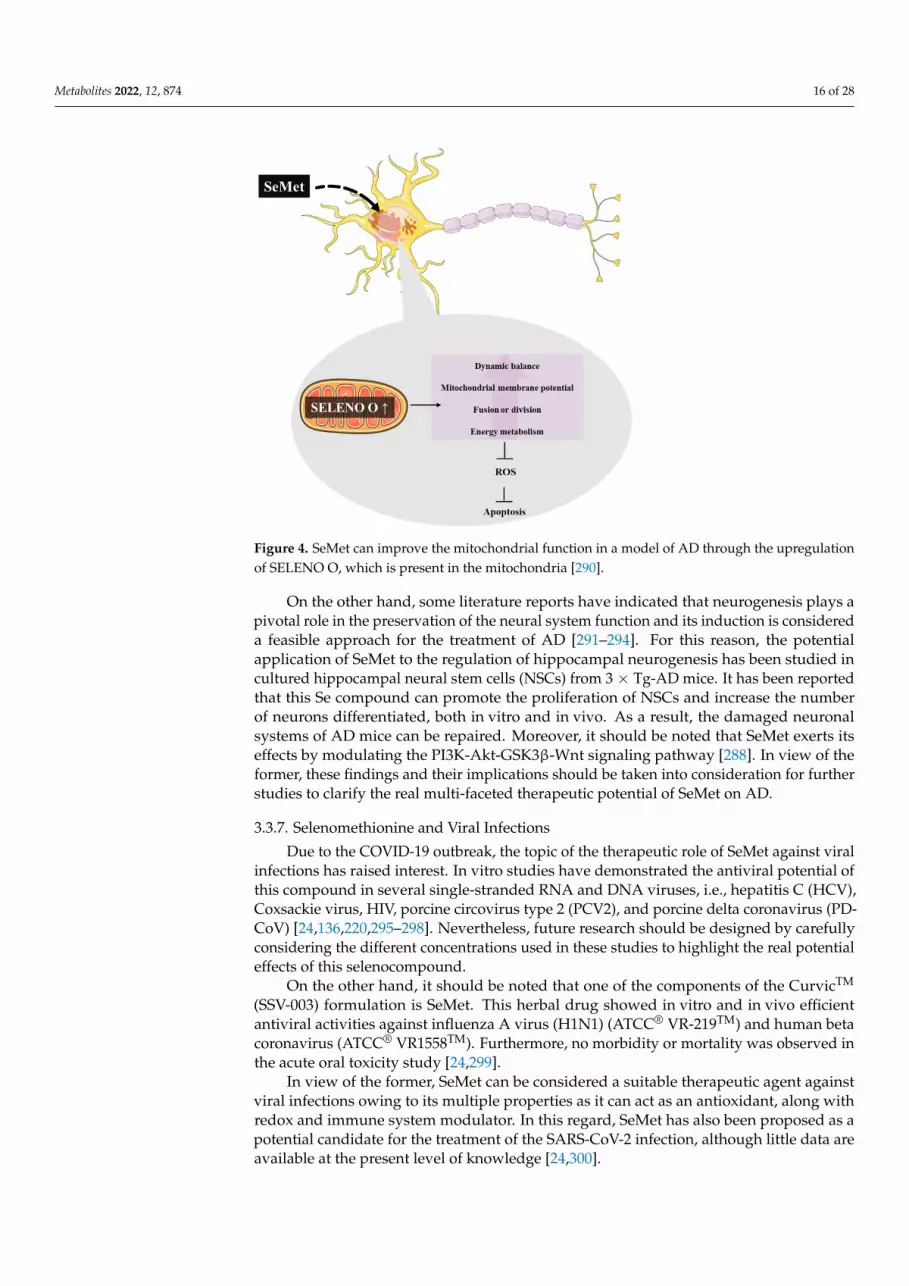
Metabolites 2022, 12, 874 16 of 28Metabolites 2022, 12, x FOR PEER REVIEW 8 of 34
Figure 4. SeMet can improve the mitochondrial function in a model of AD through the upregula-tion of SELENO O, which is present in the mitochondria [290].
3.3.7. Selenomethionine and Viral Infections Due to the COVID-19 outbreak, the topic of the therapeutic role of SeMet against
viral infections has raised interest. In vitro studies have demonstrated the antiviral po-tential of this compound in several single-stranded RNA and DNA viruses, i.e., hepatitis C (HCV), Coxsackie virus, HIV, porcine circovirus type 2 (PCV2), and porcine delta coronavirus (PDCoV) [24,136,220,295–298]. Nevertheless, future research should be de-signed by carefully considering the different concentrations used in these studies to highlight the real potential effects of this selenocompound.
On the other hand, it should be noted that one of the components of the CurvicTM (SSV-003) formulation is SeMet. This herbal drug showed in vitro and in vivo efficient antiviral activities against influenza A virus (H1N1) (ATCC® VR-219TM) and human beta coronavirus (ATCC® VR1558TM). Furthermore, no morbidity or mortality was observed in the acute oral toxicity study [24,299].
In view of the former, SeMet can be considered a suitable therapeutic agent against viral infections owing to its multiple properties as it can act as an antioxidant, along with redox and immune system modulator. In this regard, SeMet has also been proposed as a potential candidate for the treatment of the SARS-CoV-2 infection, although little data are available at the present level of knowledge [24,300].
4. Conclusions Currently, the development of new drugs presents several setbacks, such as toxicity,
solubility, and poor pharmacological profiles. In this context, the use of prodrugs is a feasible approach to overcome these issues. Among them, Se compounds that can render the two main Se-metabolites (H2Se and CH3SeH) have raised great interest in the last decade. The most studied ones are SS, MSA, MSC, and SLM, which have proven to dis-play potent biological activities towards several pathologies, including cancer, viral in-fections, and neurodegenerative and cardiovascular diseases.
The pieces of evidence gathered in this review point out that the searching for new precursors of these metabolites would be a promising toolkit to be considered in the discovery of safer and more efficient derivatives for treating a plethora of illnesses.
Figure 4. SeMet can improve the mitochondrial function in a model of AD through the upregulationof SELENO O, which is present in the mitochondria [290].
On the other hand, some literature reports have indicated that neurogenesis plays apivotal role in the preservation of the neural system function and its induction is considereda feasible approach for the treatment of AD [291–294]. For this reason, the potentialapplication of SeMet to the regulation of hippocampal neurogenesis has been studied incultured hippocampal neural stem cells (NSCs) from 3 × Tg-AD mice. It has been reportedthat this Se compound can promote the proliferation of NSCs and increase the numberof neurons differentiated, both in vitro and in vivo. As a result, the damaged neuronalsystems of AD mice can be repaired. Moreover, it should be noted that SeMet exerts itseffects by modulating the PI3K-Akt-GSK3β-Wnt signaling pathway [288]. In view of theformer, these findings and their implications should be taken into consideration for furtherstudies to clarify the real multi-faceted therapeutic potential of SeMet on AD.
3.3.7. Selenomethionine and Viral Infections
Due to the COVID-19 outbreak, the topic of the therapeutic role of SeMet against viralinfections has raised interest. In vitro studies have demonstrated the antiviral potential ofthis compound in several single-stranded RNA and DNA viruses, i.e., hepatitis C (HCV),Coxsackie virus, HIV, porcine circovirus type 2 (PCV2), and porcine delta coronavirus (PD-CoV) [24,136,220,295–298]. Nevertheless, future research should be designed by carefullyconsidering the different concentrations used in these studies to highlight the real potentialeffects of this selenocompound.
On the other hand, it should be noted that one of the components of the CurvicTM
(SSV-003) formulation is SeMet. This herbal drug showed in vitro and in vivo efficientantiviral activities against influenza A virus (H1N1) (ATCC® VR-219TM) and human betacoronavirus (ATCC® VR1558TM). Furthermore, no morbidity or mortality was observed inthe acute oral toxicity study [24,299].
In view of the former, SeMet can be considered a suitable therapeutic agent againstviral infections owing to its multiple properties as it can act as an antioxidant, along withredox and immune system modulator. In this regard, SeMet has also been proposed as apotential candidate for the treatment of the SARS-CoV-2 infection, although little data areavailable at the present level of knowledge [24,300].

Metabolites 2022, 12, 874 17 of 28
4. Conclusions
Currently, the development of new drugs presents several setbacks, such as toxicity,solubility, and poor pharmacological profiles. In this context, the use of prodrugs is afeasible approach to overcome these issues. Among them, Se compounds that can renderthe two main Se-metabolites (H2Se and CH3SeH) have raised great interest in the lastdecade. The most studied ones are SS, MSA, MSC, and SLM, which have proven to displaypotent biological activities towards several pathologies, including cancer, viral infections,and neurodegenerative and cardiovascular diseases.
The pieces of evidence gathered in this review point out that the searching for newprecursors of these metabolites would be a promising toolkit to be considered in thediscovery of safer and more efficient derivatives for treating a plethora of illnesses.
Author Contributions: Conceptualization, C.M.-S. and D.P.; writing—original draft preparation,C.M.-S.; writing—review and editing, E.A.-E., A.H.-F., C.S., A.K.S. and D.P. All authors have readand agreed to the published version of the manuscript.
Funding: This work was funded by Plan de Investigación de la Universidad de Navarra, PIUNA(2018-19). Cristina Morán-Serradilla acknowledges the support of Gobierno de Navarra for a Ph.D.fellowship. Andreina Henriquez-Figuereo acknowledges the Instituto de Salud Tropical de la Univer-sidad de Navarra (ISTUN) for a Ph.D. fellowship.
Acknowledgments: The authors acknowledge the use of Servier Medical Arts as Figures 1, 3 and 4were partially generated using Servier Medical Art provided by Servier, licensed under a CreativeCommons Attribution 3.0 unported license.
Conflicts of Interest: The authors declare no conflict of interest.
References1. Kieliszek, M. Selenium–fascinating microelement, properties and sources in food. Molecules 2019, 24, 1298. [CrossRef] [PubMed]2. Santi, C.; Bagnoli, L. Celebrating two centuries of research in selenium chemistry: State of the art and new prospective. Molecules
2017, 22, 2124. [CrossRef] [PubMed]3. Avery, J.C.; Hoffmann, P.R. Selenium, selenoproteins, and immunity. Nutrients 2018, 10, 1203. [CrossRef] [PubMed]4. Rayman, M.P. Selenium intake, status, and health: A complex relationship. Hormones 2020, 19, 9–14. [CrossRef]5. Barchielli, G.; Capperucci, A.; Tanini, D. The role of selenium in pathologies: An updated review. Antioxidants 2022, 11, 251.
[CrossRef]6. Kuršvietiene, L.; Mongirdiene, A.; Bernatoniene, J.; Šulinskiene, J.; Staneviciene, I. Selenium anticancer properties and impact on
cellular redox status. Antioxidants 2020, 9, 80. [CrossRef]7. Radomska, D.; Czarnomysy, R.; Radomski, D.; Bielawska, A.; Bielawski, K. Selenium as a bioactive micronutrient in the human
diet and its cancer chemopreventive activity. Nutrients 2021, 13, 1649. [CrossRef]8. Dos Santos, M.; da Silva Júnior, F.M.R.; Muccillo-Baisch, A.L. Selenium content of brazilian foods: A review of the literature
values. J. Food Compost. Anal. 2017, 58, 10–15. [CrossRef]9. Ferreira, R.L.U.; Sena-Evangelista, K.C.M.; de Azevedo, E.P.; Pinheiro, F.I.; Cobucci, R.N.; Pedrosa, L.F.C. Selenium in human
health and gut microflora: Bioavailability of selenocompounds and relationship with diseases. Front. Nutr. 2021, 8, 685317.[CrossRef]
10. Chuai, H.; Zhang, S.-Q.; Bai, H.; Li, J.; Wang, Y.; Sun, J.; Wen, E.; Zhang, J.; Xin, M. Small molecule selenium-containingcompounds: Recent development and therapeutic applications. Eur. J. Med. Chem. 2021, 223, 113621. [CrossRef]
11. Yang, R.; Liu, Y.; Zhou, Z. Selenium and selenoproteins, from structure, function to food resource and nutrition. Food Sci. Technol.Res. 2017, 23, 363–373. [CrossRef]
12. Tsuji, P.A.; Santesmasses, D.; Lee, B.J.; Gladyshev, V.N.; Hatfield, D.L. Historical roles of selenium and selenoproteins in healthand development: The good, the bad and the ugly. Int. J. Mol. Sci. 2021, 23, 5. [CrossRef]
13. Hariharan, S.; Dharmaraj, S. Selenium and selenoproteins: It’s role in regulation of inflammation. Inflammopharmacology 2020, 28,667–695. [CrossRef]
14. Qazi, I.H.; Angel, C.; Yang, H.; Zoidis, E.; Pan, B.; Wu, Z.; Ming, Z.; Zeng, C.J.; Meng, Q.; Han, H.; et al. Role of selenium andselenoproteins in male reproductive function: A review of past and present evidences. Antioxidants 2019, 8, 268. [CrossRef]
15. Arias-Borrego, A.; Callejón-Leblic, B.; Calatayud, M.; Gómez-Ariza, J.L.; Collado, M.C.; García-Barrera, T. Insights into cancer andneurodegenerative diseases through selenoproteins and the connection with gut microbiota—Current analytical methodologies.Expert Rev. Proteom. 2019, 16, 805–814. [CrossRef]
16. Qazi, I.H.; Angel, C.; Yang, H.; Pan, B.; Zoidis, E.; Zeng, C.-J.; Han, H.; Zhou, G.-B. Selenium, selenoproteins, and femalereproduction: A review. Molecules 2018, 23, 3053. [CrossRef]

Metabolites 2022, 12, 874 18 of 28
17. Barrett, C.W.; Short, S.P.; Williams, C.S. Selenoproteins and oxidative stress-induced inflammatory tumorigenesis in the gut. Cell.Mol. Life Sci. 2017, 74, 607–616. [CrossRef]
18. Jehan, C.; Cartier, D.; Bucharles, C.; Anouar, Y.; Lihrmann, I. Emerging roles of ER-resident selenoproteins in brain physiologyand physiopathology. Redox Biol. 2022, 55, 102412. [CrossRef]
19. Koeberle, S.C.; Gollowitzer, A.; Laoukili, J.; Kranenburg, O.; Werz, O.; Koeberle, A.; Kipp, A.P. Distinct and overlapping functionsof glutathione peroxidases 1 and 2 in limiting NF-κB-driven inflammation through redox-active mechanisms. Redox Biol. 2020, 28,101388. [CrossRef]
20. Lee, J.H.; Jang, J.K.; Ko, K.Y.; Jin, Y.; Ham, M.; Kang, H.; Kim, I.Y. Degradation of selenoprotein S and selenoprotein K throughPPARγ-mediated ubiquitination is required for adipocyte differentiation. Cell Death Differ. 2019, 26, 1007–1023. [CrossRef]
21. Bevinakoppamath, S.; Saleh Ahmed, A.M.; Ramachandra, S.C.; Vishwanath, P.; Prashant, A. Chemopreventive and anticancerproperty of selenoproteins in obese breast cancer. Front. Pharmacol. 2021, 12, 618172. [CrossRef] [PubMed]
22. Lennicke, C.; Rahn, J.; Bukur, J.; Hochgräfe, F.; Wessjohann, L.A.; Lichtenfels, R.; Seliger, B. Modulation of MHC class I surfaceexpression in B16F10 melanoma cells by methylseleninic acid. OncoImmunology 2017, 6, e1259049. [CrossRef] [PubMed]
23. Poluboyarinov, P.A.; Elistratov, D.G.; Moiseeva, I.J. Antitumor activity of selenium and search parameters for its new potentiallyactive derivatives. Russ. J. Bioorg. Chem. 2020, 46, 989–1003. [CrossRef]
24. Mal’tseva, V.N.; Goltyaev, M.V.; Turovsky, E.A.; Varlamova, E.G. Immunomodulatory and anti-inflammatory properties ofselenium-containing agents: Their role in the regulation of defense mechanisms against COVID-19. Int. J. Mol. Sci. 2022, 23, 2360.[CrossRef]
25. Kang, X.; Huang, H.; Jiang, C.; Cheng, L.; Sang, Y.; Cai, X.; Dong, Y.; Sun, L.; Wen, X.; Xi, Z.; et al. Cysteine-activatedsmall-molecule H2Se donors inspired by synthetic H2S donors. J. Am. Chem. Soc. 2022, 144, 3957–3967. [CrossRef]
26. Newton, T.D.; Bolton, S.G.; Garcia, A.C.; Chouinard, J.E.; Golledge, S.L.; Zakharov, L.N.; Pluth, M.D. Hydrolysis-based small-molecule hydrogen selenide (H2Se) donors for intracellular H2Se delivery. J. Am. Chem. Soc. 2021, 143, 19542–19550. [CrossRef]
27. Luan, D.; Zhao, Z.; Xia, D.; Zheng, Q.; Gao, X.; Xu, K.; Tang, B. Hydrogen selenide, a vital metabolite of sodium selenite, uncouplesthe sulfilimine bond and promotes the reversal of liver fibrosis. Sci. China Life Sci. 2021, 64, 443–451. [CrossRef]
28. Kuganesan, M.; Samra, K.; Evans, E.; Singer, M.; Dyson, A. Selenium and hydrogen selenide: Essential micronutrient and thefourth gasotransmitter? Intensive Care Med. Exp. 2019, 7, 71. [CrossRef]
29. Hartmann, C.; Nussbaum, B.; Calzia, E.; Radermacher, P.; Wepler, M. Gaseous mediators and mitochondrial function: The futureof pharmacologically induced suspended animation? Front. Physiol. 2017, 8, 691. [CrossRef]
30. Samra, K.; Kuganesan, M.; Smith, W.; Kleyman, A.; Tidswell, R.; Arulkumaran, N.; Singer, M.; Dyson, A. The pharmacology andtherapeutic utility of sodium hydroselenide. Int. J. Mol. Sci. 2021, 22, 3258. [CrossRef]
31. Kong, F.; Ge, L.; Pan, X.; Xu, K.; Liu, X.; Tang, B. A highly selective near-infrared fluorescent probe for imaging H2Se in livingcells and in vivo. Chem. Sci. 2016, 7, 1051–1056. [CrossRef]
32. Kong, F.; Zhao, Y.; Liang, Z.; Liu, X.; Pan, X.; Luan, D.; Xu, K.; Tang, B. Highly selective fluorescent probe for imaging H2Se inliving cells and in vivo based on the disulfide bond. Anal. Chem. 2017, 89, 688–693. [CrossRef]
33. Pan, X.; Song, X.; Wang, C.; Cheng, T.; Luan, D.; Xu, K.; Tang, B. H2Se induces reductive stress in HepG2 cells and activates cellautophagy by regulating the redox of HMGB1 protein under hypoxia. Theranostics 2019, 9, 1794–1808. [CrossRef]
34. Kim, S.J.; Choi, M.C.; Park, J.M.; Chung, A.S. Antitumor effects of selenium. Int. J. Mol. Sci. 2021, 22, 11844. [CrossRef]35. Hagemann-Jensen, M.; Uhlenbrock, F.; Kehlet, S.; Andresen, L.; Gabel-Jensen, C.; Ellgaard, L.; Gammelgaard, B.; Skov, S. The
selenium metabolite methylselenol regulates the expression of ligands that trigger immune activation through the lymphocytereceptor NKG2D. J. Biol. Chem. 2014, 289, 31576–31590. [CrossRef]
36. Dauplais, M.; Bierla, K.; Maizeray, C.; Lestini, R.; Lobinski, R.; Plateau, P.; Szpunar, J.; Lazard, M. Methylselenol produced in vivofrom methylseleninic acid or dimethyl diselenide induces toxic protein aggregation in Saccharomyces cerevisiae. Int. J. Mol. Sci.2021, 22, 2241. [CrossRef]
37. Díaz-Argelich, N.; Encío, I.; Plano, D.; Fernandes, A.P.; Palop, J.A.; Sanmartín, C. Novel methylselenoesters as antiproliferativeagents. Molecules 2017, 22, 1288. [CrossRef]
38. Dauplais, M.; Mahou, P.; Plateau, P.; Lazard, M. Exposure to the methylselenol precursor dimethyldiselenide induces a reductiveendoplasmic reticulum stress in Saccharomyces cerevisiae. Int. J. Mol. Sci. 2021, 22, 5467. [CrossRef]
39. Ebert, F.; Müller, S.M.; Meyer, S.; Schwerdtle, T. Selenium and toxicological aspects: Cytotoxicity, cellular bioavailability, andbiotransformation of Se species. In Selenium; Michalke, B., Ed.; Springer International Publishing: Cham, Switzerland, 2018; pp. 373–391.
40. Font, M.; Romano, B.; González-Peñas, E.; Sanmartín, C.; Plano, D.; Palop, J.A. Methylselenol release as a cytotoxic tool: A studyof the mechanism of the activity achieved by two series of methylselenocarbamate derivatives. Metallomics 2018, 10, 1128–1140.[CrossRef]
41. Kieliszek, M.; Lipinski, B.; Blazejak, S. Application of sodium selenite in the prevention and treatment of cancers. Cells 2017, 6, 39.[CrossRef]
42. Hiffler, L.; Rakotoambinina, B. Selenium and RNA virus interactions: Potential implications for SARS-CoV-2 infection (COVID-19).Front. Nutr. 2020, 7, 164. [CrossRef]
43. Varela, M.F.; Stephen, J.; Lekshmi, M.; Ojha, M.; Wenzel, N.; Sanford, L.M.; Hernandez, A.J.; Parvathi, A.; Kumar, S.H. Bacterialresistance to antimicrobial agents. Antibiotics 2021, 10, 593. [CrossRef]

Metabolites 2022, 12, 874 19 of 28
44. Lai, C.-C.; Chen, S.-Y.; Ko, W.-C.; Hsueh, P.-R. Increased antimicrobial resistance during the COVID-19 pandemic. Int. J. Antimicrob.Agents 2021, 57, 106324. [CrossRef]
45. Rodríguez-Baño, J.; Rossolini, G.M.; Schultsz, C.; Tacconelli, E.; Murthy, S.; Ohmagari, N.; Holmes, A.; Bachmann, T.; Goossens,H.; Canton, R.; et al. Key considerations on the potential impacts of the COVID-19 pandemic on antimicrobial resistance researchand surveillance. Trans. R. Soc. Trop. Med. Hyg. 2021, 115, 1122–1129. [CrossRef]
46. Hu, X.; Guo, J.; Xu, M.; Jiang, P.; Yuan, X.; Zhao, C.; Maimai, T.; Cao, Y.; Zhang, N.; Fu, Y. Clostridium tyrobutyricum alleviatesStaphylococcus aureus-induced endometritis in mice by inhibiting endometrial barrier disruption and inflammatory response. FoodFunct. 2019, 10, 6699–6710. [CrossRef]
47. Pellissery, A.J.; Vinayamohan, P.G.; Yin, H.B.; Mooyottu, S.; Venkitanarayanan, K. In vitro efficacy of sodium selenite in reducingtoxin production, spore outgrowth and antibiotic resistance in hypervirulent Clostridium difficile. J. Med. Microbiol. 2019, 68,1118–1128. [CrossRef]
48. Mosolygo, T.; Kincses, A.; Csonka, A.; Tonki, A.S.; Witek, K.; Sanmartin, C.; Marc, M.A.; Handzlik, J.; Kiec-Kononowicz, K.;Dominguez-Alvarez, E.; et al. Selenocompounds as novel antibacterial agents and bacterial efflux pump inhibitors. Molecules2019, 24, 1487. [CrossRef]
49. Kumar, B.S.; Tiwari, S.K.; Manoj, G.; Kunwar, A.; Amrita, N.; Sivaram, G.; Abid, Z.; Ahmad, A.; Khan, A.A.; Priyadarsini, K.I.Anti-unlcer and antimicrobial activities of sodium selenite against Helicobacter pylori: In vitro and in vivo evaluation. Scand. J.Infect. Dis. 2010, 42, 266–274. [CrossRef]
50. Boonpiyathad, T.; Sözener, Z.C.; Satitsuksanoa, P.; Akdis, C.A. Immunologic mechanisms in asthma. Semin. Immunol. 2019, 46,101333. [CrossRef]
51. Allam, V.; Paudel, K.R.; Gupta, G.; Singh, S.K.; Vishwas, S.; Gulati, M.; Gupta, S.; Chaitanya, M.; Jha, N.K.; Gupta, P.K.; et al.Nutraceuticals and mitochondrial oxidative stress: Bridging the gap in the management of bronchial asthma. Environ. Sci. Pollut.Res. Int. 2022, 29, 1–22. [CrossRef] [PubMed]
52. Ammar, M.; Bahloul, N.; Amri, O.; Omri, R.; Ghozzi, H.; Kammoun, S.; Zeghal, K.; Ben Mahmoud, L. Oxidative stress in patientswith asthma and its relation to uncontrolled asthma. J. Clin. Lab. Anal. 2022, 36, e24345. [CrossRef] [PubMed]
53. Karadogan, B.; Beyaz, S.; Gelincik, A.; Buyukozturk, S.; Arda, N. Evaluation of oxidative stress biomarkers and antioxidantparameters in allergic asthma patients with different level of asthma control. J. Asthma 2022, 59, 663–672. [CrossRef] [PubMed]
54. Shaheen, S.O.; Newson, R.B.; Rayman, M.P.; Wong, A.P.; Tumilty, M.K.; Phillips, J.M.; Potts, J.F.; Kelly, F.J.; White, P.T.; Burney,P.G. Randomised, double blind, placebo-controlled trial of selenium supplementation in adult asthma. Thorax 2007, 62, 483–490.[CrossRef] [PubMed]
55. WHO Organization. Cancer. Available online: https://www.who.int/news-room/fact-sheets/detail/cancer (accessed on 3 July 2022).56. Kumar, A.; Jaitak, V. Natural products as multidrug resistance modulators in cancer. Eur. J. Med. Chem. 2019, 176, 268–291.
[CrossRef]57. Yuan, J.; Li, X.; Zhang, Y.; Zhang, G.; Cheng, W.; Wang, W.; Lei, Y.; Song, G. USP39 attenuates the antitumor activity of cisplatin
on colon cancer cells dependent on p53. Cell Biol. Toxicol. 2021, 1–16. [CrossRef]58. Olatunde, A.; Nigam, M.; Singh, R.K.; Panwar, A.S.; Lasisi, A.; Alhumaydhi, F.A.; Jyoti Kumar, V.; Mishra, A.P.; Sharifi-Rad, J. Cancer
and diabetes: The interlinking metabolic pathways and repurposing actions of antidiabetic drugs. Cancer Cell Int. 2021, 21, 499.[CrossRef]
59. Su, Z.; Dong, S.; Zhao, S.-C.; Liu, K.; Tan, Y.; Jiang, X.; Assaraf, Y.G.; Qin, B.; Chen, Z.-S.; Zou, C. Novel nanomedicines toovercome cancer multidrug resistance. Drug Resist. Updat. 2021, 58, 100777. [CrossRef]
60. Ahmad, M.H.; Rizvi, M.A.; Fatima, M.; Mondal, A.C. Pathophysiological implications of neuroinflammation mediated HPA axisdysregulation in the prognosis of cancer and depression. Mol. Cell. Endocrinol. 2021, 520, 111093. [CrossRef]
61. Evans, S.O.; Jacobson, G.M.; Goodman, H.J.B.; Bird, S.; Jameson, M.B. Comparative safety and pharmacokinetic evaluation ofthree oral selenium compounds in cancer patients. Biol. Trace Elem. Res. 2019, 189, 395–404. [CrossRef]
62. Hu, W.; Zhao, C.; Hu, H.; Yin, S. Food sources of selenium and its relationship with chronic diseases. Nutrients 2021, 13, 1739.[CrossRef]
63. Gandin, V.; Khalkar, P.; Braude, J.; Fernandes, A.P. Organic selenium compounds as potential chemotherapeutic agents forimproved cancer treatment. Free Radic. Biol. Med. 2018, 127, 80–97. [CrossRef]
64. Rataan, A.O.; Geary, S.M.; Zakharia, Y.; Rustum, Y.M.; Salem, A.K. Potential role of selenium in the treatment of cancer and viralinfections. Int. J. Mol. Sci. 2022, 23, 2215. [CrossRef]
65. Radomska, D.; Czarnomysy, R.; Radomski, D.; Bielawski, K. Selenium compounds as novel potential anticancer agents. Int. J.Mol. Sci. 2021, 22, 1009. [CrossRef]
66. Tan, H.W.; Mo, H.Y.; Lau, A.T.Y.; Xu, Y.M. Selenium species: Current status and potentials in cancer prevention and therapy. Int. J.Mol. Sci. 2018, 20, 75. [CrossRef]
67. Stolwijk, J.M.; Garje, R.; Sieren, J.C.; Buettner, G.R.; Zakharia, Y. Understanding the redox biology of selenium in the search oftargeted cancer therapies. Antioxidants 2020, 9, 420. [CrossRef]
68. Kim, J.B.; Yang, E.Y.; Woo, J.; Kwon, H.; Lim, W.; Moon, B.I. Sodium selenite enhanced the anti-proliferative effect of MEK-ERKinhibitor in thyroid cancer cells. In Vivo 2020, 34, 185–190. [CrossRef]

Metabolites 2022, 12, 874 20 of 28
69. Domínguez-Álvarez, E.; Rácz, B.; Marc, M.A.; Nasim, M.J.; Szemerédi, N.; Viktorová, J.; Jacob, C.; Spengler, G. Selenium andtellurium in the development of novel small molecules and nanoparticles as cancer multidrug resistance reversal agents. DrugResist. Updat. 2022, 63, 100844. [CrossRef]
70. Szemerédi, N.; Dobiasová, S.; Salardón-Jiménez, N.; Kincses, A.; Nové, M.; Habibullah, G.; Sevilla-Hernández, C.; Benito-Lama,M.; Alonso-Martínez, F.-J.; Viktorová, J.; et al. Cyano- and ketone-containing selenoesters as multi-target compounds againstresistant cancers. Cancers 2021, 13, 4563. [CrossRef]
71. Valente, A.; Podolski-Renic, A.; Poetsch, I.; Filipovic, N.; López, Ó.; Turel, I.; Heffeter, P. Metal- and metalloid-based compoundsto target and reverse cancer multidrug resistance. Drug Resist. Updat. 2021, 58, 100778. [CrossRef]
72. Lendvai, G.; Szekerczes, T.; Kontsek, E.; Selvam, A.; Szakos, A.; Schaff, Z.; Bjornstedt, M.; Kiss, A. The effect of methylselenocys-teine and sodium selenite treatment on microRNA expression in liver cancer cell lines. Pathol. Oncol. Res. 2020, 26, 2669–2681.[CrossRef]
73. Jablonska, E.; Li, Q.; Reszka, E.; Wieczorek, E.; Tarhonska, K.; Wang, T. Therapeutic potential of selenium and selenium compoundsin cervical cancer. Cancer Control 2021, 28, 10732748211001808. [CrossRef] [PubMed]
74. Augustyniak, M. Selenium and cancer or adenoma related to the large bowel. Postepy Hig. Med. Dosw. 2021, 75, 426–436.[CrossRef]
75. Razaghi, A.; Poorebrahim, M.; Sarhan, D.; Björnstedt, M. Selenium stimulates the antitumour immunity: Insights to futureresearch. Eur. J. Cancer 2021, 155, 256–267. [CrossRef] [PubMed]
76. Yu, H.; Huang, Y.; Ge, Y.; Hong, X.; Lin, X.; Tang, K.; Wang, Q.; Yang, Y.; Sun, W.; Huang, Y.; et al. Selenite-inducedROS/AMPK/FoxO3a/GABARAPL-1 signaling pathway modulates autophagy that antagonize apoptosis in colorectal cancercells. Discov. Oncol. 2021, 12, 35. [CrossRef]
77. Doello, K.; Mesas, C.; Quinonero, F.; Perazzoli, G.; Cabeza, L.; Prados, J.; Melguizo, C.; Ortiz, R. The antitumor activity of sodiumselenite alone and in combination with gemcitabine in pancreatic cancer: An in vitro and in vivo study. Cancers 2021, 13, 3169.[CrossRef]
78. Soukupova, K.; Rudolf, E. Suppression of proliferation and activation of cell death by sodium selenite involves mitochondria andlysosomes in chemoresistant bladder cancer cells. J. Trace Elem. Med. Biol. 2019, 52, 58–67. [CrossRef]
79. Larrouquère, L.; Berthier, S.; Chovelon, B.; Garrel, C.; Vacchina, V.; Paucot, H.; Boutonnat, J.; Faure, P.; Hazane-Puch, F.Preclinical evaluation of sodium selenite in mice: Toxicological and tumor regression studies after striatum implantation ofhuman glioblastoma stem cells. Int. J. Mol. Sci. 2021, 22, 10646. [CrossRef]
80. Powers, M.; Liu, L.; Deemer, D.; Chen, S.; Scholl, A.; Yoshinaga, M.; Liu, Z. Selenite inhibits Notch signaling in cells and mice. Int.J. Mol. Sci. 2021, 22, 2518. [CrossRef]
81. Woo, J.; Kim, J.B.; Cho, T.; Yoo, E.H.; Moon, B.-I.; Kwon, H.; Lim, W. Selenium inhibits growth of trastuzumab-resistant humanbreast cancer cells via downregulation of Akt and beclin-1. PLoS ONE 2021, 16, e0257298. [CrossRef]
82. Shimada, B.K.; Alfulaij, N.; Seale, L.A. The impact of selenium deficiency on cardiovascular function. Int. J. Mol. Sci. 2021, 22,10713. [CrossRef]
83. Bomer, N.; Grote Beverborg, N.; Hoes, M.F.; Streng, K.W.; Vermeer, M.; Dokter, M.M.; IJmker, J.; Anker, S.D.; Cleland, J.G.F.;Hillege, H.L.; et al. Selenium and outcome in heart failure. Eur. J. Heart Fail. 2020, 22, 1415–1423. [CrossRef]
84. Shi, Y.; Yang, W.; Tang, X.; Yan, Q.; Cai, X.; Wu, F. Keshan disease: A potentially fatal endemic cardiomyopathy in remotemountains of China. Front. Pediatr. 2021, 9, 576916. [CrossRef]
85. Yan, C.; Luo, R.; Li, F.; Liu, M.; Li, J.; Hua, W.; Li, X. The epidemiological status, environmental and genetic factors in the etiologyof Keshan disease. Cardiovasc. Endocrinol. Metab. 2021, 10, 14–21. [CrossRef]
86. Xia, J.; Xia, X.; Wang, W.; Xia, J.; Li, M. Protective effect of Se-methylselenocysteine on elaidic acid-induced inflammation inhuman arterial endothelial cells. J. Nutr. Sci. Vitaminol. 2020, 66, 577–582. [CrossRef]
87. Cai, C.; Wu, Y.; Yang, L.; Xiang, Y.; Zhu, N.; Zhao, H.; Hu, W.; Lv, L.; Zeng, C. Sodium selenite attenuates balloon injury-inducedand monocrotaline-induced vascular remodeling in rats. Front. Pharmacol. 2021, 12, 618493. [CrossRef]
88. Ye, C.; Zheng, F.; Wu, N.; Zhu, G.-q.; Li, X.-z. Extracellular vesicles in vascular remodeling. Acta Pharmacol. Sin. 2022, 43,2191–2201. [CrossRef]
89. Xiao, J.; Li, N.; Xiao, S.; Wu, Y.; Liu, H. Comparison of selenium nanoparticles and sodium selenite on the alleviation of earlyatherosclerosis by inhibiting endothelial dysfunction and inflammation in apolipoprotein E-deficient mice. Int. J. Mol. Sci. 2021,22, 11612. [CrossRef]
90. Dong, H.L.; Yuan, N.; Sun, T.; Dun, A.S.; Hou, H.F. Effects of selenium supplement on atherogenesis of ApoE-knockout mice fedhigh fat diet. Zhonghua Xin Xue Guan Bing Za Zhi 2016, 44, 244–249.
91. Krohn, R.M.; Lemaire, M.; Negro Silva, L.F.; Lemarié, C.; Bolt, A.; Mann, K.K.; Smits, J.E. High-selenium lentil diet protectsagainst arsenic-induced atherosclerosis in a mouse model. J. Nutr. Biochem. 2016, 27, 9–15. [CrossRef]
92. Wang, X.; Sui, H.; Su, Y.; Zhao, S. Protective effects of sodium selenite on insulin secretion and diabetic retinopathy in rats withtype 1 diabetes mellitus. Pak. J. Pharm. Sci. 2021, 34, 1729–1735.
93. Schomburg, L. Selenium deficiency due to diet, pregnancy, severe illness, or COVID-19—A preventable trigger for autoimmunedisease. Int. J. Mol. Sci. 2021, 22, 8532. [CrossRef]
94. Ilonen, J.; Lempainen, J.; Veijola, R. The heterogeneous pathogenesis of type 1 diabetes mellitus. Nat. Rev. Endocrinol. 2019, 15,635–650. [CrossRef]

Metabolites 2022, 12, 874 21 of 28
95. Cecilia, O.M.; José Alberto, C.G.; José, N.P.; Ernesto Germán, C.M.; Ana Karen, L.C.; Luis Miguel, R.P.; Ricardo Raúl, R.R.; AdolfoDaniel, R.C. Oxidative stress as the main target in diabetic retinopathy pathophysiology. J. Diabetes Res. 2019, 2019, 8562408.[CrossRef]
96. Muller, T.E.; Nunes, M.E.; Menezes, C.C.; Marins, A.T.; Leitemperger, J.; Gressler, A.C.L.; Carvalho, F.B.; de Freitas, C.M.; Quadros,V.A.; Fachinetto, R.; et al. Sodium selenite prevents paraquat-induced neurotoxicity in zebrafish. Mol. Neurobiol. 2018, 55,1928–1941. [CrossRef]
97. Colle, D.; Santos, D.B.; de Souza, V.; Lopes, M.W.; Leal, R.B.; de Souza Brocardo, P.; Farina, M. Sodium selenite protects from3-nitropropionic acid-induced oxidative stress in cultured primary cortical neurons. Mol. Biol. Rep. 2019, 46, 751–762. [CrossRef]
98. Vicente-Zurdo, D.; Romero-Sanchez, I.; Rosales-Conrado, N.; Leon-Gonzalez, M.E.; Madrid, Y. Ability of selenium species toinhibit metal-induced Aβ aggregation involved in the development of Alzheimer’s disease. Anal. Bioanal. Chem. 2020, 412,6485–6497. [CrossRef]
99. Juszczyk, G.; Mikulska, J.; Kasperek, K.; Pietrzak, D.; Mrozek, W.; Herbet, M. Chronic stress and oxidative stress as common factorsof the pathogenesis of depression and Alzheimer’s disease: The role of antioxidants in prevention and treatment. Antioxidants2021, 10, 1439. [CrossRef]
100. Solovyev, N. Selenoprotein P and its potential role in Alzheimer’s disease. Hormones 2020, 19, 73–79. [CrossRef]101. Sharma, G.; Shin, E.J.; Sharma, N.; Nah, S.Y.; Mai, H.N.; Nguyen, B.T.; Jeong, J.H.; Lei, X.G.; Kim, H.C. Glutathione peroxidase-1
and neuromodulation: Novel potentials of an old enzyme. Food Chem. Toxicol. 2021, 148, 111945. [CrossRef]102. Zhang, Z.H.; Song, G.L. Roles of selenoproteins in brain function and the potential mechanism of selenium in Alzheimer’s disease.
Front. Neurosci. 2021, 15, 646518. [CrossRef] [PubMed]103. Ying, H.; Zhang, Y. Systems biology of selenium and complex disease. Biol. Trace Elem. Res. 2019, 192, 38–50. [CrossRef] [PubMed]104. Reichert, C.O.; de Freitas, F.A.; Sampaio-Silva, J.; Rokita-Rosa, L.; Barros, P.d.L.; Levy, D.; Bydlowski, S.P. Ferroptosis mechanisms
involved in neurodegenerative diseases. Int. J. Mol. Sci. 2020, 21, 8765. [CrossRef] [PubMed]105. Yang, S.; Xie, Z.; Pei, T.; Zeng, Y.; Xiong, Q.; Wei, H.; Wang, Y.; Cheng, W. Salidroside attenuates neuronal ferroptosis by activating
the Nrf2/HO1 signaling pathway in Aβ1-42-induced Alzheimer’s disease mice and glutamate-injured HT22 cells. Chin. Med.2022, 17, 82. [CrossRef]
106. Li, F.; Wang, H.; Chen, H.; Guo, J.; Dang, X.; Ru, Y.; Wang, H. Mechanism of ferroptosis and its role in spinal cord injury. Front.Neurol. 2022, 13, 926780. [CrossRef]
107. Chen, Y.X.; Zuliyaer, T.; Liu, B.; Guo, S.; Yang, D.G.; Gao, F.; Yu, Y.; Yang, M.L.; Du, L.J.; Li, J.J. Sodium selenite promotesneurological function recovery after spinal cord injury by inhibiting ferroptosis. Neural Regen. Res. 2022, 17, 2702–2709. [CrossRef]
108. Dang, X.; Huan, X.; Du, X.; Chen, X.; Bi, M.; Yan, C.; Jiao, Q.; Jiang, H. Correlation of ferroptosis and other types of cell death inneurodegenerative diseases. Neurosci. Bull. 2022, 38, 938–952. [CrossRef]
109. Wu, H.; Luan, Y.; Wang, H.; Zhang, P.; Liu, S.; Wang, P.; Cao, Y.; Sun, H.; Wu, L. Selenium inhibits ferroptosis and amelioratesautistic-like behaviors of BTBR mice by regulating the Nrf2/GPX4 pathway. Brain Res. Bull. 2022, 183, 38–48. [CrossRef]
110. Onukwufor, J.O.; Dirksen, R.T.; Wojtovich, A.P. Iron dysregulation in mitochondrial dysfunction and Alzheimer’s disease.Antioxidants 2022, 11, 692. [CrossRef]
111. Chen, Y.; Liu, S.; Li, J.; Li, Z.; Quan, J.; Liu, X.; Tang, Y.; Liu, B. The latest view on the mechanism of ferroptosis and its researchprogress in spinal cord injury. Oxid. Med. Cell. Longev. 2020, 2020, 6375938. [CrossRef]
112. Wu, X.; Li, R.; Hong, Q.; Chi, X. Development and validation of a novel diagnostic model for childhood autism spectrum disorderbased on ferroptosis-related genes. Front. Psychiatry 2022, 13, 886055. [CrossRef]
113. Lord, C.; Brugha, T.S.; Charman, T.; Cusack, J.; Dumas, G.; Frazier, T.; Jones, E.J.H.; Jones, R.M.; Pickles, A.; State, M.W.; et al.Autism spectrum disorder. Nat. Rev. Dis. Primers 2020, 6, 5. [CrossRef]
114. Tuo, Q.Z.; Masaldan, S.; Southon, A.; Mawal, C.; Ayton, S.; Bush, A.I.; Lei, P.; Belaidi, A.A. Characterization of seleniumcompounds for anti-ferroptotic activity in neuronal cells and after cerebral ischemia-reperfusion injury. Neurotherapeutics 2021, 18,2682–2691. [CrossRef]
115. Alim, I.; Caulfield, J.T.; Chen, Y.; Swarup, V.; Geschwind, D.H.; Ivanova, E.; Seravalli, J.; Ai, Y.; Sansing, L.H.; Ste Marie, E.J.;et al. Selenium drives a transcriptional adaptive program to block ferroptosis and treat stroke. Cell 2019, 177, 1262–1279.e1225.[CrossRef] [PubMed]
116. Capuz, A.; Karnoub, M.A.; Osien, S.; Rose, M.; Mériaux, C.; Fournier, I.; Devos, D.; Vanden Abeele, F.; Rodet, F.; Cizkova, D.; et al.The antibody dependant neurite outgrowth modulation response involvement in spinal cord injury. Front. Immunol. 2022, 13,882830. [CrossRef]
117. Zhang, L.-J.; Chen, Y.; Wang, L.-X.; Zhuang, X.-Q.; Xia, H.-C. Identification of potential oxidative stress biomarkers for spinal cordinjury in erythrocytes using mass spectrometry. Neural Regen. Res. 2021, 16, 1294–1301. [CrossRef]
118. Zhang, X.; Liu, C.-B.; Yang, D.-G.; Qin, C.; Dong, X.-C.; Li, D.-P.; Zhang, C.; Guo, Y.; Du, L.-J.; Gao, F.; et al. Dynamic changes inintramedullary pressure 72 hours after spinal cord injury. Neural Regen. Res. 2019, 14, 886–895.
119. McCreedy, D.A.; Abram, C.L.; Hu, Y.; Min, S.W.; Platt, M.E.; Kirchhoff, M.A.; Reid, S.K.; Jalufka, F.L.; Lowell, C.A. Spleen tyrosinekinase facilitates neutrophil activation and worsens long-term neurologic deficits after spinal cord injury. J. Neuroinflamm. 2021,18, 302. [CrossRef]
120. Ni, Y.; Gu, J.; Wu, J.; Xu, L.; Rui, Y. MGMT-mediated neuron apoptosis in injured rat spinal cord. Tissue Cell 2020, 62, 101311.[CrossRef]

Metabolites 2022, 12, 874 22 of 28
121. Shi, Z.; Yuan, S.; Shi, L.; Li, J.; Ning, G.; Kong, X.; Feng, S. Programmed cell death in spinal cord injury pathogenesis and therapy.Cell Prolif. 2021, 54, e12992. [CrossRef]
122. Yao, X.; Zhang, Y.; Hao, J.; Duan, H.-Q.; Zhao, C.-X.; Sun, C.; Li, B.; Fan, B.-Y.; Wang, X.; Li, W.-X.; et al. Deferoxamine promotesrecovery of traumatic spinal cord injury by inhibiting ferroptosis. Neural Regen. Res. 2019, 14, 532–541.
123. Zhou, H.; Yin, C.; Zhang, Z.; Tang, H.; Shen, W.; Zha, X.; Gao, M.; Sun, J.; Xu, X.; Chen, Q. Proanthocyanidin promotes functionalrecovery of spinal cord injury via inhibiting ferroptosis. J. Chem. Neuroanat. 2020, 107, 101807. [CrossRef] [PubMed]
124. Ge, H.; Xue, X.; Xian, J.; Yuan, L.; Wang, L.; Zou, Y.; Zhong, J.; Jiang, Z.; Shi, J.; Chen, T.; et al. Ferrostatin-1 alleviates white matterinjury via decreasing ferroptosis following spinal cord injury. Mol. Neurobiol. 2022, 59, 161–176. [CrossRef] [PubMed]
125. Zhang, Y.; Sun, C.; Zhao, C.; Hao, J.; Zhang, Y.; Fan, B.; Li, B.; Duan, H.; Liu, C.; Kong, X.; et al. Ferroptosis inhibitor SRS 16-86attenuates ferroptosis and promotes functional recovery in contusion spinal cord injury. Brain Res. 2019, 1706, 48–57. [CrossRef][PubMed]
126. Ge, M.H.; Tian, H.; Mao, L.; Li, D.Y.; Lin, J.Q.; Hu, H.S.; Huang, S.C.; Zhang, C.J.; Mei, X.F. Zinc attenuates ferroptosis andpromotes functional recovery in contusion spinal cord injury by activating Nrf2/GPX4 defense pathway. CNS Neurosci. Ther.2021, 27, 1023–1040. [CrossRef]
127. Jia, Z.-Q.; Li, S.-Q.; Qiao, W.-Q.; Xu, W.-Z.; Xing, J.-W.; Liu, J.-T.; Song, H.; Gao, Z.-Y.; Xing, B.-W.; He, X.-J. Ebselen protectsmitochondrial function and oxidative stress while inhibiting the mitochondrial apoptosis pathway after acute spinal cord injury.Neurosci. Lett. 2018, 678, 110–117. [CrossRef]
128. Luo, W.; Wang, Y.; Lin, F.; Liu, Y.; Gu, R.; Liu, W.; Xiao, C. Selenium-doped carbon quantum dots efficiently ameliorate secondaryspinal cord injury via scavenging reactive oxygen species. Int. J. Nanomed. 2020, 15, 10113–10125. [CrossRef]
129. Javdani, M.; Ghorbani, R.; Hashemnia, M. Histopathological evaluation of spinal cord with experimental traumatic injuryfollowing implantation of a controlled released drug delivery system of chitosan hydrogel loaded with selenium nanoparticle.Biol. Trace Elem. Res. 2021, 199, 2677–2686. [CrossRef]
130. Rao, S.; Lin, Y.; Lin, R.; Liu, J.; Wang, H.; Hu, W.; Chen, B.; Chen, T. Traditional Chinese medicine active ingredients-basedselenium nanoparticles regulate antioxidant selenoproteins for spinal cord injury treatment. J. Nanobiotechnol. 2022, 20, 278.[CrossRef]
131. Wang, W.; Huang, X.; Zhang, Y.; Deng, G.; Liu, X.; Fan, C.; Xi, Y.; Yu, J.; Ye, X. Se@SiO2 nanocomposites suppress microglia-mediated reactive oxygen species during spinal cord injury in rats. RSC Adv. 2018, 8, 16126–16138. [CrossRef]
132. Garcia, E.; Hernández-Ayvar, F.; Rodríguez-Barrera, R.; Flores-Romero, A.; Borlongan, C.; Ibarra, A. Supplementation withvitamin E, zinc, selenium, and copper re-establishes T-cell function and improves motor recovery in a rat model of spinal cordinjury. Cell Transplant. 2022, 31, 9636897221109884. [CrossRef]
133. Cheng, Z.; Zhi, X.; Sun, G.; Guo, W.; Huang, Y.; Sun, W.; Tian, X.; Zhao, F.; Hu, K. Sodium selenite suppresses hepatitis B virustranscription and replication in human hepatoma cell lines. J. Med. Virol. 2016, 88, 653–663. [CrossRef]
134. Bermano, G.; Méplan, C.; Mercer, D.K.; Hesketh, J.E. Selenium and viral infection: Are there lessons for COVID-19? Br. J. Nutr.2021, 125, 618–627. [CrossRef]
135. Guillin, O.M.; Vindry, C.; Ohlmann, T.; Chavatte, L. Selenium, selenoproteins and viral infection. Nutrients 2019, 11, 2101.[CrossRef]
136. Martinez, S.S.; Huang, Y.; Acuna, L.; Laverde, E.; Trujillo, D.; Barbieri, M.A.; Tamargo, J.; Campa, A.; Baum, M.K. Role of seleniumin viral infections with a major focus on SARS-CoV-2. Int. J. Mol. Sci. 2021, 23, 280. [CrossRef]
137. Lima, L.W.; Nardi, S.; Santoro, V.; Schiavon, M. The relevance of plant-derived Se compounds to human health in the SARS-CoV-2(COVID-19) pandemic era. Antioxidants 2021, 10, 1031. [CrossRef]
138. Zhu, Y.-H.; Wang, X.-F.; Yang, G.; Wei, J.; Tan, W.-H.; Wang, L.-X.; Guo, X.; Lammi, M.J.; Xu, J.-H. Efficacy of long-term seleniumsupplementation in the treatment of chronic Keshan disease with congestive heart failure. Curr. Med. Sci. 2019, 39, 237–242.[CrossRef]
139. Thompson, R.; Wood, J.G.; Tempia, S.; Muscatello, D.J. Global variation in early epidemic growth rates and reproduction numberof seasonal influenza. Int. J. Infect. Dis. 2022, 122, 382–388. [CrossRef]
140. World Health Organization. Guidelines for the Clinical Management of Severe Illness from Influenza Virus Infections; World HealthOrganization: Geneva, Switzerland, 2022.
141. Lyons, D.M.; Lauring, A.S. Mutation and epistasis in influenza virus evolution. Viruses 2018, 10, 407. [CrossRef]142. Yin, H.; Jiang, N.; Shi, W.; Chi, X.; Liu, S.; Chen, J.-L.; Wang, S. Development and effects of influenza antiviral drugs. Molecules
2021, 26, 810. [CrossRef]143. Honce, R.; Schultz-Cherry, S. Impact of obesity on influenza a virus pathogenesis, immune response, and evolution. Front.
Immunol. 2019, 10, 1071. [CrossRef]144. Gong, G.; Li, Y.; He, K.; Yang, Q.; Guo, M.; Xu, T.; Wang, C.; Zhao, M.; Chen, Y.; Du, M.; et al. The inhibition of H1N1 influenza
induced apoptosis by sodium selenite through ROS-mediated signaling pathways. RSC Adv. 2020, 10, 8002–8007. [CrossRef]145. Bai, Y.; Jones, J.C.; Wong, S.-S.; Zanin, M. Antivirals targeting the surface glycoproteins of influenza virus: Mechanisms of action
and resistance. Viruses 2021, 13, 624. [CrossRef]146. Shang, J.; Ye, G.; Shi, K.; Wan, Y.; Luo, C.; Aihara, H.; Geng, Q.; Auerbach, A.; Li, F. Structural basis of receptor recognition by
SARS-CoV-2. Nature 2020, 581, 221–224. [CrossRef]

Metabolites 2022, 12, 874 23 of 28
147. Majeed, M.; Nagabhushanam, K.; Prakasan, P.; Mundkur, L. Can selenium reduce the susceptibility and severity of SARS-CoV-2?—A comprehensive review. Int. J. Mol. Sci. 2022, 23, 4809. [CrossRef] [PubMed]
148. Zhang, J.; Saad, R.; Taylor, E.W.; Rayman, M.P. Selenium and selenoproteins in viral infection with potential relevance toCOVID-19. Redox Biol. 2020, 37, 101715. [CrossRef] [PubMed]
149. Kieliszek, M.; Lipinski, B. Selenium supplementation in the prevention of coronavirus infections (COVID-19). Med. Hypotheses2020, 143, 109878. [CrossRef] [PubMed]
150. Ali, W.; Benedetti, R.; Handzlik, J.; Zwergel, C.; Battistelli, C. The innovative potential of selenium-containing agents for fightingcancer and viral infections. Drug Discov. Today 2021, 26, 256–263. [CrossRef] [PubMed]
151. Varlamova, E.G.; Turovsky, E.A. The main cytotoxic effects of methylseleninic acid on various cancer cells. Int. J. Mol. Sci. 2021,22, 6614. [CrossRef]
152. Park, J.-M.; Kim, D.-H.; Na, H.-K.; Surh, Y.-J. Methylseleninic acid induces NAD(P)H:quinone oxidoreductase-1 expressionthrough activation of NF-E2-related factor 2 in Chang liver cells. Oncotarget 2016, 9, 3014–3028. [CrossRef]
153. Nair, D.; Rådestad, E.; Khalkar, P.; Diaz-Argelich, N.; Schröder, A.; Klynning, C.; Ungerstedt, J.; Uhlin, M.; Fernandes, A.P.Methylseleninic acid sensitizes ovarian cancer cells to T-cell mediated killing by decreasing PDL1 and VEGF levels. Front. Oncol.2018, 8, 407. [CrossRef]
154. Hu, W.; Ma, Y.; Zhao, C.; Yin, S.; Hu, H. Methylseleninic acid overcomes programmed death-ligand 1-mediated resistance ofprostate cancer and lung cancer. Mol. Carcinog. 2021, 60, 746–757. [CrossRef]
155. Wang, Y.; Liu, X.; Hu, G.; Hu, C.; Gao, Y.; Huo, M.; Zhu, H.; Liu, M.; Xu, N. EGFR-IL-6 signaling axis mediated the inhibitoryeffect of methylseleninic acid on esophageal squamous cell carcinoma. Front. Pharmacol. 2021, 12, 719785. [CrossRef]
156. Xin, M.; Gao, Q.; Xiang, X.; Xu, J.; Jiao, Y.; Li, X.; Zhang, X.; Jia, X. Autophagy inhibition enhances the anti-tumor activity ofmethylseleninic acid in cisplatin-resistance human lung adenocarcinoma cells. Front. Pharmacol. 2022, 13, 890974. [CrossRef]
157. Lafin, J.T.; Sarsour, E.H.; Kalen, A.L.; Wagner, B.A.; Buettner, G.R.; Goswami, P.C. Methylseleninic acid induces lipid peroxidationand radiation sensitivity in head and neck cancer cells. Int. J. Mol. Sci. 2019, 20, 225. [CrossRef]
158. Liu, T.; Sun, Y.; Yang, S.; Liang, X. Inhibitory effect of selenium on esophagus cancer cells and the related mechanism. J. Nutr. Sci.Vitaminol. 2020, 66, 456–461. [CrossRef]
159. Zhang, W.; Hu, C.; Wang, X.; Bai, S.; Cao, S.; Kobelski, M.; Lambert, J.R.; Gu, J.; Zhan, Y. Role of GDF15 in methylseleninicacid-mediated inhibition of cell proliferation and induction of apoptosis in prostate cancer cells. PLoS ONE 2019, 14, e0222812.[CrossRef]
160. Zhang, H.; Zheng, Y.; Han, D.; Lu, J.; Yin, S.; Hu, H.; Zhao, C. Combination of palmitic acid and methylseleninic acid inducesmitochondria-dependent apoptosis via attenuation of the IRE1α arm and enhancement of CHOP in hepatoma. ACS Omega 2021,6, 15708–15715. [CrossRef]
161. Sundaram, S.; Yan, L. Dietary supplementation with methylseleninic acid inhibits mammary tumorigenesis and metastasis inmale MMTV-PyMT mice. Biol. Trace Elem. Res. 2018, 184, 186–195. [CrossRef]
162. Cai, Z.; Dong, L.; Song, C.; Zhang, Y.; Zhu, C.; Zhang, Y.; Ling, Q.; Hoffmann, P.R.; Li, J.; Huang, Z.; et al. Methylseleninic acidprovided at nutritional selenium levels inhibits angiogenesis by down-regulating integrin β3 signaling. Sci. Rep. 2017, 7, 9445.[CrossRef]
163. Goltyaev, M.V.; Varlamova, E.G.; Novoselov, S.V.; Fesenko, E.E. Activation of signal pathways of apoptosis under conditions ofprolonged ER-stress caused by exposure of mouse testicular teratoma cells to selenium-containing compounds. Dokl. Biochem.Biophys. 2020, 490, 9–11. [CrossRef]
164. Durgan, J.; Florey, O. Cancer cell cannibalism: Multiple triggers emerge for entosis. Biochim. Biophys. Acta Mol. Cell Res. 2018,1865, 831–841. [CrossRef]
165. Khalkar, P.; Díaz-Argelich, N.; Antonio Palop, J.; Sanmartín, C.; Fernandes, A.P. Novel methylselenoesters induce programed celldeath via entosis in pancreatic cancer cells. Int. J. Mol. Sci. 2018, 19, 2849. [CrossRef]
166. Mlynarczuk-Bialy, I.; Dziuba, I.; Sarnecka, A.; Platos, E.; Kowalczyk, M.; Pels, K.K.; Wilczynski, G.M.; Wojcik, C.; Bialy, L.P.Entosis: From cell biology to clinical cancer pathology. Cancers 2020, 12, 2481. [CrossRef]
167. Lobb, R.J.; Jacobson, G.M.; Cursons, R.T.; Jameson, M.B. The interaction of selenium with chemotherapy and radiation on normaland malignant human mononuclear blood cells. Int. J. Mol. Sci. 2018, 19, 3167. [CrossRef] [PubMed]
168. Qiao, X.; Wu, L.; Liu, D.; Pei, T.; Huang, W. Existence of chronic endometritis and its influence on pregnancy outcomes in infertilewomen with minimal/mild endometriosis. Int. J. Gynaecol. Obstet. 2022. [CrossRef] [PubMed]
169. Puente, E.; Alonso, L.; Laganà, A.S.; Ghezzi, F.; Casarin, J.; Carugno, J. Chronic endometritis: Old problem, novel insights andfuture challenges. Int. J. Fertil. Steril. 2020, 13, 250–256. [PubMed]
170. Liu, Y.; Qiu, C.; Li, W.; Mu, W.; Li, C.; Guo, M. Selenium plays a protective role in Staphylococcus aureus-induced endometritis inthe uterine tissue of rats. Biol. Trace Elem. Res. 2016, 173, 345–353. [CrossRef] [PubMed]
171. Kieliszek, M. Selenium in the prevention of SARS-CoV-2 and other viruses. Biol. Trace Elem. Res. 2022. [CrossRef]172. Gopalakrishna, R.; Gundimeda, U.; Zhou, S.; Zung, K.; Forell, K.; Holmgren, A. Imbalance in protein thiol redox regulation and
cancer-preventive efficacy of selenium. React. Oxyg. Species 2016, 2, 272–289. [CrossRef]173. Gopalakrishna, R.; Gundimeda, U.; Zhou, S.; Bui, H.; Holmgren, A. Redox regulation of protein kinase C by selenometabolites
and selenoprotein thioredoxin reductase limits cancer prevention by selenium. Free Radic. Biol. Med. 2018, 127, 55–61. [CrossRef]

Metabolites 2022, 12, 874 24 of 28
174. World Health Organization. Global Tuberculosis Report 2021. Available online: https://www.who.int/publications/i/item/9789240037021 (accessed on 3 July 2022).
175. Park, S.; Jung, J.; Kim, J.; Han, S.B.; Ryoo, S. Investigation of clofazimine resistance and genetic mutations in drug-resistantMycobacterium tuberculosis isolates. J. Clin. Med. 2022, 11, 1927. [CrossRef]
176. Shah, I.; Poojari, V.; Meshram, H. Multi-drug resistant and extensively-drug resistant tuberculosis. Indian J. Pediatr. 2020, 87,833–839. [CrossRef]
177. Li, H.; Yuan, J.; Duan, S.; Pang, Y. Resistance and tolerance of Mycobacterium tuberculosis to antimicrobial agents—How M.tuberculosis can escape antibiotics. WIREs Mech. Dis. 2022, e1573. [CrossRef]
178. Perveen, S.; Kumari, D.; Singh, K.; Sharma, R. Tuberculosis drug discovery: Progression and future interventions in the wake ofemerging resistance. Eur. J. Med. Chem. 2022, 229, 114066. [CrossRef]
179. Chen, W.; Liu, Z.; Zheng, Y.; Wei, B.; Shi, J.; Shao, B.; Wang, D. Selenium donor restricts the intracellular growth of Mycobacteriumtuberculosis through the induction of c-Jun-mediated both canonical autophagy and LC3-associated phagocytosis of alveolarmacrophages. Microb. Pathog. 2021, 161, 105269. [CrossRef]
180. Hussain, M.I.; Ahmed, W.; Nasir, M.; Mushtaq, M.H.; Sheikh, A.A.; Shaheen, A.Y.; Ali, S.; Iqbal, S. Immune modulatory andanti-oxidative effect of selenium against pulmonary tuberculosis. Pak. J. Pharm. Sci. 2019, 32, 779–784.
181. Ribeiro, R.C.B.; de Marins, D.B.; Di Leo, I.; da Silva Gomes, L.; de Moraes, M.G.; Abbadi, B.L.; Villela, A.D.; da Silva, W.F.; da Silva,L.C.R.P.; Machado, P.; et al. Anti-tubercular profile of new selenium-menadione conjugates against Mycobacterium tuberculosisH37Rv (ATCC 27294) strain and multidrug-resistant clinical isolates. Eur. J. Med. Chem. 2021, 209, 112859. [CrossRef]
182. Estevez, H.; Palacios, A.; Gil, D.; Anguita, J.; Vallet-Regi, M.; González, B.; Prados-Rosales, R.; Luque-Garcia, J.L. Antimycobacte-rial effect of selenium nanoparticles on Mycobacterium tuberculosis. Front. Microbiol. 2020, 11, 800. [CrossRef]
183. Muzembo, B.A.; Mbendi, N.C.; Ngatu, N.R.; Suzuki, T.; Wada, K.; Ikeda, S. Serum selenium levels in tuberculosis patients: Asystematic review and meta-analysis. J. Trace Elem. Med. Biol. 2018, 50, 257–262. [CrossRef]
184. Du, X.; Shi, Q.; Zhao, Y.; Xie, Y.; Li, X.; Liu, Q.; Iqbal, J.; Zhang, H.; Liu, X.; Shen, L. Se-methylselenocysteine (SMC) improvescognitive deficits by attenuating synaptic and metabolic abnormalities in Alzheimer’s mice model: A proteomic study. ACS Chem.Neurosci. 2021, 12, 1112–1132. [CrossRef]
185. Rao, S.; Gou, Y.; Yu, T.; Cong, X.; Gui, J.; Zhu, Z.; Zhang, W.; Liao, Y.; Ye, J.; Cheng, S.; et al. Effects of selenate on Se, flavonoid, andglucosinolate in broccoli florets by combined transcriptome and metabolome analyses. Food Res. Int. 2021, 146, 110463. [CrossRef]
186. Ding, J.; Qi, C.; Li, J.; Huang, C.; Zhang, J.; Zhang, Y.; Li, Y.; Fan, B. Se-methylselenocysteine alleviates liver injury in di-ethylnitrosamine (DEN)-induced hepatocellular carcinoma rat model by reducing liver enzymes, inhibiting angiogenesis, andsuppressing nitric oxide (NO)/nitric oxide synthase (NOS) signaling pathway. Med. Sci. Monit. 2021, 27, e929255. [CrossRef]
187. Wang, J.; Zhang, J.; Zhong, Y.; Qin, L.; Li, J. Sex-dimorphic distribution and anti-oxidative effects of selenomethionine andSe-methylselenocysteine supplementation. J. Food Sci. 2021, 86, 5424–5438. [CrossRef]
188. Selvam, A.K.; Björnstedt, M. A novel assay method to determine the β-elimination of Se-methylselenocysteine to monomethylse-lenol by kynurenine aminotransferase 1. Antioxidants 2020, 9, 139. [CrossRef]
189. Xie, Y.; Liu, Q.; Zheng, L.; Wang, B.; Qu, X.; Ni, J.; Zhang, Y.; Du, X. Se-methylselenocysteine ameliorates neuropathology andcognitive deficits by attenuating oxidative stress and metal dyshomeostasis in Alzheimer model mice. Mol. Nutr. Food Res. 2018,62, e1800107. [CrossRef]
190. Ma, J.; Huang, J.; Sun, J.; Zhou, Y.; Ji, X.; Guo, D.; Liu, C.; Li, J.; Zhang, J.; Song, H. L-Se-methylselenocysteine sensitizes lungcarcinoma to chemotherapy. Cell Prolif. 2021, 54, e13038. [CrossRef]
191. Selvam, A.K.; Björnstedt, M.; Misra, S. Therapeutic potential of selenium compounds in the treatment of cancer. In Selenium;Michalke, B., Ed.; Springer International Publishing: Cham, Switzerland, 2018; pp. 251–269.
192. Adimulam, T.; Arumugam, T.; Foolchand, A.; Ghazi, T.; Chuturgoon, A.A. The effect of organoselenium compounds on histonedeacetylase inhibition and their potential for cancer therapy. Int. J. Mol. Sci. 2021, 22, 12952. [CrossRef]
193. Evans, S.O.; Khairuddin, P.F.; Jameson, M.B. Optimising selenium for modulation of cancer treatments. Anticancer Res. 2017, 37,6497–6509. [PubMed]
194. Gonçalves Correa, N.D.; Silva, F.D.; Vieira, D.P.; Soares, C.R.J.; de Queiroz, A.A.A. In vitro cytotoxic data on Se-methylselenocysteine conjugated to dendritic poly(glycerol) against human squamous carcinoma cells. J. Biomater. Sci.Polym. Ed. 2022, 33, 651–667. [CrossRef] [PubMed]
195. Fang, M.; Ohman Strickland, P.A.; Kang, H.-G.; Zarbl, H. Uncoupling genotoxic stress responses from circadian control increasessusceptibility to mammary carcinogenesis. Oncotarget 2017, 8, 32752–32768. [CrossRef] [PubMed]
196. Evans, S.O.; Jacobson, G.M.; Goodman, H.J.B.; Bird, S.; Jameson, M.B. Comparison of three oral selenium compounds in cancerpatients: Evaluation of differential pharmacodynamic effects in normal and malignant cells. J. Trace Elem. Med. Biol. 2020, 58,126446. [CrossRef]
197. Rustum, Y.M.; Chintala, S.; Durrani, F.A.; Bhattacharya, A. Non-coding micro RNAs and hypoxia-inducible factors are seleniumtargets for development of a mechanism-based combination strategy in clear-cell renal cell carcinoma—bench-to-bedside therapy.Int. J. Mol. Sci. 2018, 19, 3378. [CrossRef]
198. Zhou, X.; Li, M.; Cheng, Q.; Shao, Y.; Wang, W.; Du, Q.; Liu, J.; Yang, Y. Methylselenocysteine potentiates etoposide-inducedcytotoxicity by enhancing gap junction activity. Biol. Pharm. Bull. 2022, 45, 467–476. [CrossRef]

Metabolites 2022, 12, 874 25 of 28
199. Farr, A.; Effendy, I.; Frey Tirri, B.; Hof, H.; Mayser, P.; Petricevic, L.; Ruhnke, M.; Schaller, M.; Schaefer, A.P.A.; Sustr, V.; et al.Guideline: Vulvovaginal candidosis (AWMF 015/072, level S2k). Mycoses 2021, 64, 583–602. [CrossRef]
200. Pereira, L.C.; Correia, A.F.; da Silva, Z.D.L.; de Resende, C.N.; Brandão, F.; Almeida, R.M.; de Medeiros Nóbrega, Y.K. Vulvovaginalcandidiasis and current perspectives: New risk factors and laboratory diagnosis by using MALDI TOF for identifying species inprimary infection and recurrence. Eur. J. Clin. Microbiol. Infect. Dis. 2021, 40, 1681–1693. [CrossRef]
201. Mohankumar, B.; Shandil, R.K.; Narayanan, S.; Krishnan, U.M. Vaginosis: Advances in new therapeutic development andmicrobiome restoration. Microb. Pathog. 2022, 168, 105606. [CrossRef]
202. Jafarzadeh, L.; Ranjbar, M.; Nazari, T.; Naeimi Eshkaleti, M.; Aghaei Gharehbolagh, S.; Sobel, J.D.; Mahmoudi, S. Vulvovaginalcandidiasis: An overview of mycological, clinical, and immunological aspects. J. Obstet. Gynaecol. Res. 2022, 48, 1546–1560.[CrossRef]
203. Sobel, J.D.; Sobel, R. Current treatment options for vulvovaginal candidiasis caused by azole-resistant Candida species. ExpertOpin. Pharmacother. 2018, 19, 971–977. [CrossRef]
204. Denning, D.W.; Kneale, M.; Sobel, J.D.; Rautemaa-Richardson, R. Global burden of recurrent vulvovaginal candidiasis: Asystematic review. Lancet Infect. Dis. 2018, 18, e339–e347. [CrossRef]
205. Yang, X.; Wang, M.; Kang, X.; Mo, F.; Si, P.; Ma, J.; Zhang, P.; Zheng, S.; Li, J.; Wang, Y.; et al. L-Se-methylselenocysteine loadedmucoadhesive thermogel for effective treatment of vulvar candidiasis. Int. J. Pharm. 2022, 622, 121851. [CrossRef]
206. Thapa, K.; Khan, H.; Kanojia, N.; Singh, T.G.; Kaur, A.; Kaur, G. Therapeutic insights on ferroptosis in Parkinson’s disease. Eur. J.Pharmacol. 2022, 930, 175133. [CrossRef] [PubMed]
207. Peeples, E.S.; Genaro-Mattos, T.C. Ferroptosis: A promising therapeutic target for neonatal hypoxic-ischemic brain injury. Int. J.Mol. Sci. 2022, 23, 7420. [CrossRef] [PubMed]
208. Liang, J.; Shen, Y.; Wang, Y.; Huang, Y.; Wang, J.; Zhu, Q.; Tong, G.; Yu, K.; Cao, W.; Wang, Q.; et al. Ferroptosis participatesin neuron damage in experimental cerebral malaria and is partially induced by activated CD8+T cells. Mol. Brain 2022, 15, 57.[CrossRef] [PubMed]
209. Fang, J.; Yuan, Q.; Du, Z.; Fei, M.; Zhang, Q.; Yang, L.; Wang, M.; Yang, W.; Yu, J.; Wu, G.; et al. Ferroptosis in brain microvascularendothelial cells mediates blood-brain barrier disruption after traumatic brain injury. Biochem. Biophys. Res. Commun. 2022, 619, 34–41.[CrossRef] [PubMed]
210. Fuhrmann, D.C.; Brüne, B. A graphical journey through iron metabolism, micrornas, and hypoxia in ferroptosis. Redox Biol. 2022,54, 102365. [CrossRef]
211. Shi, Y.; Han, L.; Zhang, X.; Xie, L.; Pan, P.; Chen, F. Selenium alleviates cerebral ischemia/reperfusion injury by regulatingoxidative stress, mitochondrial fusion and ferroptosis. Neurochem. Res. 2022, 47, 2992–3002. [CrossRef]
212. Moholkar, D.N.; Sadalage, P.S.; Peixoto, D.; Paiva-Santos, A.C.; Pawar, K.D. Recent advances in biopolymer-based formulationsfor wound healing applications. Eur. Polym. J. 2021, 160, 110784. [CrossRef]
213. Kim, D.; Ku, B.; Choi, E.M. Se-methylselenocysteine stimulates migration and antioxidant response in HaCaT keratinocytes:Implications for wound healing. J. Trace Elem. Med. Biol. 2020, 58, 126426. [CrossRef]
214. Zhong, J.; Wang, H.; Yang, K.; Wang, H.; Duan, C.; Ni, N.; An, L.; Luo, Y.; Zhao, P.; Gou, Y.; et al. Reversibly immortalized ker-atinocytes (iKera) facilitate re-epithelization and skin wound healing: Potential applications in cell-based skin tissue engineering.Bioact. Mater. 2022, 9, 523–540. [CrossRef]
215. Haensel, D.; Dai, X. Epithelial-to-mesenchymal transition in cutaneous wound healing: Where we are and where we are heading.Dev. Dyn. 2018, 247, 473–480. [CrossRef]
216. Tardáguila-García, A.; García-Morales, E.; García-Alamino, J.M.; Álvaro-Afonso, F.J.; Molines-Barroso, R.J.; Lázaro-Martínez, J.L.Metalloproteinases in chronic and acute wounds: A systematic review and meta-analysis. Wound Repair Regen. 2019, 27, 415–420.[CrossRef]
217. Victor, P.; Sarada, D.; Ramkumar, K.M. Pharmacological activation of Nrf2 promotes wound healing. Eur. J. Pharmacol. 2020, 886,173395. [CrossRef]
218. Zhang, Y.; Roh, Y.J.; Han, S.-J.; Park, I.; Lee, H.M.; Ok, Y.S.; Lee, B.C.; Lee, S.-R. Role of selenoproteins in redox regulation ofsignaling and the antioxidant system: A review. Antioxidants 2020, 9, 383. [CrossRef]
219. Vozza, G.; Khalid, M.; Byrne, H.J.; Ryan, S.M.; Frias, J.M. Nutraceutical formulation, characterisation, and in-vitro evaluationof methylselenocysteine and selenocystine using food derived chitosan: Zein nanoparticles. Food Res. Int. 2019, 120, 295–304.[CrossRef]
220. Nasim, M.J.; Zuraik, M.M.; Abdin, A.Y.; Ney, Y.; Jacob, C. Selenomethionine: A pink trojan redox horse with implications in agingand various age-related diseases. Antioxidants 2021, 10, 882. [CrossRef]
221. Alcântara, D.B.; Dionísio, A.P.; Artur, A.G.; Silveira, B.K.S.; Lopes, A.F.; Guedes, J.A.C.; Luz, L.R.; Nascimento, R.F.; Lopes, G.S.;Hermsdorff, H.H.M.; et al. Selenium in brazil nuts: An overview of agronomical aspects, recent trends in analytical chemistry,and health outcomes. Food Chem. 2022, 372, 131207. [CrossRef]
222. Zhang, L.; Hu, B.; Deng, K.; Gao, X.; Sun, G.; Zhang, Z.; Li, P.; Wang, W.; Li, H.; Zhang, Z.; et al. NRT1.1B improves seleniumconcentrations in rice grains by facilitating selenomethinone translocation. Plant Biotechnol. J. 2019, 17, 1058–1068. [CrossRef]
223. Gao, L.; Yuan, J.; Cheng, Y.; Chen, M.; Zhang, G.; Wu, J. Selenomethionine-dominated selenium-enriched peanut proteinameliorates alcohol-induced liver disease in mice by suppressing oxidative stress. Foods 2021, 10, 2979. [CrossRef]

Metabolites 2022, 12, 874 26 of 28
224. Kieliszek, M.; Błazejak, S. Current knowledge on the importance of selenium in food for living organisms: A review. Molecules2016, 21, 609. [CrossRef]
225. Santos, L.R.; Neves, C.; Melo, M.; Soares, P. Selenium and selenoproteins in immune mediated thyroid disorders. Diagnostics 2018,8, 70. [CrossRef]
226. Gorini, F.; Sabatino, L.; Pingitore, A.; Vassalle, C. Selenium: An element of life essential for thyroid function. Molecules 2021, 26,7084. [CrossRef]
227. Ragusa, F.; Fallahi, P.; Elia, G.; Gonnella, D.; Paparo, S.R.; Giusti, C.; Churilov, L.P.; Ferrari, S.M.; Antonelli, A. Hashimotos’thyroiditis: Epidemiology, pathogenesis, clinic and therapy. Best Pract. Res. Clin. Endocrinol. Metab. 2019, 33, 101367. [CrossRef]
228. Ventura, M.; Melo, M.; Carrilho, F. Selenium and thyroid function. In Selenium; Michalke, B., Ed.; Springer InternationalPublishing: Cham, Switzerland, 2018; pp. 157–173.
229. Antonelli, A.; Ferrari, S.M.; Ragusa, F.; Elia, G.; Paparo, S.R.; Ruffilli, I.; Patrizio, A.; Giusti, C.; Gonnella, D.; Cristaudo, A.; et al.Graves’ disease: Epidemiology, genetic and environmental risk factors and viruses. Best Pract. Res. Clin. Endocrinol. Metab. 2020,34, 101387. [CrossRef]
230. Ventura, M.; Melo, M.; Carrilho, F. Selenium and thyroid disease: From pathophysiology to treatment. Int. J. Endocrinol. 2017,2017, 1297658. [CrossRef]
231. Winther, K.H.; Rayman, M.P.; Bonnema, S.J.; Hegedus, L. Selenium in thyroid disorders—Essential knowledge for clinicians. Nat.Rev. Endocrinol. 2020, 16, 165–176. [CrossRef]
232. Ye, R.; Huang, J.; Wang, Z.; Chen, Y.; Dong, Y. The role and mechanism of essential selenoproteins for homeostasis. Antioxidants2022, 11, 973. [CrossRef]
233. Davcheva, D.M.; Kirova, G.K.; Miteva, M.Z.; Tzvetkova, T.Z.; Orbetzova, M.M.; Nonchev, B.I.; Simitchiev, K.K.; Kmetov, V.J.Serum selenium concentration in patients with autoimmune thyroid disease. Folia Med. 2022, 64, 443–449. [CrossRef]
234. Kochman, J.; Jakubczyk, K.; Bargiel, P.; Janda-Milczarek, K. The influence of oxidative stress on thyroid diseases. Antioxidants2021, 10, 1442. [CrossRef] [PubMed]
235. Ruggeri, R.M.; CampennÌ, A.; Giuffrida, G.; Casciaro, M.; Barbalace, M.C.; Hrelia, S.; Trimarchi, F.; CannavÒ, S.; Gangemi,S. Oxidative stress as a key feature of autoimmune thyroiditis: An update. Minerva Endocrinol. 2020, 45, 326–344. [CrossRef][PubMed]
236. Tian, X.; Li, N.; Su, R.; Dai, C.; Zhang, R. Selenium supplementation may decrease thyroid peroxidase antibody titer via reducingoxidative stress in euthyroid patients with autoimmune thyroiditis. Int. J. Endocrinol. 2020, 2020, 9210572. [CrossRef] [PubMed]
237. Ferrari, S.M.; Ragusa, F.; Elia, G.; Paparo, S.R.; Mazzi, V.; Baldini, E.; Benvenga, S.; Antonelli, A.; Fallahi, P. Precision medicine inautoimmune thyroiditis and hypothyroidism. Front. Pharmacol. 2021, 12, 750380. [CrossRef]
238. Savastano, G.; Del Negro, V.; Pompeo, D.; Sorrenti, S.; Galoppi, P.; Brunelli, R.; Piccioni, M.G. Thyroid disease, pregnancy, andselenium supplementation. Curr. Obstet. Gynecol. Rep. 2021, 10, 115–123. [CrossRef]
239. Filipowicz, D.; Majewska, K.; Kalantarova, A.; Szczepanek-Parulska, E.; Ruchała, M. The rationale for selenium supplementationin patients with autoimmune thyroiditis, according to the current state of knowledge. Endokrynol. Pol. 2021, 72, 153–162.[CrossRef]
240. Cannarella, R.; Condorelli, R.A.; Calogero, A.E.; Bagnara, V.; Aversa, A.; Greco, E.A.; Brunetti, A.; La Vignera, S. Effects ofselenium supplementation on sperm parameters and DNA-fragmentation rate in patients with chronic autoimmune thyroiditis. J.Clin. Med. 2021, 10, 3755. [CrossRef]
241. Ferrari, S.M.; Fallahi, P.; Di Bari, F.; Vita, R.; Benvenga, S.; Antonelli, A. Myo-inositol and selenium reduce the risk of developingovert hypothyroidism in patients with autoimmune thyroiditis. Eur. Rev. Med. Pharmacol. Sci. 2017, 21, 36–42.
242. Pace, C.; Tumino, D.; Russo, M.; Le Moli, R.; Naselli, A.; Borzì, G.; Malandrino, P.; Frasca, F. Role of selenium and myo-inositolsupplementation on autoimmune thyroiditis progression. Endocr. J. 2020, 67, 1093–1098. [CrossRef]
243. Benvenga, S.; Ferrari, S.M.; Elia, G.; Ragusa, F.; Patrizio, A.; Paparo, S.R.; Camastra, S.; Bonofiglio, D.; Antonelli, A.; Fallahi, P.Nutraceuticals in thyroidology: A review of in vitro, and in vivo animal studies. Nutrients 2020, 12, 1337. [CrossRef]
244. Benvenga, S.; Elia, G.; Ragusa, F.; Paparo, S.R.; Sturniolo, M.M.; Ferrari, S.M.; Antonelli, A.; Fallahi, P. Endocrine disruptors andthyroid autoimmunity. Best Pract. Res. Clin. Endocrinol. Metab. 2020, 34, 101377. [CrossRef]
245. Kyrgios, I.; Giza, S.; Kotanidou, E.P.; Kleisarchaki, A.; Tsinopoulou, V.R.; Papadopoulou, A.; Markantonatou, A.M.; Kanellidou,E.; Giannakou, A.; Galli-Tsinopoulou, A. L-selenomethionine supplementation in children and adolescents with autoimmunethyroiditis: A randomized double-blind placebo-controlled clinical trial. J. Clin. Pharm. Ther. 2019, 44, 102–108. [CrossRef]
246. Mantovani, G.; Isidori, A.M.; Moretti, C.; Di Dato, C.; Greco, E.; Ciolli, P.; Bonomi, M.; Petrone, L.; Fumarola, A.; Campagna,G.; et al. Selenium supplementation in the management of thyroid autoimmunity during pregnancy: Results of the “SERENAstudy”, a randomized, double-blind, placebo-controlled trial. Endocrine 2019, 66, 542–550. [CrossRef]
247. Minnetti, M.; Sada, V.; Feola, T.; Giannetta, E.; Pozza, C.; Gianfrilli, D.; Isidori, A.M.; Cozzolino, A. Selenium supplementation inpregnant women with autoimmune thyroiditis: A practical approach. Nutrients 2022, 14, 2234. [CrossRef]
248. Kryczyk-Koziol, J.; Zagrodzki, P.; Prochownik, E.; Blazewska-Gruszczyk, A.; Slowiaczek, M.; Sun, Q.; Schomburg, L.; Ochab, E.;Bartyzel, M. Positive effects of selenium supplementation in women with newly diagnosed Hashimoto’s thyroiditis in an areawith low selenium status. Int. J. Clin. Pract. 2021, 75, e14484. [CrossRef] [PubMed]
249. Krysiak, R.; Kowalcze, K.; Okopien, B. The effect of selenomethionine on thyroid autoimmunity in euthyroid men with Hashimotothyroiditis and testosterone deficiency. J. Clin. Pharmacol. 2019, 59, 1477–1484. [CrossRef] [PubMed]

Metabolites 2022, 12, 874 27 of 28
250. Krysiak, R.; Szkróbka, W.; Okopien, B. The effect of vitamin D and selenomethionine on thyroid antibody titers, hypothalamic-pituitary-thyroid axis activity and thyroid function tests in men with Hashimoto’s thyroiditis: A pilot study. Pharmacol. Rep. 2019,71, 243–247. [CrossRef] [PubMed]
251. Pirola, I.; Rotondi, M.; Cristiano, A.; Maffezzoni, F.; Pasquali, D.; Marini, F.; Coperchini, F.; Paganelli, M.; Apostoli, P.; Chiovato,L.; et al. Selenium supplementation in patients with subclinical hypothyroidism affected by autoimmune thyroiditis: Results ofthe SETI study. Endocrinol. Diabetes Nutr. 2020, 67, 28–35. [CrossRef]
252. Bartalena, L.; Kahaly, G.J.; Baldeschi, L.; Dayan, C.M.; Eckstein, A.; Marcocci, C.; Marinò, M.; Vaidya, B.; Wiersinga, W.M. The2021 European Group on Graves’ orbitopathy (EUGOGO) clinical practice guidelines for the medical management of Graves’orbitopathy. Eur. J. Endocrinol. 2021, 185, G43–G67. [CrossRef]
253. Korbut, E.; Ptak-Belowska, A.; Brzozowski, T. Inhibitory effect of selenomethionine on carcinogenesis in the model of humancolorectal cancer in vitro and its link to the Wnt/β-catenin pathway. Acta Biochim. Pol. 2018, 65, 359–366. [CrossRef]
254. Pons, D.G.; Moran, C.; Alorda-Clara, M.; Oliver, J.; Roca, P.; Sastre-Serra, J. Micronutrients selenomethionine and selenocysteinemodulate the redox status of MCF-7 breast cancer cells. Nutrients 2020, 12, 865. [CrossRef]
255. Dai, X.; Thongchot, S.; Dokduang, H.; Loilome, W.; Khuntikeo, N.; Titapun, A.; Ungarreevittaya, P.; Yongvanit, P.; Techasen, A.;Namwat, N. Potential of selenium compounds as new anticancer agents for cholangiocarcinoma. Anticancer Res. 2016, 36, 5981.[CrossRef]
256. Zhao, M.-J.; Yuan, S.; Zi, H.; Gu, J.-M.; Fang, C.; Zeng, X.-T. Oxidative stress links aging-associated cardiovascular diseases andprostatic diseases. Oxid. Med. Cell. Longev. 2021, 2021, 5896136. [CrossRef]
257. Steven, S.; Frenis, K.; Oelze, M.; Kalinovic, S.; Kuntic, M.; Bayo Jimenez, M.T.; Vujacic-Mirski, K.; Helmstädter, J.; Kröller-Schön, S.;Münzel, T.; et al. Vascular inflammation and oxidative stress: Major triggers for cardiovascular disease. Oxid. Med. Cell. Longev.2019, 2019, 7092151. [CrossRef]
258. Dubois-Deruy, E.; Peugnet, V.; Turkieh, A.; Pinet, F. Oxidative stress in cardiovascular diseases. Antioxidants 2020, 9, 864.[CrossRef]
259. Sha Zhao, C.K.C.; Zhang, C.-L.; Huang, Y. Interplay between oxidative stress, cyclooxygenases, and prostanoids in cardiovasculardiseases. Antioxid. Redox Signal. 2021, 34, 784–799. [CrossRef]
260. Reyes, L.; Bishop, D.P.; Hawkins, C.L.; Rayner, B.S. Assessing the efficacy of dietary selenomethionine supplementation in thesetting of cardiac ischemia/reperfusion injury. Antioxidants 2019, 8, 546. [CrossRef]
261. Sharifi-Rad, M.; Anil Kumar, N.V.; Zucca, P.; Varoni, E.M.; Dini, L.; Panzarini, E.; Rajkovic, J.; Tsouh Fokou, P.V.; Azzini, E.; Peluso,I.; et al. Lifestyle, oxidative stress, and antioxidants: Back and forth in the pathophysiology of chronic diseases. Front. Physiol.2020, 11, 694. [CrossRef]
262. Wang, W.; Kang, P.M. Oxidative stress and antioxidant treatments in cardiovascular diseases. Antioxidants 2020, 9, 1292. [CrossRef]263. Flouda, K.; Gammelgaard, B.; Davies, M.J.; Hawkins, C.L. Modulation of hypochlorous acid (HOCl) induced damage to vascular
smooth muscle cells by thiocyanate and selenium analogues. Redox Biol. 2021, 41, 101873. [CrossRef]264. Carroll, L.; Pattison, D.I.; Fu, S.; Schiesser, C.H.; Davies, M.J.; Hawkins, C.L. Catalytic oxidant scavenging by selenium-containing
compounds: Reduction of selenoxides and N-chloramines by thiols and redox enzymes. Redox Biol. 2017, 12, 872–882. [CrossRef]265. Nogueira, C.W.; Barbosa, N.V.; Rocha, J.B.T. Toxicology and pharmacology of synthetic organoselenium compounds: An update.
Arch. Toxicol. 2021, 95, 1179–1226. [CrossRef]266. Zhang, Y.; Cartland, S.P.; Henriquez, R.; Patel, S.; Gammelgaard, B.; Flouda, K.; Hawkins, C.L.; Rayner, B.S. Selenomethionine
supplementation reduces lesion burden, improves vessel function and modulates the inflammatory response within the setting ofatherosclerosis. Redox Biol. 2020, 29, 101409. [CrossRef]
267. Karaye, K.M.; Sa’idu, H.; Balarabe, S.A.; Ishaq, N.A.; Sanni, B.; Abubakar, H.; Mohammed, B.L.; Abdulsalam, T.; Tukur, J.;Mohammed, I.Y. Selenium supplementation in patients with peripartum cardiomyopathy: A proof-of-concept trial. BMCCardiovasc. Disord. 2020, 20, 457. [CrossRef]
268. Agrawal, A.; Jain, D.; Ram, P.; Leon, J.L.P.; Rangaswami, J. Anticoagulation for intra-cardiac thrombi in peripartum cardiomyopa-thy: A review of the literature. RCM 2019, 20, 53–58.
269. Gupta, D.; Wenger, N.K. Peripartum cardiomyopathy: Status 2018. Clin. Cardiol. 2018, 41, 217–219. [CrossRef]270. Nadatani, Y.; Watanabe, T.; Shimada, S.; Otani, K.; Tanigawa, T.; Fujiwara, Y. Microbiome and intestinal ischemia/reperfusion
injury. J. Clin. Biochem. Nutr. 2018, 63, 26–32. [CrossRef] [PubMed]271. Barzegar, M.; Kaur, G.; Gavins, F.N.E.; Wang, Y.; Boyer, C.J.; Alexander, J.S. Potential therapeutic roles of stem cells in ischemia-
reperfusion injury. Stem Cell Res. 2019, 37, 101421. [CrossRef]272. Li, G.; Wang, S.; Fan, Z. Oxidative stress in intestinal ischemia-reperfusion. Front. Med. 2022, 8, 750731. [CrossRef]273. Liu, Z.; Mou, G.; Liang, Z.; Zhao, R.; Jin, C.; Wu, R. Selenomethionine alleviates intestinal ischemia-reperfusion injury in mice
through the Bax-caspase pathway. Biol. Trace Elem. Res. 2021, 200, 3205–3214. [CrossRef]274. Li, G.; Zhang, Y.; Fan, Z. Cellular signal transduction pathways involved in acute lung injury induced by intestinal ischemia-
reperfusion. Oxid. Med. Cell. Longev. 2021, 2021, 9985701. [CrossRef] [PubMed]275. Wang, Z.; Sun, R.; Wang, G.; Chen, Z.; Li, Y.; Zhao, Y.; Liu, D.; Zhao, H.; Zhang, F.; Yao, J.; et al. SIRT3-mediated deacetylation of
PRDX3 alleviates mitochondrial oxidative damage and apoptosis induced by intestinal ischemia/reperfusion injury. Redox Biol.2020, 28, 101343. [CrossRef] [PubMed]
Metabolites 2022, 12, 874 28 of 28
276. Chain, E. Panel o.C.i.t.F.; Schrenk, D.; Bodin, L.; Chipman, J.K.; del Mazo, J.; Grasl-Kraupp, B.; Hogstrand, C.; Hoogenboom, L.;Leblanc, J.-C.; Nebbia, C.S.; et al. Risk assessment of ochratoxin A in food. EFSA J. 2020, 18, e06113.
277. Wang, L.; Hua, X.; Shi, J.; Jing, N.; Ji, T.; Lv, B.; Liu, L.; Chen, Y. Ochratoxin A: Occurrence and recent advances in detoxification.Toxicon 2022, 210, 11–18. [CrossRef]
278. Khoi, C.S.; Chen, J.H.; Lin, T.Y.; Chiang, C.K.; Hung, K.Y. Ochratoxin A-induced nephrotoxicity: Up-to-date evidence. Int. J. Mol.Sci. 2021, 22, 11237. [CrossRef]
279. Stoev, S.D. Balkan endemic nephropathy—Still continuing enigma, risk assessment and underestimated hazard of joint mycotoxinexposure of animals or humans. Chem. Biol. Interact. 2017, 261, 63–79. [CrossRef]
280. Mao, X.; Li, H.; Ge, L.; Liu, S.; Hou, L.; Yue, D.; Du, H.; Pan, C.; Gan, F.; Liu, Y.; et al. Selenomethionine alleviated ochratoxin Ainduced pyroptosis and renal fibrotic factors expressions in MDCK cells. J. Biochem. Mol. Toxicol. 2022, 36, e22933. [CrossRef]
281. Lan, M.; Zhang, Y.; Wan, X.; Pan, M.-H.; Xu, Y.; Sun, S.-C. Melatonin ameliorates ochratoxin A-induced oxidative stress andapoptosis in porcine oocytes. Environ. Pollut. 2020, 256, 113374. [CrossRef]
282. Li, H.; Malyar, R.M.; Zhai, N.; Wang, H.; Liu, K.; Liu, D.; Pan, C.; Gan, F.; Huang, K.; Miao, J.; et al. Zinc supplementationalleviates OTA-induced oxidative stress and apoptosis in MDCK cells by up-regulating metallothioneins. Life Sci. 2019, 234,116735. [CrossRef]
283. Hou, L.; Zhou, X.; Gan, F.; Liu, Z.; Zhou, Y.; Qian, G.; Huang, K. Combination of selenomethionine and N-acetylcysteine alleviatesthe joint toxicities of aflatoxin B1 and ochratoxin A by ERK MAPK signal pathway in porcine alveolar macrophages. J. Agric. FoodChem. 2018, 66, 5913–5923. [CrossRef]
284. Le, G.; Yang, L.; Du, H.; Hou, L.; Ge, L.; Sylia, A.; Muhmood, A.; Chen, X.; Han, B.; Huang, K. Combination of zinc and seleniumalleviates ochratoxin A-induced fibrosis via blocking ROS-dependent autophagy in HK-2 cells. J. Trace Elem. Med. Biol. 2022, 69,126881. [CrossRef]
285. Hou, L.; Lin, Z.; Xu, A.; Le, G.; Ge, L.; Liu, S.; Muhmood, A.; Gan, F.; Huang, K. Combined protective effects of icariin andselenomethionine on novel chronic tubulointerstitial nephropathy models in vivo and in vitro. Br. J. Nutr. 2022, 127, 12–22.[CrossRef]
286. Zhang, Z.H.; Wu, Q.Y.; Chen, C.; Zheng, R.; Chen, Y.; Ni, J.Z.; Song, G.L. Comparison of the effects of selenomethionine andselenium-enriched yeast in the triple-transgenic mouse model of Alzheimer’s disease. Food Funct. 2018, 9, 3965–3973. [CrossRef]
287. Zhang, Z.H.; Chen, C.; Wu, Q.Y.; Zheng, R.; Liu, Q.; Ni, J.Z.; Hoffmann, P.R.; Song, G.L. Selenomethionine reduces the depositionof beta-amyloid plaques by modulating β-secretase and enhancing selenoenzymatic activity in a mouse model of Alzheimer’sdisease. Metallomics 2016, 8, 782–789. [CrossRef] [PubMed]
288. Zheng, R.; Zhang, Z.-H.; Chen, C.; Chen, Y.; Jia, S.-Z.; Liu, Q.; Ni, J.-Z.; Song, G.-L. Selenomethionine promoted hippocampalneurogenesis via the PI3K-Akt-GSK3β-Wnt pathway in a mouse model of Alzheimer’s disease. Biochem. Biophys. Res. Commun.2017, 485, 6–15. [CrossRef] [PubMed]
289. Zhang, Z.H.; Wu, Q.Y.; Chen, C.; Zheng, R.; Chen, Y.; Liu, Q.; Ni, J.Z.; Song, G.L. Selenomethionine attenuates the amyloid-β levelby both inhibiting amyloid-β production and modulating autophagy in Neuron-2a/AβPPswe cells. J. Alzheimers Dis. 2017, 59,591–602. [CrossRef] [PubMed]
290. Chen, C.; Chen, Y.; Zhang, Z.H.; Jia, S.Z.; Chen, Y.B.; Huang, S.L.; Xu, X.W.; Song, G.L. Selenomethionine improves mitochondrialfunction by upregulating mitochondrial selenoprotein in a model of Alzheimer’s disease. Front. Aging Neurosci. 2021, 13, 750921.[CrossRef] [PubMed]
291. Moradi, H.R.; Hajali, V.; Khaksar, Z.; Vafaee, F.; Forouzanfar, F.; Negah, S.S. The next step of neurogenesis in the context ofAlzheimer’s disease. Mol. Biol. Rep. 2021, 48, 5647–5660. [CrossRef] [PubMed]
292. Sung, P.-S.; Lin, P.-Y.; Liu, C.-H.; Su, H.-C.; Tsai, K.-J. Neuroinflammation and neurogenesis in Alzheimer’s disease and potentialtherapeutic approaches. Int. J. Mol. Sci. 2020, 21, 701. [CrossRef]
293. Essa, H.; Peyton, L.; Hasan, W.; León, B.E.; Choi, D.-S. Implication of adult hippocampal neurogenesis in Alzheimer’s disease andpotential therapeutic approaches. Cells 2022, 11, 286. [CrossRef]
294. Zheng, J. Hippocampal neurogenesis and pro-neurogenic therapies for Alzheimer’s disease. Anim. Model. Exp. Med. 2022, 5, 3–14.[CrossRef]
295. Muzembo, B.A.; Ngatu, N.R.; Januka, K.; Huang, H.L.; Nattadech, C.; Suzuki, T.; Wada, K.; Ikeda, S. Selenium supplementationin HIV-infected individuals: A systematic review of randomized controlled trials. Clin. Nutr. ESPEN 2019, 34, 1–7. [CrossRef]
296. Qian, G.; Liu, D.; Hu, J.; Gan, F.; Hou, L.; Zhai, N.; Chen, X.; Huang, K. SeMet attenuates OTA-induced PCV2 replicationpromotion by inhibiting autophagy by activating the AKT/mTOR signaling pathway. Vet. Res. 2018, 49, 15. [CrossRef]
297. Ren, Z.; Jia, G.; He, H.; Ding, T.; Yu, Y.; Zuo, Z.; Hu, Y.; Zhong, Z.; Yu, S.; Deng, H.; et al. Antiviral effect of selenomethionine onporcine deltacoronavirus in pig kidney epithelial cells. Front. Microbiol. 2022, 13, 846747. [CrossRef]
298. Pecoraro, B.M.; Leal, D.F.; Frias-De-Diego, A.; Browning, M.; Odle, J.; Crisci, E. The health benefits of selenium in food animals: Areview. J. Anim. Sci. Biotechnol. 2022, 13, 58. [CrossRef]
299. Dound, Y.A.; Sehgal, R. Preclinical efficacy and safety studies of formulation SSV-003, a potent anti-viral herbal formulation. J.Exp. Pharmacol. 2021, 13, 913–921. [CrossRef]
300. Sagaama, A.; Brandan, S.A.; Ben Issa, T.; Issaoui, N. Searching potential antiviral candidates for the treatment of the 2019 novelcoronavirus based on DFT calculations and molecular docking. Heliyon 2020, 6, e04640. [CrossRef]


