Embed Size (px)
Citation preview
SelSA, Selenium Analogs of SAHA As Potent Histone DeacetylaseInhibitors
Dhimant Desai*, Ugur Salli, Kent E. Vrana, and Shantu AminDepartment of Pharmacology, H072, The Pennsylvania State Hershey College of Medicine,Hershey, PA 17033, USA
Several etiological pathways like genetic, epigenetic, and cytogenetic process play a role inthe transformation of normal cells to cancer cells. Regulation of transcription factors, as wellas modification of chromatin structures (also known as epigenetic regulation) are criticalregulatory steps in the cellular response to trauma and inflammation. 1, 2 The reversibleacetylation of the side chain of specific histone lysine residues by histone deacetylase (HDACs)and histone acetyl transferase (HATs) is one of the most widely studied chromatinmodifications. 3 The HDACs can be divided into two families, (1) the Zn+2-dependent HDACfamily composed of Class I, Class IIa/b, and Class IV and (2) Zn+2-independent NAD-dependent class III enzymes. Class I comprises HDAC1, 2, 3, and 8 which are located in thenuclei of the cells. Class IIa/b contains HDAC4, 5, 6, 7, 9, and 10 primarily localized to thecytoplasm. HDAC11 has a conserved domain in the catalytic region of both Class I and ClassII enzymes and it has been grouped to Class IV. Class III HDACs are NAD+-dependentdeacetylase with non-histone protein as substrate and have been linked to regulation of caloricutilization of cells. 4, 5
HDAC catalyzes deacetylation of ε-amino group in lysines located near the N-terminal of corehistone proteins. 6, 7 Specific HDAC activity results in hypoacetylation that is associated withsubsequent gene silencing, whereas histone hyperacetylation is associated with unwinding ofthe DNA and transcriptional activation. 8, 9 Studies have shown that inhibition of HDAC elicitsanticancer effects in several tumor cells by inhibition of cell growth, and induction of terminaldifferentiation in tumor cells. This has led to the development of HDAC inhibitors for anti-cancer chemotherapy 10 mainly directed at Zn2+-dependent Class I and II HDACs. Structural-activity relationships (SAR) and reviews of different HDAC inhibitors and analogs have beenpreviously published. 2, 11–20 Most of these HDAC inhibitors were designed to have ahydrophobic cap that blocks the entrance to the active site, a polar site, and a hydroxamic acidtype zinc-binding active site. 15
Hydroxamic acids are the broadest class of inhibitors with high affinity for HDAC that hasbeen shown to inhibit both Class I and Class II HDACs. Trichostatin A (TSA) belonging tohydroximates is one of the first natural product possess HDAC inhibitory activity and it iswidely used as reference compound. 21– 23 TSA blocks proliferation, inhibits cell growth,decreases differentiation in ovarian cancer cells, and suppresses growth of pancreaticadenocarcinoma cells at naonmolar concentrations. 24, 25 A second generation HDACinhibitor, Suberoylanilide hydroxamic acid (SAHA) inhibits secretion of TNF-α, IL-1β, IL-6,and IFN-γ in LPS-induced PBMC cells, inhibits there in vivo production as shown in an LPSinduced animal model, as well as prevents formation of tumors in mice and rats. 26 – 28 SAHA(Vorinostat) is under clinical trials in both hematological and nonhematological malignancies
*To whom correspondence should be addressed. Dhimant Desai, Department of Pharmacology, CH72, Penn State Hershey College ofMedicine, 500 University Drive, Hershey, PA 17033, USA, Phone: 717-531-6805, Fax: 717-531-0244, [email protected].
NIH Public AccessAuthor ManuscriptBioorg Med Chem Lett. Author manuscript; available in PMC 2010 June 28.
Published in final edited form as:Bioorg Med Chem Lett. 2010 March 15; 20(6): 2044–2047. doi:10.1016/j.bmcl.2009.07.068.
NIH
-PA Author Manuscript
NIH
-PA Author Manuscript
NIH
-PA Author Manuscript
and is approved for treatment of cutaneous T-cell lymphoma. 29, 30 Another class of HDACinhibitors includes a group of synthetic benzamide derivatives such as MS-275 and CI-994that are effective inhibitors of solid tumors in a murine model, but did not inhibit HDACdirectly. 31 This class of compounds inhibits both histone deacetylation and cellularproliferation at the G1-S phase. 32 MS-275 and CCI-994 are undergoing clinical trials. 33, 34
Another class, a cyclic peptide natural product include Trapoxin, having epoxide group mayact by chemically modifying an active site nucleophile with the epoxide group and forminghydrogen bonds through the ketone. 35 Trapoxin is supposed to trap HDACs through thereaction of the epoxide moiety with the zinc cation or an amino acid in the binding pocket.36 – 38 FK228 (also referred as depsipeptide) is a natural product derived fromChromobacterium violaceum, inhibit HDACs at nanomolar concentrations, and exhibits potentantitumor activity. 39 The mechanism of action of FK228 is unknown; however, according toone hypothesis, a disulfide bond is reduced inside the cell or organism and themercaptobutyenyl residue then fits inside the HDAC catalytic pocket. 35 FK228 is currentlyundergoing evaluation in clinical trials. 40 – 42 Thus HDACs have been suggested to be apotential targets for anticancer drug development and many other non-malignant diseases suchas rheumatoid arthritis and osteoporosis. 43 Therefore, demand for new HDAC inhibitorshaving strong inhibitory action is increasing. In this communication, we report the synthesesof two newly developed selenium-based HDAC inhibitors (namely, SelSA-1 and SelSA-2)and evaluation of their HDAC activity compare to SAHA and TSA, a known HDAC inhibitors.
Several structurally diverse HDAC inhibitors have been reported and many of them belong tothe family of hydroxamic acid derivatives. 44 Metabolic instability and pharmacokineticproblems such as glucuronide and sulfate conjugates that could result in short half life of thedrug in biological systems. Therefore, several new non-hydroxamic HDAC inhibitors havebeen reported in the literature. 9 However, they have a reduced potency compared tohydroxamate inhibitors. A cyclic peptide HDAC inhibitor FK228 is a potent HDAC inhibitorhaving a disulfide bond in the molecule that is reduced in the cellular environment releasingthe free thiol analog as the active species. 45 Therefore, in a similar manner we hypothesizethat in the cellular environment, the selenium dimer (SelSA-1) and selenocyanide (SelSA-2)will be reduced and free SeH will be release as the active species that will bind to the acetategroup and cause the potent HDAC inhibitory activities. Based on our hypothesis, we havesynthesized two selenium compounds. Synthesis of SelSA-1 was accomplished as illustratedin Scheme 1.
The amino group of aniline was acetylated with the appropriate acid chloride to give the amide1 in quantitative yield. 46, 47 Amide 1 was treated with selenium powder under basic conditionin a biphase system using phase transfer catalyst to give the desired dimer SelSA-1 in 60%yield 48.
Synthesis of SelSA-2 was accomplished by reacting of amide 1 with KSeCN in CH3CN asshown in Scheme 2. 49 Acetonitrile was the solvent of choice for the reaction to avoid sideproducts as reported earlier with other solvent. 50
HeLa cell nuclear extract was used as the source of the HDAC activity with HDAC1 andHDAC2 being the major contributors.51, 52 We found that SelSA-1 and SelSA-2 inhibitedHDAC activity approximately by 81% and 95% at 50nM, respectively. The inhibitory activityof SelSA-2 was statistically significant higher than TSA (90%) at the same concentrations(50nM). 53
Both SelSA-2 and TSA showed higher inhibitory activity than SAHA (77%) at 50nM whichwas not different than SelSA-1 at 50nM. Based on these findings, we assessed the IC50 of
Desai et al. Page 2
Bioorg Med Chem Lett. Author manuscript; available in PMC 2010 June 28.
NIH
-PA Author Manuscript
NIH
-PA Author Manuscript
NIH
-PA Author Manuscript
SelSA-2, TSA and SAHA. The IC50 concentrations of SelSA-2, TSA and SAHA were 8.9,28.9 and 196nM, respectively.
In summary, we have developed novel selenium based HDAC inhibitors and evaluated theirinhibitory effect on HDACs. Both selenium compounds are superior in their inhibitory effect(more than 20 fold) on HDAC than the known inhibitor, SAHA. Indeed, SAHA is currentlyin clinical use for lymphoma and under active evaluation for other indications. 5 However,these selenium- based small molecules may play an important role in the fight against cancer.Currently, we are pursuing the structural activity relationship (SAR) studies with analogs ofSelSA as HDAC inhibitors.
AcknowledgmentsThe authors would like to thank Dr. Jyh-Ming Lin from the Penn State Hershey Cancer Institute InstrumentationFacility for NMR spectra and Jenny Dai for performing the MS analysis. This study was supported by NCI contractN02-CB-56603, and funds from Penn State Hershey Cancer Institute.
References1. Alam HB, Rhee P. Surg Clin North Am 2007;87:55. [PubMed: 17127123]2. Bouchain G, Leit S, Frechette S, Khalil EA, Lavoie R, Moradei O, Woo SH, Fournel M, Yan PT, Kalita
A, Trachy-Bourget MC, Beaulieu C, Li Z, Robert MF, MacLeod AR, Besterman JM, Delorme D. JMed Chem 2003;46:820. [PubMed: 12593661]
3. Grunstein M. Nature 1997;389:349. [PubMed: 9311776]4. Bolden JE, Peart MJ, Johnstone RW. Nat Rev Drug Discov 2006;5:769. [PubMed: 16955068]5. Glaser KB. Biochem Pharmacol 2007;74:659. [PubMed: 17498667]6. Archer SY, Hodin RA. Curr Opin Genet Dev 1999;9:171. [PubMed: 10322142]7. Kouzarides T. Curr Opin Genet Dev 1999;9:40. [PubMed: 10072350]8. Hassig CA, Schreiber SL. Curr Opin Chem Biol 1997;1:300. [PubMed: 9667866]9. Suzuki T, Kouketsu A, Matsuura A, Kohara A, Ninomiya S, Kohda K, Miyata N. Bioorg Med Chem
Lett 2004;14:3313. [PubMed: 15149697]10. Johnstone RW. Nat Rev Drug Discov 2002;1:287. [PubMed: 12120280]11. Minucci S, Pelicci PG. Nat Rev Cancer 2006;6:38. [PubMed: 16397526]12. Catley L, Weisberg E, Tai YT, Atadja P, Remiszewski S, Hideshima T, Mitsiades N, Shringarpure
R, LeBlanc R, Chauhan D, Munshi NC, Schlossman R, Richardson P, Griffin J, Anderson KC. Blood2003;102:2615. [PubMed: 12816865]
13. Chen H, Alam HB, Querol RI, Rhee P, Li Y, Koustova E. J Trauma 2006;60:701. [PubMed: 16612289]14. Curtin ML, Garland RB, Heyman HR, Frey RR, Michaelides MR, Li J, Pease LJ, Glaser KB, Marcotte
PA, Davidsen SK. Bioorg Med Chem Lett 2002;12:2919. [PubMed: 12270175]15. Drummond DC, Noble CO, Kirpotin DB, Guo Z, Scott GK, Benz CC. Annu Rev Pharmacol Toxicol
2005;45:495. [PubMed: 15822187]16. Kouraklis G, Theocharis S. Curr Med Chem Anticancer Agents 2002;2:477. [PubMed: 12678732]17. Pandolfi PP. Cancer Chemother Pharmacol 2001;1:S17. [PubMed: 11587360]18. Plumb JA, Steele N, Finn PW, Brown R. Biochem Soc Trans 2004;32:1095. [PubMed: 15506976]19. Remiszewski SW. Curr Opin Drug Discov Devel 2002;5:487.20. Remiszewski SW, Sambucetti LC, Atadja P, Bair KW, Cornell WD, Green MA, Howell KL, Jung
M, Kwon P, Trogani N, Walker H. J Med Chem 2002;45:753. [PubMed: 11831887]21. Yoshida M, Horinouchi S, Beppu T. Bioessays 1995;17:423. [PubMed: 7786288]22. Yoshida M, Kijima M, Akita M, Beppu T. J Biol Chem 1990;265:17174. [PubMed: 2211619]23. Yoshida M, Furumai R, Nishiyama M, Komatsu Y, Nishino N, Horinouchi S. Cancer Chemother
Pharmacol 2001;1(48 Suppl):S20. [PubMed: 11587361]
Desai et al. Page 3
Bioorg Med Chem Lett. Author manuscript; available in PMC 2010 June 28.
NIH
-PA Author Manuscript
NIH
-PA Author Manuscript
NIH
-PA Author Manuscript
24. Hoshikawa Y, Kwon HJ, Yoshida M, Horinouchi S, Beppu T. Exp Cell Res 1994;214:189. [PubMed:8082721]
25. Donadelli M, Costanzo C, Faggioli L, Scupoli MT, Moore PS, Bassi C, Scarpa A, Palmieri M. MolCarcinog 2003;38:59. [PubMed: 14502645]
26. Leoni F, Zaliani A, Bertolini G, Porro G, Pagani P, Pozzi P, Dona G, Fossati G, Sozzani S, Azam T,Bufler P, Fantuzzi G, Goncharov I, Kim SH, Pomerantz BJ, Reznikov LL, Siegmund B, DinarelloCA, Mascagni P. Proc Natl Acad Sci U S A 2002;99:2995. [PubMed: 11867742]
27. Cohen LA, Amin S, Marks PA, Rifkind RA, Desai D, Richon VM. Anticancer Res 1999;19:4999.[PubMed: 10697502]
28. Desai D, Das A, Cohen L, El-Bayoumy K, Amin S. Anticancer Res 2003;23:499. [PubMed:12680257]
29. Zhang C, Richon V, Ni X, Talpur R, Duvic M. J Invest Dermatol 2005;125:1045. [PubMed:16297208]
30. Marks PA. Oncogene 2007;26:1351. [PubMed: 17322921]31. Belien A, De Schepper S, Floren W, Janssens B, Marien A, King P, Van Dun J, Andries L, Voeten
J, Bijnens L, Janicot M, Arts J. Mol Cancer Ther 2006;5:2317. [PubMed: 16985066]32. Jaboin J, Wild J, Hamidi H, Khanna C, Kim CJ, Robey R, Bates SE, Thiele CJ. Cancer Res
2002;62:6108. [PubMed: 12414635]33. Saito A, Yamashita T, Mariko Y, Nosaka Y, Tsuchiya K, Ando T, Suzuki T, Tsuruo T, Nakanishi O.
Proc Natl Acad Sci U S A 1999;96:4592. [PubMed: 10200307]34. Prakash S, Foster BJ, Meyer M, Wozniak A, Heilbrun LK, Flaherty L, Zalupski M, Radulovic L,
Valdivieso M, LoRusso PM. Invest New Drugs 2001;19:1. [PubMed: 11291827]35. Acharya MR, Sparreboom A, Venitz J, Figg WD. Mol Pharmacol 2005;68:917. [PubMed: 15955865]36. Kijima M, Yoshida M, Sugita K, Horinouchi S, Beppu T. J Biol Chem 1993;268:22429. [PubMed:
8226751]37. Kosugi H, Towatari M, Hatano S, Kitamura K, Kiyoi H, Kinoshita T, Tanimoto M, Murate T,
Kawashima K, Saito H, Naoe T. Leukemia 1999;13:1316. [PubMed: 10482980]38. Komatsu Y, Tomizaki KY, Tsukamoto M, Kato T, Nishino N, Sato S, Yamori T, Tsuruo T, Furumai
R, Yoshida M, Horinouchi S, Hayashi H. Cancer Res 2001;61:4459. [PubMed: 11389076]39. Piekarz R, Bates S. Curr Pharm Des 2004;10:2289. [PubMed: 15279609]40. Kwon HJ, Kim MS, Kim MJ, Nakajima H, Kim KW. Int J Cancer 2002;97:290. [PubMed: 11774279]41. Byrd JC, Marcucci G, Parthun MR, Xiao JJ, Klisovic RB, Moran M, Lin TS, Liu S, Sklenar AR,
Davis ME, Lucas DM, Fischer B, Shank R, Tejaswi SL, Binkley P, Wright J, Chan KK, Grever MR.Blood 2005;105:959. [PubMed: 15466934]
42. Marshall JL, Rizvi N, Kauh J, Dahut W, Figuera M, Kang MH, Figg WD, Wainer I, Chaissang C, LiMZ, Hawkins MJ. J Exp Ther Oncol 2002;2:325. [PubMed: 12440223]
43. Yi T, Baek JH, Kim HJ, Choi MH, Seo SB, Ryoo HM, Kim GS, Woo KM. Exp Mol Med 2007;39:213.[PubMed: 17464183]
44. Vigushin DM, Coombes RC. Anticancer Drugs 2002;13:1. [PubMed: 11914636]45. Furumai R, Matsuyama A, Kobashi N, Lee KH, Nishiyama M, Nakajima H, Tanaka A, Komatsu Y,
Nishino N, Yoshida M, Horinouchi S. Cancer Res 2002;62:4916. [PubMed: 12208741]46. General synthesis information: Melting points were recorded on a Fisher-Johnson melting point
apparatus and are uncorrected. Unless stated otherwise, proton NMR spectra were recorded inCDCl3 using a Bruker 500 MHz instrument. The chemical shifts are reported in ppm downfield fromTMS. MS were run on 4000 Q trap hybrid triple quadrupole/linear ion trap instrument (AppliedBiosystems/MDS Sciex) at the proteomic facility of the Penn State Hershey Cancer Institute, Collegeof Medicine, Hershey, PA. High-resolution MS were determined at the Instrument Center, Universityof Buffalo, Buffalo, NY. Thin-layer chromatography (TLC) was performed on aluminum-supported,pre-coated silica gel plates (EM Industries, Gibbstown, NJ). All starting materials and reagents wereobtained from Aldrich Chemical Co. (Milwaukee, WI) and used without further purification.
47. Preparation of 7-Bromoheptanoic acid phenylamide (1): A flame-dried round bottom flask wascharged with aniline (1g, 10.7mmol) in dry methylene chloride (20ml) and triethylamine (2.95 mL,21.48 mmol). The mixture was cooled on ice for 10 minutes. Through a dropping funnel, a solution
Desai et al. Page 4
Bioorg Med Chem Lett. Author manuscript; available in PMC 2010 June 28.
NIH
-PA Author Manuscript
NIH
-PA Author Manuscript
NIH
-PA Author Manuscript
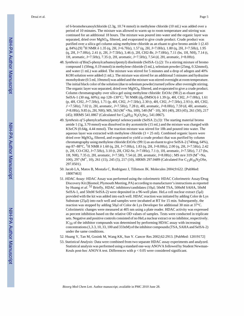
of 6-bromohexanoylchloride (2.3g, 10.74 mmol) in methylene chloride (10 mL) was added over aperiod of 10 minutes. The mixture was allowed to warm up to room temperature and stirring wascontinued for an additional 18 hours. The mixture was poured into water and the organic layer wasseparated, dried over MgSO4, filtered, and evaporated to give crude product. Crude product waspurified over a silica gel column using methylene chloride as an eluant to give bromo amide 1 (2.43g, 84%).[9] 1H NMR δ 1.35 (q, 2H, J=6.7Hz), 1.57 (q, 2H, J=7.0Hz), 1.80 (q, 2H, J=7.5Hz), 1.95(q, 2H, J=7.0Hz), 2.41 (t, 2H, J=7.5Hz), 3.46 (t, 2H, CH2-Br, J=7.0Hz), 7.11 (bs, 1H, NH), 7.14 (t,1H, aromatic, J=7.5Hz), 7.35 (t, 2H, aromatic, J=7.5Hz), 7.54 (d, 2H, aromatic, J=8.0Hz).
48. Synthesis of Bis(5-phenylcarbamoylpentyl) diselenide (SelSA-1) (2): To a stirring mixture of bromocompound 1 (50mg, 0.19 mmol) in methylene chloride (5 mL), selenium powder (25mg, 0.32mmol),and water (1 mL) was added. The mixture was stirred for 5 minutes and a drop of adogen and 40%KOH solution were added (1 mL). The mixture was stirred for an additional 5 minutes and hydrazinemonohydrate (0.5 ml, 10mmol) was added and the mixture was stirred overnight at room temperature.The initial black color of the solution (due to selenium powder) turned yellow after overnight stirring.The organic layer was separated, dried over MgSO4, filtered, and evaporated to give a crude product.Column chromatography over silica gel using methylene chloride: EtOAc (98:2) as eluant gaveSelSA-1 (30 mg, 60%); mp 128–130°C; 1H NMR (d6-DMSO) δ 1.39 (p, 4H, CH2, J=7.0Hz), 1.61(p, 4H, CH2, J=7.5Hz), 1.71 (p, 4H, CH2, J=7.5Hz), 2.30 (t, 4H, CH2, J=7.5Hz), 2.93 (t, 4H, CH2,J =7.5Hz), 7.02 (t, 2H, aromatic, J=7.5Hz), 7.28 (t, 4H, aromatic, J=8.0Hz), 7.59 (d, 4H, aromatic,J=8.0Hz), 9.85 (s, 2H, NH); MS, 563 (M++Na, 100), 540 (M+, 10), 301 (40), 283 (45), 261 (50), 217(45); HRMS 541.0867 (Calculated for C24H32 N2O2Se2, 541.0867).
49. Synthesis of 5-phenylcarbamoylpentyl selenocyanide (SelSA 2) (3): The starting material bromoamide 1 (1g, 3.70 mmol) was dissolved in dry acetonitrile (15 mL) and the mixture was charged withKSeCN (0.64g, 4.44 mmol). The reaction mixture was stirred for 18h and poured into water. Theaqueous layer was extracted with methylene chloride (3 × 25 ml). Combined organic layers weredried over MgSO4, filtered, and evaporated to yield a crude product that was purified by columnchromatography using methylene chloride:EtOAc (99:1) as an eluent to give SelSA-2 (740mg, 64%),mp 87–88°C, 1H NMR δ 1.60 (q, 2H, J=7.0Hz), 1.83 (q, 2H, J=8.0Hz), 2.00 (q, 2H, J=7.5Hz), 2.42(t, 2H, CO-CH2, J=7.5Hz), 3.10 (t, 2H, CH2-Se, J=7.0Hz), 7.1 (t, 1H, aromatic, J=7.5Hz), 7.17 (bs,1H, NH), 7.35 (t, 2H, aromatic, J=7.5H), 7.54 (d, 2H, aromatic, J=8.0Hz) ; MS m/e 319 (M++Na,100), 297 (M+, 10), 261 (15), 245 (5), 217 (10), HRMS 297.0489 (Calculated For C13H16N2OSe,297.0501).
50. Jacob LA, Matos B, Mostafa C, Rodriguez J, Tillotson JK. Molecules 2004;9:622. [PubMed:18007463]
51. HDAC Assay: HDAC Assay was performed using the colorimetric HDAC Colorimetric Assay/DrugDiscovery Kit (Biomol; Plymouth Meeting, PA) according to manufacturer’s instructions as reportedby Huang et al. 52 Briefly, HDAC inhibitors/candidates (10μl; 50nM TSA, 500nM SAHA, 50nMSelSA-1, and 50nM SelSA-2) were deposited in a 96-well plate. HeLa cell nuclear extract (5μl)provided with the kit was added into each well. HDAC reaction was initiated by adding Color de LysSubstrate (25μl) into each well and samples were incubated at RT for 15 min. Subsequently, thereaction was stopped by adding 50μl of Color de Lys Developer for additional 30 min at 37°C.Colorimetric changes were measured at 405 nm using a plate reader. HDAC activity was expressedas percent inhibition based on the relative OD values of samples. Tests were conducted in triplicatesets. Negative and positive controls consisted of no HeLa nuclear extract or no inhibitor, respectively.IC50 of the inhibitor compounds was determined by performing HDAC assay with increasingconcentrations (1,3.3, 10, 33, 100 and 333nM) of the inhibitor compounds (TSA, SAHA and SelSA-2)under the same conditions.
52. Huang Y, Tan M, Gosink M, Wang KK, Sun Y. Cancer Res 2002;62:2913. [PubMed: 12019172]53. Statistical Analysis: Data were combined from two separate HDAC assay experiments and analyzed.
Statistical analysis was performed using a standard one-way ANOVA followed by Student Newman-Keuls post-hoc ANOVA test. Differences with p < 0.05 were considered significant.
Desai et al. Page 5
Bioorg Med Chem Lett. Author manuscript; available in PMC 2010 June 28.
NIH
-PA Author Manuscript
NIH
-PA Author Manuscript
NIH
-PA Author Manuscript
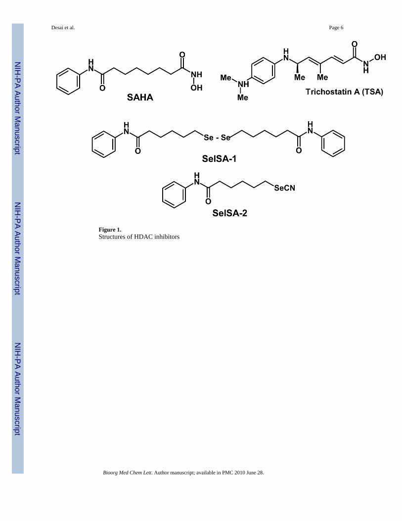
Figure 1.Structures of HDAC inhibitors
Desai et al. Page 6
Bioorg Med Chem Lett. Author manuscript; available in PMC 2010 June 28.
NIH
-PA Author Manuscript
NIH
-PA Author Manuscript
NIH
-PA Author Manuscript
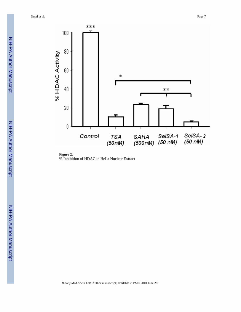
Figure 2.% Inhibition of HDAC in HeLa Nuclear Extract
Desai et al. Page 7
Bioorg Med Chem Lett. Author manuscript; available in PMC 2010 June 28.
NIH
-PA Author Manuscript
NIH
-PA Author Manuscript
NIH
-PA Author Manuscript
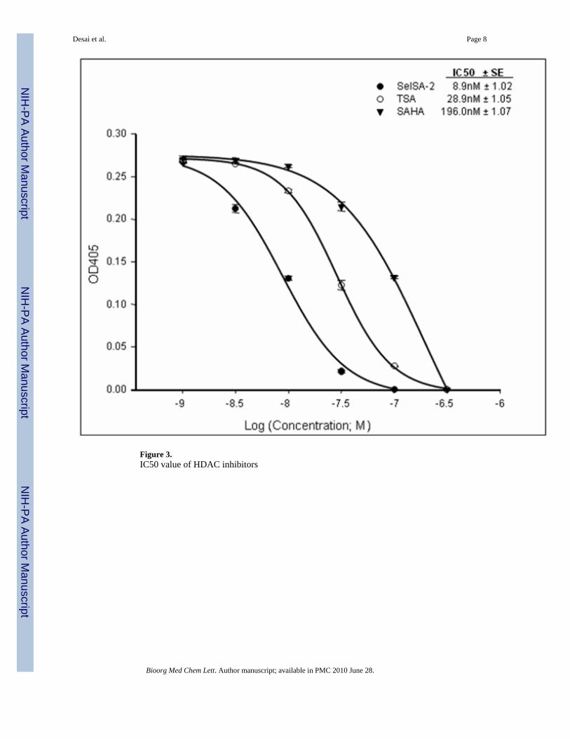
Figure 3.IC50 value of HDAC inhibitors
Desai et al. Page 8
Bioorg Med Chem Lett. Author manuscript; available in PMC 2010 June 28.
NIH
-PA Author Manuscript
NIH
-PA Author Manuscript
NIH
-PA Author Manuscript
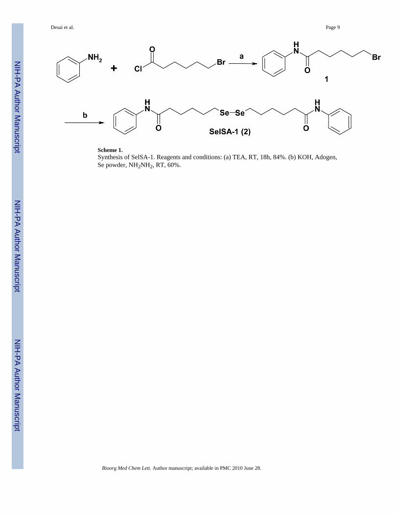
Scheme 1.Synthesis of SelSA-1. Reagents and conditions: (a) TEA, RT, 18h, 84%. (b) KOH, Adogen,Se powder, NH2NH2, RT, 60%.
Desai et al. Page 9
Bioorg Med Chem Lett. Author manuscript; available in PMC 2010 June 28.
NIH
-PA Author Manuscript
NIH
-PA Author Manuscript
NIH
-PA Author Manuscript
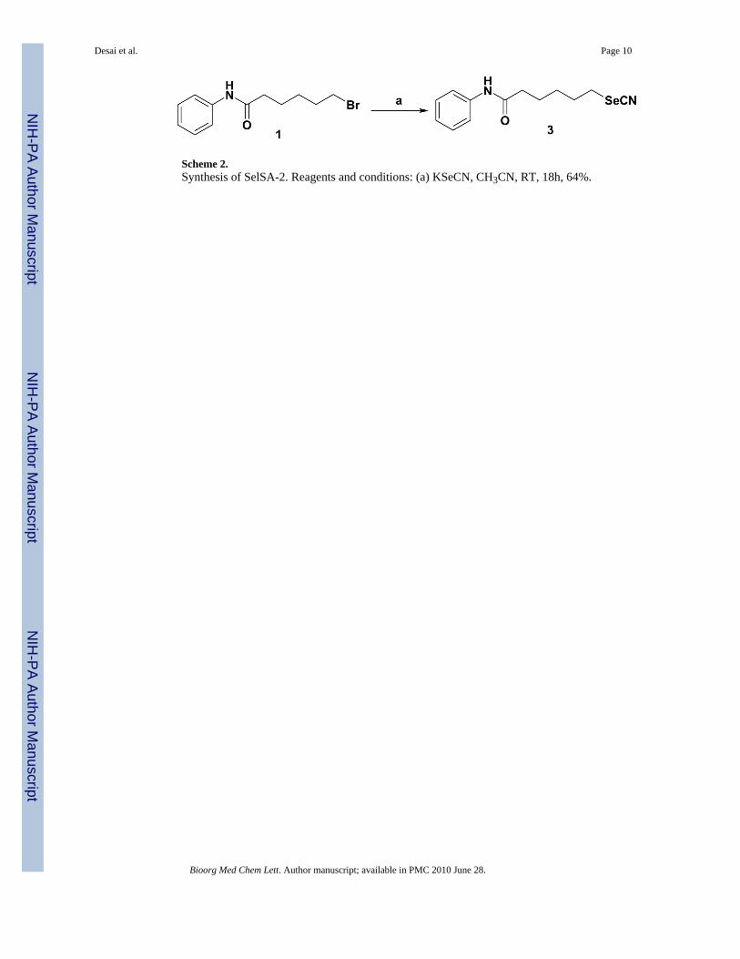
Scheme 2.Synthesis of SelSA-2. Reagents and conditions: (a) KSeCN, CH3CN, RT, 18h, 64%.
Desai et al. Page 10
Bioorg Med Chem Lett. Author manuscript; available in PMC 2010 June 28.
NIH
-PA Author Manuscript
NIH
-PA Author Manuscript
NIH
-PA Author Manuscript


