Embed Size (px)
Citation preview

TITLE PAGE
Serum B-type natriuretic peptide in the initial workup of patients with new onset
ascites: a diagnostic accuracy study
Authors:
Alberto Queiroz Farias1 ([email protected])
Odilson Marcos Silvestre2 ([email protected])
Guadalupe Garcia-Tsao3 ([email protected])
Luis Fernando Bernal da Costa Seguro2 ([email protected])
Daniel Ferraz de Campos Mazo1 ([email protected])
Fernando Bacal2 ([email protected])
José Lázaro Andrade4 ([email protected])
Luciana Lofego Gonçalves5 ([email protected])
Célia Strunz2 ([email protected])
Danusa Ramos1 ([email protected])
Demerson Polli6 ([email protected])
Vincenzo Pugliese1 ([email protected])
Ana Clara Tude Rodrigues4 ([email protected])
Meive Santos Furtado4 ([email protected])
Flair José Carrilho1 ([email protected])
Luiz Augusto Carneiro D Albuquerque1 ([email protected])
Affiliations:
1- Department of Gastroenterology. University of Sao Paulo School of Medicine
2- Heart Institute. University of São Paulo School of Medicine
3- Yale University, Section of Digestive Diseases, and VA-CT Healthcare System, West
Haven, CT, United States.
4- Institute of Radiology. University of Sao Paulo School of Medicine
5- Federal University of Espirito Santo
6- Department of Statistics. University of Brasilia
Key words: cirrhosis, heart failure, peritoneal disease, liver, natriuretic peptides
Hepatology
This article has been accepted for publication and undergone full peer review but has not beenthrough the copyediting, typesetting, pagination and proofreading process which may lead todifferences between this version and the Version of Record. Please cite this article asdoi: 10.1002/hep.26643

2
Corresponding author
Alberto Queiroz Farias, M.D.
Hospital das Clínicas da USP. Av. Dr. Eneas de Carvalho Aguiar 255, sala 9117.
São Paulo, SP, Brazil. Zip code 05403-000
Phone: +55-11-30674238
Email:[email protected]
List of abbreviations:
HF: heart failure
SAAG: serum-ascites albumin gradient
BNP: B-type natriuretic peptide
NPV: negative predictive value
PPV: positive predictive value
LR: likelihood ratio
STARD: Standards for reporting Studies of Diagnostic Accuracy
NT-proBNP: N-terminal proB-type natriuretic peptide
Financial support: FAPESP – Fundação de Amparo à Pesquisa do Estado de São Paulo
(Sao Paulo Research Foundation), grant number 2011/09484-5
Page 2 of 27
Hepatology
Hepatology
3
Abstract
Background and rationale: Heart failure (HF) is, after cirrhosis, the second most
common cause of ascites. Serum B-type natriuretic peptide (BNP) plays an important
role in the diagnosis of HF. We therefore hypothesized that BNP would be useful in the
differential diagnosis of ascites. Consecutive patients with new onset ascites were
prospectively enrolled in this cross-sectional study. All patients had measurements of
serum-ascites albumin gradient (SAAG), total protein concentration in ascitic fluid,
serum and ascites BNP. Results: We enrolled 218 consecutive patients with ascites due
to HF (n=44), cirrhosis (n=162), peritoneal disease (n=10) and constrictive pericarditis
(n=2). Compared to SAAG and/or total protein concentration in ascites, the test that
best discriminated HF-related ascites from other causes of ascites was serum BNP. A
cutoff of >364 pg/mL (sensitivity 98%, specificity 99%, diagnostic accuracy 99%) had
the highest positive likelihood ratio (168.1), that is, it was the best to rule-in HF-related
ascites. Conversely, a cutoff ≤182 pg/mL had the lowest negative likelihood ratio (0.0)
and was the best to rule-out HF-related ascites. These findings were confirmed in a 60-
patient validation cohort. Conclusions: Serum BNP is more accurate than ascites
analyses in the diagnosis of HF-related ascites. The workup of patients with new onset
ascites could be streamlined by obtaining serum BNP as an initial testand could forego
the need for diagnostic paracentesis, particularly in cases where the cause of ascites in
uncertain and/or could be due to heart failure.
Page 3 of 27
Hepatology
Hepatology

4
Introduction
Ascites secondary to heart failure (HF) is, after cirrhosis, the second most common
cause of ascites.(1) The pathophysiology of ascites in both HF and cirrhosis is hepatic
sinusoidal hypertension and therefore the serum-ascites albumin gradient (SAAG) is
greater than ≥1.1 g/dl in both conditions.(2) Since the hepatic sinusoids are normal
(leaky i.e. without significant collagen deposition in the space of Disse) in HF and are
abnormal in cirrhosis (less leaky due to capillarization of sinusoids) (3) ascites total
protein content is higher in HF-related ascites than in cirrhotic ascites and has been used
to help in the differential diagnosis between these two entities, with a ascites protein of
>2.5 mg/dL suggesting the presence of ascites related to HF. However, a significant
number of cases are still misclassified.(2,4) Even the standard of care represented by
medical history, physical examination and echocardiography may overlook the
diagnosis of HF, because clinical manifestations may be quite similar to other
conditions.(5,6)
B-type natriuretic peptide (BNP) is a biologically active 32 aminoacid peptide resulting
from the cleavage of the pro-BNP, which is secreted by heart ventricle myocytes in
response to volume expansion and pressure overload.(7,8) BNP testing facilitates the
diagnosis of HF in patients with dyspnea beyond clinical, radiographic and
echocardiographic data, with a sensitivity of 90%, specificity of 76%, and a negative
predictive value of 96%.(9-12)
Patients with cirrhosis may have a combination of findings, such as increased cardiac
output, attenuated systolic contraction and diastolic relaxation, electrophysiological
repolarization abnormalities, and reduced response to beta-1 adrenergic stimulation.(13)
Thus, BNP levels may also increase in serum of patients with cirrhosis, correlating with
the severity of liver disease.(14,15)
Page 4 of 27
Hepatology
Hepatology
5
Nevertheless, since BNP is increased in several body fluids of patients with heart
dysfunction (16-18) and is considered the initial test in the workup of patient with
suspected heart failure (19), we hypothesized that testing for this marker would be a
useful diagnostic tool in the diagnosis of HF-related ascites.
The aim of the study was to compare the diagnostic accuracy of serum BNP and ascites
BNP to standard used tests, mainly SAAG ± ascites total protein, in the differential
diagnosis among the three main causes of ascites (HF, cirrhosis and peritoneal disease)
in patients with new onset ascites.
Methods
Study design and population – From June 2010 to November 2011, all patients with
new onset ascites that were admitted to the University of Sao Paulo School of Medicine
were assessed for eligibility. Inclusion criteria were clinically-detectable ascites, age
over 18 years and consent to participate. The only exclusion criterion was a creatinine
>2.5 mg/dL (possible unreliability of BNP testing per manufacturer). All patients
underwent a stringent protocol that included clinical history, physical examination, rest
echocardiography, and collection of blood and ascitic fluid samples for analysis. The
study was conducted according to the principles of the Declaration of Helsinki; the
protocol was approved by the institutional ethics board review and registered at
www.clinicaltrials.gov (NCT01150916). A written informed consent was obtained from
patients previous to enrollment.
Adjudicated final diagnosis and diagnostic criteria – Two independent staff
cardiologists and two staff hepatologists interviewed and examined all patients before
diagnostic tests (echocardiography, ultrasound, endoscopy) were performed. They were
Page 5 of 27
Hepatology
Hepatology

6
blinded to the results of ascitic fluid biochemistry and BNP, which was not measured
for clinical purposes. In order to adjudicate the correct final diagnosis of each patient,
all necessary clinical records, laboratory tests, and imaging findings, including
echocardiography (performed in all patients), were reviewed until an agreement was
reached. The consensus among the experts was considered the gold standard for
diagnosis. The whole cohort had a mean follow-up of 13.1 ± 10.9 (median 9.3) months,
and patient follow-up helped confirm the final diagnosis in all but 8 patients (see below).
The diagnosis of cirrhosis was established with a liver biopsy or was based on a
compatible clinical history, specifically a history of chronic liver disease, physical exam
and/or laboratory abnormalities and, importantly, the presence of signs of cirrhosis
and/or portal hypertension on imaging studies (nodular liver, splenomegaly and/or
collaterals). HF was diagnosed according to current diagnostic guidelines.(19,20)
Patients had to fulfill both Framingham and Boston criteria (21,22) and have a
compatible rest echocardiography (systolic right- or left-ventricle dysfunction with
ejection fraction below 50%).
SAAG, protein concentration in ascites and BNP – All samples were sent to laboratory
immediately after venipuncture and paracentesis. SAAG and protein in ascites were
assessed using standard methods. BNP measurements were carried out according to
manufacturer’s instructions (ADVIA Centaur BNP Siemens Inc, San Diego, CA, USA).
This test is a fully automated two-site sandwich immunoassay, based on
chemiluminescent technology, and standardized with synthetic purified protein
preparation of human BNP (aminoacid 77 to 108), within the range of <2.0 to 5,000
pg/ml. The inter- and intra-assay coefficients of variation at different concentrations
Page 6 of 27
Hepatology
Hepatology

7
were, respectively: 2.5% and 2.1% at 48.5 pg/mL; 1.5% and 2.0% at 458 pg/mL; 0.5%
and 2.0% at 1,452 pg/mL.
According to the manufacturer, BNP assay is reliable in presence of increased values of
several biochemical parameters that can be elevated in patients with decompensated
cirrhosis. No interference in measurements has been reported with urea values up to 200
mg/dL, creatinine up to 2.5 mg/dL, unconjugated bilirubin up to 25 mg/dL, conjugated
bilirubin up to 25 mg/dL, triglycerides up to 800 mg/dL, cholesterol up to 1,000 mg/dL.
For whole blood testing, a 4 ml sample was collected in an EDTA containing tube. For
ascitic fluid testing, a 10 ml sample was collected at the same time and before infusion
of albumin or volume overload. Laboratory staff was unaware of both the clinical
diagnosis and routine laboratory results. Kits were purchased from the manufacturers,
who had no role in the study design, analysis of data or writing the manuscript.
Standard Echocardiography – This was performed at rest in all patients, according to
the recommendations of the American Society of Echocardiography.(23) The following
parameters were assessed: left-atrial diameter, systolic left ventricle diameter, diastolic
left ventricle diameter, left ventricular posterior wall, right ventricular diameter,
interventricular septum. Ventricular ejection fraction was estimated from the biplane
Simpson method.(23) We used tricuspid regurgitation velocity to estimate pulmonary
artery systolic pressures and assessed tricuspid regurgitation velocity in the parasternal
long- and short-axis and apical 4-chamber views. Diastolic function was assessed
according to the American Society of Echocardiography guideline.(24)
All parameters were recorded in three cycles, and the mean of the measurements was
taken for analysis. The examiner was blinded for the clinical diagnosis of the patient.
Page 7 of 27
Hepatology
Hepatology

8
Validation cohort - a second set of patients with new onset ascites was recruited for the
validation cohort. These patients were prospectively enrolled between January and
March 2012 from three secondary and tertiary hospitals in Brazil (Federal University of
Espirito Santo, Heart Institute and Central Institute of Sao Paulo University School of
Medicine), according to the same protocol and the same inclusion and exclusion criteria
used for the training cohort. Similar clinical and laboratory baseline data were available
for analysis.
Statistical analysis – Continuous variables were presented as medians, with the
interquartile range, and categorical variables as numbers and percentages. Sensitivity,
specificity, diagnostic accuracy, positive and negative predictive values were calculated
with cutoffs defined by choosing the highest likelihood ratio (to rule-in cardiac ascites)
and the lowest negative likelihood ratio (to rule out cardiac ascites). For the validation
set, a sample size of at least 50 patients was calculated, assuming a sensitivity of 93%
and a 95% confidence interval of width ± 7%. All hypothesis testing were two-tailed,
and p values of less than 0.05 were considered statistically significant. Statistical
analyses were performed with the SPSS software, version 19.0 (IBM, New York, USA).
Results
Training cohort - In the study period, 281 consecutive patients with new onset ascites
were admitted and considered for participation in the study. Sixty-three patients were
excluded, leaving 218 patients for analysis (Figure 1). As noted in Figure 1, in eight
patients the experts could not adjudicate the cause of ascites; these were patients with
evidence of both chronic liver disease and heart disease but a hepatic venous pressure
gradient measurement was not performed to confirm the source of ascites.
Page 8 of 27
Hepatology
Hepatology

9
Of the 218 patients included in the study, the cause of ascites was HF in 44, cirrhosis in
162, peritoneal disease in 10 and constrictive pericarditis in 2. Notably, in nine of the
44 patients with HF, the diagnosis was not entirely certain because of the presence of a
low serum albumin suggestive of the presence of cirrhosis; however, six-month follow-
up of these patients continued to reveal no evidence of cirrhosis and hypoalbuminemia
was attributed to cardiac cachexia.
Two patients with ascites secondary to constrictive pericarditis are described separately
because they did not fit in any of our three main categories.
Of 44 patients with HF, the etiology was ischemic (n=11), Chagas disease (n=10),
valvular (n=9), idiopathic (n=7), hypertensive (n=5), alcoholic (n=1), and peripartum
(n=1). Eleven patients were found to have class IV functional capacity, 21 class III and
12 class II, according to the New York Heart Association.(25) No patient with HF had
esophageal varices or indirect signs of portal hypertension on ultrasound.
All 162 patients with cirrhosis had clinically significant portal hypertension (esophageal
varices at endoscopy, n=119; venous collaterals identified by imaging methods, n=43)
The etiology was hepatitis C (n=56), alcoholic (n=48), hepatitis B (n=6), non-alcoholic
steatohepatitis (n=7), autoimmune hepatitis (n=5), biliary disease (n=2) and others
(n=38). None of them had any evidence of heart disease or HF. 113 were classified
Child B and 49 Child C.
Peritoneal disease was diagnosed in ten patients (peritoneal tuberculosis n=3,
carcinomatosis n=6, pancreatic ascites n=1). No patient in this group was found to have
cirrhosis or HF.
Characteristics of patients and laboratory tests at the time of inclusion in the study are
shown in Table 1. As expected, arterial hypertension, dyslipidemia and
echocardiographic abnormalities were more common in the HF group, while albumin
Page 9 of 27
Hepatology
Hepatology

10
was lower and bilirubin was higher in the group with cirrhosis. Groups were
comparable regarding age and glomerular filtration rates, factors that could influence
the results of BNP measurements. Table 2 shows the echocardiography findings in the
study groups.
Diagnostic accuracy of SAAG, ascites protein concentration, serum BNP and ascites
BNP in the training cohort. Figure 2 shows the individual results of SAAG, ascites
total protein, serum BNP and ascites BNP in the study groups.
A SAAG≥ 1.1 was present in all 162 patients with cirrhosis while an ascites protein
concentration ≤2.5 mg/dL was present in 142/162 (87.7%) of the patients with cirrhosis.
Of the 20 patients with an ascites protein concentration >2.5 mg/dL, 18 were Child B
and only 2 belonged to Child C classification. In fact, patients with an ascites protein
>2.5 had a significantly better liver synthetic function than those with an ascites protein
<2.5 as evidenced by a higher serum albumin (3.5±0.5 vs. 3.0±0.6 g/dL;p=0.0003), a
lower serum bilirubin (1.4±1.5 vs. 3.0±3.6 mg/dL; p=0.001) and a lower INR (1.3±0.3
vs. 1.4±0.4;p=0.0023). In the group of patients with HF a SAAG<1.1 was present in
9/44 (20%) patients, and this subset of patients characteristically had lower serum
albumin values when compared with patients with HF and a SAAG >1.1 (2.8±0.8 vs
3.5±0.5; p=0.0031). In the group of patients with peritoneal disease, a SAAG<1.1 was
present in 9/10 (90%) of the patients and ascites protein concentration >2.5 mg/dL was
present in 8/10 (80%). The two patients with constrictive pericarditis had a SAAG of
1.2 and 1.1; ascites protein of 4.3 and 5.8 mg/dL; serum BNP of 167 and 31 pg/mL, and
ascites BNP of 54 and 38 pg/mL, respectively.
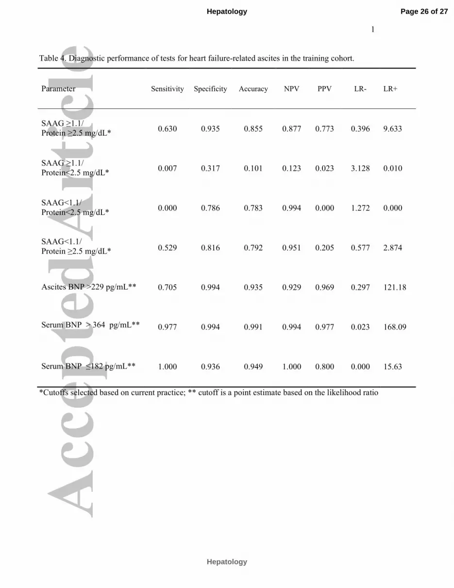
Table 3 shows the frequency of patients in different groups, according to the cutoff of
SAAG, protein in ascites, serum BNP and BNP in ascites. The sensitivity, specificity,
Page 10 of 27
Hepatology
Hepatology

11
diagnostic accuracy, negative (NPV) and positive predictive value (PPV), and the
positive (LR + ) and negative likelihood ratio (LR - ) are shown in table 4.
The test that best discriminated HF-related ascites from other causes of ascites was
serum BNP levels. Serum BNP at a cutoff of 364 pg/mL had a sensitivity of 98%,
specificity of 99%, diagnostic accuracy of 99%, NPV 99% and PPV 98%. In fact, only
one patient with cirrhosis and none of the patients with a peritoneal process as cause for
ascites had a BNP >364 pg/mL and this patient with cirrhosis had a borderline elevation
of serum BNP at 384 pg/mL. A cutoff of >364 pg/mL had the highest positive
likelihood ratio (168.1), that is, it was the best to rule-in HF-related ascites. Conversely,
a cutoff <182 pg/mL had the lowest negative likelihood ratio (0.0) and was the best to
rule-out HF-related ascites. Neither ascites BNP levels nor a combination of SAAG
>1.1 and ascites total protein >2.5 mg/dL were as effective as serum BNP in
establishing the diagnosis of ascites due to HF. However, ascites BNP >229 ng/mL had
a higher positive likelihood ratio (121.18) than the combination of high SAAG and high
ascites protein that is currently used to suggest a diagnosis of cardiac ascites (positive
likelihood ratio 9.6)
Complications - Minor complications of paracentesis were observed in 8 (3.7%) out of
the 218 patients (ascitic fluid leakage n=7; abdominal wall hematoma n=1). No
hospitalization was required for treatment. No death related to the procedure
occurred.Validation cohort - Sixty consecutive patients with new onset ascites
constituted the validation cohort. Of these, HF was the cause of ascites in 15 and
cirrhosis the cause in 45. As shown in Table 3, none of the patients with cirrhosis had a
BNP >364 and none of the patients with HF had a BNP <182. This yields a sensitivity
and specificity of 100% (Table 5).
Page 11 of 27
Hepatology
Hepatology

12
These results confirm the findings of the training cohort in that serum BNP is the best
test that discriminates HF as a cause of ascites from other causes of ascites and also
confirms that the cutoff of 362 pg/mL is the best to “rule in” ascites secondary to HF
and that <182 pg/mL rules out HF-related ascites.
Discussion
Ascites secondary to HF is the second most common cause of ascites in the Western
world. Making the differential diagnosis among the three most common causes of
ascites, i.e., HF, cirrhosis and peritoneal processes, is important as the diagnostic
workup and management will be different depending on the probable diagnosis.
The diagnosis of HF is initially based on clinical findings (history, physical exam and
routine tests) but is often difficult to establish with certainty. Although current
guidelines for HF (20,21) indicate that the diagnosis should be made with a combination
of typical signs and symptoms, this approach is limited by low accuracy rates (25% to
50%) (7,26,27) when compared to echocardiography results. BNP testing is largely
used in the primary practice setting, avoiding unnecessary investigations of HF. A
systematic review (28) examined the results of twenty studies of BNP testing for the
diagnosis of clinically defined HF, and showed a consistently high sensitivity but a
variable specificity for the diagnosis of HF, concluding that an elevated BNP does not
confirm the diagnosis of clinically defined HF, but normal levels rule out the diagnosis.
This concept has been adopted by the British National Institute for Health and Clinical
Excellence clinical practice guideline that recommends that patients without previous
myocardial infarction should undergo measurement of serum BNP as the first step, with
subsequent echocardiography, and specialist evaluation is indicated if these levels are
elevated.(29)
Page 12 of 27
Hepatology
Hepatology

13
Ascites findings are very useful to make a differential diagnosis. As mentioned
previously a high ascites protein level in the setting of an elevated serum-ascites
albumin gradient (SAAG) favors a diagnosis of HF. In cirrhosis, although SAAG is
also elevated, ascites protein is low, while in ascites due to local peritoneal processes,
the SAAG is low (because there is no sinusoidal hypertension) but the ascites protein is
elevated. However, there is a significant number of cases in which results are borderline
and we have to resort to measurements of hepatic venous pressure gradient to make the
final diagnosis. In cirrhotic ascites the hepatic venous pressure gradient is high, in
cardiac ascites the wedged hepatic venous pressure is increased but the gradient is
normal (because of elevated systemic pressures) and in ascites secondary to peritoneal
processes, both the wedge and the gradient are normal.(30)
Given the diagnostic accuracy of BNP in diagnosis HF, its widespread use and the fact
that results can be obtained in 18 minutes (8-13), the present study sought out to
determine whether serum and/or ascites BNP is more accurate than the SAAG, the
ascites protein or the SAAG/ascites protein combination in establishing the cause of
ascites in patients with new onset ascites.
Our study shows that serum BNP accurately identified HF as the cause of ascites with a
higher sensitivity, specificity and diagnostic accuracy when compared to the standard
methods, SAAG and/or ascites total protein. These results are strengthened by similar
findings in the validation cohort. We could also establish cutoff levels that will reliably
rule in the diagnosis of HF-related ascites (serum BNP >364 pg/mL) and to rule out this
diagnosis (serum BNP ≤182 pg/mL). Notably, none of the patients in the training
cohort fell in the “grey zone” (serum BNP between 183 and 363 pg/mL) and only 4 in
the validation cohort did so, but they were all patients with cirrhotic ascites, all with
levels below 363 pg/mL (216, 224, 285 and 334, respectively).
Page 13 of 27
Hepatology
Hepatology

14
Regarding the standard tests, we could confirm that SAAG ≥ 1.1 is present in nearly all
patients with cirrhosis, but low SAAG values were present in 20% of patients with HF,
particularly in the presence of hypoalbuminemia.
We also confirmed that most patients with HF have an ascites protein concentration
>2.5 g/dL.(2) However, we also found that 12% of patients with cirrhosis had a high
ascites protein, indicating that sinusoids are normal (“leaky”). Because this could
indicate that capillarization was not extensive (and therefore cirrhosis was “milder”) we
looked at these patients with cirrhosis and ascites protein >2.5 g/dL and found that they
in fact had a milder liver disease as evidenced by higher serum albumin, lower serum
bilirubin and lower INR (.
When combining SAAG and total ascites protein, we still found that the diagnostic
accuracy was not optimal (85%), with 63% sensitivity and 93% specificity.
Ascites BNP, although not as accurate as serum BNP, had a greater diagnostic accuracy
than standard tests. In ascites resulting from portal hypertension, collagen deposition in
the Disse space leads to varying degrees in loss of sinusoidal endothelial cell
fenestration and formation of basement membrane. The obliteration of this opening
between the sinusoidal lumen and the Disse space is expected to affect the filtration of
particles by the liver.(31-33) One could speculate that the explanation for the higher
performance of ascites BNP testing may be related to the lower molecular weight of this
peptide (4 kDa) when compared with human serum albumin (67 kDa), used in SAAG
calculation, which could lead to better diffusion of the former.
Interestingly, both patients with ascites secondary to constrictive pericarditis had SAAG
≥ 1.1 and very high ascites protein levels (4.3 and 5.8 mg/dL). Notably, both patients
had a low serum BNP (31 and 167 pg/mL, respectively), confirming that BNP becomes
elevated when there is heart enlargement. In this context, it is important to note that all
Page 14 of 27
Hepatology
Hepatology
15
patients included in the study had new onset ascites and none had received intravascular
volume replacement prior to performing the diagnostic tests. It is conceivable that a
patient with cirrhosis who is undergoing vigorous volume replacement (as would occur
in an intensive care unit) could have elevated serum BNP.
Data on the usefulness of natriuretic peptides testing in distinguishing ascites due to
cirrhosis from ascites due to HF are scanty in the literature. This is the first prospective
study that included consecutive patients with ascites and a wide spectrum of disease,
fulfilling the STARD (34) (Standards for reporting Studies of Diagnostic Accuracy)
initiative requirements. According to it, a valid diagnostic study must comply with a list
of 25 requirements, that includes assembling an appropriate number and spectrum of
patients with well defined clinical diagnosis of the disease entities, application of both
the diagnostic (BNP testing) and reference standard (SAAG and protein in ascites) to all
of them, blind interpretation of the results from the clinical diagnosis and validation in a
second set of patients. Importantly, the gold standard was a clinical diagnosis
established by adjudication by experts, and based on well-established clinical
parameters.
A previous retrospective small study of banked serum and ascites samples in 58 patients
with cirrhosis and 18 with HF, showed that, using the cutoff of 1,000 pg/mL, serum NT-
proBNP had a sensitivity of 100%, specificity of 97% and positive predictive value of
90% and that ascites NT-proBNP had a sensitivity of 93.3% and specificity of 97.5% in
predicting HF as the cause for ascites.(35) However, the study was retrospective and
patients were not consecutive, thereby with the potential of introducing bias.
We chose to measure BNP rather than NT-proBNP, which may be more utilized in
some centers, because the latter is more dependent on sex, age and renal dysfunction
(36) and because it is a more expensive test. We excluded patients with serum
Page 15 of 27
Hepatology
Hepatology

16
creatinine >2.5 mg/dL because the manufacturer stated that BNP determination is not
reliable at these levels. However, recent data shows that BNP continues to be the
strongest predictor of the presence of HF when holding all other predictors equal,
including renal function.(37)
Apart from the typical HF setting, a number of factors, unmet by the patients of the
current study, have been associated with higher natriuretic peptide levels. Conditions,
such as, advanced age, sepsis, hyperthyroidism, acute coronary syndromes, acute
respiratory distress syndrome and pulmonary embolism can be associated with a rise in
BNP blood levels even in the absence of concomitant HF.(38) Thus, the findings of this
study may not be applicable to all patients with high BNP. However, these clinical
scenarios are easily acknowledged and are not associated with ascites, helping to clarify
what might otherwise seem to be a discordant result.
In this study we have shown that, in patients with new onset ascites, serum BNP is more
accurate in establishing the diagnosis of HF-related ascites than ascites analysis. The
workup of patients with new onset ascites could be streamlined by obtaining serum
BNP as an initial test and could potentially forego the need for diagnostic paracentesis,
particularly in cases where the cause of ascites is uncertain or when HF-related ascites is
suspected. Even if the diagnosis of HF-related ascites is established via serum BNP, a
paracentesis would still be indicated if a superimposed infection is suspected or for
therapeutic purposes (relief of patient discomfort).
Acknowledgments: The authors thank to Rafael Ximenes, M.D., for referring patients
for the study.
Page 16 of 27
Hepatology
Hepatology

17
References
1. Runyon BA. Management of adult patients with ascites due to cirrhosis: An
Update. Hepatology 2009; 49:2087-2107.
2. Runyon BA, Montano AA, Akriviadis EA, Antillon MR, Irving MA,
McHutchison JG. The serum-ascites albumin gradient is superior to the exudate-
transudate concept in the differential diagnosis of ascites. Ann Intern Med 1992;
117:215-220.
3. Henriksen JH, Horn T, Christoffersen P. The blood-lymph barrier in the liver. A
review based on morphological and functional concepts of normal and cirrhotic
liver. Liver 1984; 4:221–232.
4. Christou L, Economou M, Economou G, Kolettis M, Tsianos EV.
Characteristics of ascitic fluid in cardiac ascites. Scandinavian J of
Gastroenterology 2007; 42:1102-1105.
5. Stevenson LW, Perloff JK. The limited reliability of physical signs for
estimating hemodynamics in chronic heart failure. JAMA 1989; 261:884-888.
6. Remes J, Miettinen H, Reunanen A, Pyorala K. Validity of clinical diagnosis of
heart failure in primary health care. Eur Heart J 1991; 12:315-321.
7. de Lemos JA, McGuirre DK, Drazner MH. B-type natriuretic peptide in
cardiovascular disease. Lancet 2003; 362:316-322.
8. Suttner SW, Boldt J. Natriuretic peptide system: physiology and clinical utility.
Curr Opin Crit Care 2004; 10:336-341.
9. Maisel A, Krishnaswamy P, Nowak RM, McCord J, Hollander J, Duc P, et al.
Rapid Measurement of B-type natriuretic peptide in the emergency diagnosis of
heart failure. N Engl J Med 2002; 347:161-167.
10. Maisel AS, McCord J, Nowak RM, Hollander JE, Wu AH, Duc P, et al. Bedside
B-type natriuretic peptide in the emergency diagnosis of heart failure with
reduced or preserved ejection fraction. J Am Coll Cardiol 2003; 41:2010–2017.
11. Maisel A, Mueller C, Adams K Jr., Anker SD, Aspromonte N, Cleland JGF, et
al. State of the art: Using natriuretic peptide levels in clinical practice. Eur J
Heart Fail 2008; 10:824-839.
12. Mueller C, Scholer A, Laule-Kilian K, Martina B, Schindler C, Buser P, et al.
Use of B-type natriuretic peptide in the evaluation and management of acute
dyspnea. New Engl J Med 2004; 350:647-654.
Page 17 of 27
Hepatology
Hepatology
18
13. Zardi EM, Abbate A, Zardi DM, Dobrina A, Margiotta D, Van Tassell BW, et al.
Cirrhotic cardiomyopathy. J Am Coll Cardiol 2010; 56:539-549.
14. Yildiz R, Yildirim B, Karincaoglu M, Harputluoglu M, Hilmioglu F. Brain
natriuretic peptide and severity of disease in non-alcoholic cirrhotic patients. J
Gastroenterol Hepatol 2005; 20:1115-1120.
15. Henriksen JH, Gøtze JP, Fuglsang S, Christensen E, Bendtsen F, Møller S.
Increased circulating pro-brain natriuretic peptide (proBNP) and brain natriuretic
peptide (BNP) in patients with cirrhosis: relation to cardiovascular dysfunction
and severity of disease. Gut 2003; 52:1511-1517.
16. Porcel JM, Martínez-Alonso M, Cao G, Bielsa S, Sopena A, Esquerda A.
Biomarkers of heart failure in pleural fluid. Chest 2009; 136:671-677.
17. Kolditz M, Halank M, Schiemanck CS, Schmeisser A, Höffken G. High
diagnostic accuracy of NT-proBNP for cardiac origin of pleural effusions. Eur
Respir J 2006; 28:144-150.
18. Tomcsányi J, Nagy E, Somlói M, Moldvay J, Bezzegh A, Bózsik B, et al. NT-
brain natriuretic peptide levels in pleural fluid distinguish between pleural
transudates and exudates. Eur J Heart Fail 2004; 6:753-756.
19. Jessup M, Abraham W, Casey DE, Feldman A, Francis GS, Ganiats TG, et al.
2009 focused update: ACCF/AHA Guidelines for the diagnosis and management
of heart failure in adults: a report of the American College of Cardiology
Foundation/American Heart Association Task Force on Practice Guidelines:
developed in collaboration with the International Society for Heart and Lung
Transplantation. Circulation 2009; 119:1977-2016.
20. Dickstein K, Cohen-Solal A, Filippatos G, McMurray JJ, Ponikowski P, Poole-
Wilson PA, et al. ESC Guidelines for the diagnosis and treatment of acute and
chronic heart failure. Eur Heart J 2008; 29:2388–2442.
21. McKee PA, Castelli WP, McNamara PM, Kannel WB. The natural history of
congestive heart failure. The Framingham Study. N Engl J Med 1971;
285:1441–1446.
22. Carlson KJ, Lee DC, Goroll AH, Leahy M, Johnson RA. An analysis of
physicians’ reasons for prescribing long-term digitalis therapy in outpatients. J
Chronic Dis 1985; 38: 733-739.
Page 18 of 27
Hepatology
Hepatology

19
23. Lang RM, Bierig M, Devereux RB, Flachskampf FA, Foster E, Pellikka PA, et
al. Recommendations for chamber quantification: a report from the American
Society of Echocardiography’s Guidelines and Standards Committee and the
Chamber Quantification Writing Group, developed in conjunction with the
European Association of Echocardiography, a branch of the European Society of
Cardiology. J Am Soc Echocardiogr 2005;18:1440 -1463.
24. Nagueh SF, Appleton CP, Gillebert TC, Marino PN, Oh JK, Smiseth OA, et al.
Recommendations for the Evaluation of Left Ventricular Diastolic Function by
Echocardiography. J Am Soc Echocardiogr 2009; 22:107-212.
25. The Criteria Committee of the New York Heart Association. Nomenclature and
Criteria for Diagnosis of Diseases of the Heart and Great Vessels. 9th ed. Boston,
Mass: Little, Brown & Co; 1994:253-256.
26. Wheeldon NM, MacDonald TM, Flucker CJ, McKendrick AD, McDevitt DG,
Struthers AD. Echocardiography in chronic heart failure in the community. Q J
Med 1993; 86:17-23.
27. Davis RC, Hobbs FD, Kenkre JE, Roalfe AK, Hare R, Lancashire RJ, et al.
Prevalence of heart failure in high risk groups: community based
epidemiological study. BMJ 2002; 325:1156.
28. Mant J, Doust J, Roalfe A, Barton P, Cowie MR, Glasziou P, et al. Systematic
review and individual patients data meta-analysis of diagnosis of heart failure,
with modeling of implications of different diagnostic strategies in primary care.
Health Technol Assess 2009; 13: 1-207.
29. Mant J, Al-Mohammad A, Swain S, Laramee P. Management of chronic heart
failure in adults: synopsis of the National Institute for Health and Clinical
Excellence. Ann Intern Med 2011; 155: 252-259.
30. Garcia-Tsao G. Ascites. In: Boyer TD, Manns MP, Sanyal AJ, eds. Zakim and
Boyer’s Hepatology. A Textbook of Liver Disease. 6th Edition. Elsevier
Saunders, Philadelphia, PA, 2012:283-295.
31. Rider H, Meyer zum Büschenfelde KH, Ramadori G. Functional spectrum of
sinusoidal endothelial liver cells: Filtration, endocytosis, synthetic capacities and
intercellular communication. J Hepatol 1992; 15:237–250.
32. Wisse E, De Zanger RB, Charels K, Van Der Smissen P, McCuskey RS. The
Liver Sieve: Considerations concerning the structure and function of endothelial
Page 19 of 27
Hepatology
Hepatology

20
fenestrae, the sinusoidal wall and the space of Disse. Hepatology 1985; 5:683–
692.
33. Fraser R, Dobbs BR, Rogers GW. Lipoproteins and the liver sieve: the role of
the fenestrated sinusoidal endothelium in lipoprotein metabolism,
atherosclerosis, and cirrhosis. Hepatology 1995; 21:863–74.
34. Bossuyt PM, Reitsma JB, Bruns DE, Gatsonis CA, Glasziou PP, Irwig LM, et al.
The STARD statement for reporting studies of diagnostic accuracy: explanation
and elaboration. Ann Intern Med 2003; 138: W1–W12.
35. Sheer TA, Joo E, Runyon BA. Usefulness of serum N-terminal-ProBNP in
distinguishing ascites due to cirrhosis from ascites due to heart failure. J Clin
Gastroenterol 2010; 44:e23–e26.
36. Manzano-Fernández S, Januzzi JL, Boronat-García M, Pastor P, Albaladejo-
Otón MD, Garrido IP, et al. Impact of kidney dysfunction on plasma and urinary
N-terminal pro-B-type natriuretic peptide in patients with acute heart failure.
Congest Heart Fail 2010;16:214-220.
37. McCullough PA, Duc P, Omland T, McCord J, Nowak RM, Hollander JE, et al.
B-type natriuretic peptide and renal function in the diagnosis of heart failure: an
analysis from the Breathing Not Properly Multinational Study. Am J Kidney Dis
2003; 41:571-579.
38. Daniels LB, Maisel AS. Natriuretic peptides. J Am Coll Cardiol 2007; 50: 2357-
2368.
Page 20 of 27
Hepatology
Hepatology
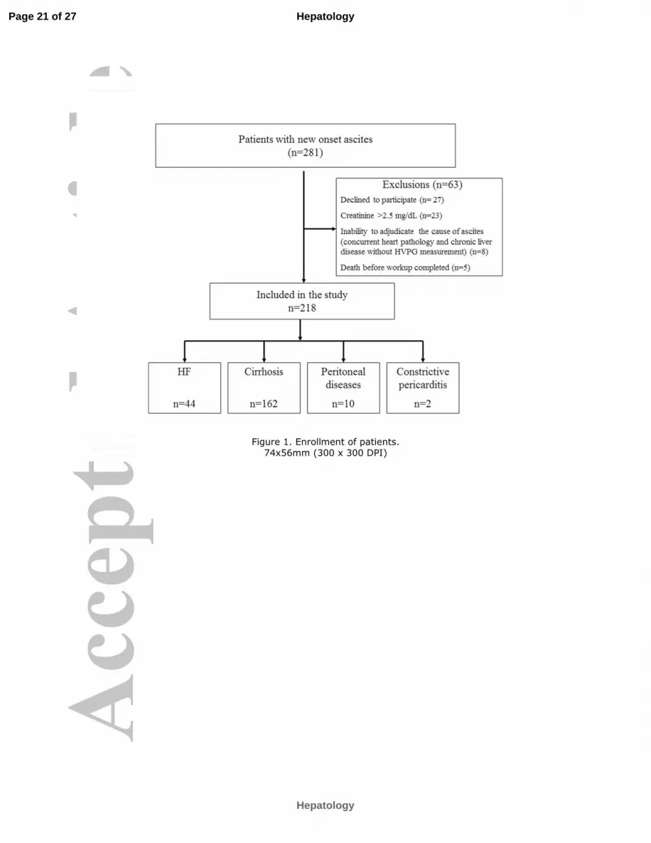
Figure 1. Enrollment of patients. 74x56mm (300 x 300 DPI)
Page 21 of 27
Hepatology
Hepatology

Figure 2. SAAG (A), protein concentration in ascites fluid (B), serum BNP (C) and BNP in ascitic fluid (D), according to the cause of ascites in the training cohort. Note that the scale for serum and ascites BNP is a
semilogarithmic scale.
74x56mm (300 x 300 DPI)
Page 22 of 27
Hepatology
Hepatology

1
Table 1. Baseline characteristics of the patients and laboratory tests.
Continuous variables are expressed as median (interquartile ranges).
Parameter HF Cirrhosis Peritoneal diseases
n=44 n=162 n=10
Age – years 55 (49 – 62) 57 (49 - 63) 42 (37 - 66)
Medical history - no. (%)
Diabetes mellitus 15 (34.1%) 52 (33.3%) 2 (20.0%)
Arterial hypertension 25 (56.8%) 37 (26.8%) 3 (30.0%)
Heavy alcohol intake 7 (15.9%) 45 (26.1%) 4 (40.0%)
Current smoker 14 (31.8%) 64 (41.1%) 3 (30.0%)
Dyslipidemia 12 (27.3%) 4 (2.6%) 1 (10.0%)
Clinical findings
Heart rate - beats/min 72 (64 - 84) 80 (68 - 84) 92 (80 - 100)
Mean arterial pressure - mmHg 80 (70 - 85) 86 (80 - 93) 96 (83 - 100)
Biochemistry
Aspartate aminotransferase - IU/L 30 (20-40) 49 (36 – 76) 26 (18 – 50)
Alanine aminotransferase - IU/L 28 (20 – 33) 32 (22 – 46) 27 (10 - 39)
Alkaline phosphatase - IU/L 145 (113 – 193) 129 (96 – 193) 122 (104 – 140)
Bilirubin - mg/dL 1.4 (0.9 - 1.8) 1.7 (1.1 - 3.1) 0.5 (0.4 – 0.8)
Serum protein - mg/dL 6.8 (6.4-7.3) 6.9 (6.3-7.4) 6.6 (5.8-7.9)
Albumin - g/dL 3.4 (3.0 - 3.8) 3.0 (2.6 - 3.5) 3.1 (2.2 - 4.1)
International normalized ratio 1.4 (1.2 – 1.5) 1.3 (1.2 – 1.6) 1.0 (1.0 – 1.2)
Platelets - x 103 mm
3
208 (153 – 250) 109 (72 – 174) 255 (189 – 415)
Cholesterol - mg/dL 122 (100 - 155) 127 (101 - 151) 181 (125 - 199)
Creatinine - mg/dL 1.45 (1.09 - 1.93) 0.97 (0.82 - 1.33) 0.78 (0.52 - 0.98)
Glomerular filtration rate - ml/min/1.73m2 63 (47 - 91) 76 (58 - 107) 89 (54 - 108)
Page 23 of 27
Hepatology
Hepatology

1
Table 2. Echocardiography findings of the patients.
Parameter HF Cirrhosis Peritoneal diseases
n=44 n=162 n=10
Rest Echocardiography
Ejection fraction - % 29 (20 - 45) 67 (64 - 70) 66 (62 - 69)
Left-atrial diameter - mm 50 (48 - 55) 40 (36 - 43) 39 (34 - 43)
Systolic left ventricle diameter - mm 54 (41 - 60) 29 (26 - 32) 26 (22 - 30)
Diastolic left ventricle diameter - mm 63 (54 - 70) 46 (42 - 50) 41 (37 - 46)
Right ventricular diameter - mm 34 (22 - 43) 22 (20 - 25) 20 (18 - 27)
Diastolic dysfunction -%
Grade 0 1 (5.6%) 65 (44.8%) 4 (50.0%)
Grade 1 and 2 5 (27.8%) 80 (55.2%) 4 (50.0%)
Grade 3 and 4 12 (66.7%) 0 (0.0%) 0 (0.0%)
Pulmonary artery systolic pressure -mmHg 49 (37 - 60) 30 (27 - 34) 31 (31 - 51)
Continuous variables are expressed as median (interquartile ranges).
Page 24 of 27
Hepatology
Hepatology

1
Table 3. Frequency of patients in different groups in the training and validation cohorts, according
to the cutoff of SAAG, protein in ascites, serum BNP and BNP in ascites.
*Cutoffs selected based on current practice; ** cutoff is a point estimate based on the likelihood ratio
Training cohort Validation cohort
Parameter HF
n=44
Cirrhosis
n=162
Peritoneal
disease
n=10
HF
n=15
Cirrhosis
n=45
SAAG ≥1.1 /Protein ≥2.5 mg/dL* 35 (79.5%) 20 (12.3%) 0 8 (53.3%) 6 (13.3%)
SAAG ≥1.1/ Protein <2.5 mg/dL* 0 142 (87.7%) 1 (10%) 2 (13.3%) 39 (86.7%)
SAAG<1.1/ Protein <2.5 mg/dL* 0 0 1 (10%) 0 0
SAAG<1.1/ Protein ≥2.5 mg/dL* 9 (20.5%) 0 8 (80%) 5 (33.3%) 0
Ascites BNP >229 pg/mL** 31 (70.5%) 1 (0.6%) 0 13 (86.7%) 1 (2.2%)
Serum BNP > 364 pg/mL** 43 (97.7%) 1 (0.6%) 0 15 (100%) 0
Serum BNP ≤182 pg/mL** 1 (2.3%) 153 (94.4%) 9 (90%) 0 41 (91.1%)
Page 25 of 27
Hepatology
Hepatology
1
Table 4. Diagnostic performance of tests for heart failure-related ascites in the training cohort.
*Cutoffs selected based on current practice; ** cutoff is a point estimate based on the likelihood ratio
Parameter Sensitivity Specificity Accuracy NPV PPV LR- LR+
SAAG ≥1.1/
Protein ≥2.5 mg/dL*
0.630 0.935 0.855 0.877 0.773 0.396 9.633
SAAG ≥1.1/
Protein<2.5 mg/dL*
0.007 0.317 0.101 0.123 0.023 3.128 0.010
SAAG<1.1/
Protein<2.5 mg/dL*
0.000 0.786 0.783 0.994 0.000 1.272 0.000
SAAG<1.1/
Protein ≥2.5 mg/dL*
0.529 0.816 0.792 0.951 0.205 0.577 2.874
Ascites BNP >229 pg/mL** 0.705 0.994 0.935 0.929 0.969 0.297 121.18
Serum BNP > 364 pg/mL** 0.977 0.994 0.991 0.994 0.977 0.023
168.09
Serum BNP ≤182 pg/mL** 1.000 0.936 0.949 1.000 0.800 0.000 15.63
Page 26 of 27
Hepatology
Hepatology

1
Table5. Diagnostic performance of tests for heart failure-related ascites in the validation cohort.
*Cutoffs selected based on current practice; ** cutoff is a point estimate based on the likelihood ratio
Parameter Sensitivity Specificity Accuracy NPV PPV LR- LR+
SAAG ≥1.1
Protein ≥2.5 mg/dL
0.533 0.867 0.783 0.848 0.571 0.538 4.000
SAAG ≥1.1
Protein<2.5 mg/dL
0.133 0.133 0.133 0.316 0.049 6.500 0.154
SAAG<1.1
Protein<2.5 mg/dL
0.000 1.000 0.750 0.750 N/A 1.000 N/A
SAAG<1.1
Protein ≥2.5 mg/dL
0.333 1.000 0.833 0.818 1.000 0.670 N/A
Ascites BNP >229 pg/mL
0.867 0.978 0.950 0.957 0.929 0.136 39.000
Serum BNP > 364 pg/mL
1.000 1.000 1.000 1.000 1.000 0.000 N/A
Serum BNP ≤ 182 pg/mL 0.000 0.089 0.067 0.211 0.000 0.000 11.250
Page 27 of 27
Hepatology
Hepatology


