Embed Size (px)
Citation preview
Shape and surface effects on the cytotoxicity of nanoparticles: Goldnanospheres versus gold nanostars
Pelagie Marlene Favi,1 Ming Gao,2 Liuda Johana Sep�ulveda Arango,3 Sandra Patricia Ospina,3
Mariana Morales,4 Juan Jose Pavon,5 Thomas Jay Webster1,6
1Department of Chemical Engineering, Northeastern University, Boston, Massachusetts2Department of Pharmaceutical Sciences, School of Pharmacy, Bouv�e College of Health Sciences, Northeastern University,
Boston, Massachusetts3Institute of Biology, University Research Headquarters, University of Antioquia, Medell�ın, Antioquia, Colombia4Department of Medicine, University of Antioquia, Medell�ın, Antioquia, Colombia5Advanced Biomaterials and Regenerative Medicine – ABRM, Bioengineering Programme, University of Antioquia, Medell�ın,
Antioquia, Colombia6Center of Excellence for Advanced Materials Research, King Abdulaziz University, Jeddah, Saudi Arabia
Received 16 November 2014; revised 7 April 2015; accepted 8 April 2015
Published online 6 May 2015 in Wiley Online Library (wileyonlinelibrary.com). DOI: 10.1002/jbm.a.35491
Abstract: Gold nanoparticles are materials with unique optical
properties that have made them very attractive for numerous
biomedical applications. With the increasing discovery of tech-
niques to synthesize novel nanoparticles such as star-shaped
gold nanoparticles for biomedical applications, the safety and
performance of these new nanomaterials must be systemati-
cally assessed before use. In this study, gold nanostars
(AuNSTs) with multibranched surface structures were synthe-
sized, and their influence on the cytotoxicity of human skin
fibroblasts and rat fat pad endothelial cells (RFPECs) were
assessed and compared with that of gold nanospheres
(AuNSPs) with unbranched surfaces. Results showed that the
AuNSPs with diameters of approximately 61.46 nm showed
greater toxicity with fibroblast cells and RFPECs compared
with the synthesized AuNSTs with diameters of approximately
33.69 nm. The AuNSPs were lethal at concentrations of 40 lg/
mL for both cell lines, whereas the AuNSTs were less toxic at
higher concentrations (400 lg/mL). The calculated IC50 (50%
inhibitory concentration) values of the AuNSPs exposed to
fibroblast cells were greater at 1 and 4 days of culture (26.4 and
27.7 lg/mL, respectively) compared with the RFPECs (13.6 and
13.8 lg/mL, respectively), indicating that the AuNSPs have a
greater toxicity to endothelial cells. It was proposed that possi-
ble factors that could be promoting the reduced toxicity effects
of the AuNSTs to fibroblast cells and RFPECs, compared with
the AuNSPs may be size, surface chemistry, and shape of the
gold nanoparticles. The reduced cell toxicity observed with the
AuNSTs suggests that AuNSTs may be a promising material for
use in biomedical applications. VC 2015 Wiley Periodicals, Inc. J
Biomed Mater Res Part A: 103A: 3449–3462, 2015.
Key Words: gold nanostar, branched gold nanoparticles,
cytotoxicity, gold nanoparticle, gold nanosphere, endothelial
cells, fibroblast cells
How to cite this article: Marlene Favi P, Gao M, Johana Sep�ulveda Arango L, Patricia Ospina S, Morales M, Jose Pavon J, JayWebster T. 2015. Shape and surface effects on the cytotoxicity of nanoparticles: Gold nanospheres versus gold nanostars.J Biomed Mater Res Part A 2015:103A:3449–3462.
INTRODUCTION
Gold nanoparticles are materials with unique optical proper-ties that have made them very attractive for numerous bio-medical applications, including drug and gene delivery,1–3
cancer treatment,4,5 and medical imaging.6 This is becausegold nanoparticles can be easily functionalized with a varietyof biomolecules, can penetrate cells, and can respond toinfrared (IR) and other wavelengths of light to aid in diseasediagnosis and medical treatment.6–10 Moreover, because thegeometric shape of metallic nanoparticles can significantly
influence their optical properties for use in cellular trackingand in medicine, researchers have been altering the typicalmetallic nanospherical shape to possess altered geometriessuch as star-shaped gold nanoparticles (i.e., gold nanostars(AuNSTs)), for potential improvement in optical proper-ties.11,12 AuNSTs are nanoparticles with multibranched sur-face structures.11,12 Unlike spherical gold nanoparticleswhose optical properties can only be modified in the visiblelight region by altering their size,13 the optical properties ofAuNSTs can be adjusted from the visible to the near-infrared
Correspondence to: T. J. Webster; e-mail: [email protected] grant sponsor: Northeastern University’s ADVANCE Future Faculty Postdoctoral Fellowship Program sponsored by NSF; contract grant
number: 0811170 (to P. F.)
VC 2015 WILEY PERIODICALS, INC. 3449

region by altering their size and degree of branching,14–16
therefore giving AuNSTs superior optical response for poten-tial medical detection and treatment.17
However, the shape (e.g., spherical, rods, stars, andcages), size, effective surface charge, and surface chemistryof gold nanoparticles will influence their cytotoxicity prop-erties.18–21 Because cytotoxicity is a common measure forthe biocompatibility of materials, the material propertiesthat influence the cytotoxicity of gold nanoparticle alsoaffect their biocompatibility. Therefore, not all gold nanopar-ticles are biocompatible. Previous studies in our laboratoryshowed that the shape of a nanomaterial significantly affectsits cellular functions.22 In the work to determine the opti-mum alumina substrate for bone regeneration, Price et al.22
found increased osteoblast adhesion (16%–97%), prolifera-tion (29%), and calcium deposition (up to 57%) on boehmitenanofiber alumina compared with delta-phase nanosphericaland alpha-phase microspherical alumina. The increasedosteoblast functions on the nanofiber alumina compared withthe nanospherical and microspherical alumina were attrib-uted to the differences in shape, chemistry, and crystallinephase of the nanomaterials.
It has been reported that cell culture media contain pro-teins and electrolytes that can adhere to the surface ofnanoparticles and affect their surface charge, surface chem-istry, and size.23–25 With the increasing discovery of noveltechniques to synthesize nanoparticles for biomedical appli-cations, the safety and performance of each of these newnanomaterials must be systematically assessed before use.For these reasons, the objective of this study was to: 1 syn-thesize AuNSTs with multibranched surface structuresfollowing a green chemistry method, and 2 assess the influ-ence of surface charge, surface chemistry, and shape of theAuNSTs on the cytotoxicity of human skin fibroblasts andrat fat pad endothelial cells (RFPECs), compared with goldnanospheres (AuNSPs) with unbranched surfaces. Fibroblastcells were used to evaluate the cytotoxicity of the goldnanoparticles because of their ubiquitous nature, as theyconstitute the stroma of virtually all tissues in the body, andbecause of their strong adhesive properties.26–28 Endothelialcells were also chosen for this study because they representthe principal barrier and lining cell of the body, and becauseof their high growth rate.29–31 The shape, size, charge,absorption spectra, and chemistry of the gold nanoparticleswere characterized by transmission electron microscopy(TEM), dynamic light scattering (DLS), spectrophotometry,and Fourier transform infrared (FTIR) spectroscopy, respec-tively. The in vitro cytotoxic effects on fibroblast and endo-thelial cells induced by the AuNSTs and the AuNSPs wereinvestigated by assessing cell viability and morphology.
MATERIALS AND METHODS
MaterialsNanospheres of gold with a diameter of 60 nm, stock concen-tration of 4.30 3 1022 mg/mL, and in an aqueous buffer solu-tion containing sodium citrate as a stabilizer were purchasedfrom Cytodiagnostics (Burlington, ON, Canada). Hydrochloricacid, nitric acid, 70% ethanol (v/v), 4-(2-hydroxyethyl)-1-
piperzazineethane-sulfonic acid (HEPES), hydrogen tetra-chloroaurate, Dulbecco’s modified Eagle’s medium, and peni-cillin–streptomycin were purchased from Sigma-Aldrich (St.Louis, MO). Human fibroblast skin cells (fibroblast; ATTC#CCL-110) were purchased from American Type CultureCollection (Manassas, VA). RFPECs were a kind donation fromDr. Eno Ebong (Department of Chemical Engineering, North-eastern University, Boston, MA). Fetal bovine serum was pur-chased from Hyclone (Logan, UT). The Promega CellTiter 96VR
AQueous Non-Radioactive Proliferation (MTS) assay kit waspurchased from Fisher Scientific (Pittsburgh, PA), and tryp-sin/EDTA was purchased from Corning (Corning, NY). Ultra-pure Millipore 18.2 MX deionized (DI) water was used in thefollowing experiments.
Synthesis of gold nanostarsAll glassware was washed with aqua regia (hydrochloricacid:nitric acid, 3:1 v/v) and rinsed with 70% ethanol andDI water before use. The AuNSTs were prepared following amodification of a previously described method.32 Briefly,15 mL of a 100 mM HEPES solution was added to 85 mL ofDI water to create a 15 mM HEPES solution. The pH of theHEPES solution was adjusted to 7.4 using a 1M NaOH solu-tion. A volume of 2.5 mL of 20 mM hydrogen tetrachloroau-rate was added to the prepared HEPES solution. Withoutshaking, AuNSTs were formed within 1 h at 258C, and thecolor of the solution changed from colorless to greenish blue.
Characterization of gold nanospheres and goldnanostarsShape and size distribution. The shape and size of theAuNSTs and AuNSPs were analyzed by TEM. The AuNSTsand AuNSPs were drop-casted onto TEM 300-mesh carbon-coated copper grids and allowed to dry at 258C. TEM imageswere obtained with a JEOL electron microscope JEM 1010(Akishima, Tokyo, Japan) operating at 80 kV. A solution ofthe AuNSTs was frozen in a 2808C freezer for 24 h andlyophilized in a Labconco FreeZoneVR 2.5 Freeze Dry System(Kansas City, MO) for at least 24 h at 2508C and 0.60 mBar.The dried nanoparticles were suspended in a solution of15 mM HEPES, DI water, and media (Dulbecco’s modifiedEagle’s medium supplemented with 10% fetal bovine serumand 1% penicillin–streptomycin). These AuNSTs suspendedin the various solutions were drop-casted onto TEM gridsand imaged using TEM, as described earlier. The dimensionsof the AuNSTs and AuNSPs were obtained by analyzing TEMimages using NIH ImageJ 1.48 software (Shareware pro-vided by National Institutes of Health; http://rsbweb. nih.gov/ij/). The images were opened in ImageJ, and measure-ments were calibrated using the scale bar on the TEMimages. The measure tool was used to find the average corediameter of the AuNSTs, tip length of the AuNSTs, and thediameter of the AuNSPs. The core length of the AuNSTs wasobtained by measuring the distance from a tip of the parti-cle, through the center of the particle and to the end of theadjacent tip (n5 14 randomly selected AuNSTs). The tiplength of the AuNSTs was obtained by measuring the dis-tance between the tip and the center of the nanoparticle
3450 FAVI ET AL. SHAPE AND SURFACE EFFECTS ON THE CYTOTOXICITY OF GOLD NANOPARTICLES

(n5 15 randomly selected AuNST tips). The average diame-ter of 10 randomly selected AuNSPs was obtained. Themeasurements were averaged to obtain a mean value andstandard deviation of the dimensions. The concentration ofthe AuNSTs was calculated as follows: freeze-drying knownvolumes of prepared nanoparticles, weighing the freeze-dried product, and dividing the dried nanoparticle weightby the initial solution volume. The hydrodynamic diameter,polydispersity index, and zeta-potential of the AuNSTs andAuNSPs were measured using a 90Plus Particle Size Ana-lyzer instrument (Brookhaven Instruments Corp., Holtsville,NY). The measurement was performed according to stand-ard operating procedures of DLS and zeta-potential. Polydis-persity values were used to assess the size distribution ofthe nanoparticle population in solution.
Optical properties and chemical structure. The absorptionspectra of the AuNSTs and AuNSPs were collected on aSpectraMax M3VR spectrophotometer (Molecular Devices,Sunnyvale, CA). The chemical structure of the surface oflyophilized AuNSTs and AuNSPs was determined using FTIRspectroscopy. The samples were lyophilized as describedearlier. The FTIR spectra of dried samples were collected inreflectance mode with a Vertex 70/70v FT-IR spectrometers(Billerica, MA). FTIR spectra were recorded over 256 scansfor each sample with a resolution of 4 cm21. The back-ground spectra were automatically subtracted from the sam-ple spectra.
Interaction of gold nanostars and gold nanospheres withcell culture media. The interaction of AuNSTs and AuNSPswith cell culture media was determined following a modifi-cation of a previously described method.33 Briefly, a pre-pared stock of the AuNSTs and the purchased stock of theAuNSPs were each mixed at 1:1 v/v with cell culture media.After 30 min at 258C, the mixture was centrifuged (10 minat 4200 rpm), and the nanoparticle pellet was resuspendedin DI water and analyzed. The hydrodynamic size, polydis-persity index, and zeta-potential of the solution were meas-ured, and the FTIR spectra were analyzed. FTIR analysiswas performed on lyophilized samples.
Cell culture conditionsFibroblast cells and RFPECs were grown in media and main-tained at 378C and 5% CO2. Once the cells reached conflu-ency on the tissue culture flasks, they were harvested bytreatment with trypsin/EDTA. Fibroblasts and RFPECs wereused at passage numbers 3–6 and 55–60, respectively, inthis study.
Cytotoxicity testingCytotoxicity testing was performed using the MTS assay (3-(4,5-dimethylthiazol-2-yl)-5-(3-carboxymethoxy-phenyl)-2-(4-sulfophenyl)-2H-tetrazolium and phenazine methosulfate)solution, following the manufacturer’s instructions. MTS is atetrazolium-based compound that is bioreduced by cellsinto a brown formazan product that is soluble in tissue cul-ture medium; the absorbance of the formazan product at
490 nm is directly proportional to the quantity of viablecells in culture.34 Cells were dispensed into 96-well flat bot-tom tissue culture plates (TCPs) at a concentration of 5 3
103 cells/well and were incubated at 378C and 5% CO2.After allowing 24 h for cell attachment, the as-preparedAuNSTs and AuNSPs solutions were diluted in fresh mediaat various concentrations (0.2, 0.4, 2.0, 4.0, 20, 40, 200, and400 lg AuNSTs/mL and 0.2, 0.4, 2.0, 4.0, 20, and 40 lgAuNSPs/mL) and were added to the cells. Untreated controlcells were cultured without the addition of the AuNSTs andAuNSPs. The media was not changed during the incubationperiod. Following 1 and 4 days of incubation, cell viabilitywas determined by the addition of the MTS reagent (cellculture:MTS reagent at a 5:1 v/v ratio). The plates wereincubated for an additional 3 h at 378C and 5% CO2, allow-ing viable cells to bioreduce the MTS reagent into a coloredformazan product. The absorbance values for the MTS cellproduct were measured on a SpectraMax M3VR microplatefluorescence reader (Molecular Devices) at a wavelength of490 nm. Blanks were cultured with media only or withmedia and nanoparticles, without the addition of cells. Therecorded absorbance values at 490 nm were subtractedfrom the blank readings to yield the corrected absorbance.Cell viability was calculated as a percentage compared withuntreated control cells. The 50% inhibitory concentration,IC50, was calculated by second-order polynomial equationrepresenting the concentration at which 50% of the cellswere alive and 50% were dead.
Phase contrast microscope images of the cells were takento access their morphology. Images were taken after 1 and 4days following continuous exposure to the AuNSTs andAuNSPs using a Nikon Eclipse TS100 phase contrast micro-scope equipped with a PixeLINK PL-B625CU camera (Pixe-LINK, ON, Canada) and mScope photo software (PixeLINK).
Statistical analysisAll experiments were performed in triplicate (n5 3) andrepeated a minimum of three different times (n�3). Dataare expressed as mean6 standard deviation of at least threeindependent samples. Statistical comparisons betweengroups were performed with a two-tailed Student’s t-test,and p< 0.05 was considered significant.
RESULTS
Characterization of gold nanospheres and gold nanostarShape, size distribution, and surface charge. The averagecore diameter and tip length of the synthesized AuNSTs, meas-ured by TEM, were 33.696 8.45 nm and 14.6963.55 nm,respectively [Fig. 1(A)]. The concentration of the preparedAuNSTs stock suspension was 4.060.5 mg/mL. The stabilityof the AuNSTs after removal of HEPES buffer and redispersionin various media was assessed. HEPES buffer solution, a pHbuffer extensively used in tissue culturing, was removed fromthe AuNSTs solution by freeze-drying. The solid freeze-driednanoparticles were suspended in various solutions, includingHEPES buffer solution [Fig. 1(B)], DI water [Fig. 1(C)], and cellculture media [Fig. 1(D)]. The TEM images of the rehydratednanoparticles show that the AuNSTs maintained their branched
ORIGINAL ARTICLE
JOURNAL OF BIOMEDICAL MATERIALS RESEARCH A | NOV 2015 VOL 103A, ISSUE 11 3451

forms, and there were no significant morphological changes inthe material following freeze-drying and redistribution inorganic HEPES buffer solution, purified DI water, and the pro-tein- and electrolyte-rich media [Fig. 1(B–D)], compared withthe freshly prepared AuNSTs [Fig. 1(A)]. The TEM image of thefreeze-dried AuNSTs suspended in media, however, does showdistinct aggregation of the nanoparticles [Fig. 1(D)]. The pre-pared AuNSTs contained various numbers of branched tips ontheir surface, from one up to six [Fig. 1(E–J)]. A representativeTEM image of the AuNSPs is illustrated in Figure 2. Figure 2shows the nanoparticle to be sphere-like or mostly sphereswith an average diameter of 61.4664.28 nm (Fig. 2).
The mean hydrodynamic diameters of the original AuNSTsand AuNSPs were 41.5 nm (polydispersity index of 0.277) and70.8 nm (polydispersity index of 0.202), respectively[Fig. 3(A,B)]. After exposure with cell culture media, the mean
hydrodynamic diameters of the AuNSTs and AuNSPs were206.7 nm (polydispersity index of 0.317) and 102.8 nm (poly-dispersity index of 0.300), respectively [Fig. 3(C,D)]. The zeta-potentials for the original and media-exposed AuNSTs(224.526 3.84 and 223.766 5.52, respectively) and AuNSPs(232.666 7.98 and 223.986 5.64, respectively) are illus-trated in Figure 3(E). The AuNSTs and AuNSPs were originallynegatively charged, and they reached similar zeta-potentialvalue of approximately 224 mV after 30 min of exposure tocell culture media [Fig. 3(E)]. The statistical change in zeta-potential for AuNSP was not significant (p< 0.05).
Optical properties and chemical structure. The opticalspectra of the AuNSTs and AuNSPs are illustrated in Figure4(A and B), respectively. The absorption spectrum ofAuNSTs confirmed a distinct and relatively broad surfaceplasmon resonance peak at 615 nm [Fig. 4(A)]. The AuNSPsdemonstrated a distinct surface plasmon resonance peak at535 nm [Fig. 4(B)]. The surface chemistry of the originaland media-exposed AuNSTs and AuNSPs were analyzed byFTIR, and the spectra of the nanoparticles are illustrated inFigure 4(C). FTIR confirmed the absorption of proteins onthe AuNSTs and AuNSPs with the identification of amide I(�1650 cm21) and II (1600–1500 cm21) bands in the nano-particles subjected to serum protein-containing media, andpurified by centrifugation and DI water.33,35 Furthermore, theFTIR spectra of the original AuNSTs shows the followingbands: 1300–1200 cm21 (amide I), 3500–3100 cm21 (m(NH)or m(OH)), 2970 cm21 (CH3), and 2854 cm21 (CH2),
35,36
which were not present on the FTIR spectra of the originalAuNSPs [Fig. 4(C)]. The spectra of the original AuNSPs exhib-ited absorption bands at 1593 cm21 (masy(COO
2)) and1405 cm21 (msym(COO
2)).37
FIGURE 1. TEM images of AuNST: freshly prepared (after 1 h synthe-
sis) (A), after freeze-drying and resuspending in a 15 mM HEPES solu-
tion (pH 5 7.4) (B), after freeze-drying and resuspending in DI water
(C), and after freeze-drying and resuspending in cell culture media
(D). Representative TEM images of AuNST with different branch num-
bers: one (E), two (F), three (G), four (H), five (I), and six (J). Scale
bars 5 20 nm (E–J). Abbreviations: TEM, transmission electron micros-
copy; AuNST, gold nanostars.
FIGURE 2. TEM image of AuNSP. Scale bar is on the bottom of the
image. Abbreviations: TEM, transmission electron microscopy;
AuNSP, gold nanospheres.
3452 FAVI ET AL. SHAPE AND SURFACE EFFECTS ON THE CYTOTOXICITY OF GOLD NANOPARTICLES

Proliferation and morphology of fibroblasts and RFPECscultured with gold nanoparticlesThe relationship between the increasing concentrations ofAuNSTs and cell viability of the fibroblasts and the RFPECsafter 1 and 4 days of culture using the MTS assay areshown in Figure 5(A and B), respectively. Cell viability wasnot significantly reduced in a dose-dependent manner afterexposure of the fibroblasts to the AuNSTs using the MTSassay [Fig. 5(A)]. However, cell viability was significantlyreduced in a time-dependent manner, 1 day versus 4 daysof culture, after exposure of the fibroblasts to the AuNSTs[Fig. 5(A)]. Increasing the exposure time to 4 days of culturewith the AuNSTs did increase the cytotoxicity of the fibro-blasts cells, although an IC50 was not reached with theAuNSTs concentrations up to 400 lg/mL. Cell viability wasreduced in a time-dependent manner (1 day vs. 4 days ofculture) after exposure of the RFPECs to the AuNSTs usingthe MTS assay [Fig. 5(B)]. Slight cell viability reduction wasobserved at the AuNST concentration of 400 lg/mL after 4days of culture with RFPECs. Similar to the findings withthe fibroblast cells and AuNSTs [Fig. 5(A)], increasing theexposure time to 4 days of culture with the AuNSTs did
increase the cytotoxicity of the RFPECs, although an IC50was not reached with the AuNSTs concentrations up to 400lg/mL [Fig. 5(B)].
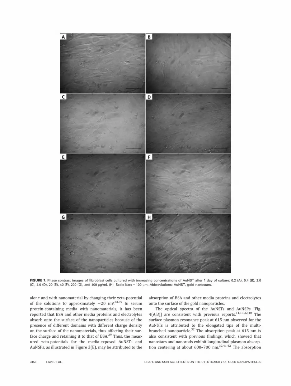
After 1 day of culture, untreated fibroblasts and RFPECsshowed healthy cell morphology as illustrated in Figure 6(Aand B), respectively. The fibroblasts featured their pheno-typic elongated and full spread-out morphology, whereasthe RFPECs depicted a round morphology common of thesecells. After 4 days of culture, untreated fibroblasts andRFPECs showed healthy cell morphology, similar to the 1day of culture (data not shown). Representative phase con-trast images of fibroblasts and RFPECs cultured withincreasing concentrations of AuNST after 1 day of cultureare illustrated in Figures 7 and 8, respectively. The phasecontrast images confirmed the MTS assay results (Fig. 5)and showed that both fibroblast cells (Fig. 7) and RFPECs(Fig. 8) maintained their morphology and density comparedwith the untreated cells (Fig. 6) after 1 day of culture. After4 days of culture, both fibroblast cells and RFPECs wereattached to the TCP at varying density compared with theuntreated cells, similar to the treated cells after 1 day ofculture (data not shown).
FIGURE 3. Hydrodynamic diameter and polydispersity of AuNST before (A) and after (C) the addition of media. Hydrodynamic diameter and pol-
ydispersity of AuNSP before (B) and after (D) the addition of media. Zeta-potential of AuNST and AuNSP before and after the addition of media
(E). The results represent the means 6 standard deviation with n 5 3 for each bar. Abbreviations: AuNST, gold nanostars; AuNSP, gold
nanospheres.
ORIGINAL ARTICLE
JOURNAL OF BIOMEDICAL MATERIALS RESEARCH A | NOV 2015 VOL 103A, ISSUE 11 3453

The relationship between the increasing concentrations ofAuNSPs and cell viability of the fibroblasts and the RFPECsafter 1 and 4 days of culture using the MTS assay are shownin Figure 9(A and B), respectively. Cell viability was signifi-cantly reduced in a dose-dependent manner after exposure ofthe fibroblasts and RFPECs to the AuNSPs using the MTSassay. By increasing the exposure time to 4 days of culture,the cytotoxicity of the fibroblast cells was not affected [Fig.
9(A)]. However, a slight cell viability increase was observedafter 4 days of RFPECs culture with AuNSPs, compared with 1day of culture [Fig. 9(B)]. Following 1 and 4 days of culture,the IC50 values of AuNSPs were 26.4 and 27.7 lg/mL forexposure with fibroblasts [Fig. 9(A)], and 13.6 and 13.8 lg/mL for exposure with RFPECs [Fig. 9(B)], respectively. TheAuNSP concentration of 40 lg/mL was a lethal concentrationfor both the fibroblast cells and the RFPECs (Fig. 9).
FIGURE 4. Absorption spectra of AuNST (A) and AuNSP (B). FTIR spectra of the original and cell culture media-exposed AuNST and AuNSP
(C). Abbreviations: AuNST, gold nanostars; AuNSP, gold nanospheres; FTIR, Fourier transform infrared spectroscopy.
FIGURE 5. Dose-dependent cellular viability of fibroblast cells (A) and RFPECs (B) cultured with increasing concentrations of AuNST after 1 and
4 days of culture, as determined using MTS assays. Data are expressed relative to that of untreated control cells (=100%) as means 6 standard
deviation with n 5 3 for each bar; the degree of significance at p< 0.05 comparing untreated control cells on the days of culture to different
concentrations (*), and the same concentrations at different days of culture (**) are indicated when appropriate. Abbreviations: AuNST, gold
nanostars; RFPECs, rat fat pad endothelial cells.
3454 FAVI ET AL. SHAPE AND SURFACE EFFECTS ON THE CYTOTOXICITY OF GOLD NANOPARTICLES

The phase contrast images of fibroblasts and RFPECscultured with increasing concentrations of AuNSP after 1day of culture are illustrated in Figures 10 and 11, respec-tively. The phase contrast images confirmed the MTS assayresults (Fig. 9) and showed that after 1 day of culture, bothfibroblast cells and RFPECs maintained their original mor-phology and density up to 4 lg/mL of AuNSPs [Figs. 10(A–D) and 11(A–D)]. Higher concentrations of AuNSP (20 and40 lg/mL), however, induced increased cytotoxicity wherethe cell did not maintain their original morphology, wereunhealthy, loosely attached to the TCPs, and had reduceddensity [Figs. 10(E,F) and 11(E,F)]. The morphologies ofboth fibroblast cells and RFPECs after 4 days of culturewith AuNSPs were similar to those observed after 1 day ofcell culture (data not shown).
DISCUSSION
In this study, TEM confirmed the shape of the biocompatibleAuNST, synthesized following a method by Xie et al.,32 topossess star-like multibranched surface structures (Fig. 1).It has been previously reported that AuNSTs can maintaintheir branched shape and size for at least a month followingfreeze-drying.32 The TEM images of these nanoparticlesillustrate that additionally, AuNSTs maintain their branchedshape and size when suspended in various organic (HEPESbuffer and cell culture media) and nonorganic (DI water)mediums following freeze-drying [Fig. 1(B–D)]. The resultsshow that the synthesized AuNSTs contained one to sixbranched tips on their surface [Fig. 1(E–J)]. The formationof tips on the surface of gold particles to produce AuNST isa random nucleation process where HEPES reduces AuCl4
–
and directs the formation of three-dimensional anisotropicgrowth on the surface of the particles, with an estimatedbranched-structure particle yield of 92%.32,38 The branchedAuNST were further studied in comparison with sphericallyshaped gold nanoparticles, a well-characterized nanoparticlein the field of biomedical science and engineering. The TEMmeasurements of the gold nanoparticles confirmed the diam-eter of the synthesized AuNST (33.6968.45 nm) to be signifi-
cantly less (p< 0.05) than the diameter of the commerciallyobtained AuNSP [61.466 4.28 nm; Figs. 1(A) and 2].
The hydrodynamic diameters for the AuNSTs (41.5 nm)and AuNSPs (70.8 nm), as measured by DLS [Fig. 3(A,B)],are higher than the average core diameter for the AuNSTs(33.6968.45 nm) and AuNSPs (61.466 4.28 nm), as meas-ured by TEM [Figs. 1(A) and 2]. The DLS instrument meas-ures the hydrodynamic diameter of a particle, which is theparticle core size and the thin solvent layer attached to theparticle, in contrast to TEM that only measures the particlecore size. The increase in measured particle diameter asobserved with the DLS data compared with the TEM data isattributed to the diameter of the thin solvent layer attachedto the nanoparticles. In general, a polydispersity index valueof approximately 0.5 or less denotes a monodispersed solu-tion. The measured polydispersity index of the synthesizedAuNST and the AuNSP stock solutions were 0.277 and0.202, respectively [Fig. 3(A,B)]. Because the measured poly-dispersity index of the nanoparticles was less than 0.5, itsignifies that the nanoparticles did not aggregate into largersized clusters but were monodispersed in the stock solu-tions. The increase in mean hydrodynamic diameter andpolydispersity index for AuNST [Fig. 3(C)] and AuNSP [Fig.3(D)] following contact with media compared with theuntreated nanoparticles [Fig. 3(A,B)] indicated that the pro-tein and electrolytes in the cell culture media adhered tothe surface of the nanoparticles, resulting in the increase oftheir size and aggregation.
The zeta-potentials for both the AuNSTs and AuNSPs fol-lowing exposure to the cell culture media were approxi-mately 224 mV [Fig. 3(E)]. Alkilany et al.33 reported thatgold nanorods coated with cationic surfactant cetyltrimethy-lammonium bromide, negatively charged polyacrylic acid,and positively charged polyelectrolyte poly(allylamine)hydrochloride adopted a negative effective surface charge ofapproximately 220 mV following the addition of serumprotein-containing media. This change was attributed to thepresence of bovine serum albumin (BSA) in the media,which has an effective surface charge of 220 mV at a physi-ological pH.33 The surface charge of BSA affects the media
FIGURE 6. Phase contrast images of untreated fibroblast cells (A) and RFPECs (B) cultured on tissue culture plates after 1 day of culture. Scale
bars 5 100 lm. Abbreviations: RFPECs, rat fat pad endothelial cells.
ORIGINAL ARTICLE
JOURNAL OF BIOMEDICAL MATERIALS RESEARCH A | NOV 2015 VOL 103A, ISSUE 11 3455
alone and with nanomaterial by changing their zeta-potentialof the solutions to approximately 220 mV.33,39 In serumprotein-containing media with nanomaterials, it has beenreported that BSA and other media proteins and electrolytesabsorb onto the surface of the nanoparticles because of thepresence of different domains with different charge densityon the surface of the nanomaterials, thus affecting their sur-face charge and retaining it to that of BSA.39 Thus, the meas-ured zeta-potentials for the media-exposed AuNSTs andAuNSPs, as illustrated in Figure 3(E), may be attributed to the
absorption of BSA and other media proteins and electrolytesonto the surface of the gold nanoparticles.
The optical spectra of the AuNSTs and AuNSPs [Fig.4(A,B)] are consistent with previous reports.11,13,32,40 Thesurface plasmon resonance peak at 615 nm observed for theAuNSTs is attributed to the elongated tips of the multi-branched nanoparticle.32 The absorption peak at 615 nm isalso consistent with previous findings, which showed thatnanostars and nanorods exhibit longitudinal plasmon absorp-tion centering at about 600–700 nm.32,41,42 The absorption
FIGURE 7. Phase contrast images of fibroblast cells cultured with increasing concentrations of AuNST after 1 day of culture: 0.2 (A), 0.4 (B), 2.0
(C), 4.0 (D), 20 (E), 40 (F), 200 (G), and 400 lg/mL (H). Scale bars 5 100 lm. Abbreviations: AuNST, gold nanostars.
3456 FAVI ET AL. SHAPE AND SURFACE EFFECTS ON THE CYTOTOXICITY OF GOLD NANOPARTICLES

peak at 335 nm may be due to the molecules on the surface ofthe AuNSTs and is consistent with previous reports.40 Theabsorption spectrum of AuNSPs showed a distinct surfaceplasmon resonance peak at 535 nm [Fig. 4(B)], which isattributed to the diameter of the nanoparticle, and is in agree-ment with experimental and theoretical data.13
The surface chemistry of the gold nanoparticles, ana-lyzed by FTIR, confirmed that the original gold nanopar-ticles had differing surface chemistries [Fig. 4(C)]. The FTIRabsorption bands confirmed that the surfaces of the synthe-
sized AuNSTs were adorned with numerous chemical func-tional groups, including amide I, m(NH), m(OH), CH3, andCH2 bands [Fig. 4(C)]. It is proposed that these chemicalbands on the surfaces of the original AuNSTs are due to thepresence of residual HEPES buffer and HEPES moleculesthat had adsorbed on the AuNSTs from the synthesis of thenanoparticle. The amide I or m(NH), m(OH), CH3, and CH2
bands detected on the FTIR spectra of the original AuNSTs[Fig. 4(C)] correspond to the organic amines, hydroxyl,methyl, and methylene groups, respectively, in the HEPES.43
FIGURE 8. Phase contrast images of RFPECs cultured with increasing concentrations of AuNST after 1 day of culture: 0.2 (A), 0.4 (B), 2.0 (C), 4.0
(D), 20 (E), 40 (F), 200 (G), and 400 lg/mL (H). Scale bars 5 100 lm. Abbreviations: AuNST, gold nanostars; RFPECs, rat fat pad endothelial cells.
ORIGINAL ARTICLE
JOURNAL OF BIOMEDICAL MATERIALS RESEARCH A | NOV 2015 VOL 103A, ISSUE 11 3457

FIGURE 9. Dose-dependent cell viability of fibroblast cells (A) and RFPECs (B) cultured with increasing concentrations of AuNSP after 1 and 4
days of culture, as determined using MTS assays. Data are expressed relative to that of untreated control cells (=100%) as means 6 standard
deviation with n 5 3 for each bar; the degree of significance at p< 0.05 comparing untreated control cells on the days of culture to different con-
centrations (*), and the same concentrations at different days of culture (**) are indicated when appropriate. Abbreviations: AuNSP, gold nano-
spheres; RFPECs, rat fat pad endothelial cells.
FIGURE 10. Phase contrast images of fibroblast cells cultured with increasing concentrations of AuNSP after 1 day of culture: 0.2 (A), 0.4 (B), 2.0
(C), 4.0 (D), 20 (E), and 40 lg/mL (F). Scale bars 5 100 lm. Abbreviations: AuNSP, gold nanospheres.
3458 FAVI ET AL. SHAPE AND SURFACE EFFECTS ON THE CYTOTOXICITY OF GOLD NANOPARTICLES

The asymmetric and symmetric COO2 absorption bandsexhibited by AuNSPs originated from the excess citrate sta-bilizing buffer and the citrate molecules that had adsorbedon the AuNSPs. This result is as expected and concurs witha previous published result by Park and Shumaker-Parry.37
As determined by FTIR, the difference in surface chemistrybetween the original AuNSTs and the AuNSPs is the pres-ence of the residual buffers, and adsorbed HEPES andcitrate molecules on the AuNSTs and AuNSPs, respectively[Fig. 4(C)]. In this study, FTIR analysis also confirmed theadsorption of proteins onto the surfaces of AuNSTs andAuNSPs after exposure to cell culture media for 30 min at258C [Fig. 4(C)]. Ultimately, the structure and compositionof adsorbed proteins on material surfaces depend on thephysicochemical properties of the nanomaterial (e.g., size,shape, composition, surface functional groups, and surface
charges), the characteristics of the physiological environ-ment (blood, interstitial fluid, cell cytoplasm, serum proteinmedia, etc.), and the length of exposure.44 Protein adsorptionto surfaces does change over time following the Vroman’seffect, with higher-binding-constant proteins replacing initialweak adsorbing and abundant proteins.45,46
The cellular proliferation and morphology results of thisstudy demonstrated that the spherically shaped gold nano-particles with diameters of 61.466 4.28 nm showed greatertoxicity after both 1 and 4 days of culture, with fibroblast cellsand RFPECs, compared with the synthesized star-shaped goldnanoparticles with diameters of 33.696 8.45 nm. TheAuNSPs were lethal at concentrations of 40 lg/mL for bothcell lines at 1 and 4 days of culture, whereas the AuNSTs wereless toxic at high concentrations (400 lg/mL). The calculatedIC50 values of AuNSP exposed to fibroblast cells were greater
FIGURE 11. Phase contrast images of RFPECs cultured with increasing concentrations of AuNSP after 1 day of culture: 0.2 (A), 0.4 (B), 2.0 (C), 4.0
(D), 20 (E), and 40 lg/mL (F). Scale bars 5 100 lm. Abbreviations: AuNSP, gold nanospheres; RFPECs, rat fat pad endothelial cells.
ORIGINAL ARTICLE
JOURNAL OF BIOMEDICAL MATERIALS RESEARCH A | NOV 2015 VOL 103A, ISSUE 11 3459

at 1 and 4 days of culture (26.4 and 27.7 lg/mL, respectively)compared with the RFPECs (13.6 and 13.8 lg/mL, respec-tively), indicating that AuNSP have a greater toxicity to endo-thelial cells. However, the IC50 values could not be measuredfor the AuNSTs because of incomplete dose–response curves.Results from this study demonstrate that there are three pos-sible factors that could be promoting the reduced toxicityeffects of AuNST to fibroblast cells and RFPECs, comparedwith AuNSP: size, surface chemistry, and shape. The followingdiscussion will explain how each of these material propertiesmight have affected these cell toxicities.
Several studies have demonstrated that the size of goldnanoparticles influences cell toxicity.47–50 Pan et al.47 haveshown that 1.4 nm gold nanoparticles induce cytotoxicity toa greater extent in four different cell lines (connective tissuefibroblasts, epithelial cells, macrophages, and melanomacells) than the corresponding 15 nm particles. Previousstudies have also found that when exposed to dermal fibro-blasts, larger gold nanoparticles (45 nm) increased the rateof apoptosis by penetrating the cells via clathrin-mediatedphagocytes, reduced proliferation, decreased extracellularmatrix protein expression, and induced cytoskeletal disrup-tion.49 Gold nanoparticles (60 nm) with comparable diame-ters as those evaluated in this study have been shown topenetrate human mesenchymal stem cells via endocytosis,and prompting necrosis by oxidative stress.47 To reduce cel-lular damage and cytotoxicity, researchers have functional-ized the surfaces of these unmodified gold nanoparticleswith materials such as neutrally charged poly(ethylene gly-col) and positive-charged ethanediamine.51,52 Freese et al.51
demonstrated that surface-modified 18, 35, and 65 nm goldnanoparticles, such as positive-charged ethanediamine, didnot induce cell toxicity up to a concentration of 250 lg/mLfollowing 2 days of culture. In this study, although both goldnanoparticles are negatively charged, FTIR analysis con-firmed that the surface chemistry of the AuNSPs andAuNSTs were different because of the residual buffers, andHEPES and citrate molecules that had adsorbed on the sur-face of the AuNSTs and AuNSPs, respectively.
Nevertheless, the shapes of gold nanoparticles have alsobeen reported to influence cellular response. In a recent invitro study comparing the effect of particle shapes on themechanisms of cellular uptake, Chu et al.21 assessed variousparticles, including large (150 nm) AuNST and AuNSP. Theauthors demonstrated that because of their sharp surfacestructures, nanostars could readily disrupt the endosomalmembranes of human liver carcinoma cells and reside inthe cytoplasm for an extended period of time, irrespectiveof their surface compositions, charges, materials, and sizes.Alternatively, following endocytosis, nanospheres wouldreside in the endosomes of the cells under stable conditions,where they would adapt with endosome maturation or beeasily excreted by exocytosis. This study by Chu et al.21 indi-cated that the two different shapes of gold nanoparticlesevaluated in this study may have been taken up by the cells,via endocytosis with the nanostars residing in the cytoplasmand the nanospheres in the endosomes of the cells. Further-more, the AuNST toxicity results in this study are supported
by a previous published result by Chen et al.40 that indi-cated that shorter, 4 h, exposure of synthesized AuNSTs(25 nm diameter, 5.0 lg/mL) to human breast and livercancer cells produced no morphological changes in the cells;however, increasing concentrations of the AuNST (0.7, 3.6,17.9, 89.3, and 446.4 lg/mL) in the breast cancer cellsgenerated a dose–response cytotoxicity behavior using MTTassay after 4 h of culture.40 Moreover, these authorsreported that following the 4 h of culture with the AuNSTdose of 446.4 lg/mL, the breast cancer cells viability wasapproximately 45%6 5%. Although the mechanism of celldeath by AuNST functionalized for targeting specific subcel-lular compartments such as the nucleus,40,53 and membranedeformation and cytoskeleton reorganization via macropino-cytosis54 have been investigated, the mechanistic details ofcell death for as-synthesized AuNSTs remains unclear. Basedon previous studies, it is hypothesized that the potentialmechanism of cell death by the as-synthesized AuNSTs isthat the nanoparticles enter the cells via endocytosis,21 con-centrate in the nuclei55 and in the cytoplasm,40,53 andinduce dose-dependent cell death via mechanical damage.56
Additional AuNST optimization for future research consider-ation is to synthesize AuNSTs with controlled size distribu-tion, branch length and number of branched tips, andanalyze the effect of these modified AuNST on cells and innative tissue. Controlling the size distribution, branchlength, and the number of branched tips of these AuNSTshas the potential to enable further tuning of the opticalproperties for the AuNSTs for potential medical detectionand treatment.
CONCLUSION
This study has demonstrated that various properties ofAuNSPs and nanostars influence the toxicity of fibroblastand endothelial cells. Additional studies will have to be per-formed to confirm the mechanisms of cellular uptake of thesegold nanoparticles and the role of AuNST branch length andnumber of branched tips on the observed biologicalresponses. The toxicity data presented in this study canpotentially be used to design an optimum gold nanoparticle-based material for numerous biomedical applications, includ-ing drug and gene delivery.
ACKNOWLEDGMENTS
Ms. Mariana Valencia is thankful for Northeastern Universityand the University of Antioquia’s Summer School Program.The authors also thank Mr. William Fowle and Dr. WentaoLiang for assistance with the TEM images, and Dr. Eno Ebongfor donation of the rat fat pad endothelial cells.
REFERENCES1. Perez-Rentero S, Grijalvo S, Penuelas G, Fabrega C, Eritja R.
Thioctic acid derivatives as building blocks to incorporate DNA
oligonucleotides onto gold nanoparticles. Molecules 2014; 19:
10495–10523.
2. Ding Y, Jiang ZW, Saha K, Kim CS, Kim ST, Landis RF, Rotello
VM. Gold nanoparticles for nucleic acid delivery. Mol Ther 2014;
22:1075–1083.
3. Jiwaji M, Sandison ME, Reboud J, Stevenson R, Daly R, Barkess
G, Faulds K, Kolch W, Graham D, Girolami MA, Cooper JM,
3460 FAVI ET AL. SHAPE AND SURFACE EFFECTS ON THE CYTOTOXICITY OF GOLD NANOPARTICLES

Pitt AR. Quantification of functionalised gold nanoparticle-
targeted knockdown of gene expression in HeLa cells. PLOS One
2014; 9:e99458.
4. Kim HJ, Takemoto H, Yi Y, Zheng M, Maeda Y, Chaya H, Hayashi
K, Mi P, Pittella F, Christie RJ, Toh K, Matsumoto Y, Nishiyama N,
Miyata K, Kataoka K. Precise engineering of siRNA delivery
vehicles to tumors using polyion complexes and gold nanopar-
ticles. ACS Nano 2014; 8:8979–8991.
5. Kim J, Lee YM, Kang Y, Kim WJ. Tumor-homing, size-tunable
clustered nanoparticles for anticancer therapeutics. ACS Nano
2014; 8:9358–9367.
6. Chen WH, Xu XD, Jia HZ, Lei Q, Luo GF, Cheng SX, Zhuo RX,
Zhang XZ. Therapeutic nanomedicine based on dual-intelligent
functionalized gold nanoparticles for cancer imaging and therapy
in vivo. Biomaterials 2013; 34:8798–8807.
7. Agasti SS, Chompoosor A, You C-C, Ghosh P, Kim CK, Rotello
VM. Photoregulated release of caged anticancer drugs from gold
nanoparticles. J Am Chem Soc 2009; 131:5728–5729.
8. Kim C, Agasti SS, Zhu Z, Isaacs L, Rotello VM. Recognition-medi-
ated activation of therapeutic gold nanoparticles inside living
cells. Nat Chem 2010; 2:962–966.
9. Saha K, Agasti SS, Kim C, Li X, Rotello VM. Gold nanoparticles inchemical and biological sensing. Chem Rev 2012; 112:2739–2779.
10. Giljohann DA, Seferos DS, Daniel WL, Massich MD, Patel PC,Mirkin CA. Gold nanoparticles for biology and medicine. AngewChem Int Ed 2010; 49:3280–3294.
11. Li WT, Sun XL, Wang Y, Niu G, Chen XY, Qian ZY, Nie LM. In
vivo quantitative photoacoustic microscopy of gold nanostar
kinetics in mouse organs. Biomed Opt Express 2014; 5:2679–2685.
12. Nie L, Wang S, Wang X, Rong P, Ma Y, Liu G, Huang P, Lu G,
Chen X. In vivo volumetric photoacoustic molecular angiography
and therapeutic monitoring with targeted plasmonic nanostars.
Small 2014; 10:1585–1593.
13. Haiss W, Thanh NTK, Aveyard J, Fernig DG. Determination of size
and concentration of gold nanoparticles from UV-vis spectra.
Anal Chem 2007; 79:4215–4221.
14. Indrasekara ASDS, Meyers S, Shubeita S, Feldman LC,
Gustafsson T, Fabris L. Gold nanostar substrates for SERS-based
chemical sensing in the femtomolar regime. Nanoscale 2014; 6:
8891–8899.
15. Kumar PS, Pastoriza-Santos I, Rodr�ıguez-Gonzalez B, Garc�ıa de
Abajo FJ, Liz-Marz�an LM. High-yield synthesis and optical
response of gold nanostars. Nanotechnology 2008; 19:015606. s
16. Verma MS, Chen PZ, Jones L, Gu FX. Branching and size of
CTAB-coated gold nanostars control the colorimetric detection of
bacteria. RSC Adv 2014; 4:10660–10668.
17. Huang X, Jain PK, El-Sayed IH, El-Sayed MA. Plasmonic photo-
thermal therapy (PPTT) using gold nanoparticles. Laser Med Sci
2008; 23:217–228.
18. Zhang XD, Wu D, Shen X, Liu PX, Yang N, Zhao B, Zhang H, Sun
YM, Zhang LA, Fan FY. Size-dependent in vivo toxicity of PEG-
coated gold nanoparticles. Int J Nanomed 2011; 6:2071–2081.
19. Lewinski N, Colvin V, Drezek R. Cytotoxicity of nanoparticles.
Small 2008; 4:26–49.
20. Hauck TS, Ghazani AA, Chan WCW. Assessing the effect of sur-
face chemistry on gold nanorod uptake, toxicity, and gene expres-
sion in mammalian cells. Small 2008; 4:153–159.
21. Chu Z, Zhang S, Zhang B, Zhang C, Fang C-Y, Rehor I, Cigler P,
Chang H-C, Lin G, Liu R, Li Q. Unambiguous observation of shape
effects on cellular fate of nanoparticles. Sci Rep 2014; 4:4495.
22. Price RL, Gutwein LG, Kaledin L, Tepper F, Webster TJ. Osteo-
blast function on nanophase alumina materials: Influence of
chemistry, phase, and topography. J Biomed Mater Res A 2003;
67A:1284–1293.
23. Lynch I, Cedervall T, Lundqvist M, Cabaleiro-Lago C, Linse S,
Dawson KA. The nanoparticle–protein complex as a biological
entity; a complex fluids and surface science challenge for the 21st
century. Adv Colloid Interface Sci 2007; 134–135:167–174.
24. Lynch I, Dawson KA. Protein-nanoparticle interactions. Nano
Today 2008; 3:40–47.
25. Aggarwal P, Hall JB, McLeland CB, Dobrovolskaia MA, McNeil SE.
Nanoparticle interaction with plasma proteins as it relates to par-
ticle biodistribution, biocompatibility and therapeutic efficacy.
Adv Drug Deliv Rev 2009; 61:428–437.
26. Flavell SJ, Hou TZ, Lax S, Filer AD, Salmon M, Buckley CD. Fibro-blasts as novel therapeutic targets in chronic inflammation. Br JPharmacol 2008; 153(Suppl 1):S241–S246.
27. Rodemann HP, Rennekampff H-O. Functional diversity of fibro-
blasts. In: Mueller MM, Fusenig NE, editors. Tumor-Associated
Fibroblasts and Their Matrix. The Netherlands: Springer; 2011. p
23–36.
28. Baxter LC, Frauchiger V, Textor M, Ap Gwynn I, Richards RG.
Fibroblast and osteoblast adhesion and morphology on calcium
phosphate surfaces. Eur Cell Mater 2002; 4:1–17.
29. Quadri SK, Bhattacharjee M, Parthasarathi K, Tanita T,
Bhattacharya J. Endothelial barrier strengthening by activation of
focal adhesion kinase. J Biol Chem 2003; 278:13342–13349.
30. Abbott NJ. Astrocyte–endothelial interactions and blood–brain
barrier permeability. J Anat 2002; 200:629–638.
31. Schniedermann J, Rennecke M, Buttler K, Richter G, Stadtler A-M,
Norgall S, Badar M, Barleon B, May T, Wilting J, Weich H. Mouse
lung contains endothelial progenitors with high capacity to form
blood and lymphatic vessels. BMC Chem Biol 2010; 11:50.
32. Xie JP, Lee JY, Wang DIC. Seedless, surfactantless, high-yield
synthesis of branched gold nanocrystals in HEPES buffer solution.
Chem Mater 2007; 19:2823–2830.
33. Alkilany AM, Nagaria PK, Hexel CR, Shaw TJ, Murphy CJ,
Wyatt MD. Cellular uptake and cytotoxicity of gold nanorods:
Molecular origin of cytotoxicity and surface effects. Small 2009;
5:701–708.
34. Barltrop JA, Owen TC, Cory AH, Cory JG. 5-(3-Carboxymethoxy-
phenyl)-2-(4,5-dimethylthiazolyl)-3-(4-sulfophenyl)tetrazolium, inner
salt (MTS) and related analogs of 3-(4,5-dimethylthiazolyl)-2,5-
diphenyltetrazolium bromide (MTT) reducing to purple water-
soluble formazans as cell-viability indicators. Bioorg Med Chem
Lett 1991; 1:611–614.
35. Scheirlinckx F, Raussens V, Ruysschaert JM, Goormaghtigh E.
Conformational changes in gastric H1/K1 - ATPase monitored by
difference Fourier-transform infrared spectroscopy and hydrogen/
deuterium exchange. Biochem J 2004; 382:121–129.
36. Bellamy LJ. The Infra-Red Spectra of Complex Molecules. Lon-
don: Chapman and Hall; 1975.
37. Park J-W, Shumaker-Parry JS. Structural study of citrate
layers on gold nanoparticles: Role of intermolecular interac-
tions in stabilizing nanoparticles. J Am Chem Soc 2014;136:
1907–1921.
38. Ivan B, Ru-Shi L. Shape and size selective synthesis of gold nano-
structures for biomedical applications. In: Ru-Shi L, editor. Con-
trolled Nanofabrication: Advances and Applications. Singapore:
Pan Stanford Publishing; 2013.
39. Rezwan K, Meier LP, Rezwan M, V€or€os J, Textor M, Gauckler LJ.
Bovine serum albumin adsorption onto colloidal Al2O3 particles:
A new model based on zeta potential and UV2vis measurements.
Langmuir 2004; 20:10055–10061.
40. Chen HY, Zhang X, Dai SH, Ma YX, Cui SS, Achilefu S, Gu YQ.
Multifunctional gold nanostar conjugates for tumor imaging and
combined photothermal and chemo-therapy. Theranostics 2013;
3:633–649.
41. Link S, El-Sayed MA. Spectral properties and relaxation
dynamics of surface plasmon electronic oscillations in gold
and silver nanodots and nanorods. J Phys Chem B 1999; 103:
8410–8426.
42. Nikoobakht B, El-Sayed MA. Preparation and growth mechanism
of gold nanorods (NRs) using seed-mediated growth method.
Chem Mater 2003; 15:1957–1962.
43. Jahromi EZ, White W, Wu Q, Yamdagni R, Gailer J. Remarkable
effect of mobile phase buffer on the SEC-ICP-AES derived Cu, Fe
and Zn-metalloproteome pattern of rabbit blood plasma. Metallo-
mics 2010; 2:460–468.
44. Rahman M, Laurent S, Tawil N, Yahia LH, Mahmoudi M. Nano-
particle and Protein Corona. Protein-Nanoparticle Interactions.
Berlin: Springer; 2013. p 21–44.
45. Vroman L, Adams AL, Fischer GC, Munoz PC. Interaction of high
molecular-weight kininogen, factor-XII, and fibrinogen in plasma
at interfaces. Blood 1980; 55:156–159.
46. Vroman L, Adams AL. Findings with the recording ellipsometer
suggesting rapid exchange of specific plasma proteins at liquid/
solid interfaces. Surf Sci 1969; 16:438–446.
ORIGINAL ARTICLE
JOURNAL OF BIOMEDICAL MATERIALS RESEARCH A | NOV 2015 VOL 103A, ISSUE 11 3461

47. Pan Y, Neuss S, Leifert A, Fischler M, Wen F, Simon U, Schmid
G, Brandau W, Jahnen-Dechent W. Size-dependent cytotoxicity of
gold nanoparticles. Small 2007; 3:1941–1949.
48. Fan JH, Li WT, Hung WI, Chen CP, Yeh JM. Cytotoxicity and differ-
entiation effects of gold nanoparticles to human bone marrow
mesenchymal stem cells. Biomed Eng App Bas C 2011; 23:
141–152.
49. Mironava T, Hadjiargyrou M, Simon M, Jurukovski V, Rafailovich
MH. Gold nanoparticles cellular toxicity and recovery: Effect of size,
concentration and exposure time. Nanotoxicology 2010; 4:120–137.
50. Soenen SJ, Manshian B, Montenegro JM, Amin F, Meermann B,
Thiron T, Cornelissen M, Vanhaecke F, Doak S, Parak WJ, De
Smedt S, Braeckmans K. Cytotoxic effects of gold nanoparticles:
A multiparametric study. ACS Nano 2012; 6:5767–5783.
51. Freese C, Gibson MI, Klok H-A, Unger RE, Kirkpatrick CJ. Size-
and coating-dependent uptake of polymer-coated gold nanopar-
ticles in primary human dermal microvascular endothelial cells.
Biomacromolecules 2012; 13:1533–1543.
52. Niidome T, Yamagata M, Okamoto Y, Akiyama Y, Takahashi H,
Kawano T, Katayama Y, Niidome Y. PEG-modified gold nanorods
with a stealth character for in vivo applications. J Control Release
2006; 114:343–347.
53. Dam DHM, Lee JH, Sisco PN, Co DT, Zhang M, Wasielewski MR,
Odom TW. Direct observation of nanoparticle–cancer cell nucleus
interactions. ACS Nano 2012; 6:3318–3326.
54. Yuan H, Fales AM, Vo-Dinh T. TAT peptide-functionalized gold
nanostars: Enhanced intracellular delivery and efficient NIR
photothermal therapy using ultralow irradiance. J Am Chem Soc
2012; 134:11358–11361.
55. Romero VH, Kereselidze Z, Egido W, Michaelides EA, Santamaria
F, Peralta XG. Nanoparticle assisted photothermal deformation of
individual neuronal organelles and cells. Biomed Opt Express
2014;5:4002–4012.
56. Kodiha M, Wang YM, Hutter E, Maysinger D, Stochaj U. Off to the
organelles—Killing cancer cells with targeted gold nanoparticles.
Theranostics 2015; 5:357–370.
3462 FAVI ET AL. SHAPE AND SURFACE EFFECTS ON THE CYTOTOXICITY OF GOLD NANOPARTICLES


