Embed Size (px)
Citation preview
Shear Stress Regulates the Flk-1/Cbl/PI3K/NF-κB Pathway ViaActin and Tyrosine Kinases
Yingxiao Wang1, Leona Flores3, Shaoying Lu1, Hui Miao2, Yi-Shuan Li2, and Shu Chien21Department of Bioengineering and The Beckman Institute for Advanced Science and Technology,University of Illinois, Urbana-Champaign, Urbana, IL 61801, USA2Department of Bioengineering and The Whitaker Institute of Biomedical Engineering, University ofCalifornia, San Diego, La Jolla, CA 92093-0427, USA3Biomedical Sciences Graduate Program, University of California, San Diego, La Jolla, CA92093-0427, USA
AbstractVascular endothelial cells (ECs) are continuously exposed to mechanical stimuli (e.g., shear stress).Our previous study has shown that the shear-induced nuclear factor-κB (NF-κB) activation ismediated by integrins [Bhullar, I. S., Y. S. Li, H. Miao, E. Zandi, M. Kim, et al. J. Biol. Chem.273:30544–30549, 1998]. The shear-activated integrins can also transactivate Flk-1 (a receptor forvascular endothelial growth factor (VEGF)) [Wang, Y., H. Miao, S. Li, K. D. Chen, Y. S. Li, et al.Am. J. Physiol. Cell Physiol. 283:C1540–C1547, 2002], which subsequently recruits Casitas B-lineage lymphoma (Cbl) to regulate inhibitor of κB protein kinase (IKK) [Wang, Y., J. Chang, Y. C.Li, Y. S. Li, J. Y. Shyy, and S. Chien. Am. J. Physiol. Heart Circ. Physiol. 286:H685–H692, 2004],an upstream molecule of NF-κB. Therefore, shear stress may likely utilize the Flk-1/Cbl pathway inregulating NF-κB. In this paper, we confirmed that the inhibition of Flk-1 by its specific inhibitorSU1498 blocked the shear-induced NF-κB translocation. The inhibition of Cbl (an adaptor proteinwhich binds to Flk-1 upon shear) by using a negative mutant (Cblnm) also blocked the promoteractivity of NF-κB, and the inhibition of the Cbl-downstream molecule phosphatidylinositol-3-kinase(PI3K) abolished the NF-κB translocation. Further experiments revealed that the disruption of actincytoskeleton inhibited the Flk-1 and Cbl interaction and NF-κB translocation. The inhibition of focaladhesion kinase (FAK) and Src family kinases, which are involved in the integrin-mediated focaladhesion complex, also blocked the shear-induced NF-κB translocation. Together with our previousfindings that integrins mediate the shear-induced activation of Flk-1 and NF-κB [Bhullar, I. S., Y.S. Li, H. Miao, E. Zandi, M. Kim, et al. J. Biol. Chem. 273:30544–30549, 1998; Wang, Y., H. Miao,S. Li, K. D. Chen, Y. S. Li, et al. Am. J. Physiol. Cell Physiol. 283:C1540–C1547, 2002], the presentresults suggest that Flk-1, Cbl, and PI3K act upstream to NF-κB in response to shear stress. ThisFlk-1/Cbl/PI3K/NF-κB signaling pathway may be originated from integrins and transmitted by keytyrosine kinases and actin cytoskeleton. These results shed new lights on the molecular mechanismby which mechanical shear stress activates the NF-κB signaling pathway, which is critical for vascularinflammatory responses and atherosclerosis.
© 2009 Biomedical Engineering SocietyAddress correspondence to Shu Chien, Department of Bioengineering and The Whitaker Institute of Biomedical Engineering, Universityof California, San Diego, La Jolla, CA 92093-0427, USA. [email protected]. Wang, L. Flores contributed equally to this work.
NIH Public AccessAuthor ManuscriptCell Mol Bioeng. Author manuscript; available in PMC 2009 December 8.
Published in final edited form as:Cell Mol Bioeng. 2009 September 1; 2(3): 341–350. doi:10.1007/s12195-009-0069-3.
NIH
-PA Author Manuscript
NIH
-PA Author Manuscript
NIH
-PA Author Manuscript
KeywordsShear stress; Flk-1; Cbl; PI3K; NF-κB; Actin; Tyrosine kinases
INTRODUCTIONVascular endothelial cells (ECs) are continuously exposed to mechanical stimuli (e.g., the shearstress resulting from blood flow), which play an important role in circulatory regulation inhealth and disease.12 Atherosclerotic lesions, which are characterized by the patchy deposit offatty materials in the arterial walls and the subsequent reduced/blocked blood flow, occurpreferentially at vascular curvatures and branch sites where the vessel walls are exposed todisturbed flow but not at the straight parts of vessels where laminar flow dominates.11 It hasbeen well documented that shear stress can activate a variety of signaling cascades and geneexpressions to modulate EC functions and pathophysiological processes, includingatherosclerosis.4,14,26 A wide range of signaling and structural molecules, including the plasmamembrane,7,38 membrane proteins/receptors such as integrins19,40 and VEGF receptor 2(Flk-1),8,44 and cytoskeletal filaments25 have been shown to play important roles inmechanotransduction, i.e., transducing shear stress into biochemical signaling cascades.However, there is a need to further elucidate the molecular mechanism by which cells perceiveand subsequently transduce shear stress into intracellular molecular activities to modulatepathophysiological processes such as atherosclerosis.
NF-κB, which belongs to the NF-κB/REL transcription factor family and plays important rolesin vascular inflammatory responses,9,32 is sequestered in the cytoplasm by the inhibitor of κBprotein (IκB). Pro-inflammatory stimuli, e.g., tumor necrosis factor (TNF)-α, activate IκBkinase (IKK) to phosphorylate IκB.2,21 The phosphorylated IκB subsequently undergoespolyubiquination and degradation to release NF-κB for its translocation into the nucleus topromote the expression of multiple genes,2,21 and the up-regulation of inflammatory genessuch as NF-κB can be atherogenic.32 Shear stress has been shown to activate NF-κB in theendothelium.3,24 Our previous studies have shown that this shear-induced NF-κB is mediatedby integrins.3 We have further shown that the membrane receptors integrin and Flk-1 caninteract with each other upon shear application, with integrins acting upstream to Flk-1.44 Theresultant activation and phosphorylation of Flk-1 can then recruit an adapter protein, Cbl,3which regulates downstream signaling events.30,36 Upon shear stimulation, both Flk-1 and Cblwere shown to regulate Akt (protein kinase B) and subsequently IKK,43 an upstream moleculefor NF-κB.33,34 Therefore, it is expected that shear stress can regulate NF-κB via the Flk-1/Cbl pathway. However, it has not been established as to how the integrin signaling can betransmitted to Flk-1 to activate the NF-κB signaling pathway.
In this work, we first confirmed the roles of Flk-1 and Cbl in the shear stress-induced activationof NF-κB and its translocation into the nucleus. We extended our study to show that PI3K, amolecule acting in between Cbl and Akt, is also essential for the shear-induced NF-κBactivation. Furthermore, our results demonstrate that actin cytoskeleton and key tyrosinekinases in the adhesion complex, including Src family kinases and FAK, are involved in theshear-induced activation of NF-κB. Therefore, our results established the expected roles ofFlk-1 and Cbl in regulating the shear-induced NF-κB activation, demonstrated that PI3K actsbetween Cbl and Akt in the signaling pathway, and identified actin cytoskeleton and integrin-associated tyrosine kinases as the mediators transmitting the shear-activated integrin signalsto Flk-1/Cbl and subsequently NF-κB. Together with our previous findings, our current resultsindicate that shear stress regulates NF-κB via the activation of the membrane receptor Flk-1to recruit the adapter protein Cbl for the subsequent activation of PI3K and Akt. The shear-
Wang et al. Page 2
Cell Mol Bioeng. Author manuscript; available in PMC 2009 December 8.
NIH
-PA Author Manuscript
NIH
-PA Author Manuscript
NIH
-PA Author Manuscript
induced signaling initiated from integrins is transmitted by integrin-associated tyrosine kinasesand the actin cytoskeleton to modulate the Flk-1/Cbl/PI3K/NF-κB pathway.
MATERIALS AND METHODSCell Culture
Cell culture reagents were obtained from GIBCO BRL (Grand Island, New York, USA).Bovine aortic endothelial cells (BAECs) were isolated from bovine aorta with collagenase andcultured in a humidified 95% air, 5% CO2 incubator at 37 °C. The culture medium wasDulbecco's modified Eagle's medium (DMEM) supplemented with 10% fetal bovine serum, 2mM L-glutamine, 1 unit/mL penicillin, 100 μg/mL streptomycin, and 1 mM sodium pyruvate.All experiments were conducted with BAEC cultures prior to passage 10 as previouslydescribed.3
Shear Stress ExperimentsA parallel-plate flow system was used to impose shear stress on the cultured ECs as describedby Frangos et al.17 In all experiments, BAECs were starved for 12 h in 0.5% serum followedby 2 h in serum-free medium before being subjected to shear stress (12 dyn/cm2).
DNA Plasmids and Transient TransfectionA hemagglutinin (HA) epitope-tagged wild type Cbl (HA-Cblwt) and an HA epitope-taggednegative mutant of Cbl (HA-Cblnm) were gifts from Dr. Alexander Y. Tsygankov. In HA-Cblnm, tyrosine → phenylalanine mutations were introduced at positions 700, 731, and 774,which are the major tyrosine phosphorylation sites of c-Cbl.16 HIV(LTR)-Luc encodes aluciferase reporter driven by the human immunodeficiency virus (HIV) long terminal repeat(LTR) that contains two binding sites for NF-κB.31 The pSV-β-galactosidase plasmid containsa β-galactosidase gene driven by the SV40 promoter and enhancer. The various plasmids weretransfected into BAECs at 80% confluence using the lipofectamine method as described bythe vendor (GIBCO BRL).
Luciferase Activity AssayHA-Cblwt or HA-Cblnm were cotransfected with HIV(LTR)-Luc and the pSV-β-galactosidaseplasmid into BAECs for the NF-κB transcriptional activation assays with HIV(LTR)-Luc toreport NFκB activity and the pSV-β-galactosidase plasmid to monitor transfection efficiency.The luciferase and β-gal assays were conducted as described previously.3
Immunoprecipitation and ImmunoblottingThe antibodies used for immunoprecipitation and immunoblotting were polyclonal anti-Cbl,anti-Flk-1, and PY20 monoclonal anti-phosphotyrosine (Santa Cruz Biotechnology, SantaCruz, CA, USA). For immunoprecipitation, the cells were scraped into a lysis buffer (25 mMTris–HCl, pH 7.5, 150 mM NaCl, 1% Triton-X-100, 5 mM NaF, 1 mM Na3VO4, 1 mM PMSF,and 10 μg/mL Leupeptin). The lysate was centrifuged, and the supernatant wasimmunoprecipitated with the appropriate antibodies and protein A-sepharose beads(Amersham Pharmacia Biotech) at 4 °C overnight. The immunoprecipitated complexes werewashed and used for immunoblotting. After SDS-PAGE, proteins in the gel were transferredto a nitrocellulose membrane for immunoblotting. The membrane was blocked with 5% bovineserum albumin followed by incubation with the primary antibody in 10 mM Tris–HCl, pH 7.4,150 mM NaCl, and 0.05% Tween 20 containing 0.1% bovine serum albumin. The boundprimary antibodies were detected by using a goat anti-mouse or goat anti-rabbit IgG-horseradish peroxidase conjugate (Santa Cruz Biotechnology) and the ECL detection system(Amersham Pharmacia Biotech).
Wang et al. Page 3
Cell Mol Bioeng. Author manuscript; available in PMC 2009 December 8.
NIH
-PA Author Manuscript
NIH
-PA Author Manuscript
NIH
-PA Author Manuscript
Immunostaining and Fluorescence MicroscopyFor the inhibition experiments, BAECs were pretreated for 1 h with DMSO as the controlsolvent or with 5 μM SU1498, 100 nM Wortmannin, 1 μM Cytochalasin D, 100 μM genistein,10 μM PP2, or 20 μM AG82 (Sigma) to inhibit Flk-1, PI3K, actin filaments, tyrosine kinases,Src family kinases, or FAK kinase, respectively. The treated cells were subsequently subjectedto flow application for 30 min. The translocation of NF-κB was then studied byimmunostaining. In brief, confluent monolayers of BAECs were fixed in methanol at –20 °Cfor 5 min and incubated with 3% goat serum at 4 °C overnight. The specimens were washedthree times with phosphate-buffered saline (PBS) followed by incubation with the polyclonalanti-NF-κB p65 antibody (Santa Cruz Biotechnology) to detect the distribution of NF-κB. Theimages were collected using a confocal microscopy system (MRC-1000, Bio-Rad) andanalyzed using NIH Image and Microsoft Excel software.
StatisticsThe various experiments were performed independently at least three times. Comparisons ofresults between two groups were performed by using unpaired two-tailed t-tests assumingunequal population variance. Multiple group comparisons were done with analysis of variance(ANOVA), and statistical significance among multiple groups was determined by usingDunnett's Method. p-Values of <0.05 were considered statistically significant.
RESULTSThe Shear-Induced Translocation of NF-κB is Dependent on the Enzymatic Activity of Flk-1
We have previously shown that shear stress can induce the activation of NF-κB, which can berepresented by the translocation of NF-κB into the nucleus.3 This shear effect on NF-κBtranslocation is mediated by integrins.3 Since the shear-activated integrins were shown totransactivate Flk-1 which can regulate IKK, an upstream molecule of NF-κB,41,43,44 we askedwhether Flk-1 can also mediate the shear-induced translocation of NF-κB. BAECs weresubjected to various time periods of shear stress (12 dyn/cm2) or kept as static control.Fluorescence immunostaining revealed that p65 (a subunit of NF-κB) clearly translocated tothe nucleus upon shear application as shown in the control groups treated with solvent vehicles(Fig. 1a). In contrast, SU1498, an inhibitor of Flk-1 tyrosine phosphorylation, blocked thetranslocation of p65 to the nucleus in response to shear stress (Fig. 1b). These results suggestthat the enzymatic activity of Flk-1 is essential for the shear-induced NF-κB translocation.
Cbl Mediates the Shear-Induced Activation of NF-κBWe then asked whether Cbl, which contains multiple protein docking sites and has been shownto bind to Flk-1 in regulating IKK upon shear application,43,44 is involved in the regulation ofthe shear-induced NF-κB activation. Since the activated NF-κB can translocate to the nucleusand serve as a transcription factor,2 the level of NF-κB binding to target promoters to activategene expression was measured to represent the transcriptional activation of NF-κB. An HIV(LTR)-Luc construct, containing two repeats of NF-κB binding sites in the promoter regionlinked to a luciferase gene, was designed to report NF-κB transcriptional activity. The amountof luciferase produced can be quantitatively measured by the luciferase activity assay. A controlreporter construct, pSV-β-galactosidase driven by SV40 promoter, was co-transfected withHIV(LTR)-Luc to quantify the transfection efficiency by measuring the production of β-galactosidase via a β-gal assay as previously described.3 The ratio of detected luciferase andβ-galactosidase can hence represent a normalized NF-κB activity (Fig. 2a). HA-Cblwt (wildtype Cbl) or HA-Cblnm (negative mutant of Cbl) was co-transfected with HIV(LTR)-Luc andpSV-β-galactosidase into BAECs before the application of flow. As shown in Fig. 2b, HA-
Wang et al. Page 4
Cell Mol Bioeng. Author manuscript; available in PMC 2009 December 8.
NIH
-PA Author Manuscript
NIH
-PA Author Manuscript
NIH
-PA Author Manuscript
Cblnm significantly inhibited the shear-induced NF-κB transcriptional activity. These resultsindicate that Cbl acts upstream to NF-κB signaling in response to shear stress.
PI3K Mediates the Activation of NF-κB Induced by Shear StressCblnm, which inhibited the shear-induced NF-κB activation (Fig. 2b), has an impaired interfacein binding to PI3K.16 Since PI3K is a major protein associated with Cbl and plays a role inregulating the NF-κB pathway,13 we hypothesized that PI3K may act downstream to Flk-1 andCbl in mediating the shear-induced NF-κB activation. To examine the role of PI3K, BAECswere pretreated with a PI3K inhibitor, Wortmannin (100 nM), for 1 h before being subjectedto various time periods of shear stress (12 dyn/cm2) or kept as static control. The fluorescenceimmunostaining revealed that Wortmannin significantly attenuated the shear-inducedtranslocation of NF-κB (Fig. 3a). Quantification of the results and statistical analysis furtherconfirmed the immunostaining observations (Fig. 3b). While Wortmannin clearly attenuatedthe shear-induced NF-κB translocation and can completely block the PI3K activity and the Aktactivation in response to shear stress,5,22 it is of note that shear stress still caused a minorinduction of NF-κB translocation in the presence of Wortmannin. These results indicate thatthe shear-induced translocation of NF-κB is mainly mediated by PI3K, although other signalingpathways may bypass PI3K in regulating NF-κB upon shear application.
Actin Cytoskeleton is Essential for the Interaction Between Flk-1 and Cbl in Response toShear Stress
It has been shown that integrins are crucial in mechanotransduction4 and mediate the shear-induced complex formation, and interaction between Flk-1 and Cbl.44 Since the actin-basedcytoskeleton is closely linked to integrins and their associated focal adhesion complex,35 wetested the hypothesis that the integrity of the actin-based cytoskeleton is essential in mediatingthe transactivation from integrins to Flk-1 and its subsequent interaction with Cbl in responseto shear stress. BAECs were pre-incubated with Cytochalasin D, which is a reagent that disruptsthe actin filament network, or its solvent DMSO as control followed by the application of shearstress (12 dyn/cm2) for 5 min. Cell lysates were immunoprecipitated with an anti-Cbl antibodyfollowed by immunoblotting with anti-PY20 and anti-Flk-1 antibodies to measure the degreeof Flk-1 tyrosine phosphorylation and the amount of Cbl-bound Flk-1, respectively. Both theshear-induced tyrosine phosphorylation (top gel panel) and the amount of Cbl-bound Flk-1(middle gel panel) were blocked by Cytochalasin D (Fig. 4a). We then examined the role ofactin cytoskeleton on the subsequent Cbl tyrosine phosphorylation. As shown in Fig. 4b,Cytochalasin D abolished the shear-induced tyrosine phosphorylation of Cbl. These resultsshow that actin filaments are important for both the Flk-1-Cbl interaction and Cbl tyrosinephosphorylation in response to shear stress. Together with our previous study demonstratingthat integrins mediate the shear-induced activation of Flk-1 and Cbl,44 these results indicatethat the actin cytoskeleton may provide a link to transmit the signals between the two membranereceptors integrin and Flk-1 to regulate downstream molecular events, e.g., Cblphosphorylation.
Actin Cytoskeleton, FAK, and Src Family Kinases at Focal Adhesions are Important for theShear-Induced NF-κB Activation
Since the actin cytoskeleton is essential for the shear-induced Cbl phosphorylation (Fig. 4b),which may lead to the recruitment of PI3K and subsequent regulation of NF-κB, we furthertested the role of the actin cytoskeleton in shear-induced NF-κB activation. BAECs werepretreated with Cytochalasin D for 1 h before being subjected to shear stress. As shown in Fig.5, Cytochalasin D clearly abolished the shear-induced translocation of NF-κB (first image fromleft). In addition, since tyrosine kinases, such as focal adhesion kinase (FAK) and Src familykinases, have been shown to play critical roles for the integrin-mediated focal adhesion
Wang et al. Page 5
Cell Mol Bioeng. Author manuscript; available in PMC 2009 December 8.
NIH
-PA Author Manuscript
NIH
-PA Author Manuscript
NIH
-PA Author Manuscript
dynamics and the actin-mediated mechanotransduction,20,25 we reasoned that these tyrosinekinases may be essential for the shear-induced NF-κB translocation. Indeed, genistein, AG82or PP2, which inhibit general tyrosine kinases, FAK or Src family kinases, respectively,blocked the shear-induced NF-κB translocation (Fig. 5). These results confirmed that tyrosinekinases, including FAK and Src family kinases, play important roles in mediating the shear-induced NF-κB activation. Therefore, the integrin-mediated focal adhesion and its associatedtyrosine kinase activities may transmit the mechanical cues via the actin cytoskeleton toactivate the Flk-1/Cbl/PI3K pathway to regulate NF-κB signaling.
DISCUSSIONVascular ECs are continuously exposed to mechanical forces, including shear stress.Remarkable progress has been made in advancing our understanding on how these mechanicalforces can modulate EC functions.10 NF-κB in ECs is sensitive to mechanical stimulation andhas been shown to play critical roles in inflammatory responses and atherosclerosis.9,32 Ourprevious results suggested that shear stress can activate integrins to regulate NF-κB.3 The shear-activated integrins was also shown to transactivate Flk-1, which recruits Cbl to regulate thedownstream molecules Akt and IKK.43,44 While it is likely that Flk-1 and Cbl are involved inthe shear-induced NF-κB activation, it was unclear as to how the integrin signaling istransmitted to Flk-1. The results in the current paper indicate that key integrin-associatedtyrosine kinases, actin cytoskeleton, and the Flk-1/Cbl/PI3K pathway play important roles inregulating the shear-induced activation of NF-κB. This study also shows that actin cytoskeletonplays a critical role in mediating the interaction between Flk-1 and Cbl in response to shearstress.
Integrins and their associated cytoskeleton have been well documented to play important rolesin mechanotransduction.1,44 Our previous findings suggest that integrins can transactivateFlk-1 and its associated adapter protein Cbl upon shear stimulation.44 In this report, we havefurther shown that the actin cytoskeleton plays a significant role in mediating thistransactivation. Thus, the disruption of actin filaments by Cytochalasin D blocked the shear-induced Flk-1 and Cbl interaction, as well as the Cbl phosphorylation (Fig. 4), though wecannot rule out the possibility that these effects may be mediated via blocking the integrin-dependent phosphorylation of Flk-1. Cytochalasin D also inhibited the shear-induced signalingevent of NF-κB activation, downstream of Flk-1 and Cbl (Fig. 5). Interestingly, inhibition ofFAK and Src family kinases, the key tyrosine kinases involved in the integrin-mediated focaladhesion dynamics, blocked the shear-induced NF-κB activation (Fig. 5). Integrin engagementhas been shown to activate FAK via its FERM domain,29 with FAK undergoing aconformational change to expose its kinase domain, which subsequently auto-phosphorylatesthe intramolecular Y397 site.27 This phosphorylated Y397 can provide a docking site to recruitSrc family kinases via their SH2 domain to the focal adhesion sites.39 The localized Src familykinases can phosphorylate actin-interacting molecules, including cortactin, to regulate theconnection between focal adhesion sites and actin cytoskeleton.39 Therefore, FAK and Srcfamily kinases may provide a regulation mechanism that connects integrins to actin network,which can then transmit the mechanical impact to Flk-1 and Cbl. Indeed, it has been welldocumented that the actin-based cytoskeleton can transmit biochemical signals to allow fastresponses when cells are exposed to mechanical stimulation.18,42 Several reports indicate thatintegrins can form physical complexes with receptor tyrosine kinases (RTKs) such asFlk-1.6,37 This association between integrins and Flk-1 may also depend on the integrity of theactin cytoskeleton. Both integrin αvβ3 and an activated RTK, PDGFR-β, have been shown toassociate with a cytoskeletal (NP-40 insoluble) fraction in the PDGF-stimulated humanforeskin fibroblasts.35 It is possible that the actin cytoskeleton is required inmechanotransduction by providing the structural connection and support, as well as organizingthe spatial distribution of signaling molecules. The assessment of the internal structure of the
Wang et al. Page 6
Cell Mol Bioeng. Author manuscript; available in PMC 2009 December 8.
NIH
-PA Author Manuscript
NIH
-PA Author Manuscript
NIH
-PA Author Manuscript
cell using cytoskeletal depolymerization agents via a microfluidics system may directly addressthis question at the subcellular level.23
Both Flk-1 and Cbl appear essential for the NF-κB activation in response to shear stress (Figs.1 and 2), consistent with the expectation based on our previous findings that integrins not onlymediate the shear-activated NF-κB, but also transactivate Flk-1 to regulate Cbl andsubsequently IKK.3,43,44 It is possible that shear stress, via integrins and actin network, cancause changes in the conformation and localization of Flk-1, thus altering the clustering andautophosphorylation of Flk-1. The phosphorylated cytoplasmic tail of Flk-1 can then recruitCbl directly through its PTB domain or indirectly through other adapter proteins such as Grb2and Shc, which are known to associate with Flk-1.33 The receptor-bound Cbl can then undergophosphorylation and recruit signaling molecules such as PI3K. Indeed, the phosphorylatedtyrosine 731 of Cbl is a binding site for p85, the regulatory subunit of PI3K.28
While it has been well established that PI3K regulates NF-κB signaling,33,34 the detailedmolecular mechanism linking PI3K and NF-κB is not clear. One possibility is that the activatedPI3K can produce PIP3 at the plasma membrane which recruits Akt via its pleckstrin homologydomain.15 This membrane localization of Akt will allow its interaction with the upstreamphosphoinositide-dependent kinase (PDK) to lead to Akt activation.45 The subsequent complexformation between Akt and IKK can then cause the phosphorylation and degradation of IκBand the subsequent nuclear translocation of NF-κB for transcriptional activation.33,34
In summary, our results demonstrate that shear stress regulates the NF-κB activation via apathway in which signals are transmitted from integrins, via FAK and Src family kinases atfocal adhesions and actin cytoskeleton, to activate Flk-1, followed by Cbl and PI3K (see Fig.6 for illustration). These results help advance our understanding on how ECs coordinateintracellular signaling molecules to adjust and respond to external mechanical cues in healthand disease, e.g., atherosclerosis.
AcknowledgmentsThis article is part of the celebration dedicated to Dr. Van C. Mow's 70th birthday, for his outstanding leadership andmarvelous contributions in the field of biomechanics and mechanobiology. We thank Dr. Alexander Y. Tsygankow(Temple University) for providing HA-Cblwt, and HA-Cblnm. This work was supported in part by NIH researchGrants HL080518, HL085195, and HL064382 (S. Chien), NCI139272, NSF0846429, and the Beckman Laser Institute,Inc. (Y. Wang).
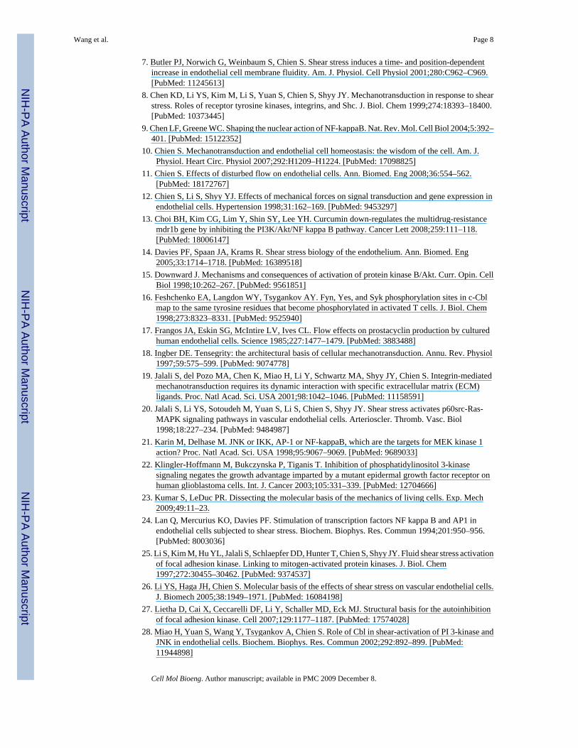
REFERENCES1. Alenghat FJ, Ingber DE. Mechanotransduction: all signals point to cytoskeleton, matrix, and integrins.
Sci. STKE 2002:PE6. [PubMed: 11842240]2. Baldwin AS Jr. The NF-kappa B and I kappa B proteins: new discoveries and insights. Annu. Rev.
Immunol 1996;14:649–683. [PubMed: 8717528]3. Bhullar IS, Li YS, Miao H, Zandi E, Kim M, Shyy JY, Chien S. Fluid shear stress activation of I kappaB
kinase is integrin-dependent. J. Biol. Chem 1998;273:30544–30549. [PubMed: 9804824]4. Boo YC, Jo H. Flow-dependent regulation of endothelial nitric oxide synthase: role of protein kinases.
Am. J. Physiol. Cell Physiol 2003;285:C499–C508. [PubMed: 12900384]5. Boo YC, Sorescu G, Boyd N, Shiojima I, Walsh K, Du J, Jo H. Shear stress stimulates phosphorylation
of endothelial nitric-oxide synthase at Ser1179 by Akt-independent mechanisms: role of protein kinaseA. J. Biol. Chem 2002;277:3388–3396. [PubMed: 11729190]
6. Borges E, Jan Y, Ruoslahti E. Platelet-derived growth factor receptor beta and vascular endothelialgrowth factor receptor 2 bind to the beta 3 integrin through its extracellular domain. J. Biol. Chem2000;275:39867–39873. [PubMed: 10964931]
Wang et al. Page 7
Cell Mol Bioeng. Author manuscript; available in PMC 2009 December 8.
NIH
-PA Author Manuscript
NIH
-PA Author Manuscript
NIH
-PA Author Manuscript
7. Butler PJ, Norwich G, Weinbaum S, Chien S. Shear stress induces a time- and position-dependentincrease in endothelial cell membrane fluidity. Am. J. Physiol. Cell Physiol 2001;280:C962–C969.[PubMed: 11245613]
8. Chen KD, Li YS, Kim M, Li S, Yuan S, Chien S, Shyy JY. Mechanotransduction in response to shearstress. Roles of receptor tyrosine kinases, integrins, and Shc. J. Biol. Chem 1999;274:18393–18400.[PubMed: 10373445]
9. Chen LF, Greene WC. Shaping the nuclear action of NF-kappaB. Nat. Rev. Mol. Cell Biol 2004;5:392–401. [PubMed: 15122352]
10. Chien S. Mechanotransduction and endothelial cell homeostasis: the wisdom of the cell. Am. J.Physiol. Heart Circ. Physiol 2007;292:H1209–H1224. [PubMed: 17098825]
11. Chien S. Effects of disturbed flow on endothelial cells. Ann. Biomed. Eng 2008;36:554–562.[PubMed: 18172767]
12. Chien S, Li S, Shyy YJ. Effects of mechanical forces on signal transduction and gene expression inendothelial cells. Hypertension 1998;31:162–169. [PubMed: 9453297]
13. Choi BH, Kim CG, Lim Y, Shin SY, Lee YH. Curcumin down-regulates the multidrug-resistancemdr1b gene by inhibiting the PI3K/Akt/NF kappa B pathway. Cancer Lett 2008;259:111–118.[PubMed: 18006147]
14. Davies PF, Spaan JA, Krams R. Shear stress biology of the endothelium. Ann. Biomed. Eng2005;33:1714–1718. [PubMed: 16389518]
15. Downward J. Mechanisms and consequences of activation of protein kinase B/Akt. Curr. Opin. CellBiol 1998;10:262–267. [PubMed: 9561851]
16. Feshchenko EA, Langdon WY, Tsygankov AY. Fyn, Yes, and Syk phosphorylation sites in c-Cblmap to the same tyrosine residues that become phosphorylated in activated T cells. J. Biol. Chem1998;273:8323–8331. [PubMed: 9525940]
17. Frangos JA, Eskin SG, McIntire LV, Ives CL. Flow effects on prostacyclin production by culturedhuman endothelial cells. Science 1985;227:1477–1479. [PubMed: 3883488]
18. Ingber DE. Tensegrity: the architectural basis of cellular mechanotransduction. Annu. Rev. Physiol1997;59:575–599. [PubMed: 9074778]
19. Jalali S, del Pozo MA, Chen K, Miao H, Li Y, Schwartz MA, Shyy JY, Chien S. Integrin-mediatedmechanotransduction requires its dynamic interaction with specific extracellular matrix (ECM)ligands. Proc. Natl Acad. Sci. USA 2001;98:1042–1046. [PubMed: 11158591]
20. Jalali S, Li YS, Sotoudeh M, Yuan S, Li S, Chien S, Shyy JY. Shear stress activates p60src-Ras-MAPK signaling pathways in vascular endothelial cells. Arterioscler. Thromb. Vasc. Biol1998;18:227–234. [PubMed: 9484987]
21. Karin M, Delhase M. JNK or IKK, AP-1 or NF-kappaB, which are the targets for MEK kinase 1action? Proc. Natl Acad. Sci. USA 1998;95:9067–9069. [PubMed: 9689033]
22. Klingler-Hoffmann M, Bukczynska P, Tiganis T. Inhibition of phosphatidylinositol 3-kinasesignaling negates the growth advantage imparted by a mutant epidermal growth factor receptor onhuman glioblastoma cells. Int. J. Cancer 2003;105:331–339. [PubMed: 12704666]
23. Kumar S, LeDuc PR. Dissecting the molecular basis of the mechanics of living cells. Exp. Mech2009;49:11–23.
24. Lan Q, Mercurius KO, Davies PF. Stimulation of transcription factors NF kappa B and AP1 inendothelial cells subjected to shear stress. Biochem. Biophys. Res. Commun 1994;201:950–956.[PubMed: 8003036]
25. Li S, Kim M, Hu YL, Jalali S, Schlaepfer DD, Hunter T, Chien S, Shyy JY. Fluid shear stress activationof focal adhesion kinase. Linking to mitogen-activated protein kinases. J. Biol. Chem1997;272:30455–30462. [PubMed: 9374537]
26. Li YS, Haga JH, Chien S. Molecular basis of the effects of shear stress on vascular endothelial cells.J. Biomech 2005;38:1949–1971. [PubMed: 16084198]
27. Lietha D, Cai X, Ceccarelli DF, Li Y, Schaller MD, Eck MJ. Structural basis for the autoinhibitionof focal adhesion kinase. Cell 2007;129:1177–1187. [PubMed: 17574028]
28. Miao H, Yuan S, Wang Y, Tsygankov A, Chien S. Role of Cbl in shear-activation of PI 3-kinase andJNK in endothelial cells. Biochem. Biophys. Res. Commun 2002;292:892–899. [PubMed:11944898]
Wang et al. Page 8
Cell Mol Bioeng. Author manuscript; available in PMC 2009 December 8.
NIH
-PA Author Manuscript
NIH
-PA Author Manuscript
NIH
-PA Author Manuscript
29. Mitra SK, Schlaepfer DD. Integrin-regulated FAK-Src signaling in normal and cancer cells. Curr.Opin. Cell Biol 2006;18:516–523. [PubMed: 16919435]
30. Miyake S, Lupher ML Jr. Andoniou CE, Lill NL, Ota S, Douillard P, Rao N, Band H. The Cblprotooncogene product: from an enigmatic oncogene to center stage of signal transduction. Crit. Rev.Oncog 1997;8:189–218. [PubMed: 9570294]
31. Nabel G, Baltimore D. An inducible transcription factor activates expression of humanimmunodeficiency virus in T cells. Nature 1987;326:711–713. [PubMed: 3031512]
32. Orr AW, Hahn C, Blackman BR, Schwartz MA. p21-activated kinase signaling regulates oxidant-dependent NF-kappa B activation by flow. Circ. Res 2008;103:671–679. [PubMed: 18669917]
33. Ozes ON, Mayo LD, Gustin JA, Pfeffer SR, Pfeffer LM, Donner DB. NF-kappaB activation by tumournecrosis factor requires the Akt serine-threonine kinase. Nature 1999;401:82–85. [PubMed:10485710]
34. Romashkova JA, Makarov SS. NF-kappaB is a target of AKT in anti-apoptotic PDGF signalling.Nature 1999;401:86–90. [PubMed: 10485711]
35. Schneller M, Vuori K, Ruoslahti E. Alphavbeta3 integrin associates with activated insulin andPDGFbeta receptors and potentiates the biological activity of PDGF. EMBO J 1997;16:5600–5607.[PubMed: 9312019]
36. Smit L, Borst J. The Cbl family of signal transduction molecules. Crit. Rev. Oncog 1997;8:359–379.[PubMed: 9622055]
37. Soldi R, Mitola S, Strasly M, Defilippi P, Tarone G, Bussolino F. Role of alphavbeta3 integrin in theactivation of vascular endothelial growth factor receptor-2. EMBO J 1999;18:882–892. [PubMed:10022831]
38. Thi MM, Tarbell JM, Weinbaum S, Spray DC. The role of the glycocalyx in reorganization of theactin cytoskeleton under fluid shear stress: a “bumper-car” model. Proc. Natl Acad. Sci .USA2004;101:16483–16488. [PubMed: 15545600]
39. Thomas SM, Brugge JS. Cellular functions regulated by Src family kinases. Annu. Rev. Cell Dev.Biol 1997;13:513–609. [PubMed: 9442882]
40. Tzima E, del Pozo MA, Shattil SJ, Chien S, Schwartz MA. Activation of integrins in endothelial cellsby fluid shear stress mediates Rho-dependent cytoskeletal alignment. EMBO J 2001;20:4639–4647.[PubMed: 11532928]
41. Tzima E, Irani-Tehrani M, Kiosses WB, Dejana E, Schultz DA, Engelhardt B, Cao G, DeLisser H,Schwartz MA. A mechanosensory complex that mediates the endothelial cell response to fluid shearstress. Nature 2005;437:426–431. [PubMed: 16163360]
42. Wang Y, Botvinick EL, Zhao Y, Berns MW, Usami S, Tsien RY, Chien S. Visualizing the mechanicalactivation of Src. Nature 2005;434:1040–1045. [PubMed: 15846350]
43. Wang Y, Chang J, Li YC, Li YS, Shyy JY, Chien S. Shear stress and VEGF activate IKK via theFlk-1/ Cbl/Akt signaling pathway. Am. J. Physiol. Heart Circ. Physiol 2004;286:H685–H692.[PubMed: 14551058]
44. Wang Y, Miao H, Li S, Chen KD, Li YS, Yuan S, Shyy JY, Chien S. Interplay between integrins andFLK-1 in shear stress-induced signaling. Am. J. Physiol. Cell Physiol 2002;283:C1540–C1547.[PubMed: 12372815]
45. Wymann MP, Pirola L. Structure and function of phosphoinositide 3-kinases. Biochim. Biophys.Acta 1998;1436:127–150. [PubMed: 9838078]
Wang et al. Page 9
Cell Mol Bioeng. Author manuscript; available in PMC 2009 December 8.
NIH
-PA Author Manuscript
NIH
-PA Author Manuscript
NIH
-PA Author Manuscript
FIGURE 1.The shear-induced translocation of NF-κB is dependent on the enzymatic activity of Flk-1. (a)BAECs were treated with a control solvent 0.1% DMSO for 1 h before being subjected to shearstress (right) or kept under static incubation (left) for 30 min. (b) BAECs were treated withSU1498 (5 μM) for 1 h before being subjected to shear stress (right) or kept under staticincubation (left) for 30 min. In both (a) and (b), immunostaining was performed after fixationwith an anti-NF-κB p65 antibody, which was then probed with fluorescein-labeled goat anti-rabbit IgG. The subcellular localization of NF-κB was visualized by the distribution offluorescein. (c) Bar graphs represent mean ± SEM of the percentile of BAECs with NF-κB
Wang et al. Page 10
Cell Mol Bioeng. Author manuscript; available in PMC 2009 December 8.
NIH
-PA Author Manuscript
NIH
-PA Author Manuscript
NIH
-PA Author Manuscript
localization inside the nucleus. Asterisk indicates a significant difference (p < 0.05) betweenthe indicated groups (n = 3). Scale bar, 20 μm.
Wang et al. Page 11
Cell Mol Bioeng. Author manuscript; available in PMC 2009 December 8.
NIH
-PA Author Manuscript
NIH
-PA Author Manuscript
NIH
-PA Author Manuscript
FIGURE 2.Cbl mediates the shear-activated NF-κB. (a) A cartoon scheme depicting the assays formeasuring the transcriptional activity of NF-κB. HIV(LTR)-Luc and pSV-β-galactosidasewere co-transfected into BAECs to report the transcriptional activity of NF-κB by luciferaseproduction and the transfection efficiency for normalization by β-galactosidase production,respectively. The ratio of luciferase (measured by luciferase assay) to β-galactosidase(measured by β-gal assay) represents the normalized NF-κB activity. (b) HA-Cblwt or HA-Cblnm was co-transfected with HIV(LTR)-Luc and pSV-β-galactosidase into BAECs. Thetransfected cells were kept as static controls or subjected to shear stress for 8 h, followed byluciferase and β-galactosidase activity assays. The luminometer readings of luciferase activitywere normalized for transfection efficiency by β-galactosidase activity. Bar graphs representthe normalized NF-κB transcriptional activity showing mean ± SEM from three separateexperiments. Asterisk indicates a significant difference (p < 0.05) between groups as indicated.
Wang et al. Page 12
Cell Mol Bioeng. Author manuscript; available in PMC 2009 December 8.
NIH
-PA Author Manuscript
NIH
-PA Author Manuscript
NIH
-PA Author Manuscript
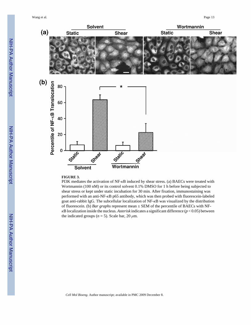
FIGURE 3.PI3K mediates the activation of NF-κB induced by shear stress. (a) BAECs were treated withWortmannin (100 nM) or its control solvent 0.1% DMSO for 1 h before being subjected toshear stress or kept under static incubation for 30 min. After fixation, immunostaining wasperformed with an anti-NF-κB p65 antibody, which was then probed with fluorescein-labeledgoat anti-rabbit IgG. The subcellular localization of NF-κB was visualized by the distributionof fluorescein. (b) Bar graphs represent mean ± SEM of the percentile of BAECs with NF-κB localization inside the nucleus. Asterisk indicates a significant difference (p < 0.05) betweenthe indicated groups (n = 5). Scale bar, 20 μm.
Wang et al. Page 13
Cell Mol Bioeng. Author manuscript; available in PMC 2009 December 8.
NIH
-PA Author Manuscript
NIH
-PA Author Manuscript
NIH
-PA Author Manuscript
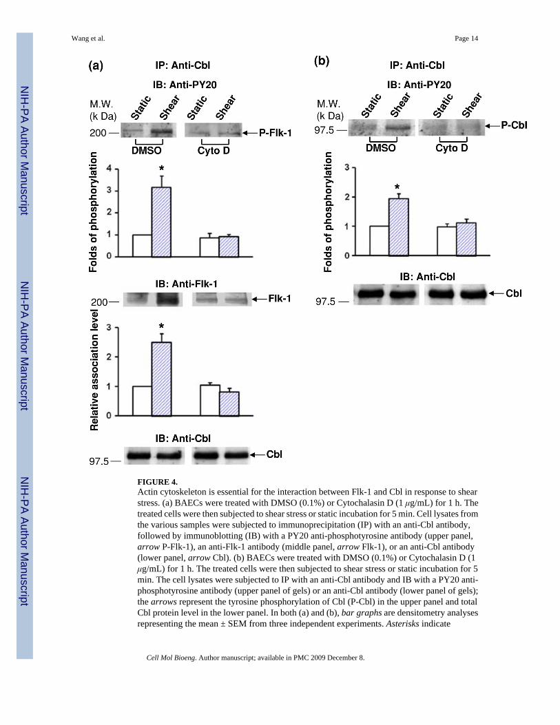
FIGURE 4.Actin cytoskeleton is essential for the interaction between Flk-1 and Cbl in response to shearstress. (a) BAECs were treated with DMSO (0.1%) or Cytochalasin D (1 μg/mL) for 1 h. Thetreated cells were then subjected to shear stress or static incubation for 5 min. Cell lysates fromthe various samples were subjected to immunoprecipitation (IP) with an anti-Cbl antibody,followed by immunoblotting (IB) with a PY20 anti-phosphotyrosine antibody (upper panel,arrow P-Flk-1), an anti-Flk-1 antibody (middle panel, arrow Flk-1), or an anti-Cbl antibody(lower panel, arrow Cbl). (b) BAECs were treated with DMSO (0.1%) or Cytochalasin D (1μg/mL) for 1 h. The treated cells were then subjected to shear stress or static incubation for 5min. The cell lysates were subjected to IP with an anti-Cbl antibody and IB with a PY20 anti-phosphotyrosine antibody (upper panel of gels) or an anti-Cbl antibody (lower panel of gels);the arrows represent the tyrosine phosphorylation of Cbl (P-Cbl) in the upper panel and totalCbl protein level in the lower panel. In both (a) and (b), bar graphs are densitometry analysesrepresenting the mean ± SEM from three independent experiments. Asterisks indicate
Wang et al. Page 14
Cell Mol Bioeng. Author manuscript; available in PMC 2009 December 8.
NIH
-PA Author Manuscript
NIH
-PA Author Manuscript
NIH
-PA Author Manuscript
significant differences (p < 0.05) between the indicated groups and the untreated, staticcontrols.
Wang et al. Page 15
Cell Mol Bioeng. Author manuscript; available in PMC 2009 December 8.
NIH
-PA Author Manuscript
NIH
-PA Author Manuscript
NIH
-PA Author Manuscript
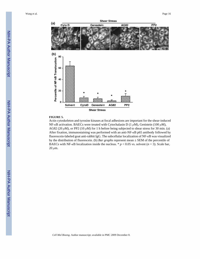
FIGURE 5.Actin cytoskeleton and tyrosine kinases at focal adhesions are important for the shear-inducedNF-κB activation. BAECs were treated with Cytochalasin D (1 μM), Genistein (100 μM),AG82 (20 μM), or PP2 (10 μM) for 1 h before being subjected to shear stress for 30 min. (a)After fixation, immunostaining was performed with an anti-NF-κB p65 antibody followed byfluorescein-labeled goat anti-rabbit IgG. The subcellular localization of NF-κB was visualizedby the distribution of fluorescein. (b) Bar graphs represent mean ± SEM of the percentile ofBAECs with NF-κB localization inside the nucleus. * p < 0.05 vs. solvent (n = 3). Scale bar,20 μm.
Wang et al. Page 16
Cell Mol Bioeng. Author manuscript; available in PMC 2009 December 8.
NIH
-PA Author Manuscript
NIH
-PA Author Manuscript
NIH
-PA Author Manuscript
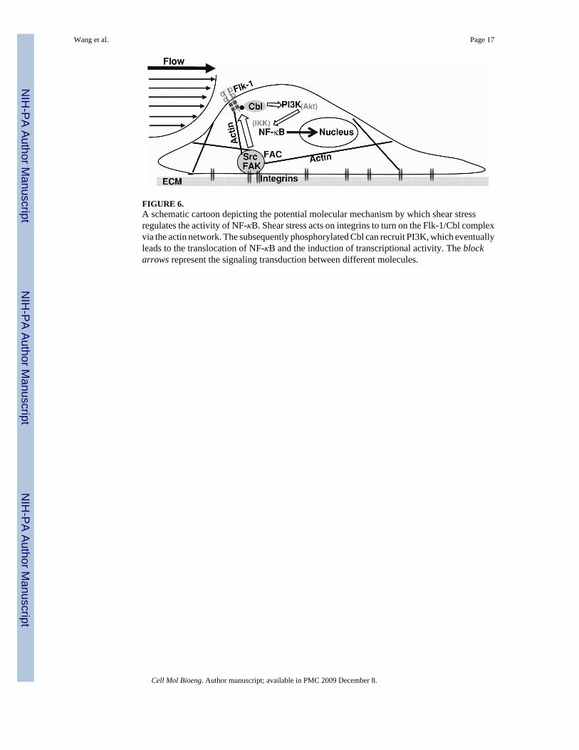
FIGURE 6.A schematic cartoon depicting the potential molecular mechanism by which shear stressregulates the activity of NF-κB. Shear stress acts on integrins to turn on the Flk-1/Cbl complexvia the actin network. The subsequently phosphorylated Cbl can recruit PI3K, which eventuallyleads to the translocation of NF-κB and the induction of transcriptional activity. The blockarrows represent the signaling transduction between different molecules.
Wang et al. Page 17
Cell Mol Bioeng. Author manuscript; available in PMC 2009 December 8.
NIH
-PA Author Manuscript
NIH
-PA Author Manuscript
NIH
-PA Author Manuscript


