Embed Size (px)
Citation preview
Shiga toxin-producing Escherichia coli in humans
and the food chain in Bangladesh
Mohammad Aminul Islam
Promotor:
Prof. Dr. Ir. M. H. Zwietering
Hoogleraar Levensmiddelenmicrobiologie
Wageningen Universiteit
Co-promotor:
Dr. Ir. A. E. Heuvelink
Levensmiddelenmicrobioloog
Voedsel en Waren Autoriteit (VWA)
Zutphen
Promotie commissie:
Prof. Dr. F. van Knapen, Universiteit Utrecht
Prof. Dr. Ir. M. C. M. de Jong, Wageningen Universiteit
Prof. Dr. A. Cravioto, ICDDR,B, Bangladesh
Dr. N. C. A. J. van de Kar, Universitair Medisch Centrum St Radboud, Nijmegen
Dit onderzoek is uitgevoerd binnen de onderzoekschool VLAG
(Food Technology, Agrobiotechnology, Nutrition and Health Sciences)
Shiga toxin-producing Escherichia coli in humans
and the food chain in Bangladesh
Mohammad Aminul Islam
Proefschrift
Ter verkrijging van de graad van doctor op gezag van de rector magnificus
van Wageningen Universiteit Prof. Dr. M. J. Kropff
in het openbaar te verdedigen op maandag 26 januari 2009
des namiddags te 13:30 in de Aula

Mohammad Aminul Islam, 2009 Shiga toxin-producing Escherichia coli in humans and the food chain in Bangladesh. PhD thesis, Wageningen University, the Netherlands With references - with summaries in English and Dutch. Keywords: Shiga toxin-producing Escherichia coli, immunoconcentration, rfbEO157 PCR, prevalence, diarrheal patients, slaughter animals, raw meats, antibody response, healthy population. ISBN: 978-90-8585-291-9
Abstract Shiga toxin (Stx)-producing Escherichia coli (STEC) are one of the most important foodborne pathogens. Infection with STEC in humans can lead to mild diarrhea, bloody diarrhea or even in certain cases to the severe hemolytic-uremic syndrome (HUS). Domestic ruminants appear to be important reservoirs for STEC and play a significant role in the epidemiology of human infections. The main objective of the research described in this thesis was to gain insight in the epidemiology of STEC infection in humans and the ecology of STEC in the human food chain in Bangladesh. We found that the prevalence of STEC among diarrheal patients was relatively low compared with other enteric pathogens. Around 0.5% (n = 2) of patients in the hospital and 1.9% (n = 3) of patients in the community were found positive for STEC with no case (0/570) of STEC O157 infection. To identify an effective technique for isolation of STEC O157 from animal and food sources, we evaluated the efficiency of different methods. Immunomagnetic separation (IMS) using Dynabeads anti-E. coli O157 proved to be more efficient than the Vitek Immunodiagnostic Assay System (VIDAS) Immuno-Concentration E. coli O157 (ICE) kit (VIDAS ICE) in case of animal feces and the use of CHROMagar O157 with cefixime (0.025 mg/l) and tellurite (1.25 mg/l) as plating media resulted in more positive samples than sorbitol–MacConkey agar with cefixime (0.05 mg/l) and tellurite (2.5 mg/l). In addition, IMS was found more sensitive than PCR to detect STEC O157. In order to estimate the prevalence of these organisms in animal feces and foods we used both IMS and PCR techniques. Of the fecal samples collected from buffalo (n = 174), cows (n = 139), and goats (n = 110), 82.2%, 72.7%, and 11.8% tested positive for stx1 and/or stx2, respectively. STEC could be isolated from 37.9%, 20.1%, and 10.0% of the buffalo, cows, and goats, respectively. STEC O157 strains were isolated from 14.4% of the buffalo, 7.2% of the cows, and 9.1% of the goats. In case of foods, more than 71% of the raw meats, 10% of the raw milk and 8% of the fresh juice samples were found positive for the stx genes. STEC O157 strains were isolated from 8% (n = 7) of the meat samples. In order to explain the lack of STEC O157 infection in humans, we investigated the presence of antibodies against these organisms among the healthy population in Bangladesh. We collected serum samples from different groups of people comprising butchers, and people with other occupations living in urban and rural areas. We found that around 50% (n = 116) of the samples were positive for antibodies (IgG, IgA and/or IgM) to E. coli O157 lipopolysaccharide. Using separate analysis, we found that all the 116 samples positive for polyvalent antibodies were positive for the IgG-class and 87 were also positive for IgM-class antibodies. A statistically significantly higher number of nonbutcher participants (63%; n = 57) were positive than the butcher participants (41%; n = 54) (P < 0.05). No statistically significant difference in antibody response was found between the urban and rural populations. Finally, we compared the occurrences of STEC in diarrheal patients, animal reservoirs and foods in Bangladesh with similar data available in other countries from both developed and developing parts of the world in order to get a global perspective. A major difference between our findings and findings from developed countries was observed in the prevalence of STEC O157-associated human infections. Unlike developed countries, no case of STEC O157 infection was found among diarrheal patients in Bangladesh. We concluded that the lack of STEC O157 infection among Bangladeshi population might be attributable to the protective immunity against these pathogens acquired by the frequent exposure to the antigens.

Acknowledgements
Over the years since I enrolled in this PhD research project, I have received support from
many people and many institutions. I highly appreciate every contribution, small or big,
supporting me in achieving this final result. Thank you, all.
My appreciation and deepest esteem go to my promoter, Prof. Dr. Ir. Marcel Zwietering.
Marcel, thank you so much for your unreserved commitment to bring me to the completion of
this thesis. I count myself very fortunate to have been your student and benefited from your
critical guidance invoking critical reflections on my work thereby improving its quality. Your
scientific credence are hallmarks that I will always reckon in my scientific career.
Dr. Ir. Annet Heuvelink, my co-promoter and supervisor. Truly speaking, I never felt like
she was supervising me rather we used to have a lot of friendly discussion on a topic to come
to a decision. She was the most important person whom I used to rely on for any scientific
issues concerned with my study. I learned a lot from her, especially on how to think critically
but clearly. She is a prolific editor. She always checked every single detail of my work.
Whenever I was over enthusiastic or too much optimistic, she informed me where it could go
wrong. She pointed out the checkpoints in my path that immensely helped me to rethink in
advance and take the right path or to be prepared to cross those checkpoints in my research.
Thank you very much Annet for your support to complete my thesis.
I am deeply indebted to my supervisor Ir. Enne de Boer, whom I met at my first step into
the Netherlands. It is his effort that created the opportunity for me to do PhD in Wageningen
University. I learned so many things from him that it is literally impossible to express
everything in words. He was not only my supervisor but also my local guardian in Holland as
well as a very good friend. With his fatherly guidance and teaching, I learned how to survive
in the unpredictable Dutch weather and to enjoy my living in Holland. Although he is a very
busy person, I always found him beside me at any situation when needed. In every difficult
situation, either in my academic or in personal life I still can feel his warmth presence beside
me. Without his support it was not possible for me to complete this study. I also express my
deepest appreciation to Enne’s wife Eveline and their daughters Inge and Sanne for giving me
the feeling’s that I was not alone in Holland and I have a family there.
I like to show my deepest gratitude to my supervisor in Bangladesh, Dr. Kaisar Ali
Talukder. He gave me the opportunity to do PhD research in the Netherlands. It is him who
inspired me to develop a research proposal to be submitted to the NWO-WOTRO for
funding. I have progressed to this level because of his inspiration to build a scientific career
already during my MSc. I am grateful for his relentless support and encouragement.
I deeply acknowledge the guidance and encouragement from my supervisor Dr. Rijkelt
Beumer. This thesis could not have been completed without his interest and support. I
appreciate his cooperation, guidance and friendly atmosphere that existed between us. I
sincerely thank him for the English-Dutch translation of the summary of this thesis. Although
it was not his job, he sacrificed his time to do it as fast as he could and it reads well. Rijkelt, I
appreciate your positive attitude and words of encouragement that kept me going.
This research was financed (partly) by the Netherlands Foundation for the Advancement
of Tropical Research (NWO-WOTRO), for which I would like to express my gratitude. In
addition, I gratefully acknowledge the financial support from the Food and Consumer
Product Safety Authority (VWA), Zutphen, the Netherlands, International Centre for
Diarrhoeal Disease Research, Bangladesh (ICDDR,B), Dhaka, Bangladesh and Laboratory of
Food Microbiology, Wageningen University, The Netherlands.
I wish to mention the names of the people at VWA where I carried out most of my
research work. I want to thank Jeroen for helping me with technical matters in the molecular
biology works. Caroliene and Ans for their patient support in the microbiology laboratory.
Ben Wit kindly took the time for statistical analysis also to fix my old bicycle when it was
broken. I would also like to thank Ingeborg Boxman for having very refreshing discussion on
various topics including career, science, culture, politics and family matters. Thanks to
Nathalie for her extensive help in doing Real-time PCR and sequencing. Harman in the
routine microbiology lab for his patience to collect cow fecal samples from his own cattle
farm early in the morning for my artificial contamination study. Benny, for his extended help
to prepare the culture media whenever I requested him. Geke for her patience and sincere
help for doing western blotting. In addition, I am indebted to Wilma and her family at
Bronsbergen, Zutphen. I stayed with her family for the first eight months of my study. I
enjoyed the living and appreciate their extraordinary hospitality and support.
Throughout the study, sample collection was a significant part. Many people had many
efforts in this part and without their sincere efforts it was impossible to complete the study.
The assistance provided by my young and energetic assistants especially Pavel, Salam,
Yunhee Kang, and Musarrat was immense. I would like to thank the Robin da, who took the
most trouble to collect the rectal excisions of animals from the slaughterhouses very early in
the morning. My special thanks go to Razu, who did his master thesis within my PhD project
and helped me to kick start the research on antibody responses.

I would like to express my gratitude to my colleagues at ICDDR,B. Dr. G. B. Nair
(former director, LSD), his extraordinary personality and dedication to science that inspired
me to work hard. My special thanks to Dr. Hubert Endtz (current director, LSD) for his
understanding on my study and care that he provided me to cope up with the critical situation
at the tailor-end of my study. My sincere appreciations also go to Dilip da, Zhahir, Bijay, Lisa
and Aslam for their commitment to creating a favorable environment for me to do my
research at ICDDR,B and giving me the feelings that they are always beside me at any
critical situation. I like to show my sincere gratitude to Mr. Ansaruzzaman and Dr. Munir for
their inspiration, informal chats and suggestions during my study. I like to thank other
members of Enteric Laboratory, especially Hasan, Shampa, Bhuiya bhai, Shamim, Lina,
Shumon, Mahmuda, Dr. Belal, Khohinur apa, Hossein, Lucky, Atik, Roman, and many more
who made my working environment so pleasant.
It was my pleasure to work together with the Group of Food Microbiology in
Wageningen. Thanks a lot to Professor Tjakko Abee, Dr. Rob Nout and Dr. Martine Reij for
their encouragement. I like to express my gratitude to Gerda for her endurance to wait for me
until the last minute and taking the trouble to arrange all the documents ready in time. I
would also like to thank my colleagues in the group: Mark, Maarten, Zeus, Heidy, Petra,
Lidia, Polycarp, Loveness, Ingrid, Stijn, Armand, Jianfen, Wilma, Marcel and Menno.
Many thanks to Dr. Henrik Chart (Laboratory of Enteric Pathogens, Central Public Health
Laboratory, London, UK) for many good advices and kind help with implementation of LPS
serology in our Laboratory. I am extremely grateful to you Henrik for your extraordinary
support to carry out the antibody testing experiments. My special thanks are extended to
Professor Alejandro Cravioto, Executive Director of ICDDR,B and Dr. Armando Navarro
(Universidad Nacional Autónoma de México) for their enormous support for analysis of the
antibody results presented in Chapter 8 of this thesis.
Life in Wageningen without my Bangladeshi friends would have been a little difficult. I
thank all my Bangladeshi friends Muntaseer Billah, Mukit Billah, Anowar kamal, Mizan,
Chayan, Titu, Qumrul, Atahar, Munni, Alam, Rahman, Hazi, and many more (sorry, if I
missed somebody) for sharing the joy of friendship, very nice Bangladeshi food and music
whenever we met. Special thanks also to Jwel bhai, Reza bhai, and Henrike for their personal
care, love and affection, and suggestions while taking some crucial decision in life.
I like to thank the butchers in the slaughterhouses who allowed us to collect the rectal
sample during slaughtering of the animals early in the morning. I thank them for being
patience and bearing all our disturbances during sample collection. I also like to thank all
participants who donated their blood and allotted their precious time to answer all our
questions.
Without the family support, this dream would have been unattainable. My parents, Md.
Akter Hossain Talukder, who is no more in this world and Mrs. Huria Begum; my brothers,
Mizan and Zahid; and sisters Khusbu and Shilu and all other family members, always wished
me success in all my endeavors. This not only gave me moral support and strength but also
determinations to climb up each step in my life. I thank them all for their wishes and
blessings. Special acknowledgements go to my father-in-law Mr. A. M. Alamgir, who gave
me always his unreserved and invaluable support.
Last but not least, I want to thank my beloved wife Nadia. While doing my PhD I could
not afford to think about marriage, as my top most priority was to first accomplish my PhD
programme. Nonetheless, at the middle of my PhD, Nadia came to my life. She took the most
pain living alone while I stayed in Holland at the last phase of my study. Without her
understanding and support, I could not have persisted to complete the study. Thank you so
much my dear for your attention, care and love.
All of the above, I thank the Almighty Allah for giving me the strength to make my
dreams come true all the time in my life journey.
Mohammad Aminul Islam
Wageningen, January 2009

Dedicated to my late father Md. Akter Hossain Talukder

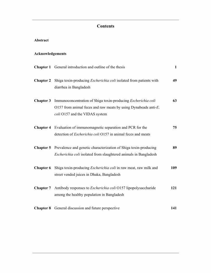
Contents
Abstract
Acknowledgements
Chapter 1 General introduction and outline of the thesis
1
Chapter 2 Shiga toxin-producing Escherichia coli isolated from patients with
diarrhea in Bangladesh
49
Chapter 3 Immunoconcentration of Shiga toxin-producing Escherichia coli
O157 from animal feces and raw meats by using Dynabeads anti-E.
coli O157 and the VIDAS system
63
Chapter 4 Evaluation of immunomagnetic separation and PCR for the
detection of Escherichia coli O157 in animal feces and meats
75
Chapter 5 Prevalence and genetic characterization of Shiga toxin-producing
Escherichia coli isolated from slaughtered animals in Bangladesh
89
Chapter 6 Shiga toxin-producing Escherichia coli in raw meat, raw milk and
street vended juices in Dhaka, Bangladesh
109
Chapter 7 Antibody responses to Escherichia coli O157 lipopolysaccharide
among the healthy population in Bangladesh
121
Chapter 8 General discussion and future perspective
141
Summary
169
Samenvatting
173
Curriculum vitae
177
List of publications
178
Overview of completed training activities
181
General introduction and outline of the thesis
Chapter 1
Chapter 1
2
Escherichia coli is probably the most studied organism in microbiology. Since its first
description (48, 49), the bacterium has become the model organism for much microbiological
research, such that it is often forgotten that its main ecological niche is the alimentary tract of
humans and most warm-blooded animals. The bacterium is shed in the feces of warm-
blooded animals and humans; however, they only comprise a very small percentage of the
total fecal flora. The organisms typically colonize the gastrointestinal tract of human infants
within a few hours after birth (44). Usually E. coli and its human hosts coexist in good health
and with mutual benefits. The niche of commensal E. coli is the mucous layer of the
mammalian colon. The bacterium is a highly successful competitor at this crowded site,
comprising the most abundant facultative anaerobe of the human intestinal microflora. E. coli
usually remains harmlessly confined to the intestinal lumen; however, in the debilitated or
immunosuppressed host, or when gastrointestinal barriers are violated, even normal
"nonpathogenic" strains of E. coli can cause infection.
Over the last half-century it has become increasingly obvious that there are a number of
different enteropathogenic groups of E. coli. At least six known pathotypes associated with
gastrointestinal infections have been recognized, apart from those opportunistic
“nonpathogenic strains” causing urinary tract infections, septicemia, and meningitis in
humans and a number of similar diseases in animals. The pathotypes associated with
gastrointestinal infections currently recognized are:
• Enteropathogenic E. coli (EPEC)
• Enterotoxigenic E. coli (ETEC)
• Enterohemorrhagic E. coli (EHEC), which are a subgroup of Verocytotoxigenic E. coli
(VTEC) or Shiga toxin-producing E. coli (STEC)
• Enteroinvasive E. coli (EIEC)
• Enteroaggregative E. coli (EAggEC)
• Diffuse-adherent E. coli (DAEC)
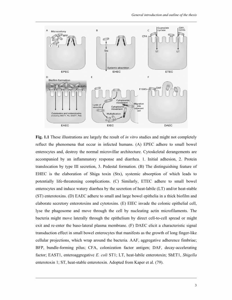
Each of these pathotypes has unique features in their interaction with eukaryotic cells,
which is schematically represented in Fig. 1.1.
General introduction and outline of the thesis
3
Fig. 1.1 These illustrations are largely the result of in vitro studies and might not completely
reflect the phenomena that occur in infected humans. (A) EPEC adhere to small bowel
enterocytes and, destroy the normal microvillar architecture. Cytoskeletal derangements are
accompanied by an inflammatory response and diarrhea. 1. Initial adhesion, 2. Protein
translocation by type III secretion, 3. Pedestal formation. (B) The distinguishing feature of
EHEC is the elaboration of Shiga toxin (Stx), systemic absorption of which leads to
potentially life-threatening complications. (C) Similarly, ETEC adhere to small bowel
enterocytes and induce watery diarrhea by the secretion of heat-labile (LT) and/or heat-stable
(ST) enterotoxins. (D) EAEC adhere to small and large bowel epithelia in a thick biofilm and
elaborate secretory enterotoxins and cytotoxins. (E) EIEC invade the colonic epithelial cell,
lyse the phagosome and move through the cell by nucleating actin microfilaments. The
bacteria might move laterally through the epithelium by direct cell-to-cell spread or might
exit and re-enter the baso-lateral plasma membrane. (F) DAEC elicit a characteristic signal
transduction effect in small bowel enterocytes that manifests as the growth of long finger-like
cellular projections, which wrap around the bacteria. AAF, aggregative adherence fimbriae;
BFP, bundle-forming pilus; CFA, colonization factor antigen; DAF, decay-accelerating
factor; EAST1, enteroaggregative E. coli ST1; LT, heat-labile enterotoxin; ShET1, Shigella
enterotoxin 1; ST, heat-stable enterotoxin. Adopted from Kaper et al. (79).
Chapter 1
4
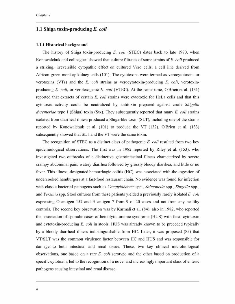
1.1 Shiga toxin-producing E. coli
1.1.1 Historical background The history of Shiga toxin-producing E. coli (STEC) dates back to late 1970, when
Konowalchuk and colleagues showed that culture filtrates of some strains of E. coli produced
a striking, irreversible cytopathic effect on cultured Vero cells, a cell line derived from
African green monkey kidney cells (101). The cytotoxins were termed as verocytotoxins or
verotoxins (VTs) and the E. coli strains as verocytotoxin-producing E. coli, verotoxin-
producing E. coli, or verotoxigenic E. coli (VTEC). At the same time, O'Brien et al. (131)
reported that extracts of certain E. coli strains were cytotoxic for HeLa cells and that this
cytotoxic activity could be neutralized by antitoxin prepared against crude Shigella
dysenteriae type 1 (Shiga) toxin (Stx). They subsequently reported that many E. coli strains
isolated from diarrheal illness produced a Shiga-like toxin (SLT), including one of the strains
reported by Konowalchuk et al. (101) to produce the VT (132). O'Brien et al. (133)
subsequently showed that SLT and the VT were the same toxin.
The recognition of STEC as a distinct class of pathogenic E. coli resulted from two key
epidemiological observations. The first was in 1982 reported by Riley et al. (153), who
investigated two outbreaks of a distinctive gastrointestinal illness characterized by severe
crampy abdominal pain, watery diarrhea followed by grossly bloody diarrhea, and little or no
fever. This illness, designated hemorrhagic colitis (HC), was associated with the ingestion of
undercooked hamburgers at a fast-food restaurant chain. No evidence was found for infection
with classic bacterial pathogens such as Campylobacter spp., Salmonella spp., Shigella spp.,
and Yersinia spp. Stool cultures from these patients yielded a previously rarely isolated E. coli
expressing O antigen 157 and H antigen 7 from 9 of 20 cases and not from any healthy
controls. The second key observation was by Karmali et al. (84), also in 1982, who reported
the association of sporadic cases of hemolytic-uremic syndrome (HUS) with fecal cytotoxin
and cytotoxin-producing E. coli in stools. HUS was already known to be preceded typically
by a bloody diarrheal illness indistinguishable from HC. Later, it was proposed (85) that
VT/SLT was the common virulence factor between HC and HUS and was responsible for
damage to both intestinal and renal tissue. These, two key clinical microbiological
observations, one based on a rare E. coli serotype and the other based on production of a
specific cytotoxin, led to the recognition of a novel and increasingly important class of enteric
pathogens causing intestinal and renal disease.
General introduction and outline of the thesis
5
1.1.2 Nomenclature
In the past decade the understanding of the VT/SLT has rapidly increased and the
identification, purification, cloning and sequencing of several immunologically related toxin
variants led to the insight that these toxins constitute a family with major sequence homology
at both the nucleotide and peptide level. Therefore, Calderwood et al. (30) proposed a new,
more rational nomenclature, in which VT and SLT have been renamed Stx, after the
prototype toxin of the family, and the related VTEC and SLT-producing E. coli (SLTEC)
organisms have been renamed STEC. VTEC, SLTEC, and STEC are equivalent terms, and
all three refer to E. coli strains that produce one or more toxins of the Stx family. These
names are used interchangeably but the term STEC is used throughout this thesis.
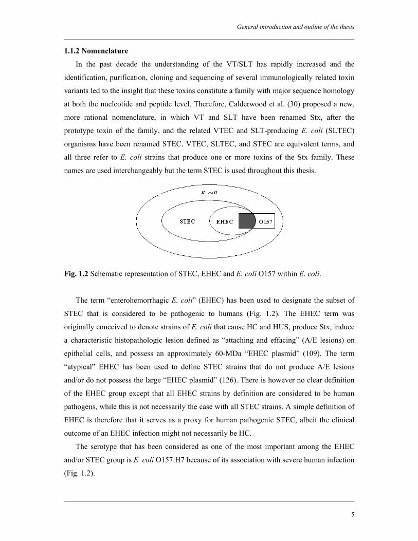
Fig. 1.2 Schematic representation of STEC, EHEC and E. coli O157 within E. coli.
The term “enterohemorrhagic E. coli” (EHEC) has been used to designate the subset of
STEC that is considered to be pathogenic to humans (Fig. 1.2). The EHEC term was
originally conceived to denote strains of E. coli that cause HC and HUS, produce Stx, induce
a characteristic histopathologic lesion defined as “attaching and effacing” (A/E lesions) on
epithelial cells, and possess an approximately 60-MDa “EHEC plasmid” (109). The term
“atypical” EHEC has been used to define STEC strains that do not produce A/E lesions
and/or do not possess the large “EHEC plasmid” (126). There is however no clear definition
of the EHEC group except that all EHEC strains by definition are considered to be human
pathogens, while this is not necessarily the case with all STEC strains. A simple definition of
EHEC is therefore that it serves as a proxy for human pathogenic STEC, albeit the clinical
outcome of an EHEC infection might not necessarily be HC.
The serotype that has been considered as one of the most important among the EHEC
and/or STEC group is E. coli O157:H7 because of its association with severe human infection
(Fig. 1.2).
Chapter 1
6
1.2 Methods for detection of STEC
There are no common biochemical characteristics associated with the great majority of
STEC serotypes. The only way to identify all types of STEC in any kind of test sample is the
detection of Stx produced by the bacteria. STEC can also indirectly be detected by examining
E. coli strains or samples for the genes encoding Stx. The gold standard for the detection of
Stx employs Vero cells, but other cell lines can also be used, e.g., HeLa cells (132). Toxin
production can also be detected by the use of immunological methods. Since a relatively
small number of STEC serotypes are responsible for the majority of human STEC infections,
serotype-specific detection methods have been developed, where strains are isolated on the
basis of their O-antigen and are subsequently analyzed for Stx production or presence of stx
genes. Generally, the diagnosis of STEC is laborious, and currently there are no simple,
inexpensive methods available for routine isolation of all STEC serotypes.
1.2.1 Tissue culture methods
The Vero cell assay has been used widely for the detection of free Stx in fecal specimens
and in enrichment cultures inoculated with foods, animal feces or environmental samples.
However, the most common application of the Vero cell assay is for confirmation of toxin
production by pure cultures. The Vero cell assay is performed by addition of cell-free
supernatants to tissue culture monolayers and preliminary results are obtained after 24 h with
final results after 3-4 days. The specificity of the tissue culture cytotoxicity tests can be
considerably improved by employment of Stx1 and Stx2 specific neutralizing antisera in
order to determine if the cytotoxic effect is caused by Stxs or by another nonneutralizable
toxin present in the sample (88). This assay is sensitive and regarded as the ‘gold standard’ to
which other methods should be validated. However, since maintenance of tissue culture is
costly, specialized in nature and labor-intensive, immunological and DNA-based methods
have been largely supplanted the Vero cell assay for confirmation of Stx production.
1.2.2 Immunological methods
Over the years, a number of immunological methods for the detection of Stxs have been
developed such as enzyme immunoassays, colony blot and passive agglutination assays. The
methods utilize Stx specific poly- or monoclonal antibodies. These assays can be applied to
pure and mixed cultures (enrichment cultures of food or feces, usually incubated overnight).
General introduction and outline of the thesis
7
When Stx is detected, the broth can be subcultured onto isolation media and pure or pooled
colonies can be further examined. Immunoassays are generally reliable and most assays are
easy to implement in laboratories and do not require expensive equipment. Several
immunological assays are today available as commercial ‘ready to use test kits’, which is of
advantage for routine microbiological laboratories. The most commonly used commercially
available test kits are Premier EHEC (Meridian Diagnostics Inc., Cincinnati, Ohio), VTEC-
RPLA toxin detection kit (a reverse passive latex agglutination test for the detection of Stx1
and Stx2) (Denka Seiken Co. Ltd., Japan), the Ridascreen Verotoxin enzyme immunoassay
technique (R-Biopharm, Darmstadt, Germany), the ProSpecT Shiga toxin microplate assay
(Alexon-Trend, Ramsey, Minn.) and the VTEC-Screen ‘SEIKEN’ (Denka Seiken). Most of
these tests are used for nondiscriminative detection of Stx in supernatants from stool, or from
bacterial cultures on microtitre plates coated with Stx1/Stx2-specific (monoclonal)
antibodies.
1.2.3 DNA-based methods
DNA-based methods are considered as a good alternative to the culture or immunological
methods because these methods detect the presence of small amounts of species- or strain-
specific DNA rather than unique aspects of target organisms to be in a specific physiological
state. For STEC, presence of stx-specific gene sequences in the fecal or food samples is an
indication that STEC is present in the sample, which should always be confirmed by
detecting the gene in subsequently isolated organisms. The presence of a particular serotype
of STEC is determined by targeting serotype-specific gene fragments in the organism or in
the sample. Detection of these targets is attained by the use of DNA-DNA hybridization
probes or by amplification of target-specific DNA. Numerous DNA-DNA hybridization
assays, using oligo- or polynucleotide probes have been described and different formats have
been used, including dot-blot and replica assays, liquid-based assays, and more recently
micro-array chips. Amplification of specific DNA is most frequently achieved by polymerase
chain reaction (PCR), but other DNA amplification techniques like Nucleic Acid Sequence-
based Amplification (NASBA) are also applicable for STEC detection. A range of different
PCR formats is being used, and additional techniques to ensure the identity of the amplicon
beyond measurement of the size of the amplified DNA are necessary. These techniques
include the use of internal sequence-based probes (particularly in real-time PCR), full DNA
sequencing, and the use of fragment analysis following restriction endonuclease digestion.
Chapter 1
8
DNA-based detection methods can be applied to nucleic acid from pure or mixed cultures
(enrichment cultures of food or feces), as well as colonies growing on solid isolation media.
DNA probes are used to detect STEC by the use of replica plating techniques, whereas PCR-
based methods can be used to investigate single colonies or pools of colonies. Generally, the
amplification-based techniques are rapid and will give a result within hours (after enrichment
or directly from colonies). However, when testing mixed cultures the detected genes might
not originate from the same STEC strain. DNA-based methods have the disadvantage of
being unable to distinguish between DNA from viable and nonviable cells, although this may
only be important in specific situations.
1.3 Isolation and enrichment of STEC O157
1.3.1 Isolation Usually STEC do not possess phenotypic characteristics that are distinguishable from
those of other E. coli. However, an important exception to this is that STEC O157 are usually
both unable to ferment sorbitol within 24 h of incubation and lack β-D-glucuronidase activity
(120, 151). These characteristics are utilized in the routine selective isolation of STEC O157.
The most widely used solid medium for the isolation of non-sorbitol-fermenting STEC O157
is sorbitol-MacConkey (SMAC) agar. This medium contains 1% sorbitol in place of lactose
in the standard MacConkey agar. Because of the inability to ferment sorbitol, STEC O157
grow after overnight incubation as colorless colonies and can be distinguished from most of
the remaining intestinal E. coli strains (around 75 to 94%) that ferment sorbitol and grow as
pink colonies (120). However, some Enterobacteriaceae present in human stools, such as
Proteus spp., Providencia spp., Hafnia spp., Enterobacter spp., and Escherichia hermanii,
also grow in colorless colonies (120). Moreover, some of these species share common
epitopes with the E. coli O157 antigen (113).
The inability of the majority of STEC O157 strains to produce β-D-glucuronidase is
exploited by supplementation of agar media, e.g., SMAC agar, with the fluorogenic 4-
methylumbelliferyl-β-D-glucuronide (MUG) or with the chromogenic 5-bromo-6-chloro-3-
indolyl-β-D-glucuronide (BCIG). Cleaving of MUG or BCIG by β-D-glucuronidase-positive
strains results in the formation of fluorescent (at 365 nm) and blue-colored colonies,
respectively. No fluorescence or change in color is seen for strains negative for β-D-
glucuronidase. Commercially available agar media based on this property are Rainbow Agar
General introduction and outline of the thesis
9
O157 (Biolog, Inc., Hayward, Calif.), Fluorocult E. coli O157:H7 agar (Merck, Darmstadt,
Germany) and CHROMagar O157. In a recent study, CHROMagar O157 has been shown to
have a higher sensitivity (96.3%) and negative predictive value (100%) and a better
diagnostic efficiency than SMAC agar for the isolation of STEC O157 from human stool
samples (36).
The selectivity of solid media can be improved by the use of selective supplements. The
most frequently used supplements are: cefixime, a third generation cephalosporine; and
potassium tellurite (e.g., in CT-SMAC) (193). Cefixime inhibits Proteus spp. at a
concentration not inhibitory to E. coli and tellurite inhibits many other non-sorbitol-
fermenters such as Aeromonas spp., Pleisomonas spp., Morganella spp., Providencia spp.,
and most other E. coli strains. However, some STEC O157 strains are sensitive to cefixime
and potassium tellurite and therefore may not be detected on CT-SMAC agar (118).
The use of only CT-SMAC for isolation of STEC O157 has become contentious with
recent isolation of sorbitol-fermenting STEC O157 from patients with HUS or diarrhea in
many countries, including Germany, Austria, and the Czech Republic (83). Such strains can
be overlooked by the diagnostic procedures recommended for the isolation of non-sorbitol-
fermenting STEC O157 strains.
Conventional culture methods are both time-consuming and laborious. Their sensitivity
can significantly be increased by the application of an immunoconcentration step. A
commonly used immunoconcentration method, which is also implemented in the
international standard for the detection of E. coli O157 in food and feeding stuffs (6), is
immunomagnetic separation (IMS). The procedure involves mixing of enrichment cultures
with paramagnetic particles coated with anti-O157 antibodies. The target organisms in the
sample bind to the immunomagnetic beads, which are then separated from other sample
material and microorganisms in a magnetic field. Following an extensive washing procedure
to remove nonspecifically bound bacteria and sample particles, the beads are plated onto solid
media. The IMS procedure can be performed both manually and automatically. IMS increases
the sensitivity by relatively concentrating E. coli O157 compared with background
microflora, which may overgrow or mimic STEC O157 cells on selective agars. Example of a
fully automatic immunoconcentration system is the Vitek Immunodiagnostic Assay System
(VIDAS) (bioMérieux, Marcy l'Etoile, France).
Recently, a relatively new microbial capture system called “Recirculating Immuno
Magnetic-capture System (RIMS)” has been developed for the detection of very low numbers
Chapter 1
10
of pathogens, including E. coli O157, in food and environmental samples. Conventional IMS,
which has been employed in the analysis of food and environmental samples, only processes
1 ml of enrichment broth. To maximize the chance of microbial capture, the RIMS procedure
re-circulates the entire volume of enrichment broth past anti-O157-conjugated paramagnetic
beads at 37°C for 30 min. However, there are several protocols available depending on the
amount of sample to be examined and the accepted time to the results. The target organisms
can be detected and isolated from the captured and concentrated sample by direct plating onto
appropriate selective media or using for example the colortrix (a colorimetric assay), fluratrix
(fluorescence microscopy), serology, PCR, ELISA, and/or DNA probes. In previous studies,
it has been shown that RIMS coupled with real-time PCR can result in high recovery rates
with low levels of E. coli O157:H7 in ground beef (7), fresh leafy produce and surface water
after short enrichment periods (66). The method provided detection and isolation of E. coli
O157:H7 at levels as low as 0.07 CFU/g of romaine lettuce and 0.1 CFU/g of spinach and
isolation of E. coli O157:H7 in water samples at levels of 6 CFU/100 ml of surface water
(66).
1.3.2 Enrichment
While human clinical stool specimens are examined mostly by direct plating onto
selective and differential agars, animal feces, food and environmental samples usually
contain low numbers of STEC O157 together with an abundant microbial flora, and therefore
require a selective enrichment step. However, enrichment methods may also be applied to
human feces, which can contain low levels of STEC. The most widely used media for the
enrichment of STEC O157 are tryptone soya broth (TSB) (mainly for food) and buffered
peptone water (for human and animal feces) (181). These broths may be supplemented with
different selective agents such as bile salts, novobiocin, vancomycin, cefsulodin, and
cefixime (33). Bile salts inhibit the growth of non-Enterobacteriaceae strains, which make up
the majority of food background microflora (181). Among the antibiotics, novobiocin has
been used most widely. Novobiocin is mainly active against Gram-positive bacteria
(especially Gram-positive cocci) and against some Gram-negative bacteria frequently present
as background microflora in various samples (181). STEC O157 organisms are generally
resistant to novobiocin, which might also explain its relatively higher use in enrichment
broths compared with other antibiotics. Vancomycin, cefsulodin and cefixime suppress the
growth of Gram-positive bacteria, Aeromonas spp. and Proteus spp., respectively (107).
General introduction and outline of the thesis
11
The occurrence of heat-, freeze-, acid-, or salt-stressed STEC O157 in food makes it
important to be able to detect cells that are in a stressed state, since injured cells mostly retain
their pathogenic properties. The detection and isolation of stressed STEC O157 by direct
selective enrichment or direct plating onto selective agar may not allow the recovery of these
strains (3, 169). Probably the best approach for the recovery of stressed STEC O157 cells is a
nonselective pre-enrichment for at least 18 to 24 h (169).
There is currently no consensus on optimal incubation temperature (37°C versus 41.5°C)
and time (6-8 h incubation versus overnight incubation) for all types of samples. The
incubation period required will depend on the competing microflora. Standard methods for
food include the analysis of both the 6- and 18-h incubating enrichment cultures (6, 181). A
6-8 h incubation of the enrichment broth increases the sensitivity when analyzing matrices
with a high number of background flora. However, when stressed or sublethally injured
STEC O157 are present there are difficulties in reaching a detectable level after 6-8 h of
enrichment. Therefore, this short period of incubation can only be recommended when testing
matrices where E. coli has a short-lag time before onset of growth, as for example with
minced meat products.
1.4 Isolation and enrichment of STEC non-O157
There is no internationally accepted standard method for the isolation of STEC non-
O157. Over 200 O:H serotypes have been recognized as STEC from different sources (45).
Unlike STEC O157 most of the other serotypes of STEC show similar biochemical
characteristics as commensal E. coli. This limits the development of selective culture media
applicable for the isolation of all serotypes of STEC.
Following successful application in STEC O157 isolation, methods based on IMS have
been developed for a few predominant STEC non-O157 serogroups. Magnetic beads coated
with antibodies for STEC non-O157 have been developed for serogroups O26, O103, O111,
and O145. IMS-based detection of serogroups other than O157 is similar to that for the
detection of E. coli O157; enrichment, and IMS followed by plating onto selective indicative
agars. However, these methods have not yet been sufficiently validated.
There is no recommended selective enrichment or plating medium for STEC non-O157.
Selective agents to improve the isolation of STEC O157 (e.g., novobiocin) may inhibit the
growth of some STEC non-O157 (182). Several studies showed that some STEC non-O157
Chapter 1
12
serotypes (O5:H-, O26:H-, O26:H11, O91:H21, O111:H-, O111:H8, O104:H11, O113:H21
and O157:H8) are capable of growing on media supplemented with vancomycin, cefixime,
and cefsulodin (68, 107).
A nonselective, but differential plating medium is enterohemolysin agar (Oxoid Ltd.,
Basingstoke, United Kingdom) (washed sheep blood agar supplemented with calcium), which
may be suitable for isolation of all human pathogenic STEC strains, including STEC O157
(14). Nearly all (ca. 90%) STEC O157 strains and proportion of human pathogenic STEC
non-O157 strains (ca. 70%) produce enterohemolysin. Enterohemolytic E. coli are
characterized on this medium by small turbid zones of hemolysis around the colonies
occurring after 18 to 24 h incubation at 37°C. α-hemolytic E. coli form large, clear zones of
hemolysis after only 3 to 6 h of incubation. To improve the selectivity of the medium,
antibiotics such as novobiocin and cefsulodin may be used. By combining culture on
enterohemolysin agar and Stx detection using the VTEC-RPLA kit, Beutin et al. (15) were
able to isolate STEC O157:H7 and non-O157 strains from stools of HUS patients that
constituted as little as 0.03% of the total coliform flora. Enterohemolysin agar is easy to use
and commercially available (Oxoid), and therefore suitable for routine application. However,
it has some limitations: 1) enterohemolysin-positive colonies must be tested for Stx
production (15); 2) a proportion of STEC non-O157 (14) and sorbitol-fermenting STEC
O157 (16) fail to produce the enterohemolytic phenotype and can be missed; and 3) the
presence of a large number of non-STEC α-hemolytic colonies or overgrowth with other
enteric bacteria (Proteus spp., Pseudomonas spp., and Gram-positive cocci) can interfere
with the detection of fewer enterohemolytic colonies (15).
1.5 Characterization and typing of STEC
Characterization of STEC isolates is extremely valuable since this allows comparisons
between isolates from human, animal and food origin and also provides information on
changes in their prevalence over time and in different geographical locations.
Characterization of STEC with respect to the presence of a range of virulence properties may
further identify markers that confer the capacity to cause serious infections and so identify
strains with increased risk of causing disease. These data are essential for an evolving
definition of human pathogenic STEC. Some typing and fingerprinting methods inform
General introduction and outline of the thesis
13
epidemiological investigations that link human cases to each other and to specific sources of
infection.
1.5.1 Serotyping
The serotype of an E. coli isolate is based on the O-(Ohne) antigen determined by the
polysaccharide portion of cell wall lipopolysaccharide (LPS) and the H-(Hauch) antigen due
to flagella protein. It is an important basis for differentiating STEC and is often the starting
point in characterization. Strains of E. coli, including STEC are serotyped by an
internationally recognized and evolving scheme comprising over 180 O-types and 56 H-
types. Full serotyping is generally performed in national reference laboratories although
antisera for some common STEC O-groups are available commercially. Agglutination kits,
generally based on antibody-coated latex particles, are used widely in the identification of
presumptive STEC, particularly O157, isolated from human and non-human samples.
Due to restricted use of antisera-based serotyping of E. coli in some reference
laboratories, DNA-based serotyping has been evolved, which attempts to avoid the
dependency on antisera and make serotype characterization more widely available. This
method targets unique sequences involved in the biosynthesis of O-antigens specific for O-
groups such as O157, O26, O111, O113 and O145 by PCR. PCR-RFLP (PCR-restriction
fragment length polymorphism) and PCR combined with sequencing have also been used.
Determination of the H-type has been directed mainly at the fliC gene that is present even if
the isolate is nonmotile. The large number of O-types of E. coli means that in the short to
medium term, DNA-based tests are unlikely to replace conventional serotyping in the
reference laboratory setting for comprehensive characterization of isolates. Such
developments require sequence data to become available on a more extensive range of O-
groups than the present.
1.5.2 Stx production
STEC are defined by their ability to produce either one or both antigenically-distinct
Shiga toxins termed Stx1 and Stx2 that were first recognized by their ability to cause an
irreversible cytopathic effect on Vero cells and other cell lines in culture (101). Although
very good tests in general, cell line assays are labour-intensive and time consuming and not
really appropriate for many routine diagnostic laboratories, which may lack tissue culture
facilities. Moreover, neutralizing antisera against Stxs are not commercially available. It is
however essential for reference laboratories to continue to use this test, because it will reveal
Chapter 1
14
the presence of unknown variants of Stxs, which the exclusive use of specific immunological
or DNA-based methods would miss.
1.5.3 Stx typing and subtyping
Typing and subtyping of Stxs has a great importance in the epidemiology and ecology of
these organisms. Two major types of Stxs, called Stx1 and Stx2, which share 56% homology
to each other, have been described (141). The genetic analysis of the stx genes found in
different STEC isolates resulted in the detection of an increasing number of genetic variants
of both stx1 and stx2. A large number of PCR assays have been evolved for the detection of
stx1 and stx2 genes and their variants. Until now, five genetic variants of stx1 and 12 variants
of stx2 have been described and were summarized in a new proposal for an adapted
nomenclature for the Stx family by Scheutz et al. (158). This nomenclature organizes 6
groups of toxin types (1, 2, 2c, 2d, 2e and 2f) according to antigenic variability, differences in
toxicity for cells or animals, capacity to be activated by mouse elastase (mucus) and by
differences in DNA or amino acid sequences.
1.5.4 Presence of other virulence genes
Most STEC included in the EHEC group colonize the intestinal mucosa with a
mechanism that subverts the epithelial cell function (52) and induces A/E lesions. The
complex mechanism of A/E lesions is genetically governed by a large pathogenicity island
defined as the Locus of Enterocyte Effacement (LEE) (126). A/E lesions have been
characterized routinely by the presence of the eae gene, which encodes for the adherence
factor intimin. eae exists in a large number of sequence subtypes due to variation at the C-
terminal end. PCR assays are usually directed at the conserved region of the sequence. The
presence of the eae gene has been shown to be strongly associated with some STEC
serotypes that can cause serious complications, including HC and HUS (19).
LEE-negative STEC strains have also been associated with serious illness indicating that
other factors enhance the virulence potential of these strains. Several studies have
investigated the role of other genomic islands identified in STEC O157 for their potential
contribution to virulence of other STEC. One island, termed O122, is contiguous to LEE in
many strains and is strongly associated with E. coli that can cause A/E lesions. In STEC
O157, it contains the 5′ end of the efa1 (EHEC factor for adherence) gene but this region is
complete in many STEC non-O157 and in sorbitol-fermenting STEC O157 and may be
linked to colonization of the bovine intestine (45, 89).
General introduction and outline of the thesis
15
Sorbitol-fermenting STEC O157 strains that have caused outbreaks of HUS are
characterized by the presence of plasmid-coded fimbriae and the plasmid-borne pilin subunit
gene (sfpA) has been used as a potential diagnostic marker for these strains by PCR.
Most of the STEC O157 and some STEC non-O157 strains contain a virulence plasmid
called “pO157”. This plasmid contains several genes encoding for proteins implicated in
STEC pathogenesis. These include: hlyEHEC encoding for EHEC hemolysin; espP encoding
for an autotransported serine protease involved in the cleavage of human coagulation factor V
(27); toxB encoding for a 362-kDa protein sharing amino acid sequence similarity with the
large Clostridium toxin family, which is involved in adherence to epithelial cells in culture
(173); saa encoding for STEC autoagglutinating adhesin; and stcE encoding for a zinc
metalloprotease. StcE is secreted by the etp type II secretion system, cleaves the C1 esterase
inhibitor (C1-INH) of the complement pathway, has mucinase activity, and is thought to be
involved in colonization and tissue damage (60, 103).
1.5.5 Phage typing
Phage typing is an important internationally standardized subtyping method, which is
used only for STEC O157. Phage typing schemes are not available for STEC non-O157.
Phage types are determined by the lysis pattern obtained when a test isolate is subjected to a
panel of established standard lytic phages. STEC O157 strains are differentiated into about 90
types by a scheme of 16 bacteriophages (96). The method is rapid, epidemiologically
valuable in real time and gives information about the emergence and distribution of new
strains. It is performed routinely in a limited number of laboratories worldwide. Although
phage typing is relatively easy to perform, the biggest drawback of using phage typing as the
sole typing method is the occurrence of common phage types. There is a close association
between phage type and the presence of stx1 and stx2 gene subtypes. Some phage types (e.g.,
PT2, PT21/28) appeared to be more strongly associated with HUS than others. Generally, the
same phage types that cause human illness are recovered from animals, although the
proportions may differ. To obtain maximum strain discrimination, it has been recommended
to use phage typing in conjunction with stx subtyping and pulsed-field gel electrophoresis
(PFGE) analysis (154).
1.5.6 Subtyping and fingerprinting for epidemiology and population studies
One of the most widely applied methods of subtyping is PFGE, a technique in which
fragments of the bacterial chromosome generated by digestion with a restriction enzyme
Chapter 1
16
selected to cut the DNA into 20 to 25 pieces are separated by agarose gel electrophoresis (58,
176). The procedure for this technique is relatively similar to performing a standard gel
electrophoresis except that instead of constantly running the voltage in one direction, the
voltage is periodically switched among three directions. One that runs through the central
axis of the gel and two that run at an angle of 120° either side. The pulse times are equal for
each direction resulting in a net forward migration of the DNA. Numerical analysis of
digitally captured banding patterns can be carried out to build dendrograms, with the ultimate
potential of using the technique comparatively to trace and identify outbreaks through a
central database (172). PFGE has been applied to both STEC O157 and non-O157. However,
there is considerably more information on the application of PFGE to STEC O157.
In 1995, the Centers for Disease Control and Prevention (CDC) set up "PulseNet"
(www.cdc.gov/ncidod/dbmd/pulsenet/pulsenet.html), a national network of public health
laboratories in the United States for rapid comparison of fingerprints generated by PFGE
subtyping of E. coli O157 isolates with an electronic database at the CDC. Rigorous
standardization of a protocol involving digestion of genomic DNA with the enzyme XbaI has
resulted in patterns that may be compared across the PulseNet system in the United States
(58) and in other countries (176). Strong epidemiological data are essential in the application
of PFGE as strains that share profiles may be obtained from unlinked cases separated
geographically and over long periods of time.
Sequence-based typing methods include multi-locus variable number of tandem repeat
analysis (MLVA) has been evaluated as an alternative to PFGE for STEC O157 (72). It has
the advantage that the output appears as a numeric sequence that can be generated
automatically. MLVA methods have not yet been applied to STEC non-O157 strains because
of lack of sequence data. Multi-locus sequence typing (MLST), another sequence-based
typing method is more appropriate for studying the relationships between E. coli populations
rather than for typing and epidemiological analysis (130).
1.5.7 Seropathotype classification of STEC
The concept of seropathotype that has recently been proposed classifies STEC into five
groups based on the incidence of serotypes in human disease, association with outbreaks
versus sporadic infection, their capacity to cause HUS or HC, and the presence of virulence
markers (90). This approach attempts to combine these inputs to understand better the
apparent differences in virulence of different STEC. Seropathotype A strains (STEC O157)
have a high relative incidence, commonly cause outbreaks and are associated with HUS.
General introduction and outline of the thesis
17
STEC O26:H11, O103:H2/NM, O111:NM and O145:NM together with O121:H19 fall into
seropathotype B, as they have a moderate incidence and are uncommon in outbreaks but are
associated with HUS. Seropathotype C includes STEC O91, O104 and O113 strains, all of H-
type 21, are associated with HUS, but these strains are of low incidence and rarely cause
outbreaks. Seropathotypes A and B possess LEE and genes of O-island 122 but strains
belonging to seropathotype C may be LEE-negative and only some of the strains possess
O122 virulence genes. Seropathotypes D and E are not HUS-associated and are uncommon in
human or found only in nonhuman sources. Surveys targeting isolation of STEC (not
specifically O157) from nonhuman sources generally result in isolates from groups C and D.
Substantial differences in virulence potential have been observed among isolates
belonging to the same STEC serotypes. For example, studies based on subtyping of stx from
STEC O157 isolated from human patients and healthy cattle have indicated that there are
differences among the relative frequency of the seropathotypes that are predominating among
patients with severe disease (HC and HUS) and seropathotypes that are predominating in the
bovine reservoir (155). The same observation is made for STEC O26. Strains of STEC
O26:H11 that cause HUS are usually identified as Stx2- and eae-positive whereas infections
caused by Stx1- and eae-positive STEC O26:H11 usually are characterized by causing
relatively mild diarrheal symptoms in most patients (50). The concept of seropathotype is
likely to be further refined and will provide a valuable tool in the future for the assessment of
the human pathogenic potential of different STEC serotypes.
1.6 Epidemiology of STEC infection
The epidemiology of STEC infections has remarkably changed during the past ten years.
The organisms have been reported in a large variety of domestic and wild animal species, and
an increasing number of unusual food vehicles have been associated with human infections.
New routes of transmission have emerged, like contact with animals during farm visits and a
wide variety of environment-related exposures.
STEC infections are still a much greater problem in developed countries than in
developing countries. The true incidence of STEC-associated illness is difficult to estimate
since many persons presenting only mild symptoms may not seek medical attention. The vast
majority of reported outbreaks and sporadic cases of HUS in humans have been caused by
serotype O157:H7. However, more than 100 different STEC serotypes have been associated
Chapter 1
18
with disease in humans (159, 190) and STEC non-O157 serotypes (especially O26:H11,
O103:H2, O111:H-, and O145:H-) are increasingly being associated with outbreaks of
foodborne illness and HUS (126).
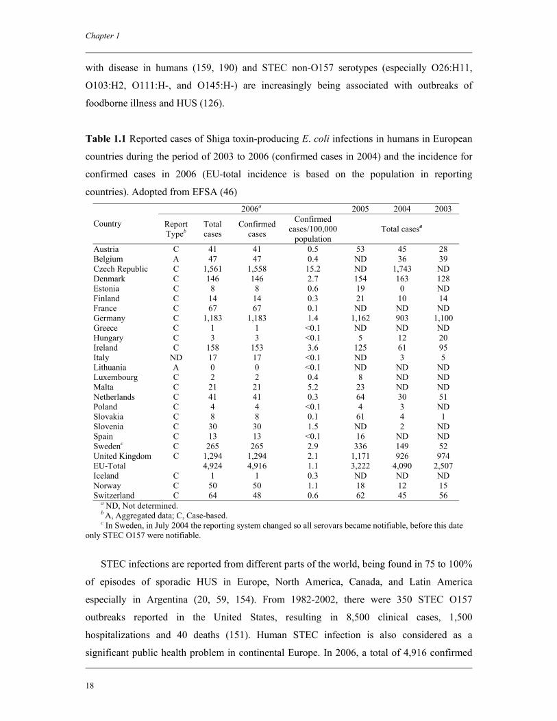
Table 1.1 Reported cases of Shiga toxin-producing E. coli infections in humans in European
countries during the period of 2003 to 2006 (confirmed cases in 2004) and the incidence for
confirmed cases in 2006 (EU-total incidence is based on the population in reporting
countries). Adopted from EFSA (46) 2006a 2005 2004 2003
Country Report Typeb
Total cases
Confirmed cases
Confirmed cases/100,000
population Total casesa
Austria C 41 41 0.5 53 45 28 Belgium A 47 47 0.4 ND 36 39 Czech Republic C 1,561 1,558 15.2 ND 1,743 ND Denmark C 146 146 2.7 154 163 128 Estonia C 8 8 0.6 19 0 ND Finland C 14 14 0.3 21 10 14 France C 67 67 0.1 ND ND ND Germany C 1,183 1,183 1.4 1,162 903 1,100 Greece C 1 1 <0.1 ND ND ND Hungary C 3 3 <0.1 5 12 20 Ireland C 158 153 3.6 125 61 95 Italy ND 17 17 <0.1 ND 3 5 Lithuania A 0 0 <0.1 ND ND ND Luxembourg C 2 2 0.4 8 ND ND Malta C 21 21 5.2 23 ND ND Netherlands C 41 41 0.3 64 30 51 Poland C 4 4 <0.1 4 3 ND Slovakia C 8 8 0.1 61 4 1 Slovenia C 30 30 1.5 ND 2 ND Spain C 13 13 <0.1 16 ND ND Swedenc C 265 265 2.9 336 149 52 United Kingdom C 1,294 1,294 2.1 1,171 926 974 EU-Total 4,924 4,916 1.1 3,222 4,090 2,507 Iceland C 1 1 0.3 ND ND ND Norway C 50 50 1.1 18 12 15 Switzerland C 64 48 0.6 62 45 56
a ND, Not determined. b A, Aggregated data; C, Case-based. c In Sweden, in July 2004 the reporting system changed so all serovars became notifiable, before this date
only STEC O157 were notifiable.
STEC infections are reported from different parts of the world, being found in 75 to 100%
of episodes of sporadic HUS in Europe, North America, Canada, and Latin America
especially in Argentina (20, 59, 154). From 1982-2002, there were 350 STEC O157
outbreaks reported in the United States, resulting in 8,500 clinical cases, 1,500
hospitalizations and 40 deaths (151). Human STEC infection is also considered as a
significant public health problem in continental Europe. In 2006, a total of 4,916 confirmed
General introduction and outline of the thesis
19
human STEC cases were reported from 22 member states of the EU (46), giving an incidence
of 1.1 per 100,000 populations. There has been a statistically significant decreasing trend in
the EU incidence since 2004 (Table 1.1). The most commonly identified STEC serogroup
was O157. Overall, more than one half of the reported STEC cases occurred in 0-4 year old
children. There was, however, increased reporting of human cases caused by non-O157
serogroups.
In Australia and New Zealand the annual incidence of HUS is approximately 1.0 to 1.3
per 100,000 children less than five years old. Interestingly, the predominant STEC serotypes
associated with HUS in these two countries differ. In New Zealand, STEC O157 strains make
up around half of the isolates, and in Australia STEC O111 strains account for most HUS
cases, with STEC O157 being associated with fewer than 20% (108).
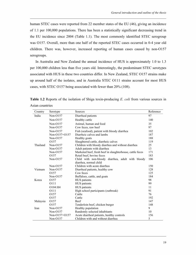
Table 1.2 Reports of the isolation of Shiga toxin-producing E. coli from various sources in
Asian countries Country Serotype Sources Reference
Non-O157 Diarrheal patients 97 Non-O157 Healthy cattle 140 Non-O157 Animal, human and food 35 Non-O157 Cow feces, raw beef 97 Non-O157 Fish (seafood), patient with bloody diarrhea 102 Non-O157+O157 Diarrheic calves and lambs 187 Non-O157 Healthy goats 188
India
O157 Slaughtered cattle, diarrheic calves 119 Non-O157 Children with bloody diarrhea and without diarrhea 25 Non-O157 Adult patients with diarrhea 13 Non-O157 Marketed beef, fresh beef in slaughterhouse, cattle feces 171 O157 Retail beef, bovine feces 183 Non-O157 Child with non-bloody diarrhea, adult with bloody
diarrhea, normal child 106
Thailand
Non-O157 Children with acute diarrhea 150 Non-O157 Diarrheal patients, healthy cow 128 O157 Cow feces 125
Vietnam
Non-O157 Buffaloes, cattle, and goats 184 O157 HUS patients 98 O111 HUS patients 99 O104:H4 HUS patients 11 O111 High school participants (outbreak) 91 O157 Cattle 76
Korea
O157 Cattle 192 O157 Beef 147 Malaysia O157 Tenderloin beef, chicken burger 148 Non-O157 Healthy population 9 Non-O157 Randomly selected inhabitants 10 Non-O157+O157 Acute diarrheal patients, healthy controls 156
Iran
Non-O157 Children with and without diarrhea 5
Chapter 1
20
In most of the Asian countries, STEC are not yet considered as a major health problem,
except in Japan, where 29 outbreaks were reported during the period 1991 to 1995 (123).
Several STEC outbreaks were also reported from China and Korea, where cases of HUS
patients infected with STEC O157 have been reported. Xu et al. reviewed the isolation of
STEC from different places in China (191). A review of literature on STEC isolation from
Asian countries is given in Table 1.2.
1.7 Reservoirs of STEC
STEC represent the only pathogenic group of E. coli that has a definite zoonotic origin,
although not all STEC strains have been demonstrated to cause disease in humans. STEC
rarely cause disease in animals, and ruminants are recognized as their main natural reservoir.
Cattle are considered to be the major animal source of STEC that are virulent to humans, in
particular STEC O157, and the ecology of these microorganisms in cattle farming have been
extensively studied (32). Cattle are asymptomatic excretors of STEC O157, which are
transient members of their gut microflora. The presence of STEC O157 appears to be
influenced by the age of the animals and by the season. Shedding is usually longer and more
intense in calves than in adult cattle, and increases after weaning. It is also much higher
during the summer period (32). The reported prevalence of STEC and/or STEC O157 in
cattle is also clearly influenced by the sampling and detection methods adopted in the
investigations. STEC O157 and other serotypes associated with human infection have also
been isolated frequently from the intestinal content of other ruminant species, including
sheep, goat, water buffalo, and wild ruminants, while pigs and poultry have not been
identified to be major sources of STEC.
Fecal testing of dairy cattle worldwide showed prevalence rates for STEC O157 and
STEC non-O157 ranging from 0.2 to 48.8% and 0.4 to 74.0%, respectively (70). Global
testing of beef cattle feces revealed prevalence rates for STEC O157 and STEC non-O157 of
0.2 to 27.8% and 2.1 to 70.1%, respectively (69).
1.8 Transmission of STEC
Although the ultimate source of STEC is the feces of ruminants, there are four main
transmission routes whereby these organisms may be transmitted to humans: 1) foodborne
General introduction and outline of the thesis
21
transmission; 2) waterborne transmission; 3) person-to-person transmission; and 4) direct
contact with animals.
During the 1980s, most of the outbreaks of STEC O157 infection were foodborne and
food vehicles implicated were mostly inadequately cooked hamburgers or other beef
products, and unpasteurized milk (32). Over the years numerous studies on transmission
routes of human pathogenic STEC have identified many other types of food vehicles for these
organisms. In addition to foods of bovine origin, several outbreaks have been associated with
low pH products like fermented salami, apple juice/cider, mayonnaise and yogurt (122). This
has highlighted the tolerance of STEC O157 to acidic pH and its ability to survive the
processes of fermentation and drying. In addition, waterborne outbreaks and outbreaks
associated with other types of environment related exposures have been increasingly reported
(124). The dispersion of untreated manure in the environment can cause the contamination of
different items, which can then act as secondary vehicles for human infections (32). A
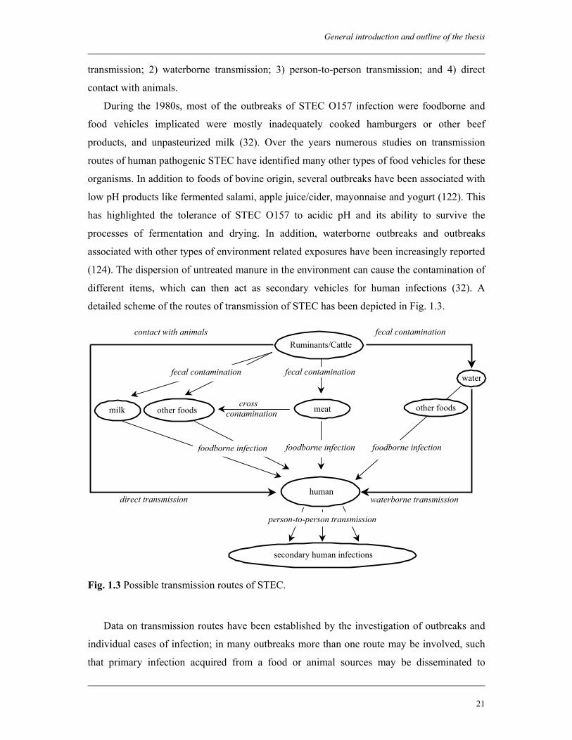
detailed scheme of the routes of transmission of STEC has been depicted in Fig. 1.3.
Fig. 1.3 Possible transmission routes of STEC.
Data on transmission routes have been established by the investigation of outbreaks and
individual cases of infection; in many outbreaks more than one route may be involved, such
that primary infection acquired from a food or animal sources may be disseminated to
secondary human infections
Ruminants/Cattle fecal contamination contact with animals
milk meat
human direct transmission
other foods
foodborne infection
cross contamination
fecal contamination
waterborne transmission
waterfecal contamination
foodborne infection
person-to-person transmission
foodborne infection
other foods
Chapter 1
22
secondary cases in families or the wider community. The high infectivity and intrinsic
properties of STEC (e.g., acid tolerance and the ability to survive well in the environment)
have made investigation of infection increasingly complex. Case-control studies of varying
design have been performed to assess risk factors for sporadic infection with STEC (mainly
O157) in several European and other countries. Results show differences between countries
and the risk factors may be age-related. In several studies specific food vehicles were not
identified but contact with animals and/or the rural environment were identified as the major
risk factor for STEC O157 infection (115, 136). A risk analysis study conducted on STEC
O157 outbreaks in Scotland from 1994 to 2003 showed that approximately 40% of the
outbreaks were foodborne, 54% were environmental, and 6% involved both transmission
routes (170).
1.8.1 Foodborne transmission
Descriptive epidemiology of STEC infection in Ontario, Canada during the period of
1996-2005 identified that food (35%) was the most frequent mode of transmission of STEC,
followed by person-to-person (5%) and waterborne (3%) transmission (114). In the United
States, 52% of the outbreaks of STEC O157 infection between 1982 and 2002 were
associated with consumption of contaminated food (149). The food vehicle for 75 (41%)
outbreaks was ground beef, and for 38 (21%) outbreaks, produce (149).
Contamination of carcasses with STEC usually occurs during slaughter and subsequent
processing through fecal material originating directly or indirectly from the rectal-caecal area
(47). Dairy products (e.g., milk, cheese, cream) associated with infection have included those
that are unpasteurized, have had a pasteurization failure or have been contaminated post-
pasteurization. Ready-to-eat foods have also been associated with infection, particularly
cooked meats contaminated by raw materials during processing, in catering establishments, at
retail sale and in the home.
Vegetables and fruits, fertilized with ruminants’ manure or contaminated during
irrigation, harvesting or processing have also been implicated in transmitting STEC
especially STEC O157 (32). Examples are lettuce, potatoes, radish sprouts, alfalfa sprouts,
cantaloupe, and unpasteurized fruit juice. Such ‘produce’ items are now recognized in the
United States as a major cause of outbreaks requiring increased biosecurity and changes in
processing practices.
STEC O157 can survive for substantial periods of time on stainless steel (51) and plastic
(4). Hence, these surfaces can serve as intermediate sources of contamination during food
General introduction and outline of the thesis
23
processing operations. Processing equipment and utensils used in the preparation of fruit
juices have also been linked to cross-contamination events. A meat grinding unit can also be
a significant contributor to cross contamination resulting from multiple contacts with surfaces
in the mixing, blending, cutting and forming actions (47).
In 2006, a multistate outbreak of STEC O157:H7 infection occurred in the United States
(39). A total of 183 persons infected with the outbreak strain of E. coli O157:H7 had been
reported to the CDC, from 26 states. Among the ill persons, 95 (52%) were hospitalized, 29
(16%) had HUS, and one person died. Fresh spinach was identified as the source of the
outbreak. One hundred and twenty-three of the 130 patients (95%) reported consuming
uncooked fresh spinach within the 10 days before illness onset. In addition, STEC O157:H7
with a PFGE pattern matching the outbreak strain had been isolated from three open packages
of fresh spinach consumed by patients.
1.8.2 Waterborne transmission
Water is a very efficient vehicle for the dissemination of STEC. Surface waters may be
subjected to STEC contamination through run-off from organic wastes applied to agricultural
land and from direct fecal deposition. Fresh water close to livestock farming systems may
therefore represent a potential reservoir for enteric pathogens, allowing cycles of livestock re-
infection and increasing the potential for the organism to spread (121).
STEC may be present in manure heaps and surrounding run-off puddles, which provide
another opportunity for the pathogen to be washed into surface waters. Besides in many
countries, river water is readily contaminated with huge load of treated and untreated sewage
(178). Recently, E. coli O157:H7 has been detected in the Ganges River, which is an
extensively used water source in India (63).
There have been a number of water-associated outbreaks following contamination of
water by STEC O157 (34). Swimming-associated outbreaks (1, 144) and outbreaks linked
with contamination of private water supplies have been described (8). The largest STEC
O157 outbreak in the United States occurred in 1999 at a county fair due to contaminated
drinking water (37). A total of 921 persons reported diarrhea after attending the fair. Stool
cultures yielded E. coli O157:H7 from 116 persons; 13 of these persons were co-infected
with Campylobacter jejuni. Sixty-five persons have been hospitalized; 11 children have
developed HUS; and two persons died: a 3-year-old girl from HUS and, a 79-year-old man
from HUS/thrombotic thrombocytopenic purpura. The implicated water was from a
temporary unregulated well at the fairground. Recently, one small-scale outbreak of STEC
Chapter 1
24
O157 infection has been reported in England, which was associated with unchlorinated water
in a swimming pool (180).
1.8.3 Person-to-person transmission
Because of the low infectious dose (1 to 100 CFU) of STEC O157 (141), person-to-
person fecal-oral transmission can easily occur in settings of poor hygiene and close contacts.
Accordingly, person-to-person transmission has emerged as the predominant route of
infection in outbreaks of STEC O157 infection in daycare facilities (137), and in institutional
settings (2, 86). During outbreaks, transmission from asymptomatically infected individuals
may also be a source of secondary infections and can further amplify the outbreak (2). This
mode of transmission is also considered responsible for the spreading of STEC infection
within families (165). However, rates of STEC transmission by person-to-person and
resultant illness are largely unknown (117).
1.8.4 Contact with animals
During the past decennia, the role of animals in health and social care has significantly
increased. Animals are brought in nursing homes and hospitals and increasing number of
farms combine agriculture and care (65). Certain venues encourage or permit the public to
contact animals, resulting in millions of human-animal interactions each year. Contact with
animals in public settings provides opportunities for entertainment and education. These
settings include county or state fairs, petting zoos, animal swap meets, pet stores, zoological
institutions, circuses, carnivals, farm tours, livestock-birthing exhibits, educational exhibits at
schools and wildlife photo opportunities. Although multiple benefits of human-animal
contact exist, inadequate understanding of disease transmission and animal behavior can
increase the likelihood of zoonotic infectious diseases in these settings (127).
Transmission through direct animal contact has been documented in outbreaks and
sporadic infections by E. coli O157:H7 (38, 64). Although reports often document cattle,
sheep, or goats as sources for infection, poultry, rodents (93), and other domestic and wild
animals also are potential sources. The primary mode of transmission for enteric pathogens is
fecal-oral. Because animal fur, hair, skin, and saliva (92) can become contaminated with fecal
organisms, transmission can occur when people touch, feed, or are licked by animals.
General introduction and outline of the thesis
25
1.9 Pathological features of STEC disease
It is now recognized that STEC are associated with a very broad spectrum of clinical
manifestations ranging from symptom-free infection through mild uncomplicated diarrhea, to
severe HC and HUS. The most extensive clinical observations have been made with STEC of
serogroup O157. The average interval between exposure and illness is three to four days,
although incubation times as long as five to eight days or as short as one to two days have
been described in some outbreaks (141). In uncomplicated cases of infection, the diarrheal
symptoms usually resolve within two weeks. Excretion of the organisms usually continues
for about one to two weeks. However, prolonged fecal shedding of organisms has also been
observed (80, 163). Some individuals infected with STEC may be completely asymptomatic,
in spite of the presence of large numbers of organisms as well as free toxin in the feces (23).
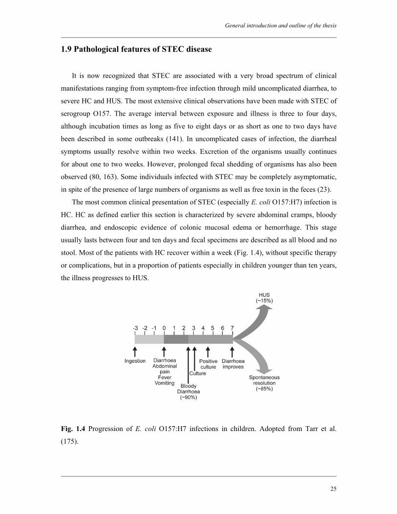
The most common clinical presentation of STEC (especially E. coli O157:H7) infection is
HC. HC as defined earlier this section is characterized by severe abdominal cramps, bloody
diarrhea, and endoscopic evidence of colonic mucosal edema or hemorrhage. This stage
usually lasts between four and ten days and fecal specimens are described as all blood and no
stool. Most of the patients with HC recover within a week (Fig. 1.4), without specific therapy
or complications, but in a proportion of patients especially in children younger than ten years,
the illness progresses to HUS.
Fig. 1.4 Progression of E. coli O157:H7 infections in children. Adopted from Tarr et al.
(175).
Chapter 1
26
Three to 15 percent of patients infected with STEC O157:H7 can progress to HUS, but
this figure may reach 50% if antibiotics are used (162). Young children and older persons
with altered immune response (189) are particularly vulnerable. The classic triad of features
for HUS consists of microangiopathic hemolytic anaemia, thrombocytopenia, and acute renal
failure (152). Clinical features identifying patients at high risk for HUS are vague and may
mimic common gastroenteritis, including bloody diarrhea occurring from three days to more
than two weeks before HUS is diagnosed. Ten percent of cases are associated with rectal
prolapse and colitis (152).
HUS cannot be diagnosed without evidence of hemolytic anaemia. Hematologic findings
include destruction and fragmentation of erythrocytes that results in microangiopathic
hemolytic anaemia. This develops in all patients within around a day of infection and may
result in respiratory and cardiovascular compromise. Mean hemoglobin concentration of 6 g
per dL (60 g per L) is common and requires red blood cell transfusion (152). Ninety-two
percent of patients with HUS develop thrombocytopenia, which results from entrapment of
platelets in the organs (152). Clotting times are normal, and petechiae and purpura are
uncommon features of HUS (152). Acute renal failure results when microthrombi are
deposited in kidney parenchyma. This manifests in the form of hypertension associated with
oliguria and anuria. Approximately 12% of patients who contract HUS either develop end-
stage renal disease or die (57). The central nervous system is another organ system that could
become involved. Thirty-three percent of patients with HUS experience neurologic
complaints such as irritability, seizures, and altered mental status (152). Approximately 10%
of patients with HUS develop central nervous system problems and subsequent coma,
hemiparesis, or stroke (57, 173). Currently, the overall mortality rate for patients with HUS is
around 10% (152).
1.10 Pathogenesis
The pathogenesis of STEC is considered to be a multistep process, involving a complex
interaction between a range of bacterial and host factors.
1.10.1 Survival in the acidic environment
Orally ingested STEC must initially survive the harsh environment of the stomach and
then compete with other microorganisms in the gut to establish intestinal colonization. There
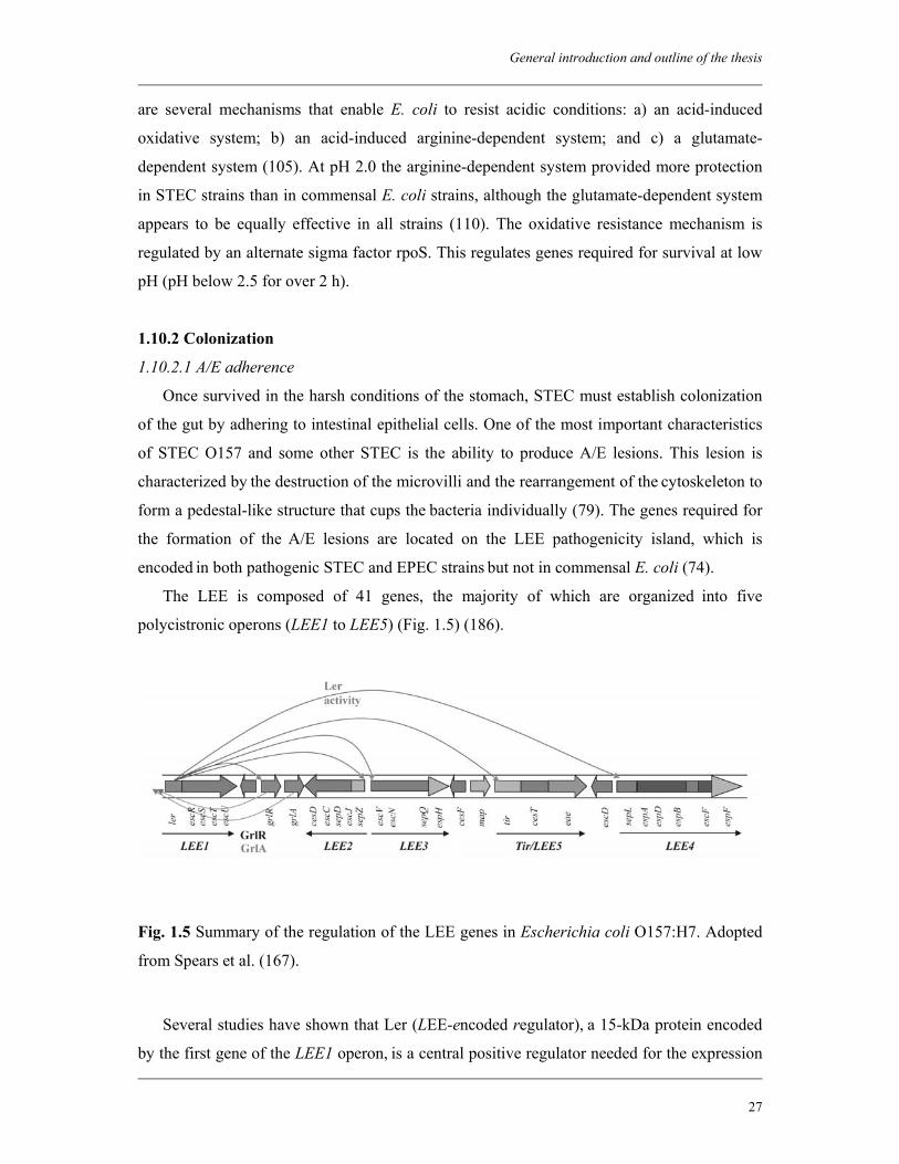
General introduction and outline of the thesis
27
are several mechanisms that enable E. coli to resist acidic conditions: a) an acid-induced
oxidative system; b) an acid-induced arginine-dependent system; and c) a glutamate-
dependent system (105). At pH 2.0 the arginine-dependent system provided more protection
in STEC strains than in commensal E. coli strains, although the glutamate-dependent system
appears to be equally effective in all strains (110). The oxidative resistance mechanism is
regulated by an alternate sigma factor rpoS. This regulates genes required for survival at low
pH (pH below 2.5 for over 2 h).
1.10.2 Colonization
1.10.2.1 A/E adherence
Once survived in the harsh conditions of the stomach, STEC must establish colonization
of the gut by adhering to intestinal epithelial cells. One of the most important characteristics
of STEC O157 and some other STEC is the ability to produce A/E lesions. This lesion is
characterized by the destruction of the microvilli and the rearrangement of the cytoskeleton to
form a pedestal-like structure that cups the bacteria individually (79). The genes required for
the formation of the A/E lesions are located on the LEE pathogenicity island, which is
encoded in both pathogenic STEC and EPEC strains but not in commensal E. coli (74).
The LEE is composed of 41 genes, the majority of which are organized into five
polycistronic operons (LEE1 to LEE5) (Fig. 1.5) (186).
Fig. 1.5 Summary of the regulation of the LEE genes in Escherichia coli O157:H7. Adopted
from Spears et al. (167).
Several studies have shown that Ler (LEE-encoded regulator), a 15-kDa protein encoded
by the first gene of the LEE1 operon, is a central positive regulator needed for the expression
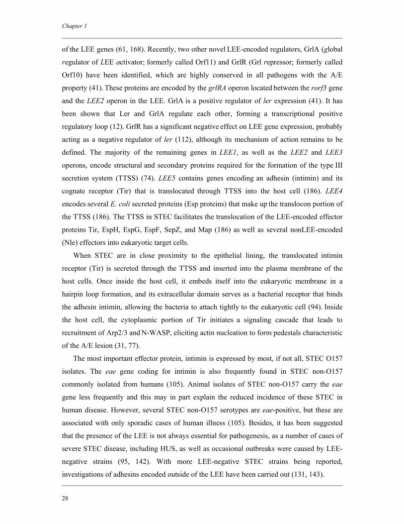
Chapter 1
28
of the LEE genes (61, 168). Recently, two other novel LEE-encoded regulators, GrlA (global
regulator of LEE activator; formerly called Orf11) and GrlR (Grl repressor; formerly called
Orf10) have been identified, which are highly conserved in all pathogens with the A/E
property (41). These proteins are encoded by the grlRA operon located between the rorf3 gene
and the LEE2 operon in the LEE. GrlA is a positive regulator of ler expression (41). It has
been shown that Ler and GrlA regulate each other, forming a transcriptional positive
regulatory loop (12). GrlR has a significant negative effect on LEE gene expression, probably
acting as a negative regulator of ler (112), although its mechanism of action remains to be
defined. The majority of the remaining genes in LEE1, as well as the LEE2 and LEE3
operons, encode structural and secondary proteins required for the formation of the type III
secretion system (TTSS) (74). LEE5 contains genes encoding an adhesin (intimin) and its
cognate receptor (Tir) that is translocated through TTSS into the host cell (186). LEE4
encodes several E. coli secreted proteins (Esp proteins) that make up the translocon portion of
the TTSS (186). The TTSS in STEC facilitates the translocation of the LEE-encoded effector
proteins Tir, EspH, EspG, EspF, SepZ, and Map (186) as well as several nonLEE-encoded
(Nle) effectors into eukaryotic target cells.
When STEC are in close proximity to the epithelial lining, the translocated intimin
receptor (Tir) is secreted through the TTSS and inserted into the plasma membrane of the
host cells. Once inside the host cell, it embeds itself into the eukaryotic membrane in a
hairpin loop formation, and its extracellular domain serves as a bacterial receptor that binds
the adhesin intimin, allowing the bacteria to attach tightly to the eukaryotic cell (94). Inside
the host cell, the cytoplasmic portion of Tir initiates a signaling cascade that leads to
recruitment of Arp2/3 and N-WASP, eliciting actin nucleation to form pedestals characteristic
of the A/E lesion (31, 77).
The most important effector protein, intimin is expressed by most, if not all, STEC O157
isolates. The eae gene coding for intimin is also frequently found in STEC non-O157
commonly isolated from humans (105). Animal isolates of STEC non-O157 carry the eae
gene less frequently and this may in part explain the reduced incidence of these STEC in
human disease. However, several STEC non-O157 serotypes are eae-positive, but these are
associated with only sporadic cases of human illness (105). Besides, it has been suggested
that the presence of the LEE is not always essential for pathogenesis, as a number of cases of
severe STEC disease, including HUS, as well as occasional outbreaks were caused by LEE-
negative strains (95, 142). With more LEE-negative STEC strains being reported,
investigations of adhesins encoded outside of the LEE have been carried out (131, 143).
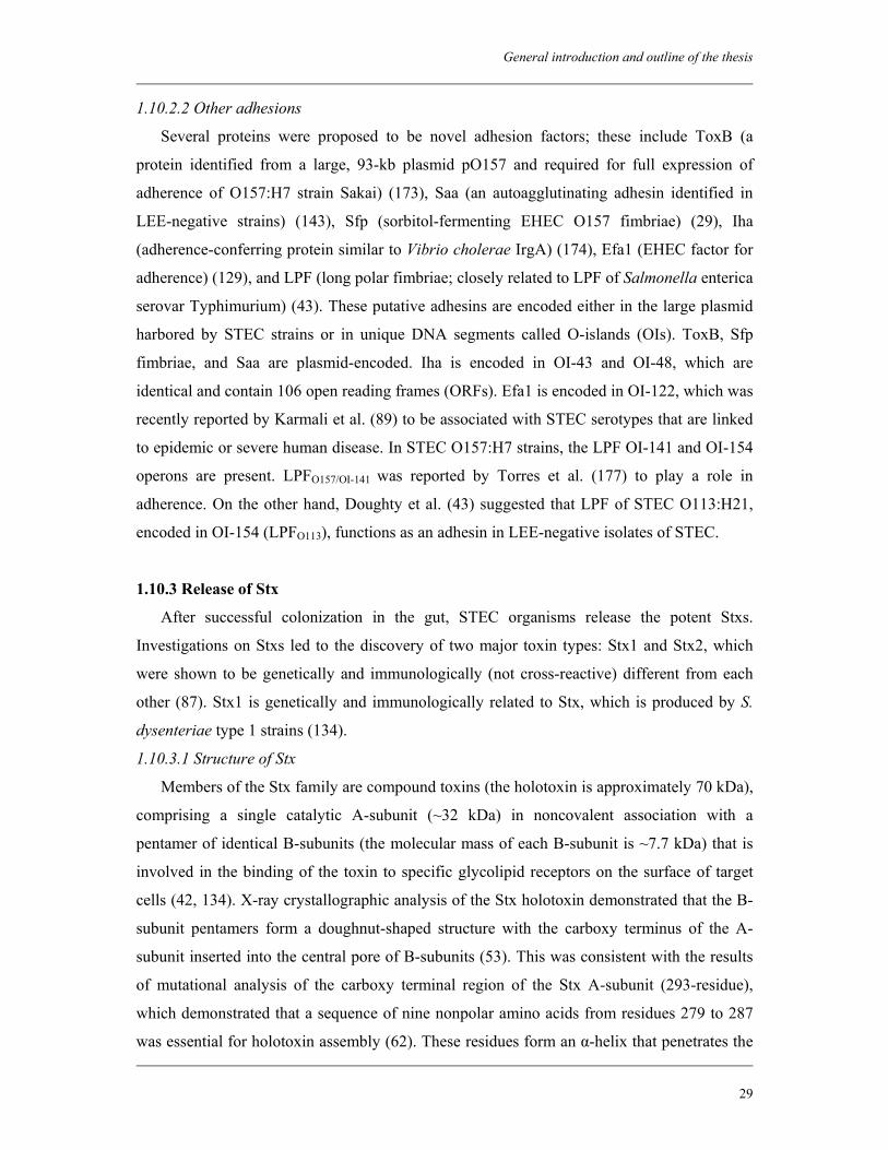
General introduction and outline of the thesis
29
1.10.2.2 Other adhesions
Several proteins were proposed to be novel adhesion factors; these include ToxB (a
protein identified from a large, 93-kb plasmid pO157 and required for full expression of
adherence of O157:H7 strain Sakai) (173), Saa (an autoagglutinating adhesin identified in
LEE-negative strains) (143), Sfp (sorbitol-fermenting EHEC O157 fimbriae) (29), Iha
(adherence-conferring protein similar to Vibrio cholerae IrgA) (174), Efa1 (EHEC factor for
adherence) (129), and LPF (long polar fimbriae; closely related to LPF of Salmonella enterica
serovar Typhimurium) (43). These putative adhesins are encoded either in the large plasmid
harbored by STEC strains or in unique DNA segments called O-islands (OIs). ToxB, Sfp
fimbriae, and Saa are plasmid-encoded. Iha is encoded in OI-43 and OI-48, which are
identical and contain 106 open reading frames (ORFs). Efa1 is encoded in OI-122, which was
recently reported by Karmali et al. (89) to be associated with STEC serotypes that are linked
to epidemic or severe human disease. In STEC O157:H7 strains, the LPF OI-141 and OI-154
operons are present. LPFO157/OI-141 was reported by Torres et al. (177) to play a role in
adherence. On the other hand, Doughty et al. (43) suggested that LPF of STEC O113:H21,
encoded in OI-154 (LPFO113), functions as an adhesin in LEE-negative isolates of STEC.
1.10.3 Release of Stx
After successful colonization in the gut, STEC organisms release the potent Stxs.
Investigations on Stxs led to the discovery of two major toxin types: Stx1 and Stx2, which
were shown to be genetically and immunologically (not cross-reactive) different from each
other (87). Stx1 is genetically and immunologically related to Stx, which is produced by S.
dysenteriae type 1 strains (134).
1.10.3.1 Structure of Stx
Members of the Stx family are compound toxins (the holotoxin is approximately 70 kDa),
comprising a single catalytic A-subunit (~32 kDa) in noncovalent association with a
pentamer of identical B-subunits (the molecular mass of each B-subunit is ~7.7 kDa) that is
involved in the binding of the toxin to specific glycolipid receptors on the surface of target
cells (42, 134). X-ray crystallographic analysis of the Stx holotoxin demonstrated that the B-
subunit pentamers form a doughnut-shaped structure with the carboxy terminus of the A-
subunit inserted into the central pore of B-subunits (53). This was consistent with the results
of mutational analysis of the carboxy terminal region of the Stx A-subunit (293-residue),
which demonstrated that a sequence of nine nonpolar amino acids from residues 279 to 287
was essential for holotoxin assembly (62). These residues form an α-helix that penetrates the
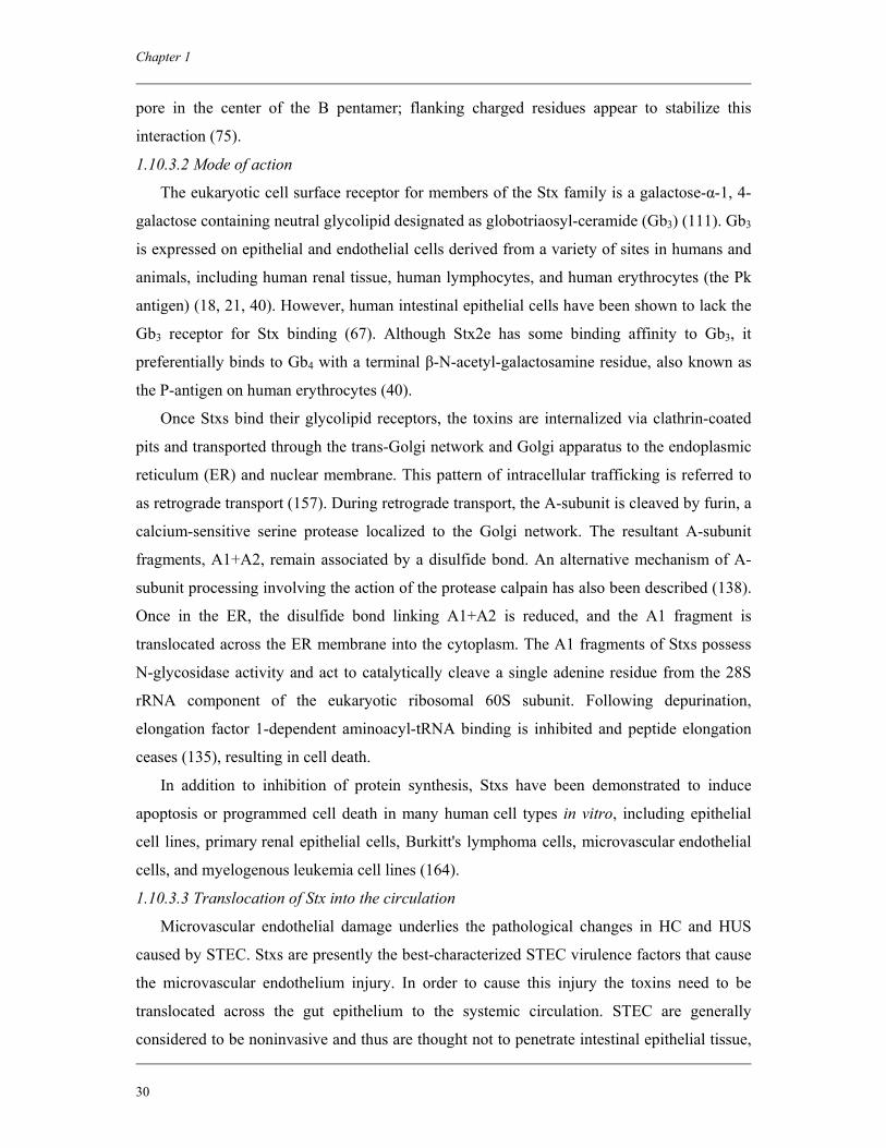
Chapter 1
30
pore in the center of the B pentamer; flanking charged residues appear to stabilize this
interaction (75).
1.10.3.2 Mode of action
The eukaryotic cell surface receptor for members of the Stx family is a galactose-α-1, 4-
galactose containing neutral glycolipid designated as globotriaosyl-ceramide (Gb3) (111). Gb3
is expressed on epithelial and endothelial cells derived from a variety of sites in humans and
animals, including human renal tissue, human lymphocytes, and human erythrocytes (the Pk
antigen) (18, 21, 40). However, human intestinal epithelial cells have been shown to lack the
Gb3 receptor for Stx binding (67). Although Stx2e has some binding affinity to Gb3, it
preferentially binds to Gb4 with a terminal β-N-acetyl-galactosamine residue, also known as
the P-antigen on human erythrocytes (40).
Once Stxs bind their glycolipid receptors, the toxins are internalized via clathrin-coated
pits and transported through the trans-Golgi network and Golgi apparatus to the endoplasmic
reticulum (ER) and nuclear membrane. This pattern of intracellular trafficking is referred to
as retrograde transport (157). During retrograde transport, the A-subunit is cleaved by furin, a
calcium-sensitive serine protease localized to the Golgi network. The resultant A-subunit
fragments, A1+A2, remain associated by a disulfide bond. An alternative mechanism of A-
subunit processing involving the action of the protease calpain has also been described (138).
Once in the ER, the disulfide bond linking A1+A2 is reduced, and the A1 fragment is
translocated across the ER membrane into the cytoplasm. The A1 fragments of Stxs possess
N-glycosidase activity and act to catalytically cleave a single adenine residue from the 28S
rRNA component of the eukaryotic ribosomal 60S subunit. Following depurination,
elongation factor 1-dependent aminoacyl-tRNA binding is inhibited and peptide elongation
ceases (135), resulting in cell death.
In addition to inhibition of protein synthesis, Stxs have been demonstrated to induce
apoptosis or programmed cell death in many human cell types in vitro, including epithelial
cell lines, primary renal epithelial cells, Burkitt's lymphoma cells, microvascular endothelial
cells, and myelogenous leukemia cell lines (164).
1.10.3.3 Translocation of Stx into the circulation
Microvascular endothelial damage underlies the pathological changes in HC and HUS
caused by STEC. Stxs are presently the best-characterized STEC virulence factors that cause
the microvascular endothelium injury. In order to cause this injury the toxins need to be
translocated across the gut epithelium to the systemic circulation. STEC are generally
considered to be noninvasive and thus are thought not to penetrate intestinal epithelial tissue,
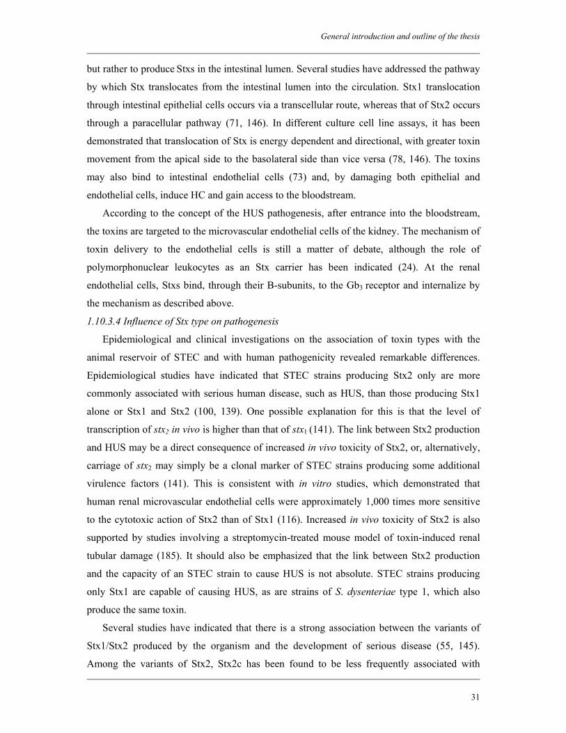
General introduction and outline of the thesis
31
but rather to produce Stxs in the intestinal lumen. Several studies have addressed the pathway
by which Stx translocates from the intestinal lumen into the circulation. Stx1 translocation
through intestinal epithelial cells occurs via a transcellular route, whereas that of Stx2 occurs
through a paracellular pathway (71, 146). In different culture cell line assays, it has been
demonstrated that translocation of Stx is energy dependent and directional, with greater toxin
movement from the apical side to the basolateral side than vice versa (78, 146). The toxins
may also bind to intestinal endothelial cells (73) and, by damaging both epithelial and
endothelial cells, induce HC and gain access to the bloodstream.
According to the concept of the HUS pathogenesis, after entrance into the bloodstream,
the toxins are targeted to the microvascular endothelial cells of the kidney. The mechanism of
toxin delivery to the endothelial cells is still a matter of debate, although the role of
polymorphonuclear leukocytes as an Stx carrier has been indicated (24). At the renal
endothelial cells, Stxs bind, through their B-subunits, to the Gb3 receptor and internalize by
the mechanism as described above.
1.10.3.4 Influence of Stx type on pathogenesis
Epidemiological and clinical investigations on the association of toxin types with the
animal reservoir of STEC and with human pathogenicity revealed remarkable differences.
Epidemiological studies have indicated that STEC strains producing Stx2 only are more
commonly associated with serious human disease, such as HUS, than those producing Stx1
alone or Stx1 and Stx2 (100, 139). One possible explanation for this is that the level of
transcription of stx2 in vivo is higher than that of stx1 (141). The link between Stx2 production
and HUS may be a direct consequence of increased in vivo toxicity of Stx2, or, alternatively,
carriage of stx2 may simply be a clonal marker of STEC strains producing some additional
virulence factors (141). This is consistent with in vitro studies, which demonstrated that
human renal microvascular endothelial cells were approximately 1,000 times more sensitive
to the cytotoxic action of Stx2 than of Stx1 (116). Increased in vivo toxicity of Stx2 is also
supported by studies involving a streptomycin-treated mouse model of toxin-induced renal
tubular damage (185). It should also be emphasized that the link between Stx2 production
and the capacity of an STEC strain to cause HUS is not absolute. STEC strains producing
only Stx1 are capable of causing HUS, as are strains of S. dysenteriae type 1, which also
produce the same toxin.
Several studies have indicated that there is a strong association between the variants of
Stx1/Stx2 produced by the organism and the development of serious disease (55, 145).
Among the variants of Stx2, Stx2c has been found to be less frequently associated with
Chapter 1
32
bloody diarrhea and HUS than Stx2 (55). Stx2 and the elastase (mucus)-activatable Stx2d
type, are associated with the high virulence of STEC and with HC and HUS (17). Other toxin
variants, such as Stx1c, Stx2-O118 (formerly Stx2d-Ount), Stx2e, and Stx2f, were found to be
associated mainly with uncomplicated diarrhea or asymptomatic excretion (55, 56). It has
also been demonstrated that there is an association between the type of Stx and the origin of
the strains. For example, STEC strains producing Stx1c and/or Stx2d-O118 were associated
with sheep (22, 179), Stx2e with pigs (54), and Stx2f with pigeons (166) as natural reservoirs.
1.10.4 Release of other potential virulence factors
1.10.4.1 Enterohemolysin
A high proportion of STEC strains posses a novel hemolytic phenotype, which is distinct
from that associated with the E. coli α-hemolysin (hly) (14). Nearly all STEC O157 strains
and a wide range of STEC non-O157 strains are positive for enterohemolysin (subsequently
designated EHEC-hly) (82). Strains producing this enterohemolysin are not hemolytic on
standard blood agar but produce small, turbid hemolytic zones on washed sheep red blood
cell agar (supplemented with calcium) after overnight incubation. Unlike α-hemolysin, which
is chromosomally encoded, EHEC-hly is encoded by the 60-MDa “virulence plasmid” of the
STEC O157 (pO157). The role of enterohemolysin in the pathogenesis of STEC infection is
still subject to speculation. One possibility is that hemoglobin released by the action of
EHEC-hly provides a source of iron, thereby stimulating the growth of STEC in the gut
(104).
1.10.4.2 EHEC type II secretion system (EtpD)
Upstream of the EHEC-hly operon in pO157 is a gene cluster showing similarities to
genes of members of the type II secretion pathway systems of Gram-negative bacteria (81).
The highest similarities were found to the pullulanase secretion pathway of Klebsiella
oxytoca and Klebsiella pneumoniae in which at least 14 genes and proteins are involved (81).
The genetic determinants of this type II secretion pathway system are encoded in 13 ORFs.
These ORFs are termed as etpC through etpO. The significance of this gene cluster in the
pathogenesis of STEC infection is yet to be elucidated. The distribution of the genetic
determinants of this type II secretion pathway system is not uniform. Karch et al. (81)
showed that all 50 isolates of STEC O157:H7 (100%) tested possessed this pathway, but that
it was present in only 52% of 50 STEC non-O157. In another study, the presence of one of
the genes in the pathway, etpD, was detected in all the 13 STEC O103 strains but not in any
of the STEC strains tested belonging to O26 or O111 (161). The etp genes were rarely
General introduction and outline of the thesis
33
detected in STEC isolated from bovine feces (160). Moreover, it was found not to occur in
other pathotypes of E. coli (160).
1.10.4.3 The bifunctional catalase peroxidase (KatP)
The STEC bifunctional catalase peroxidase is termed as KatP (26). The functional role of
this enzyme remains unclear. It has been suggested that bacterial catalases and superoxide
dismutases detoxify cytotoxic oxidant products during the oxidative bursts of macrophages
and neutrophils, thereby assisting the bacterium in escaping host defenses (26). The
distribution of KatP genetic determinants is not uniform. Brunder et al. (26) found that KatP
was present in 66% of the STEC O157:H7 isolates and 38% of the STEC non-O157 isolates.
Besides, absence of KatP in STEC isolates causing HC or HUS suggested that the enzyme
does not have a major role in virulence.
1.10.4.4 The secreted serine protease (EspP)
One of the most important virulence factors of STEC strains is the secreted serine
protease (EspP), which is also encoded on the large plasmid (pO157) but is not involved in
the production of the A/E lesions (27). E. coli has several secreted proteins (Esps). The entire
sequence of EspP was homologous to that of EspC (110 kDa protein secreted by EPEC
strains), SepA (the major extracellular protein of Shigella flexneri) and Tsh (a haemagglutinin
from an avian pathogenic E. coli strain) (81). The EspP is subjected to N- and C-terminal
processing during the secretion process. EspP is known as an autotransporter as it mediates
its own secretion through the outer membrane. It also has proteolytic activity against a
narrow range of substrates, one of which is human coagulation factor V (81). This surprising
activity indicates that EspP secreted by STEC may influence the blood clotting cascade.
Cleavage of factor V could result in a decreased coagulation reaction leading to prolonged
bleeding. Local degradation of factor V by STEC attached to the intestinal mucosa could
increase hemorrhage in the gastrointestinal tract (81). Children suffering from STEC
infection produce an antibody response to EspP indicating its expression in vivo (27),
although this does not necessarily indicate a role in pathogenesis. The distribution of the
EspP toxin in STEC strains causing infection is not uniform (27, 28). Therefore, the
significance of EspP in disease is unclear, but its frequent absence in STEC O157:H7 and
non-O157 strains indicates that it is not vital to the development of the disease process in
humans.
Chapter 1
34
1.11 Outline of the thesis
The main objective of the work presented in this thesis was to gain insight in the
epidemiology of STEC infection in humans and the ecology of STEC in the human food
chain in Bangladesh. Although Bangladesh is an endemic zone for diarrheal diseases caused
by different enteric pathogens, no systematic study has yet been done on the epidemiology of
STEC infection in diarrheal patients. In chapter 2 we described the prevalence and
characteristics of STEC isolated from patients with diarrhea attending the hospital as well as
a community clinic in Dhaka city in Bangladesh. The next two chapters deal with the
evaluation of detection and isolation methods for STEC O157 from animal feces and raw
meats. There are several methods available for the detection and isolation of STEC O157 but
a single method may not be equally effective for all different kinds of samples. In chapter 3,
we compared the efficiency of two commonly used immunoconcentration methods,
Dynabeads anti-E. coli O157 and the Vitek Immunodiagnostic Assay System (VIDAS)
Immuno-Concentration E. coli O157 (ICE) kit, by analyzing retrospectively the results of
isolation of STEC O157 from animal feces and raw meats. In this study, we also compared
two commonly used selective plating media CT-SMAC and 1/2CT-CHROMagar for isolation
of STEC O157 in combination with the Dynabeads anti-E. coli O157 method and the VIDAS
ICE method. When the Dynabeads anti-E. coli O157 method turned out to be more effective
than the VIDAS ICE method for the isolation of STEC O157 from animal feces, we further
compared this method with a PCR-based method and this has been described in chapter 4. In
order to study the occurrence of STEC in the food chain, we investigated the prevalence of
this organism in the reservoirs and in food. We focused on animals and food, which have the
greatest chance of being positive for STEC and therefore can play an important role in the
transmission of these pathogens to humans. In chapter 5, we described the prevalence and
characteristics of STEC in different slaughtered animals in Bangladesh, including buffalo,
cows and goats. Similarly in chapter 6, we described the occurrence and characteristics of
STEC in different types of food, including raw meat, raw milk and freshly prepared juices in
Bangladesh. In order to interlink the results obtained from the investigations of STEC in each
of the components of the food chain, we analyzed the antibody responses against STEC O157
among the healthy population in Bangladesh, which has been described in chapter 7. Finally,
in chapter 8, the findings of this thesis were summarized with a more general discussion and
placed in perspective with future directions of the research.
General introduction and outline of the thesis
35
References
1. Ackman, D., S. Marks, P. Mack, M. Caldwell, T. Root, and G. Birkhead. 1997. Swimming associated haemorrhagic colitis due to Escherichia coli O157:H7 infection: evidence of prolonged contamination of a fresh water lake. Epidemiol. Infect. 119:1–8.
2. Afza, M., J. Hawker, H. Thurston, K. Gunn, and J. Orendi. 2006. An outbreak of Escherichia coli O157 gastroenteritis in a care home for the elderly. Epidemiol. Infect. 134:1276–1281.
3. Ahmed, N. M., and D. E. Conner. 1995. Evaluation of various media for recovery of thermally-injured Escherichia coli O157:H7. J. Food Prot. 58:357–360.
4. Ak, N. O., D. O. Cliver, and C. W. Kaspari. 1994. Cutting boards of plastic and wood contaminated experimentally with bacteria. J. Food Prot. 57:16-22.
5. Alikhani, M. Y., A. Mirsalehian, B. Fatollahzadeh, M. R. Pourshafie, and M. M. Aslani. 2007. Prevalence of enteropathogenic and shiga toxin-producing Escherichia coli among children with and without diarrhoea in Iran. J. Health Popul. Nutr. 25:88-93.
6. Anonymous. 2001. Microbiology of food and animal feeding stuffs—Horizontal method for the detection of Escherichia coli O157 (EN ISO 16654). International Organization for Standardization, Geneva, Switzerland.
7. Arthur, T. M., J. M. Bosilevac, X. Nou, and M. Koohmaraie. 2005. Evaluation of culture-and PCR-based detection methods for Escherichia coli O157:H7 in inoculated ground beef. J. Food Prot. 68:1566–1574.
8. Artz, R. E., and K. Killham. 2002. Survival of Escherichia coli O157:H7 in private drinking water wells: influences of protozoan grazing and elevated copper concentrations. FEMS Microbiol. Lett. 216:117–122.
9. Aslani, M. M., N. Badami, M. Mahmmoodi, and S. Bouzari. 1998. Verotoxin-producing Escherichia coli (VTEC) infection in randomly selected population of Ilam Province (Iran). Scand. J. Infect. Dis. 30:473-476.
10. Aslani, M. M., and S. Bouzari. 2003. An epidemiological study on Verotoxin-producing Escherichia coli (VTEC) infection among population of northern region of Iran (Mazandaran and Golestan provinces). Eur. J. Epidemiol. 18:345-349.
11. Bae, W. K., Y. K. Lee, M. S. Cho, S. K. Ma, S. W. Kim, N. H. Kim, and K. C. Choi. 2006. A case of hemolytic uremic syndrome caused by Escherichia coli O104:H4. Yonsei Med. J. 47:437-439.
12. Barba, J., V. H. Bustamante, M. A. Flores-Valdez, W. Deng, B. B. Finlay, and J. L. Puente. 2005. A positive regulatory loop controls expression of the locus of enterocyte effacement-encoded regulators Ler and GrlA. J. Bacteriol. 187:7918-7930.
13. Bettelheim, K. A., J. E. Brown, S. Lolekha, and P. Echeverria. 1990. Serotypes of Escherichia coli that hybridized with DNA probes for genes encoding Shiga-like toxin I, Shiga-like toxin II, and serogroup O157 enterohemorrhagic E. coli fimbriae isolated from adults with diarrhea in Thailand. J. Clin. Microbiol. 28:293-295.
14. Beutin, L., M. A. Montenegro, I. Ørskov, F. Ørskov, J. Prada, S. Zimmermann, and R. Stephan. 1989. Close association of verocytotoxin (Shiga-like toxin) production with enterohemolysin production in strains of Escherichia coli. J. Clin. Microbiol. 27:2559–2564.
15. Beutin, L., S. Zimmermann, and K. Gleier. 1996. Rapid detection and isolation of Shiga-like toxin (verocytotoxin)-producing Escherichia coli by direct testing of individual enterohemolytic colonies from washed sheep blood agar plates in the VTEC-RPLA assay. J. Clin. Microbiol. 34:2812–2814.
Chapter 1
36
16. Bielaszewska, M., H. Schmidt, M. A. Kamali, R. Khakhria, J. Janda, K. Blahova, and H. Karch. 1998. Isolation and characterization of sorbitol-fermenting Shiga toxin-producing Escherichia coli O157:H- strains in the Czech Republic. J. Clin. Microbiol. 36:2135–2137.
17. Bielaszewska, M., A. W. Friedrich, T. Aldick, R. Schurk-Bulgrin, and H. Karch. 2006. Shiga toxin activatable by intestinal mucus in Escherichia coli isolated from humans: predictor for a severe clinical outcome. Clin. Infect. Dis. 43:1160–1167.
18. Bitzan, M., S. Richardson, C. Huang, B. Boyd, M. Petric, and M. A. Karmali. 1994. Evidence that verotoxins (Shiga-like toxins) from Escherichia coli bind to P blood group antigens of human erythrocytes in vitro. Infect. Immun. 62:3337-3347.
19. Boerlin, P., S. A. McEwen, F. Boerlin-Petzold, J. B. Wilson, R. P. Johnson, and C. L. Gyles. 1999. Associations between virulence factors of Shiga toxin-producing Escherichia coli and disease in humans. J. Clin. Microbiol. 37:497–503.
20. Boyce, T. G., D. L. Swerdlow, and P. M. Griffin. 1995. Escherichia coli O157:H7 and the haemolytic-uraemic syndrome. N. Engl. J. Med. 333:364-368.
21. Boyd, B., and C. Lingwood. 1989. Verotoxin receptor glycolipid in human renal tissue. Nephron. 51:207-210. Erratum in: Nephron 1989; 51:582.
22. Brett, K. N., V. Ramachandran, M. A. Hornitzky, K. A. Bettelheim, M. J. Walker, and S. P. Djordjevic. 2003. Stx1c is the most common Shiga toxin 1 subtype among Shiga toxin-producing Escherichia coli isolates from sheep but not among isolates from cattle. J. Clin. Microbiol. 41:926–936.
23. Brian, M. J., M. Frosolono, B. E. Murray, A. Miranda, E. L. Lopez, H. F. Gomez, and T. G. Cleary. 1992. Polymerase chain reaction for diagnosis of enterohemorrhagic Escherichia coli infection and hemolytic-uremic syndrome. J. Clin. Microbiol. 30:1801–1806.
24. Brigotti, M., D. Carnicelli, E. Ravanelli, S. Barbieri, F. Ricci, A. Bontadini, A. E. Tozzi, G. Scavia, A. Caprioli, and P. L. Tazzari. 2008. Interactions between Shiga toxins and human polymorphonuclear leukocytes. J. Leukoc. Biol. 84:1019-1027.
25. Brown, J. E., P. Echeverria, D. N. Taylor, J. Seriwatana, V. Vanapruks, U. Lexomboon, R. N. Neill, and J. W. Newland. 1989. Determination by DNA hybridization of Shiga-like-toxin-producing Escherichia coli in children with diarrhea in Thailand. J. Clin. Microbiol. 27:291-294.
26. Brunder, W., H. Schmidt, and H. Karch. 1996. KatP, a novel catalase-peroxidase encoded by the large plasmid of enterohaemorrhagic Escherichia coli O157:H7. Microbiology 142:3305-3315.
27. Brunder, W., H. Schmidt, and H. Karch. 1997. EspP, a novel extracellular serine protease of enterohaemorrhagic Escherichia coli O157:H7 cleaves human coagulation factor V. Mol. Microbiol. 24:767-778.
28. Brunder, W., H. Schmidt, M. Frosch, and H. Karch. 1999. The large plasmids of Shiga-toxin-producing Escherichia coli (STEC) are highly variable genetic elements. Microbiology 145:1005-1014.
29. Brunder, W., A. S. Khan, J. Hacker, and H. Karch. 2001. Novel type of fimbriae encoded by the large plasmid of sorbitol-fermenting enterohemorrhagic Escherichia coli O157:H–. Infect. Immun. 69:4447-4457.
30. Calderwood, S. B., D. W. K. Acheson, G. T. Keusch, T. J. Barrett, P. M. Griffin, N. A. Strockbine, B. Swaminathan, J. B. Kaper, M. M. Levine, B. S. Kaplan, H. Karch, A. D. O’Brien, T. G. Obrig, Y. Takeda, P. I. Tarr, and I. K. Wachsmuth. 1996. Proposed new nomenclature for SLT (VT) family. ASM News 62:118-119.
General introduction and outline of the thesis
37
31. Campellone, K. G., and J. M. Leong. 2003. Tails of two Tirs: actin pedestal formation by enteropathogenic E. coli and enterohemorrhagic E. coli O157:H7. Curr. Opin. Microbiol. 6:82-90.
32. Caprioli, A., S. Morabito, H. Brugere, and E. Oswald. 2005. Enterohaemorrhagic Escherichia coli: emerging issues on virulence and modes of transmission. Vet. Res. 36:289-311.
33. Catarame, T. M. G., K. A. O’Hanlon, G. Duffy, J. J. Sheridan, I. S. Blair, and D. A. McDowell. 2003. Optimisation of enrichment and plating procedures for the recovery of Escherichia coli O111 and O26 from minced beef. J. Appl. Microbiol. 95:949–957.
34. Chalmers, R. M., H. Aird, and F. J. Bolton. 2000. Waterborne Escherichia coli O157. J. Appl. Microbiol. 88:124S–132S.
35. Chattopadhyay, U. K., S. Dutta, and A. Deb. 2001. Verotoxin-producing Escherichia coli–an environment-induced emerging zoonosis in and around Calcutta. Int. J. Environ. Health Res. 1:107–112.
36. Church, D. L., D. Emshey, H. Semeniuk, T. Lloyd, and J. D. Pitout. 2007. Evaluation of BBL CHROMagar O157 versus sorbitol-MacConkey medium for routine detection of Escherichia coli O157 in a centralized regional clinical microbiology laboratory. J. Clin. Microbiol. 45:3098-3100.
37. Centers for Disease Control and Prevention (CDC). 1999. Outbreak of Escherichia coli O157:H7 and Campylobacter among attendees of the Washington County Fair-New York. MMWR Morb. Mortal. Wkly. Rep. 48:803-805.
38. Centers for Disease Control and Prevention (CDC). 2005. Outbreaks of Escherichia coli O157:H7 associated with petting zoos—North Carolina, Florida, and Arizona, 2004 and 2005. MMWR Morb. Mortal. Wkly. Rep. 54:1277-1280.
39. Centers for Disease Control and Prevention (CDC). 2006. Ongoing multistate outbreak of Escherichia coli serotype O157:H7 infections associated with consumption of fresh spinach-United States, September 2006. MMWR Morb. Mortal. Wkly. Rep. 55:1045-1046.
40. DeGrandis, S., H. Law, J. Brunton, C. Gyles, and C. A. Lingwood. 1989. Globotetraosylceramide is recognized by the pig edema disease toxin. J. Biol. Chem. 264:12520-12525.
41. Deng, W., J. L. Puente, S. Gruenheid, Y. Li, B. A. Vallance, A. Vazquez, J. Barba, J. A. Ibarra, P. O'Donnell, P. Metalnikov, K. Ashman, S. Lee, D. Goode, T. Pawson, and B. B. Finlay. 2004. Dissecting virulence: systematic and functional analyzes of a pathogenicity island. Proc. Natl. Acad. Sci. USA 101:3597-3602.
42. Donohue-Rolfe, A., G. T. Keusch, C. Edson, D. Thorley-Lawson, and M. Jacewicz. 1984. Pathogenesis of Shigella diarrhea. IX. Simplified high yield purification of Shigella toxin and characterization of subunit composition and function by the use of subunit-specific monoclonal and polyclonal antibodies. J. Exp. Med. 160:1767–1781.
43. Doughty, S., J. Sloan, V. Bennet-Wood, M. Robertson, R. M. Robins-Browne, and E. L. Hartland. 2002. Identification of a novel fimbrial gene cluster related to long polar fimbriae in locus of enterocyte effacement-negative strains of enterohemorrhagic Escherichia coli. Infect. Immun. 70:6761-6769.
44. Drasar, B. S., and M. J. Hill. 1974. The distribution of bacterial flora in the intestine, p. 36-43. In B. S. Drasar and M. J. Hill (ed.), Human intestinal microflora. Academic Press, London.
Chapter 1
38
45. EFSA. 2007. Scientific opinion of the panel on biological hazards on monitoring of verotoxigenic Escherichia coli (VTEC) and identification of human pathogenic VTEC types. EFSA J. 579:1-61.
46. EFSA. 2007. The community summary report on trends and sources of zoonoses, zoonotic agents, antimicrobial resistance and foodborne outbreaks in the European Union in 2006. EFSA J. 130:151-164.
47. Erickson, M. C., and M. P. Doyle. 2007. Food as a vehicle for transmission of Shiga toxin-producing Escherichia coli. J. Food Prot. 70:2426-2449.
48. Escherich, T. 1988. The intestinal bacteria of the neonate and breast-fed infant. 1884. Rev. Infect. Dis. 10:1220–1225.
49. Escherich, T. 1989. The intestinal bacteria of the neonate and breast-fed infant. 1885. Rev. Infect. Dis. 11:352–356.
50. Ethelberg, S., K. E. Olsen, F. Scheutz, C. Jensen, P. Schiellerup, J. Enberg, A. M. Petersen, B. Olesen, P. Gerner-Smidt, and K. Molbak. 2004. Virulence factors for hemolytic uremic syndrome, Denmark. Emerg. Infect. Dis. 10:842-847.
51. Farrell, B. L., A. B. Ronner, and A. C. Wong. 1998. Attachment of Escherichia coli O157:H7 in ground beef to meat grinders and survival after sanitation with chlorine and peroxyacetic acid. J. Food Prot. 61:817-822.
52. Frankel, G., A. D. Phillips, I. Rosenshine, G. Dougan, J. B. Kaper, and S. Knutton. 1998. Enteropathogenic and enterohaemorrhagic Escherichia coli: more subversive elements. Mol. Microbiol. 30:911–921.
53. Fraser, M. E., M. M. Chernaia, Y. V. Kozlov, and M. N. James. 1994. Crystal structure of the holotoxin from Shigella dysenteriae at 2.5 A resolution. Nat. Struct. Biol. 1:59–64.
54. Fratamico, P. M., L. K. Bagi, E. J. Bush, and B. T. Solow. 2004. Prevalence and characterization of Shiga toxin-producing Escherichia coli in swine feces recovered in the National Animal Health Monitoring System’s Swine 2000 study. Appl. Environ. Microbiol. 70:7173–7178.
55. Friedrich, A. W., M. Bielaszewska, W. L. Zhang, M. Pulz, T. Kuczius, A. Ammon, and H. Karch. 2002. Escherichia coli harboring Shiga toxin 2 gene variants: frequency and association with clinical symptoms. J. Infect. Dis. 185:74–84.
56. Friedrich, A. W., J. Borell, M. Bielaszewska, A. Fruth, H. Tschape, and H. Karch. 2003. Shiga toxin 1c-producing Escherichia coli strains: phenotypic and genetic characterization and association with human disease. J. Clin. Microbiol. 41:2448–2453.
57. Garg, A. X., R. S. Suri, N. Barrowman, F. Rehman, D. Matsell, M. P. Rosas-Arellano, M. Salvadori, R. B. Haynes, and W. F. Clark. 2003. Long-term renal prognosis of diarrhea-associated hemolytic uremic syndrome: a systematic review, meta-analysis, and meta-regression. JAMA 290:1360-1370.
58. Gerner-Smidt, P., K. Hise, J. Kincaid, S. Hunter, S. Rolando, E. Hyytia-Trees, E. M. Ribot, and B. Swaminathan; Pulsenet Taskforce. 2006. PulseNet USA: A five-year update. Foodborne Pathog. Dis. 3:9–19.
59. Griffin, P. M., and R. V. Tauxe. 1991. The epidemiology of infections caused by Escherichia coli O157:H7, other enterohemorrhagic E. coli, and the associated haemolytic uraemic syndrome. Epidemiol. Rev. 13:60-98.
60. Grys, T. E., M. B. Siegel, W. W. Lathem, and R. A. Welch. 2005. The StcE protease contributes to intimate adherence of enterohemorrhagic Escherichia coli O157:H7 to host cells. Infect. Immun. 73:1295-1303.
General introduction and outline of the thesis
39
61. Haack, K. R., C. L. Robinson, K. J. Miller, J. W. Fowlkes, and J. L. Mellies. 2003. Interaction of Ler at the LEE5 (tir) operon of enteropathogenic Escherichia coli. Infect. Immun. 71:384-392.
62. Haddad, J. E., and M. P. Jackson. 1993. Identification of the Shiga toxin A-subunit residues required for holotoxin assembly. J. Bacteriol. 175:7652–7657.
63. Hamner, S., S. C. Broadaway, V. B. Mishra, A. Tripathi, R. K. Mishra, E. Pulcini, B. H. Pyle, and T. E. Ford. 2007. Isolation of potentially pathogenic Escherichia coli O157:H7 from the Ganges River. Appl. Environ. Microbiol. 73:2369-2372.
64. Heuvelink, A. E., C. van Heerwaarden, J. T. Zwartkruis-Nahuis, R. van Oosterom, K. Edink, Y. T. van Duynhoven, and E. de Boer. 2002. Escherichia coli O157 infection associated with a petting zoo. Epidemiol. Infect. 129:295-302.
65. Heuvelink, A. E., S. M. Valkenburgh, J. J. Tilburg, C. van Heerwaarden, J. T. Zwartkruis-Nahuis, and E. de Boer. 2007. Public farms: hygiene and zoonotic agents. Epidemiol. Infect. 135:1174-1183.
66. Himathongkham, S., M. L. Dodd, J. K. Yee, D. K. Lau, R. G. Bryant, A. S. Badoiu, H. K. Lau, L. S. Guthertz, L. Crawford-Miksza, and M. A. Soliman. 2007. Recirculating immunomagnetic separation and optimal enrichment conditions for enhanced detection and recovery of low levels of Escherichia coli O157:H7 from fresh leafy produce and surface water. J. Food Prot. 70:2717-2724.
67. Holgersson, J., P. A. Jovell, and M. E. Breimer. 1991. Glycosphingolipids of the human large intestine: detailed structural characterization with special reference to blood group compounds and bacterial receptor structures. J. Biochem. 110:120-131.
68. Hornitzky, M. A., K. A. Bettelheim, and S. P. Djordjevic. 2001. The detection of Shiga toxin-producing Escherichia coli in diagnostic bovine faecal samples using vancomycin-cefixime-cefsulodin blood agar and PCR. FEMS Microbiol. Lett. 198:17-22.
69. Hussein, H. S., and L. M. Bollinger. 2005. Prevalence of Shiga toxin-producing Escherichia coli in beef cattle. J. Food Prot. 68:2224-2241.
70. Hussein, H. S., and T. Sakuma. 2005. Prevalence of Shiga toxin-producing Escherichia coli in dairy cattle and their products. J. Dairy Sci. 88:450–465.
71. Hurley, B. P., M. Jacewicz, M. Thorpe, L. L. Lincicome, A. J. King, G. T. Keusch, and D. W. K. Acheson. 1999. Shiga toxins 1 and 2 translocate differently across polarized intestinal epithelial cells. Infect. Immun. 67:6670–6677.
72. Hyytia-Trees, E., S. C. Smole, P. A. Fields, B. Swaminathan, and E. M. Ribot. 2006. Second generation subtyping: a proposed PulseNet protocol for multiple-locus variable-number tandem repeat analysis of Shiga toxin-producing Escherichia coli O157 (STEC O157). Foodborne Pathog. Dis. 3:118–131.
73. Jacewicz, M. S., D. W. K. Acheson, D. G. Binion, G. A. West, L. L. Lincicome, C. Fiocchi, and G. T. Keusch. 1999. Response of human intestinal microvascular endothelial cells to Shiga toxins 1 and 2 and pathogenesis of hemorrhagic colitis. Infect. Immun. 67:1439–1444.
74. Jarvis, K. G., J. A. Giron, A. E. Jerse, T. K. McDaniel, M. S. Donnenberg, and J. B. Kaper. 1995. Enteropathogenic Escherichia coli contains a putative type III secretion system necessary for the export of proteins involved in attaching and effacing lesion formation. Proc. Natl. Acad. Sci. USA 92:7996-8000.
75. Jemal, C., J. E. Haddad, D. Begum, and M. P. Jackson. 1995. Analysis of Shiga toxin subunit association by using hybrid A polypeptides and site-specific mutagenesis. J. Bacteriol. 177:3128–3132.
76. Jo, M. Y., J. H. Kim, J. H. Lim, M. Y. Kang, H. B. Koh, Y. H. Park, D. Y. Yoon, J. S. Chae, S. K. Eo, and J. H. Lee. 2004. Prevalence and characteristics of
Chapter 1
40
Escherichia coli O157 from major food animals in Korea. Int. J. Food Microbiol. 95:41-49.
77. Kalman, D., O. D. Weiner, D. L. Goosney, J. W. Sedat, B. B. Finlay, A. Abo, and J. M. Bishop. 1999. Enteropathogenic E. coli acts through WASP and Arp2/3 complex to form actin pedestals. Nat. Cell Biol. 1:389-391.
78. Kaper, J. B. 1998. Enterohemorrhagic Escherichia coli. Curr. Opin. Microbiol. 1:103-108.
79. Kaper, J. B., J. P. Nataro, and H. L. Mobley. 2004. Pathogenic Escherichia coli. Nat. Rev. Microbiol. 2:123-140.
80. Karch, H., H. Rüssmann, H. Schmidt, A. Schwarzkopf, and J. Heesemann. 1995. Long-term shedding and clonal turnover of enterohemorrhagic Escherichia coli O157 in diarrheal diseases. J. Clin. Microbiol. 33:1602-1605.
81. Karch, H., H. Schmidt, and W. Brunder. 1998. Plasmid-encoded determinants of Escherichia coli O157:H7, p. 183-94. In J. B. Kaper, and A. D. O'Brien, (ed.), Escherichia coli O157:H7 and other Shiga toxin-producing E. coli strains. ASM press, Washington DC.
82. Karch, H., M. Bielaszewska, M. Bitzan, and H. Schmidt. 1999. Epidemiology and diagnosis of Shiga toxin-producing Escherichia coli infections. Diagn. Microbiol. Infect. Dis. 34:229-243.
83. Karch, H., and M. Bielaszewska. 2001. Sorbitol-fermenting Shiga toxin-producing Escherichia coli O157:H- strains: epidemiology, phenotypic and molecular characteristics, and microbiological diagnosis. J. Clin. Microbiol. 39:2043–2049.
84. Karmali, M. A., B. T. Steele, M. Petric, and C. Lim. 1983. Sporadic cases of haemolytic uraemic syndrome associated with faecal cytotoxin and cytotoxin-producing Escherichia coli in stools. Lancet 1:619–620.
85. Karmali, M. A., M. Petric, C. Lim, P. C. Fleming, and B. T. Steele. 1983. Escherichia coli cytotoxin, haemolytic uraemic syndrome, and haemorrhagic colitis. Lancet 2:1299-1300.
86. Karmali, M. A., G. S. Arbus, M. Petric, M. L. Patrick, M. Roscoe, J. Shaw, and H. Lior. 1988. Hospital-acquired Escherichia coli O157:H7 associated haemolytic uraemic syndrome in a nurse. Lancet 1:526.
87. Karmali, M. A. 1989. Infection by verocytotoxin-producing Escherichia coli. Clin. Microbiol. Rev. 2:5-38.
88. Karmali, M. A., M. Petric, and M. Bielaszewska. 1999. Evaluation of a microplate latex agglutination method (verotox-F-assay) for detecting and characterizing verotoxins (Shiga toxins) in Escherichia coli. J. Clin. Microbiol. 37:396–399.
89. Karmali, M. A., M. Mascarenhas, S. Shen, K. Ziebell, S. Johnson, R. Reid-Smith, J. Isaac-Renton, C. Clark, K. Rahn, and J. B. Kaper. 2003. Association of genomic O island 122 of Escherichia coli EDL 933 with verocytotoxin-producing Escherichia coli seropathotypes that are linked to epidemic and/or serious disease. J. Clin. Microbiol. 41:4930-4940.
90. Karmali, M. A. 2003. The medical significance of Shiga toxin-producing Escherichia coli infections. An overview. Methods Mol. Med. 73:1-7.
91. Kato, K., R. Shimoura, K. Nashimura, K. Yoshifuzi, K. Shiroshita, N. Sakurai, H. Kodama, and S. Kuramoto. 2005. Outbreak of enterohemorrhagic Escherichia coli O111 among high school participants in excursion to Korea. Jpn. J. Infect. Dis. 58:332-333.
92. Keen, J. E., and R. O. Elder. 2002. Isolation of Shiga-toxigenic Escherichia coli O157 from hide surfaces and the oral cavity of finished beef feedlot cattle. J. Am. Vet. Med. Assoc. 220:756-763.
General introduction and outline of the thesis
41
93. Keen, J. E., T. E. Wittum, J. R. Dunn, J. L. Bono, and L. M. Durso. 2006. Shiga-toxigenic Escherichia coli O157 in agricultural fair livestock, United States. Emerg. Infect. Dis. 12:780-786.
94. Kenny, B., R. DeVinney, M. Stein, D. J. Reinscheid, E. A. Frey, and B. B. Finlay. 1997. Enteropathogenic E. coli (EPEC) transfers its receptor for intimate adherence into mammalian cells. Cell 91:511-520.
95. Keskimäki, M., R. Ikäheimo, P. Kärkkäinen, F. Scheutz, R. L. Puohiniemi, and A. Siitonen. 1997. Shiga toxin producing Escherichia coli serotype OX3:H21 causing hemolytic uremic syndrome. Clin. Infect. Dis. 24:1278-1279.
96. Khakhria, R., D. Duck, and H. Lior. 1990. Extended phage-typing scheme for Escherichia coli O157:H7. Epidemiol. Infect. 105:511-520.
97. Khan, A., S. Yamasaki, T. Sato, T. Ramamurthy, A. Pal, S. Datta, N. R. Chowdhury, S. C. Das, A. Sikdar, T. Tsukamoto, S. K. Bhattacharya, Y. Takeda, and G. B. Nair. 2002. Prevalence and genetic profiling of virulence determinants of non-O157 Shiga toxin producing Escherichia coli isolated from cattle, beef and human cases in Calcutta, India. Emerg. Infect. Dis. 8:54–62.
98. Kim, Y. B., J. Okuda, C. Matsumoto, T. Morigaki, N. Asai, H. Watanabe, and M. Nishibuchi. 1998. Isolation of an Escherichia coli O157:H7 strain producing Shiga toxin 1 but not Shiga toxin 2 from a patient with haemolytic uraemic syndrome in Korea. FEMS Microbiol. Lett. 166:43-48.
99. Kim, J. Y., S. H. Kim, N. H. Kwon, W. K. Bae, J. Y. Lim, H. C. Koo, J. M. Kim, K. M. Noh, W. K. Jung, K. T. Park, and Y. H. Park. 2005. Isolation and identification of Escherichia coli O157:H7 using different detection methods and molecular determination by multiplex PCR and RAPD. J. Vet. Sci. 6:7-19.
100. Kleanthous, H., H. R. Smith, S. M. Scotland, R. J. Gross, B. Rowe, C. M. Taylor, and D. V. Milford. 1990. Haemolytic uraemic syndromes in the British Isles, 1985–8: association with verocytotoxin-producing Escherichia coli. Part 2: Microbiological aspects. Arch. Dis. Child. 65:722–727.
101. Konowalchuk, J., J. I. Speirs, and S. Stavric. 1977. Vero response to a cytotoxin of Escherichia coli. Infect. Immun. 18:775–779.
102. Kumar, H. S., I. Karunasagar, T. Teizou, K. Shima, and S. Yamasaki. 2004. Characterization of Shiga toxin-producing Escherichia coli (STEC) isolated from seafood and beef. FEMS Microbiol. Lett. 233:173-178.
103. Lathem, W. W., T. E. Grys, S. E. Witowski, A. G. Torres, J. B. Kaper, P. I. Tarr, and R. A. Welch. 2002. StcE, a metalloprotease secreted by Escherichia coli O157:H7, specifically cleaves C1 esterase inhibitor. Mol. Microbiol. 45:277-288.
104. Law, D., and J. Kelly. 1995. Use of heme and hemoglobin by Escherichia coli O157 and other Shiga-like-toxin-producing E. coli serogroups. Infect. Immun. 63:700–702.
105. Law, D. 2000. Virulence factors of Escherichia coli O157 and other Shiga toxin-producing E. coli. J. Appl. Microbiol. 88:729-745.
106. Leelaporn, A., M. Phengmak, B. Eampoklap, S. Manatsathit, S. Tritilanunt, S. Siritantikorn, K. Nagayama, T. Iida, C. Niyasom, and P. Komolpit. 2003. Shiga toxin- and enterotoxin-producing Escherichia coli isolated from subjects with bloody and non-bloody diarrhea in Bangkok, Thailand. Diagn. Microbiol. Infect. Dis. 46:173-180.
107. Lehmacher, A., H. Meier, S. Aleksic, and J. Bockenmohl. 1998. Detection of hemolysin variants of Shiga toxin-producing Escherichia coli by PCR and culture on vancomycin-cefixime-cefsulodin blood agar. Appl. Environ. Microbiol. 64:2449-2453.
108. Leotta, G. A., E. S. Miliwebsky, I. Chinen, E. M. Espinosa, K. Azzopardi, S. M. Tennant, R. M. Robins-Browne, and M. Rivas. 2008. Characterization of Shiga
Chapter 1
42
toxin-producing Escherichia coli O157 strains isolated from humans in Argentina, Australia and New Zealand. BMC Microbiol. 8:46.
109. Levine, M. M. 1987. Escherichia coli that cause diarrhea: enterotoxigenic, enteropathogenic, enteroinvasive, enterohemorrhagic, and enteroadherent. J. Infect. Dis. 155:377–389.
110. Lin, J., M. P. Smith, K. C. Chapin, H. S. Baik, G. N. Bennett, and J. W. Foster. 1996. Mechanisms of acid resistance in enterohemorrhagic Escherichia coli. Appl. Environ. Microbiol. 62:3094-3100.
111. Lingwood, C. A., H. Law, S. Richardson, M. Petric, J. L. Brunton, S. DeGrandis, and M. Karmali. 1987. Glycolipid binding of natural and cloned Escherichia coli produced verotoxin in vitro. J. Biol. Chem. 262:8834–8839.
112. Lio, J. C., and W. J. Syu. 2004. Identification of a negative regulator for the pathogenicity island of enterohemorrhagic Escherichia coli O157:H7. J. Biomed. Sci. 11:855-863.
113. Lior, H., and A. A. Borczyk. 1987. False positive identifications of Escherichia coli O157. Lancet 1:333.
114. Liu, Z., S. Calvin, and D. Middleton. 2007. Descriptive epidemiology of verotoxin-producing E. coli reported in Ontario, 1996-2005. Can. Commun. Dis. Rep. 33:69-76.
115. Locking, M. E., S. J. O'Brien, W. J. Reilly, E. M. Wright, D. M. Campbell, J. E. Coia, L. M. Browning, and C. N. Ramsay. 2001. Risk factors for sporadic cases of Escherichia coli O157 infection: the importance of contact with animal excreta. Epidemiol. Infect. 127:215-220.
116. Louise, C. B., and T. G. Obrig. 1995. Specific interaction of Escherichia coli O157:H7-derived Shiga-like toxin II with human renal endothelial cells. J. Infect. Dis. 172:1397–1401.
117. Ludwig, K., V. Sarkim, M. Bitzan, M. A. Karmali, C. Bobrowski, H. Ruder, R. Laufs, I. Sobottka, M. Petric, H. Karch, and D. E. Müller-Wiefel. 2002. Shiga toxin-producing Escherichia coli infection and antibodies against Stx2 and Stx1 in household contacts of children with enteropathic hemolytic-uremic syndrome. J. Clin. Microbiol. 40:1773-1782.
118. MacRae, M., T. Rebate, M. Johnston, and I. D. Ogden. 1997. The sensitivity of Escherichia coli O157 to some antimicrobials by conventional and conductance assays. Lett. Appl. Microbiol. 25:135-137.
119. Manna, S. K., M. P. Brahmane, C. Manna, K. Batabyal, and R. Das. 2006. Occurrence, virulence characteristics and antimicrobial resistance of Escherichia coli O157 in slaughtered cattle and diarrhoeic calves in West Bengal, India. Lett. Appl. Microbiol. 43:405–409.
120. March, S. B., and S. Ratnam. 1986. Sorbitol-MacConkey medium for detection of Escherichia coli O157:H7 associated with hemorrhagic colitis. J. Clin. Microbiol. 23:869-872.
121. McGee, P., D. J. Bolton, J. J. Sheridan, B. Earley, G. Kelly, and N. Leonard. 2002. Survival of Escherichia coli O157:H7 in farm water: its role as a vector in the transmission of the organism within herds. J. Appl. Microbiol. 93:706–713.
122. Meng, J., and M. P. Doyle. 1998. Microbiology of Shiga toxin-producing Escherichia coli in foods, p. 92-108. In J. B. Kaper, and A. D. O'Brien, (ed.), Escherichia coli O157:H7 and other Shiga toxin-producing E. coli strains. ASM press, Washington DC.
123. Michino, H., K. Araki, S. Minami, T. Nakayama, Y. Ejima, K. Hiroe, H. Tanaka, N. Fujita, S. Usami, M. Yonekawa, K. Sadamoto, S. Takaya, and N. Sakai. 1998. Recent outbreaks of infections caused by Escherichia coli O157:H7 in Japan, p. 73-81.
General introduction and outline of the thesis
43
In J. B. Kaper, and A. D. O'Brien, (ed.), Escherichia coli O157:H7 and other Shiga toxin-producing E. coli strains. ASM press, Washington DC.
124. Muniesa, M., J. Jofre, C. García-Aljaro, and A. R. Blanch. 2006. Occurrence of Escherichia coli O157:H7 and other enterohemorrhagic Escherichia coli in the environment. Environ. Sci. Technol. 40:7141-7149.
125. Nakasone, N., H. H. Tran, M. B. Nguyen, N. Higa, C. Toma, T. Song, Y. Ichinose, and M. Iwanaga. 2005. Isolation of Escherichia coli O157:H7 from fecal samples of cows in Vietnam. Am. J. Trop. Med. Hyg. 73:586-587.
126. Nataro, J. P., and J. B. Kaper. 1998. Diarrheagenic Escherichia coli. Clin. Microbiol. Rev. 11:142-201.
127. National Association of State Public Health Veterinarians, Inc. (NASPHV); Centers for Disease Control and Prevention (CDC); Council of State and Territorial Epidemiologists; American Veterinary Medical Association. 2007. Compendium of measures to prevent disease associated with animals in public settings, 2007: National Association of State Public Health Veterinarians, Inc. (NASPHV). MMWR Recomm. Rep. 56:1-14.
128. Nguyen, T. V., P. Le Van, C. Le Huy, K. N. Gia, and A. Weintraub. 2005. Detection and characterization of diarrheagenic Escherichia coli from young children in Hanoi, Vietnam. J. Clin. Microbiol. 43:755-760.
129. Nicholls, L., T. H. Grant, and R. M. Robins-Browne. 2000. Identification of a novel genetic locus that is required for in vitro adhesion of a clinical isolate of enterohaemorrhagic Escherichia coli to epithelial cells. Mol. Microbiol. 35:275-288.
130. Noller, A. C., M. C. McEllistrem, A. G. Pacheco, D. J. Boxrud, and L. H. Harrison. 2003. Multilocus variable-number tandem repeat analysis distinguishes outbreak and sporadic Escherichia coli O157:H7 isolates. J. Clin. Microbiol. 41:5389-5397.
131. O’Brien, A. D., G. D. LaVeck, D. E. Griffin, and M. R. Thompson. 1980. Characterization of Shigella dysenteriae 1 (Shiga) toxin purified by anti-Shiga toxin affinity chromatography. Infect. Immun. 30:170–179.
132. O’Brien, A. D., G. D. LaVeck, M. R. Thompson, and S. B. Formal. 1982. Production of Shigella dysenteriae type 1-like cytotoxin by Escherichia coli. J. Infect. Dis. 146:763–769.
133. O’Brien, A. D., T. A. Lively, M. E. Chen, S. W. Rothman, and S. B. Formal. 1983. Escherichia coli O157:H7 strains associated with haemorrhagic colitis in the United States produce a Shigella dysenteriae 1 (Shiga) like cytotoxin. Lancet 1:702.
134. O'Brien, A. D, and R. K. Holmes. 1987. Shiga and Shiga-like toxins. Microbiol. Rev. 51:206-220.
135. O’Brien, A. D., V. L. Tesh, A. Donohue-Rolfe, M. P. Jackson, S. Olsnes, K. Sandvig, A. A. Lindberg, and G. T. Keusch. 1992. Shiga toxins: biochemistry, genetics, mode of action and role in pathogenesis. Curr. Top. Microbiol. Immunol. 180:65-94.
136. O'Brien, S. J., G. K. Adak, and C. Gilham. 2001. Contact with farming environment as a major risk factor for Shiga toxin (Vero cytotoxin)-producing Escherichia coli O157 infection in humans. Emerg. Infect. Dis. 7:1049-1051.
137. O'Donnell, J. M., L. Thornton, E. B. McNamara, T. Prendergast, D. Igoe, and C. Cosgrove. 2002. Outbreak of Vero cytotoxin-producing Escherichia coli O157 in a child day care facility. Commun. Dis. Public Health. 5:54-58.
138. O’Loughlin, E. V., and R. M. Robins-Browne. 2001. Effect of Shiga toxin and Shiga-like toxins on eukaryotic cells. Microb. Infect. 3:493-507.
Chapter 1
44
139. Ostroff, S. M., P. I. Tarr, M. A. Neill, J. H. Lewis, N. Hargrett-Bean, and J. M. Kobayashi. 1989. Toxin genotypes and plasmid profiles as determinants of systemic sequelae in Escherichia coli O157:H7 infections. J. Infect. Dis. 160:994–999.
140. Pal, A., S. Ghosh, and T. Ramamurthy. 1999. Shiga-toxin producing Escherichia coli from healthy cattle in a semi-urban community in Calcutta, India. Indian J. Med. Res. 110:83–85.
141. Paton, J. C., and A. W. Paton. 1998. Pathogenesis and diagnosis of Shiga toxin producing Escherichia coli infections. Clin. Microbiol. Rev. 11:450-479.
142. Paton, A. W., M. C. Woodrow, R. M. Doyle, J. A. Lanser, and J. C. Paton. 1999. Molecular characterization of a Shiga-toxigenic Escherichia coli O113:H21 strain lacking eae responsible for a cluster of cases of hemolytic uremic syndrome. J. Clin. Microbiol. 37:3357-3361.
143. Paton, A. W., P. Srimanote, M. C. Woodrow, and J. C. Paton. 2001. Characterization of Saa, a novel autoagglutinating adhesin produced by locus of enterocyte effacement-negative Shiga-toxigenic Escherichia coli strains that are virulent for humans. Infect. Immun. 69:6999-7009.
144. Paunio, M., R. Pebody, M. Keskimäki, M. Kokki, P. Ruutu, S. Oinonen, V. Vuotari, A. Siitonen, E. Lahti, and P. Leinikki. 1999. Swimming-associated outbreak of Escherichia coli O157:H7. Epidemiol. Infect. 122:1–5.
145. Persson, S., K. E. Olsen, S. Ethelberg, and F. Scheutz. 2007. Subtyping method for Escherichia coli shiga toxin (verocytotoxin) 2 variants and correlations to clinical manifestations. J. Clin. Microbiol. 45:2020-2024.
146. Philpott, D. J., C. A. Ackerley, A. J. Kiliaan, M. A. Karmali, M. H. Perdue, and P. M. Sherman. 1997. Translocation of verotoxin 1 across T84 monolayers: mechanisms of bacterial toxin penetration of epithelium. Am. J. Physiol. 273:G1349–G1358.
147. Radu, S., S. A. Mutalib, R. G. Z. Ahmad, T. Morigaki, N. Asai, Y. B. Kim, J. Okuda, and M. Nishibuchi. 1998. Detection of Escherichia coli O157:H7 in the beef marketed in Malaysia. Appl. Environ. Microbiol. 64:1153-1156.
148. Radu, S., O. W. Ling, G. Rusul, M. I. Karim, and M. Nishibuchi. 2001. Detection of Escherichia coli O157:H7 by multiplex PCR and their characterization by plasmid profiling, antimicrobial resistance, RAPD and PFGE analyses. J. Microbiol. Methods 46:131-139.
149. Rangel, J. M., P. H. Sparling, C. Crowe, P. M. Griffin, and D. L. Swerdlow. 2005. Epidemiology of Escherichia coli O157:H7 outbreaks, United States, 1982-2002. Emerg. Infect. Dis. 11:603-609.
150. Ratchtrachenchai, O. A., S. Subpasu, H. Hayashi, and W. Ba-Thein. 2004. Prevalence of childhood diarrhoea-associated Escherichia coli in Thailand. J. Med. Microbiol. 53:237-243.
151. Ratnam, S., S. B. March, R. Ahmed, G. S. Bezanson, and S. Kasatiya. 1988. Characterization of Escherichia coli serotype O157:H7. J. Clin. Microbiol. 26:2006-2012.
152. Razzaq, S. 2006. Hemolytic uremic syndrome: an emerging health risk. Am. Fam. Physician 74:991-996.
153. Riley, L. W., R. S. Remis, S. D. Helgerson, H. B. McGee, J. G. Wells, B. R. Davis, R. J. Hebert, E. S. Olcott, L. M. Johnson, N. T. Hargrett, P. A. Blake, and M. L. Cohen. 1983. Hemorrhagic colitis associated with a rare Escherichia coli serotype. N. Engl. J. Med. 308:681–685.
154. Rivas, M., E. Miliwebsky, I. Chinen, C. D. Roldan, L. Balbi, B. Garcia, G. Fiorilli, S. Sosa-Estani, J. Kincaid, J. Rangel, and P. M. Griffin; The case-control study group. 2006. Characterization and epidemiologic subtyping of Shiga toxin-producing
General introduction and outline of the thesis
45
Escherichia coli strains isolated from hemolytic uremic syndrome and diarrhea cases in Argentina. Foodborne Pathog. Dis. 3:88–96.
155. Roldgaard, B. B., F. Scheutz, J. Boel, S. Aabo, A. C. Schultz, T. Cheasty, E. M. Nielsen, K. E. Olsen, and B. B. Christensen. 2004. VTEC O157 subtypes associated with the most severe clinical symptoms in humans constitute a minor part of VTEC O157 isolates from Danish cattle. Int. J. Med. Microbiol. 294:255-259.
156. Salmanzadeh-Ahrabi, S., E. Habibi, F. Jaafari, and M. R. Zali. 2005. Molecular epidemiology of Escherichia coli diarrhea in children in Tehran. Ann. Trop. Paediatr. 25:35-39.
157. Sandvig, K., and B. van Deurs. 2002. Transport of protein toxins into cells: pathways used by ricin, cholera toxin and Shiga toxin. FEBS Lett. 529:49-53.
158. Scheutz, F., L. Beutin, D. Pierard, and H. Smith. 2001. Nomenclature of verocytotoxins. p. 447–452. In G. Duffy, P. Garvey, and M. D. Trumbull (ed.), Verocytotoxigenic E. coli, Food and Nutrition Press, Cunnicticut.
159. Scheutz, F., and N. A. Strockbine. 2005. Genus 1. Escherichia, p. 607-623. In D. J. Brenner, N. R. Krieg, and J. T. Staley, (ed.), Bergey's Manual of Systematic Bacteriology, 2nd ed., Springer and Bergey's Manual Trust, Michigan.
160. Schmidt, H., B. Henkel, and H. Karch. 1997. A gene cluster closely related to type II secretion pathway operons of gram-negative bacteria is located on the large plasmid of enterohemorrhagic Escherichia coli O157 strains. FEMS Microbiol. Lett. 148:265-272.
161. Schmidt, H., C. Geitz, P. I. Torr, M. Frosch, and H. Karch. 1999. Non-O157:H7 pathogenic Shiga toxin-producing Escherichia coli: phenotypic and genetic profiling of virulence traits and evidence for clonality. J. Infect. Dis. 179:115-123.
162. Serna, A. 4th., and E. C. Boedeker. 2008. Pathogenesis and treatment of Shiga toxin-producing Escherichia coli infections. Curr. Opin. Gastroenterol. 24:38-47.
163. Shah, S., R. Hoffman, P. Shillam, and B. Wilson. 1996. Prolonged fecal shedding of Escherichia coli O157:H7 during an outbreak at a day care center. Clin. Infect. Dis. 23:835-836.
164. Siegler, R. L. 1992. Central nervous system involvement in the hemolytic uremic syndrome, p. 113-129. In B. S. Kaplan, R. S. Trompeter, and J. L. Moake, (ed.), Hemolytic Uremic Syndrome and Thrombotic Thrombocytopenic Purpura, Marcel Dekker, New York, NY.
165. Siegler, R. L., J. R. Sherbotie, N. D. Denkers, and A. T. Pavia. 2003. Clustering of post-diarrheal (Shiga toxin-mediated) hemolytic uremic syndrome in families. Clin. Nephrol. 60:74-79.
166. Sonntag, A. K., E. Zenner, H. Karch, and M. Bielaszewska. 2005. Pigeons as a possible reservoir of Shiga toxin 2f-producing Escherichia coli pathogenic to humans. Berl. Muench. Tieraerztl. Wochenschr. 118:464–470.
167. Spears, K. J., A. J. Roe, and D. L. Gally. 2006. A comparison of enteropathogenic and enterohaemorrhagic Escherichia coli pathogenesis. FEMS Microbiol. Lett. 255:187-202.
168. Sperandio, V., J. L. Mellies, R. M. Delahay, G. Frankel, J. A. Crawford, W. Nguyen, and J. B. Kaper. 2000. Activation of enteropathogenic Escherichia coli (EPEC) LEE2 and LEE3 operons by Ler. Mol. Microbiol. 38:781-793.
169. Stephens, P. J., and J. A. Joynson. 1998. Direct inoculation into media containing bile salts and antibiotics is unsuitable for the detection of acid/salt stressed Escherichia coli O157:H7. Lett. Appl. Microbiol. 27:147-151.
Chapter 1
46
170. Strachan, N. J., G. M. Dunn, M. E. Locking, T. M. Reid, and I. D. Ogden. 2006. Escherichia coli O157: burger bug or environmental pathogen? Int. J. Food Microbiol. 112:129-137.
171. Suthienkul, O., J. E. Brown, J. Seriwatana, S. Tienthongdee, S. Sastravaha, and P. Echeverria. 1990. Shiga-like-toxin-producing Escherichia coli in retail meats and cattle in Thailand. Appl. Environ. Microbiol. 56:1135-1139.
172. Swaminathan, B., T. J. Barrett, S. B. Hunter, and R. V. Tauxe. 2001. PulseNet: the molecular subtyping network for foodborne bacterial disease surveillance, United States. Emerg. Infect. Dis. 7:382-389.
173. Tatsuno, I., M. Horie, H. Abe, T. Miki, K. Makino, H. Shinagawa, H. Taniguchi, S. Kamiya, T. Hayashi, and C. Sasakawa. 2001. toxB gene on pO157 of enterohemorrhagic Escherichia coli O157:H7 is required for full epithelial cell adherence phenotype. Infect. Immun. 69:6660-6669.
174. Tarr, P. I., S. S. Bilge, J. C. Vary, Jr. S. Jelacic, R. L. Habeeb, T. R. Ward, M. R. Baylor, and T. E. Besser. 2001. Iha: a novel Escherichia coli O157:H7 adherence-conferring molecule encoded on a recently acquired chromosomal island of conserved structure. Infect. Immun. 68:1400-1407.
175. Tarr, P. I., C. A. Gordon, and W. L. Chandler. 2005. Shiga-toxin-producing Escherichia coli and haemolytic uraemic syndrome. Lancet 365:1073-1086.
176. Terajima, J., H. Izumiya, S. Iyoda, J. Mitobe, M. Miura, and H. Watanabe. 2006. Effectiveness of pulsed-field gel electrophoresis for the early detection of diffuse outbreaks due to Shiga toxin-producing Escherichia coli in Japan. Foodborne Pathog. Dis. 3:68–73.
177. Torres, A. G., J. A. Giron, N. T. Perna, V. Burland, F. R. Blattner, F. Avelino-Flores, and J. B. Kaper. 2002. Identification and characterization of lpfABCC'DE, a fimbrial operon of enterohemorrhagic Escherichia coli O157:H7. Infect. Immun. 70:5416-5427.
178. Tyrrel, S. F. 1999. The microbiological quality of water used for irrigation, Irrig. News. 27:39–42.
179. Urdahl, A. M., L. Beutin, E. Skjerve, S. Zimmermann, and Y. Wasteson. 2003. Animal host associated differences in Shiga toxin-producing Escherichia coli isolated from sheep and cattle on the same farm. J. Appl. Microbiol. 95:92–101.
180. Verma, A., F. J. Bolton, D. Fiefield, P. Lamb, E. Woloschin, N. Smith, and R. McCann. 2007. An outbreak of E. coli O157 associated with a swimming pool: an unusual vehicle of transmission. Epidemiol. Infect. 135:989-992.
181. Vimont, A., C. Vernozy-Rozand, and M. L. Delignette-Muller. 2006. Isolation of E. coli O157:H7 and non-O157 STEC in different matrices: review of the most commonly used enrichment protocols. Lett. Appl. Microbiol. 42:102–108.
182. Vimont, A., M. L. Delignette-Muller, and C. Vernozy-Rozand. 2007. Supplementation of enrichment broths by novobiocin for detecting Shiga toxin-producing Escherichia coli from food: a controversial use. Lett. Appl. Microbiol. 44:326-331.
183. Vuddhakul, V., N. Patararungrong, P. Pungrasamee, S. Jitsurong, T. Morigaki, N. Asai, and M. Nishibuchi. 2000. Isolation and characterization of Escherichia coli O157 from retail beef and bovine faeces in Thailand. FEMS Microbiol. Lett. 182:343-347.
184. Vu-Khac, H., and N. A. Cornick. 2008. Prevalence and genetic profiles of Shiga toxin-producing Escherichia coli strains isolated from buffaloes, cattle, and goats in central Vietnam. Vet. Microbiol. 126:356-363.
General introduction and outline of the thesis
47
185. Wadolkowski, E. A., J. A. Burris, and A. D. O’Brien. 1990. Mouse model for colonization and disease caused by enterohemorrhagic Escherichia coli O157:H7. Infect. Immun. 58:2438–2445.
186. Walters, M., and V. Sperandio. 2006. Autoinducer 3 and Epinephrine Signaling in the Kinetics of Locus of Enterocyte Effacement Gene Expression in Enterohemorrhagic Escherichia coli. Infect. Immun. 74:5445-5455.
187. Wani, S. A., M. A. Bhat, I. Samanta, Y. Nishikawa, and A. S. Buchh. 2003. Isolation and characterization of Shiga toxin-producing Escherichia coli (STEC) and enteropathogenic Escherichia coli (EPEC) from calves and lambs with diarrhoea in India. Lett. Appl. Microbiol. 37:121-126.
188. Wani, S. A., I. Samanta, Z. H. Munshi, M. A. Bhat, and Y. Nishikawa. 2006. Shiga toxin-producing Escherichia coli and enteropathogenic Escherichia coli in healthy goats in India: occurrence and virulence properties. J. Appl. Microbiol. 100:108-113.
189. Westerholt, S., A. K. Pieper, M. Griebel, H. D. Volk, T. Hartung, and R. Oberhoffer. 2003. Characterization of the cytokine immune response in children who have experienced an episode of typical hemolytic uremic syndrome. Clin. Diagn. Lab. Immunol. 10:1090-1095.
190. World Health Organization. 1999. Zoonotic non-O157 Shiga toxin-producing Escherichia coli (STEC), p. 1–30. In Report of a WHO Scientific Working Group Meeting, Berlin, Germany, 22-23 June, 1998. WHO/CSR/APH/98.8. World Health Organization, Geneva, Switzerland.
191. Xu, J. G., B. K. Cheng, and H. Q. Jing. 1999. Escherichia coli O157:H7 and Shiga-like-toxin-producing Escherichia coli in China. World J. Gastroenterol. 5:191-194.
192. You, J. Y., B. M. Moon, I. G. Oh, B. K. Baek, L. G. Li, B. S. Kim, B. D. Stein, and J. H. Lee. 2006. Antimicrobial resistance of Escherichia coli O157 from cattle in Korea. Int. J. Food Microbiol. 106:74-78.
193. Zadik, P. M., P. A. Chapman, and C. A. Siddons. 1993. Use of tellurite for the selection of verocytotoxigenic Escherichia coli O157. J. Med. Microbiol. 39:155-158.

Shiga toxin-producing Escherichia coli isolated from
patients with diarrhea in Bangladesh
Journal of Medical Microbiology 2007; 56:380–385
M. Aminul Islam A. E. Heuvelink E. de Boer P. D. Strum R. R. Beumer M. H. Zwietering A. S. G. Faruque R. Haque D. A. Sack K. A. Talukder
Chapter 2
Chapter 2
50
Abstract
The prevalence of Shiga toxin-producing Escherichia coli (STEC) and its characteristics
were determined among hospitalized patients with diarrhea and children with diarrhea in an
urban slum community of Dhaka city using sensitive culture and PCR methods. Stool
samples were collected from 410 patients with diarrhea enrolled in the 2% surveillance
system (every 50th patient attending the hospital with diarrheal disease is included) at the
ICDDR,B hospital and from 160 children of 2–5 years of age with diarrhea living in an urban
slum in Dhaka, between September 2004 and April 2005. Shiga toxin genes (stx) were
detected by multiplex PCR in the enrichment broth of nine samples (2.2%) from hospitalized
patients and 11 samples (6.9%) from the community patients. STEC were isolated from five
stool samples with positive PCR results using a colony patch technique. All five isolates were
positive in the Vero cell assay and PCR fragments of stx genes were confirmed by
sequencing. Two isolates were positive for the E. coli attaching and effacing (eae) gene and
four were positive for the enterohemolysin (hlyEHEC) gene and enterohemolysin production.
The five isolates belonged to five different serotypes: O32:H25, O2:H45, O76:H19,
ONT:H25 and ONT:H19. It can be concluded that STEC is not a common pathogen in
Bangladesh among hospitalized patients with diarrhea nor among mild cases of diarrhea in
the community.
STEC in diarrheal patients
51
2.1 Introduction
Shiga toxin-producing Escherichia coli (STEC) are important causative agents of
hemorrhagic colitis and diarrhea-associated hemolytic–uremic syndrome (HUS) with or
without neurological complications (13, 22, 26). STEC are serologically diverse group of
foodborne, zoonotic pathogens. Serotype O157:H7 has been the predominant type worldwide
(34). However, altogether more than 200 STEC serotypes have been reported and more than
100 have been linked with human infection (7). In some geographic areas, STEC non-O157
are more commonly isolated from persons with diarrhea or HUS than STEC O157 strains
(28). Although most sporadic cases and outbreaks have been reported from developed
countries, human infections associated with STEC strains have also been described in Latin
America, India and other developing countries (11, 17). In Bangladesh, the predominant
group of E. coli associated with childhood diarrhea is enterotoxigenic E. coli, accounting for
approximately 20% of all diarrheal cases (29). The burden of STEC-associated diarrhea in
Bangladesh is unknown. In the present study, we investigated the prevalence of STEC among
hospitalized patients with diarrhea as well as among patients with diarrhea attending the
community clinics. The STEC isolates were characterized in detail.
2.2 Materials and methods
2.2.1 Stool samples
Between September 2004 and April 2005, stool samples were collected from patients
with diarrhea enrolled in an ongoing active surveillance system at the Dhaka treatment centre
operated by the International Centre for Diarrhoeal Disease Research, Bangladesh
(ICDDR,B). In this surveillance system, every 50th patient attending the hospital with
diarrheal disease is included. A detailed history of the patients is obtained, including
information on the age, sex and clinical presentation. Stool samples were collected in sterile
McCartney bottles and transported to the laboratory within 1 h.
In the same period, stool samples were collected from children having complaints of
diarrhea attending the Centre’s community clinic at Mirpur, an urban slum in Dhaka. The
inhabitants of Mirpur are of Bihari ethnic origin and settled there after the separation of
Bangladesh from Pakistan in 1971. The area is densely populated and has poor sanitary and
hygiene conditions. Clinical information was collected by health care workers who visited the
Chapter 2
52
children and their parents every other day. Stool samples were kept on ice, and transported to
the laboratory for processing within 2–4 h after collection.
2.2.2 Isolation procedure for STEC O157
A loopful of stool sample was inoculated into 3 ml modified tryptone soy broth (Oxoid
Ltd., Basingstoke, United Kingdom) and incubated overnight at 37°C. Immunomagnetic
separation using Dynabeads anti-E. coli O157 (Dynal, Oslo, Norway) was performed with 1
ml broth culture, following the manufacturer’s instructions. The immunoconcentrated
samples were inoculated onto two selective isolation media: sorbitol-MacConkey agar
(Oxoid) supplemented with cefixime (0.05 mg/l) and tellurite (2.5 mg/l) (Oxoid) (CT-
SMAC); and CHROMagar O157 (ITK Diagnostics BV, the Netherlands) with cefixime
(0.025 mg/l) and tellurite (1.25 mg/l) (1/2CT-CHROMagar). The agar plates were incubated
for 18–24 h at 37°C. Typical colonies (colorless colonies on CT-SMAC and mauve on
1/2CT-CHROMagar), up to 12 per plate, were selected and streaked onto tryptone soy agar
(TSA) plates. The colonies on TSA plates were tested for agglutination with an E. coli O157
latex test kit (Murex) and a PCR for the rfbEO157 gene, which is specific for E. coli O157
(Table 2.1).
2.2.3 Detection and isolation of STEC non-O157 types
The overnight incubated enrichment broth was also examined by PCR using stx1 and stx2
primers (Table 2.1). DNA was extracted from 1 ml broth culture by thermal cell lysis using
Chelex-100 resin (Bio-Rad Laboratories Ltd., Richmond, Calif.) (21) and 5 µl of the DNA
extract was used in the PCR. Touchdown multiplex PCR for stx1 and stx2 was carried out in a
PTC-200 peltier thermal cycler (Bio-Rad). After initial incubation at 94°C for 5 min, a 40-
cycle amplification protocol was implemented as follows: 94°C for 30 s, 64°C for 30 s and
72°C for 60 s for two cycles followed by eight cycles with decreasing annealing temperatures
of 2°C in every two cycles. When the annealing temperature of 54°C was reached at cycle 10,
the PCR was continued with these cycling parameters followed by a final extension of 10 min
at 72°C. All PCR fragments for stx1 and stx2 genes were subjected to sequencing. After
electrophoresis, bands of PCR products were extracted and purified using the PCR product
purification kit (Roche Diagnostics, Mannheim, Germany). Subsequently, a cycle-sequence
reaction was performed using a kit (BigDye Terminator v 3.1 cycle-sequencing kit; Applied
Biosystems) according to the manufacturer’s protocol. Purified PCR products were
sequenced on an automated sequencer (ABI Prism 3100-Avant Genetic Analyzer; Applied
STEC in diarrheal patients
53
Biosystems). The chromatogram sequencing files were inspected using Chromas 2.23, and
contigs were prepared using SeqMan II (DNASTAR). Nucleotide sequence similarity
searches were performed using the National Center for Biotechnology Information (NCBI)
Basic Local Alignment Search Tool (BLAST) server on the GenBank database, release
138.0. Broth cultures that yielded positive PCR results for stx1 and stx2 or both were streaked
onto tryptone bile x-glucuronide (TBX) agar (Oxoid) supplemented with cefixime (20 mg/l),
cefsulodin (3 mg/l) and vancomycin (30 mg/l) (CCV-TBX). Single colonies, including
different morphological types were transferred from the CCV-TBX and plated onto Luria
agar (Difco, BD diagnostics, USA) to create a grid pattern of 96 colonies (12×8). The plates
were incubated overnight at 37°C. The isolates were subjected to PCR for the detection of
stx1 and stx2 as described above. The number of PCR reactions was reduced to 20 by pooling
the colonies per row and per column in 500 µl sterile distilled water. DNA was extracted by
boiling the suspensions for 10 min and 5 µl of the supernatant was used in the PCR. The
isolates with positive PCR results were identified as E. coli by an API 20E test (bioMérieux,
Marcy l'Etoile, France).
2.2.4 Characterization of STEC isolates
All isolates were sent to the National Institute of Public Health and the Environment
(RIVM) in Bilthoven, the Netherlands, for O and H typing. Production of Shiga toxin was
determined by Vero cell culture assay (14). The isolates were tested by PCR for the presence
of the E. coli attaching and effacing (eae) and the enterohemolysin (hlyEHEC) genes (10, 35).
The oligonucleotide primers used for PCR are listed in Table 2.1. Enterohemolytic activity
was examined by streaking the isolates onto tryptone soy agar supplemented with 10 mM
CaCl2 and 5% defibrinated sheep blood cells washed three times in PBS (pH 7.2), as
described previously (3). The plates were observed for hemolysis after 4 h (α-hemolysis) and
after overnight incubation in ambient air at 37°C (enterohemolysis or nonhemolysis).
2.2.5 Isolation of other enteric pathogens
All samples from hospitalized patients were examined for other enteric pathogens in
addition to STEC, including Vibrio cholerae, Salmonella spp., Shigella spp. and Aeromonas
spp., using standard microbiology procedures (36).
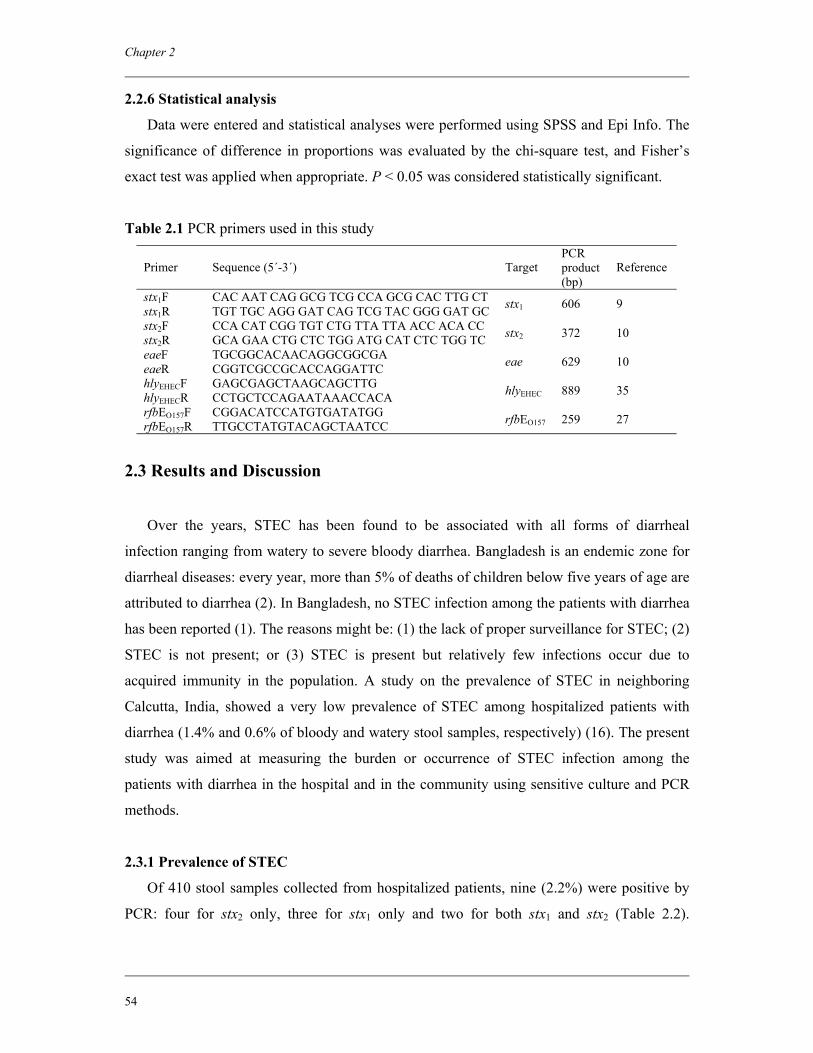
Chapter 2
54
2.2.6 Statistical analysis
Data were entered and statistical analyses were performed using SPSS and Epi Info. The
significance of difference in proportions was evaluated by the chi-square test, and Fisher’s
exact test was applied when appropriate. P < 0.05 was considered statistically significant.
Table 2.1 PCR primers used in this study
Primer Sequence (5´-3´) Target PCR product (bp)
Reference
stx1F CAC AAT CAG GCG TCG CCA GCG CAC TTG CT stx1R TGT TGC AGG GAT CAG TCG TAC GGG GAT GC stx1 606 9
stx2F CCA CAT CGG TGT CTG TTA TTA ACC ACA CC stx2R GCA GAA CTG CTC TGG ATG CAT CTC TGG TC stx2 372 10
eaeF TGCGGCACAACAGGCGGCGA eaeR CGGTCGCCGCACCAGGATTC eae 629 10
hlyEHECF GAGCGAGCTAAGCAGCTTG hlyEHECR CCTGCTCCAGAATAAACCACA hlyEHEC 889 35
rfbEO157F CGGACATCCATGTGATATGG rfbEO157R TTGCCTATGTACAGCTAATCC rfbEO157 259 27
2.3 Results and Discussion
Over the years, STEC has been found to be associated with all forms of diarrheal
infection ranging from watery to severe bloody diarrhea. Bangladesh is an endemic zone for
diarrheal diseases: every year, more than 5% of deaths of children below five years of age are
attributed to diarrhea (2). In Bangladesh, no STEC infection among the patients with diarrhea
has been reported (1). The reasons might be: (1) the lack of proper surveillance for STEC; (2)
STEC is not present; or (3) STEC is present but relatively few infections occur due to
acquired immunity in the population. A study on the prevalence of STEC in neighboring
Calcutta, India, showed a very low prevalence of STEC among hospitalized patients with
diarrhea (1.4% and 0.6% of bloody and watery stool samples, respectively) (16). The present
study was aimed at measuring the burden or occurrence of STEC infection among the
patients with diarrhea in the hospital and in the community using sensitive culture and PCR
methods.
2.3.1 Prevalence of STEC
Of 410 stool samples collected from hospitalized patients, nine (2.2%) were positive by
PCR: four for stx2 only, three for stx1 only and two for both stx1 and stx2 (Table 2.2).
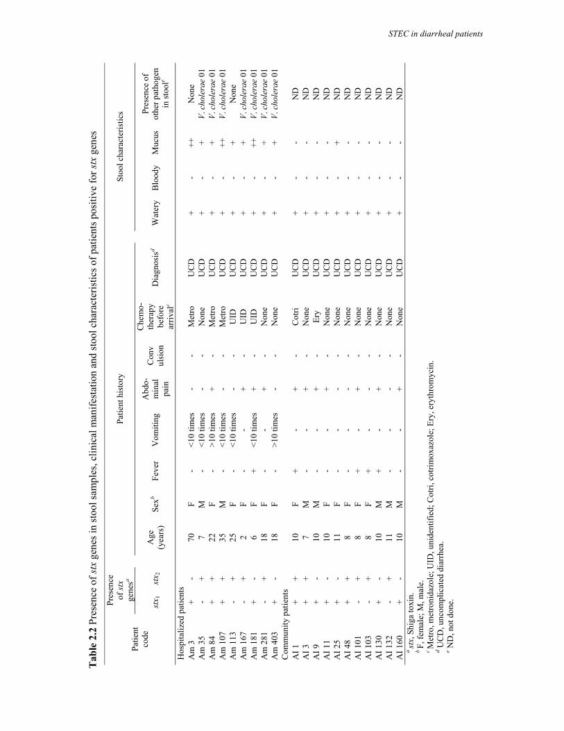
Tab
le 2
.2 P
rese
nce
of st
x ge
nes i
n st
ool s
ampl
es, c
linic
al m
anife
stat
ion
and
stoo
l cha
ract
eris
tics o
f pat
ient
s pos
itive
for s
tx g
enes
Pr
esen
ce
of st
x ge
nesa
Pa
tient
his
tory
Stoo
l cha
ract
eris
tics
Patie
nt
code
st
x 1
stx 2
Age
(y
ears
) Se
xb Fe
ver
Vom
iting
A
bdo-
min
al
pain
Con
vul
sion
Che
mo-
ther
apy
befo
re
arriv
alc
Dia
gnos
isd
W
ater
y B
lood
y M
ucus
Pr
esen
ce o
f ot
her p
atho
gen
in st
oole
Hos
pita
lized
pat
ient
s
A
m 3
+
-
70
F -
<10
times
-
- M
etro
U
CD
+ -
++
Non
e A
m 3
5 -
+
7 M
-
<10
times
-
- N
one
UC
D
+
- +
V. c
hole
rae
01
Am
84
+ +
22
F
- >1
0 tim
es
+ -
Met
ro
UC
D
+
- +
V. c
hole
rae
01
Am
107
+
+
35
M
- <1
0 tim
es
- -
Met
ro
UC
D
+
- ++
V.
cho
lera
e 01
A
m 1
13
- +
25
F
- <1
0 tim
es
- -
UID
U
CD
+ -
+ N
one
Am
167
-
+
2 F
- -
+ -
UID
U
CD
+ -
+ V.
cho
lera
e 01
A
m 1
81
+ -
6
F +
<10
times
+
- U
ID
UC
D
+
- ++
V.
cho
lera
e 01
A
m 2
81
- +
18
F
- -
+ -
Non
e U
CD
+ -
+ V.
cho
lera
e 01
A
m 4
03
+ -
18
F
- >1
0 tim
es
- -
Non
e U
CD
+ -
+ V.
cho
lera
e 01
C
omm
unity
pat
ient
s
A
I 1
+ +
10
F
+ -
+ -
Cot
ri U
CD
+ -
- N
D
AI 3
+
+
7 M
-
- +
- N
one
UC
D
+
- -
ND
A
I 9
+ -
10
M
-
- +
- Er
y U
CD
+ -
- N
D
AI 1
1 +
-
10
F -
- +
- N
one
UC
D
+
- -
ND
A
I 25
+ -
11
F
- -
- -
Non
e U
CD
+ -
+ N
D
AI 4
8 +
+
8 F
- -
- -
Non
e U
CD
+ -
- N
D
AI 1
01
- +
8
F +
- +
- N
one
UC
D
+
- -
ND
A
I 103
-
+
8 F
+ -
- -
Non
e U
CD
+ -
- N
D
AI 1
30
+ -
10
M
+
- +
- N
one
UC
D
+
- -
ND
A
I 132
-
+
11
M
- -
- -
Non
e U
CD
+ -
- N
D
AI 1
60
+ -
10
M
-
- +
- N
one
UC
D
+
- -
ND
a st
x, S
higa
toxi
n.
b F,
fem
ale;
M, m
ale.
c M
etro
, met
roni
dazo
le; U
ID, u
nide
ntifi
ed; C
otri,
cot
rimox
azol
e; E
ry, e
ryth
rom
ycin
.
d UC
D, u
ncom
plic
ated
dia
rrhe
a.
e N
D, n
ot d
one.
STEC in diarrheal patients
Chapter 2
56
No statistically significant correlation between the age of patients and the presence of stx
genes was observed. All stx positive patients were clinically diagnosed as having
uncomplicated diarrhea. All patients had watery diarrhea and mucus was present in the stool
samples. Vomiting was recorded for seven patients (78%). Interestingly, seven of the nine
patients were primarily diagnosed with a V. cholerae O1 infection (Table 2.2). The relative
frequency of other enteric pathogens detected in the hospital samples is shown in Table 2.3.
Table 2.3 Prevalence of other bacterial enteropathogens in stool samples from hospitalized
patients examined for Shiga toxin-producing E. coli Enteropathogen Prevalence (%) Vibrio cholerae O1 El Tor Inaba 19.0 Vibrio cholerae O1 El Tor Ogawa 18.0 Shigella flexneri 2.2 Shigella boydii 0.7 Shigella dysenteriae 0.2 Salmonella Typhi 0.2 Salmonella paratyphi B 0.2 Salmonella Group C1 0.7 Salmonella Group D 0.2 Salmonella Group C2 0.2 STEC O157 0.0 STEC non-O157 0.5
Of 160 samples collected from the community patients with diarrhea, 11 (6.9%) were
found to be positive: five for stx1, three for stx2 and three for both stx1 and stx2 (Table 2.2).
All patients had mild diarrhea and were diagnosed as having uncomplicated diarrhea. STEC
could be isolated from two (22%) among nine PCR positive samples from the hospitalized
patients and three (27%) of 11 PCR positive samples from community patients. The
prevalence of STEC in hospitalized patients with diarrhea was very low (0.5%), and was
somewhat higher among the community patients with diarrhea (1.9%). Failure to isolate the
STEC non-O157 from PCR positive stool samples is still a common problem encountered by
researchers around the world (5). According to previous studies, the nonisolation of viable
STEC from PCR-positive samples might be due to the presence of very low numbers of
bacteria (16), the presence of free stx phages in the sample (15) and loss of stx genes upon
subcultivation of strains (12). In routine diagnostics, there is no definitive biochemical
characteristic, such as sorbitol fermentation in the case of serogroup O157, which can
identify STEC irrespective of serotypes from the commensal flora. Therefore, selection of
suitable culture media for isolation of STEC non-O157 serogroups is always difficult. In this
study, we used TBX agar; this medium allows basic differentiation of E. coli colonies but
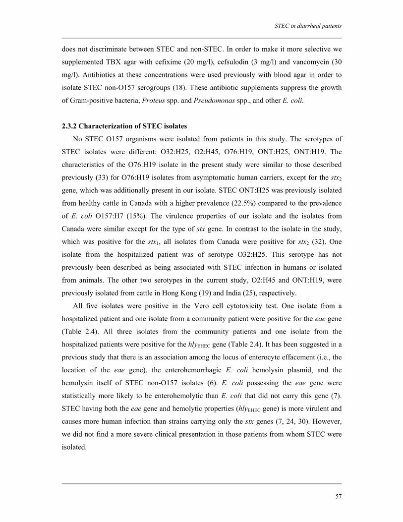
STEC in diarrheal patients
57
does not discriminate between STEC and non-STEC. In order to make it more selective we
supplemented TBX agar with cefixime (20 mg/l), cefsulodin (3 mg/l) and vancomycin (30
mg/l). Antibiotics at these concentrations were used previously with blood agar in order to
isolate STEC non-O157 serogroups (18). These antibiotic supplements suppress the growth
of Gram-positive bacteria, Proteus spp. and Pseudomonas spp., and other E. coli.
2.3.2 Characterization of STEC isolates
No STEC O157 organisms were isolated from patients in this study. The serotypes of
STEC isolates were different: O32:H25, O2:H45, O76:H19, ONT:H25, ONT:H19. The
characteristics of the O76:H19 isolate in the present study were similar to those described
previously (33) for O76:H19 isolates from asymptomatic human carriers, except for the stx2
gene, which was additionally present in our isolate. STEC ONT:H25 was previously isolated
from healthy cattle in Canada with a higher prevalence (22.5%) compared to the prevalence
of E. coli O157:H7 (15%). The virulence properties of our isolate and the isolates from
Canada were similar except for the type of stx gene. In contrast to the isolate in the study,
which was positive for the stx1, all isolates from Canada were positive for stx2 (32). One
isolate from the hospitalized patient was of serotype O32:H25. This serotype has not
previously been described as being associated with STEC infection in humans or isolated
from animals. The other two serotypes in the current study, O2:H45 and ONT:H19, were
previously isolated from cattle in Hong Kong (19) and India (25), respectively.
All five isolates were positive in the Vero cell cytotoxicity test. One isolate from a
hospitalized patient and one isolate from a community patient were positive for the eae gene
(Table 2.4). All three isolates from the community patients and one isolate from the
hospitalized patients were positive for the hlyEHEC gene (Table 2.4). It has been suggested in a
previous study that there is an association among the locus of enterocyte effacement (i.e., the
location of the eae gene), the enterohemorrhagic E. coli hemolysin plasmid, and the
hemolysin itself of STEC non-O157 isolates (6). E. coli possessing the eae gene were
statistically more likely to be enterohemolytic than E. coli that did not carry this gene (7).
STEC having both the eae gene and hemolytic properties (hlyEHEC gene) is more virulent and
causes more human infection than strains carrying only the stx genes (7, 24, 30). However,
we did not find a more severe clinical presentation in those patients from whom STEC were
isolated.
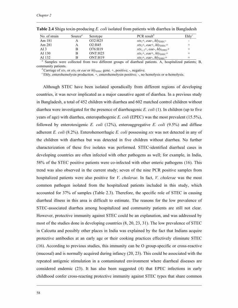
Chapter 2
58
Table 2.4 Shiga toxin-producing E. coli isolated from patients with diarrhea in Bangladesh No. of strain Sourcea Serotype PCR resultb Ehlyc Am 181 A O32:H25 stx1+, eae-, hlyEHEC- - Am 281 A O2:H45 stx2+, eae+, hlyEHEC+ + AI 3 B O76:H19 stx1, 2+, eae-, hlyEHEC+ + AI 130 B ONT:H25 stx1+, eae+, hlyEHEC+ + AI 132 B ONT:H19 stx2+, eae-, hlyEHEC+ +
a Samples were collected from two different groups of diarrheal patients. A, hospitalized patients; B, community patients. b Carriage of stx1 or stx2 or eae or hlyEHEC gene. +, positive; -, negative. c Ehly, enterohemolysin production. +, enterohemolysin positive; -, no hemolysis or α-hemolysis.
Although STEC have been isolated sporadically from different regions of developing
countries, it was never implicated as a major causative agent of diarrhea. In a previous study
in Bangladesh, a total of 452 children with diarrhea and 602 matched control children without
diarrhea were investigated for the presence of diarrheagenic E. coli (1). In children (up to five
years of age) with diarrhea, enteropathogenic E. coli (EPEC) was the most prevalent (15.5%),
followed by enterotoxigenic E. coli (12%), enteroaggregative E. coli (9.5%) and diffuse
adherent E. coli (8.2%). Enterohemorrhagic E. coli possessing stx was not detected in any of
the children with diarrhea but was detected in five children without diarrhea. No further
characterization of these five isolates was performed. STEC-identified diarrheal cases in
developing countries are often infected with other pathogens as well; for example, in India,
58% of the STEC positive patients were co-infected with other enteric pathogens (16). This
trend was also observed in the current study; seven of the nine PCR positive samples from
hospitalized patients were also positive for V. cholerae. In fact, V. cholerae was the most
common pathogen isolated from the hospitalized patients included in this study, which
accounted for 37% of samples (Table 2.3). Therefore, the specific role of STEC in causing
diarrheal illness in this area is difficult to estimate. The reasons for the low prevalence of
STEC-associated diarrhea among hospitalized and community patients are still not clear.
However, protective immunity against STEC could be an explanation, and was addressed by
most of the studies done in developing countries (8, 20, 23, 31). The low prevalence of STEC
in Calcutta and possibly other places in India was explained by the fact that Indians acquire
protective antibodies at an early age or their cooking practices effectively eliminate STEC
(16). According to previous studies, this immunity can be O group-specific or cross-reactive
(mucosal) and is normally acquired during infancy (20, 23). This could be associated with the
repeated antigenic stimulation in a contaminated environment where diarrheal diseases are
considered endemic (23). It has also been suggested (4) that EPEC infections in early
childhood confer cross-reacting protective immunity against STEC types that share common
STEC in diarrheal patients
59
antigens (such as LPS and intimin) with classical EPEC strains. However, a detailed study of
the immune status of patients with diarrhea as well as healthy controls in areas where enteric
pathogens are considered to be endemic should be carried out in order to explain these
phenomena.
Acknowledgements
This research was funded by the ICDDR,B: Centre for Health and Population Research
and by the Netherlands Foundation for the Advancement of Tropical Research (NWO-
WOTRO) grant (award number WB 93-415). ICDDR,B acknowledges with gratitude the
commitment of the Netherlands Foundation for the Advancement of Tropical Research
(NWO-WOTRO) grant to the Centre’s research efforts. ICDDR,B also gratefully
acknowledges these donors who provide unrestricted support to the Centre’s research efforts:
Australian International Development Agency (AusAID), Government of Bangladesh,
Canadian International Development Agency (CIDA), The Kingdom of Saudi Arabia (KSA),
Government of the Netherlands, Government of Sri Lanka, Swedish International
Development Cooperative Agency (SIDA), Swiss Development Cooperation (SDC) and
Department for International Development, UK (DFID).
References
1. Albert, M. J., S. M. Faruque, A. S. Faruque, P. K. Neogi, M. Ansaruzzaman, N. A. Bhuiyan, K. Alam, and M. S. Akbar. 1995. Controlled study of Escherichia coli diarrheal infections in Bangladeshi children. J. Clin. Microbiol. 33:973–977.
2. Arifeen, S. E., T. Akhter, H. R. Chowdhury, K. M. Rahman, and E. K. Chowdhury. 2005. Causes of death in children under five years of age, p. 125–133. In National Institute of Population Research and Training (NIPORT), Bangladesh Demographic and Health Survey 2004. National Institute of Population Research and Training, Dhaka, Bangladesh.
3. Beutin, L., M. A. Montenegro, I. Orskov, F. Orskov, J. Prada, S. Zimmermann, and R. Stephan. 1989. Close association of verotoxin (Shiga-like toxin) production with enterohemolysin production in strains of Escherichia coli. J. Clin. Microbiol. 27:2559–2564.
4. Beutin, L., S. Zimmermann, and K. Gleier. 1998. Human infections with Shiga toxin-producing Escherichia coli other than serogroup O157 in Germany. Emerg. Infect. Dis. 4:635–639.
5. Blanco, J. E., M. Blanco, M. P. Alonso, A. Mora, G. Dahbi, M. A. Coira, and J. Blanco. 2004. Serotypes, virulence genes, and intimin types of Shiga toxin
Chapter 2
60
(verotoxin)-producing Escherichia coli isolates from human patients: prevalence in Lugo, Spain, from 1992 through 1999. J. Clin. Microbiol. 42:311–319.
6. Boerlin, P., S. Chen, J. K. Colbourne, R. Johnson, S. De Grandis, and C. L. Gyles. 1998. Evolution of enterohemorrhagic Escherichia coli hemolysin plasmids and the locus for enterocyte effacement in Shiga toxin-producing E. coli. Infect. Immun. 66:2553–2561.
7. Eklund, M., F. Scheutz, and A. Siitonen. 2001. Clinical isolates of non-O157 Shiga toxin-producing Escherichia coli: serotypes, virulence characteristics, and molecular profiles of strains of the same serotype. J. Clin. Microbiol. 39:2829–2834.
8. Gianantonio, C., M. Vitacco, F. Mendilaharzu, A. Rutty, and J. Mendilaharzu 1964. The haemolytic-uraemic syndrome. J. Pediatr. 64:478–491.
9. Heuvelink, A. E., E. de Boer, and K. Wernars. 1994. Detection of Escherichia coli O157:H7 in raw beef by polymerase chain reaction. De Ware(n)- Chemicus 24:25–43 (in Dutch).
10. Heuvelink, A. E., N. C. van de Kar, J. F. Meis, L. A. Monnens, and W. J. Melchers. 1995. Characterization of verocytotoxin-producing Escherichia coli O157 isolates from patients with haemolytic uraemic syndrome in Western Europe. Epidemiol. Infect. 115:1–14.
11. Kaddu-Mulindw, D. H., T. Aisu, K. Gleier, S. Zimmermann, and L. Beutin. 2001. Occurrence of Shiga toxin-producing Escherichia coli in fecal samples from children with diarrhoea and from healthy zebu cattle in Uganda. Int. J. Food Microbiol. 66:95–101.
12. Karch, H., T. Meyer, H. Russmann, and J. Heesemann. 1992. Frequent loss of Shiga-like toxin genes in clinical isolates of Escherichia coli upon subcultivation. Infect. Immun. 60:3464–3467.
13. Karmali, M. A. 1989. Infection by verotoxin-producing Escherichia coli. Clin. Microbiol. Rev. 2:15–38.
14. Karmali, M. A., M. Petric, C. Lim, P. C. Fleming, G. S. Arbus, and H. Lior. 1985. The association between idiopathic hemolytic uremic syndrome and infection by verotoxin-producing Escherichia coli. J. Infect. Dis. 151:775–782.
15. Karmali, M. A., M. Petric, C. Lim, R. Cheung, and G. S. Arbus. 1985. Sensitive method for detecting low numbers of verotoxin-producing Escherichia coli in mixed cultures by use of colony sweeps and polymyxin extraction of verotoxin. J. Clin. Microbiol. 22:614–619.
16. Khan, A., S. Yamasaki, T. Sato, T. Ramamurthy, A. Pal, S. Datta, N. R. Chowdhury, S. C. Das, A. Sikdar, T. Tsukamoto, S. K. Bhattacharya, Y. Takeda, and G. B. Nair. 2002. Prevalence and genetic profiling of virulence determinants of non-O157 Shiga toxin-producing Escherichia coli isolated from cattle, beef, and humans, Calcutta, India. Emerg. Infect. Dis. 8:54–62.
17. Leelaporn, A., M. Phengmak, B. Eampoklap, S. Manatsathit, S. Tritilanunt, S. Siritantikorn, K. Nagayama, T. Iida, C. Niyasom, and P. Komolpit. 2003. Shiga toxin- and enterotoxin-producing Escherichia coli isolated from subjects with bloody and non-bloody diarrhea in Bangkok, Thailand. Diagn. Microbiol. Infect. Dis. 46:173–180.
18. Lehmacher, A., H. Meier, S. Aleksic, and J. Bockemuhl. 1998. Detection of hemolysin variants of Shiga toxin-producing Escherichia coli by PCR and culture on vancomycin-cefixime-cefsulodin blood agar. Appl. Environ. Microbiol. 64:2449–2453.
STEC in diarrheal patients
61
19. Leung, P. H., J. S. Peiris, W. W. Ng, R. M. Robins-Browne, K. A. Bettelheim, and W. C. Yam. 2003. A newly discovered verotoxin variant, VT2g, produced by bovine verocytotoxigenic Escherichia coli. Appl. Environ. Microbiol. 69:7549–7553.
20. Lopez, E. L., M. Diaz, S. Grinstein, S. Devoto, F. Mendilaharzu, B. E. Murray, S. Ashkenazi, E. Rubeglio, M. Woloj, and M. Vasque. 1989. Hemolytic uremic syndrome and diarrhea in Argentine children: the role of Shiga-like toxins. J. Infect. Dis. 160:469–475.
21. Malorny, B., J. Hoorfar, M. Hugas, A. Heuvelink, P. Fach, L. Ellerbroek, C. Bunge, C. Dorn, and R. Helmuth. 2003. Interlaboratory diagnostic accuracy of Salmonella specific PCR based method. Int. J. Food Microbiol. 89:241–249.
22. Nataro, J. P., and J. B. Kaper. 1998. Diarrheagenic Escherichia coli. Clin. Microbiol. Rev. 11:142–201.
23. Navarro, A., C. Eslava, U. Hernandez, J. L. Navarro-Henze, M. Aviles, G. Garcia-de la Torre, and A. Cravioto. 2003. Antibody responses to Escherichia coli O157 and other lipopolysaccharides in healthy children and adults. Clin. Diagn. Lab. Immunol. 10:797–801.
24. Oswald, E., H. Schmidt, S. Morabito, H. Karch, O. Marches, and A. Caprioli. 2000. Typing of intimin genes in human and animal enterohemorrhagic and enteropathogenic Escherichia coli: characterization of a new intimin variant. Infect. Immun. 68:64–71.
25. Pal, A., S. Ghosh, T. Ramamurthy, S. Yamasaki, T. Tsukamoto, S. K. Bhattacharya, G. B. Nair, and Y. Takeda. 1999. Shiga toxin-producing Escherichia coli from healthy cattle in a semi-urban community in Calcutta, India. Indian J. Med. Res. 110:83–85.
26. Paton, J. C., and A. W. Paton. 1998. Pathogenesis and diagnosis of Shiga toxin-producing Escherichia coli infections. Clin. Microbiol. Rev. 11:450–479.
27. Paton, J. C., and A. W. Paton. 1998. Detection and characterization of Shiga toxigenic Escherichia coli by using multiplex PCR assays for stx1, stx2, eaeA, enterohemorrhagic E. coli hlyA, rfbO111, and rfbO157. J. Clin. Microbiol. 36:598–602.
28. Pradel, N., V. Livrelli, C. De Champs, J. B. Palcox, A. Reynaud, F. Scheutz, J. Sirot, B. Joly, and C. Forestier. 2000. Prevalence and characterization of Shiga toxin-producing Escherichia coli isolated from cattle, food, and children during a one-year prospective study in France. J. Clin. Microbiol. 38:1023–1031.
29. Qadri, F., A. M. Svennerholm, A. S. Faruque, and R. B. Sack. 2005. Enterotoxigenic Escherichia coli in developing countries: epidemiology, microbiology, clinical features, treatment and prevention. Clin. Microbiol. Rev. 18:465–483.
30. Schmidt, H., L. Beutin, and H. Karch. 1995. Molecular analysis of the plasmid-encoded hemolysin of Escherichia coli O157:H7 strain EDL 933. Infect. Immun. 63:1055–1061.
31. Seriwatana, J., J. E. Brown, P. Echeverria, D. N. Taylor, O. Suthienkul, and J. Newland. 1988. DNA probes to identify Shiga-like toxin I- and II-producing enteric bacterial pathogens isolated from patients with diarrhea in Thailand. J. Clin. Microbiol. 26:1614–1615.
32. Sheng, H., M. A. Davis, H. J. Knecht, D. D. Hancock, J. Van Donkersgoed, and C. J. Hovde. 2005. Characterization of a Shiga toxin-, intimin-, and enterotoxin hemolysin-producing Escherichia coli ONT:H25 strain commonly isolated from healthy cattle. J. Clin. Microbiol. 43:3213–3220.
Chapter 2
62
33. Stephan, R., and F. Untermann. 1999. Virulence factors and phenotypical traits of verotoxin-producing Escherichia coli strains isolated from asymptomatic human carriers. J. Clin. Microbiol. 37:1570–1572.
34. Tarr, P. I., C. A. Gordon, and W. L. Chandler. 2005. Shiga toxin-producing Escherichia coli and haemolytic uraemic syndrome. Lancet 365:1073–1086.
35. Wieler, L. H., M. Tigges, F. Ebel, S. Schaferkordt, S. Djafari, T. Schlapp, G. Baljer, and T. Chakraborty. 1996. The enterohemolysin phenotype of bovine Shiga-like toxin-producing Escherichia coli (SLTEC) is encoded by the EHEC-hemolysin gene. Vet. Microbiol. 52:153–164.
36. World Health Organization.1987. Programme for control of diarrhoeal diseases, p. 9–20. In Manual for Laboratory Investigation of Acute Enteric Infections. CDD/93.3, rev.1. World Health Organization, Geneva, Switzerland.
Immunoconcentration of Shiga toxin-producing
Escherichia coli O157 from animal feces and raw
meats by using Dynabeads anti-E. coli O157 and the
VIDAS system
International Journal of Food Microbiology 2006; 109:151–156
M. Aminul Islam A. E. Heuvelink K. A. Talukder E. de Boer
Chapter 3
Chapter 3
64
Abstract
To identify the reservoirs and routes of transmission of Shiga toxin-producing
Escherichia coli (STEC) O157, sensitive detection and isolation methods are necessary. The
sensitivity of traditional culture methods can be improved significantly by the inclusion of an
immunoconcentration step, resulting in less false-negatives. In this report, we evaluated the
results of two commercially available test systems: Dynabeads anti-E. coli O157 and the
Vitek Immunodiagnostic Assay System (VIDAS) Immuno-Concentration E. coli O157 (ICE)
kit. Additionally, we compared two selective isolation media for STEC O157. Statistical
analysis of the results obtained for animal fecal samples (n = 637) examined by both
immunoconcentration methods showed that by the manual Dynabeads anti-E. coli O157
procedure systematically more samples were identified as positive than by the VIDAS ICE.
In case of meat samples (n = 360), no difference between the results of the two methods was
found. In addition to being accurate, the Dynabeads anti-E. coli O157 method is a less
expensive method than the VIDAS ICE. But, the Dynabeads method is laborious and there is
a risk of cross-contamination. The VIDAS ICE procedure on the other hand is fully
automated with a standardized performance; fast and safe for the user. Irrespective of the type
of sample (feces or meat) and the immunoconcentration technique applied (Dynabeads anti-
E. coli O157 or VIDAS ICE) more samples were found positive after plating onto
CHROMagar O157 with cefixime (0.025 mg/l) and tellurite (1.25 mg/l) than after plating
onto sorbitol–MacConkey agar with cefixime (0.05 mg/l) and tellurite (2.5 mg/l). However,
only in case of meat samples examined by the VIDAS ICE the difference between the
isolation media was not statistically significant.
Immunoconcentration of STEC O157 by using Dynabeads anti-E. coli O157 and the VIDAS system
65
3.1 Introduction
Since the early 1980s, it is known that an infection with Shiga toxin-producing
Escherichia coli (STEC) strains of serogroup O157 are the major cause of hemorrhagic
colitis and the hemolytic–uremic syndrome (5, 9, 14). The infections frequently have been
associated with the consumption of undercooked (minced) beef and raw milk (15).
Epidemiological studies have shown that cattle, but also other farm animals can excrete the
bacteria with their feces, without showing any clinical symptoms (11). In addition to (fecally
contaminated) foods, direct transmission from animal to human can result in infection (13).
To elucidate further the reservoirs of STEC O157, sensitive methods are needed since
these pathogens may be present in food and environmental samples in only small numbers.
Furthermore, sensitive and rapid detection methods are necessary for the food industry to
ensure a safe supply of foods. Conventional culture methods are both time-consuming and
laborious. Their sensitivity can significantly be increased by the application of an
immunoconcentration step, which will result in less false-negatives. A commonly used
method, which is also implemented in the international standard for the detection of E. coli
O157 in food and feeding stuffs (2), is immunomagnetic separation (IMS). Prior to plating
onto selective isolation media, E. coli O157 cells present in the enrichment culture are
selectively concentrated by magnetic beads with E. coli O157-specific antibodies covalently
bound onto their surface. The IMS procedure can be performed both manually and
automatically. Another example of immunoconcentration is the fully automated Vitek
Immunodiagnostic Assay System (VIDAS) (bioMérieux, Marcy l'Etoile, France). The
VIDAS Immuno-Concentration E. coli O157 (ICE) kit includes two ready-to-use
components. One is a Solid Phase Receptacle (SPR), which serves as the solid phase as well
as the pipetting device for the assay. The interior of the SPR is coated with anti-E. coli O157
antibodies adsorbed on its surface. The other component is a strip which contains all the wash
and release solutions. An aliquot of the enrichment culture is manually transferred into the
strip and subsequently the sample is cycled in and out of the SPR. E. coli O157 present in the
broth will bind to the anti-E. coli O157 antibodies coating the interior of the SPR. Unbound
sample components are then washed away. A final enzymatic step releases the captured E.
coli O157 into a specific well from which they can be plated onto selective agar.
In our laboratory, samples of animal feces and meats had been examined regularly for the
presence of STEC O157 by using a manual IMS procedure as well as the VIDAS ICE
Chapter 3
66
system. In this study, we retrospectively evaluated the performance of both methods.
Additionally, two selective agars used to finally isolate STEC O157 were compared.
3.2 Materials and methods
3.2.1 Samples
The following samples were examined for the presence of STEC O157 by performing a
manual IMS procedure as well as the VIDAS ICE system: (1) feces from farm animals that
previously tested positive and had been stored at -20°C (n = 144); (2) rectal contents of
different farm animals in petting zoos sampled individually by rectal palpation (n = 73); (3)
fecal droppings from different animals collected at different petting zoos (n = 95); (4) rectal
contents of sheep collected at the slaughterline (n = 325); (5) swab samples from sheep
carcasses (n = 325); and (6) different retail raw meats that previously tested positive and had
been stored at -20°C (n = 35).
For the comparison of two selective isolation media, in addition to the results obtained for
the samples listed above, results of another 679 samples of animal feces were included, which
were analyzed with either IMS or VIDAS ICE. These samples, both rectal contents and
droppings, had been collected from different animals at petting zoos and cattle farms, either
in order to trace sources of human infection (n = 177) or to study the occurrence of STEC
O157 in petting zoos (n = 502).
With the exception of the positive feces and meat samples that were kept in the freezer,
all samples were fresh and the microbiological examination had been started within 72 h after
their collection. As a rule, the carcass swabs were processed within the day of sampling.
3.2.2 Selective enrichment
The samples were, if applicable after being thawed at 2 to 4°C, diluted 10 times in
modified tryptone soy broth (Oxoid Ltd., Basingstoke, United Kingdom) supplemented with
novobiocin (20 mg/l) (Sigma Chemical Co., St. Louis, MO) (mTSB+n). To the plastic
stomacher bags containing the sponges used to sample the sheep carcasses, 90 ml of mTSB
without novobiocin was added. After homogenization (1 min) in a stomacher, the samples
were incubated at 41.5±0.5°C for 18 to 24 h. In case of the sheep feces collected at slaughter,
after 6 to 8 h of incubation about 5 ml of enrichment culture was taken for the purpose of the
IMS procedure. For all the other analyzes overnight cultures were used.
Immunoconcentration of STEC O157 by using Dynabeads anti-E. coli O157 and the VIDAS system
67
3.2.3 Immunoconcentration
3.2.3.1 Dynabeads anti-E. coli O157
For the IMS procedure Dynabeads anti-E. coli O157 (Dynal, Oslo, Norway) were used
and the instructions of the manufacturer were followed.
3.2.3.2 VIDAS Immuno-Concentration E. coli O157
The manufacturer of the VIDAS ICE kit (bioMérieux, Marcy l'Etoile, France) prescribes
a first selective enrichment of the sample in mTSB+n (6–7 h at 41±1°C) followed by a
second selective enrichment (1:10 dilution) in MacConkey broth supplemented with cefixime
(0.05 mg/l) and tellurite (2.5 mg/l) (18±1 h at 35–37°C) before testing with the VIDAS ICE
kit. Deviating from this protocol, 500 µl from the overnight cultures in mTSB+n was pipetted
into the strips. This choice has been made based upon the results of an extensive study on
methods for detection and isolation of E. coli O157, previously performed in our laboratory
(16).
3.2.4 Selective isolation and confirmation
The immunoconcentrated samples were inoculated onto two selective isolation media:
sorbitol–MacConkey agar (Oxoid) supplemented with cefixime (0.05 mg/l) and tellurite (2.5
mg/l) (Oxoid) (CT-SMAC) and CHROMagar O157 (ITK Diagnostics BV, Uithoorn, The
Netherlands) with cefixime (0.025 mg/l) and tellurite (1.25 mg/l) (1/2CT-CHROMagar). The
agar plates were incubated for 18 to 24 h at 37°C. Typical colonies (colorless colonies on CT-
SMAC and purple on 1/2CT-CHROMagar), up to 12 per plate, were selected and inoculated
onto Levine's eosin methylene blue agar (Oxoid) (L-EMB) agar and sorbitol–MacConkey
agar with 4-methylumbelliferyl-β-D-glucuronide (0.1 g/l) (Sigma) (SMAC-MUG) agar. The
plates were read after 18 to 24 h of incubation at 37°C. Presumptive STEC O157 isolates
(those with a typical E. coli metallic sheen on L-EMB, being both sorbitol-nonfermenting and
β-glucuronidase-negative on SMAC-MUG) were tested for agglutination with an E. coli
O157 latex test kit (Murex Biotech Ltd., Dartford, Kent, United Kingdom). Isolates that gave
a positive latex test result were confirmed to be E. coli by using an API 20E biochemical test
strip (bioMérieux). Finally, the isolates were confirmed with a number of PCR tests specific
for identification of several characteristics of STEC O157, including the presence of stx genes
(stx1 and stx2) (7), the eae gene (7), the enterohemolysin gene (hlyEHEC) (17) and a specific
portion of the rfb (O-antigen encoding) region of E. coli O157 lipopolysaccharide (rfbEO157)
(12).
Chapter 3
68
3.2.5 Statistical analysis
Since the international standard for the detection of E. coli O157 is based on IMS, the
Dynabeads anti-E. coli O157 procedure was considered as the reference method for the
statistical analysis. The relative sensitivity, specificity and diagnostic accuracy were
calculated with the following formulas (3):
Relative sensitivity=100%×(no. of samples positive with both methods)/(total
no. of samples positive with the Dynabeads anti-E. coli O157 method).
Relative specificity=100%×(no. of samples negative with both methods)/(total
no. of samples negative with the Dynabeads anti-E. coli O157 method).
Relative accuracy=100%×(no. of samples positive and negative with both
methods)/(total no. of samples examined by both methods).
The diagnostic accuracy takes into account the target and non-target microorganisms in
the presence of a biological matrix and consists of the terms sensitivity and specificity (10).
The relative sensitivity is the ability of the alternative method (here the VIDAS ICE) to detect
the target when it is also detected with the reference method. The relative specificity is the
ability of the alternative method not to detect the target organism when it is also not detected
with the reference method. The relative accuracy is the degree of correspondence between the
responses obtained by the alternative method and the reference method on identical samples.
Finally, it was calculated whether the methods were statistically significantly different,
according to Annex F of EN ISO 16140:2003(E) (3). The same calculations were performed
to compare the isolation media CT-SMAC and 1/2CT-CHROMagar using CT-SMAC as the
reference.
3.3 Results and discussion
Since their first identification as an enteropathogen, strains of STEC O157 have emerged
as a serious threat to public health, especially in developed countries (15). Various factors
may affect the prevalence findings of E. coli O157 in different kind of samples, of which the
method of detection and isolation has a major impact. A total of 997 samples had been
examined by both the Dynabeads anti-E. coli O157 method and the VIDAS ICE. Of the 637
fecal samples, 50 (7.8%) were found positive: 29 with both methods, 17 only with the
Dynabeads anti-E. coli O157 method, and four only with the VIDAS ICE. Therefore, by
using the Dynabeads anti-E. coli O157 method systematically more samples were identified
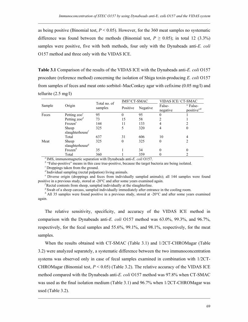
Immunoconcentration of STEC O157 by using Dynabeads anti-E. coli O157 and the VIDAS system
69
as being positive (Binomial test, P < 0.05). However, for the 360 meat samples no systematic
difference was found between the methods (Binomial test, P ≥ 0.05); in total 12 (3.3%)
samples were positive, five with both methods, four only with the Dynabeads anti-E. coli
O157 method and three only with the VIDAS ICE.
Table 3.1 Comparison of the results of the VIDAS ICE with the Dynabeads anti-E. coli O157
procedure (reference method) concerning the isolation of Shiga toxin-producing E. coli O157
from samples of feces and meat onto sorbitol–MacConkey agar with cefixime (0.05 mg/l) and
tellurite (2.5 mg/l) IMSa/CT-SMAC VIDAS ICE/ CT-SMAC
Sample Origin Total no. of samples Positive Negative False-
negative “ False- positive”b
Feces Petting zooc 95 0 95 0 1 Petting zood 73 15 58 2 1 Frozene 144 11 133 4 2 Sheep 325 5 320 4 0 slaughterhousef Total 637 31 606 10 4 Meat Sheep 325 0 325 0 2 slaughterhouseg Frozenh 35 1 34 0 0 Total 360 1 359 0 2
a IMS, immunomagnetic separation with Dynabeads anti-E. coli O157. b “False-positive” means in this case true-positive, because the target bacteria are being isolated. c Droppings taken from the ground. d Individual sampling (rectal palpation) living animals. e Diverse origin (droppings and feces from individually sampled animals); all 144 samples were found positive in a previous study, stored at -20°C and after some years examined again. f Rectal contents from sheep, sampled individually at the slaughterline. g Swab of a sheep carcass, sampled individually immediately after entrance in the cooling room. h All 35 samples were found positive in a previous study, stored at -20°C and after some years examined again.
The relative sensitivity, specificity, and accuracy of the VIDAS ICE method in
comparison with the Dynabeads anti-E. coli O157 method was 63.0%, 99.3%, and 96.7%,
respectively, for the fecal samples and 55.6%, 99.1%, and 98.1%, respectively, for the meat
samples.
When the results obtained with CT-SMAC (Table 3.1) and 1/2CT-CHROMagar (Table
3.2) were analyzed separately, a systematic difference between the two immunoconcentration
systems was observed only in case of fecal samples examined in combination with 1/2CT-
CHROMagar (Binomial test, P < 0.05) (Table 3.2). The relative accuracy of the VIDAS ICE
method compared with the Dynabeads anti-E. coli O157 method was 97.8% when CT-SMAC
was used as the final isolation medium (Table 3.1) and 96.7% when 1/2CT-CHROMagar was
used (Table 3.2).
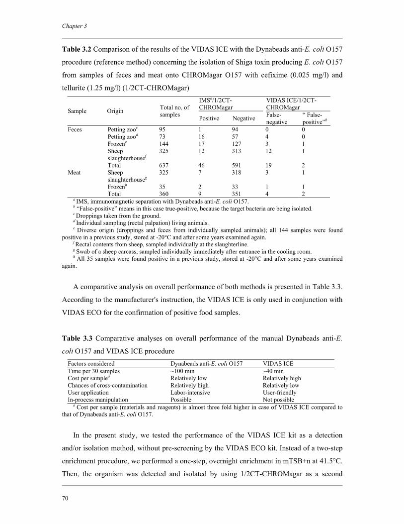
Chapter 3
70
Table 3.2 Comparison of the results of the VIDAS ICE with the Dynabeads anti-E. coli O157
procedure (reference method) concerning the isolation of Shiga toxin producing E. coli O157
from samples of feces and meat onto CHROMagar O157 with cefixime (0.025 mg/l) and
tellurite (1.25 mg/l) (1/2CT-CHROMagar) IMSa/1/2CT- CHROMagar
VIDAS ICE/1/2CT- CHROMagar Sample Origin Total no. of
samples Positive Negative False- negative
“ False- positive”b
Feces Petting zooc 95 1 94 0 0 Petting zood 73 16 57 4 0 Frozene 144 17 127 3 1 Sheep 325 12 313 12 1 slaughterhousef Total 637 46 591 19 2 Meat Sheep 325 7 318 3 1 slaughterhouseg Frozenh 35 2 33 1 1 Total 360 9 351 4 2
a IMS, immunomagnetic separation with Dynabeads anti-E. coli O157. b “False-positive” means in this case true-positive, because the target bacteria are being isolated. c Droppings taken from the ground. d Individual sampling (rectal palpation) living animals. e Diverse origin (droppings and feces from individually sampled animals); all 144 samples were found positive in a previous study, stored at -20°C and after some years examined again. f Rectal contents from sheep, sampled individually at the slaughterline. g Swab of a sheep carcass, sampled individually immediately after entrance in the cooling room. h All 35 samples were found positive in a previous study, stored at -20°C and after some years examined again.
A comparative analysis on overall performance of both methods is presented in Table 3.3.
According to the manufacturer's instruction, the VIDAS ICE is only used in conjunction with
VIDAS ECO for the confirmation of positive food samples.
Table 3.3 Comparative analyses on overall performance of the manual Dynabeads anti-E.
coli O157 and VIDAS ICE procedure Factors considered Dynabeads anti-E. coli O157 VIDAS ICE Time per 30 samples ~100 min ~40 min Cost per samplea Relatively low Relatively high
Chances of cross-contamination Relatively high Relatively low User application Labor-intensive User-friendly In-process manipulation Possible Not possible
a Cost per sample (materials and reagents) is almost three fold higher in case of VIDAS ICE compared to that of Dynabeads anti-E. coli O157.
In the present study, we tested the performance of the VIDAS ICE kit as a detection
and/or isolation method, without pre-screening by the VIDAS ECO kit. Instead of a two-step
enrichment procedure, we performed a one-step, overnight enrichment in mTSB+n at 41.5°C.
Then, the organism was detected and isolated by using 1/2CT-CHROMagar as a second
Immunoconcentration of STEC O157 by using Dynabeads anti-E. coli O157 and the VIDAS system
71
plating medium. Although the VIDAS system has been developed and validated for the
examination of food samples, we also evaluated its performance for animal feces. All the
changes that have been introduced in the VIDAS ICE in the present study were either
preceding or following the immunoconcentration step implying the robustness of the system
on one hand as well as its flexibility on the other. By using the VIDAS ICE, the entire
immunoconcentration process is performed automatically within 40 min, for a maximum of
30 samples (including controls) in one run. The concentration of 30 samples with the
Dynabeads anti-E. coli O157 procedure takes about 100 min (when using two magnetic
particle concentrators, fitting six samples each). The Dynabeads procedure consists of several
handlings, including a number of washing steps to remove non-(specifically) bound bacteria
and matrix components. The risk of cross-contamination is being reduced by the use of
filtertips and so-called tube-openers. Alternatively the BeadRetriever (Dynal) can be used
which is an automated IMS instrument. With the BeadRetriever 15 samples can be tested
within 20 min, not taking into account the time for pipetting the beads and the wash buffer.
Advantages of automated systems like the BeadRetriever and the VIDAS are the standardized
performance, which does not require any manipulation once the assay has started and the
safety of the user.
The international standard for the detection of E. coli O157 prescribes the final
immunoconcentrates being transferred to CT-SMAC and a second selective agar of own
choice (2). In one of our previous studies, CHROMagar O157 turned out to be the best
isolation medium (16). Compared with several other media, STEC O157 colonies could be
best distinguished from background flora on CHROMagar O157. An additional benefit of the
use of chromogenic media is the ability to recognize sorbitol-fermenting STEC O157 strains,
while these cannot be distinguished from nonpathogenic E. coli on media containing sorbitol.
These sorbitol-fermenting strains are increasingly being isolated from patients in Europe and
sporadically in Australia (1, 4, 6, 8). A comparison between the performance of CT-SMAC
and 1/2CT-CHROMagar was done for a total of 1676 samples: 1316 fecal and 360 meat
samples (Table 3.4). In case of feces, systematically more samples were found positive when
1/2CT-CHROMagar was used as the plating medium (McNemar test, P < 0.05), irrespective
of the method of immunoconcentration carried out. For meat samples, the use of 1/2CT-
CHROMagar also resulted in more positives than CT-SMAC. However, this difference
between the two plating media was statistically significant only in combination with the
Dynabeads anti-E. coli O157 (Binomial test, P < 0.05) but not with the VIDAS ICE
(Binomial test, P ≥ 0.05). The relative accuracy of 1/2CT-CHROMagar compared with CT-
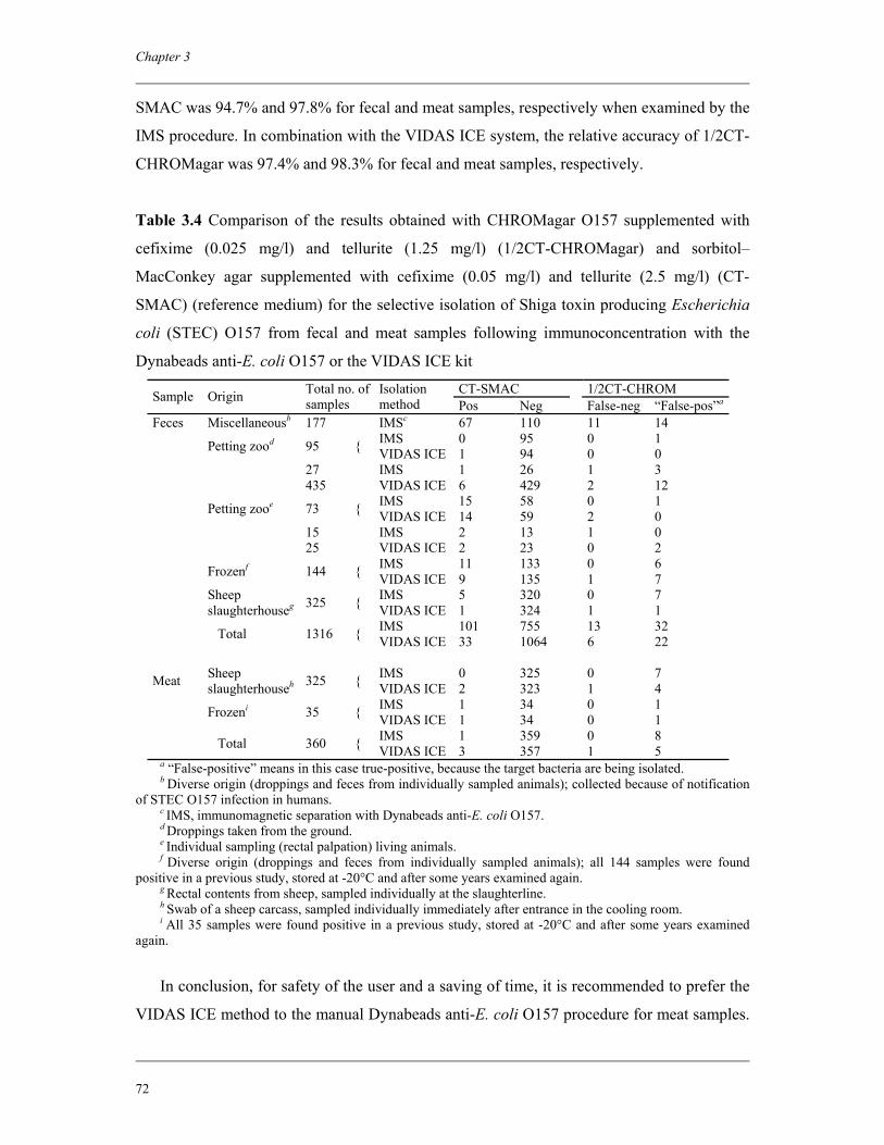
Chapter 3
72
SMAC was 94.7% and 97.8% for fecal and meat samples, respectively when examined by the
IMS procedure. In combination with the VIDAS ICE system, the relative accuracy of 1/2CT-
CHROMagar was 97.4% and 98.3% for fecal and meat samples, respectively.
Table 3.4 Comparison of the results obtained with CHROMagar O157 supplemented with
cefixime (0.025 mg/l) and tellurite (1.25 mg/l) (1/2CT-CHROMagar) and sorbitol–
MacConkey agar supplemented with cefixime (0.05 mg/l) and tellurite (2.5 mg/l) (CT-
SMAC) (reference medium) for the selective isolation of Shiga toxin producing Escherichia
coli (STEC) O157 from fecal and meat samples following immunoconcentration with the
Dynabeads anti-E. coli O157 or the VIDAS ICE kit
CT-SMAC 1/2CT-CHROM Sample Origin Total no. of samples
Isolation method Pos Neg False-neg “False-pos”a
Feces Miscellaneousb 177 IMSc 67 110 11 14 IMS 0 95 0 1 Petting zood 95 { VIDAS ICE 1 94 0 0 27 IMS 1 26 1 3 435 VIDAS ICE 6 429 2 12 IMS 15 58 0 1 Petting zooe 73 { VIDAS ICE 14 59 2 0 15 IMS 2 13 1 0 25 VIDAS ICE 2 23 0 2 IMS 11 133 0 6 Frozenf 144 { VIDAS ICE 9 135 1 7 IMS 5 320 0 7
Sheep slaughterhouseg 325 { VIDAS ICE 1 324 1 1
IMS 101 755 13 32 Total 1316 { VIDAS ICE 33 1064 6 22
IMS 0 325 0 7 Meat Sheep slaughterhouseh 325 { VIDAS ICE 2 323 1 4
IMS 1 34 0 1 Frozeni 35 { VIDAS ICE 1 34 0 1 IMS 1 359 0 8 Total 360 { VIDAS ICE 3 357 1 5
a “False-positive” means in this case true-positive, because the target bacteria are being isolated. b Diverse origin (droppings and feces from individually sampled animals); collected because of notification of STEC O157 infection in humans. c IMS, immunomagnetic separation with Dynabeads anti-E. coli O157. d Droppings taken from the ground. e Individual sampling (rectal palpation) living animals. f Diverse origin (droppings and feces from individually sampled animals); all 144 samples were found positive in a previous study, stored at -20°C and after some years examined again. g Rectal contents from sheep, sampled individually at the slaughterline. h Swab of a sheep carcass, sampled individually immediately after entrance in the cooling room. i All 35 samples were found positive in a previous study, stored at -20°C and after some years examined again.
In conclusion, for safety of the user and a saving of time, it is recommended to prefer the
VIDAS ICE method to the manual Dynabeads anti-E. coli O157 procedure for meat samples.
Immunoconcentration of STEC O157 by using Dynabeads anti-E. coli O157 and the VIDAS system
73
To our knowledge, this is the first effort so far to use the VIDAS ICE system as a diagnostic
method for routine isolation of E. coli O157 from the animal fecal sample. But, it can be a
good choice only in case of prevalence studies at the farm level, where testing of more than
one sample from the same origin (same farm, same herd etc.) is a prerequisite. Furthermore,
it is recommended to use 1/2CT-CHROMagar as a standard isolation medium in addition to
CT-SMAC, that currently counts as the golden standard according to the ISO 16654:2001 (E)
(2), following immunoconcentration with both the Dynabeads anti-E. coli O157 and the
VIDAS ICE kit.
Acknowledgements
This research was supported by the Netherlands Foundation for the Advancement of
Tropical Research (NWO-WOTRO) (Award number WB 93-415).
References 1. Alison, L. 2003. HUS due to a sorbitol-fermenting verotoxigenic E. coli O157 in
Scotland. Eur. Surveill. 6:2–3. 2. Anonymous. 2001. Microbiology of food and animal feeding stuffs—Horizontal
method for the detection of Escherichia coli O157 (EN ISO 16654). International Organization for Standardization, Geneva, Switzerland.
3. Anonymous. 2003. Microbiology of food and animal feeding stuffs—Protocol for the validation of alternative methods (EN ISO 16140). International Organization for Standardization, Geneva, Switzerland.
4. Bettelheim, K. A., M. Whipp, S. P. Djordjevic, and V. Ramachandran. 2002. First isolation outside Europe of sorbitol-fermenting verocytotoxigenic Escherichia coli (VTEC) belonging to O group O157. J. Med. Microbiol. 51:713–714.
5. Cleary, T. G. 2004. The role of Shiga toxin-producing Escherichia coli in hemorrhagic colitis and haemolytic uraemic syndrome. Sem. Pediatr. Infect. Dis. 15:260–265.
6. Friedrich, A. W., M. Bielaszewska, W. L. Zhang, M. Pulz, T. Kuczius, A. Ammon, and H. Karch. 2002. Escherichia coli harboring Shiga toxin 2 gene variants: frequency and association with clinical symptoms. J. Infect. Dis. 185:74–84.
7. Heuvelink, A. E., N. C. van de Kar, J. F. Meis, L. A. Monnens, and W. J. Melchers. 1995. Characterization of verocytotoxin-producing Escherichia coli O157 isolates from patients with haemolytic uraemic syndrome in Western Europe. Epidemiol. Infect. 115:1–14.
8. Karch, H., and M. Bielaszewska. 2001. Sorbitol-fermenting Shiga toxin-producing Escherichia coli O157:H strains: epidemiology, phenotypic and molecular characteristics, and microbiological diagnosis. J. Clin. Microbiol. 39:2043–2049.
Chapter 3
74
9. Karmali, M. A., M. Petric, C. Lim, P. C. Fleming, G. S. Arbus, and H. Lior. 1985. The association between idiopathic hemolytic uremic syndrome and infection by verotoxin-producing Escherichia coli. J. Infect. Dis. 151:775–782.
10. Malorny, B., J. Hoorfar, M. Hugas, A. Heuvelink, P. Fach, L. Ellerbroek, C. Bunge, C. Dorn, and R. Helmuth. 2003. Interlaboratory diagnostic accuracy of a Salmonella specific PCR-based method. Int. J. Food Microbiol. 89:241–249.
11. Moxley, R. A. 2004. Escherichia coli O157:H7: an update on intestinal colonization and virulence mechanisms. Anim. Health Res. Rev. 5:15–33.
12. Paton, J. C., and A. W. Paton. 1998. Detection and characterization of Shiga toxigenic Escherichia coli by using multiplex PCR assays for stx1, stx2, eaeA, enterohemorrhagic E. coli hlyA, rfbO111, and rfbO157. J. Clin. Microbiol. 36:598–602.
13. Paton, J. C., and A. W. Paton. 1998. Pathogenesis and diagnosis of Shiga toxin-producing Escherichia coli infections. Clin. Microbiol. Rev. 11:450–479.
14. Riley, L. W., R. S. Remis, S. D. Helgerson, H. B. McGee, J. G. Wells, B. R. Davis, R. J. Hebert, E. S. Olcott, L. M. Johnson, N. T. Hargrett, P. A. Blake, and M. L. Cohen. 1983. Hemorrhagic colitis associated with a rare Escherichia coli serotype. N. Eng. J. Med. 308:681–685.
15. Tarr, P. I., C. A. Gordon, and W. L. Chandler. 2005. Shiga-toxin-producing Escherichia coli and haemolytic uraemic syndrome. Lancet 365:1073–1086.
16. Tilburg, J. J. H. C., H. van der Zee, O. Creemers, A. E. Heuvelink, and E. de Boer. 1999. Vergelijking van methoden voor detectie en isolatie van E. coli O157. De Ware(n)-Chemicus 29:158–176 (in Dutch).
17. Wieler, L. H., M. Tigges, F. Ebel, S. Schaferkordt, S. Djafari, T. Schlapp, G. Baljer, and T. Chakraborty. 1996. The enterohemolysin phenotype of bovine Shiga-like toxin-producing Escherichia coli (SLTEC) is encoded by the EHEC hemolysin gene. Vet. Microbiol. 52:153–164.
Evaluation of immunomagnetic separation and PCR
for the detection of Escherichia coli O157 in animal
feces and meats
Journal of Food Protection 2006; 69:2865-2869
M. Aminul Islam A. E. Heuvelink K. A. Talukder M. H. Zwietering E. de Boer
Chapter 4
Chapter 4
76
Abstract
Series of animal feces and meat samples artificially contaminated with strains of
Escherichia coli O157 isolated from different sources were tested by both an
immunomagnetic separation (IMS)-based method and a PCR method using primers specific
for a portion of the rfbE gene of E. coli O157 (rfbEO157). IMS is laborious and time
consuming but ends up with the isolation of the pathogen. PCR is fast and less laborious, but
it can only be used for screening purposes, so a further culture step is required to isolate the
organism. For both fecal and meat samples, the IMS method was found to be more sensitive
than the PCR. Furthermore, the detection efficiency of the PCR was influenced by the origin
of the fecal sample and the type of meat. For sheep feces, the efficiency of the PCR appeared
to be systematically lower than for cattle feces. And the efficiency of the PCR in detecting E.
coli O157 in spiked samples of raw minced beef and dry-fermented sausages was
systematically lower than in samples of filet americain. Based on this study, it can be
concluded that both for animal feces and meat, IMS can be used more successfully to detect
E. coli O157 than PCR, because IMS showed to be more sensitive and the outcome was not
influenced by the type of animal feces or meat.
Evaluation of immunomagnetic separation and PCR for the detection of E. coli O157
77
4.1 Introduction
Shiga toxin-producing Escherichia coli (STEC) are pathogens capable of causing
sporadic and epidemic infections in humans. Symptoms of STEC infections range from mild
diarrhea to the life-threatening hemolytic-uremic syndrome (17, 21, 25). STEC have zoonotic
potential and a wide range of animals have been recognized as a transient reservoir for these
pathogens (7). E. coli O157:H7 is still by far the most important serotype of STEC due to its
high virulence profile and severe manifestation of illness. Contamination of meat with animal
feces during slaughtering is one of the major routes of transmission of E. coli O157 into the
human food chain (10, 13).
With the increase in reports of E. coli O157 cases of human infection, greater attention
has been given to develop methods for detecting this pathogen, including cultural,
immunological, and molecular methods. Traditional culture methods for the detection of E.
coli O157 are time consuming, taking approximately three to four days and are often
inefficient due to the large numbers of other flora that either overgrow or mimic E. coli O157
colonies (9, 22). The sensitivity of culture-based methods has significantly been increased by
the application of an immunomagnetic separation (IMS) step (12), which is also adopted by
the International Organization for Standardization for the detection of E. coli O157 in food
and animal feeding stuffs (5). Prior to plating onto selective isolation media, E. coli O157
cells present in the enrichment culture are selectively concentrated by magnetic beads with E.
coli O157–specific antibodies covalently bound onto their surface.
Although IMS offers significant advantages in the detection and isolation of E. coli O157,
the successful application of this culture-based method is still dependent on the expression of
the physiological characteristics and/or metabolic activities of the bacterium (26). One of the
difficulties can be that bacteria suffer damage during food processing and preservation, which
can inhibit expression of the phenotypic characteristics essential for their growth and ultimate
detection (27).
PCR-based methods are considered as a good alternative because the presence of small
amounts of species- or strain-specific DNA is being detected rather than unique aspects of
target organisms to be in a specific physiological state. Recently, the O157 O antigen gene
cluster has been sequenced containing certain genes highly specific to E. coli O157 (29) and a
diagnostic PCR assay has been developed based on the amplification of sequences of the
Chapter 4
78
rfbEO157 gene (2). This PCR assay was validated toward an international standard, by means
of a multicenter validation study (1).
In the present study, we evaluated the performance of an IMS-based method and the PCR
assay targeting the rfbEO157 gene by testing series of animal feces and meat samples
artificially contaminated with strains of E. coli O157 isolated from different sources.
4.2 Materials and Methods
4.2.1 Test strains and samples
Seven E. coli O157 strains were used as follows: one isolate from cattle feces, one from
minced beef, one from sheep feces, one from lamb meat, and three clinical isolates, including
NCTC 12900. As negative control strain, we used the E. coli non-O157 strain ATCC 25922.
With the exception of the two strains of the official culture collections, all of the strains were
from our own collection (VWA, Zutphen, the Netherlands).
For the spiking experiments, we collected five samples of cattle feces and three samples
of sheep feces on two different farms in The Netherlands, ruminants being the main reservoir
of STEC O157. In addition, we purchased three samples of minced beef, two samples of dry-
fermented sausages, and two samples of filet americain (minced beef with added spices and
mayonnaise) from retail sellers. All of these types of meat products previously have been
linked with human infection.
4.2.2. Artificial contamination
The complete scheme of the artificial contamination and detection strategy has been
described in Fig. 4.1. The test strains were cultured in brain heart infusion broth (Oxoid Ltd.,
Basingstoke, United Kingdom) at 37°C for 18 to 20 h. Except for the negative control strain
ATCC 25922, 10-fold serial dilutions of each of the overnight broth cultures were made in
0.1% peptone water. For each of the seven E. coli O157 strains one replicate of sample
(animal feces or meat) was divided into five portions of 10 g each. Each portion was mixed
with 90 ml of modified tryptone soy broth (Oxoid) supplemented with novobiocin (20 mg/l)
(Sigma Chemical Co., St. Louis, Mo.). After homogenization in a stomacher for 1 min (eight
strokes per s), each portion was inoculated with 1 ml of each dilution of the test strains
ranging from dilution 10-5 to 10-9. For every replicate of a sample, two more suspensions
were prepared as described above; one was inoculated with 1 ml of overnight culture of the
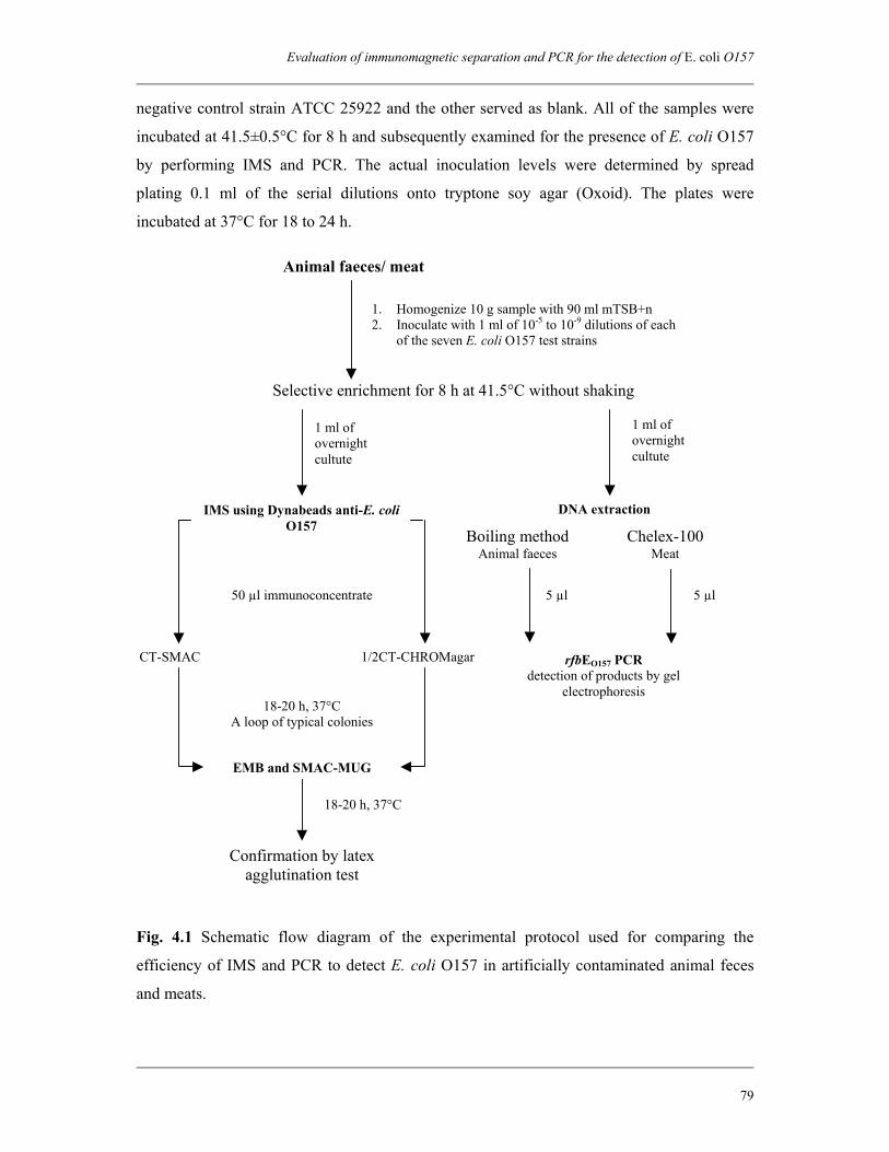
Evaluation of immunomagnetic separation and PCR for the detection of E. coli O157
79
negative control strain ATCC 25922 and the other served as blank. All of the samples were
incubated at 41.5±0.5°C for 8 h and subsequently examined for the presence of E. coli O157
by performing IMS and PCR. The actual inoculation levels were determined by spread
plating 0.1 ml of the serial dilutions onto tryptone soy agar (Oxoid). The plates were
incubated at 37°C for 18 to 24 h.
Fig. 4.1 Schematic flow diagram of the experimental protocol used for comparing the
efficiency of IMS and PCR to detect E. coli O157 in artificially contaminated animal feces
and meats.
CT-SMAC
1. Homogenize 10 g sample with 90 ml mTSB+n 2. Inoculate with 1 ml of 10-5 to 10-9 dilutions of each
of the seven E. coli O157 test strains
Selective enrichment for 8 h at 41.5°C without shaking
IMS using Dynabeads anti-E. coli O157
1 ml of overnight cultute
1/2CT-CHROMagar
18-20 h, 37°C A loop of typical colonies
EMB and SMAC-MUG
18-20 h, 37°C
Confirmation by latex agglutination test
Chelex-100 Meat
5 µl
rfbEO157 PCR detection of products by gel
electrophoresis
Boiling methodAnimal faeces
Animal faeces/ meat
DNA extraction
1 ml of overnight cultute
5 µl 50 µl immunoconcentrate

Chapter 4
80
4.2.3 Immunomagnetic separation
After 8 h incubation, IMS with Dynabeads anti-E. coli O157 (Dynal, Oslo, Norway) was
performed using 1 ml aliquot of each enriched culture and following the instructions of the
manufacturer. The concentrated samples were inoculated onto sorbitol–MacConkey agar
(Oxoid) supplemented with cefixime (0.05 mg/l) and tellurite (2.5 mg/l) (Oxoid) (CT-SMAC)
and CHROMagar O157 (ITK Diagnostics BV, Uithoorn, the Netherlands) with cefixime
(0.025 mg/l) and tellurite (1.25 mg/l) (1/2CT-CHROMagar), 50 µl onto each plate. The plates
were incubated at 37°C for 18 to 20 h. From each plate, up to 8 typical colonies (non-
sorbitol-fermenting colonies on CT-SMAC; β-glucuronidase-negative colonies on 1/2CT-
CHROMagar) were selected and inoculated onto eosine methylene blue (Oxoid) and sorbitol-
MacConkey agar supplemented with 4-methylumbelliferyl-β-D-glucuronide (SMAC-MUG).
Typical colonies with green metallic shine on eosine methylene blue and fluorescence-
negative colonies on SMAC-MUG were then subjected to agglutination using an E. coli
O157 latex agglutination kit (Murex Biotech Ltd., Dartford, Kent, United Kingdom).
4.2.4 DNA extraction
Based on the results of preliminary experiments comparing different methods of DNA
extraction, we selected a simple boiling method to extract DNA from the enrichment cultures
of animal feces and thermal cell lysis using Chelex-100 resin (Bio-Rad Laboratories Ltd.,
Richmond, Calif.) to extract DNA from the enrichment cultures of meat samples. In case of
the boiling method, 1 ml of enrichment culture was centrifuged at 14,000× g for 5 min and
the pellet was washed with 1 ml of fresh modified tryptic soy broth (14,000× g for 5 min).
After discarding the supernatant, the pellet was resuspended in 200 µl of milliQ water, boiled
for 10 min, and then immediately put on ice. The suspension was centrifuged (14,000× g, 10
min at 4°C) and 5 µl of the supernatant was used in the PCR. In case of the Chelex-100
method, we also used 1 ml of the enrichment cultures and followed the procedure described
by Malorny et al. (20).
4.2.5 PCR assay
The E. coli O157-specific PCR assay evaluated is based on amplification of sequences of
the rfbEO157 gene. The specific oligonucleotide primers GiO157-I and GiO157-II have been
developed by Abdulmawjood et al. (2). For each sample, 25 µl of reaction mix was prepared
containing 0.2 µl of each primer (50 pmol/µl), 0.5 µl of each deoxynucleoside triphosphate
(10 mM) (Roche Diagnostics, Mannheim, Germany), 2.5 µl of 10 Fast Start buffer (Roche
Evaluation of immunomagnetic separation and PCR for the detection of E. coli O157
81
Diagnostics), 1.5 µl of MgCl2 (25 mM) (Roche Diagnostics), 1 µl (5.23 fg) of rfbEO157
internal amplification control (3), 0.2 µl of Fast Start Taq DNA polymerase (5 U/µl) (Roche
Diagnostics), 2.5 µl of 10 mg/ml bovine serum albumin (BSA) fraction V, and 11.4 µl of
molecular biology grade water. Finally, 5 µl of template DNA solution was added to each
reaction mix. The PCR was carried out in a thermal cycler (Eppendorf Mastercycler Gradient,
VWR, Amsterdam, The Netherlands) with the following program: denaturation at 94°C for 5
min, 30 cycles of 15 s at 94°C, 15 s at 60°C, and 30 s at 72°C, followed by a final extension
incubation of 72°C for 5 min. The presence of PCR products was determined by
electrophoresis of 10 µl of the reaction product in a 1.5% agarose gel with Tris-borate EDTA
buffer. A 100-bp DNA ladder marker XIV was used as molecular marker (Roche
Diagnostics).
4.2.6 Statistical analysis
To calculate whether the outcome (i.e., the total number of positive replicates of animal
feces or meat divided by the total number of replicates of animal feces or meat examined,
respectively) of the IMS and the PCR method were statistically significantly different, we
performed the Fisher’s exact test (double-sided). Furthermore, we analyzed whether the
outcome of the IMS and the PCR method were influenced by the origin of the fecal sample or
the type of meat by the Fisher’s exact test (double-sided).
4.3 Results
For the detection of E. coli O157 in both animal feces and meat, IMS proved to be more
sensitive than the PCR (Table 4.1). For example, when 101 CFU of the seven strains were
inoculated per 10 g of animal feces, the organisms were recovered from 95% of the replicates
of both cattle and sheep feces by using IMS and from only 33 and 5%, respectively, by using
the PCR-based method (Table 4.1). The lowest level of inoculation at which at least 95% of
the replicates of cattle and sheep feces were found positive by PCR was 103 CFU of E. coli
O157 per 10 g of sample (Table 4.1). Whereas the outcome of the IMS was not influenced by
the origin of the fecal samples, the detection efficiency of the PCR was systematically lower
for sheep feces than for cattle feces (Table 4.1).
When 101 CFU of any of the seven strains were inoculated per 10 g of meat, the
organisms were recovered from 100% of the replicates of both minced beef and dry-
Chapter 4
82
fermented sausages by using IMS and from only 74 and 57%, respectively, by using the PCR
(Table 4.1). The lowest level of inoculation at which all sample replicates of minced beef and
dry-fermented sausages were detected as positive by the PCR-based method was 102 CFU per
10 g (Table 4.1). Although the overall result for meat showed the IMS method to be clearly
more sensitive than the PCR, the methods showed similar detection efficiency when the E.
coli O157 test strains were inoculated into filet americain, with all sample replicates detected
as positive by both methods at the inoculation level of 101 CFU per 10 g (Table 4.1). Again,
the outcome of IMS was not influenced by the type of meat, whereas the detection efficiency
of the PCR was systematically lower for raw minced beef and dry-fermented sausages than
for filet americain (Table 4.1). None of the samples of animal feces nor meat was found
positive either by using IMS or the PCR when spiked with less than 100 CFU of organism per
10 g sample (Table 4.1). Results obtained for the uninoculated portions of animal feces and
meat and the portions spiked with the negative control strain were all negative.
4.4 Discussion
With the increased interest in the occurrence of STEC O157, the demand for more
reliable and easy detection methods for diversified sources has been intensified. To date, IMS
and PCR have been evaluated as the most effective methods for routine diagnosis of E. coli
O157 from a variety of sources (11, 12). IMS is laborious and time consuming but ends up
with the isolation of the pathogen. On the other hand, PCR is fast and less laborious but it can
only be used for screening purposes, so a further culture step is required to isolate the
organism. The objective of this study was to evaluate the performance of IMS and PCR in
detecting E. coli O157 in animal feces and meats. Although IMS is considered as the official
standard method for detection and isolation of E. coli O157 from food and animal feeding
stuffs (5), no standardized protocol has yet been described for animal feces.
For a PCR-based method to be completely suitable for implementation as an analytical
tool, its diagnostic accuracy must be thoroughly evaluated and demonstrated (15, 20). The
sensitivity of PCR detection systems depends on the choice of reaction conditions and
interactions with the particular nature of the sample matrix (26). PCR is inhibited by
numerous compounds often found in feces and food (6), providing a practical hindrance to
the molecular-based detection of small numbers of contaminating pathogens (4, 23).
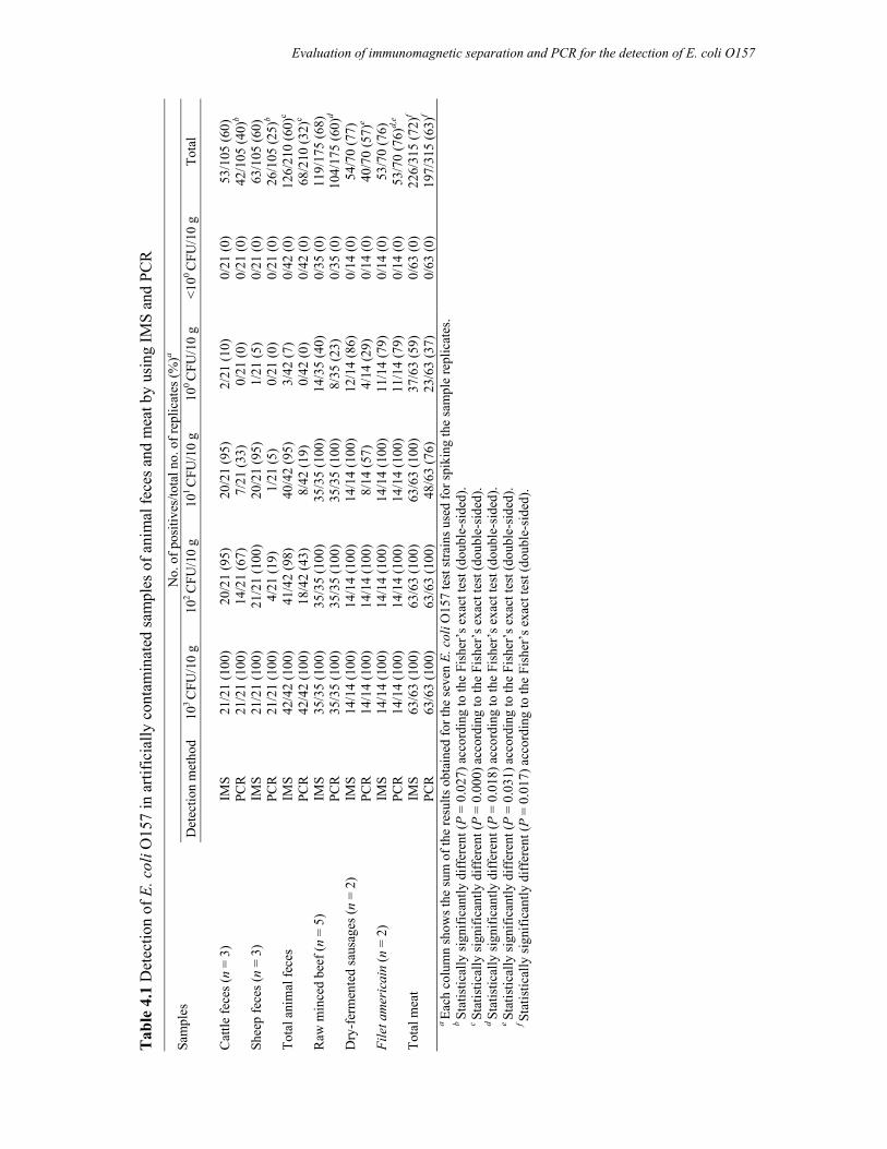
Tab
le 4
.1 D
etec
tion
of E
. col
i O15
7 in
arti
ficia
lly c
onta
min
ated
sam
ples
of a
nim
al fe
ces a
nd m
eat b
y us
ing
IMS
and
PCR
N
o. o
f pos
itive
s/to
tal n
o. o
f rep
licat
es (%
)a Sa
mpl
es
Det
ectio
n m
etho
d10
3 C
FU/1
0 g
102 C
FU/1
0 g
101 C
FU/1
0 g
100 C
FU/1
0 g
<100
CFU
/10
g To
tal
Cat
tle fe
ces (
n =
3)
IMS
21/2
1 (1
00)
20/2
1 (9
5)
20/2
1 (9
5)
2/21
(10)
0/
21 (0
) 53
/105
(60)
PCR
21
/21
(100
) 14
/21
(67)
7/
21 (3
3)
0/21
(0)
0/21
(0)
42/1
05 (4
0)b
Shee
p fe
ces (
n =
3)
IMS
21/2
1 (1
00)
21/2
1 (1
00)
20/2
1 (9
5)
1/21
(5)
0/21
(0)
63/1
05 (6
0)
PC
R
21/2
1 (1
00)
4/21
(19)
1/
21 (5
) 0/
21 (0
) 0/
21 (0
) 26
/105
(25)
b
Tota
l ani
mal
fece
s IM
S 42
/42
(100
) 41
/42
(98)
40
/42
(95)
3/
42 (7
) 0/
42 (0
) 12
6/21
0 (6
0)c
PC
R
42/4
2 (1
00)
18/4
2 (4
3)
8/42
(19)
0/
42 (0
) 0/
42 (0
) 68
/210
(32)
c
Raw
min
ced
beef
(n =
5)
IMS
35/3
5 (1
00)
35/3
5 (1
00)
35/3
5 (1
00)
14/3
5 (4
0)
0/35
(0)
119/
175
(68)
PCR
35
/35
(100
) 35
/35
(100
) 35
/35
(100
) 8/
35 (2
3)
0/35
(0)
104/
175
(60)
d
Dry
-fer
men
ted
saus
ages
(n =
2)
IMS
14/1
4 (1
00)
14/1
4 (1
00)
14/1
4 (1
00)
12/1
4 (8
6)
0/14
(0)
54/7
0 (7
7)
PC
R
14/1
4 (1
00)
14/1
4 (1
00)
8/14
(57)
4/
14 (2
9)
0/14
(0)
40/7
0 (5
7)e
File
t am
eric
ain
(n =
2)
IMS
14/1
4 (1
00)
14/1
4 (1
00)
14/1
4 (1
00)
11/1
4 (7
9)
0/14
(0)
53/7
0 (7
6)
PC
R
14/1
4 (1
00)
14/1
4 (1
00)
14/1
4 (1
00)
11/1
4 (7
9)
0/14
(0)
53/7
0 (7
6)d,
e
Tota
l mea
t IM
S 63
/63
(100
) 63
/63
(100
) 63
/63
(100
) 37
/63
(59)
0/
63 (0
) 22
6/31
5 (7
2)f
PC
R
63/6
3 (1
00)
63/6
3 (1
00)
48/6
3 (7
6)
23/6
3 (3
7)
0/63
(0)
197/
315
(63)
f
a Ea
ch c
olum
n sh
ows t
he su
m o
f the
resu
lts o
btai
ned
for t
he se
ven
E. c
oli O
157
test
stra
ins u
sed
for s
piki
ng th
e sa
mpl
e re
plic
ates
.
b St
atis
tical
ly si
gnifi
cant
ly d
iffer
ent (
P =
0.02
7) a
ccor
ding
to th
e Fi
sher
’s e
xact
test
(dou
ble-
side
d).
c St
atis
tical
ly si
gnifi
cant
ly d
iffer
ent (
P =
0.00
0) a
ccor
ding
to th
e Fi
sher
’s e
xact
test
(dou
ble-
side
d).
d St
atis
tical
ly si
gnifi
cant
ly d
iffer
ent (
P =
0.01
8) a
ccor
ding
to th
e Fi
sher
’s e
xact
test
(dou
ble-
side
d).
e St
atis
tical
ly si
gnifi
cant
ly d
iffer
ent (
P =
0.03
1) a
ccor
ding
to th
e Fi
sher
’s e
xact
test
(dou
ble-
side
d).
f St
atis
tical
ly si
gnifi
cant
ly d
iffer
ent (
P =
0.01
7) a
ccor
ding
to th
e Fi
sher
’s e
xact
test
(dou
ble-
side
d).
Evaluation of immunomagnetic separation and PCR for the detection of E. coli O157
Chapter 4
84
One way to address these limitations is the use of effective sample processing as well as
DNA extraction techniques, the area where many of the efforts have been put into (14, 16,
19). Nevertheless, none of these techniques is ideal, and in many cases a technique optimized
for one matrix or microorganism is not readily adaptable for use with others. Moreover,
because purification adds to the time and expense of sample preparation, as well as to the loss
of target nucleic acids, a more satisfying approach to the problem of PCR inhibition would be
to reduce interference rather than attempt to remove all of the offending substances. To this
end, various additives have been included in the PCR mixture to minimize inhibition. Among
these, BSA is widely being used (18). We also found that BSA at the concentration of 1
mg/ml in the PCR mixture significantly reduced the effects of inhibitory substances present
in both feces and meats. This was confirmed by using an internal amplification control in the
PCRs (results not shown). The use of an internal amplification control in the PCR can detect
the level of reaction inhibition and therefore eliminates the chances of false-negative results
(1).
Preliminary experiments comparing different techniques of DNA extraction showed a
simple boiling method to be the most effective for animal feces and thermal lysis using
Chelex-100 resin for meats. Once the DNA extraction method and PCR conditions were
optimized, the recovery of the E. coli O157 test strains by the PCR was compared with that of
the IMS method. IMS was found to be more effective than the PCR for both samples of
animal feces and meat. Hence, use of PCR alone in the routine diagnosis of E. coli O157 in
animal feces and meats might result in a lower number of positives. Furthermore, the
detection efficiency of the PCR was dependent on the origin of the fecal samples. According
to a previous study, sheep feces were found to possess more substances inhibitory to PCR
than feces from other animals, including cattle (18). The detection efficiency of the IMS
procedure was not affected by the origin of the fecal sample used for spiking. Like the results
of the spiking experiments with feces, for meat samples it was observed that the detection
efficiency of the IMS was not influenced by the type of meat whereas the detection efficiency
of the PCR was. Based on the higher detection efficiency of the PCR for samples of filet
americain and IMS and PCR showing the same sensitivity, filet americain seems to contain
less PCR-inhibitory substances than raw minced beef and dry-fermented sausages.
One of the set backs of IMS is that it is sometimes insensitive due to the overgrowth of
background flora, which can mimic the characteristics of target organisms (8). This can
happen particularly for those samples that contain a high load of indigenous flora, for
example, animal feces. But this limitation was overridden in this study by means of a
Evaluation of immunomagnetic separation and PCR for the detection of E. coli O157
85
selective enrichment step preceding IMS and by using a second selective plating medium
(1/2CT-CHROMagar) in addition to CT-SMAC.
The concentration at which E. coli O157 is shed in feces varies from animal to animal
(24), where a range from 102 to 105 CFU/g was observed. An even wider range of fecal
shedding of E. coli O157 in naturally infected cattle was found in another study (30).
Animals excreting >104 CFU/g of E. coli O157 are normally considered as high shedding and
those excreting <102 CFU/g are low shedding. In order to understand the epidemiology of E.
coli O157 infection in cattle and to inform quantitative microbial risk assessments, detailed
quantitative information on the patterns and concentration of organisms shed by cattle into
the environment is required. Although animals can carry E. coli O157 in high numbers, only
a few organisms can already give rise to human infection. This accentuates the need for
enforcing zero tolerance of this organism in foods and for markedly decreasing the risk of
contamination of raw meat (10, 28). Therefore, the demand of a sensitive and reliable
detection and isolation technique is obvious. According to this study, both for animal feces
and meat IMS can be used more successfully to detect E. coli O157 than PCR, being more
sensitive, not influenced by the type of animal feces or meat, and resulting in the isolation of
the pathogen.
Acknowledgments
This research was supported by the Netherlands Foundation for the Advancement of
Tropical Research (NWO-WOTRO) (Award Number WB 93-415). We thank Dr. Amir
Abdulmawjood (Institute of Veterinary Food Science, Justus-Liebig-University, Giessen,
Germany) for kindly providing the rfbEO157 internal amplification control.
References
1. Abdulmawjood, A., M. Bulte, N. Cook, S. Roth, H. Schonenbrucher, and J. Hoorfar. 2003. Toward an international standard for PCR based detection of food borne Escherichia coli O157. Part 1. Assay development and multi-center validation. J. Microbiol. Methods 55:775–786.
2. Abdulmawjood, A., M. Bulte, S. Roth, H. Schonenbrucher, N. Cook, A. E. Heuvelink, and J. Hoorfar. 2004. Development, validation, and standardization of polymerase chain reaction-based detection of E. coli O157. J. AOAC Int. 87:596–603.
Chapter 4
86
3. Abdulmawjood, A., S. Roth, and M. Bulte. 2002. Two methods for construction of internal amplification controls for the detection of Escherichia coli O157 by polymerase chain reaction. Mol. Cell. Probes 16:335–339.
4. Andersen, M. R., and C. J. Omiecinski. 1992. Direct extraction of bacterial plasmids from food for polymerase chain reaction amplification. Appl. Environ. Microbiol. 58:4080–4082.
5. Anonymous. 2001. Microbiology of food and animal feeding stuffs-Horizontal method for the detection of Escherichia coli O157 (EN ISO 16654). International Organization for Standardization, Geneva, Switzerland.
6. Brian, M. J., M. Frosolono, B. E. Murray, A. Miranda, E. L. Lopez, H. F. Gomez, and T. G. Cleary. 1992. Polymerase chain reaction for diagnosis of enterohemorrhagic Escherichia coli infection and hemolytic-uremic syndrome. J. Clin. Microbiol. 30:1801–1806.
7. Caprioli, A., S. Morabito, H. Brugereb, and E. Oswald. 2005. Enterohaemorrhagic Escherichia coli: emerging issues on virulence and modes of transmission. Vet. Res. 36:289–311.
8. Clifton-Hadley, F. A. 2000. Detection and diagnosis of Escherichia coli O157 and other verotoxigenic E. coli in animal faeces. Rev. Med. Microbiol. 11:47–58.
9. Fitzmaurice, J., G. Duffy, B. Kilbride, J. J. Sheridan, C. Carroll, and M. Maher. 2004. Comparison of a membrane surface adhesion recovery method with an IMS method for use in a polymerase chain reaction method to detect Escherichia coli O157:H7 in minced beef. J. Microbiol. Methods 59:243–252.
10. Heuvelink, A. E., G. L. Roessink, K. Bosboom, and E. de Boer. 2001. Zero-tolerance for faecal contamination of carcasses as a tool in the control of O157 VTEC infections. Int. J. Food Microbiol. 66:13–20.
11. Heuvelink, A. E., F. L. van den Biggelaar, E. de Boer, R. G. Herbes, W. J. Melchers, J. H. Huis in ’t Veld, and L. A. Monnens. 1998. Isolation and characterization of verocytotoxin-producing Escherichia coli O157 strains from Dutch cattle and sheep. J. Clin. Microbiol. 36:878–882.
12. Heuvelink, A. E., K. Wernars, and E. de Boer. 1996. Occurrence of Escherichia coli O157 and other verocytotoxin-producing E. coli in retail raw meats in The Netherlands. J. Food Prot. 59:1267–1272.
13. Heuvelink, A. E., J. T. Zwartkruis-Nahuis, R. R. Beumer, and E. de Boer. 1999. Occurrence and survival of verocytotoxin-producing Escherichia coli O157 in meats obtained from retail outlets in The Netherlands. J. Food Prot. 62:1115–1122.
14. Holland, J. L., L. Louie, A. E. Simor, and M. Louie. 2000. PCR detection of Escherichia coli O157:H7 directly from stools: evaluation of commercial extraction methods for purifying fecal DNA. J. Clin. Microbiol. 38:4108–4113.
15. Hoorfar, J., and N. Cook. 2003. Critical aspects of standardization of PCR. Methods Mol. Biol. 216:51–64.
16. Jinneman, K. C., J. H. Wetherington, W. E. Hill, A. M. Adams, J. M. Johnson, B. J. Tenge, N. L. Dang, R. L. Manger, and M. M. Wekell. 1998. Template preparation for PCR and RFLP of amplification products for the detection and identification of Cyclospora sp. and Eimeria spp. oocysts directly from raspberries. J. Food Prot. 61:497–503.
17. Karmali, M. A. 1989. Infection by verotoxin-producing Escherichia coli. Clin. Microbiol. Rev. 2:15–38.
18. Kreader, C. A. 1996. Relief of amplification inhibition in PCR with bovine serum albumin or T4 gene 32 protein. Appl. Environ. Microbiol. 62:1102–1106.
Evaluation of immunomagnetic separation and PCR for the detection of E. coli O157
87
19. Lantz, P. G., W. Abu Al-Soud, R. Knutsson, B. Hahn-Hagerdal, and P. Radstrom. 2000. Biotechnical use of the polymerase chain reaction for microbial analysis of biological samples, p. 87–130. In M. R. El- Gewely (ed.), Biotechnology Annual Review. Elsevier Science Publishing, Amsterdam, The Netherlands
20. Malorny, B., P. T. Tassios, P. Radstrom, N. Cook, M. Wagner, and J. Hoorfar. 2003. Standardization of diagnostic PCR for the detection of food borne pathogens. Int. J. Food Microbiol. 83:39–48.
21. Nataro, J. P., and J. B. Kaper. 1998. Diarrheagenic Escherichia coli. Clin. Microbiol. Rev. 11:142–201.
22. Oberst, R. D., M. P. Hays, L. K. Bohra, R. K. Phebus, C. T. Yamashiro, C. Paszko-Kolva, J. A. Flood, J. M. Sargeant, and J. R. Gillespie. 1998. PCR-based DNA amplification and presumptive detection of Escherichia coli O157:H7 with an internal fluorogenic probe and the 5’nuclease (TaqMan) assay. Appl. Environ. Microbiol. 64:3389–3396.
23. Oliveira, S. D., L. R. Santos, D. M. Schuch, A. B. Silva, C. T. Salle, and C. W. Canal. 2002. Detection and identification of salmonellas from poultry-related samples by PCR. Vet. Microbiol. 87:25–35.
24. Omisakin, F., M. MacRae, I. D. Ogden, and N. J. Strachan. 2003. Concentration and prevalence of Escherichia coli O157 in cattle feces at slaughter. Appl. Environ. Microbiol. 69:2444–2447.
25. Paton, J. C., and A. W. Paton. 1998. Pathogenesis and diagnosis of Shiga toxin-producing Escherichia coli infections. Clin. Microbiol. Rev. 11:450–479.
26. Scheu, P. M., K. Berghof, and U. Stahl. 1998. Detection of pathogenic and spoilage microorganisms in food with the polymerase chain reaction. Food Microbiol. 15:13–31.
27. Smith, J. L., and S. A. Palumbo. 1982. Microbial injury reviewed for the sanitarian. Dairy Food Environ. Sanit. 2:57–63.
28. Tuttle, J., T. Gomez, M. P. Doyle, J. G. Wells, T. Zhao, R. V. Tauxe, and P. M. Griffin. 1999. Lessons from a large outbreak of Escherichia coli O157:H7 infections: insights into the infectious dose and method of widespread contamination of hamburger patties. Epidemiol. Infect. 122:185–192.
29. Wang, L., and P. R. Reeves. 1998. Organization of Escherichia coli O157 O antigen gene cluster and identification of its specific genes. Infect. Immun. 66:3545–3551.
30. Widiasih, D. A., N. Ido, K. Omoe, S. Sugii, and K. Shinagawa. 2004. Duration and magnitude of faecal shedding of Shiga toxin-producing Escherichia coli from naturally infected cattle. Epidemiol. Infect. 132:67–75.

Prevalence and genetic characterization of Shiga
toxin-producing Escherichia coli isolated from
slaughtered animals in Bangladesh
Applied and Environmental Microbiology 2008; 74:5414-5421
M. Aminul Islam A. Salam Mondol E. de Boer R. R. Beumer M. H. Zwietering K. A. Talukder A. E. Heuvelink
Chapter 5
Chapter 5
90
Abstract
To determine the prevalence of Shiga toxin (Stx)-producing Escherichia coli (STEC) in
slaughter animals in Dhaka, Bangladesh, we collected rectal contents immediately after
animals were slaughtered. Of the samples collected from buffalo (n = 174), cows (n = 139),
and goats (n = 110), 82.2%, 72.7%, and 11.8% tested positive for stx1 and/or stx2,
respectively. STEC could be isolated from 37.9%, 20.1%, and 10.0% of the buffalo, cows,
and goats, respectively. STEC O157 strains were isolated from 14.4% of the buffalo, 7.2% of
the cows, and 9.1% of the goats. More than 93% (n = 42) of the STEC O157 isolates were
positive for the stx2, eae, katP, etpD, and enterohemorrhagic E. coli hly (hlyEHEC) virulence
genes. STEC O157 isolates were characterized by seven recognized phage types, of which
types 14 (24.4%) and 31 (24.4%) were predominant. Subtyping of the 45 STEC O157
isolates by pulsed-field gel electrophoresis showed 37 distinct restriction patterns, suggesting
a heterogeneous clonal diversity. In addition to STEC O157, 71 STEC non-O157 strains were
isolated from 60 stx-positive samples from 23.6% of the buffalo, 12.9% of the cows, and
0.9% of the goats. The STEC non-O157 isolates belonged to 36 different O groups and 52
O:H serotypes. Unlike STEC O157, most of the STEC non-O157 isolates (78.9%) were
positive for stx1. Only 7.0% (n = 5) of the isolates were positive for hlyEHEC, and none was
positive for eae, katP, and etpD. None of the isolates was positive for the iha, toxB, and efa1
putative adhesion genes. However, 35.2% (n = 25), 11.3% (n = 8), 12.7% (n = 9), and 12.7%
(n = 9) of the isolates were positive for the lpfO113, saa, lpfAO157/01-141, and lpfAO157/OI-154 genes,
respectively. The results of this study provide the first evidence that slaughtered animals like
buffalo, cows, and goats in Bangladesh are reservoirs for STEC, including the potentially
virulent STEC strain O157.
STEC in slaughtered animals in Bangladesh
91
5.1 Introduction
Shiga toxin (Stx)-producing Escherichia coli (STEC) organisms, also called
verocytotoxin (VT)-producing E. coli (VTEC), are one of the most important groups of
foodborne pathogens (2, 32). Infection can cause gastroenteritis that may be complicated by
hemorrhagic colitis or the hemolytic-uremic syndrome (HUS), which is the main cause of
acute renal failure in children. STEC strains causing human infections belong to a large, still-
increasing number of O:H serotypes. Most outbreaks and sporadic cases of hemorrhagic
colitis and HUS have been attributed to the STEC O157 strains (39). However, infections
caused by some non-O157 serotypes have also been frequently associated with severe illness
in humans. In some geographic areas, STEC non-O157 strains are more commonly isolated
from persons with diarrhea or HUS than STEC O157 strains (33).
STEC are defined by the production of one or more types of Shiga toxin (Stx1 or Stx2 or
their variants), which inhibit the protein synthesis of host cells, leading to cell death. Stx1 and
Stx2 are encoded by alleles in the genome of temperate, lambdoid bacteriophages that are
integrated in the E. coli chromosome (38). Besides the stx gene(s), human pathogenic STEC
strains often carry the eae gene, encoding the adherence factor intimin, which is an outer
membrane protein (32). The eae gene is carried by a pathogenicity island in the chromosome
called the locus of enterocyte effacement (LEE), which is required for intimate attachment to
the host intestinal mucosa (32). Furthermore, human pathogenic STEC strains often harbor a
large plasmid encoding possible additional virulence traits such as the enterohemorrhagic E.
coli (EHEC) hemolysin (hlyEHEC) gene, which acts as a pore-forming cytolysin on eukaryotic
cells (36); the bifunctional catalase peroxidase (katP) gene (7); a secreted serine protease
(espP) gene, which can cleave human coagulation factor V (8); and the etpD gene cluster,
which probably encodes a type II secretion pathway (37). With more LEE-negative STEC
strains being reported, investigations of adhesins encoded outside of the LEE have been
carried out (40). Several proteins were proposed to be novel adhesion factors; these include
ToxB (a protein identified from the large, 93-kb plasmid pO157 and required for full
expression of adherence of O157:H7 strain Sakai), Saa (an autoagglutinating adhesin
identified in the LEE-negative strains), Iha (an adherence-conferring protein similar to the
Vibrio cholerae IrgA protein), Efa1 (an EHEC factor for adherence), and LPF (the long polar
fimbriae protein, closely related to LPF of Salmonella enterica serovar Typhimurium) (40).
Chapter 5
92
Domestic ruminants, mainly cattle, sheep, and goats, have been established as major
natural reservoirs for STEC and play a significant role in the epidemiology of human
infections (16). During the processing of the carcasses, fecal contamination or transfer of
bacteria from the animal’s hide to the carcass can facilitate transmission of pathogenic E. coli
to the meat (15).
In most of the developing and underdeveloped countries, hygienic conditions are severely
compromised, and living with domestic animals within the same premises is a common
practice in both rural and urban areas. Although the socioeconomic status and living style of
the people in developing countries support the prevalence of STEC infection, surprisingly,
until recently, only a few studies have been done in this part of the world. Human infections
associated with STEC strains have been described in Latin America, India, and other
developing countries (24, 26). In Bangladesh, the predominant group of E. coli associated
with childhood diarrhea is enterotoxigenic E. coli, accounting for approximately 20% of all
diarrheal cases (34). Recently, we determined the prevalence of STEC and the organisms’
characteristics among hospitalized diarrheal patients and children with diarrhea in the urban
slum community of Dhaka, Bangladesh. The prevalence of STEC was 0.5% among the
hospitalized patients, which was 1.9% of the cases of community patients. No STEC O157
organisms were isolated in the study (23). This result is comparable with data from other
developing countries. The low prevalence of STEC compared with other enteric pathogens in
diarrheal patients can be explained by different facts, the first of which is the absence of any
potential reservoir for this organism. Limited studies of the ecology of STEC have been
reported, particularly from developing countries. In Bangladesh, no study of the occurrence
of STEC in animals had been done. Therefore, the primary objective of the present study was
to determine the prevalence of STEC in feces collected from buffalo, cows, and goats
slaughtered for meat production.
5.2 Materials and methods
5.2.1 Sampling
In the period from 1 January to 1 May 2006, we collected fecal samples from 174 buffalo,
139 cows, and 110 goats at a slaughterhouse in Dhaka of Mohakhali area in Bangladesh. The
animals sampled were randomly selected. The origin of the animals could not be recorded
due to the lack of information. However, according to the local information, animals were
STEC in slaughtered animals in Bangladesh
93
received from different places across the country. Immediately after slaughter, a piece of
large intestine containing fecal material (~5 cm), 1 to 1.5 cm away from the rectum was
excised aseptically and kept at 4 to 8°C. The samples were transported immediately to the
laboratory, where the microbiological examination was started within 8 h.
5.2.2 Isolation of STEC O157
From each sample, a 25-g portion of feces was collected from a piece of intestine (~5
cm), which was aseptically excised from the animal immediately after slaughter and added to
225 ml of modified tryptone soy broth (Oxoid Ltd., Basingstoke, United Kingdom). After
homogenization in a stomacher for 1 min, the samples were incubated for 18-20 h at 37°C.
STEC O157 organisms were isolated as described previously (22), by using the
immunomagnetic separation (IMS) technique and presumptive isolates were confirmed by an
agglutination test using E. coli O157 latex agglutination kit (Murex Biotech Ltd., Dartford,
Kent, United Kingdom) and a PCR for the rfb (O-antigen-encoding) region of E. coli O157
(rfbEO157) (31).
5.2.3 Isolation of STEC non-O157
The overnight enrichment cultures were screened by PCR for the presence of stx1 and stx2
genes. DNA was extracted from 1 ml of broth culture by thermal cell lysis using Chelex-100
resin (Bio-Rad Laboratories Ltd., Richmond, Calif.) and 5 µl of the DNA extract was used in
the PCR. Multiplex PCR for stx1 and stx2 was carried out in a PTC-200 peltier thermal cycler
(Bio-Rad) as described previously (23). Broth cultures that yielded positive PCR results for
stx1 and/or stx2 were streaked onto tryptone bile x-glucuronide (TBX) agar (Oxoid)
supplemented with cefixime (20 µg/l), cefsulodin (3 mg/l), and vancomycin (30 mg/l) (CCV-
TBX). STEC colonies were identified by a colony patch technique described previously (23).
5.2.4 Serotyping
All isolates were sent to the National Institute of Public Health and the Environment
(RIVM) in Bilthoven, The Netherlands, for O and H typing.
5.2.5 Phage typing
Isolates confirmed to be STEC O157 were phage typed at the laboratory of Enteric
Pathogens, Central Public Health Laboratory, London, United Kingdom.
Chapter 5
94
5.2.6 PCR detection of putative virulence and adhesin genes
Isolates were tested for both chromosomally (e.g., stx1, stx2, and eae)- and plasmid (e.g.,
hlyEHEC, katP, and etpD)-encoded virulence genes by PCR according to the procedure
described earlier (23). The presence of eight putative adhesin genes in STEC non-O157
strains (iha, toxB, saa, efa1, lpfAO113, lpfAO157/OI-141, and lpfAO157/OI-154) was tested using the
primer sets and reaction conditions as described by Toma et al. (40).
5.2.7 PFGE
All STEC O157 and STEC non-O157 isolates that belonged to the same serotype were
selected to be analyzed by PFGE. PFGE was performed following the standardized protocol
developed by PulseNet for E. coli O157:H7 (35). Analysis of the TIFF images was carried
out by the BioNumerics software (Applied Maths, Belgium) using the dice coefficient and
unweighted-pair group method using average linkages to generate dendrograms with 1.0%
tolerance values.
5.2.8 Statistical methods
The chi-square test with Yates’ correction or, if necessary, Fisher’s exact test, was used as
indicated to compare the test results. Statistical testing was performed using Epi Info version
3.3.2 on a standard personal computer. Statistically, P values < 0.05 were considered
significant for comparisons.
5.3 Results
5.3.1 Prevalence of STEC
Of the fecal samples collected from buffalo (n = 174), cows (n = 139), and goats (n =
110), 82.2% (n = 143), 72.7% (n = 101), and 11.8% (n = 13) tested positive for stx1 and/or
stx2, respectively. Higher numbers of samples from buffalo (66.7%; n = 116) and cows
(45.3%; n = 63) were positive for both the stx1 and the stx2 genes than were positive for only
the stx1 or the stx2 gene (P < 0.001). Only stx1 was positively identified in 8.6% (n = 15)
buffalo, 12.9% (n = 18) cow and 3.6% (n = 4) of goat samples. Only stx2 was positively
identified in 6.9% (n = 12), 14.4% (n = 20) and 7.3% (n = 8) of buffalo, cow and goat
samples, respectively. STEC could be isolated from 37.9% (n = 66), 20.1% (n = 28) and
10.0% (n = 11) of the buffalo, cow and goat samples, respectively. Significantly higher
STEC in slaughtered animals in Bangladesh
95
numbers of buffalo samples were positive for STEC than cow and goat samples (P < 0.001).
STEC O157 strains were isolated from 14.4% (n = 25) of the buffalo, 7.2% (n = 10) of the
cows, and 9.1% (n = 10) of the goats. STEC non-O157 strains were isolated from 23.6% (n =
41) of the buffalo, 12.9% (n = 18) of the cows, and 0.9% (n = 1) of the goats. Sometimes
more than one serotype was isolated from a single sample, so the number of samples with
STEC non-O157 isolates was 41 (49 different isolates from 41 stx-positive samples) for
buffalo and 18 (21 different isolates from 18 stx-positive samples) for cows.
5.3.2 Characterization of STEC O157
The results of the PCR assays for different virulence genes are presented in Table 5.1.
Among 45 STEC O157 isolates, seven phage types (PT) were identified. Ten isolates from
buffalo could not be characterized by any recognized PT, five of which were identified as
untypeable and the remaining were reacts but not confirmed. Digestion of genomic DNA
from the 45 isolates with XbaI and analysis by contour-clamped homogenous electric field
(CHEF)-PFGE revealed 37 distinct restriction profiles (Fig. 5.1).
5.3.3 Characterization of STEC non-O157
The results of the PCR assays for different virulence and (putative) adhesin genes are
presented in Table 5.1. None of the isolates was positive for putative virulence and adhesion
genes, including eae, katP, etpD, iha, toxB, and efa1 (results not shown). More than 30% (n =
15) of the isolates from buffalo and 47% (n = 10) of the isolates from cows were positive for
the lpfO113 gene. The saa gene was present in 8.1% (n = 4) and 19.0% (n = 4) of the isolates
from buffalo and cows, respectively. Seven isolates from buffalo and two isolates from cows
were positive for both the lpfAO157/01-141 and lpfAO157/OI-154 genes. The goat isolate was
negative for all adhesin genes.
Serotyping of the STEC non-O157 isolates revealed that the isolates belonged to 36
different O-groups (grouping isolates with an untypeable O serogroup, as serogroup ONT)
and 52 O:H serotypes (Table 5.2). Isolates of different serotypes were found in the same
sample; at the same time, isolates of the same serotype were found in different samples of
same/different origins (Table 5.2). Isolates belonging to the same serotype isolated from
same/different samples were analyzed by PFGE, which showed a clonal relationship among
different isolates of the same serotype (results not shown). However, polymorphism was also
observed between isolates of the same serotype.
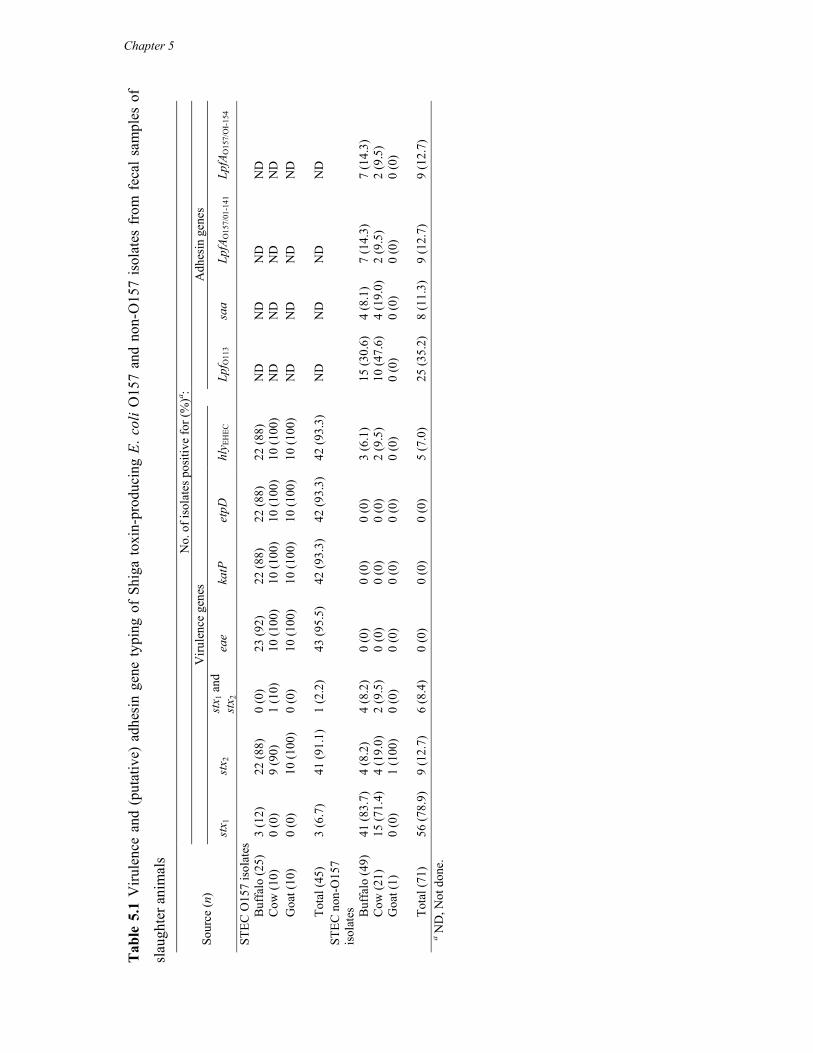
Tab
le 5
.1 V
irule
nce
and
(put
ativ
e) a
dhes
in g
ene
typi
ng o
f Sh
iga
toxi
n-pr
oduc
ing
E. c
oli O
157
and
non-
O15
7 is
olat
es f
rom
fec
al s
ampl
es o
f
slau
ghte
r ani
mal
s N
o. o
f iso
late
s pos
itive
for (
%)a :
Viru
lenc
e ge
nes
A
dhes
in g
enes
So
urce
(n)
stx 1
st
x 2
stx 1
and
st
x 2
eae
katP
et
pD
hly E
HEC
Lpf O
113
saa
LpfA
O15
7/01
-141
Lp
fAO
157/
OI-
154
STEC
O15
7 is
olat
es
Buf
falo
(25)
3
(12)
22
(88)
0
(0)
23 (9
2)
22 (8
8)
22 (8
8)
22 (8
8)
N
D
ND
N
D
ND
C
ow (1
0)
0 (0
) 9
(90)
1
(10)
10
(100
) 10
(100
) 10
(100
) 10
(100
)
ND
N
D
ND
N
D
Goa
t (10
) 0
(0)
10 (1
00)
0 (0
) 10
(100
) 10
(100
) 10
(100
) 10
(100
)
ND
N
D
ND
N
D
To
tal (
45)
3 (6
.7)
41 (9
1.1)
1
(2.2
) 43
(95.
5)
42 (9
3.3)
42
(93.
3)
42 (9
3.3)
ND
N
D
ND
N
D
STEC
non
-O15
7 is
olat
es
Buf
falo
(49)
41
(83.
7)
4 (8
.2)
4 (8
.2)
0 (0
) 0
(0)
0 (0
) 3
(6.1
)
15 (3
0.6)
4
(8.1
) 7
(14.
3)
7 (1
4.3)
C
ow (2
1)
15 (7
1.4)
4
(19.
0)
2 (9
.5)
0 (0
) 0
(0)
0 (0
) 2
(9.5
)
10 (4
7.6)
4
(19.
0)
2 (9
.5)
2 (9
.5)
Goa
t (1)
0
(0)
1 (1
00)
0 (0
) 0
(0)
0 (0
) 0
(0)
0 (0
)
0 (0
) 0
(0)
0 (0
) 0
(0)
To
tal (
71)
56 (7
8.9)
9
(12.
7)
6 (8
.4)
0 (0
) 0
(0)
0 (0
) 5
(7.0
)
25 (3
5.2)
8
(11.
3)
9 (1
2.7)
9
(12.
7)
a ND
, Not
don
e.
Chapter 5
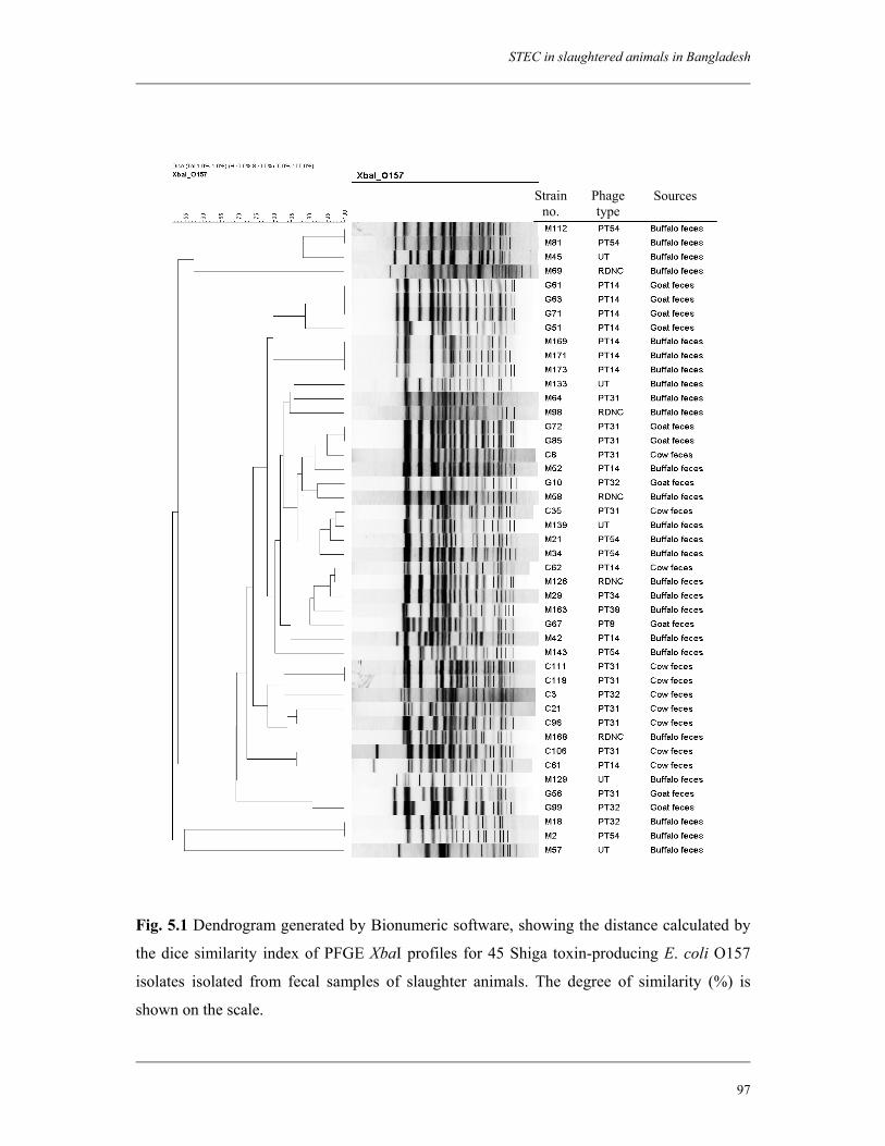
STEC in slaughtered animals in Bangladesh
97
Strain no.
Phage type
Sources
Fig. 5.1 Dendrogram generated by Bionumeric software, showing the distance calculated by
the dice similarity index of PFGE XbaI profiles for 45 Shiga toxin-producing E. coli O157
isolates isolated from fecal samples of slaughter animals. The degree of similarity (%) is
shown on the scale.
Chapter 5
98
Three isolates of serotype O80:H19 and two isolates of serotype O125:H19 isolated from
both cow and buffalo feces showed identical PFGE patterns. Likewise, three isolates of
serotype O149:H8 and two isolates of serotype O141:H21 isolated from buffalo feces
generated identical patterns.
5.4 Discussion
This study provides the first evidence that slaughtered animals in Bangladesh are
reservoirs for STEC, including STEC O157. Around 37.9% of buffalo, 20.1% of cows and
10.0% of goats were positive for STEC. STEC O157 strains were isolated from 14.4% of
buffalo, 7.2% of cows, and 9.1% of goats. The prevalence of STEC, including STEC O157,
was significantly higher in buffalo feces than in cow or goat feces (P < 0.05). Within similar
geographical locations, a few studies of the occurrence of STEC have been done, and most of
the studies were done in India and Thailand (26, 28). Recently, a study was done in central
Vietnam that found a prevalence of STEC of 27% in buffalo, 23% in cows, and 38.5% in
goats. Although a high number of isolates were recovered and characterized based on the
virulence determinants, no STEC O157 could be isolated, and little information was provided
on the serotypic diversity of the isolates (41). In India, Manna et al. reported that the
prevalence of STEC O157 in fecal samples from slaughtered cattle and diarrheic calves was
2.0% and 7.6%, respectively (28). STEC O157 strains have also been isolated in India from
foods of cattle origin, namely, raw minced beef samples (9%; n = 22) (14), beef surface
swabs (3.7%; n = 27), and milk samples (2.4%; n = 81) (28). In China, STEC O157:H7 was
isolated from 10 to 20% of the animals in the villages, including pigs, cattle, goats and
chickens (43).
The results of the investigations of the prevalence of STEC O157 in cattle are clearly
influenced by the sampling and detection methods adopted. As a matter of fact, the use of
specific immunoconcentration procedures for STEC O157 (21, 22) strongly enhances the
sensitivity of the isolation methods. Hence, studies based on such procedures reported
prevalence rates for STEC O157 that were much higher than those reported with the use of
conventional methods (9). It is therefore difficult to determine whether the results reported
reflect true differences in isolation rates or are the consequence of the different
methodologies adopted.
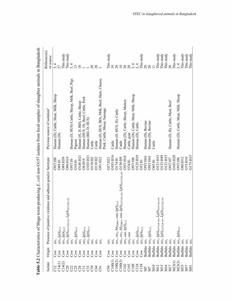
Tab
le 5
.2 C
hara
cter
istic
s of S
higa
toxi
n-pr
oduc
ing
E. c
oli n
on-O
157
isol
ates
from
feca
l sam
ples
of s
laug
hter
ani
mal
s in
Ban
glad
esh
Isol
ate
Orig
in
Pres
ence
of p
utat
ive
viru
lenc
e an
d ad
hesi
n ge
ne(s
)Se
roty
pe
Prev
ious
sour
ce o
f iso
latio
na R
efer
ence
(s)
or so
urce
C
12
Cow
st
x 1
ON
T:H
8
Hum
an (D
), C
attle
, Mea
t, M
ilk, S
heep
3,
4
C14
(1)
Cow
st
x 1, l
pfA 0
113
O89
:H-
Hum
an (D
) 27
C
14(2
) C
ow
stx 1
, lpf
A 011
3 O
89:H
38
Th
is st
udy
C20
C
ow
stx 1
, lpf
A O15
7/O
I-14
1, Lp
fAO
157/
OI-
154
O80
:H19
This
stud
y C
22
Cow
st
x 1
ON
T:H
- H
uman
(D, H
US)
Cat
tle, S
heep
, Milk
, Bee
f, Pi
gs
3, 4
C
25
Cow
st
x 1, l
pfA 0
113
O18
:H-
Pige
on
13
C26
C
ow
stx 1
O
146:
H21
H
uman
(H, D
, BD
), C
attle
, She
ep
18
C31
C
ow
stx 2
, lpf
A 011
3 O
8:H
19
Hum
an (H
US,
D),
Bee
f, C
attle
, Por
k 1
C33
C
ow
stx 2
, lpf
A 011
3 O
103:
H21
H
uman
(BD
, D, H
US)
1
C59
C
ow
stx 1
O
110:
H2
Cat
tle
20
C64
C
ow
stx 1
O
110:
H2
Cat
tle
20
C91
C
ow
stx 1
O
91:H
21
Hum
an (D
, HU
S, B
D),
Milk
, Bee
f, H
am, C
hees
e,
Pork
, Cat
tle, S
heep
, Sau
sage
1
C94
C
ow
stx 1
O
57:H
21
Th
is st
udy
C10
0(1)
C
ow
stx 1
O
103:
H16
C
attle
19
C
100(
2)
Cow
st
x 1, s
tx2,
hly E
HEC
, saa
, lpf
A 011
3 O
174:
H-
Hum
an (D
, HU
S, H
), C
attle
20
C
100(
3)
Cow
st
x 1, s
tx2,
hly E
HEC
, saa
, lpf
A O15
7/O
I-14
1, Lp
fAO
157/
OI-
154
O13
0:H
9 C
attle
19
C
102
Cow
st
x 2, s
aa, l
pfA 0
113
ON
T:H
16
Hum
an (D
), C
attle
, She
ep, M
utto
n 20
C
103
Cow
st
x 2, s
aa
O74
:H-
Cat
tle, g
oat
20
C10
9 C
ow
stx 1
O
NT:
H8
Hum
an (D
), C
attle
, Mea
t, M
ilk, S
heep
3,
4
C12
4 C
ow
stx 1
, lpf
A 011
3 O
125:
H19
H
uman
(D),
Cat
tle
2, 4
C
138
Cow
st
x 1, l
pfA 0
113
O52
:H-
Th
is st
udy
M5
Buf
falo
stx
1 O
NT:
H41
H
uman
(D),
Bov
ine
20
M7
Buf
falo
stx
2, lp
fA01
13
ON
T:H
41
Hum
an (D
), B
ovin
e 20
M
10
Buf
falo
stx
1 O
65:H
- C
attle
11
M
12
Buf
falo
stx
1, lp
fAO
157/
OI-
141,
LpfA
O15
7/O
I-15
4 O
121:
H15
This
stud
y M
15
Buf
falo
stx
1, lp
fAO
157/
OI-
141,
LpfA
O15
7/O
I-15
4 O
132:
H49
This
stud
y M
16
Buf
falo
stx
1 O
121:
H15
This
stud
y M
17
Buf
falo
stx
1 O
NT:
H7
H
uman
(D, H
), C
attle
, Mea
t, B
eef
20
M23
(1)
Buf
falo
stx
1 O
103:
H28
This
stud
y M
23(2
) B
uffa
lo s
tx1,
lpfA
0113
O
NT:
H8
Hum
an (D
), C
attle
, Mea
t, M
ilk, S
heep
3,
4
M35
B
uffa
lo s
tx1
O80
:H12
This
stud
y M
37
Buf
falo
stx
1 O
75:H
10
Th
is st
udy
M41
B
uffa
lo s
tx1
O17
5:H
15
Th
is st
udy
STEC in slaughtered animals in Bangladesh
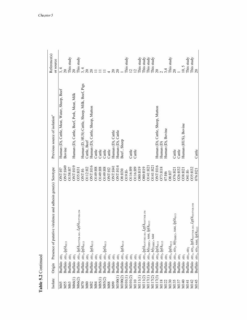
Tab
le 5
.2 C
ontin
ued
Isol
ate
Orig
in
Pres
ence
of p
utat
ive
viru
lenc
e an
d ad
hesi
n ge
ne(s
)Se
roty
pe
Prev
ious
sour
ce o
f iso
latio
na R
efer
ence
(s)
or so
urce
M
45
Buf
falo
stx
1 O
NT:
H?
Hum
an (D
), C
attle
, Mea
t, W
ater
, She
ep, B
eef
3, 4
M
55
Buf
falo
stx
1, lp
fA01
13
ON
T:H
49
Bov
ine
20
M63
B
uffa
lo s
tx1
O15
8:H
?
This
stud
y M
66(1
) B
uffa
lo s
tx1
ON
T:H
19
Hum
an (D
), C
attle
, Bee
f, Po
rk, M
eat,
Milk
20
M
66(2
) B
uffa
lo s
tx1,
lpfA
O15
7/O
I-14
1, Lp
fAO
157/
OI-
154
O35
:H31
This
stud
y M
70
Buf
falo
stx
1, lp
fA01
13
ON
T:H
- H
uman
(D, H
US)
Cat
tle, S
heep
, Milk
, Bee
f, Pi
gs
3, 4
M
80
Buf
falo
stx
1 O
112:
H2
Cat
tle, B
eef
20
M82
B
uffa
lo s
tx1,
lpfA
0113
O
NT:
H16
H
uman
(D),
Cat
tle, S
heep
, Mut
ton
20
M84
B
uffa
lo s
tx1
O14
9:H
8 C
attle
11
M
85(1
) B
uffa
lo s
tx1
O14
9:H
8 C
attle
11
M
85(2
) B
uffa
lo s
tx1,
lpfA
0113
O
149:
H8
Cat
tle
11
M88
B
uffa
lo s
tx1
ON
T:H
2
Cat
tle
4 M
99
Buf
falo
stx
1 O
NT:
H41
H
uman
(D),
Cat
tle
20
M10
0(1)
Buf
falo
stx
1 O
NT:
H14
H
uman
(D),
Cat
tle
20
M10
0(2)
Buf
falo
stx
1, lp
fA01
13
O8:
H30
B
eef ,
She
ep
1 M
101(
1) B
uffa
lo s
tx1
O19
:H-
Th
is st
udy
M10
1(2)
Buf
falo
stx
1 O
116:
H9
Cat
tle
12
M11
0 B
uffa
lo s
tx1
O11
6:H
9 C
attle
12
M
111(
1) B
uffa
lo s
tx1
O80
:H19
This
stud
y M
111(
2) B
uffa
lo s
tx1,
lpfA
O15
7/O
I-14
1, Lp
fAO
157/
OI-
154
O80
:H19
This
stud
y M
117(
1) B
uffa
lo s
tx1,
hly E
HEC
, saa
, lpf
A 011
3 O
141:
H21
This
stud
y M
117(
2) B
uffa
lo s
tx1,
hly E
HEC
, saa
O
141:
H21
This
stud
y M
117(
3) B
uffa
lo s
tx2,
lpfA
0113
O
NT:
H16
H
uman
(D),
Cat
tle, S
heep
, Mut
ton
20
M11
8 B
uffa
lo s
tx2,
lpfA
0113
O
73:H
18
Hum
an (D
) 2
M12
2 B
uffa
lo s
tx1
07:H
6 H
uman
(D),
Bov
ine
3,4
M13
0 B
uffa
lo s
tx1,
lpfA
0113
O
8:H
7
This
stud
y M
135
Buf
falo
stx
1, st
x 2, h
lyEH
EC, s
aa, l
pfA 0
113
O76
:H21
C
attle
20
M
137
Buf
falo
stx
1 O
26:H
32
Cat
tle
1 M
140
Buf
falo
stx
1 O
38:H
21
Hum
an (H
US)
, Bov
ine
10, 5
M
141
Buf
falo
stx
1, st
x 2
O41
:H-
Th
is st
udy
M14
2 B
uffa
lo s
tx1,
stx 2
, lpf
A O15
7/O
I-14
1, Lp
fAO
157/
OI-
154
O35
:H32
This
stud
y M
145
Buf
falo
stx
1, st
x 2, s
aa, l
pfA 0
113
076:
H21
C
attle
20
Cha pter 5
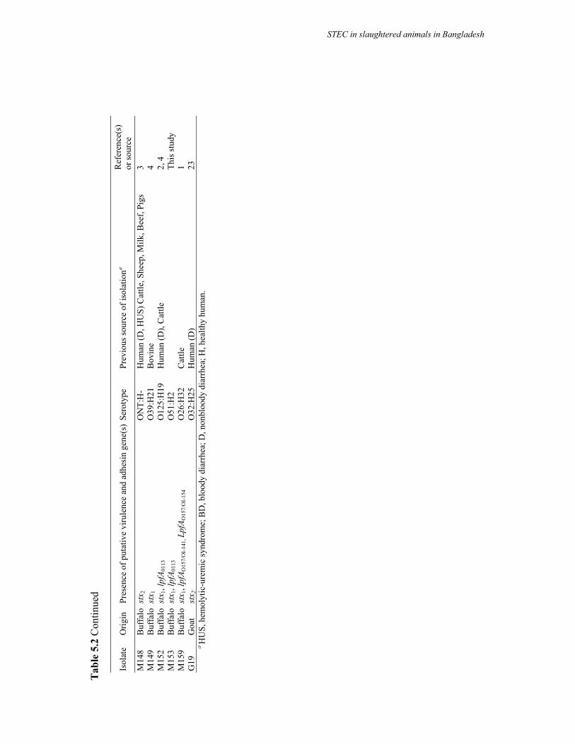
Tab
le 5
.2 C
ontin
ued
Isol
ate
Orig
in
Pres
ence
of p
utat
ive
viru
lenc
e an
d ad
hesi
n ge
ne(s
)Se
roty
pe
Prev
ious
sour
ce o
f iso
latio
na R
efer
ence
(s)
or so
urce
M
148
Buf
falo
stx
2 O
NT:
H-
Hum
an (D
, HU
S) C
attle
, She
ep, M
ilk, B
eef,
Pigs
3
M14
9 B
uffa
lo s
tx1
O39
:H21
B
ovin
e 4
M15
2 B
uffa
lo s
tx1,
lpfA
0113
O
125:
H19
H
uman
(D),
Cat
tle
2, 4
M
153
Buf
falo
stx
1, lp
fA01
13
O51
:H2
Th
is st
udy
M15
9 B
uffa
lo s
tx1,
lpfA
O15
7/O
I-14
1, L
pfA O
157/
OI-
154
O26
:H32
C
attle
1
G19
G
oat
stx 2
O
32:H
25
Hum
an (D
) 23
a H
US,
hem
olyt
ic-u
rem
ic sy
ndro
me;
BD
, blo
ody
diar
rhea
; D, n
onbl
oody
dia
rrhe
a; H
, hea
lthy
hum
an.
STEC in slaughtered animals in Bangladesh
Chapter 5
102
When buffalo samples were tested for the presence of stx genes by PCR, around 80% of
the samples were positive. Of the cows and goats, around 73% and 12%, respectively, were
positive. STEC non-O157 strains could be isolated from 23.6%, 12.9% and 0.9% of the
buffalo, cow and goat samples, respectively. Failure to isolate the STEC non-O157 strains
from PCR-positive fecal samples is a common problem (3). In India, viable STEC were
recovered from 24 to 40% of PCR-positive samples, depending on the origin of the sample.
The nonisolation of viable STEC from PCR-positive samples might be due to the presence of
very low numbers of bacteria, the presence of free stx phages in the sample, and the loss of
stx genes upon subcultivation of strains (23). Under routine diagnostics, there is no definitive
biochemical characteristic, like sorbitol fermentation in the case of serogroup O157, which
can identify STEC irrespective of the serotypes from commensal flora.
Most of the STEC O157 isolates (91.1%) were positive for the stx2 gene only. In humans,
epidemiologic data suggest that E. coli O157 strains that express Stx2 are more important
than Stx1 in the development of HUS (17) and that strains that express Stx2 alone are more
likely to be associated with the progression to HUS than are strains that produce both Stx1
and Stx2 (16). When the presence of other virulence genes was analyzed, most of the strains
were found to be positive for the eae (95.5%), hlyEHEC (93.3%), katP (93.3%), and etpD
(93.3%) genes (Table 5.1). Thus, STEC O157 isolates in this study were found to be carrying
additional virulence factors, clearly associated with increased human pathogenicity. In our
previous study of patients in Bangladesh, we found the prevalence of STEC in diarrheal
patients was relatively low compared to that of other enteric pathogens, including V. cholerae
and Shigella spp. (23). STEC O157 were not found in any of the patients. The higher
prevalence of STEC in cattle, in tandem with a low isolation rate in diarrheal patients in
Bangladesh, can be explained by the presence of the high prevalence of antibodies in the
population, which prevent infection and/or clinical manifestations of STEC infection.
However, further studies are needed to establish this explanation.
At least 90 PT have been reported for STEC O157 (25), but only seven of these (PT2,
PT4, PT8, PT14, PT21/28, PT32, and PT54) account for the majority (>75%) of the human
strains isolated in Europe and Canada (30). More than 77% (n = 35) of the STEC O157
isolates in this study were characterized by seven recognized PT. PT14 (24.4%), -31 (24.4%),
and -32 (8.9%) were commonly found in all three types of animals, with differences in
prevalence. Other PTs that were found in this study were PT8 for goat isolates and PT34,
PT38, and PT54 for buffalo isolates. PT14 has commonly been found among STEC O157
strains from different sources, including cattle feces, meat, and human patients (25).
STEC in slaughtered animals in Bangladesh
103
Furthermore, a significant association has been detected between PT14 and the presence of
acute pathologies (30). PT8 is one of the PTs most frequently found among STEC O157:H7
strains from humans and bovines in many European countries, including Spain, Belgium,
Finland, Germany, Italy, England, and Scotland (25). PT34 and PT54 have also been found
among strains from both human and animal origin but appeared to be more frequent among
strains from animal origins (30). PT32 was detected in STEC O157 strains associated with
several large outbreaks, including a multistate outbreak in the United States (6) and a recent
one in Ireland (29). PT31 was found in both human and non-human sources (meat and
slaughterhouses) in Canada, suggesting a foodborne transmission in human cases (25).
A high degree of polymorphism was observed for STEC O157 isolates analyzed by
PFGE. A total of 37 distinct profiles were obtained among 45 isolates. Strains which had
identical PFGE patterns belonged to the same type of animal (Fig. 5.1). No PFGE profiles
were found to be commonly generated by isolates from different types of animals (Fig. 5.1).
STEC non-O157 isolates have been shown to be important pathogens, despite being
severely underreported, because in many laboratories, the facilities to isolate, identify, and
characterize them do not exist. Where facilities do exist, STEC non-O157 isolates are found
(1). In the present study, we have characterized 71 STEC non-O157 isolates. These isolates
belonged to 36 O serogroups and 52 O:H serotypes (Table 5.2), of which 32 were isolated
from buffalo only, 14 from cow only, 1 from goat only, and 5 from both cow and buffalo. All
non-O157 serotypes in the study were compared with the serotypes isolated in previous
studies from different sources. The majority of STEC isolates belonged to serotypes (42.3%;
n = 22) previously found among STEC of human origin, of which 31.8% (n = 7) were of
serotypes associated with severe infections, including bloody diarrhea and HUS (Table 5.2).
In addition, 17 serotypes found in this study were found previously among STEC strains
isolated from multiple origins, including those of human patients, cattle, and milk/meat
(Table 5.2). Interestingly, 18 serotypes in this study could not be matched with the
recognized STEC serotypes reported so far (Table 5.2).
When virulence genes were analyzed, 78.9% of the isolates were found to be positive for
stx1 only, 12.7% were positive for stx2 only, and 8.4% were positive for both stx1 and stx2.
Higher prevalence rates of stx1 have been reported in strains from animal origin in previous
studies (42), which corroborates the results of the present study. Only 7% of the isolates were
found to be positive for the hlyEHEC gene (Table 5.1), and no isolates were positive for other
virulence genes, including eae, katP, and etpD. STEC strains, which are positive for eae that
is harbored by the large pathogenicity island LEE are considered to be highly virulent in
Chapter 5
104
humans (40). The LEE appears to confer enhanced virulence; however, the presence of the
LEE is not essential for pathogenesis, since a number of cases of severe STEC disease,
including HUS, as well as occasional outbreaks, were caused by the LEE-negative strains
(31). It has been suggested that additional virulence factors, including adhesions encoded
outside of the LEE, may play a role in STEC pathogenesis (40). Since all non-O157 isolates
in this study were negative for eae, the isolates were tested for the presence of several novel
adhesin genes. About 30% (n = 15) of the buffalo and 47% (n = 10) of the cow isolates were
positive for the lpfO113 gene (Table 5.1). This is in accordance with a previous study where
lpfO113 was found to be one of the most prevalent adhesin genes among STEC non-O157
strains (40). More than 8% (n = 4) of the buffalo and 19% (n = 4) of the cow isolates
possessed the saa gene. In the study previously referred to, it was shown that there is a
correlation between the presence of saa and hlyEHEC for certain serotypes (40). In the present
study, similar results were found; among eight isolates positive for saa, five (62.5%) were
positive for hlyEHEC.
Although a high degree of serotypic diversity was found among STEC non-O157 isolates
in this study, isolates of the same serotype were found in different samples from different
animal types. In addition, more than one isolate belonging to the same serotype with different
virulence profiles was isolated from the same sample. PFGE analysis was applied to find out
if there was any genetic relationship among isolates of the same serotype. Identical PFGE
patterns were found among isolates belonging to the same serotype but isolated from
different samples, and at the same time, different PFGE patterns were found in the case of
some serotypes.
In our previous study, we found a low prevalence of STEC in diarrheal patients in
Bangladesh, with no cases of STEC O157 infection (23). In this study, we found a
significantly higher prevalence of both STEC O157 and non-O157 serotypes in the animal
population. Therefore, there must be a limiting factor existing in the transmission route of this
pathogen from the reservoir to the human population or there might be natural protection
against STEC infection among the general population in Bangladesh. Further studies are
being carried out to confirm these assumptions.
STEC in slaughtered animals in Bangladesh
105
Acknowledgments
This research was funded by the International Centre for Diarrhoeal Disease Research,
Bangladesh (ICDDR,B) and by the Netherlands Foundation for the Advancement of Tropical
Research (NWO-WOTRO) grant (Award number WB 93-415). ICDDR,B acknowledges
with gratitude the commitment of the Netherlands Foundation for the Advancement of
Tropical Research (NWO-WOTRO) grant to the Centre's research efforts. ICDDR,B also
gratefully acknowledges these donors who provide unrestricted support to the Centre's
research efforts: Australian International Development Agency (AusAID), Government of
Bangladesh, Canadian International Development Agency (CIDA), The Kingdom of Saudi
Arabia (KSA), Government of the Netherlands, Government of Sri Lanka, Swedish
International Development Cooperative Agency (SIDA), Swiss Development Cooperation
(SDC) and Department for International Development, UK (DFID).
References
1. Bettelheim, K. A. 2007. The non-O157 Shiga-toxigenic (verocytotoxigenic) Escherichia coli; under-rated pathogens. Crit. Rev. Microbiol. 33:67-87.
2. Beutin, L., G. Krause, S. Zimmermann, S. Kaulfuss, and K. Gleier. 2004. Characterization of Shiga toxin-producing Escherichia coli strains isolated from human patients in Germany over a 3-year period. J. Clin. Microbiol. 42:1099-1108.
3. Blanco, J. E., M. Blanco, M. P. Alonso, A. Mora, G. Dahbi, M. A. Coira, and J. Blanco. 2004. Serotypes, virulence genes, and intimin types of Shiga toxin (verotoxin)-producing Escherichia coli isolates from human patients: prevalence in Lugo, Spain, from 1992 through 1999. J. Clin. Microbiol. 42:311-319.
4. Blanco, M., J. E. Blanco, A. Mora, G. Dahbi, M. P. Alonso, E. A. Gonzalez, M. I. Bernardez, and J. Blanco. 2004. Serotypes, virulence genes, and intimin types of Shiga toxin (verotoxin)-producing Escherichia coli isolates from cattle in Spain and identification of a new intimin variant gene (eae-ζ). J. Clin. Microbiol. 42:645-651.
5. Boerlin, P., S. A. McEwen, F. Boerlin-Petzold, J. B. Wilson, R. P. Johnson, and C. L. Gyles. 1999. Associations between virulence factors of Shiga toxin-producing Escherichia coli and disease in humans. J. Clin. Microbiol. 37:497–503.
6. Breuer, T., D. H. Benkel, R. L. Shapiro, W. N. Hall, M. M. Winnett, M. J. Linn, J. Neimann, T. J. Barrett, S. Dietrich, F. P. Downes, D. M. Toney, J. L. Pearson, H. Rolka, L. Slutsker, and P. M. Griffin. 2001. A multi-state outbreak of Escherichia coli O157:H7 infections linked to alfalfa sprouts grown from contaminated seeds. Emerg. Infect. Dis. 7:977-982.
7. Brunder, W., H. Schmidt, and H. Karch. 1996. KatP, a novel catalase-peroxidase encoded by the large plasmid of enterohaemorrhagic Escherichia coli O157:H7. Microbiology 142:3305-3315.
Chapter 5
106
8. Brunder, W., H. Schmidt, and H. Karch. 1997. EspP, a novel extracellular serine protease of enterohaemorrhagic Escherichia coli O157:H7 cleaves human coagulation factor V. Mol. Microbiol. 24:767-778.
9. Caprioli, A., S. Morabito, H. Brugere, and E. Oswald. 2005. Enterohaemorrhagic Escherichia coli: emerging issues on virulence and modes of transmission. Vet. Res. 36:289-311.
10. Cho, S., F. Diez-Gonzalez, C. P. Fossler, S. J. Wells, C. W. Hedberg, J. B. Kaneene, P. L. Ruegg, L. D. Warnick, and J. B. Bender. 2006. Prevalence of Shiga toxin-encoding bacteria and Shiga toxin-producing Escherichia coli isolates from dairy farms and county fairs. Vet. Microbiol. 118:289-298.
11. Cookson, A. L., S. C. Taylor, J. Bennett, F. Thomson-Carter, and G. T. Attwood. 2006. Serotypes and analysis of distribution of Shiga toxin-producing Escherichia coli from cattle and sheep in the lower North Island, New Zealand. N. Z. Vet. J. 54:78-84.
12. Cookson, A. L., J. Bennett, F. Thomson-Carter, and G. T. Attwood. 2007. Intimin subtyping of Escherichia coli: concomitant carriage of multiple intimin subtypes from forage-fed cattle and sheep. FEMS Microbiol. Lett. 272:163-171.
13. Dell'Omo, G., S. Morabito, R. Quondam, U. Agrimi, F. Ciuchini, A. Macrì, and A. Caprioli. 1998. Feral pigeons as a source of verocytotoxin-producing Escherichia coli. Vet. Rec. 142:309-310.
14. Dutta, S., A. Deb, U. K. Chattopadhyay, and T. Tsukamoto. 2000. Isolation of Shiga toxin-producing Escherichia coli including O157:H7 strains from dairy cattle and beef samples marketed in Calcutta, India. J. Med. Microbiol. 49:765-767.
15. Elder, R. O., J. E. Keen, G. R. Siragusa, G. A. Barkocy-Gallagher, M. Koohmaraie, and W. W. Laegreid. 2000. Correlation of enterohemorrhagic Escherichia coli O157 prevalence in feces, hides, and carcasses of beef cattle during processing. Proc. Natl. Acad. Sci. USA. 97:2999-3003.
16. Griffin, P. M., and R. V. Tauxe. 1991. The epidemiology of infections caused by Escherichia coli O157:H7, other enterohemorrhagic E. coli, and the associated haemolytic uraemic syndrome. Epidemiol. Rev. 13:60-97.
17. Griffin, P. M. 1995. Escherichia coli O157:H7 and other enterohemorrhagic Escherichia coli. p. 739–761. In M. J. Blaser, P. D. Smith, J. I. Rovin, H. B. Greenberg, and R. L. Guerrant (ed.), Infections of the gastrointestinal tract. Raven Press, New York, NY.
18. Gyles, C. L. 2007. Shiga toxin-producing Escherichia coli: an overview. J. Anim. Sci. 13 (Suppl.):E45-E62.
19. Hussein, H. S., and T. Sakuma. 2005. Prevalence of Shiga toxin-producing Escherichia coli in dairy cattle and their products. J. Dairy Sci. 88:450-465.
20. Hussein, H. S. 2007. Prevalence and pathogenicity of Shiga toxin-producing Escherichia coli in beef cattle and their products. J. Anim. Sci. 85:E63-E72.
21. Islam, M. A., A. E. Heuvelink, K. A. Talukder, and E. de Boer. 2006. Immunoconcentration of Shiga toxin-producing Escherichia coli O157 from animal faeces and raw meats by using Dynabeads anti-E. coli O157 and the VIDAS system. Int. J. Food Microbiol. 109:151-156.
22. Islam, M. A., A. E. Heuvelink, K. A. Talukder, M. H. Zwietering, and E. de Boer. 2006. Evaluation of immunomagnetic separation and PCR for the detection of Escherichia coli O157 in animal feces and meats. J. Food Prot. 69:2865-2869.
23. Islam, M. A., A. E. Heuvelink, E. de Boer, P. D. Sturm, R. R. Beumer, M. H. Zwietering, A. S. Faruque, R. Haque, D. A. Sack, and K. A. Talukder. 2007. Shiga toxin-producing Escherichia coli isolated from patients with diarrhoea in Bangladesh. J. Med. Microbiol. 56:380-385.
STEC in slaughtered animals in Bangladesh
107
24. Kaddu-Mulindw, D. H., T. Aisu, K. Gleier, S. Zimmermann, and L. Beutin. 2001. Occurrence of Shiga toxin-producing Escherichia coli in fecal samples from children with diarrhoea and from healthy zebu cattle in Uganda. Int. J. Food Microbiol. 66:95-101.
25. Khakria, R., D. Duck, and H. Lior. 1990. Extended phage-typing scheme for Escherichia coli O157:H7. Epidemiol. Infect. 105:511-520.
26. Leelaporn, A., M. Phengmak, B. Eampoklap, S. Manatsathit, S. Tritilanunt, S. Siritantikorn, K. Nagayama, T. Iida, C. Niyasom, and P. Komolpit. 2003. Shiga toxin- and enterotoxin-producing Escherichia coli isolated from subjects with bloody and non-bloody diarrhea in Bangkok, Thailand. Diagn. Microbiol. Infect. Dis. 46:173-180.
27. Lehmacher, A., H. Meier, S. Aleksic, and J. Bockemühl. 1998. Detection of hemolysin variants of Shiga toxin-producing Escherichia coli by PCR and culture on vancomycin-cefixime-cefsulodin blood Agar. Appl. Environ. Microbiol. 64: 2449–2453.
28. Manna, S. K., M. P. Brahmane, C. Manna, K. Batabyal, and R. Das. 2006. Occurrence, virulence characteristics and antimicrobial resistance of Escherichia coli O157 in slaughtered cattle and diarrhoeic calves in West Bengal, India. Lett. Appl. Microbiol. 43:405–409.
29. Mannix, M., D. Whyte, E. McNamara, N. O Connell, R. Fitzgerald, M. Mahony, T. Prendiville, T. Norris, A. Curtin, A. Carroll, E. Whelan, J. Buckley, J. McCarthy, M. Murphy, and T. Greally. 2007. Large outbreak of E. coli O157 in 2005, Ireland. Eur. Surveill. 12(2):article 3.
30. Mora, A., M. Blanco, J. E. Blanco, M. P. Alonso, G. Dahbi, F. Thompson-Carter, M. A. Usera, R. Bartolomé, G. Prats, and J. Blanco. 2004. Phage types and genotypes of human and animal Shiga toxin-producing Escherichia coli O157:H7 in Spain: identification of two predominating phage types (PT2 and PT8). J. Clin. Microbiol. 42:4007-4015.
31. Paton, J. C., and A. W. Paton. 1998. Pathogenesis and diagnosis of Shiga toxin-producing Escherichia coli infections. Clin. Microbiol. Rev. 11:450-479.
32. Paton, A. W., M. C. Woodrow, R. M. Doyle, J. A. Lanser, and J. C. Paton. 1999. Molecular characterization of a Shiga toxigenic Escherichia coli O113:H21 strain lacking eae responsible for a cluster of cases of hemolytic-uremic syndrome. J. Clin. Microbiol. 37:3357–3361.
33. Pradel, N., V. Livrelli, C. De Champs, J. B. Palcox, A. Reynaud, F. Scheutz, J. Sirot, B. Joly, and C. Forestier. 2000. Prevalence and characterization of Shiga toxin-producing Escherichia coli isolated from cattle, food, and children during a one-year prospective study in France. J. Clin. Microbiol. 38:1023-1031.
34. Qadri, F., A. M. Svennerholm, A. S. Faruque, and R. B. Sack. 2005. Enterotoxigenic Escherichia coli in developing countries: epidemiology, microbiology, clinical features, treatment and prevention. Clin. Microbiol. Rev. 18:465-483.
35. Ribot, E. M., M. A. Fair, R. Gautom, D. N. Cameron, S. B. Hunter, B. Swaminathan, and T. J. Barrett. 2006. Standardization of pulsed-field gel electrophoresis protocols for the subtyping of Escherichia coli O157:H7, Salmonella, and Shigella for PulseNet. Foodborne Pathog. Dis. 3:59-67.
36. Schmidt, H., L. Beutin, and H. Karch. 1995. Molecular analysis of the plasmid-encoded hemolysin of Escherichia coli O157:H7 strain EDL 933. Infect. Immun. 63:1055–1061.
Chapter 5
108
37. Schmidt, H., B. Henkel, and H. Karch. 1997. A gene cluster closely related to type II secretion pathway operons of gram-negative bacteria is located on the large plasmid of enterohemorrhagic Escherichia coli O157 strains. FEMS Microbiol. Lett. 148:265-272.
38. Strockbine, N. A., L. R. Marques, J. W. Newland, H. W. Smith, R. K. Holmes, and A. D. O'Brien. 1986. Two toxin-converting phages from Escherichia coli O157:H7 strain 933 encode antigenically distinct toxins with similar biologic activities. Infect. Immun. 53:135-140.
39. Tarr, P. I., C. A. Gordon, and W. L. Chandler. 2005. Shiga-toxin-producing Escherichia coli and haemolytic uraemic syndrome. Lancet 365:1073-1086.
40. Toma, C., E. M. Espinosa, T. Song, E. Miliwebsky, I. Chinen, S. Iyoda, M. Iwanaga, and M. Rivas. 2004. Distribution of putative adhesins in different seropathotypes of Shiga toxin-producing Escherichia coli. J. Clin. Microbiol. 42:4937–4946.
41. Vu-Khac, H., and N. A. Cornick. 2008. Prevalence and genetic profiles of Shiga toxin-producing Escherichia coli strains isolated from buffaloes, cattle, and goats in central Vietnam. Vet. Microbiol. 126:356-63.
42. Wieler, L. H., M. Tigges, F. Ebel, S. Schaferkordt, S. Djafari, T. Schlapp, G. Baljer, and T. Chakraborty. 1996. The enterohemolysin phenotype of bovine Shiga-like toxin-producing Escherichia coli (SLTEC) is encoded by the EHEC-hemolysin gene. Vet. Microbiol. 52:153-164.
43. Zheng, H., H. Jing, H. Wang, S. Xia, W. Hu, S. Cui, Z. Bi, J. Yang, B. Pang, G. Zhao, J. Zhang, H. Li, and J. Xu. 2005. stx2vha is the dominant genotype of Shiga toxin-producing Escherichia coli O157:H7 isolated from patients and domestic animals in three regions of China. Microbiol. Immunol. 49:1019-1026.
Shiga toxin-producing Escherichia coli in raw meat,
raw milk and street vended juices in Dhaka,
Bangladesh
Submitted for publication
M. Aminul Islam A. Salam Mondol I. Jahan Azmi E. de Boer R. R. Beumer M. H. Zwietering A. E. Heuvelink K. A. Talukder
Chapter 6
Chapter 6
110
Abstract
The prevalence of Shiga toxin (Stx)-producing Escherichia coli (STEC) has been
investigated in 210 food samples, including 87 raw meat samples collected from
slaughterhouses, 20 raw milk samples from domestic cattle, and 103 fresh juice samples from
street vendors in Dhaka city. More than 71% (n = 62) of the raw meat samples were positive
for the stx gene(s); 34% (n = 21) of buffalo meats and 66% (n = 41) of beef. Approximately
10% (n = 2) of the raw milk and 8% (n = 8) of the fresh juice samples were positive for stx.
STEC O157 were isolated from seven meat samples (two buffalo meats and five beef). No
other STEC serotypes could be isolated. STEC could not be isolated from any of the stx-
positive raw milk and juice samples. The STEC O157 isolates from raw meats were positive
for the stx2, eae, katP, etpD and enterohemorrhagic E. coli hly (hlyEHEC) virulence genes, and
belonged to three different phage types: 8 (14.3%), 31 (42.8%), and 32 (42.8%). PFGE
subtyping revealed six distinct restriction patterns suggesting a heterogeneous clonal
diversity. This is the first report of the isolation of STEC O157 from raw meat samples and
the presence of Shiga toxin-producing organisms in different food samples in Bangladesh.
STEC in raw meat, raw milk and street vended juices in Bangladesh
111
6.1 Introduction
Shiga toxin-producing Escherichia coli (STEC) organisms, also called verocytotoxin
(VT)-producing E. coli (VTEC), are one of the most important groups of foodborne
pathogens (14, 21). They are a major cause of gastroenteritis that may be complicated by
hemorrhagic colitis (HC) or the hemolytic-uremic syndrome (HUS), which is the main cause
of acute renal failure in children. STEC are a serologically diverse group of zoonotic
pathogens. Serotype O157:H7 has been the predominate type worldwide (23). Since its
identification as a pathogen in 1982, STEC O157:H7 has been the cause of a series of
outbreaks in Canada, Japan, the United Kingdom, the United States and other countries (14,
21, 25). However, more than 200 STEC serotypes have been reported and more than 100
have been linked with human infection (5).
The pathogenicity of STEC strains is associated with various virulence factors. The main
factor is the capacity to form two potent phage-encoded cytotoxins called Shiga toxins (Stx1,
Stx2 and their variants) or verocytotoxins (VT1 and VT2) (21). Another virulence factor
associated with clinical STEC isolates is the protein intimin, encoded by the eae gene and
responsible for the intimate attachment of STEC strains to the intestinal epithelial cells and
causing the formation of attaching and effacing (A/E) lesions in the intestinal mucosa (12).
Enterohemolysin, also called enterohemorrhagic E. coli (EHEC) hemolysin has also been
identified as a possible STEC virulence factor (2).
STEC represent the only pathogenic group of E. coli that has a definite zoonotic origin,
although not all the STEC strains have been demonstrated to cause disease in humans. STEC
strains rarely cause disease in animals, and ruminants are recognized as their main natural
reservoir. Cattle are considered to be the major animal source of STEC that are virulent to
humans, in particular STEC O157 (3). STEC O157 and other serotypes associated with
human infections have also frequently been isolated from the intestinal content of other
ruminant species, including sheep, goat, water buffalo, and wild ruminants. Contamination of
carcasses with STEC usually occurs during slaughter and subsequent processing through
fecal material originating directly or indirectly from the rectal-cecal area (6). Dairy products
(milk, cheese, cream) associated with infection have included those that are unpasteurized,
have had a pasteurization failure or have been contaminated post-pasteurization. Ready-to-eat
foods have also been associated with infection, particularly cooked meats contaminated by
raw materials during processing, in catering establishments, at retail sale, and in the home. In
Chapter 6
112
developed countries, STEC O157 outbreaks associated with beef have caused concern among
public health workers, clinicians, and the public, prompting major changes in clinical and
laboratory practice, meat production, and food preparation (17).
Like many of the developing and underdeveloped countries, the hygienic conditions in
Bangladesh are severely compromised, and living with domestic animals within the same
premises is a common practice. In our previous study we found that the prevalence of STEC
in slaughter animals was 24%, and 43% of these STEC isolates belonged to serogroup O157
(11). This is in contrast to our study of patients in Bangladesh where we found the prevalence
of STEC was 0.5% among the hospitalized diarrheal patients, which was 1.9% of the cases of
community patients. No STEC O157 organisms were isolated in the study (10). From these
two studies, it can be concluded that although there is a high prevalence of STEC, including
serogroup O157 in animal reservoirs, these organisms are rarely associated with human
infections in Bangladesh. One of the reasons might be the absence of any food vehicles that
can transmit these organisms from animal reservoirs to humans. In Bangladesh, no study of
the occurrence of STEC in food sources had been done. Therefore, the objective of this study
was to determine the occurrence of STEC in different types of food samples, including raw
meat, raw milk and freshly prepared juices collected in Dhaka city.
6.2 Materials and methods
6.2.1 Sampling
In the period from 1 July 2006 to 31 January 2007, we collected raw meat samples from
28 buffalo, and 59 beef carcasses at slaughterhouses in Dhaka of the different areas in
Bangladesh. The origin of the animals could not be recorded due to the lack of information.
In the same period, we collected 20 raw milk samples from individual domestic dairy cows at
different suburbs of Dhaka city. In addition, we collected 103 freshly prepared (untreated)
juice samples, including mixed herbal juice (n = 49), sugar cane juice (n = 40), wood apple
juice (n = 7) and some other types of juice (three milk shake, one lemon, two mix fruit, and
one pineapple) from street vendors at different places of Dhaka city. Samples were
transported to the laboratory maintaining the cool chain (4 to 8°C) and microbiological
examination was started within 4 h.
STEC in raw meat, raw milk and street vended juices in Bangladesh
113
6.2.2 Isolation of STEC O157
For meat samples, a 25-g portion, and for raw milk and juice samples, a 25-ml portion
was added to 225 ml of modified tryptone soy broth (Oxoid Ltd., Basingstoke, United
Kingdom). After homogenization in a stomacher for 1 min, the samples were incubated for
18-20 h at 37°C. STEC O157 strains were isolated as described previously (9), by using the
immunomagnetic separation (IMS) technique and presumptive isolates were confirmed by an
agglutination test using E. coli O157 latex agglutination kit (Murex Biotech Ltd., Dartford,
Kent, United Kingdom) and a PCR for the rfb (O-antigen-encoding) region of E. coli O157
(rfbEO157) (21).
6.2.3 Isolation procedure for STEC non-O157
The overnight enrichment cultures were screened by PCR for the presence of stx1 and stx2
using the primers as described earlier (10). DNA was extracted from 1 ml of broth culture by
thermal cell lysis using Chelex-100 resin (Bio-Rad Laboratories Ltd., Richmond, Calif.) and
5 µl of the DNA extract was used in the PCR (18). Multiplex PCR for stx1 and stx2 was
carried out in a PTC-200 peltier thermal cycler (Bio-Rad) using the program as described
previously (10). DNA extracted from E. coli O157:H7 strain NCTC 12079 and E. coli strain
ATCC 25922 were used as positive and negative controls, respectively in each PCR reaction.
Broth cultures that yielded positive PCR results for stx1 and/or stx2 were streaked onto
tryptone bile x-glucuronide (TBX) agar (Oxoid) supplemented with cefixime (20 mg/l),
cefsulodin (3 mg/l) and vancomycin (30 mg/l) (CCV-TBX). Single colonies, including
different morphological types were transferred from the CCV-TBX and plated onto Luria
agar (Difco, BD diagnostics, USA) to create a grid pattern of 96 colonies (12×8). The plates
were incubated overnight at 37°C. The isolates were subjected to PCR for the detection of
stx1 and stx2 as described above. The number of PCR reactions was reduced to 20 by pooling
the colonies per row and per column in 500 µl sterile distilled water. DNA was extracted by
boiling the suspensions for 10 min and 5 µl of the supernatant was used in the PCR. The
isolates with positive PCR results were identified as E. coli by an API 20E test (bioMérieux,
Marcy l'Etoile, France).
6.2.4 Phage typing
Isolates confirmed to be STEC O157 were phage typed at the laboratory of Enteric
Pathogens, Central Public Health Laboratory, London, United Kingdom.
Chapter 6
114
6.2.5 PCR detection of virulence factors
PCR for detecting both chromosome (e.g., stx1, stx2, and eae)- and plasmid (e.g., hlyEHEC,
katP, and etpD)-encoded virulence genes was performed as described earlier (10) in a total
volume of 25 µl containing 2.5 µl of 10× Fast Start buffer with 1.5 mM MgCl2 (Applied
Biosystems, Weiterstadt, Germany), 200 µM concentration of each deoxynucleoside
triphosphate, a 0.2 pmol of each primer, and 1 U of Fast start Taq DNA polymerase (Roche,
Mannheim, Germany) using a PTC-200 peltier thermal cycler (Bio-Rad).
6.2.6 PFGE
PFGE was performed following the standardized protocol developed by PulseNet for E.
coli O157:H7 (22). Analysis of the TIFF images was carried out by the BioNumerics
software (Applied Maths, Belgium) using the dice coefficient and unweighted-pair group
method using average linkages to generate dendrograms with 1.0% tolerance values.
6.3 Results
6.3.1 Prevalence of STEC
Of the 87 meat samples collected from the slaughterhouses, 62 (71%) were positive for
stx gene(s); 21 samples of buffalo meats, and 41 samples of beef (Table 6.1). STEC O157
strains were isolated from seven samples; two samples of buffalo meats, and five samples of
beef (Table 6.1). No STEC non-O157 strains were isolated. Of the 20 raw milk samples, two
(10%) were positive for stx gene(s) (Table 6.1). Of the 103 fresh juices, eight (8%) samples
were stx-positive; six samples of mixed herbal juices, and two samples of wood apple juices
(Table 6.1). However, no STEC could be isolated from stx-positive positive samples of raw
milk and street vended juices.
6.3.2 Characterization of STEC O157
All seven STEC O157 isolates were positive for the stx2, eae, etpD, katP and hlyEHEC
genes. The strains could be grouped into three different phage types: phage type 31 (n = 4),
phage type 32 (n = 2), and phage type 8 (n = 1). PFGE analysis revealed six distinct
restriction profiles (Fig. 6.1).
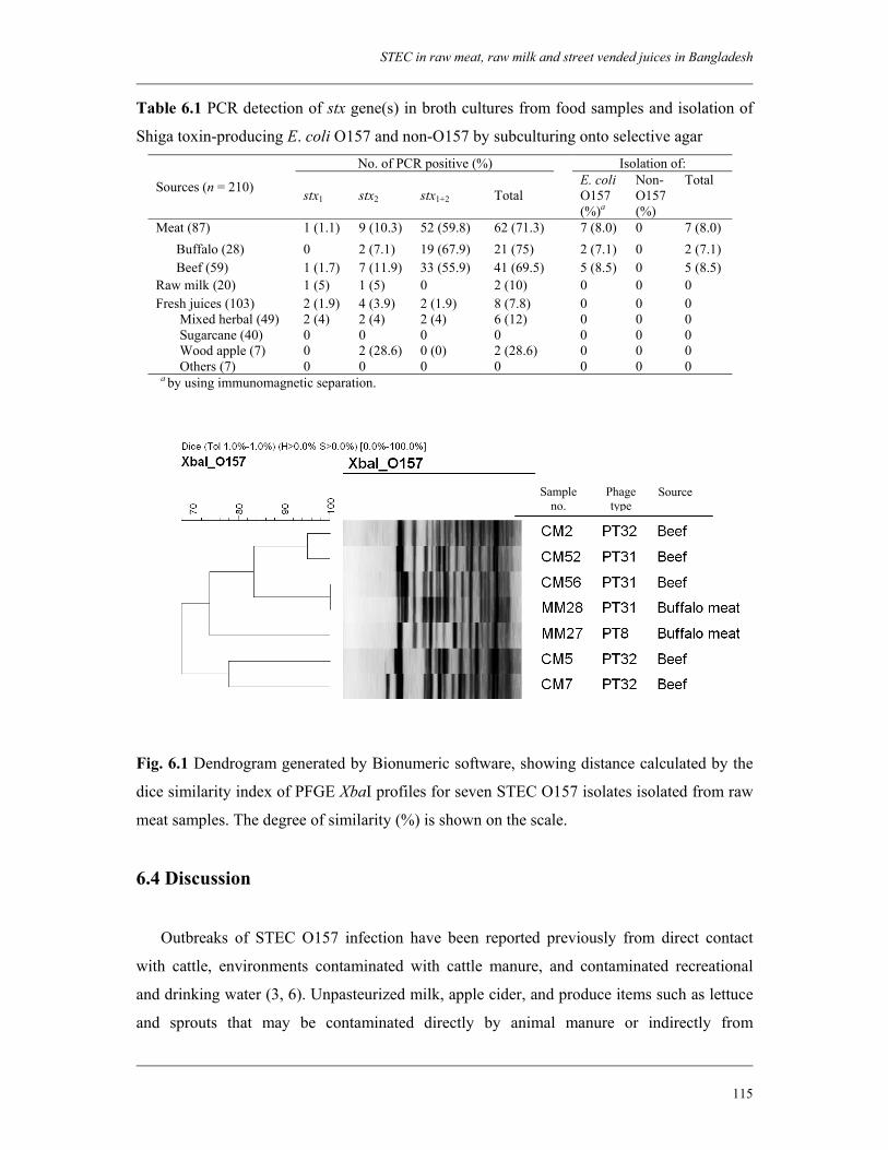
STEC in raw meat, raw milk and street vended juices in Bangladesh
115
Sample no.
Phage type
Source
Table 6.1 PCR detection of stx gene(s) in broth cultures from food samples and isolation of
Shiga toxin-producing E. coli O157 and non-O157 by subculturing onto selective agar No. of PCR positive (%) Isolation of:
Sources (n = 210) stx1 stx2 stx1+2 Total E. coli
O157 (%)a
Non-O157 (%)
Total
Meat (87) 1 (1.1) 9 (10.3) 52 (59.8) 62 (71.3) 7 (8.0) 0 7 (8.0) Buffalo (28) 0 2 (7.1) 19 (67.9) 21 (75) 2 (7.1) 0 2 (7.1) Beef (59) 1 (1.7) 7 (11.9) 33 (55.9) 41 (69.5) 5 (8.5) 0 5 (8.5) Raw milk (20) 1 (5) 1 (5) 0 2 (10) 0 0 0 Fresh juices (103) 2 (1.9) 4 (3.9) 2 (1.9) 8 (7.8) 0 0 0 Mixed herbal (49) 2 (4) 2 (4) 2 (4) 6 (12) 0 0 0 Sugarcane (40) 0 0 0 0 0 0 0 Wood apple (7) 0 2 (28.6) 0 (0) 2 (28.6) 0 0 0 Others (7) 0 0 0 0 0 0 0
a by using immunomagnetic separation.
Fig. 6.1 Dendrogram generated by Bionumeric software, showing distance calculated by the
dice similarity index of PFGE XbaI profiles for seven STEC O157 isolates isolated from raw
meat samples. The degree of similarity (%) is shown on the scale.
6.4 Discussion
Outbreaks of STEC O157 infection have been reported previously from direct contact
with cattle, environments contaminated with cattle manure, and contaminated recreational
and drinking water (3, 6). Unpasteurized milk, apple cider, and produce items such as lettuce
and sprouts that may be contaminated directly by animal manure or indirectly from
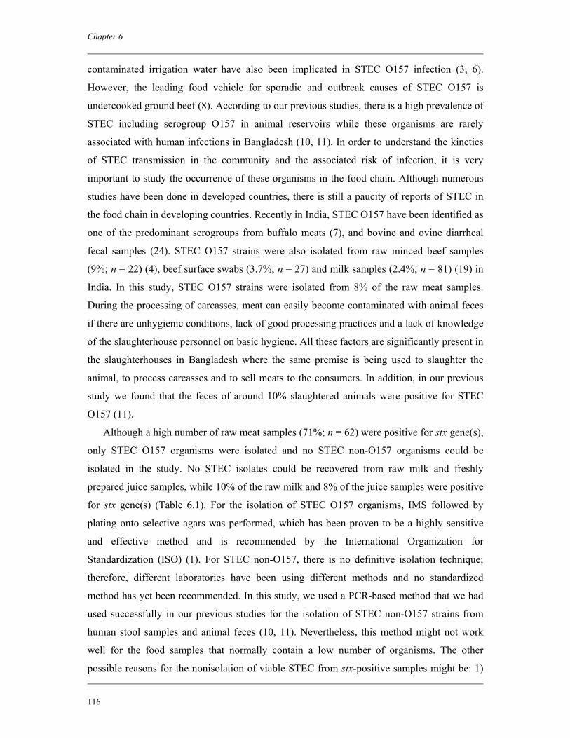
Chapter 6
116
contaminated irrigation water have also been implicated in STEC O157 infection (3, 6).
However, the leading food vehicle for sporadic and outbreak causes of STEC O157 is
undercooked ground beef (8). According to our previous studies, there is a high prevalence of
STEC including serogroup O157 in animal reservoirs while these organisms are rarely
associated with human infections in Bangladesh (10, 11). In order to understand the kinetics
of STEC transmission in the community and the associated risk of infection, it is very
important to study the occurrence of these organisms in the food chain. Although numerous
studies have been done in developed countries, there is still a paucity of reports of STEC in
the food chain in developing countries. Recently in India, STEC O157 have been identified as
one of the predominant serogroups from buffalo meats (7), and bovine and ovine diarrheal
fecal samples (24). STEC O157 strains were also isolated from raw minced beef samples
(9%; n = 22) (4), beef surface swabs (3.7%; n = 27) and milk samples (2.4%; n = 81) (19) in
India. In this study, STEC O157 strains were isolated from 8% of the raw meat samples.
During the processing of carcasses, meat can easily become contaminated with animal feces
if there are unhygienic conditions, lack of good processing practices and a lack of knowledge
of the slaughterhouse personnel on basic hygiene. All these factors are significantly present in
the slaughterhouses in Bangladesh where the same premise is being used to slaughter the
animal, to process carcasses and to sell meats to the consumers. In addition, in our previous
study we found that the feces of around 10% slaughtered animals were positive for STEC
O157 (11).
Although a high number of raw meat samples (71%; n = 62) were positive for stx gene(s),
only STEC O157 organisms were isolated and no STEC non-O157 organisms could be
isolated in the study. No STEC isolates could be recovered from raw milk and freshly
prepared juice samples, while 10% of the raw milk and 8% of the juice samples were positive
for stx gene(s) (Table 6.1). For the isolation of STEC O157 organisms, IMS followed by
plating onto selective agars was performed, which has been proven to be a highly sensitive
and effective method and is recommended by the International Organization for
Standardization (ISO) (1). For STEC non-O157, there is no definitive isolation technique;
therefore, different laboratories have been using different methods and no standardized
method has yet been recommended. In this study, we used a PCR-based method that we had
used successfully in our previous studies for the isolation of STEC non-O157 strains from
human stool samples and animal feces (10, 11). Nevertheless, this method might not work
well for the food samples that normally contain a low number of organisms. The other
possible reasons for the nonisolation of viable STEC from stx-positive samples might be: 1)
STEC in raw meat, raw milk and street vended juices in Bangladesh
117
the presence of other organisms that possess stx genes; 2) the presence of free bacteriophages
in the sample that contain stx genes (16); or 3) due to the loss of stx genes upon
subcultivation of strains (13).
STEC O157 strains that were isolated from raw meat samples were further characterized.
In addition to stx, the STEC O157 isolates were found to carry other virulence genes,
including eae, katP, etpD and hlyEHEC and therefore, appeared to be potential human
pathogens. Phage types (PT) 31 and 32 encompassed more than 85% (n = 6) of the isolates.
Interestingly, these two phage types were found to be the predominant PTs among STEC
O157 strains isolated previously from animal feces (11). It has been reported from many
countries that these phage types are commonly found in both human and non-human sources
(meat and slaughterhouses) suggesting a foodborne transmission in human cases (15). Phage
type 32 was detected in STEC O157 strains associated with the largest outbreak ever
recorded in Ireland (20). One strain in this study was typed as phage type 8, which is one of
the most frequently found phage types among STEC O157 strains from humans and bovine in
many European countries, including Spain, Belgium, Finland, Germany, Italy, England and
Scotland (15). PFGE analysis of the seven STEC O157 isolates revealed six distinct patterns,
suggesting a genetically heterogeneous group. Two strains, one from beef and one from
buffalo meat belonged to the same clonal type (Fig. 6.1).
To our knowledge, this is the first report of the occurrence of STEC O157 in meat
samples in Bangladesh. The results of this study highlight the poor hygienic conditions of
slaughterhouses in Dhaka and the lack of good processing practices. Although STEC
organisms could not be isolated from any raw milk and juice samples, the presence of stx
genes in the samples indicated that these might be contaminated with Shiga toxin-producing
organisms. Street vended juices are becoming increasingly popular in Bangladesh due to
rapid urbanization. These juices are commonly drunk in raw, exposing consumers to a
significant risk of infection with foodborne disease. Historically, people in Bangladesh prefer
to eat hot meals and extensive boiling is a common means of cooking. Therefore, raw meats
contaminated with STEC does not pose a potential risk to public health unless the issue of
cross-contamination of prepared food is taken into account. However, given the abundance of
STEC in the animal reservoirs (11), high risk of contamination of raw meats with STEC
O157 and the poor socioeconomic status of the people in Bangladesh, it is evident that STEC
are present at a significant rate in the food chain but are rarely causing infections in human
(10). This accentuates the need for further research on the antibody response to STEC,
especially to serogroup O157 among the healthy populations in Bangladesh.
Chapter 6
118
Acknowledgments
This research was funded by the International Centre for Diarrhoeal Disease Research,
Bangladesh (ICDDR,B) and by the Netherlands Foundation for the Advancement of Tropical
Research (NWO-WOTRO) grant (Award number WB 93-415). ICDDR,B acknowledges
with gratitude the commitment of the Netherlands Foundation for the Advancement of
Tropical Research (NWO-WOTRO) grant to the Centre's research efforts. ICDDR,B also
gratefully acknowledges these donors who provide unrestricted support to the Centre's
research efforts: Australian International Development Agency (AusAID), Government of
Bangladesh, Canadian International Development Agency (CIDA), The Kingdom of Saudi
Arabia (KSA), Government of the Netherlands, Government of Sri Lanka, Swedish
International Development Cooperative Agency (SIDA), Swiss Development Cooperation
(SDC) and Department for International Development, UK (DFID).
References
1. Anonymous. 2001. Microbiology of food and animal feeding stuffs—Horizontal method for the detection of Escherichia coli O157 (EN ISO 16654). International Organization for Standardization, Geneva, Switzerland.
2. Beutin, L., M. A. Montenegro, I. Orskov, F. Orskov, J. Prada, S. Zimmermann, and R. Stephan. 1989. Close association of verotoxin (Shiga-like toxin) production with enterohemolysin production in strains of Escherichia coli. J. Clin. Microbiol. 27:2559–2564.
3. Caprioli, A., S. Morabito, H. Brugère, and E. Oswald. 2005. Enterohaemorrhagic Escherichia coli: emerging issues on virulence and modes of transmission. Vet. Res. 36:289-311.
4. Dutta, S., A. Deb, U. K. Chattopadhyay, and T. Tsukamoto. 2000. Isolation of Shiga toxin-producing Escherichia coli including O157:H7 strains from dairy cattle and beef samples marketed in Calcutta, India. J. Med. Microbiol. 49:765-767.
5. Eklund, M., F. Scheutz, and A. Siitonen. 2001. Clinical isolates of non-O157 Shiga toxin-producing Escherichia coli: serotypes, virulence characteristics, and molecular profiles of strains of the same serotype. J. Clin. Microbiol. 39:2829-2834.
6. Erickson, M. C., and M. P. Doyle. 2007. Food as a vehicle for transmission of Shiga toxin-producing Escherichia coli. J. Food Prot. 70:2426-2449.
7. Hazarika, R. A., D. K. Singh, K. N. Kapoor, R. K. Agarwal, A. B. Pandey, and Purusottam. 2007. Verotoxic Escherichia coli (STEC) from beef and its products. Indian J. Exp. Biol. 45:207-211.
8. Hussein, H. S. 2007. Prevalence and pathogenicity of Shiga toxin-producing Escherichia coli in beef cattle and their products. J. Anim. Sci. 85 Suppl 13:E63-72.
9. Islam, M. A., A. E. Heuvelink, K. A. Talukder, M. H. Zwietering, and E. de Boer. 2006. Evaluation of immunomagnetic separation and PCR for the detection of Escherichia coli O157 in animal feces and meats. J. Food Prot. 69:2865-2869.
STEC in raw meat, raw milk and street vended juices in Bangladesh
119
10. Islam, M. A., A. E. Heuvelink, E. de Boer, P. D. Sturm, R. R. Beumer, M. H. Zwietering, A. S. Faruque, R. Haque, D. A. Sack, and K. A. Talukder. 2007. Shiga toxin-producing Escherichia coli isolated from patients with diarrhoea in Bangladesh. J. Med. Microbiol. 56:380-385.
11. Islam, M. A., A. S. Mondol, E. de Boer, R. R. Beumer, M. H. Zwietering, K. A. Talukder, and A. E. Heuvelink. 2008. Prevalence and genetic characterization of Shiga toxin-producing Escherichia coli isolated from slaughtered animals in Bangladesh. Appl. Environ. Microbiol. 74:5414-5421.
12. Kaper, J. B., S. Elliott, V. Sperandio, N. T. Perna, G. F. Mayhew, and F. R. Blattner. 1998. Attaching and effacing intestinal histopathology and the locus of enterocyte effacement, p. 163-182. In J. B. Kaper and A. D. O'Brien (ed.), Escherichia coli O157:H7 and other Shiga toxin-producing E. coli strains. ASM press, Washington DC.
13. Karch, H., T. Meyer, H. Rüssmann, and J. Heesemann. 1992. Frequent loss of Shiga-like toxin genes in clinical isolates of Escherichia coli upon subcultivation. Infect. Immun. 60:3464-3467.
14. Karmali, M. A. 1989. Infection by verocytotoxin-producing Escherichia coli. Clin. Microbiol. Rev. 2:5-38.
15. Khakria, R., D. Duck, and H. Lior. 1990. Extended phage-typing scheme for Escherichia coli O157:H7. Epidemiol. Infect. 105:511-520.
16. Khan, A., S. Yamasaki, T. Sato, T. Ramamurthy, A. Pal, S. Datta, N. R. Chowdhury, S. C. Das, A. Sikdar, T. Tsukamoto, S. K. Bhattacharya, Y. Takeda, and G. B. Nair. 2002. Prevalence and genetic profiling of virulence determinants of non-O157 Shiga toxin producing Escherichia coli isolated from cattle, beef and human cases in Calcutta, India. Emerg. Infect. Dis. 8:54–62.
17. Mahon, B. E., P. M. Griffin, P. S. Mead, and R. V. Tauxe. 1997. Hemolytic uremic syndrome surveillance to monitor trends in infection with Escherichia coli O157:H7 and other shiga toxin-producing E. coli. Emerg. Infect. Dis. 3:409-412.
18. Malorny, B., J. Hoorfar, M. Hugas, A. Heuvelink, P. Fach, L. Ellerbroek, C. Bunge, C. Dorn, and R. Helmuth. 2003. Interlaboratory diagnostic accuracy of Salmonella specific PCR-based method. Int. J. Food Microbiol. 89:241-249.
19. Manna, S. K., M. P. Brahmane, C. Manna, K. Batabyal, and R. Das. 2006. Occurrence, virulence characteristics and antimicrobial resistance of Escherichia coli O157 in slaughtered cattle and diarrhoeic calves in West Bengal, India. Lett. Appl. Microbiol. 43:405–409.
20. Mannix, M., D. Whyte, E. McNamara, N. O Connell, R. Fitzgerald, M. Mahony, T. Prendiville, T. Norris, A. Curtin, A. Carroll, E. Whelan, J. Buckley, J. McCarthy, M. Murphy, and T. Greally. 2007. Large outbreak of E. coli O157 in 2005, Ireland. Eur. Surveill. 12 (2):article 3.
21. Paton, J. C., and A. W. Paton. 1998. Pathogenesis and diagnosis of Shiga toxin-producing Escherichia coli infections. Clin. Microbiol. Rev. 11:450-479.
22. Ribot, E. M., M. A. Fair, R. Gautom, D. N. Cameron, S. B. Hunter, B. Swaminathan, and T. J. Barrett. 2006. Standardization of pulsed-field gel electrophoresis protocols for the subtyping of Escherichia coli O157:H7, Salmonella, and Shigella for PulseNet. Foodborne Pathog. Dis. 3:59-67.
23. Tarr, P. I., C. A. Gordon, and W. L. Chandler. 2005. Shiga-toxin-producing Escherichia coli and haemolytic uraemic syndrome. Lancet 365:1073-1086.
24. Wani, S. A., M. A. Bhat, I. Samanta, Y. Nishikawa, and A. S. Buchh. 2003. Isolation and characterization of Shiga toxin-producing Escherichia coli (STEC) and
Chapter 6
120
enteropathogenic Escherichia coli (EPEC) from calves and lambs with diarrhoea in India. Lett. Appl. Microbiol. 37:121-126.
25. Willshaw, G. A., T. Cheasty, H. R. Smith, S. J. O'Brien, and G. K. Adak. 2001. Verocytotoxin-producing Escherichia coli (VTEC) O157 and other VTEC from human infections in England and Wales: 1995-1998. J. Med. Microbiol. 50:135-142.
Antibody responses to Escherichia coli O157
lipopolysaccharide among the healthy population in
Bangladesh
Submitted for publication
M. Aminul Islam Md. M. Rahaman A. E. Heuvelink E. de Boer M. H. Zwietering H. Chart A. Navarro A. Cravioto K. A. Talukder
Chapter 7
Chapter 7
122
Abstract
An investigation was conducted to determine the serum antibody response against
Escherichia coli O157 lipopolysaccharide (LPS) among the healthy population in
Bangladesh. Participants from different occupations, including butchers and from different
locations, including rural and urban areas were included. Of the 233 serum samples, 116
(50%) had antibodies (IgG, IgA and/or IgM) to E. coli O157 LPS by Western blot analysis.
Antibody classification showed that all 116 samples positive for polyvalent antibodies were
positive for IgG and 87 were also positive for IgM. Among nine household members of
butchers involved in other occupations, five showed positive antibody response. Of the 133
serum samples collected from butchers, 54 (41%) were antibody positive. No significant
difference in antibody response was found between the butchers from urban (33%, n = 14)
and rural areas (44%, n = 40). Among participants other than butchers and their family, 57
(63%) showed a positive antibody response, which was significantly higher than that of
butchers (P < 0.05). The prevalence of IgM-class antibody was found to be also higher
among nonbutcher participants (42%, n = 38) than butchers (33%, n = 44); however, the
difference was not statistically significant. For all categories of participants, no significant
association was found between the age and antibody response results. Use of water from
municipal water supply system or other contaminated water sources for drinking and washing
was associated with a positive antibody response to E. coli O157 significantly among urban
population than rural population (P < 0.001). This study concludes that the lack of STEC
O157 infection among Bangladeshi population might be attributable to the protective
immunity against these pathogens acquired by the frequent exposure to the antigens.
Antibody responses to E. coli O157 LPS among Bangladeshi population
123
7.1 Introduction
Shiga toxin (Stx)-producing Escherichia coli (STEC) are significant zoonotic pathogens
that are capable of causing severe gastrointestinal disease that can lead to a number of
sequelae, including hemorrhagic colitis (HC), hemolytic-uremic syndrome (HUS) and
thrombotic thrombocytopenic purpura (TTP) (31). HUS is the most common cause of acute
renal failure in children. The association between HUS and STEC infection has been well
documented over the past two decades (27) and is due to the production of one or more Stx’s
produced by the organisms (26).
Cattle are considered as the primary reservoir of STEC and infection may be transmitted
to humans through contaminated food, water or by direct animal contact (5). Rural
populations have been postulated to be at greater risk for exposure to STEC by virtue of
increased exposure to animals or their excreta (33). Dairy farm visits have been implicated as
a source for infection in many countries and animal contacts are a risk factor for the
development of HUS (17).
Most outbreaks and sporadic cases of HC and HUS have been associated with STEC
O157 infection (42) and reported from developed countries. Studies conducted in developing
areas of the world have shown little involvement of STEC strains as causal agents of
diarrheal disease in these countries (42). Interestingly, the conditions for transmission of
these strains from animals to humans exist, as do the inherent problems relating to basic
sanitation (16). Furthermore, studies in developing countries showed that STEC is
significantly present in animal reservoirs and in the food chain (18, 25, 43, 47). Therefore, the
question that remains to be answered is why there is such a lack of contribution of STEC as a
cause of major outbreaks of disease in these areas of the world. The reason is not always due
to a lack of technical capacity in participating laboratories or to inadequate surveillance
systems in these areas. One of the explanations would be the protective immunity against this
pathogen, which has already been suggested by several studies as a probable cause of low
incidence of STEC infection in developing countries (32, 46).
In general, patients infected with STEC produce serum antibodies to various components,
including Stx, secreted virulence factors and lipopolysaccharide (LPS) specific to a particular
serogroup (9, 22, 29, 30). Many studies have been done on the detection of antibodies to the
O157 LPS antigen, which is considered the basis for routine serological testing to provide
evidence of recent infection even in the absence of fecal E. coli O157 (12). Antibodies to E.
Chapter 7
124
coli O157 LPS may be found in healthy persons (20). A study in Canada showed that 6.3% of
apparently healthy people associated with cattle carrying fecal STEC also had fecal STEC,
including the serogroup O157, and suggested that occupational exposure to cattle harboring
STEC may lead to subclinical infections in humans (49). This study also found higher
frequencies of antibodies to O157 LPS among residents of rural areas compared with
residents of urban areas. It has been suggested that repeated and cumulative exposure to
STEC O157 as in the case of dairy farm residents or rural populations provides adequate
antigenic stimulation (a booster effect) to induce a systemic antibody response against this
pathogen (38).
There have been numerous investigations on the systemic immune response towards
STEC O157, especially in countries where its prevalence is high. Little research has been
done in countries having endemic and epidemic burden of various enteric pathogens with a
low incidence rate of STEC infection. No study has yet been carried out to investigate the
antibody response to E. coli O157 in a population having occupational exposure to animals in
a developing country. In our previous studies, we found that the prevalence of STEC in
diarrheal patients is significantly lower in Bangladesh compared with other enteric pathogens
(24). There have been no reports of HUS or HC in association with STEC O157 infection. In
contrast, there is a significant prevalence of potentially virulent STEC, including those of
serogroup O157 in food and food producing animals (25, M. A. Islam, A. S. Mondol, I. J.
Azmi, E. de Boer, R. R. Beumer, M. H. Zwietering, A. E. Heuvelink and K. A. Talukder,
Submitted for publication). This urges the necessity to test the antibody response against
STEC O157 among the healthy population, especially those who are in frequent contact with
animals compared to those who are not.
Therefore, the aim of this study was to determine the antibody response against E. coli
O157 LPS among the healthy Bangladeshi population, including people who are working in
slaughterhouses located in both urban and rural areas.
7.2 Materials and methods
7.2.1 Human sera
Sera were obtained from a total of 233 blood samples collected from different groups of
people, including butchers (people who work in the slaughterhouses) from both urban (n =
42) and rural areas (n = 91), household members of butchers (n = 9), and people with other
Antibody responses to E. coli O157 LPS among Bangladeshi population
125
occupations (n = 91), including businessmen, students, service holders and housewives. Five
ml of blood sample from each person were collected using a sterile syringe and kept at room
temperature for 2 h for separation of the blood cells and serum. Serum was collected from the
upper layer after centrifuging at 3,000 rpm for 15 min. Informed consent for participation in
this research was obtained from participants or their guardians. A standard questionnaire was
completed by all participants at the time of blood sampling and provided information on age,
sex, blood group, occupation, any intestinal problem from two weeks before sample
collection, characteristics of stool and contact with animals. Sera from healthy volunteers
from Mexico known to contain antibodies to E. coli O157 were used as positive controls, and
sera from anonymous blood donors were used as negative controls for Western blot analysis.
Sera were stored at –20°C until tested.
7.2.2 LPS prepared from whole bacteria
LPS was prepared by digesting whole bacteria with proteinase K (9) from a reference
STEC O157:H7 strain (NCTC 12079), which is positive for stx1, stx2 and eae genes. Bacteria
were grown overnight on TSA plates and the cells were suspended in SDS-PAGE sample
buffer in pre-weighted eppendorf tubes. Cell concentration was adjusted to 1 mg per 30 µl
prior to incubation at 100°C for 10 min. After cooling, samples were mixed with an equal
volume of SDS-PAGE buffer containing 100 µg proteinase K (Sigma Chemical Co., St.
Louis, Mo) per 30 µl prior to incubation at 60°C for 1 h.
7.2.3 Purification of LPS
LPS was prepared from bacterial outer membranes based on the hot-phenol method of
Westphal and Jann (48). Briefly, STEC O157:H7 reference strain (NCTC 12079) was grown
on TSA plates and the cells were harvested from overnight cultures and suspended in 5 ml 25
mM Tris-HCl (pH 7.4) containing 1 mM EDTA-Na2. Bacterial cells were disrupted by
sonication and cell suspensions were poured into a pre-cooled volume of 20 ml of the same
buffer and centrifuged to sediment any residual whole bacteria (5,000× g, 30 min, 4°C).
Supernatants, containing bacterial envelopes were transferred into new tubes and centrifuged
(45,000× g, 1 h, 4°C). Pellets were re-suspended in 1 ml of 25 mM Tris-HCl (pH 7.4) and
then added to 19 ml of the same buffer. The approximate protein concentration in the
suspension was determined by measuring the OD (at 280 nm). The protein was mixed with
Sarkosyl (20 µl/mg) and incubated for 30 min at room temperature. Then, outer membranes
Chapter 7
126
were sedimented by centrifugation (45,000× g, 1 h, 4°C). After removing the supernatant, the
pellets were re-suspended in 1 ml of 25 mM Tris-HCl (pH 7.4) and added to an ice-cold
volume of 29 ml of the same buffer and centrifuged as before. After centrifugation, the outer
membrane pellets were re-suspended in 200 µl of the same buffer and stored at –20°C.
7.2.3.1 Large-scale LPS purification
This method is a modification of the hot-phenol procedure of Westphal and Jann (48). For
each outer membrane preparation, a 5 ml-volume of 80% aqueous phenol was dispensed into
a centrifuge tube and kept in a 68°C water bath to equilibrate. The outer membrane
preparation was suspended in 5 ml of deionized water and then added to the pre-heated
phenol solutions and the two solutions were mixed with a Pasteur pipette to form a uniform
‘milky’ emulsion. Then, it was incubated at 68°C for 15 min followed by centrifugation at
68°C (3,000× g for 45 min). After centrifugation, the upper aqueous phase was harvested and
transferred to a clean glass container. The remaining phenol phase was mixed with 5 ml
deionized water, incubated at 68°C for 15 min and centrifuged (as above). The aqueous phase
was removed and mixed with the initial aqueous phase fraction prior to dialysis (three times)
against 5 L deionized water at 4°C. The aqueous fraction was lyophilized and the LPS was
weighed. The LPS was analyzed by sodium dodecyl sulfate-polyacrylamide gel
electrophoresis (SDS-PAGE) alongside LPS prepared from whole bacteria (mentioned
above). Gels were stained with silver to show LPS profiles (45) and to show that the purified
LPS was representative of whole-cell LPS.
7.2.4 SDS-PAGE and Western blotting
SDS-PAGE of O157 LPS was carried out as described earlier (28). Samples (LPS) were
loaded (10 µg/lane) onto gels comprising a 4.5% acrylamide stacking gel and a 12.5%
acrylamide separation gel. Electrophoresis was performed using the mini-protean III system
(Bio-Rad Laboratories Ltd., Richmond, Calif.) with a current of 12 mA for first 12-15 min
then 15 mA for 1 h.
Western blotting analysis of the O157 LPS with the serum samples was performed by the
method described previously (44). Briefly, profiles were transferred onto nitrocellulose
membrane paper (NCP) by electroblotting (with a constant current of 80 V for 2 h).
Membranes were cut into strips and blocked with PBS containing 3% skim milk by
incubating at room temperature for 30 min. Strips were reacted with 30 µl of human sera in 5
ml of PBS containing 3% of skim milk and incubated at room temperature for 2 h. Thereafter,
the strips were washed with PBS+Tween 20 three times for 10 min each. Then the strips were
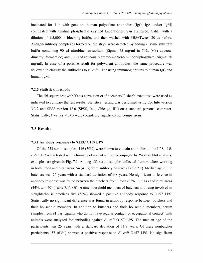
Antibody responses to E. coli O157 LPS among Bangladeshi population
127
incubated for 1 h with goat anti-human polyvalent antibodies (IgG, IgA and/or IgM)
conjugated with alkaline phosphatase (Zymed Laboratories, San Francisco, Calif.) with a
dilution of 1:5,000 in blocking buffer, and then washed with PBS+Tween 20 as before.
Antigen-antibody complexes formed on the strips were detected by adding enzyme substrate
buffer containing 90 µl nitroblue tetrazolium (Sigma; 75 mg/ml in 70% (v/v) aqueous
dimethyl formamide) and 70 µl of aqueous 5-bromo-4-chloro-3-indolylphosphate (Sigma; 50
mg/ml). In case of a positive result for polyvalent antibodies, the same procedure was
followed to classify the antibodies to E. coli O157 using immunoglobulins to human IgG and
human IgM.
7.2.5 Statistical methods
The chi-square test with Yates correction or if necessary Fisher’s exact test, were used as
indicated to compare the test results. Statistical testing was performed using Epi Info version
3.3.2 and SPSS version 12.0 (SPSS, Inc., Chicago, III.) on a standard personal computer.
Statistically, P values < 0.05 were considered significant for comparisons.
7.3 Results
7.3.1 Antibody responses to STEC O157 LPS
Of the 233 serum samples, 116 (50%) were shown to contain antibodies to the LPS of E.
coli O157 when tested with a human polyvalent antibody-conjugate by Western blot analysis;
examples are given in Fig. 7.1. Among 133 serum samples collected from butchers working
in both urban and rural areas, 54 (41%) were antibody positive (Table 7.1). Median age of the
butchers was 26 years with a standard deviation of 9.8 years. No significant difference in
antibody response was found between the butchers from urban (33%; n = 14) and rural areas
(44%; n = 40) (Table 7.1). Of the nine household members of butchers not being involved in
slaughterhouse practices five (56%) showed a positive antibody response to O157 LPS.
Statistically no significant difference was found in antibody response between butchers and
their household members. In addition to butchers and their household members, serum
samples from 91 participants who do not have regular contact (or occupational contact) with
animals were analyzed for antibodies against E. coli O157 LPS. The median age of the
participants was 25 years with a standard deviation of 11.8 years. Of these nonbutcher
participants, 57 (63%) showed a positive response to E. coli O157 LPS. No significant
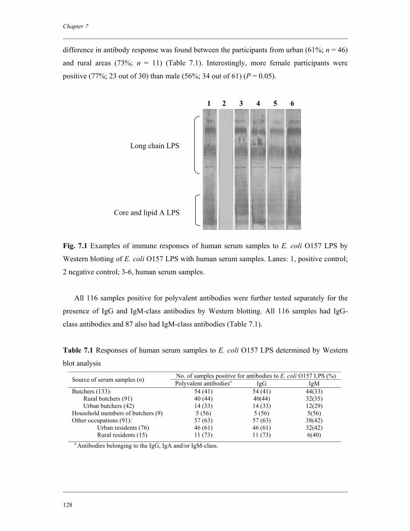
Chapter 7
128
difference in antibody response was found between the participants from urban (61%; n = 46)
and rural areas (73%; n = 11) (Table 7.1). Interestingly, more female participants were
positive (77%; 23 out of 30) than male (56%; 34 out of 61) (P = 0.05).
Fig. 7.1 Examples of immune responses of human serum samples to E. coli O157 LPS by
Western blotting of E. coli O157 LPS with human serum samples. Lanes: 1, positive control;
2 negative control; 3-6, human serum samples.
All 116 samples positive for polyvalent antibodies were further tested separately for the
presence of IgG and IgM-class antibodies by Western blotting. All 116 samples had IgG-
class antibodies and 87 also had IgM-class antibodies (Table 7.1).
Table 7.1 Responses of human serum samples to E. coli O157 LPS determined by Western
blot analysis No. of samples positive for antibodies to E. coli O157 LPS (%) Source of serum samples (n) Polyvalent antibodiesa IgG IgM
Butchers (133): 54 (41) 54 (41) 44(33) Rural butchers (91) 40 (44) 40(44) 32(35) Urban butchers (42) 14 (33) 14 (33) 12(29) Household members of butchers (9) 5 (56) 5 (56) 5(56) Other occupations (91): 57 (63) 57 (63) 38(42) Urban residents (76) 46 (61) 46 (61) 32(42) Rural residents (15) 11 (73) 11 (73) 6(40)
a Antibodies belonging to the IgG, IgA and/or IgM-class.
1 2 3 4 5 6
Long chain LPS
Core and lipid A LPS
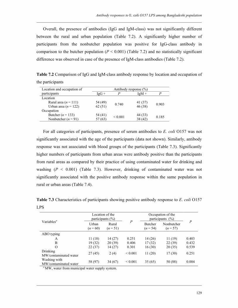
Antibody responses to E. coli O157 LPS among Bangladeshi population
129
Overall, the presence of antibodies (IgG and IgM-class) was not significantly different
between the rural and urban population (Table 7.2). A significantly higher number of
participants from the nonbutcher population was positive for IgG-class antibody in
comparison to the butcher population (P < 0.001) (Table 7.2) and no statistically significant
difference was observed in case of the presence of IgM-class antibodies (Table 7.2).
Table 7.2 Comparison of IgG and IgM-class antibody response by location and occupation of
the participants Antibody response (%) Location and occupation of
participants IgG + P IgM + P Location Rural area (n = 111) 54 (49) 41 (37) Urban area (n = 122) 62 (51) 0.740
46 (38) 0.903
Occupation Butcher (n = 133) 54 (41) 44 (33) Nonbutcher (n = 91) 57 (63) < 0.001 38 (42) 0.185
For all categories of participants, presence of serum antibodies to E. coli O157 was not
significantly associated with the age of the participants (data not shown). Similarly, antibody
response was not associated with blood groups of the participants (Table 7.3). Significantly
higher numbers of participants from urban areas were antibody positive than the participants
from rural areas as compared by their practice of using contaminated water for drinking and
washing (P < 0.001) (Table 7.3). However, drinking of contaminated water was not
significantly associated with the positive antibody response within the same population in
rural or urban areas (Table 7.4).
Table 7.3 Characteristics of participants showing positive antibody response to E. coli O157
LPS Location of the participants (%)
Occupation of the participants (%)
Variablesa Urban (n = 60)
Rural (n = 51)
P Butcher (n = 54)
Nonbutcher (n = 57)
P
ABO typing A 11 (18) 14 (27) 0.251 14 (26) 11 (19) 0.403 B 19 (32) 20 (39) 0.406 17 (32) 22 (39) 0.432 O 22 (37) 14 (27) 0.301 16 (30) 20 (35) 0.539
Drinking MW/contaminated water 27 (45) 2 (4) < 0.001 11 (20) 17 (30) 0.251
Washing with MW/contaminated water 58 (97) 34 (67) < 0.001 35 (65) 50 (88) 0.004
a MW, water from municipal water supply system.
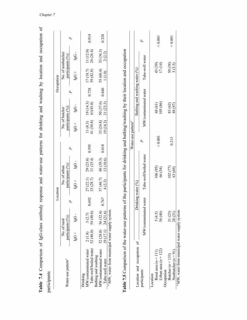
Tab
le 7
.4 C
ompa
rison
of
IgG
-cla
ss a
ntib
ody
resp
onse
and
wat
er-u
se p
atte
rns
for
drin
king
and
was
hing
by
loca
tion
and
occu
patio
n of
parti
cipa
nts
Loca
tion
O
ccup
atio
n N
o. o
f rur
al
parti
cipa
nts (
%):
No.
of u
rban
pa
rtici
pant
s (%
): N
o. o
f but
cher
pa
rtici
pant
s (%
): N
o. o
f non
butc
her
parti
cipa
nts (
%):
Wat
er-u
se p
atte
rna
IgG
+
IgG
- P
IgG
+
IgG
- P
Ig
G +
Ig
G -
P Ig
G +
Ig
G -
P
Drin
king
MW
/con
tam
inat
ed w
ater
2
(1.8
) 3
(2.7
) 27
(22.
1)
29 (2
3.8)
11
(8.3
) 19
(14.
3)
17 (1
8.7)
11
(12.
0)
Tube
-wel
l/boi
led
wat
er
52 (4
6.8)
54
(48.
6)
0.69
2 35
(28.
7)
31 (2
5.4)
0.
595
41
(30.
8)
61(4
5.8)
0.
728
39 (4
2.8)
24
(26.
4)
0.91
4
Bat
hing
and
was
hing
MW
/con
tam
inat
ed w
ater
32
(28.
8)
36 (3
2.4)
57
(46.
7)
48 (3
9.3)
33
(24.
8)
50 (3
7.6)
55
(60.
4)
33 (3
6.3)
Tu
be-w
ell w
ater
19
(17.
1)
24 (2
1.6)
0.
767
4 (3
.3)
13 (1
0.6)
0.
018
19
(14.
3)
31 (2
3.3)
0.
840
1 (1
.0)
2 (2
.2)
0.32
8
a MW
, wat
er fr
om m
unic
ipal
wat
er su
pply
syst
em.
T
able
7.5
Com
paris
on o
f the
wat
er-u
se p
atte
rns o
f the
par
ticip
ants
for d
rinki
ng a
nd b
athi
ng/w
ashi
ng b
y th
eir l
ocat
ion
and
occu
patio
n W
ater
-use
pat
tern
a D
rinki
ng w
ater
(%)
Bat
hing
and
was
hing
wat
er (%
) Lo
catio
n an
d oc
cupa
tion
of
parti
cipa
nts
MW
/con
tam
inat
ed w
ater
Tu
be-w
ell/b
oile
d w
ater
P
MW
/con
tam
inat
ed w
ater
Tu
be-w
ell w
ater
P
Loca
tion
R
ural
are
a (n
= 1
11)
5 (4
.5)
106
(95)
68
(61)
43
(39)
Urb
an a
rea
(n =
122
) 56
(46)
66
(54)
<
0.00
1 10
5 (8
6)
17 (1
4)
< 0.
001
Occ
upat
ion
B
utch
er (n
= 1
33)
31 (2
3)
102
(77)
83
(62)
50
(38)
Non
butc
her (
n =
91)
28 (3
1)
63 (6
9)
0.21
3 88
(97)
3
(3.3
) <
0.00
1
a M
W, w
ater
from
mun
icip
al w
ater
supp
ly sy
stem
.
Chapter 7
Antibody responses to E. coli O157 LPS among Bangladeshi population
131
Water-use pattern for bathing and washing had no significant effect on antibody response
among rural population but in urban areas a significantly higher number of people who used
MW for bathing and washing were positive for antibodies than people who used tube-well
water (Table 7.4). No significant difference in antibody response was observed between the
butcher and nonbutcher populations by their practice of using contaminated water for
drinking (Table 7.4). However, significantly higher numbers of nonbutcher participants were
antibody positive than butcher participants who used contaminated water for bathing and
washing (Table 7.3).
Very few numbers of participants complained on intestinal problems such as diarrhea and
dysentery during the last two weeks before sampling: five butchers, and eight other
occupations. No statistically significant association was observed between the occurrence of
diarrhea and dysentery and antibody response in the case of butchers (data not shown).
7.4 Discussion
The complete (smooth) LPS structure of E. coli and related species may be divided into
three distinct regions: (1) the hydrophobic lipid A moiety, responsible for the endotoxic
properties of LPS; (2) the core oligosaccharide; and (3) the O-polysaccharide antigen which is
easily accessible to the host immune system and leads to the generation of O-specific immune
responses, thus the term O-antigen is commonly used. Detection of O-antigen specific
antibodies has been used widely for routine diagnosis of STEC O157 infection when stool
samples from patients are culture-negative (12). Any subclinical infection among the healthy
population due to E. coli O157 may also be identified by serodiagnosis (detection of O-
antigen specific IgM antibodies) in conjunction with culture identification.
Strains of E. coli express LPS with one of five core structures, termed R1, R2, R3, R4 and
K-12 and many, but not all, strains of STEC, including O157, express an R3 LPS-core (3).
Patients infected with STEC O157 may produce serum antibodies binding to epitopes on the
R3 LPS-core (3). This binding to the R3 LPS-core is not always serogroup specific. It has
been demonstrated that an R3 LPS-core is widely distributed within the species E. coli and is
not always associated with a specific pathotype (15). In addition to different STEC serotypes,
R3 LPS-core is also found in enteroaggregative (EAEC) and enteropathogenic E. coli (EPEC)
(15). Thus, based on binding with only the R3 LPS-core, it cannot be concluded that
antibodies to E. coli O157 are present in serum samples collected from individuals suspected
Chapter 7
132
with STEC O157 infection. Several studies showed that serum samples from STEC O157-
infected HUS patients bind specifically to the long chain LPS region of STEC O157 in
Western blotting and do not show nonspecific binding with other LPS antigens (13, 21).
In this study, we determined the serum samples positive for antibodies to E. coli O157
only when we detected reaction against both core components and long chain LPS (Fig. 7.1).
We analyzed all the samples by Western blotting and did not apply ELISA or micro-
agglutination assays. It has been suggested by previous studies that such assays should
always be confirmed by Western blotting (13, 14).
Of the 233 sera, 116 (50%) reacted to both core and long chain LPS epitopes of STEC
O157 when tested with anti-human polyvalent antibodies (IgG, IgA and/or IgM).
Classification of antibody response to E. coli O157 LPS revealed that all samples gave a
positive response with the polyvalent antibody (50%; n = 116), were positive for IgG, which
is indicative of repeated exposure to the antigen. Around 37% (n = 87) of the samples were
also positive for IgM-class antibodies, which is indicative of recent exposure to the antigen.
Significantly higher numbers (63%) of participants other than butchers were positive for IgG-
class antibodies than butchers (41%) (P < 0.001) (Table 7.2). However, no statistically
significant difference was observed between butcher and nonbutcher populations with respect
to the presence of IgM-class antibodies (Table 7.2).
In this study, we found no statistically significant association between the antibody
response and age of the participants. Similar results were observed in studies conducted
among rural Wisconsin population in the United States (6) and among healthy people in
Thailand (46). In these studies, O157 LPS antibodies were not associated with increasing age,
sex, manure contact, or sheep contact.
Our data suggest that people in Bangladesh are frequently exposed to an antigen or
antigens that produce antibodies to the E. coli O157 LPS antigen. However, occupational
exposure to animals is not significantly associated with the positive antibody response against
E. coli O157 among the Bangladeshi population. High seropositivity may be due to cross-
reacting antibodies of many other intestinal pathogens that are very common in Bangladesh
in addition to E. coli O157. It has been shown that the O157 LPS antigen can be expressed by
different organisms, including nonpathogenic E. coli (39), Citrobacter freundii (7),
Escherichia hermanii (36), Yersinia entercolitica O9 (10), Salmonella O:30 (35) and
Brucella abortus (36). It has also been reported that the serum of patients with HUS
associated with E. coli O157 reacted with strains of Vibrio cholerae O1 Inaba. In another
study with serum from individuals vaccinated against V. cholerae O1 (11), the sera were seen
Antibody responses to E. coli O157 LPS among Bangladeshi population
133
to react strongly against O157 LPS. V. cholerae O1 is endemic in Bangladesh, occurring
twice yearly in epidemic form (1). It has been identified as the leading cause of cholera in
Bangladesh (40). Contaminated water is the main source of transmission of V. cholerae in
regions where clean drinking water is not available to local populations (40). According to
previous study, V. cholerae are native to both marine and fresh water environments in
Bangladesh indicating that people are repeatedly exposed to the organisms either directly or
indirectly (40).
Cross-reacting antibodies were also described in situations where enteric pathogens
sharing common epitopes are endemically present (34). For example, infections with EPEC
in early childhood confer cross-reacting protective immunity against STEC types that share
common antigens (such as LPS and intimin) with classical EPEC strains (4). There is a huge
burden of EPEC infection in Bangladesh and children below five years of age are most
susceptible (2). EPEC infections occur rarely in adults and this can be explained by repeated
exposure in early childhood conferring to protection later in life (4).
It would have been interesting if we could analyze the serum samples for antibodies
against cross-reacting antigens. Nevertheless, antibodies to the E. coli O157 LPS antigen
possibly represent exposure to E. coli O157, especially as examples exist of asymptomatic
carriage of E. coli O157 inducing an antibody response to O157 LPS (8, 49). This can be
further explained by our previous studies, where we found that more than 82% of slaughtered
cattle in Dhaka were positive for stx genes (25). More than 10% of the cattle were positive for
STEC O157 and 14% for STEC non-O157 (25). Raw meats in the slaughterhouses were also
highly contaminated with STEC O157 (M. A. Islam, A. S. Mondol, I. J. Azmi, E. de Boer, R.
R. Beumer, M. H. Zwietering, A. E. Heuvelink and K. A. Talukder, Submitted for
publication). Therefore, people who buy raw meats from the slaughterhouses and those who
prepare the meat for cooking at home are also exposed to the organism. In Bangladesh,
women are mainly responsible for preparing the food at home, and therefore, woman are
more exposed than man within the same population who do not have direct contact with
animals. This has been reflected by the data of the present study where we found more female
(77%) were antibody positive than male (56%) among participants other than butchers (P =
0.05).
STEC infection occurs in humans through acquisition of the bacteria via consumption of
contaminated food and/or drink. Raw or undercooked foods (ground meat or vegetables)
from animal origin have been recognized as the most important vehicle for transmission of
STEC O157 (19). However, people in Bangladesh are not accustomed to eating raw or
Chapter 7
134
undercooked meat. Thus, direct contamination of raw foods with bacterial pathogens,
including STEC O157 does not pose a significant threat to human health. Nevertheless, lack
of knowledge on good hygienic practices and unsanitary situations in food preparation can
cause cross-contamination of food and water. Moreover, people living in rural and semi
urban areas in Bangladesh are directly exposed to cattle feces regularly as cattle farming are
still a domestic practice in Bangladesh. Although urban residents are not exposed to animals
directly, they live in a contaminated environment where many enteric pathogens are
considered to be endemic. Besides, due to lack of proper drainage systems in city abattoirs
large amount of feces are daily exposed to the environment, including water bodies.
Dhaka, the capital city of Bangladesh is one of the mega cities in the world where 11.3
million people are living within 227.8 km2 of metropolitan area and 80% of the total
population is dependent on municipal water supply systems. It has been found that the
municipal water at initial points were acceptable for drinking according to WHO guidelines
in microbiological point of view but water collected from the points of use (consumer taps)
was contaminated with total coliform and fecal coliform and were unacceptable for drinking
(M. S. Islam, International Centre for Diarrhoeal Disease Research, Bangladesh, unpublished
data). The municipal piped water might be contaminated with sewage and depleting
groundwater through leaky joints of inadequately maintained pipes facilitated by low
pressure, intermittent delivery systems (41). Only 20% of people in Dhaka are connected to
sewer and most of the rest use septic tanks that discharge into drains or the river. Storm water
drains are blocked and malfunctioning and most water runs through natural channels into
rivers. In a previous study it has been recommended that interventions to secure a high-
quality municipal water supply for Dhaka’s population are critical for preventing the spread
of enteric pathogens through piped water sources (37).
In this study, we found that a significantly higher number of participants from urban areas
was antibody positive than the participants from rural areas when compared by their practice
of using contaminated water for drinking and washing (P < 0.001) (Table 7.3). This may be
due to the fact that a significantly higher number of people in urban areas used
MW/contaminated water for drinking and washing than the people in rural areas (Table 7.5).
In fact, the only source of drinking water in rural Bangladesh is tube-well and water from this
source is considered to be free of fecal contamination. In addition, the majority of the urban
population (86%) is dependent on MW for bathing and washing, while a significant
percentage (39%) of the rural population used tube-well water (Table 7.5). This was further
explained by analyzing the data separately from rural and urban areas where we found a
Antibody responses to E. coli O157 LPS among Bangladeshi population
135
significant association between the use of MW/contaminated water and positive antibody
response among urban population but not among rural population (Table 7.4).
Drinking of MW/contaminated water had no significant association with positive
antibody response between the butcher and nonbutcher population. However, significantly
higher numbers of nonbutcher participants were antibody positive than butcher participants
who used contaminated water for bathing and washing (Table 7.3). This may be due to the
inclusion of higher number of nonbutcher residents from urban areas (n = 76) than from rural
areas (n = 15) in the study. When tested separately, antibody response was not significantly
associated with water-use pattern for drinking and washing/bathing within butcher or
nonbutcher population (Table 7.4).
Although there is no report of contamination of surface water with STEC O157 in
Bangladesh, it would be interesting to test the common sources of water for this pathogen.
Recently, in India STEC O157:H7 has been isolated from the river Ganges, which is
regularly used for personal hygiene, laundry, and utensil washing (23).
The results of the study are in concordance with the previous studies done in other
developing countries, including Thailand and Mexico where high levels of serum antibodies
against E. coli O157 LPS have been reported in healthy people (in Thailand: 23% positive for
IgG and 12% for IgM; in Mexico: 20% positive for IgG) (32, 46).
The results of the present study suggest that Bangladeshi population, irrespective of the
extent of exposure to animals are being exposed to the antigens that are capable of
stimulating the production of antibodies against STEC O157. According to our previous
study, prevalence of STEC infection among diarrheal patients is low (0.5%) compared with
other enteric pathogens and there has been no report of HUS cases associated with STEC
O157 infection in Bangladesh (24). The ubiquitous presence of antibodies to STEC O157
LPS among the mass population in Bangladesh can be the reason why there is a lack of STEC
O157 infection, including HUS or HC in this country compared with other enteric infections.
Acknowledgments
This research was funded by the International Centre for Diarrhoeal Disease Research,
Bangladesh (ICDDR,B) and by the Netherlands Foundation for the Advancement of Tropical
Research (NWO-WOTRO) grant (Award number WB 93-415). ICDDR,B acknowledges
with gratitude the commitment of the Netherlands Foundation for the Advancement of
Chapter 7
136
Tropical Research (NWO-WOTRO) grant to the Centre's research efforts. ICDDR,B also
gratefully acknowledges these donors who provide unrestricted support to the Centre's
research efforts: Australian Agency for International Development (AusAID), Government of
Bangladesh, Canadian International Development Agency (CIDA), Embassy of the Kingdom
of the Netherlands, Swedish International Development Cooperation Agency (Sida), Swiss
Agency for Development and Cooperation (SDC) and Department for International
Development, UK (DFID).
References
1. Alam, M., M. Sultana, G. B. Nair, R. B. Sack, D. A. Sack, A. K. Siddique, A. Ali, A. Huq, and R. R. Colwell. 2006. Toxigenic Vibrio cholerae in the aquatic environment of Mathbaria, Bangladesh. Appl. Environ. Microbiol. 72:2849-2855.
2. Albert, M. J., A. S. Faruque, S. M. Faruque, R. B. Sack, and D. Mahalanabis. 1999. Case-control study of enteropathogens associated with childhood diarrhea in Dhaka, Bangladesh. J. Clin. Microbiol. 37:3458-3464.
3. Amor, K., D. E. Heinrichs, E. Frirdich, K. Ziebell, R. P. Johnson, and C. Whitfield. 2000. Distribution of core oligosaccharide types in lipopolysaccharides from Escherichia coli. Infect. Immun. 68:1116–1124.
4. Beutin, L., S. Zimmermann, and K. Gleier. 1998. Human infections with Shiga toxin-producing Escherichia coli other than serogroup O157 in Germany. Emerg. Infect. Dis. 4:635-639.
5. Beutin, L. 2006. Emerging enterohaemorrhagic Escherichia coli, causes and effects of the rise of a human pathogen. J. Vet. Med. B. Infect. Dis. Vet. Public Health. 53:299-305.
6. Belongia, E. A., P. H. Chyou, R. T. Greenlee, G. Perez-Perez, W. F. Bibb, and E. O. DeVries. 2003. Diarrhea incidence and farm-related risk factors for Escherichia coli O157:H7 and Campylobacter jejuni antibodies among rural children. J. Infect. Dis. 187:1460-1468.
7. Bettelheim, K. A., H. Evangelidis, J. L. Pearce, E. Sowers, and N. A. Strockbine. 1993. Isolation of a Citrobacter freundii strain which carries the Escherichia coli O157 antigen. J. Clin. Microbiol. 31:760-761.
8. Bielaszewska, M., J. Janda, K. Bláhová, H. Minaríková, E. Jíková, M. A. Karmali, J. Laubová, J. Sikulová, M. A. Preston, R. Khakhria, H. Karch, H. Klazarová, and O. Nyc. 1997. Human Escherichia coli O157:H7 infection associated with the consumption of unpasteurized goat’s milk. Epidemiol. Infect. 119:299–305.
9. Chart, H., S. M. Scotland, and B. Rowe. 1989. Serum antibodies to Escherichia coli serotype O157:H7 in patients with hemolitic uremic syndrome. J. Clin. Microbiol. 27:285–290.
10. Chart, H., O. A. Okubadejo, and B. Rowe. 1992. The serological relationship between Escherichia coli O157 and Yersinia enterocolitica O9 using sera from patients with brucellosis. Epidemiol. Infect. 108:77-85.
11. Chart, H., and B. Rowe. 1993. Antibody cross-reactions with lipopolysaccharide from E. coli O157 after cholera vaccination. Lancet 341:1282.
Antibody responses to E. coli O157 LPS among Bangladeshi population
137
12. Chart, H., and C. Jenkins. 1999. Serodiagnosis of infection with verocytotoxin-producing Escherichia coli. J. Appl. Microbiol. 86:569–575.
13. Chart, H., T. Cheasty, and G. A. Willshaw. 2002. Production of serum antibodies that recognize epitopes located on the R3 region of Escherichia coli core lipopolysaccharides by patients infected with strains of enterohaemorrhagic E. coli. J. Med. Microbiol. 51:1050–1054.
14. Chart, H., and N. T. Perry. 2004. The serological response to verocytotoxigenic Escherichia coli in patients with haemolytic uraemic syndrome. Lett. Appl. Microbiol. 38:351-354.
15. Chart, H., N. T. Perry, and C. Jenkins. 2004. The expression of an R3 lipopolysaccharide-core by pathotypes of Escherichia coli. J. Appl. Microbiol. 96:982-986.
16. Cravioto, A. 2005. Importance of Escherichia coli strains producing verotoxins. J. Health Popul. Nutr. 23:303-304.
17. Crump, J. A., A. C. Sulka, A. J. Langer, C. Schaben, A. S. Crielly, R. Gage, M. Baysinger, M. Moll, G. Withers, D. M. Toney, S. B. Hunter, M. Hoekstra, S. K. Wong, P. M. Griffin, and T. J. Van Gilder. 2002. An outbreak of Escherichia coli O157:H7 infections among visitors to a dairy farm. N. Engl. J. Med. 347:555–560.
18. Das, S. C., A. Khan, P. Panja, S. Datta, A. Sikdar, S. Yamasaki, Y. Takeda, S. K. Bhattacharya, T. Ramamurthy, and G. B. Nair. 2005. Dairy farm investigation on Shiga toxin-producing Escherichia coli (STEC) in Kolkata, India with emphasis on molecular characterization. Epidemiol. Infect. 133:617-626.
19. Erickson, M. C., and M. P. Doyle. 2007. Food as a vehicle for transmission of Shiga toxin-producing Escherichia coli. J. Food Prot. 70:2426-2449.
20. Evans, J., R. M. Chalmers, H. Chart, R. L. Salmon, S. M. Kench, T. J. Coleman, D. Meadows, P. Morgan-Capner, P. Softley, M. Sillis, and D. R. Thomas. 2000. Evidence of persisting serum antibodies to Escherichia coli O157 lipopolysaccharide and verocytotoxin in members of rural communities in England. Eur. J. Epidemiol. 16:885-889.
21. Gibbs, R. J., J. Stewart and I. R. Poxton. 2004. The distribution of, and antibody response to, the core lipopolysaccharide region of Escherichia coli isolated from the faeces of healthy humans and cattle. J. Med. Microbiol. 53:959–964.
22. Greatorex, J. S., and G. M. Thorne. 1994. Humoral immune responses to Shiga-like toxins and Escherichia coli O157 lipopolysaccharide in hemolytic-uremic syndrome patients and healthy subjects. J. Clin. Microbiol. 32:1172–1178.
23. Hamner, S., S. C. Broadaway, V. B. Mishra, A. Tripathi, R. K. Mishra, E. Pulcini, B. H. Pyle, and T. E. Ford. 2007. Isolation of potentially pathogenic Escherichia coli O157:H7 from the Ganges River. Appl. Environ. Microbiol. 73:2369-2372.
24. Islam, M. A., A. E. Heuvelink, E. de Boer, P. D. Sturm, R. R. Beumer, M. H. Zwietering, A. S. Faruque, R. Haque, D. A. Sack, and K. A. Talukder. 2007. Shiga toxin-producing Escherichia coli isolated from patients with diarrhoea in Bangladesh. J. Med. Microbiol. 56:380-385.
25. Islam, M. A., A. S. Mondol, E. de Boer, R. R. Beumer, M. H. Zwietering, K. A. Talukder, and A. E. Heuvelink. 2008. Prevalence and genetic characterization of Shiga toxin-producing Escherichia coli isolated from slaughtered animals in Bangladesh. Appl. Environ. Microbiol. 74:5414-5421.
26. Karmali, M. A., M. Petric, C. Lim, P. C. Fleming, G. S. Arbus, and H. Lior. 2004. The association between idiopathic hemolytic uremic syndrome and infection by verotoxin-producing Escherichia coli. J. Infect. Dis. 189:556–563.
Chapter 7
138
27. Kulkarni, H., P. N. Goldwater, A. Martin, and K. A. Bettelheim. 2002. Escherichia coli ‘O’ group serological responses and clinical correlations in epidemic HUS patients. Comp. Immunol. Microbiol. Infect. Dis. 25:249–268.
28. Laemmli, U. K. 1970. Cleavage of structural proteins during the assembly of the head of bacteriophage T4. Nature 227:680–685.
29. Li, Y., E. Frey, A. M. Mackenzie, and B. B. Finlay. 2000. Human response to Escherichia coli O157:H7 infection: antibodies to secreted virulence factors. Infect. Immun. 68:5090-5095.
30. Ludwig, K., M. A. Karmali, V. Sarkim, C. Bobrowski, M. Petric, H. Karch, and D. E. Müller-Wiefel; Arbeitsgemeinschaft für Pädiatrische Nephrologie. 2001. Antibody response to Shiga toxins Stx2 and Stx1 in children with enteropathic hemolytic-uremic syndrome. J. Clin. Microbiol. 39:2272-2279.
31. Nataro, J. P., and J. B. Kaper. 1998. Diarrheagenic Escherichia coli. Clin. Microbiol. Rev. 11:142-201.
32. Navarro, A., C. Eslava, U. Hernandez, J. L. Navarro-Henze, M. Aviles, G. Garcia-de la Torre, and A. Cravioto. 2003. Antibody responses to Escherichia coli O157 and other lipopolysaccharides in healthy children and adults. Clin. Diagn. Lab. Immunol. 10:797–801.
33. O’Brien, S. J., G. K. Adak, and C. Gilham. 2001. Contact with farming environment as a major risk factor for Shiga toxin (verocytotoxin)–producing Escherichia coli O157 infection in humans. Emerg. Infect. Dis. 7:1049–1051.
34. Palmeira, P., L. Yu Ito, C. Arslanian, and M. M. Carneiro-Sampaio. 2007. Passive immunity acquisition of maternal anti-enterohemorrhagic Escherichia coli (EHEC) O157:H7 IgG antibodies by the newborn. Eur. J. Pediatr. 166:413-419.
35. Perry, M. B., D. R. Bundle, L. MacLean, J. A. Perry, and D. W. Griffith. 1986. The structure of the antigenic lipopolysaccharide O chain produced by Salmonella urbana and Salmonella godesberg. Carbohydr. Res. 156:107-122.
36. Perry, M. B., and D. R. Bundle. 1990. Antigenic relationships of the lipopolysaccharides of Escherichia hermanii strains with those of Escherichia coli O157:H7, Brucella melitensis and Brucella abortus. Infect. Immun. 58:1391-1395.
37. Ram, P. K., A. Naheed, W. A. Brooks, M. A. Hossain, E. D. Mintz, R. F. Breiman, and S. P. Luby. 2007. Risk factors for typhoid fever in a slum in Dhaka, Bangladesh. Epidemiol. Infect. 135:458-465.
38. Reymond, D., R. P. Johnson, M. A. Karmali, M. Petric, M. Winkler, S. Johnson, K. Rahn, S. Renwick, J. Wilson, R. C. Clarke, and J. Spika. 1996. Neutralizing antibodies to Escherichia coli Vero cytotoxin 1 and antibodies to O157 lipopolysaccharide in healthy farm family members and urban residents. J. Clin. Microbiol. 34:2053-2057.
39. Rice, E. W., E. G. Sowers, C. H. Johnson, M. E. Dunnigan, N. A. Strockbine, and S. C. Edberg. 1992. Serological cross-reaction between Escherichia coli O157 and other species of the genus Escherichia. J. Clin. Microbiol. 30:1315-1316.
40. Sack, R. B., A. K. Siddique, I. M. Jr. Longini, A. Nizam, M. Yunus, M. S. Islam, J. G. Jr. Morris, A. Ali, A. Huq, G. B. Nair, F. Qadri, S. M. Faruque, D. A. Sack, and R. R. Colwell. 2003. A 4-year study of the epidemiology of Vibrio cholerae in four rural areas of Bangladesh. J. Infect. Dis. 187:96-101.
41. Sirajul Islam, M., A. Brooks, M. S. Kabir, I. K. Jahid, M. Shafiqul Islam, D. Goswami, G. B. Nair, C. Larson, W. Yukiko, and S. Luby. 2007. Faecal contamination of drinking water sources of Dhaka city during the 2004 flood in Bangladesh and use of disinfectants for water treatment. J. Appl. Microbiol. 103:80-87.
Antibody responses to E. coli O157 LPS among Bangladeshi population
139
42. Tarr, P. I., C. A. Gordon, and W. L. Chandler. 2005. Shiga-toxin-producing Escherichia coli and haemolytic uraemic syndrome. Lancet 365:1073-1086.
43. Timm, C. D., K. Irino, T. A. Gomes, M. M. Vieira, B. E. Guth, T. M. Vaz, C. N. Moreira, and J. A. Aleixo. 2007. Virulence markers and serotypes of Shiga toxin-producing Escherichia coli, isolated from cattle in Rio Grande do Sul, Brazil. Lett. Appl. Microbiol. 44:419-425.
44. Towbin, H., T. Staehelin, and J. Gordon. 1979. Electrophoretic transfer of proteins from polyacrylamide gels to nitrocellulose sheets: procedure and some applications. Proc. Natl. Acad. Sci. USA 76:4350-4354.
45. Tsai, C. M., and C. E. Frasch. 1982. A sensitive silver stain for detecting lipopolysaccharide in polyacrylamide gels. Anal. Biochem. 119:115-119.
46. Voravuthikunchai, S. P., C. Chaowana, P. Perepat, T. Iida, and T. Honda. 2005. Antibodies among healthy population of developing countries against enterohaemorrhagic Escherichia coli O157:H7. J. Health Popul. Nutr. 23:305-310.
47. Vuddhakul, V., N. Patararungrong, P. Pungrasamee, S. Jitsurong, T. Morigaki, N. Asai, and M. Nishibuchi. 2000. Isolation and characterization of Escherichia coli O157 from retail beef and bovine faeces in Thailand. FEMS Microbiol. Lett. 182:343-347.
48. Westphal, O., and K. Jann. 1965. Bacterial lipopolysaccharide extraction with phenol water and further application of the procedure, p. 83-91. In R. L. Whistler (ed.), Methods in carbohydrate chemistry. Academic Press, New York, NY.
49. Wilson, J. B., R. C. Clarke, S. A. Renwick, K. Rahn, R. P. Johnson, M. A. Karmali, H. Lior, D. Alves, C. L. Gyles, K. S. Sandhu, S. A. McEwen, and J. S. Spika. 1996. Vero cytotoxigenic Escherichia coli infection in dairy farm families. J. Infect. Dis. 174:1021-1027.


General discussion and future perspective
Chapter 8
Chapter 8
142
8.1 Introduction
Shiga toxin (Stx)-producing Escherichia coli (STEC) were first recognized as a human
pathogens in 1982, in the United States, when strains of serotype O157:H7 caused two
outbreaks of hemorrhagic colitis (74). In 1983, the association of E. coli O157:H7 and of
several other STEC serotypes with sporadic cases of the classical hemolytic-uremic
syndrome (HUS) was first described (43) and subsequently confirmed in a prospective study
(44). Since then, epidemiological studies from different parts of the world established STEC
as the major cause of bloody diarrhea and HUS in temperate climates and as an important
cause of uncomplicated watery diarrhea in some geographic areas (85).
Over the last decade, remarkable changes in the epidemiology of human infection and in
the transmission routes of STEC have occurred. Although serotype O157:H7 is
predominantly associated with human infections, more than 200 other STEC serotypes have
been identified of which over 100 have been associated with disease in humans (23).
Ruminants are considered as the most important reservoir for STEC. There are many routes
by which STEC organisms can be transmitted from ruminants to humans, but still foodborne
transmission remains the most important cause of infection resulting from fecal
contamination of the food.
The majority of studies have been done in the industrialized countries where STEC are
considered as a major problem. There is still a gap of knowledge on the epidemiology of
STEC in human infections, their sources and the transmission routes in developing countries.
It is only in recent years that laboratories in developing countries have started to find out the
importance of these pathogens as part of the spectrum of diarrheal diseases.
Bangladesh is an endemic zone for diarrheal diseases. Apart from a few sporadic efforts
to find out the presence of STEC in diarrheal patients, no systematic study had been done on
the epidemiology and ecology of STEC in Bangladesh. Therefore, the primary focus of this
thesis was to gain insight in both the epidemiology of STEC infection in humans and the
occurrence of STEC in the human food chain in Bangladesh. Because the use of appropriate
techniques for detection and isolation is a prerequisite for revealing good estimates of
prevalence rates, we also evaluated three commonly used methods for the detection and
isolation of STEC O157 from animal feces and raw meats.
General discussion and future perspective
143
8.2 STEC in diarrheal patients
Acute infectious diarrhea is the second most common cause of death in children living in
developing countries, surpassed only by acute respiratory diseases accounting for
approximately 20% of all childhood deaths (48). Despite the use of oral rehydration therapy,
deaths due to diarrhea in children aged less than five years are still estimated to be about two
million per year (16). Besides mortality, the long-term effects of diarrheal illness on
childhood health are extremely serious and include malnutrition and growth faltering (15).
Aetiology of diarrhea is variable according to geographic and climatic conditions, host
factors, and socioeconomic situations. The major aetiologic agents of acute diarrhea are
diarrheagenic Escherichia coli, rotavirus, Vibrio cholerae, and Shigella spp. (36, 48). All are
known to be endemic in essentially all developing countries. A study done recently on the
incidence of aetiology-specific diarrhea in a birth cohort (n = 252) in rural Bangladesh has
shown that enterotoxigenic Escherichia coli (ETEC), enteroadherent E. coli (EAEC),
Shigella spp., Campylobacter jejuni, Giardia spp., and rotavirus were the most commonly
isolated pathogens (32). A similar but more comprehensive study done in a hospital setting in
Bangladesh found that like studies conducted in other developing countries, the major
pathogens of childhood diarrhea were rotavirus, C. jejuni, ETEC, enteropathogenic
Escherichia coli (EPEC), Shigella spp., and V. cholerae. Among six pathotypes of
diarrheagenic E. coli, ETEC was found to be the most prevalent in both of these studies.
However, a study done previously in the same hospital setting found that in children (up to
five years of age) with diarrhea, enteropathogenic E. coli (EPEC) were the most prevalent
(15.5%), followed by ETEC (12%), enteroaggregative E. coli (EAggEC) (9.5%) and
diffusely-adherent E. coli (DAEC) (8.2%) (2). STEC were not detected in any of the children
with diarrhea but were detected in five children without diarrhea; however, these isolates
were not characterized further. These studies illustrate that the aetiology of diarrheal disease
and the prevalence of diarrheagenic pathogens can change over time in an endemic setting for
diarrhea like Bangladesh. These changes may be attributable for example to the emergence of
new pathogens or climate change. But changes in detected prevalences can also be the result
of improved surveillance systems and advances in microbiological detection techniques.
As far as STEC concern, none of the previous studies done in Bangladesh identified
STEC as diarrheal pathogens. The reasons might be: 1) the lack of proper surveillance for
STEC; 2) lack of technical capacity for identification of STEC; 3) STEC is not present; 4)
Chapter 8
144
STEC is present but relatively few infections occur due to acquired immunity in the
population; or 5) consumer habits intervene the transmission of STEC from sources to
humans (cooking, hygiene).
In chapter 2, we described the prevalence and characteristics of STEC in patients with
diarrhea in a hospital and in a community clinic in Bangladesh. A total of 570 stool samples
collected from patients with diarrhea were tested for the presence of stx genes. Of these
patients, 410 patients were hospitalized due to severe diarrhea and the remaining 160 patients
attended the community clinic in an urban slum of Dhaka city. The prevalence of stx in stool
samples was 2.2% (n = 9) among hospitalized patients and 7% (n = 11) among patients in the
community. STEC strains could be isolated from 0.5% (n = 2) and 1.9% (n = 3) of the
samples from hospitalized and community patients, respectively. No statistically significant
association between the age of the patients and the presence of stx genes was observed. All
stx-positive patients were clinically diagnosed as having uncomplicated diarrhea. Seven of
the nine patients who were hospitalized and stx-positive were primarily diagnosed with a V.
cholerae O1 infection. Therefore, the role of STEC in causing the diarrheal infection is still
to be speculated. The five STEC isolates belonged to five different serotypes; no STEC O157
strains were isolated. All five isolates were positive in the Vero cell cytotoxicity test. In
addition to the stx, one isolate from a hospitalized patient and one from a community patient
were positive for the eae gene. All three isolates from the community patients and one isolate
from the hospitalized patients were positive for the hlyEHEC gene. It has been suggested by
previous studies that STEC having both the eae and hlyEHEC genes are more virulent and
causing more often severe human infection than strains carrying only the stx genes (25, 68,
79). However, no significant difference in clinical symptoms was observed for patients from
whom these more virulent STEC were isolated (n = 2).
The findings of this study corroborated with previous studies done in other South Asian
countries, including India, Thailand and Vietnam where similar prevalences of STEC were
found in patients with diarrhea (47, 52, 63). Based on the results of this study, it can be
concluded that STEC are not a significant problem compared with other enteric pathogens in
Bangladesh. However, this study generates some interesting questions concerning the
absence of STEC infection in humans in Bangladesh where many enteric infections are
considered to be endemic. These are: 1) is there any potential reservoir for STEC in
Bangladesh?; 2) does there any intervention exist in the transmission route of STEC from
their reservoirs to human patients?; 3) are the strains in animal reservoirs or in foods, if
present, virulent enough to cause human infection?; or 4) is there a high prevalence of
General discussion and future perspective
145
antibodies against STEC, including serogroup O157 among the Bangladeshi population?
These issues have been discussed in detail in the following parts of the thesis.
8.3 Detection and isolation methods for STEC O157
The low prevalence of STEC in patients with diarrhea compared to other diarrhea causing
pathogens prompted us to estimate the prevalence of STEC in food and food producing
animals in Bangladesh. Unlike the clinical samples, it is very difficult to isolate and identify
STEC from food samples because of the presence of very low numbers of these organisms in
samples with an abundance of background microflora. Although many studies have been
done on the development of detection and isolation methods for STEC O157, only a few data
are available on the comparison of different methods. Besides, the growing number of
methods and the variety of commercially available test-kits make it difficult for a diagnostic
laboratory to choose the most satisfying assay system if no evaluation data on these tests are
publicly available (10). As a consequence, the prices of the tests remain an important
criterion, which can be applied to all commercially available assay systems. The use of test
systems that are not evaluated for the specificity and sensitivity with types of samples that the
laboratory is mainly concerned might result in a risk of obtaining false-positive and/or false-
negative results (10).
Traditional culture methods for the detection of STEC O157 are often inefficient due to
the large numbers of other flora that either overgrow or mimic STEC O157. The sensitivity of
the conventional culture method can significantly be increased by the application of an
immunoconcentration step, which will result in less false-negatives. A commonly used
method, which is also implemented in the international standard for the detection of STEC
O157 in food and feeding stuffs (5), is immunomagnetic separation (IMS). Prior to plating
onto selective isolation media, STEC O157 cells present in the enrichment culture are
selectively concentrated by magnetic beads with O157-specific antibodies covalently bound
onto their surface. The IMS procedure can be performed both manually and automatically.
In chapter 3, we described the evaluation of two commercially available test systems for
STEC O157, both based on immunoconcentration of these bacteria: a manual IMS method
using Dynabeads anti-E. coli O157 and the Vitek Immunodiagnostic Assay System (VIDAS)
Immunoconcentration E. coli O157 (ICE) kit. Additionally, we compared two selective
isolation media for STEC O157; sorbitol–MacConkey agar with cefixime (0.05 mg/l) and
Chapter 8
146
tellurite (2.5 mg/l) (CT-SMAC), and CHROMagar O157 with cefixime (0.025 mg/l) and
tellurite (1.25 mg/l) (1/2CT-CHROMagar). A total of 997 samples comprised of animal feces
and raw meats have been examined by both the Dynabeads anti-E. coli O157 method and the
VIDAS ICE method. By using the Dynabeads anti-E. coli O157 method systematically more
fecal samples were identified as being positive (Binomial test, P < 0.05). However, for meat
samples, no systematic difference was found between the methods (Binomial test, P ≥ 0.05).
The relative sensitivity, specificity, and accuracy of the VIDAS ICE method in comparison
with the Dynabeads anti-E. coli O157 method was 63.0%, 99.3%, and 96.7%, respectively,
for fecal samples and 55.6%, 99.1%, and 98.1%, respectively, for meat samples. When the
results obtained with CT-SMAC and 1/2CT-CHROMagar were analyzed separately, a
systematic difference between the two immunoconcentration systems was observed only in
case of fecal samples examined in combination with 1/2CT-CHROMagar (Binomial test, P <
0.05). The relative accuracy of the VIDAS ICE method compared with the Dynabeads anti-E.
coli O157 method was 97.8% when CT-SMAC was used as the final isolation medium and
96.7% when 1/2CT-CHROMagar was used. By using the VIDAS ICE method, the entire
immunoconcentration process is performed automatically within 40 min, for a maximum of
30 samples (including controls) in one run. The concentration of 30 samples with the
Dynabeads anti-E. coli O157 procedure takes about 100 min. For the safety of the user and a
saving of time, in case of meat samples it is recommended to prefer the VIDAS ICE method
to the manual Dynabeads anti-E. coli O157 procedure. Although the performance of the
Dynabeads anti-E. coli O157 procedure was statistically significantly better than the VIDAS
ICE method for animal feces, the latter method can be a good choice in case of prevalence
studies at the farm level, when more than one sample from the same origin (same farm, same
herd etc.) is examined. One of the major drawbacks of the VIDAS ICE method is the high
price. The cost per sample (materials and reagents) is almost three fold higher in case of the
VIDAS ICE method compared with that of the Dynabeads anti-E. coli O157 method.
Comparison of the performance of CT-SMAC and 1/2CT-CHROMagar revealed that for
animal feces, systematically more samples were found positive when 1/2CT-CHROMagar
was used as the plating medium (McNemar test, P < 0.05), irrespective of the method of
immunoconcentration carried out. For meat samples, the use of 1/2CT-CHROMagar also
resulted in more positives than CT-SMAC. However, this difference between the two plating
media was statistically significant only in combination with the Dynabeads anti-E. coli O157
(Binomial test, P < 0.05) but not with the VIDAS ICE method (Binomial test, P ≥ 0.05).
Therefore, it is recommended to use 1/2CT-CHROMagar as a standard isolation medium in
General discussion and future perspective
147
addition to CT-SMAC, following immunoconcentration with either the Dynabeads anti-E.
coli O157 or the VIDAS ICE kit.
Although IMS offers significant advantages in the detection and isolation of STEC O157,
the successful application of this culture-based method is still dependent on the expression of
the physiological characteristics and/or metabolic activities of the bacterium (78). PCR-based
methods are considered as a good alternative because the presence of small amounts of
species- or strain-specific DNA is being detected rather than unique aspects of target
organisms to be in a specific physiological state. For STEC O157, Abdulmawjood et al. (1)
developed a diagnostic PCR assay based on the amplification of sequences for the rfb (O-
antigen-encoding) region of E. coli O157 (rfbEO157) gene. The diagnostic application of this
PCR assay was subsequently validated by means of a multicenter validation study (1). In
chapter 4, we described the evaluation of the performance of the IMS-based method using
Dynabeads anti-E. coli O157 and the rfbEO157 PCR assay by testing series of animal feces and
raw meats artificially contaminated with strains of STEC O157 isolated from different
sources. IMS is laborious and time-consuming but ends up with the isolation of the pathogen.
On the other hand, PCR is fast and less laborious but it can only be used for screening
purposes, so a further culture step is required to isolate the organism. Although IMS is
considered as the official standard method for detection and isolation of STEC O157 from
food and animal feeding stuffs (5), no standardized protocol has yet been described for
animal feces.
The sensitivity of PCR detection systems depends on the choice of reaction conditions
and interactions with the particular sample matrix (78). PCR is inhibited by numerous
compounds often found in feces and food (14), providing a practical hindrance to the
molecular-based detection of small numbers of contaminating pathogens (4, 66). One way to
address these limitations is the use of effective sample processing as well as DNA extraction
techniques. However, in many cases a technique optimized for one matrix or microorganism
is not readily adaptable for use with others. Moreover, because purification adds to the time
and expense of sample preparation, as well as to the loss of target nucleic acids, a more
satisfying approach to the problem of PCR inhibition would be to reduce interference rather
than attempt to remove all of the offending substances. To this end, various additives have
been included in the PCR mixture to minimize inhibition. Among these, bovine serum
albumin (BSA) is widely being used (49). We also found that BSA at a concentration of 1
mg/ml in the PCR mixture significantly reduced the effects of inhibitory substances present
in both feces and meats. This was confirmed by using an internal amplification control in the
Chapter 8
148
PCRs. The use of an internal amplification control in the PCR can detect the level of reaction
inhibition and therefore eliminates the chances of false-negative results (1).
IMS was found to be more effective than PCR for both animal feces and meat samples.
Hence, use of only PCR in the routine diagnosis of STEC O157 in animal feces and meats
might result in a lower number of positives. Furthermore, the detection efficiency of the PCR
method was dependent on the origin of the fecal samples. The detection efficiency of the IMS
method was not affected by the origin of the fecal and meat sample used for spiking.
However, it is sometimes insensitive due to the overgrowth of background flora, which can
mimic the characteristics of target organisms (20). This can happen particularly for those
samples that contain a high load of indigenous flora, for example, animal feces. Detection of
stx genes by PCR does not necessarily indicate that a sample has live, infectious bacteria
because dead cells or free DNA would also be detected. In contrast, culture-based methods
may result in false-negatives as STEC O157 organisms have been reported to enter a viable,
nonculturable state under certain environmental conditions (92). Viable, nonculturable STEC
O157, which may be potentially infectious, would not be detected by culture methods but
would be detected by PCR. Therefore, the best approach to detect and isolate STEC O157 is
to use both PCR and an IMS-based culture method, simultaneously.
8.4 STEC in the food chain
STEC have been shown to be present in the intestinal tract of many domestic and wild
animals, with carriage rates being especially high in ruminants (28). Transmission of the
organisms generally by a fecal route can lead to contamination of foods derived from these
animals as well as other foods. It has been shown many years ago that E. coli isolated from
the feces of animals at slaughter can be isolated throughout the food chain (82, 83). Most of
the large outbreaks of STEC infection across the world have been associated with
contaminated foods. Due to the very small infectious dose, a few organisms (< 100 CFU)
present in the food can cause infection (69). The following two chapters described the results
of surveys on the occurrence of STEC in rectal contents of slaughtered animals, raw meats,
unpasteurized milk and fresh juices.
General discussion and future perspective
149
8.4.1 STEC in slaughtered animals
In order to get insight in the ecology of STEC in the food chain, the first step is to
investigate the prevalence of these organisms in animal reservoirs. Numerous studies have
been done in developed countries, where STEC are considered as emerging foodborne
pathogens. However, there is still a paucity of information from the developing parts of the
world and in Bangladesh no study had been done. In chapter 5, we described the results of
investigation of fecal samples for STEC collected from buffalo (n = 174), cows (n = 139) and
goats (n = 110) in a slaughterhouse located in Dhaka. We focused on these animal species,
since ruminants are considered as the primary reservoirs for STEC. In order to avoid cross-
contamination, a piece of large intestine containing fecal material (~5 cm), 1-1.5 cm away
from the rectum was excised aseptically. The highest prevalence of stx genes was found in
buffalo (82.2%), followed by cows (72.6%) and goats (11.8%). STEC could be isolated from
37.9%, 20.1% and 10.0% of the buffalo, cow and goat samples, respectively. STEC O157
strains were isolated from 14.4% of the buffalo, 7.2% of the cows, and 9.1% of the goats.
STEC non-O157 strains were isolated from 23.6% of the buffalo, 12.9% of the cows, and
0.9% of the goats. From the majority of the samples positive for stx genes, no STEC could be
isolated, indicating the absence of viable cells in the sample or the limitations of suitable
culture media that are capable of isolating all STEC non-O157 serogroups. Most of the STEC
O157 (91%) strains were positive for the stx2 gene only. In toxin-profiling studies of clinical
STEC O157:H7 isolates, Ostroff et al. (67) demonstrated that patients infected with isolates
carrying only stx2 were 6.8 times more likely to develop severe disease than those infected
with isolates carrying stx1 or both stx1 and stx2. Another study showed that the carriage of stx2
by an isolate increased its association with HC and HUS fivefold (13). Subtyping of the stx
genes has a great importance in characterizing STEC isolates for their virulence potential to
cause human infections. It has been found that STEC isolates carrying certain stx subtypes
are more commonly associated with human infections (71). Therefore, it would be interesting
to characterize the STEC O157 strains isolated in the present study by stx subtyping. Most of
the STEC O157 strains in the study were also found to be positive for other virulence genes
that are associated with increased human pathogenicity, including the eae (96%), hlyEHEC
(93%), katP (93%), and etpD (93%). Phage typing results gave an additional indication that
these isolates can be potentially pathogenic for humans. More than 77% (n = 35) of the STEC
O157 isolates could be classified into seven phage types of which phage type 14 (24.4%) and
31(24.4%) were predominant. According to previous studies, phage type 14 is commonly
found among STEC O157 strains from different sources, including human patients with HUS
Chapter 8
150
(46, 60). Phage type 31 has been found in both human and nonhuman sources (meat and
slaughterhouses) in Canada (46).
The STEC non-O157 isolates belonged to 36 O serogroups and 52 O:H serotypes, of
which 32 were isolated from buffalo only, 14 from cows only, one from a goat and five from
both cows and buffalo. The majority (42.3%, n = 22) of the STEC non-O157 isolates
belonged to serotypes previously isolated from human patients, of which 32% (n = 7) were of
serotypes associated with severe infections, including bloody diarrhea and HUS. In addition,
17 serotypes in this study were previously isolated from multiple origins, including human
patients, cattle, milk or meat. Interestingly, 18 serotypes in this study belonged to serotypes
not previously recognized as STEC serotypes.
Unlike the STEC O157 isolates, the majority (79%) of the STEC non-O157 isolates were
positive for stx1 only. Only 7% of the isolates were positive for the hlyEHEC gene and no
isolates were positive for other virulence genes, including eae, katP, and etpD. Although
these virulence genes are essentially important for pathogenesis, also STEC isolates lacking
these genes have been reported to cause human infection. Many studies have been done on
the role of the functional intimin encoded by the eae gene in the pathogenesis of STEC in
human infection (69). The eae gene located on a chromosomal pathogenicity island, termed
the locus of enterocyte effacement (LEE) is expressed by most, if not all, STEC O157 as well
as STEC non-O157 isolates commonly isolated from humans (51). Animal isolates of STEC
non-O157 carry the eae gene less frequently and this may in part explain the reduced
incidence of these isolates in human disease. It has been suggested that the presence of the
LEE is not always essential for pathogenesis, as a number of cases of severe STEC disease,
including HUS, as well as occasional outbreaks were caused by LEE-negative strains (45,
70). Since all STEC non-O157 strains in this study were negative for eae, the isolates were
tested for the presence of several novel adhesin genes, including toxB, saa, sfp, iha, efa1 and
Lpf. About 30% of the buffalo and 45% of the cow isolates were positive for the LpfO113 gene.
This is in accordance with a previous study where LpfO113 was found as one of the most
prevalent adhesin genes among STEC non-O157 strains (86). More than 8% (n = 4) of the
buffalo and 18% (n = 4) of the cow isolates possessed the saa gene. Interestingly, 62.5% of
the saa-positive isolates were also positive for hlyEHEC, which corroborated with a previous
study remarking that there is a correlation between the presence of saa and hlyEHEC for certain
serotypes (86).
Further characterization of STEC strains that belonged to the same serogroup (including
STEC O157) by PFGE analysis revealed that there was no clonal relationship among the
General discussion and future perspective
151
strains. This was expected since animals delivered to the slaughterhouses normally arrive
from different locations throughout Bangladesh. Animals within the same herd might have
higher chances of carrying the same STEC type due to similar feeding habit (or regime),
grazing conditions, and horizontal transfer of the organisms. However, even in a defined
ecosystem such as a farm, the repertoire of E. coli strains is subject to continuous amendment
and may contain several distinct DNA subtypes of a particular serogroup such as STEC O157
(31). New E. coli strains can be introduced by movement of cattle and other livestock into a
farm, or possibly by animal feeds. Similarly water may play a key role in disseminating
STEC due to high volume of daily intake. Water troughs may serve as a long-term reservoir
where STEC O157 can persist at least several months, and especially in warm weather it may
even multiply in this environment (53). Based on this study, it can be concluded that
ruminants, such as buffalo, cows and goats serve as reservoirs for STEC in Bangladesh and
can be potential sources of human infection.
8.4.2 STEC in foods
Contaminated food continues to be the principal vehicle for transmission of STEC O157
and other STEC to humans. The high prevalence of STEC, including those of serogroup
O157 in slaughtered animals in Bangladesh prompted us to examine food samples, especially
foods that are at risk of direct fecal contamination. A large variety of foods, including those
associated with outbreaks (e.g., beef, fresh produce, unpasteurized milk and juices), have
been the focus of intensive studies in the past few years to assess the prevalence of STEC
(O157) and to identify effective intervention and inactivation treatments for these pathogens
(26). One of the major routes of STEC transmission is via consuming undercooked ground
beef. A recent review on the global testing of beef showed wide ranges of prevalence rates of
STEC O157 (from 0.01% to 54.2%) and STEC non-O157 (from 1.7% to 62.5%) (37).
Milk and milk products have also been implicated in outbreaks of STEC infection (91, 94,
95). These outbreaks are generally caused by improperly pasteurized products, post-
pasteurization contamination, or by raw milk and raw milk products. According to a recent
review, prevalence of STEC in raw milk ranges from 0.75 to 16.2% (38). Foods from other
origins can become contaminated passively by contaminated foods of animal origin. Produce
can also become contaminated with STEC via manure applied to the fields, the use of
contaminated irrigation or process water, poor worker hygiene, and poor equipment
sanitation (11). Salad, lettuce, juice, melon and sprouts are the most frequently implicated
food items in produce-associated outbreaks (84).
Chapter 8
152
In chapter 6, we investigated the occurrence of STEC in three different categories of food,
including raw meats (n = 87), raw milk (n = 20) and fresh juices (n = 103). More than 71% (n
= 62) of the raw meat samples, 10% (n = 2) of the raw milk samples and 7.8% (n = 8) of the
juice samples were positive for stx genes. Although high numbers of samples were positive
for stx, only STEC O157 strains could be isolated from 8% (n = 7) of the raw meat samples.
All isolates were positive for stx2 and other virulence genes, including the eae, etpD, katP and
hlyEHEC. The isolates belonged to three phage types (PT), including PT8 (14.3%), PT31
(42.8%) and PT32 (42.8%), and were genetically heterogeneous by PFGE analysis.
In Bangladesh, slaughterhouses are part of a big (open) market and no special premises
are used for slaughtering the animals. Animals are eviscerated at the same place where
carcasses are processed. Intestinal contents are dumped on the floors of the slaughterhouse
after evisceration. Contamination of carcasses with feces is very common during processing.
In most of the slaughterhouses, there is a shortage of water supply and lack of proper
drainage systems. The high number of stx-positive raw meat samples in this study are
therefore corresponding to the high prevalence of STEC in animal feces (chapter 6).
Dairy cattle farming are still a domestic practice in Bangladesh. Although there is a
number of milk processing industries, which supply pasteurized milk all over the country, yet
people in the rural areas and many city dwellers prefer to collect raw milk from small
suppliers for their daily intake. Industries also collect bulk amount of milk from these small
suppliers through cooperatives. Livelihood of many of these small suppliers is based on one
or two dairy cows. People are used to milk the cows by hands. Milk can become
contaminated with STEC during the milking process as a result of fecal contamination of the
(exterior of the) udder. Personal hygiene and good hygienic practices during the milking
process are very important to avoid contamination, which are often compromised due to lack
of knowledge. Although no viable STEC were isolated from raw milk samples in this study,
10% of the samples were positive for stx genes.
For both raw meat and unpasteurized milk, bacterial contamination at the production level
does not pose a significant threat to public health in Bangladesh. This is due to the food
habits; people in Bangladesh are not accustomed to eat/drink raw or undercooked meat or
milk. Normally meats are cooked with spices at boiling temperature at least for half an hour
before it is eaten. Raw milk and even pasteurized milk are boiled before drinking. This
cooking practice widely exists all over the country irrespective of the location, religion and
social status. Therefore, raw meat or raw milk contaminated with STEC will not cause any
human infection unless cross-contamination occurs to other foods especially the processed or
General discussion and future perspective
153
cooked ones. Nevertheless, people who are working at the slaughterhouses, doing domestic
farming, or taking care of the animals might be at the risk of STEC infection and be the
sources for person-to-person transmission.
Juice samples tested in this study were collected from street vendors from different areas
within Dhaka city. People are used to drink these juices in raw. These vendors were selected
because they operated under perceived high-risk conditions with respect to juice preparation,
holding and serving practices and exhibited a noticeable lack of personal hygiene. Vendors
usually congregate in overcrowded areas where there are high numbers of potential
customers. Such areas usually provide limited access to basic sanitary facilities such as
running water, garbage disposal and clean toilets. Such conditions have given rise to many
concerns regarding the sanitary standards of street vending operations, especially because
consumers are concerned about the price of food rather than its safety and hygiene in many
cases. Although no viable STEC were isolated from juice samples, around 8% of the samples
were positive for stx genes.
In this study, no STEC organisms were isolated from the raw milk and juice samples
although a significant percentage of samples were positive for stx genes. The nonisolation of
viable STEC might be due to: 1) the presence of a very low number of organisms in the
samples, which could not be isolated by the methods used in the study; 2) the presence of
only stx DNA or dead cells containing stx genes in the samples that gave positive results in
the PCR; or 3) the presence of other organisms in the samples that carried the stx genes.
Based on this study it can be concluded that raw meats are readily contaminated with
STEC O157 in slaughterhouses in Bangladesh. This highlights the poor hygienic conditions
of slaughterhouses and the lack of good processing practices. Although the organisms have
not been isolated, the detection of the stx genes in raw meats, raw milk and raw juices
indicated the presence of STEC or other Shiga toxin-producing organisms in the samples.
Therefore, the risk of human infection due to STEC cannot be ruled out if preventive
measures such as thorough cooking of the food prior consumption or hygienic practices
during food preparation are not taken into account.
8.5 Antibody responses to E. coli O157
Given the high prevalence of STEC O157 in animal reservoirs, and raw meats in the
slaughterhouses, it can be stated that these organisms are present in the human food chain but
Chapter 8
154
are not associated with any severe human infections in Bangladesh. According to our studies,
it is clear that the conditions for transmission of these organisms from animals to humans
exist, as do the inherent problems relating to basic sanitation. Therefore, the explanation that
fits into this situation is the high prevalence of antibodies among human population in
Bangladesh, which has also been suggested by other studies as a probable cause of the lack of
STEC O157 infection in developing countries (62, 89).
The majority of enteric infections give rise to serotype-specific antibodies where LPS
plays the key role. It has been suggested that the presence of anti-LPS antibodies might play a
protective role in cases of STEC infections without extra-intestinal complications such as
HUS (56). The protective role that these antibodies play is suggested in populations having
frequent contact with cattle, such as dairy farm workers, who have higher serum titers of
antibodies to E. coli O157 and a lower risk of symptomatic infection than does the general
population (30, 73). However, in other studies, O157 LPS antibodies among the healthy
population were found not to be associated with manure contact, and contact with animal (7,
89).
In chapter 7, we described the antibody responses against E. coli O157 LPS among
healthy Bangladeshi population, including the people who are working in the slaughterhouses
(butchers). A total of 233 sera were tested for antibodies against E. coli O157 LPS by
Western blot analysis of which 116 (50%) were positive for polyvalent antibodies (IgG, IgA
and/or IgM). Positive antibody response was found among 41% of the butcher participants
and 63% of the participants with other professions (P < 0.05).
Further classification of the antibody response showed that all samples positive for
polyvalent antibodies (50%; n = 116) were positive for IgG-class antibodies, of which 75% (n
= 87) were positive for IgM-class antibodies. Overall, 37% (n = 87) of the samples were
positive for IgM-class antibodies and no statistically significant difference was observed in
IgM-class antibody response between the butcher and nonbutcher populations. These results
suggest that Bangladeshi population, irrespective of their living standards and the extent of
exposure to animals are being exposed to the antigens that are capable of stimulating the
production of antibodies against STEC O157. Exposure to the antigens might be attributed
either to the infection with STEC O157 or to other organisms present in the environment that
express the O157 LPS antigens.
In our previous study, we found that more than 82% of the cattle, including buffalo, cows
and goats in the slaughterhouses in Dhaka were positive for stx genes (40). More than 10% of
the cattle were positive for STEC O157 and 14% for STEC non-O157 isolates (40). In
General discussion and future perspective
155
addition, 8% of the raw meats sampled in the slaughterhouses were positive for STEC O157
(chapter 6). Therefore, people can be easily exposed to the organisms either directly through
animal contact or indirectly by consuming contaminated food or contaminated drinking and
bathing water. Antibodies to E. coli O157 LPS can also be expressed due to infection with
other organisms. According to previous studies, O157 LPS antigen can be expressed by
different organisms, including nonpathogenic E. coli, Citrobacter freundii, Escherichia
hermanii, Yersinia enterocolitica O9, Salmonella O:30, and Brucella abortus. Cross-reacting
antibodies were also described in situations where enteric pathogens sharing common
epitopes are endemically present. For example, infections with enteropathogenic E. coli
(EPEC) and Vibrio cholerae O1 serotype Inaba were shown to stimulate antibody production
that cross-reacts with STEC O157 (12, 19). It has been suggested that repeated exposure to
EPEC in early childhood might confer to cross protection against STEC infections later in life
(12). There is a huge burden of EPEC infection in Bangladesh and children below 5 years of
age are the most susceptible (3). Both EPEC and V. cholerae O1 are endemically present in
Bangladesh. V. cholerae O1, the leading cause of cholera is ubiquitously present in the
surface water in Bangladesh, where it can be present throughout the year. Therefore, people
are likely to be exposed to these organisms via drinking contaminated water or by using it for
household practices.
This study demonstrated the association between the antibody response and demographic
characteristics of the participants, including age, sex, geographical location (urban and rural),
ABO blood grouping, occupational contact with animals and water-use patterns for drinking
and washing. Statistically no significant association was observed between the antibody
response and age and blood groups of the participants. Similarly, no statistically significant
difference in antibody response was found between the participants from urban (61%; n = 46)
and rural areas (73%; n = 11). A significantly higher number of participants from the
nonbutcher population was positive for IgG-class antibodies than from the butcher population
(P < 0.001) while no statistically significant difference was observed in case of the presence
of IgM-class antibodies. This result indicated that the occupational contact with animals
alone was not significantly associated with a positive antibody response against E. coli O157
LPS among Bangladeshi population. This was explained by the repeated exposure of the
population to the contaminated environment through the use of contaminated water for
drinking and bathing/washing. It has been found that more than 45% of the people in urban
areas (Dhaka city) drink piped water supplied by a municipal water supply system (MW)
without any prior treatment. In addition, a significant proportion of urban population (86%) is
Chapter 8
156
dependent on the MW for bathing and washing. In previous studies, it has been found that the
municipal water in Dhaka city at initial points was acceptable for drinking according to WHO
guidelines from a microbiological point of view, but water collected from the points of use
(consumer taps) was contaminated with total coliform (TC) and fecal coliform (FC) and was
unacceptable for drinking (M. S. Islam, International Centre for Diarrhoeal Disease Research,
Bangladesh, unpublished data).
In this study, a significantly higher number of people in urban areas were positive for
antibodies than in rural areas as compared by their practice of using MW/contaminated water
for drinking and bathing/washing (P < 0.001). This might be due to the fact that most of the
people in rural areas (95%) drink tube-well water, which is considered to be safe for drinking.
Besides, a significant percentage of people in rural areas (39%) use tube well water for
bathing and for washing the household utensils. Water-use pattern for bathing and washing
had no significant effect on antibody response among the rural population but in urban areas
a significantly higher number of people who used MW for bathing and washing were positive
for antibodies than people who used tube-well water (P < 0.05). In a previous study, it has
been shown that bathing and washing utensils in tube-well water has a statistically significant
protective effect against cholera in rural Bangladesh (76). However, in another study it has
been shown that around 39% of tube-well water samples at the supply points do not meet the
Bangladesh and WHO standards of FC (35). In this situation, contaminated water remains an
important route of transmission of diarrheal pathogens to human populations both in urban
and rural Bangladesh. A large number of people use unsafe water sources for personal and
domestic needs due to the shortage of pure drinking water supply, lack of awareness and poor
understanding about the link between use of contaminated surface water and disease.
Although there is no report on contamination of surface water with STEC O157 in
Bangladesh, it would be interesting to test the common sources of water for these pathogens.
This study concluded that the people in Bangladesh, irrespective of their location and the
extent of animal exposure are positive for antibodies to E. coli O157, which might be
attributable to the regular exposure to common sources of pathogens, including contaminated
water. This finding explains the results of our previous study, which showed a relatively low
prevalence of STEC compared to other enteric pathogens with no cases of STEC O157
infection among diarrheal patients in Bangladesh (chapter 2). The ubiquitous presence of
antibodies to STEC O157 among the mass population in Bangladesh can be the reason why
there is a lack of STEC O157 infection in this country.
General discussion and future perspective
157
8.6 Future perspectives
8.6.1 STEC epidemiology: situation of Bangladesh in a global perspective
In order to get a global perspective, we compared our findings of the prevalence of STEC
in humans, animals and foods with similar data available in published reports from other
countries, both from developed and developing parts of the world (Tables 8.1 and 8.2). We
did not include the reports of STEC-associated HUS in the tables, since there are no such
reports available in Bangladesh. In case of STEC infection in humans, it is not possible to
compare the rates of infection directly between countries because of the variation in study
design and microbiological methods applied. In the United States and European countries,
there are active ongoing surveillance systems for monitoring of the prevalence of STEC in
humans and other sources while the published reports from the developing countries are
mostly based on the small-scale surveys. In order to make a fair comparison, we included
only those data from published reports, which are comparable with our data. A major
difference was observed in the prevalence of STEC O157-associated human infections. In our
study we did not find any STEC O157 infection among diarrheal patients, which is correlated
with similar studies done in other South Asian countries including India, Thailand and
Vietnam. In contrast, although the infection rate is not very high, there is a prevalence of
STEC O157 in patients with enteric infections or diarrhea in the United States and in
European countries.
In case of animal reservoirs, there is a relatively high prevalence of STEC O157 in
Bangladesh compared with that in other developing countries and this might be due to the
methodologies applied for the isolation of the organisms. Nevertheless, the prevalence of
STEC O157 in animals in Bangladesh is comparable with that in the developed countries.
The prevalence of STEC non-O157 in animals was found higher than the prevalence of STEC
O157 in Bangladesh and in other developing countries, which is comparable with the findings
in developed countries. The occurrence of STEC has been studied in different types of foods
depending on the risk of contamination with the organisms and a majority of these studies
have been done with raw meats and meat products. In Bangladesh, we found that around 8%
of the raw meats were contaminated with STEC O157, which is relatively higher than that
found in most of the European countries. In our study we could not isolate any STEC non-
O157 from food samples, although a high percentage of the samples were positive for stx
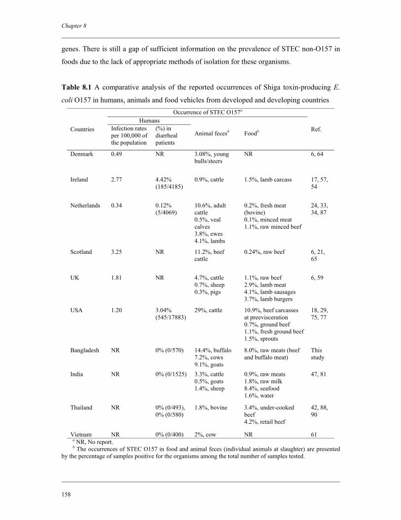
Chapter 8
158
genes. There is still a gap of sufficient information on the prevalence of STEC non-O157 in
foods due to the lack of appropriate methods of isolation for these organisms.
Table 8.1 A comparative analysis of the reported occurrences of Shiga toxin-producing E.
coli O157 in humans, animals and food vehicles from developed and developing countries Occurrence of STEC O157a
Humans Countries Infection rates
per 100,000 of the population
(%) in diarrheal patients
Animal fecesb Foodb Ref.
Denmark 0.49 NR 3.08%, young bulls/steers
NR 6, 64
Ireland 2.77 4.42% (185/4185)
0.9%, cattle 1.5%, lamb carcass 17, 57, 54
Netherlands 0.34 0.12% (5/4069)
10.6%, adult cattle 0.5%, veal calves 3.8%, ewes 4.1%, lambs
0.2%, fresh meat (bovine) 0.1%, minced meat 1.1%, raw minced beef
24, 33, 34, 87
Scotland 3.25 NR 11.2%, beef cattle
0.24%, raw beef 6, 21, 65
UK
1.81 NR 4.7%, cattle 0.7%, sheep 0.3%, pigs
1.1%, raw beef 2.9%, lamb meat 4.1%, lamb sausages 3.7%, lamb burgers
6, 59
USA 1.20 3.04% (545/17883)
29%, cattle
10.9%, beef carcasses at preevisceration 0.7%, ground beef 1.1%, fresh ground beef 1.5%, sprouts
18, 29, 75, 77
Bangladesh NR 0% (0/570) 14.4%, buffalo 7.2%, cows 9.1%, goats
8.0%, raw meats (beef and buffalo meat)
This study
India NR 0% (0/1525) 3.3%, cattle 0.5%, goats 1.4%, sheep
0.9%, raw meats 1.8%, raw milk 8.4%, seafood 1.6%, water
47, 81
Thailand NR 0% (0/493), 0% (0/580)
1.8%, bovine 3.4%, under-cooked beef 4.2%, retail beef
42, 88, 90
Vietnam NR 0% (0/400) 2%, cow NR 61 a NR, No report. b The occurrences of STEC O157 in food and animal feces (individual animals at slaughter) are presented by the percentage of samples positive for the organisms among the total number of samples tested.
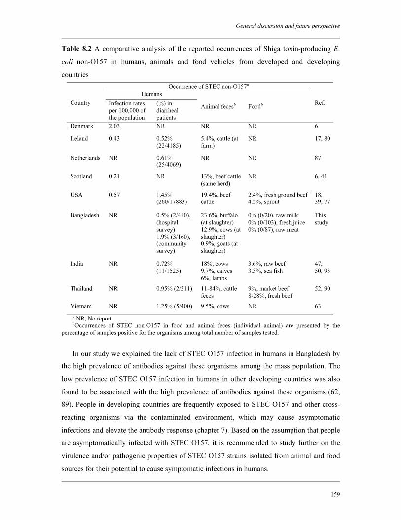
General discussion and future perspective
159
Table 8.2 A comparative analysis of the reported occurrences of Shiga toxin-producing E.
coli non-O157 in humans, animals and food vehicles from developed and developing
countries Occurrence of STEC non-O157a
Humans Country Infection rates
per 100,000 of the population
(%) in diarrheal patients
Animal fecesb Foodb Ref.
Denmark 2.03 NR NR NR 6
Ireland 0.43 0.52% (22/4185)
5.4%, cattle (at farm)
NR 17, 80
Netherlands NR 0.61% (25/4069)
NR NR 87
Scotland 0.21 NR 13%, beef cattle (same herd)
NR 6, 41
USA 0.57 1.45% (260/17883)
19.4%, beef cattle
2.4%, fresh ground beef 4.5%, sprout
18, 39, 77
Bangladesh NR 0.5% (2/410), (hospital survey) 1.9% (3/160), (community survey)
23.6%, buffalo (at slaughter) 12.9%, cows (at slaughter) 0.9%, goats (at slaughter)
0% (0/20), raw milk 0% (0/103), fresh juice 0% (0/87), raw meat
This study
India NR 0.72% (11/1525)
18%, cows 9.7%, calves 6%, lambs
3.6%, raw beef 3.3%, sea fish
47, 50, 93
Thailand NR 0.95% (2/211) 11-84%, cattle feces
9%, market beef 8-28%, fresh beef
52, 90
Vietnam NR 1.25% (5/400) 9.5%, cows NR 63 a NR, No report. bOccurrences of STEC non-O157 in food and animal feces (individual animal) are presented by the percentage of samples positive for the organisms among total number of samples tested.
In our study we explained the lack of STEC O157 infection in humans in Bangladesh by
the high prevalence of antibodies against these organisms among the mass population. The
low prevalence of STEC O157 infection in humans in other developing countries was also
found to be associated with the high prevalence of antibodies against these organisms (62,
89). People in developing countries are frequently exposed to STEC O157 and other cross-
reacting organisms via the contaminated environment, which may cause asymptomatic
infections and elevate the antibody response (chapter 7). Based on the assumption that people
are asymptomatically infected with STEC O157, it is recommended to study further on the
virulence and/or pathogenic properties of STEC O157 strains isolated from animal and food
sources for their potential to cause symptomatic infections in humans.
Chapter 8
160
8.6.2 Selective and differential culture media for the isolation of STEC non-O157
serotypes
Worldwide the number of human illness outbreaks due to STEC non-O157 infection has
increased dramatically in the past decade. Many efforts have been carried out to develop a
suitable culture medium that can detect and isolate STEC organisms irrespective of their
serotype. The major problem in detecting STEC non-O157 is that apart from producing
Stx(s), they do not differ significantly in their biochemical characteristics from typical
commensal E. coli. The only exception is the possible decreased ability to ferment
carbohydrate-like substances (9). However, this characteristic has not been used in
developing a medium for the detection of STEC in a manner similar to that of selecting most
of the STEC O157 by using SMAC agar (58). Except for sorbitol, which is an excellent
marker of sorbitol-negative O157 strains, there are no other single carbohydrate sources,
which enable a quick and clear discrimination between serotypes. Durso et al. (22) reported
that commensal strains were capable of utilizing a broader range of carbon sources than
pathogenic STEC strains. Thus, it is the desire of microbiologists in this field to be able to
select STEC non-O157 by using a specialized medium.
Recently, Possé et al. (72) has described two novel selective differential media for the
isolation of STEC serotypes from food and feces. The first one was for the isolation of STEC
non-O157 serotypes (O26, O103, O111 and O145) and the second one was for both sorbitol-
positive and -negative STEC O157 strains. These media were developed based on a
chromogenic compound to signal β-D-galactosidase activity and one or more fermentative
carbon sources. The chromogenic marker and carbohydrates were combined with a pH
indicator and several inhibitory components, which resulted in a color-based separation of
these serotypes. This study also showed that the consecutive use of a serotype-dependent
choice of confirmation media (dulcitol, L-rhamnose, D-arabinose, D-raffinose, and D-
arabinose) resulted in a very low incidence of false-positive isolates when comparing clinical
STEC strains with a collection of commensal E. coli strains (72).
8.6.3 Surveillance of STEC-associated HUS in Bangladesh
In our previous study, we estimated the prevalence of STEC infection among diarrheal
patients in Bangladesh (chapter 2). However, it is also very important to estimate the
prevalence of STEC infection among HUS patients in the country. At present, there are no
data available on the burden of HUS and therefore the burden of STEC-associated HUS in
Bangladesh is not known. In order to estimate the total burden of HUS in Bangladesh, it is
General discussion and future perspective
161
recommended to perform a prospective study involving all tertiary-level hospitals and some
randomly selected secondary-level hospitals. All HUS cases, as defined according to the
criteria described earlier (8), admitted to these hospitals should be included in the study.
Clinical history of the patients should be recorded and stool and serum samples be collected
and send to the laboratory for analysis. Stool samples should be tested for the presence of Stx,
stx genes and STEC organisms including serogroup O157. The presence of other enteric
pathogens in the stool, including Shigella spp., should also be tested. The serum samples
should be tested for the presence of antibodies against Stx and LPS of STEC O157 and other
predominant serogroups of STEC in order to detect any preceding infections. Serum samples
that we collected previously from the healthy population in Bangladesh can be used as
controls.
8.6.4 Cross reactivity of STEC O157 specific sera with other enteric pathogens
The LPS of E. coli O157 has been reported to cross-react with O-antigens of Y.
enterocolitica O9, C. freundii, E. hermannii, and B. abortus. It has also been reported that the
serum of patients with HUS associated with E. coli O157 reacted with strains of V. cholerae
O1 Inaba. Similarly, cross-reactivities between the E. coli O157 serogroup and the E. coli O7
and O116 serogroups have been described (27). It has also been described that infection with
EPEC in the early childhood may confer protection against STEC infection later in life (12).
In our study, we found a high prevalence of antibodies against E. coli O157 LPS among the
healthy population in Bangladesh (chapter 7). However, we did not test the serum samples for
antibodies against these cross-reacting antigens. Therefore, it would be interesting to test the
serum samples collected from all groups of the people for the cross-reacting antibodies. This
will potentially generate some interesting findings to explain more clearly the lack of STEC
O157 infection in humans in a situation where many other enteric pathogens are endemic.
8.6.5 Occurrence of STEC in surface water in Bangladesh
Both urban and rural water supplies throughout Bangladesh are frequently contaminated
with human fecal organisms (55). Those who can afford it buy bottled water (of dubious
quality), and the majority are left to drink the available contaminated water. The use of
contaminated water remains an important route of transmission of diarrheal pathogens to
human population in Bangladesh. In our study, we found a high prevalence of antibodies
against STEC O157 LPS among healthy population in Bangladesh, which might be
attributable to the regular exposure to common sources of pathogens, including contaminated
Chapter 8
162
water (chapter 7). There is no report of contamination of surface water with STEC in
Bangladesh. Therefore, it is recommended to test the water samples from commonly used
sources in both rural and urban areas of Bangladesh for STEC including serogroup O157 and
other organisms that express O157 antigens.
References
1. Abdulmawjood, A., M. Bulte, N. Cook, S. Roth, H. Schonenbrucher, and J. Hoorfar. 2003. Toward an international standard for PCR based detection of food borne Escherichia coli O157. Part 1. Assay development and multi-center validation. J. Microbiol. Methods 55:775–786.
2. Albert, M. J., S. M. Faruque, A. S. Faruque, P. K. Neogi, M. Ansaruzzaman, N. A. Bhuiyan, K. Alam, and M. S. Akbar. 1995. Controlled study of Escherichia coli diarrheal infections in Bangladeshi children. J. Clin. Microbiol. 33:973-977.
3. Albert, M. J., A. S. Faruque, S. M. Faruque, R. B. Sack, and D. Mahalanabis. 1999. Case-control study of enteropathogens associated with childhood diarrhea in Dhaka, Bangladesh. J. Clin. Microbiol. 37:3458-3464.
4. Andersen, M. R., and C. J. Omiecinski. 1992. Direct extraction of bacterial plasmids from food for polymerase chain reaction amplification. Appl. Environ. Microbiol. 58:4080–4082.
5. Anonymous. 2001. Microbiology of food and animal feeding stuffs—Horizontal method for the detection of Escherichia coli O157 (EN ISO 16654). International Organization for Standardization, Geneva, Switzerland.
6. Anonymous. 2007. Enter-net annual report: 2005 – surveillance of enteric pathogens in Europe and beyond. Enter-net surveillance hub, HPA, Centre for Infections, Colindale, London.
7. Belongia, E. A., P. H. Chyou, R. T. Greenlee, G. Perez-Perez, W. F. Bibb, and E. O. DeVries. 2003. Diarrhea incidence and farm-related risk factors for Escherichia coli O157:H7 and Campylobacter jejuni antibodies among rural children. J. Infect. Dis. 187:1460-1468.
8. Bennish, M. L., W. A. Khan, M. Begum, E. A. Bridges, S. Ahmed, D. Saha, M. A. Salam, D. Acheson, and E. T. Ryan. 2006. Low risk of hemolytic uremic syndrome after early effective antimicrobial therapy for Shigella dysenteriae type 1 infection in Bangladesh. Clin. Infect. Dis. 42:356-362.
9. Bettelheim, K. A. 1997. Biochemical studies on enterohemorrhagic Escherichia coli (EHEC), p. 243-248. In D. Kay, and C. Fricker (ed.), Coliforms and E. coli Problem or Solution. Atheneum Press, Gateshead, Tyne, and Wear.
10. Bettelheim, K. A., and L. Beutin. 2003. Rapid laboratory identification and characterization of verocytotoxigenic (Shiga toxin-producing) Escherichia coli (VTEC/STEC). J. Appl. Microbiol. 95:205-217.
11. Beuchat, L. R. 1996. Pathogenic microorganisms associated with fresh produce. J. Food Prot. 59:204–216.
12. Beutin, L., S. Zimmermann, and K. Gleier. 1998. Human infections with Shiga toxin-producing Escherichia coli other than serogroup O157 in Germany. Emerg. Infect. Dis. 4:635-639.
General discussion and future perspective
163
13. Boerlin, P., S. A. McEwen, F. Boerlin-Petzold, J. B. Wilson, R. P. Johnson, and C. L. Gyles. 1999. Associations between virulence factors of Shiga toxin-producing Escherichia coli and disease in humans. J. Clin. Microbiol. 37:497–503.
14. Brian, M. J., M. Frosolono, B. E. Murray, A. Miranda, E. L. Lopez, H. F. Gomez, and T. G. Cleary. 1992. Polymerase chain reaction for diagnosis of enterohemorrhagic Escherichia coli infection and hemolytic uremic syndrome. J. Clin. Microbiol. 30:1801–1806.
15. Brown, K. H. 2003. Diarrhea and malnutrition. J. Nutr. 133:328S-332S. 16. Bryce, J., C. Boschi-Pinto, K. Shibuya, R. E. Black and WHO Child Health
Epidemiology Reference Group. 2005. WHO estimates of the causes of death in children. Lancet 365:1147-1152.
17. Carroll, A. M., A. Gibson, and E. B. McNamara. 2005. Laboratory-based surveillance of human verocytotoxigenic Escherichia coli infection in the Republic of Ireland, 2002-2004. J. Med. Microbiol. 54:1163-1169.
18. Centers for Disease Control and Prevention (CDC). 2008. Preliminary FoodNet data on the incidence of infection with pathogens transmitted commonly through food-10 states, 2007. MMWR Morb. Mortal. Wkly. Rep. 57:366-370.
19. Chart, H., and B. Rowe. 1993. Antibody cross-reactions with lipopolysaccharide from E. coli O157 after cholera vaccination. Lancet 341:1282.
20. Clifton-Hadley, F. A. 2000. Detection and diagnosis of Escherichia coli O157 and other verotoxigenic E. coli in animal faeces. Rev. Med. Microbiol. 11:47–58.
21. Coia, J. E., Y. Johnston, N. J. Steers, and M. F. Hanson. 2001. A survey of the prevalence of Escherichia coli O157 in raw meats, raw cow's milk and raw-milk cheeses in South-East Scotland. Int. J. Food Microbiol. 66:63-69.
22. Durso, L. M., D. Smith, and R. W. Hutkins. 2004. Measurements of fitness and competition in commensal Escherichia coli and E. coli O157:H7 strains. Appl. Environ. Microbiol. 70:6466-6472.
23. EFSA. 2007. Scientific opinion of the panel on biological hazards on monitoring of verotoxigenic Escherichia coli (VTEC) and identification of human pathogenic VTEC types. EFSA J. 579:1-61.
24. EFSA. 2007. The community summary report on trends and sources of zoonoses, zoonotic agents, antimicrobial resistance and foodborne outbreaks in the European Union in 2006. EFSA J. 130:151-164.
25. Eklund, M., F. Scheutz, and A. Siitonen. 2001. Clinical isolates of non-O157 Shiga toxin-producing Escherichia coli: serotypes, virulence characteristics, and molecular profiles of strains of the same serotype. J. Clin. Microbiol. 39:2829-2834.
26. Erickson, M. C., and M. P. Doyle. 2007. Food as a vehicle for transmission of Shiga toxin-producing Escherichia coli. J. Food Prot. 70:2426-2449.
27. Ewing, W. H. 1986. Edwards and Ewing’s identification of Enterobacteriaceae. Elsevier Science Publishing Inc., New York, NY.
28. Fagan, P. K., M. A. Hornitzky, K. A. Bettelheim, and S. P. Djordjevic. 1999. Detection of Shiga-like toxin (stx1 and stx2), Intimin (eaeA), and enterohemorrhagic Escherichia coli (EHEC) hemolysin (EHEC hlyA) genes in animal feces by multiplex PCR. Appl. Environ. Microbiol. 65:868–872.
29. Fox, J. T., X. Shi, and T. G. Nagaraja. 2008. Escherichia coli O157 in the Rectoanal Mucosal Region of Cattle. Foodborne Pathog. Dis. 5:69-77.
30. Haack, J. P., S. Jelacic, T. E. Besser, E. Weinberger, D. J. Kirk, G. L. McKee, S. M. Harrison, K. J. Musgrave, G. Miller, T. H. Price, and P. I. Tarr. 2003. Escherichia coli O157 exposure in Wyoming and Seattle: serologic evidence of rural risk. Emerg. Infect. Dis. 9:1226–1231.
Chapter 8
164
31. Hancock, D. D., T. E. Besser, and D. H. Rice. 1998. Ecology of Escherichia coli O157:H7 in cattle and impact of management practices, p. 85–91. In J. B. Kaper and A. D. O'Brien (ed.), Escherichia coli O157:H7 and other Shiga toxin-producing E. coli strains. ASM press, Washington, DC.
32. Hasan, K. Z., P. Pathela, K. Alam, G. Podder, S. M. Faruque, E. Roy, A. K. Haque, R. Haque, M. J. Albert, A. K. Siddique, and R. B. Sack. 2006. Aetiology of diarrhoea in a birth cohort of children aged 0-2 year(s) in rural Mirzapur, Bangladesh. J. Health Popul. Nutr. 24:25-35.
33. Heuvelink, A. E., F. L. van den Biggelaar, E. de Boer, R. G. Herbes, W. J. Melchers, J. H. Huis in 't Veld, and L. A. Monnens. 1998. Isolation and characterization of verocytotoxin-producing Escherichia coli O157 strains from Dutch cattle and sheep. J. Clin. Microbiol. 36:878-882.
34. Heuvelink, A. E., J. T. Zwartkruis-Nahuis, R. R. Beumer, and E. de Boer. 1999. Occurrence and survival of verocytotoxin-producing Escherichia coli O157 in meats obtained from retail outlets in The Netherlands. J. Food Prot. 62:1115-1122.
35. Hoque, B. A., K. Hallman, J. Levy, H. Bouis, N. Ali, F. Khan, S. Khanam, M. Kabir, S. Hossain, and M. S. Alam. 2006. Rural drinking water at supply and household levels: quality and management. Int. J. Hyg. Environ. Health 209:451-460.
36. Huilan, S., L. G. Zhen, M. M. Mathan, M. M. Mathew, J. Olarte, R. Espejo, U. Khin Maung, M. A. Ghafoor, M. A. Khan, and Z. Sami. 1991. Etiology of acute diarrhoea among children in developing countries: a multicentre study in five countries. Bull. WHO 69:549–555.
37. Hussein, H. S., and L. M. Bollinger. 2005. Prevalence of Shiga toxin-producing Escherichia coli in beef. Meat Sci. 71:676–689.
38. Hussein, H. S., and T. Sakuma. 2005. Prevalence of Shiga toxin-producing Escherichia coli in dairy cattle and their products. J. Dairy Sci. 88:450–465.
39. Hussein, H. S. 2007. Prevalence and pathogenicity of Shiga toxin-producing Escherichia coli in beef cattle and their products. J. Anim. Sci. 85:E63-E72.
40. Islam, M. A., A. S. Mondol, E. de Boer, R. R. Beumer, M. H. Zwietering, K. A. Talukder, and A. E. Heuvelink. 2008. Prevalence and genetic characterization of Shiga toxin-producing Escherichia coli isolated from slaughtered animals in Bangladesh. Appl. Environ. Microbiol. 74:5414-5421.
41. Jenkins, C., M. C. Pearce, H. Chart, T. Cheasty, G. A. Willshaw, G. J. Gunn, G. Dougan, H. R. Smith, B. A. Synge, and G. Frankel. 2002. An eight-month study of a population of verocytotoxigenic Escherichia coli (VTEC) in a Scottish cattle herd. J. Appl. Microbiol. 93:944-953.
42. Kalnauwakul, S., M. Phengmak, U. Kongmuang, Y. Nakaguchi, and M. Nishibuchi. 2007. Examination of diarrheal stools in Hat Yai City, South Thailand, for Escherichia coli O157 and other diarrheagenic Escherichia coli using immunomagnetic separation and PCR method. Southeast Asian J. Trop. Med. Public Health 38:871-880.
43. Karmali, M. A., B. T. Steele, M. Petric, and C. Lim. 1983. Sporadic cases of haemolytic uraemic syndrome associated with fecal cytotoxin and cytotoxin-producing Escherichia coli. Lancet 1:619–620.
44. Karmali, M. A., M. Petric, C. Lim, P. C. Fleming, G. S. Arbus, and H. Lior. 1985. The association between idiopathic hemolytic uremic syndrome and infection by verotoxin-producing Escherichia coli. J. Infect. Dis. 151:775-782.
45. Keskimaki, M., R. Ikaheimo, P. Karkkainen, F. Scheutz, R. L. Puohiniemi, and A. Siitonen. 1997. Shiga toxin producing Escherichia coli serotype OX3:H21 causing hemolytic uremic syndrome. Clin. Infect. Dis. 24:1278–1279.
General discussion and future perspective
165
46. Khakria, R., D. Duck, and H. Lior. 1990. Extended phage-typing scheme for Escherichia coli O157:H7. Epidemiol. Infect. 105:511-520.
47. Khan, A., S. Yamasaki, T. Sato, T. Ramamurthy, A. Pal, S. Datta, N. R. Chowdhury, S. C. Das, A. Sikdar, T. Tsukamoto, S. K. Bhattacharya, Y. Takeda, and G. B. Nair. 2002. Prevalence and genetic profiling of virulence determinants of non-O157 Shiga toxin producing Escherichia coli isolated from cattle, beef and human cases in Calcutta, India. Emerg. Infect. Dis. 8:54–62.
48. Kosek, M., C. Bern, and R. L. Guerrant. 2003. The global burden of diarrhoeal disease, as estimated from studies published between 1992 and 2000. Bull. WHO. 81:197–204.
49. Kreader, C. A. 1996. Relief of amplification inhibition in PCR with bovine serum albumin or T4 gene 32 protein. Appl. Environ. Microbiol. 62:1102–1106.
50. Kumar, S. H., S. K. Otta, I. Karunasagar, and I. Karunasagar. 2001. Detection of Shiga-toxigenic Escherichia coli (STEC) in fresh seafood and meat marketed in Mangalore, India by PCR. Lett. Appl. Microbiol. 33:334–338.
51. Law, D. 2000. Virulence factors of Escherichia coli O157 and other Shiga toxin-producing E. coli. J. Appl. Microbiol. 88:729-745.
52. Leelaporn, A., M. Phengmak, B. Eampoklap, S. Manatsathit, S. Tritilanunt, S. Siritantikorn, K. Nagayama, T. Iida, C. Niyasom, and P. Komolpit. 2003. Shiga toxin- and enterotoxin-producing Escherichia coli isolated from subjects with bloody and non-bloody diarrhea in Bangkok, Thailand. Diagn. Microbiol. Infect. Dis. 46:173-180.
53. LeJeune, J. T., T. E. Besser, and D. D. Hancock. 2001. Cattle water troughs as reservoirs of Escherichia coli O157. Appl. Environ. Microbiol. 67:3053-3057.
54. Lenahan, M., S. O'Brien, K. Kinsella, T. Sweeney, and J. J. Sheridan. 2007. Prevalence and molecular characterization of Escherichia coli O157:H7 on Irish lamb carcasses, fleece and in faeces samples. J. Appl. Microbiol. 103:2401-2409.
55. Luby, S. 2008. Water quality in South Asia. J. Health Popul. Nutr. 26:123-124. 56. Ludwig, K., V. Sarkim, M. Bitzan, M. A. Karmali, C. Bobrowski, H. Ruder, R.
Laufs, I. Sobottka, M. Petric, H. Karch, and D. E. Müller-Wiefel. 2002. Shiga toxin-producing Escherichia coli infection and antibodies against Stx2 and Stx1 in household contacts of children with enteropathic hemolytic-uremic syndrome. J. Clin. Microbiol. 40:1773–1782.
57. Madden, R. H., K. A. Murray, and A. Gilmour. 2007. Carriage of four bacterial pathogens by beef cattle in Northern Ireland at time of slaughter. Lett. Appl. Microbiol. 44:115-119.
58. March, S. B., and S. Ratnam. 1986. Sorbitol-MacConkey medium for detection of Escherichia coli O157:H7 associated with hemorrhagic colitis. J. Clin. Microbiol. 23:869–872.
59. Milnes, A. S., I. Stewart, F. A. Clifton-Hadley, R. H. Davies, D. G. Newell, A. R. Sayers, T. Cheasty, C. Cassar, A. Ridley, A. J. Cook, S. J. Evans, C. J. Teale, R. P. Smith, A. McNally, M. Toszeghy, R. Futter, A. Kay, and G. A. Paiba. 2008. Intestinal carriage of verocytotoxigenic Escherichia coli O157, Salmonella, thermophilic Campylobacter and Yersinia enterocolitica, in cattle, sheep and pigs at slaughter in Great Britain during 2003. Epidemiol. Infect. 136:739-751.
60. Mora, A., M. Blanco, J. E. Blanco, M. P. Alonso, G. Dahbi, F. Thompson-Carter, M. A. Usera, R. Bartolomé, G. Prats, and J. Blanco. 2004. Phage types and genotypes of human and animal Shiga toxin-producing Escherichia coli O157:H7 in Spain: identification of two predominating phage types (PT2 and PT8). J. Clin. Microbiol. 42:4007-4015.
Chapter 8
166
61. Nakasone, N., H. H. Tran, M. B. Nguyen, N. Higa, C. Toma, T. Song, Y. Ichinose, and M. Iwanaga. 2005. Isolation of Escherichia coli O157:H7 from fecal samples of cows in Vietnam. Am. J. Trop. Med. Hyg. 73:586-587.
62. Navarro, A., C. Eslava, U. Hernandez, J. L. Navarro-Henze, M. Aviles, G. G. la Torre, and A. Cravioto. 2003. Antibody responses to Escherichia coli O157 and other lipopolysaccharides in healthy children and adults. Clin. Diagn. Lab. Immunol. 10:797–801.
63. Nguyen, T. V., P. Le Van, C. Le Huy, K. N. Gia, and A. Weintraub. 2005. Detection and characterization of diarrheagenic Escherichia coli from young children in Hanoi, Vietnam. J. Clin. Microbiol. 43:755-760.
64. Nielsen, E. M., and F. Scheutz. 2002. Characterisation of Escherichia coli O157 isolates from Danish cattle and human patients by genotyping and presence and variants of virulence genes. Vet. Microbiol. 88:259-273.
65. Ogden, I. D., M. MacRae, and N. J. Strachan. 2004. Is the prevalence and shedding concentrations of E. coli O157 in beef cattle in Scotland seasonal? FEMS Microbiol. Lett. 233:297-300.
66. Oliveira, S. D., L. R. Santos, D. M. Schuch, A. B. Silva, C. T. Salle, and C. W. Canal. 2002. Detection and identification of salmonellas from poultry-related samples by PCR. Vet. Microbiol. 87:25–35.
67. Ostroff, S. M., P. I. Tarr, M. A. Neill, J. H. Lewis, N. Hargrett-Bean, and J. M. Kobayashi. 1989. Toxin genotypes and plasmid profiles as determinants of systemic sequelae in Escherichia coli O157:H7 infections. J. Infect. Dis. 160:995–998.
68. Oswald, E., H. Schmidt, S. Morabito, H. Karch, O. Marches, and A. Caprioli. 2000. Typing of intimin genes in human and animal enterohemorrhagic and enteropathogenic Escherichia coli: characterization of a new intimin variant. Infect. Immun. 68:64-71.
69. Paton, J. C., and A. W. Paton. 1998. Pathogenesis and diagnosis of Shiga toxin producing Escherichia coli infections. Clin. Microbiol. Rev. 11:450-479.
70. Paton, A. W., M. C. Woodrow, R. M. Doyle, J. A. Lanser, and J. C. Paton. 1999. Molecular characterization of a Shiga toxigenic Escherichia coli O113:H21 strain lacking eae responsible for a cluster of cases of hemolytic-uremic syndrome. J. Clin. Microbiol. 37:3357–3361.
71. Persson, S., K. E. Olsen, S. Ethelberg, and F. Scheutz. 2007. Subtyping method for Escherichia coli shiga toxin (verocytotoxin) 2 variants and correlations to clinical manifestations. J. Clin. Microbiol. 45:2020-2024.
72. Possé, B., L. De Zutter, M. Heyndrickx, and L. Herman. 2008. Novel differential and confirmation plating media for Shiga toxin-producing Escherichia coli serotypes O26, O103, O111, O145 and sorbitol-positive and-negative O157. FEMS Microbiol. Lett. 282:124-131.
73. Reymond, D., R. D. Johnson, M. A. Karmali, M. Petric, M. Winkler, S. Johnson, K. Rahn, S. Renwick, J. Wilson, R. C. Clarke, and J. Spika. 1996. Neutralizing antibodies to Escherichia coli Vero cytotoxin 1 and antibodies to O157 lipopolysaccharide in healthy farm family members and urban residents. J. Clin. Microbiol. 34:2053–2057.
74. Riley, L. W., R. S. Remis, S. D. Helgerson, H. B. McGee, J. G. Wells, B. R. Davis, R. J. Hebert, E. S. Olcott, L. M. Johnson, N. T. Hargrett, P. A. Blake, and M. L. Cohen. 1983. Hemorrhagic colitis associated with a rare Escherichia coli serotype. N. Engl. J. Med. 308:681-685.
75. Rivera-Betancourt, M., S. D. Shackelford, T. M. Arthur, K. E. Westmoreland, G. Bellinger, M. Rossman, J. O. Reagan, and M. Koohmaraie. 2004. Prevalence of
General discussion and future perspective
167
Escherichia coli O157:H7, Listeria monocytogenes, and Salmonella in two geographically distant commercial beef processing plants in the United States. J. Food Prot. 67:295–302.
76. Sack, R. B., A. K. Siddique, I. M. Jr. Longini, A. Nizam, M. Yunus, M. S. Islam, J. G. Jr. Morris, A. Ali, A. Huq, G. B. Nair, F. Qadri, S. M. Faruque, D. A. Sack, and R. R. Colwell. 2003. A 4-year study of the epidemiology of Vibrio cholerae in four rural areas of Bangladesh. J. Infect. Dis. 187:96-101.
77. Samadpour, M., M. W. Barbour, T. Nguyen, T. M. Cao, F. Buck, G. A. Depavia, E. Mazengia, P. Yang, D. Alfi, M. Lopes, and J. D. Stopforth. 2006. Incidence of enterohemorrhagic Escherichia coli, Escherichia coli O157, Salmonella, and Listeria monocytogenes in retail fresh ground beef, sprouts, and mushrooms. J. Food Prot. 69: 441-443.
78. Scheu, P. M., K. Berghof, and U. Stahl. 1998. Detection of pathogenic and spoilage microorganisms in food with the polymerase chain reaction. Food Microbiol. 15:13–31.
79. Schmidt, H., L. Beutin, and H. Karch. 1995. Molecular analysis of the plasmid-encoded hemolysin of Escherichia coli O157:H7 strain EDL 933. Infect. Immun. 63:1055-1061.
80. Scott, L., P. McGee, C. Walsh, S. Fanning, T. Sweeney, J. Blanco, M. Karczmarczyk, B. Earley, N. Leonard, and J. J. Sheridan. 2008. Detection of numerous verotoxigenic E. coli serotypes, with multiple antibiotic resistance from cattle faeces and soil. Vet. Microbiol. Aug 22. [Epub ahead of print]
81. Sehgal, R., Y. Kumar, and S. Kumar. 2008. Prevalence and geographical distribution of Escherichia coli O157 in India: a 10-year survey. Tans. Royal Soc. Trop. Med. Hyg. 102:380-383.
82. Shooter, R. A., E. M. Cooke, S. A. Rousseau, and A. L. Breaden. 1970. Animal sources of common serotypes of E. coli in the food of hospital patients. Possible significance in urinary tract infection. Lancet 2:226–228.
83. Shooter, R. A., E. M. Cooke, S. M. O’Farrell, K. A. Bettelheim, M. E. Chandler, and F. M. Bushrod. 1974. The isolation of Escherichia coli serotypes from a poultry packing station and an abattoir. J. Hyg. Camb. 73:245–247.
84. Sivapalasingam, S., C. R. Friedman, L. Cohen, and R. V. Tauxe. 2004. Fresh produce: a growing cause of outbreaks of foodborne illness in the United States, 1973 through 1997. J. Food Prot. 67:2342-2353.
85. Tarr, P. I., C. A. Gordon, and W. L. Chandler. 2005. Shiga toxin-producing Escherichia coli and haemolytic uraemic syndrome. Lancet 365:1073-1086.
86. Toma, C., E. M. Espinosa, T. Song, E. Miliwebsky, I. Chinen, S. Iyoda, M. Iwanaga, and M. Rivas. 2004. Distribution of putative adhesins in different seropathotypes of Shiga toxin-producing Escherichia coli. J. Clin. Microbiol. 42:4937–4946.
87. van Duynhoven, Y. T., I. H. Friesema, T. Schuurman, A. Roovers, A. A. van Zwet, L. J. Sabbe, W. K. van der Zwaluw, D. W. Notermans, B. Mulder, E. J. van Hannen, F. G. Heilmann, A. Buiting, R. Jansen, and A. M. Kooistra-Smid. 2008. Prevalence, characterisation and clinical profiles of Shiga toxin-producing Escherichia coli in The Netherlands. Clin. Microbiol. Infect. 14:437-445.
88. Voravuthikunchai, S. P., O. Keisaku, T. Iida, and T. Honda. 2002. Surveillance of Enterohaemorrhagic Escherichia coli O157:H7 in Southern Thailand. J. Health Popul. Nutr. 20:189-191.
Chapter 8
168
89. Voravuthikunchai, S. P., C. Chaowana, P. Perepat, T. Iida, and T. Honda. 2005. Antibodies among healthy population of developing countries against enterohaemorrhagic Escherichia coli O157:H7. J. Health Popul. Nutr. 23:305-310.
90. Vuddhakul, V., N. Patararungrong, P. Pungrasamee, S. Jitsurong, T. Morigaki, N. Asai, and M. Nishibuchi. 2000. Isolation and characterization of Escherichia coli O157 from retail beef and bovine faeces in Thailand. FEMS Microbiol. Lett. 182:343-347.
91. Wallace, J. S. 1999. The ecological cycle of Escherichia coli O157:H7, p. 195-223. In C. S. Stewart and H. J. Flint (ed.), Escherichia coli O157 in Farm Animals. CABI Publishing, New York, NY.
92. Wang, G., and M. P. Doyle. 1998. Survival of enterohemorrhagic Escherichia coli O157:H7 in water. J. Food Prot. 61:662–667.
93. Wani, S. A., F. Pandit, I. Samanta, M. A. Bhat, and A. S. Buchh. 2004. Molecular epidemiology of Shiga toxin-producing Escherichia coli in India. Cur. Sci. 87:1345-1353.
94. Wells, J. G., L. D. Chipman, K. D. Greene, E. G. Sowers, J. H. Green, D. N. Cameron, F. P. Downes, M. L. Martin, P. M. Griffin, S. M. Ostroff, M. E. Potter, R. V. Tauxe, and J. K. Wachsmuth. 1991. Isolation of Escherichia coli serotype O157:H7 and other Shiga like toxin-producing E. coli from dairy cattle. J. Clin. Microbiol. 29:985-989.
95. Zhao, T., M. P. Doyle, J. Shere, and L. Garber. 1995. Prevalence of enterohemorrhagic Escherichia coli O157:H7 in a survey of dairy heards. Appl. Environ. Microbiol. 61:1290-1293.

Summary
Summary
170
Summary
Shiga toxin (Stx)-producing Escherichia coli (STEC) are one of the most important
groups of foodborne pathogens. Infection with STEC in humans can lead to mild diarrhea,
bloody diarrhea or the hemolytic-uremic syndrome (HUS). Domestic ruminants appear to be
important reservoirs for STEC and play a significant role in the epidemiology of human
infections. STEC infections are reported from different parts of the world, being found in 75
to 100% of episodes of sporadic HUS in Europe, North America, Canada, and Latin America
especially in Argentina. Still STEC infections are a much greater problem in developed
countries than in developing countries. Bangladesh is an endemic zone for diarrheal diseases
caused by different enteric pathogens, however, no systematic study has yet been done here
on the epidemiology of STEC infection in humans. The main objective of the research
described in this thesis was to gain a detailed insight in the epidemiology of STEC infection
in humans in Bangladesh and to find out the reservoirs and transmission routes of these
organisms.
To estimate the prevalence of STEC in humans, we tested stool samples collected from
patients with diarrhea attending a hospital (n = 410) and a community clinic (n = 160) in
Dhaka, Bangladesh (chapter 2). Around 2% of the patients with diarrhea in the hospital and
7% of the patients in the community clinic were positive for stx genes. STEC strains were
isolated from 0.5% (n = 2) of the patients in the hospital and 1.9% (n = 3) of the patients in
the community. All five isolates were positive in the Vero cell assay. Two isolates were
positive for the eae gene and four were positive for the hlyEHEC gene. The five isolates
belonged to different serotypes and no case of STEC O157 infection was identified.
The relatively low prevalence of STEC among patients with diarrhea compared to other
diarrhea-causing pathogens prompted us to estimate the prevalence of STEC in animal
reservoirs and in the food chain in Bangladesh. Unlike clinical samples, it is difficult to
isolate STEC from samples of animal faeces and food because of the presence of very low
numbers of STEC in an abundance of competitive microflora. To identify an effective
technique for isolation of STEC O157, we evaluated the efficiency of various methods
(chapter 3 and chapter 4). Immunomagnetic separation (IMS) using Dynabeads anti-E. coli
O157 proved to be more efficient than the Vitek Immunodiagnostic Assay System (VIDAS)
Immuno-Concentration E. coli O157 (ICE) kit (VIDAS ICE) in case of animal feces and the
use of CHROMagar O157 with cefixime (0.025 mg/l) and tellurite (1.25 mg/l) as plating
media resulted in more positive samples than sorbitol-MacConkey agar with cefixime (0.05
Summary
171
mg/l) and tellurite (2.5 mg/l). Comparison of IMS with PCR for the rfbEO157 gene showed
that IMS was more sensitive than PCR to detect STEC O157. In order to estimate the
prevalence of these organisms in animal feces and foods we used both IMS and PCR
techniques (chapter 5 and chapter 6).
Of the rectal contents collected from buffalo, cows, and goats, 82.2%, 72.7%, and 11.8%
tested positive for stx1 and/or stx2, respectively. We isolated STEC O157 strains from 14.4%
of the buffalo, 7.2% of the cows, and 9.1% of the goats. We isolated STEC non-O157 strains
from 23.6% of the buffalo, 12.9% of the cows, and 0.9% of the goats. The majority of the
STEC O157 isolates possessed virulence genes that are associated with increased human
pathogenicity, including stx2, eae, katP, etpD and hlyEHEC. The STEC non-O157 isolates
belonged to 36 O serogroups and 52 O:H serotypes. In case of foods, we found that more
than 71% (n = 62) of raw meats, 10% (n = 2) of raw cow’s milk and 8% (n = 8) of fresh juice
samples were positive for stx genes. We isolated STEC O157 strains from 8% (n = 7) of the
meat samples and no other STEC could be isolated from the remaining stx-positive food
samples. Subtyping of the STEC O157 isolates (n = 52) from animal feces and raw meats by
pulsed-field gel electrophoresis revealed 42 distinct restriction patterns suggesting
heterogeneous clonal diversity.
In order to explain the lack of STEC O157 infection in humans, we investigated the
presence of antibodies against O157 lipopolysaccharide (LPS) among the healthy population
in Bangladesh (chapter 7). We collected serum samples from different groups of people
living in urban and rural areas, comprising butchers, household members of butchers, and
people with other occupations. We found that around 50% (n = 116) of the samples were
positive for antibodies (IgG, IgA and/or IgM) to E. coli O157 LPS. By doing separate
analysis, we found that all 116 samples positive for polyvalent antibodies were positive for
IgG-class antibodies and 87 were also positive for IgM-class antibodies. Of the 133 serum
samples collected from butchers, 54 (41%) were antibody-positive. Among nine household
members of butchers involved in other occupations, five showed a positive antibody
response. Among participants other than butchers and their families, 57 (63%) showed a
positive antibody response, which was statistically significantly higher than that of butchers
(P < 0.05). We did not find any statistically significant difference in the prevalence of
antibody response between the urban and rural populations. The high prevalence of antibody
response might be due to the frequent exposure to STEC O157 present in the food chain or
other organisms present in the environment that share common epitopes of O157 LPS. Like
many other enteric pathogens, exposure of the Bangladeshi population to STEC O157 or
Summary
172
other cross-reacting organisms might be attributable to the use of contaminated water for
drinking and washing/bathing. We found a statistically significantly higher number of
participants from urban areas were positive for antibodies than participants from rural areas
as compared by their practice of drinking contaminated water and using it for
bathing/washing. We further confirmed this result by doing separate analysis of the results
obtained for both urban and rural areas, where we found a significant association between the
use of municipal water and positive antibody response to the E. coli O157 LPS among urban
populations but not among rural populations. Overall results of this study concluded that the
lack of STEC O157 infection among Bangladeshi population might be attributable to the
protective immunity against this pathogen acquired by the frequent exposure to the antigens.
Finally, an extensive discussion concerning chapters 2 to 7 is described in chapter 8,
which also includes suggestions for future research. The overall occurrences of STEC in
diarrheal patients, animal reservoirs and foods in Bangladesh were compared with similar
data available in other countries from both developed and developing parts of the world in
order to get a global perspective. A major difference between our findings and findings from
developed countries was observed in the prevalence of STEC O157-associated human
infections. Unlike developed countries, no case of STEC O157 infection was found among
diarrheal patients in Bangladesh. We concluded that the lack of STEC O157 infection could
be due to the high prevalence of antibodies against these organisms among the mass
population in Bangladesh.

Samenvatting
Samenvatting
174
Samenvatting
Shigatoxine (Stx) producerende Escherichia coli (STEC) is één van de belangrijkere
voedselpathogenen. STEC-infecties kunnen bij de mens milde tot bloederige diarree
veroorzaken, maar ook soms resulteren in het hemolytisch uremisch syndroom (HUS) dat
verantwoordelijk is voor nierfalen. Landbouwhuisdieren, vooral runderen en andere
herkauwers, zijn een belangrijk reservoir voor STEC en spelen een essentiële rol in de
epidemiologie van humane infecties. Rapportage van STEC-infecties komt wereldwijd voor
en in Europa, Noord-Amerika, Canada en Latijns Amerika (vooral in Argentinië) is deze
pathogeen verantwoordelijk voor 75-100% van de sporadische HUS-gevallen. STEC is een
veel groter probleem in ontwikkelde landen dan in ontwikkelingslanden. Alhoewel
Bangladesh een endemisch gebied is voor diverse enteropathogenen die diarree veroorzaken,
is er tot op heden nooit systematisch onderzoek verricht naar de epidemiologie van humane
STEC-infecties. Het belangrijkste doel van het onderzoek dat in dit proefschrift is
beschreven, was het verkrijgen van een gedetailleerd inzicht in de epidemiologie van die
infecties in de Bengaalse populatie.
Om het voorkomen van STEC te schatten, zijn faecesmonsters onderzocht van
ziekenhuispatiënten van het ICDDR,B ziekenhuis (n = 410) en van patiënten afkomstig uit
een locale kliniek in een achterstandswijk (n = 160), beide gevestigd in Dhaka, Bangladesh
(hoofdstuk 2). Faecesmonsters van ongeveer 2% van de ziekenhuispatiënten en 7% van de
patiënten uit de kliniek waren positief voor het stx-gen. Uit faeces van twee
ziekenhuispatiënten (0.5%) en drie personen uit de kliniek (1.9%) werden STEC-stammen
geïsoleerd. Alle vijf stammen waren positief in de Vero-cell test. Twee isolaten waren
positief voor het eae-gen en vier waren positief voor het hlyEHEC-gen. De vijf isolaten
behoorden tot verschillende serotypen, maar er werd geen STEC O157 infectie
waargenomen.
De relatieve lage prevalentie van STEC bij patiënten met diarree in vergelijking met
andere pathogenen die diarree veroorzaken, stimuleerde ons een schatting te doen naar het
voorkomen van STEC in dierlijke reservoirs en in de Bengaalse voedselketen. Omdat in
diermonsters en voedsel slechts lage aantallen STEC voorkomen tussen hoge aantallen van de
begeleidende microflora, is het veel moeilijker hieruit STEC te isoleren dan uit klinische
monsters. Verschillende methoden voor het aantonen van STEC O157 werden met elkaar
vergeleken om de meest effectieve methode te kunnen vinden (hoofdstuk 3 en 4). Het bleek
Samenvatting
175
dat voor het onderzoek van dierlijke faeces de immunomagnetische scheiding (IMS) met
Dynabeads anti-E. coli O157 efficiënter was dan de VIDAS-ICE methode. Door hier 1/2CT-
CHROMagar als isolatiemedium te gebruiken, werden meer verdachte kolonies verkregen
dan met CT-SMAC. Een vergelijking van IMS met PCR voor het aantonen van het rfbEO157-
gen toonde aan dat IMS een gevoeliger methode was dan PCR voor het aantonen van STEC
O157. Om het voorkomen van deze pathogenen in dierlijke faeces en levensmiddelen te
schatten, werd gebruik gemaakt van beide technieken, IMS en PCR (hoofdstuk 5 en 6)
Het stx1- en/of het stx2-gen werd aangetoond in rectale monsters verkregen van buffels
(82.2%), koeien (72.7%) en geiten (11.8%). Uit die monsters konden ook STEC-stammen
geïsoleerd worden: buffels (14.4%), koeien (7.2%) en geiten (9.1%). Verder werden STEC-
non O157 stammen geïsoleerd: uit buffels 23.6%, uit koeien 12.9% en 0.9% uit geiten. Het
grootste deel van de STEC O157 isolaten bezat virulentiegenen die geassocieerd zijn met
verhoogde humane pathogeniteit, waaronder stx2, eae, katP, etpD, en hlyEHEC. De STEC non
O157 isolaten behoorden tot 36 O-serogroepen en 52 O:H-serotypen. Van de onderzochte
levensmiddelen waren vooral vleesmonsters positief voor het stx-gen: 62 monsters van rauw
vlees (71%), 2 monsters rauwe koemelk (10%) en 8 monsters vers sap (8%). Uit zeven
vleesmonsters (8%) konden STEC O157 stammen worden geïsoleerd, maar niet uit de
resterende stx-positieve monsters. Met behulp van pulsed-field gel electroforese konden 52
STEC O157 isolaten afkomstig uit faeces en rauw vlees onderscheiden worden in 42
duidelijke restrictiepatronen. Dit duidt op een heterogene klonale diversiteit.
Om te kunnen verklaren waarom er nauwelijks humane STEC-infecties voorkomen in
Bangladesh, werd bloed van gezonde mensen getest op de aanwezigheid van antilichamen
tegen O157 LPS (hoofdstuk 7). Van verschillende groepen mensen, zowel uit de stad als van
het platteland, waaronder slagers, gezinsleden van slagers, en andere beroepsgroepen, werden
serummonsters afgenomen en onderzocht. Circa 50% van de monsters (n = 116) waren
positief voor antilichamen tegen E. coli O157 LPS (IgG, IgA en/of IgM). Uit verder
onderzoek bleek dat alle 116 monsters die positief waren voor polyvalente antilichamen, ook
positief waren tegen IgG, en 87 van de monsters waren bovendien positief tegen IgM. Van de
133 serummonsters afgenomen bij slagers, waren er 54 (41%) positief tegen antilichamen.
Van de 9 gezinsleden met een ander beroep, hadden er vijf een positieve respons tegen de
antilichamen. Bij de andere personen testten er 57 (63%) positief op de antilichamen, en dat
was statistisch significant hoger dan bij de slagers (P < 0.05). Er werd geen significant
verschil gevonden in antilichaamrespons bij slagers tussen stads- en plattelandsbevolking.
Deze hoge antibody-respons kan het gevolg zijn van het veelvuldig in contact komen met

Samenvatting
176
STEC O157 in de voedselketen, maar het kan ook veroorzaakt zijn door andere micro-
organismen, die gemeenschappelijke epitopen hebben met O157 LPS. Zoals voor veel andere
pathogenen geldt, kan de Bengaalse bevolking blootgesteld zijn aan STEC O157 (of aan
andere, kruisreagerende micro-organismen) door contact met bad- of drinkwater dat met
dierlijke uitwerpselen besmet is. Het drinken van, of wassen met besmet water in aanmerking
genomen, waren er significant meer stadsbewoners positief voor antilichamen dan bewoners
van het platteland. Dit werd ook nog eens bevestigd door een andere analyse van de data,
waar een significant verband werd gevonden tussen het gebruik van water uit publieke
bronnen en een positieve antilichaamrespons voor E. coli LPS bij stadsbewoners, maar niet
bij de plattelandsbevolking. Uit alle resultaten van dit deel van het onderzoek bleek dat het
geringe aantal STEC-infecties in de Bengaalse bevolking toegeschreven zou kunnen worden
aan de immuniteit tegen deze pathogeen, die verkregen wordt door veelvuldige blootstelling
aan antigenen.
In hoofdstuk 8 worden alle resultaten uit de hoofdstukken 2 tot en met 7 uitvoerig
bediscussieerd. In dit hoofdstuk worden ook aanbevelingen gedaan voor verder onderzoek.
Om het algemeen voorkomen van STEC in Bangladesh bij diarreepatiënten, in dierlijke
reservoirs en in levensmiddelen in een globaal perspectief te kunnen plaatsen, werden de
resultaten van dit onderzoek vergeleken met gegevens over deze pathogeen uit andere landen,
zowel gelegen in ontwikkelde als in minder ontwikkelde delen van de wereld. Een belangrijk
verschil tussen de resultaten van dit onderzoek en resultaten uit ontwikkelde landen was het
voorkomen van STEC O157-geassocieerde humane infecties. In tegenstelling tot in
ontwikkelde landen, werd er in Bangladesh geen STEC O157 infectie gevonden bij
diarreepatiënten. De reden hiervoor kan te danken zijn aan de hoge titer antilichamen in de
Bengaalse bevolking tegen deze pathogeen.
177
Curriculum vitae
Mohammad Aminul Islam was born on October 26th, 1976, in Faridpur, Bangladesh.
After completing primary, secondary and higher secondary education from Dhaka,
Bangladesh, he enrolled at the department of Microbiology, University of Dhaka and
completed his BSc. (honors) in 2000 and MSc. in 2001. He conducted his MSc. thesis
research on “phenotypic and genotypic characterization of Shigella flexneri type 4 isolated
from diarrheal patients in Bangladesh” at the Enteric Microbiology Laboratory, International
Centre for Diarrheal Disease Research, Bangladesh (ICDDR,B). After completing his MSc.
he joined the same laboratory at ICDDR,B as a research officer and worked on the molecular
epidemiology of Shigella spp., among diarrheal patients in Bangladesh. In 2003, he was
awarded a grant by the Netherlands Organization for Scientific Research (NWO-WOTRO) to
work on a PhD research proposal. He started his PhD in the beginning of 2004 at the Food
Microbiology Laboratory of Food and Consumer Product Safety Authority (VWA), Zutphen,
the Netherlands in collaboration with the Laboratory of Food Microbiology, Department of
Agrotechnology and Food Sciences, Wageningen University, the Netherlands and ICDDR,B,
Bangladesh.
List of publications
178
List of publications
1. Islam, M. A., Md. M. Rahaman, A. E. Heuvelink, E. de Boer, M. H. Zwietering, H. Chart, A. Navarro, A. Cravioto, and K. A. Talukder. Antibody responses to Escherichia coli O157 lipopolysaccharide among the healthy population in Bangladesh. Submitted.
2. Islam, M. A., A. S. Mondol, I. J. Azmi, E. de Boer, M. H. Zwietering, A. E. Heuvelink, and K. A. Talukder. Shiga toxin-producing Escherichia coli in raw meat, raw milk and street vended juices in Dhaka, Bangladesh. Submitted.
3. Islam, M. A., A. S. Mondol, E. de Boer, R. R. Beumer, M. H. Zwietering, K. A. Talukder, and A. E. Heuvelink. 2008. Prevalence and genetic characterization of Shiga toxin-producing Escherichia coli isolated from slaughtered animals in Bangladesh. Appl. Environ. Microbiol. 74:5414-5421.
4. Talukder, K. A., A. S. Mondol, M. A. Islam, Z. Islam, D. K. Dutta, B. K. Khajanchi, I. J. Azmi, M. A. Hossain, M. Rahman, T. Cheasty, A. Cravioto, G. B. Nair, and D. A. Sack. 2007. A novel serovar of Shigella dysenteriae from patients with diarrhoea in Bangladesh. J. Med. Microbiol. 56:654-658.
5. Islam, M. A., A. E. Heuvelink, E. de Boer, P. D. Sturm, R. R. Beumer, M. H. Zwietering, A. S. G. Faruque, R. Haque, D. A. Sack, and K. A. Talukder. 2007. Shiga toxin-producing Escherichia coli isolated from diarrhoeal patients in Bangladesh. J. Med. Microbiol. 56:380-385.
6. Islam, M. A., A. E. Heuvelink, K. A. Talukder, M. H. Zwietering, and E. de Boer. 2006. Evaluation of immunomagnetic separation and PCR for the detection of Escherichia coli O157 in animal feces and meats. J. Food Prot. 66:2865-2869.
7. Talukder, K. A., Z. Islam, D. K. Dutta, M. A. Islam, B. K. Khajanchi, I. J. Azmi, M. S. Iqbal, M. A. Hossain, A. S. G. Faruque, G. B. Nair, and D. A. Sack. 2006. Antibiotic resistance and genetic diversity of Shigella sonnei in diarrhoeal patients isolated between 1999 and 2003 in Bangladesh. J. Med. Microbiol. 55:1257-1263.
8. Talukder, K. A., B. K. Khajanchi, M. A. Islam, D. K. Dutta, Z. Islam, S. I. Khan, G. B. Nair, and D. A. Sack. 2006. The emerging strains of Shigella dysenteriae type 2 in Bangladesh are clonal. Epidemiol. Infect.134:1249-1256.
9. Islam, M. A., A. E. Heuvelink, K. A. Talukder, and E. de Boer. 2006. Immunoconcentration of Shiga toxin-producing Escherichia coli O157 from animal faeces and raw meats by using Dynabeads anti-E. coli O157 and the VIDAS system. Int. J. Food Microbiol. 109:151-156.
10. Talukder, K. A., B. K. Khajanchi, M. A. Islam, Z. Islam, D. K. Dutta, M. Rahman, H. Watanabe, G. B. Nair, and D. A. Sack. 2006. Fluoroquinolone resistance linked to both gyrA and parC mutations in the quinolone resistance-determining region of Shigella dysenteriae type 1. Curr. Microbiol. 52:108-111.
11. Talukder, K. A., B. K. Khajanchi, D. K. Dutta, Z. Islam, M. A. Islam, M. S. Iqbal, G. B. Nair, and D. A. Sack. 2005. An unusual cluster of dysentery due to Shigella dysenteriae type 4 in Dhaka, Bangladesh. J. Med. Microbiol. 54:511-513.
12. Talukder, K. A., B. K. Khajanchi, M. A. Islam, D. K. Dutta, Z. Islam, A. Safa, G. Y. Khan, K. Alam, M. A. Hossain, S. Malla, S. K. Niyogi, M. Rahman, H. Watanabe, G. Balakrish Nair, and D. A. Sack. 2004. Genetic relatedness of ciprofloxacin-resistant Shigella dysenteriae type 1 strains isolated in south Asia. J. Antimicrob. Chemother. 54:730-734.
13. Talukder, K. A., M. A. Islam, B. K. Khajanchi, D. K. Dutta, Z. Islam, A. Safa, K. Alam, A. Hossain, G. B. Nair, and D. A. Sack. 2003. Temporal shifts in the
List of publications
179
dominance of serotypes of Shigella dysenteriae from 1999 to 2002 in Dhaka, Bangladesh. J. Clin. Microbiol. 41:5053-5058.
14. Talukder, K. A., Z. Islam, M. A. Islam, D. K. Dutta, A. Safa, M. Ansaruzzaman, A. S. G. Faruque, S. N. Shahed, G. B. Nair, and D. A. Sack. 2003. Phenotypic and genotypic characterization of provisional serotype Shigella flexneri 1c and clonal relationships with 1a and 1b strains isolated in Bangladesh. J. Clin. Microbiol. 41:110-117.
15. Talukder, K. A., M. A. Islam, D. K. Dutta, F. Hassan, A. Safa, G. B. Nair, and D. A. Sack. 2002. Phenotypic and genotypic characterization of serologically atypical strains of Shigella flexneri type 4 isolated in Dhaka, Bangladesh. J. Clin. Microbiol. 40:2490-2497.

Overview of completed training activities
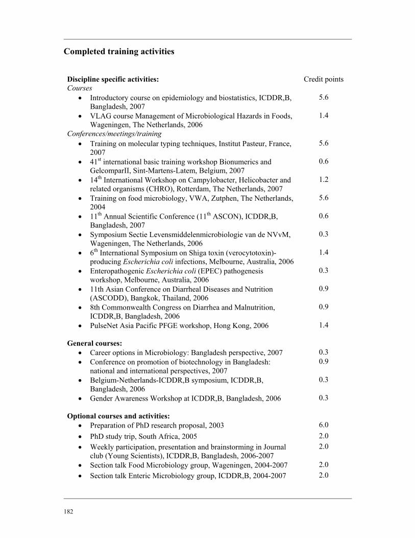
182
Completed training activities
Discipline specific activities: Credit points
Courses • Introductory course on epidemiology and biostatistics, ICDDR,B,
Bangladesh, 2007 5.6
• VLAG course Management of Microbiological Hazards in Foods, Wageningen, The Netherlands, 2006
1.4
Conferences/meetings/training • Training on molecular typing techniques, Institut Pasteur, France,
2007 5.6
• 41st international basic training workshop Bionumerics and GelcomparII, Sint-Martens-Latem, Belgium, 2007
0.6
• 14th International Workshop on Campylobacter, Helicobacter and related organisms (CHRO), Rotterdam, The Netherlands, 2007
1.2
• Training on food microbiology, VWA, Zutphen, The Netherlands, 2004
5.6
• 11th Annual Scientific Conference (11th ASCON), ICDDR,B, Bangladesh, 2007
0.6
• Symposium Sectie Levensmiddelenmicrobiologie van de NVvM, Wageningen, The Netherlands, 2006
0.3
• 6th International Symposium on Shiga toxin (verocytotoxin)-producing Escherichia coli infections, Melbourne, Australia, 2006
1.4
• Enteropathogenic Escherichia coli (EPEC) pathogenesis workshop, Melbourne, Australia, 2006
0.3
• 11th Asian Conference on Diarrheal Diseases and Nutrition (ASCODD), Bangkok, Thailand, 2006
0.9
• 8th Commonwealth Congress on Diarrhea and Malnutrition, ICDDR,B, Bangladesh, 2006
0.9
• PulseNet Asia Pacific PFGE workshop, Hong Kong, 2006
1.4
General courses: • Career options in Microbiology: Bangladesh perspective, 2007 0.3 • Conference on promotion of biotechnology in Bangladesh:
national and international perspectives, 2007 0.9
• Belgium-Netherlands-ICDDR,B symposium, ICDDR,B, Bangladesh, 2006
0.3
• Gender Awareness Workshop at ICDDR,B, Bangladesh, 2006
0.3
Optional courses and activities: • Preparation of PhD research proposal, 2003 6.0 • PhD study trip, South Africa, 2005 2.0 • Weekly participation, presentation and brainstorming in Journal
club (Young Scientists), ICDDR,B, Bangladesh, 2006-2007 2.0
• Section talk Food Microbiology group, Wageningen, 2004-2007 2.0 • Section talk Enteric Microbiology group, ICDDR,B, 2004-2007 2.0

183
Cover pictures: Md. Robiul Hasan and Md. Moniruzzaman
Cover design: Syed Hasibul Hasan and Mohammad Aminul Islam
Printed by: Ponsen & Looijen BV, Wageningen


![shiga-ronbun [Unlocked by ]](https://img.pdfslide.net/doc/110x75/63145a6d15106505030b4b46/shiga-ronbun-unlocked-by-.jpg)


