Embed Size (px)
Citation preview

Cell Physiol Biochem 2015;35:829-840DOI: 10.1159/000369741Published online: January 30, 2015
© 2015 S. Karger AG, Baselwww.karger.com/cpb 829
Nemmar/Yuvaraju/Beegam/Ali: Short-Term Cardiovascular Effects of Water-Pipe Smoking
Cellular Physiology and Biochemistry
Cellular Physiology and Biochemistry
1421-9778/15/0352-0829$39.50/0
Original Paper
Accepted: October 29, 2014
This is an Open Access article licensed under the terms of the Creative Commons Attribution-NonCommercial 3.0 Unported license (CC BY-NC) (www.karger.com/OA-license), applicable to the online version of the article only. Distribution permitted for non-commercial purposes only.
Copyright © 2015 S. Karger AG, Basel
United Arab Emirates University, College of Medicine and Health SciencesDepartment of Physiology, P.O. Box 17666, Al Ain, UAETel. +971-37137533, E-Mail [email protected], [email protected]
Prof. A. Nemmar
Short-Term Nose-Only Water-Pipe (Shisha) Smoking Exposure Accelerates Coagulation and Causes Cardiac Inflammation and Oxidative Stress in MiceAbderrahim Nemmara Priya Yuvarajua Sumaya Beegama Badreldin H Alib
aDepartment of Physiology, College of Medicine and Health Sciences, United Arab Emirates University, Al Ain, UAE, bDepartment of Pharmacology and Clinical Pharmacy, College of Medicine & Health Sciences, Sultan Qaboos University, Al-Khod, Sultanate of Oman
Key WordsWater-pipe smoking • Nose-only exposure, short-term • Heart • Oxidative stress • Inflammation • Thrombosis
AbstractBackground/Aim: Water-pipe smoking (WPS) has acquired worldwide popularity, and is disseminating particularly rapidly in Europe and North America. However, little is known about the short-term cardiovascular effects of WPS. Methods: Presently, we assessed the short-term cardiovascular effects of nose-only exposure to mainstream WPS in BALB/c mice for 30 min/day for 5 consecutive days. Control mice were exposed to air. At the end of the exposure period, several cardiovascular endpoints were measured. Results: WPS did not affect the number of leukocytes and the plasma concentrations of C-reactive protein, tumor necrosis factor α (TNFα) and interleukin-6 (IL-6). Likewise, plasma levels of lipid peroxidation (LPO), reduced glutathione (GSH) and catalase were not affected by WPS. By contrast, WPS aggravated in vivo thrombosis by shortening the thrombotic occlusion time in pial arterioles and venules. The number of circulating platelets was reduced by WPS suggesting the occurrence of platelet aggregation in vivo. Elevated concentrations of fibrinogen and plasminogen activator inhibitor-1 were seen after the exposure to WPS. Blood samples taken from mice exposed to WPS and exposed to adenosine diphosphate showed more platelet aggregation. The heart concentrations of IL-6 and TNFα were augmented by WPS. Likewise, heart levels of LPO, reactive oxygen species and the antioxidants catalase and GSH were increased by WPS. However, the systolic blood pressure and heart rate were not affected by WPS. Conclusion: It can be concluded that short-term exposure to WPS exerts procoagulatory effects and induce cardiac inflammation and oxidative stress. At the time point investigated, there was no evidence for blood inflammation or oxidative stress.
Dow
nloa
ded
by:
62.2
31.2
44.7
5 -
5/29
/201
5 5:
32:0
1 P
M

Cell Physiol Biochem 2015;35:829-840DOI: 10.1159/000369741Published online: January 30, 2015
© 2015 S. Karger AG, Baselwww.karger.com/cpb 830
Nemmar/Yuvaraju/Beegam/Ali: Short-Term Cardiovascular Effects of Water-Pipe Smoking
Cellular Physiology and Biochemistry
Cellular Physiology and Biochemistry
Introduction
Water-pipe smoking (WPS) is a fashionable practice that is commonly used in the Middle East and is now rapidly spreading in Europe and North America [1]. Its popularity results from the fact that there is a common misconception that the nicotine content in WPS is lower than that of cigarettes and that water used in this form of tobacco intake works as a filter, removing all the hazardous chemicals such as CO, nicotine and tar [1]. A recent report suggests that the harmful effects of WPS are similar to those of cigarettes, and that the water-pipe may be a passage to cigarette smoking [2]. Remarkably, numerous studies suggested that the CO content in WP smoke is much higher than in cigarette smoke (CS) [3, 4]. Additionally, WP smokers showed higher blood nicotine levels than cigarette smokers [5].
While the cardiovascular effects of cigarette smoking have been widely studied, the data on the cardiovascular effects of WPS are very limited [6]. It has been demonstrated that WPS induces a high increase in heart rate, systolic, diastolic and mean blood pressure, and markedly impaires baroreflex sensitivity in healthy normotensive subjects [7]. A single WPS session has been shown to lead to measurable transient dysfunction in cardiac autonomic regulation, and suggests an increased risk of adverse cardiac events in users [8]. Recently, it has been shown that one session of WPS resulted in significant increases in carboxyhemoglobin concentrations, systolic and diastolic blood pressure, and heart and respiratory rates [9]. WPS has also been reported to have more hazardous effects on brachial artery endothelial- dependent flow mediated vasodilation compared to CS [10].
Clinical studies have reported difficulties in studying the isolated effects of WPS because most of the smokers are also current or past cigarette smokers. Consequently, experimental studies investigating the mechanisms underlying the cardiovascular adverse effect of WPS are much needed.
Using a relevant type of WPS exposure system, namely nose-only exposure that best resembles human exposure scenarios [11-13], we have demonstrated that 1 month exposure to WPS induces an increase in systolic blood pressure (SBP) and causes inflammation and oxidative stress in the heart and prothrombotic and hypercoagulability effects in vivo and in vitro [14]. However, the short-term (5 days) cardiovascular effects of nose-only WPS have not been investigated so far. The assessment of the short-term cardiovascular impact of WPS exposure is of basic and clinical significance as it can yield more specific information and reflect the initial changes in the cardiovascular system that will eventually lead to chronic effects of WPS.
Therefore, the aim of this study is to assess the short-term early (5 days) effect of nose-only exposure to WPS on various cardiovascular parameters including (1) systemic and cardiac inflammation and oxidative stress; (2) thrombosis in pial arterioles and venules in vivo, platelet aggregation in whole blood in vitro and plasma markers of fibrinolysis; and (3) SBP and heart rate.
Materials and Methods
Animals and treatments This project was reviewed and approved by the Institutional Review Board of the United Arab Emirates
University, College of Medicine and Health Sciences, and experiments were performed in accordance with protocols approved by the Institutional Animal Care and Research Advisory Committee.
WPS exposureBoth male and female BALB/C mice (body weight: 23 ± 2 g) (Taconic Farms Inc., Germantown, NY,
USA) were housed in a conventional animal house and maintained on a 12-hour light-dark cycle (lights on at 6:00 am). The animals were placed in cages and supplied with pelleted food and water ad libitum. Following 1 week of acclimatization, animals were randomly divided into air (control) and WPS-exposed groups. Mice were placed in soft restraints and connected to the exposure tower [11, 15, 16]. They were exposed to WPS
Dow
nloa
ded
by:
62.2
31.2
44.7
5 -
5/29
/201
5 5:
32:0
1 P
M

Cell Physiol Biochem 2015;35:829-840DOI: 10.1159/000369741Published online: January 30, 2015
© 2015 S. Karger AG, Baselwww.karger.com/cpb 831
Nemmar/Yuvaraju/Beegam/Ali: Short-Term Cardiovascular Effects of Water-Pipe Smoking
Cellular Physiology and Biochemistry
Cellular Physiology and Biochemistry
or air through their noses using a nose-only exposure system (InExpose System, Scireq, Canada). Animals were exposed to mainstream WPS generated by commercially available honey flavored ``moasel`` tobacco (Al Fakher, Ajman, UAE). The tobacco was lit with instant light charcoal disk (Star, 3.5 cm diameter and 1 cm width). A standard of one puff of 2-s duration was taken once a minute, followed by 58 s of fresh air at a rate of 6 ml/s was applied. The duration of the session was 30 min/day for 5 days. At the end of the exposure period, various cardiovascular endpoints were measured.
SBP and heart rate measurementSBP and heart rate were measured using a computerized noninvasive tail-cuff manometry system
(ADInstrument, Colorado Springs, USA). To avoid procedure-induced anxiety, mice were trained for 3 consecutive days before the experimental procedure.
Blood collection and cell count Following SBP measurement, the same animals were anesthetized intraperitoneally with sodium
pentobarbital (45 mg/kg), and then blood was withdrawn from the inferior vena cava in EDTA (4 %). A sample was used for complete blood count using an ABX VET ABC Hematology Analyzer with a mouse card (ABX Diagnostics, Montpellier, France). The remaining blood was centrifuged for 15 min at 40 C at 900g, and the plasma samples obtained were stored at -80°C pending analysis.
Determination of plasma concentrations of interleukin 6 (IL-6), tumor necrosis factor alpha (TNFα), C-reactive protein (CRP), fibrinogen, plasminogen activator inhibitor-1 (PAI-1) and von Willebrand factor (vWF) The concentrations of mouse IL-6 (Duo Set, R & D systems, Minneapolis, MN, USA), TNF α (Duo Set, R &
D systems, Minneapolis, MN, USA), PAI-1 (Molecular Innovation, Southfield, MI, USA), fibrinogen (Molecular Innovation, Southfield, MI, USA), vWF (Uscn Life Science Inc, Wuhan, China) and CRP (Uscn Life Science Inc, Wuhan, China) were determined using ELISA Kits.
Determination of plasma levels of lipid peroxidation (LPO), reduced glutathione (GSH) and catalaseNADPH-dependent membrane LPO was determined using a kit that measures thiobarbituric acid
reactive substances (Cayman Chemical Company, Ann Arbor, MI, USA). Catalase was measured using a kit from Cayman Chemical Company (Ann Arbor, MI, USA). GSH was measured with kit obtained from Sigma-Aldrich Co (St Louis, MO, USA).
Measurement of IL-6 and TNFα in heartAt the end of the exposure period to WPS or air, animals were sacrificed by an overdose of sodium
pentobarbital, and their hearts were quickly collected and rinsed with ice-cold PBS (pH 7.4) before homogenization in 50 mM Tris buffer pH 7.4 containing 400 mM NaCl and 0.5 % Triton X-100 at 4oC [17]. The homogenates were centrifuged for 10 min at 3000xg to remove cellular debris, and the supernatants were used for further analysis. Protein content was measured by Bradford’s method as described before [14-16]. The concentrations of IL-6 and TNF α in the heart were determined using ELISA Kits (Duo Set, R & D systems, Minneapolis, MN, USA).
Measurement of markers of oxidative stress in heartIn separate mice, at the end of the 5 days exposure period to WPS or air, animals were sacrificed by an
overdose of sodium pentobarbital, and their hearts were quickly collected and rinsed with ice-cold PBS (pH 7.4) before homogenization in 0.1M phosphate buffer pH 7.4 containing 0.15M KCl, 0.1mM EDTA, 1mM DTT and 0.1mM phenylmethylsulfonylfluoride at 4oC. The homogenates were centrifuged for 10 min at 3000xg to remove cellular debris and supernatants were used for further analysis. Protein content was measured by Bradford’s method as described before [14-16].
Measurement of reactive oxygen species (ROS): ROS were measured in the whole cardiac tissue homogenates which were obtained as described above using 2’, 7’- Dichlorofluorescein diacetate (DCFDA; Molecular Probes, Eugene, OR, USA) as a fluorescent probe as described before [14-16]. The results were normalized as ROS produced per mg of protein.
Dow
nloa
ded
by:
62.2
31.2
44.7
5 -
5/29
/201
5 5:
32:0
1 P
M

Cell Physiol Biochem 2015;35:829-840DOI: 10.1159/000369741Published online: January 30, 2015
© 2015 S. Karger AG, Baselwww.karger.com/cpb 832
Nemmar/Yuvaraju/Beegam/Ali: Short-Term Cardiovascular Effects of Water-Pipe Smoking
Cellular Physiology and Biochemistry
Cellular Physiology and Biochemistry
NADPH-dependent membrane LPO in the heart homogenate was determined using TBARS kit purchased from Cayman Chemical Company (Ann Arbor, MI, USA). Catalase in heart homogenate was measured using a kit obtained from Cayman Chemical Company (Ann Arbor, MI, USA). Measurement of reduced glutathione (GSH) concentrations was performed with kits obtained from Sigma-Aldrich Co (St Louis, MO, USA).
Experimental pial cerebral arterioles thrombosis modelIn a separate experiment, in vivo pial arteriolar and venular thrombogenesis was measured following
WPS or air exposure, according to a previously described technique [16, 18, 19]. Briefly, the trachea was intubated after induction of anesthesia with urethane (1mg/g body weight, i.p.), and a 2F venous catheter (Portex, Hythe, UK) was inserted in the right jugular vein for the administration of fluorescein (Sigma, St. Louis, MO, USA). Thereafter, a craniotomy was first performed on the left side, using a microdrill, and the dura was stripped open. Only untraumatized preparations were used, and those showing trauma to either microvessels or underlying brain tissue were discarded. The animals were then placed on the stage of a fluorescence microscope (Olympus, Melville, NY, USA) attached to a camera and DVD recorder. A heating mat was placed under the mice and body temperature was raised to 37°C, as monitored by a rectal thermoprobe connected to a temperature reader (Physitemp Instruments, NJ, USA). The cranial preparation was moistened continuously with artificial cerebrospinal fluid of the following composition (mM): NaCl 124, KCl 5, NaH2PO4 3, CaCl 2.5, MgSO4.4, NaHCO3 23 and glucose 10, pH 7.3-7.4. A field containing arterioles and venules 15-20 µm in diameter was chosen. Such field was taped prior to and during the photochemical insult. The photochemical insult was carried out by injecting fluorescein (0.1ml/mouse of 5% solution) via the jugular vein, which was allowed to circulate for 30-40 sec. The cranial preparation was then exposed to stabilized mercury light. The combination produces endothelium injury of the arterioles and venules. This, in turn, causes platelet to adhere at the site of endothelial damage and then aggregate. Platelet aggregates and thrombus formation grow in size until complete arteriolar or venular occlusion. The time from the photochemical injury until full vascular occlusion (time to flow stop) in arterioles and venules were measured in seconds. At the end of the experiments, the animals were euthanized by an overdose of urethane.
Platelet Aggregation in mouse whole bloodIn mice exposed to WPS or air for 5 days, the platelet aggregation assay in whole blood was performed
with slight modification as described before [20]. After anesthesia, blood from separate animals was withdrawn from the inferior vena cava and placed in citrate (3.8%), and 100-µl aliquots were added to the well of Merlin coagulometer the MC 1 VET (Merlin, Lemgo, Germany). The blood samples were incubated at 37.20C with adenosine diphosphate (1µM) for 3 min, and then stirred for another 3 min. At the end of this period, 25-µl samples were removed and fixed on ice in 225 ml cellFix (Becton Dickinson, Franklin Lakes, NJ). After fixation, single platelets were counted in a VET ABX Micros with mouse card (ABX, Montpellier, France). Platelet aggregation was quantified by measuring the fall in single platelets counted due to aggregation induced by 1 µM ADP. The degree of platelet aggregation following WPS or air exposure was expressed as ℅ of that obtained in untreated (without ADP but with saline) whole blood obtained from control (unexposed) mice.
Statistics All statistical analyses were performed with GraphPad Prism Software version 5. To determine
whether parameters were normally distributed, the KS normality test was applied. Normally distributed data were analyzed using the unpaired t-test for differences between groups. Non-normally distributed data (IL-6 in plasma, platelet numbers, PAI-1, fibrinogen and catalase in heart tissue) were analyzed using Mann Whitney test for differences between groups. All the data in Figures were reported as mean ± SEM. P values < 0.05 are considered significant.
Results
Effect of WPS on leukocyte numbers and plasma concentrations of CRP, TNFα and IL-6Figure 1 illustrates the effect of WPS on systemic inflammation. In WPS-exposed
animals, we found no change in leukocyte numbers compared to air-exposed group (Fig. 1A).
Dow
nloa
ded
by:
62.2
31.2
44.7
5 -
5/29
/201
5 5:
32:0
1 P
M

Cell Physiol Biochem 2015;35:829-840DOI: 10.1159/000369741Published online: January 30, 2015
© 2015 S. Karger AG, Baselwww.karger.com/cpb 833
Nemmar/Yuvaraju/Beegam/Ali: Short-Term Cardiovascular Effects of Water-Pipe Smoking
Cellular Physiology and Biochemistry
Cellular Physiology and Biochemistry
The concentration of CRP was not affected by WPS exposure compared with air exposure (Fig. 1B). Similarly, the concentrations of TNFα and IL-6 did not change in WPS-exposed mice compared to those exposed to air.
Effect of WPS on LPO, GSH and catalase levels in plasmaFigure 2 shows the effect of WPS or air exposure on plasma markers of oxidative stress.
Following the exposure to WPS, the concentration of LPO was not affected compared with that observed in mice exposed to air (Fig. 2A). Likewise, mice exposed to WPS showed no
Fig. 1. Leukocyte numbers (A) and plasma concentra-tions of C-reactive protein (CRP, B), tumor necrosis factor α (TNFα, C) and in-terleukin-6 (IL-6, D) at end of the 5 days exposure peri-od to air (control) or water pipe smoking (WPS). Data are mean ± SEM (n = 8).
Fig. 2. Lipid peroxidation (LPO, A), reduced glutathione (GSH, B) and catalse (C) levels in plasma at end of the 5 days exposure period to air (control) or water pipe smoking (WPS). Data are mean ± SEM (n = 8).
Fig. 3. Thrombotic occlusion time in pial arterioles (A) and venules (B) at end of the 5 days exposure period to air (control) or water pipe smoking (WPS). Data are mean ± SEM (n = 8).
Dow
nloa
ded
by:
62.2
31.2
44.7
5 -
5/29
/201
5 5:
32:0
1 P
M

Cell Physiol Biochem 2015;35:829-840DOI: 10.1159/000369741Published online: January 30, 2015
© 2015 S. Karger AG, Baselwww.karger.com/cpb 834
Nemmar/Yuvaraju/Beegam/Ali: Short-Term Cardiovascular Effects of Water-Pipe Smoking
Cellular Physiology and Biochemistry
Cellular Physiology and Biochemistry
change in GSH concentration compared with air-exposed mice (Fig. 2B). Compared with air-exposed mice, WPS exposure did not affect catalase activity (Fig. 2C).
Effect of WPS on photochemically-induced thrombosis in pial arterioles and venulesNose-only exposure to WPS for 5 days induced a significant shortening of the occlusion
time in pial arterioles in a photochemically-injured vessel (Fig. 3A) compared with air-exposed mice (P<0.05). In the same way, WPS exposure caused a prothrombotic tendency in pial venules which was mirrored by a significant (P<0.05) shortening of the occlusion time compared with air-exposed group (Fig. 3B).
Effect of WPS on platelet numbers and concentrations of PAI-1, fibrinogen and vWFIn WPS-exposed animals, we observed a significant decrease of platelet numbers
compared with air-exposed group (P<0.01; Fig. 3A), indicating the occurrence of platelet aggregation in vivo. The plasma concentration of PAI-1, an endogenous factor of fibrinolysis, was significantly increased in WPS-exposed mice compared with those exposed to air (P<0.05; Fig. 4B). The concentration of fibrinogen, an acute-phase protein that increases blood viscosity and promotes thrombus formation, was significantly increased after the exposure to WPS compared to air exposure (Fig. 4C). However, the concentration of vWF was not affected in WPS-exposed mice compared with those exposed to air (Fig. 4D).
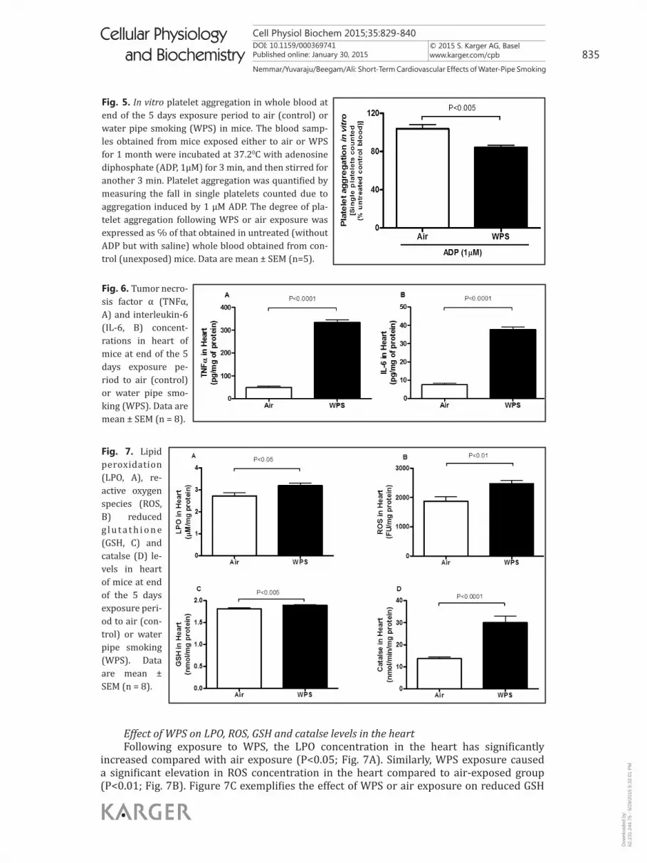
Effect of WPS on platelet aggregation in whole blood in vitroFigure 5 illustrates the effect of WPS on platelet aggregation in whole blood. Whole
blood obtained from mice exposed to WPS for 5 days and incubated with ADP (1µM) showed a significant (P<0.005) platelet aggregation compared with the blood collected from mice exposed to air (Fig. 5).
Effect of WPS on TNFα and IL-6 concentration in the heartFigure 6 shows the effect of 5 days nose-only exposure to WPS on TNFα and IL-6
concentrations in mouse heart. Compared with air-exposed mice, mice exposed to WPS showed a significant (P<0.0001) increase in TNFα concentration in the heart tissue (Fig. 6A). Likewise, IL-6 was also significantly (P<0.0001) augmented in heart tissue of WPS-exposed mice compared to those exposed to air.
Fig. 4. Platelet numbers (A) and plasma concent-rations of plasmi-nogen activator inhibitor-1 (PAI-1, B), fibrinogen (C) and von Willeb-rand Factor (vWF) (vWF, D) at end of the 5 days ex-posure period to air (control) or water pipe smo-king (WPS). Data are mean ± SEM (n = 8).
Dow
nloa
ded
by:
62.2
31.2
44.7
5 -
5/29
/201
5 5:
32:0
1 P
M
Cell Physiol Biochem 2015;35:829-840DOI: 10.1159/000369741Published online: January 30, 2015
© 2015 S. Karger AG, Baselwww.karger.com/cpb 835
Nemmar/Yuvaraju/Beegam/Ali: Short-Term Cardiovascular Effects of Water-Pipe Smoking
Cellular Physiology and Biochemistry
Cellular Physiology and Biochemistry
Fig. 5. In vitro platelet aggregation in whole blood at end of the 5 days exposure period to air (control) or water pipe smoking (WPS) in mice. The blood samp-les obtained from mice exposed either to air or WPS for 1 month were incubated at 37.20C with adenosine diphosphate (ADP, 1µM) for 3 min, and then stirred for another 3 min. Platelet aggregation was quantified by measuring the fall in single platelets counted due to aggregation induced by 1 µM ADP. The degree of pla-telet aggregation following WPS or air exposure was expressed as ℅ of that obtained in untreated (without ADP but with saline) whole blood obtained from con-trol (unexposed) mice. Data are mean ± SEM (n=5).
Fig. 6. Tumor necro-sis factor α (TNFα, A) and interleukin-6 (IL-6, B) concent-rations in heart of mice at end of the 5 days exposure pe-riod to air (control) or water pipe smo-king (WPS). Data are mean ± SEM (n = 8).
Effect of WPS on LPO, ROS, GSH and catalse levels in the heart Following exposure to WPS, the LPO concentration in the heart has significantly
increased compared with air exposure (P<0.05; Fig. 7A). Similarly, WPS exposure caused a significant elevation in ROS concentration in the heart compared to air-exposed group (P<0.01; Fig. 7B). Figure 7C exemplifies the effect of WPS or air exposure on reduced GSH
Fig. 7. Lipid peroxidation (LPO, A), re-active oxygen species (ROS, B) reduced g l u t a t h i o n e (GSH, C) and catalse (D) le-vels in heart of mice at end of the 5 days exposure peri-od to air (con-trol) or water pipe smoking (WPS). Data are mean ± SEM (n = 8).
Dow
nloa
ded
by:
62.2
31.2
44.7
5 -
5/29
/201
5 5:
32:0
1 P
M

Cell Physiol Biochem 2015;35:829-840DOI: 10.1159/000369741Published online: January 30, 2015
© 2015 S. Karger AG, Baselwww.karger.com/cpb 836
Nemmar/Yuvaraju/Beegam/Ali: Short-Term Cardiovascular Effects of Water-Pipe Smoking
Cellular Physiology and Biochemistry
Cellular Physiology and Biochemistry
concentration. WPS exposure caused a significant increase of GSH concentration compared to air-exposed group (P<0.005; Fig. 7C). Likewise, a significant increase of catalase activity was observed in the heart following acute exposure to WPS compared with air-exposed group (P<0.0001; Fig. 7D).
Effect of WPS on SBP Table 1 represents the effect of WPS or air exposure on SBP and heart rate in mice.
Compared with air-exposed group, neither SBP nor heart rate was significantly affected in mice exposed to WPS (Table 1).
Discussion
The present study provides evidence that nose-only WPS exposure for 5 consecutive days exerts procoagulatory effects and induces cardiac inflammation and oxidative stress. However, at the time point investigated, there was no evidence for blood inflammation and oxidative stress, and no effect of WPS on SBP and heart rate.
While it is well established that CS is a major independent risk factor for cardiovascular disease, including atherosclerotic vascular disease, myocardial infarction, unstable angina, sudden cardiac death, and stroke [16-18], the data on the cardiovascular effects of WPS is very scarce [2]. Despite its widespread use, only few studies to date have documented the adverse cardiovascular consequences of WPS [2]. This lack of data results from the fact that WPS is mostly a non-Western habit, and its high prevalence is a relatively recent phenomenon, lack of standardization of WPS content, and the difficulty in studying the isolated effects of WPS because most of the smokers are also current or past cigarette smokers [6]. Therefore, experimental studies investigating the pathophysiologic mechanisms underlying the cardiovascular adverse effect of WPS are critical and much needed. We have recently demonstrated that 1 month exposure to WPS increased blood pressure and thrombosis and induced inflammation and oxidative stress in the heart [4]. In the present study, we elected to study the initial cardiovascular effects related to WPS exposure. Assessing the short-term effect of WPS is essential because repetitive short-term WPS effects may constitute the underlying causal chain of reactions leading to the ultimate chronic effects of WPS.
We have recently demonstrated that one month exposure to WPS caused a significant increase in IL-6 concentrations in plasma but not that of TNFα [14]. In the present study, at the time point investigated, i.e. 5 days, the measured markers of systemic inflammation including leukocyte numbers, CRP, TNFα and IL-6 were not affected in mice exposed to WPS compared to air-exposure. The lack of increase of markers of systemic inflammation at 5 days exposure time point does not necessarily exclude their possible increase at earlier time point or later time point. Previous study found no increase in leukocyte numbers following short-term (4 days) exposure to CS [16]. On the other hand, an increase in leukocyte count has been reported in humans exposed acutely to WPS (one session of 30 min), however, no measurement of proinflammatory cytokine in plasma has been reported [21]. The discrepancy between this finding [21] and ours could be related to the time-point and/or to the fact humans exposed to WPS had previous exposure to WPS and/or cigarette that may predispose them more to develop an increase in white blood cell count following acute WPS exposure compared to mice.
Similarly to the absence of systemic inflammation, we did not observe evidence for increase of markers of oxidative stress in plasma. We found no increase of markers in
Table 1. Systolic blood pressure (SBP) and heart rate in mice at the end of the 5 days exposure period to air (control) or water pipe smoking (WPS). Data are mean ± SEM (n=8)
Dow
nloa
ded
by:
62.2
31.2
44.7
5 -
5/29
/201
5 5:
32:0
1 P
M

Cell Physiol Biochem 2015;35:829-840DOI: 10.1159/000369741Published online: January 30, 2015
© 2015 S. Karger AG, Baselwww.karger.com/cpb 837
Nemmar/Yuvaraju/Beegam/Ali: Short-Term Cardiovascular Effects of Water-Pipe Smoking
Cellular Physiology and Biochemistry
Cellular Physiology and Biochemistry
oxidative stress in plasma after a short-term exposure (4 days) to CS in mice (unpublished data). It has been reported that systemic oxidative stress is elevated in smokers compared with non-smokers [22]. However, in that study no significant increase was observed in the levels of systemic oxidative stress as an acute effect of exposure to CS [22]. A recent study reported no increase in serum oxidative stress in healthy smokers but it increased in asthmatic smokers [23].
While it is well established that CS causes alterations in platelet function, antithrombotic/prothrombotic factors, and fibrinolytic factors [24], little is known about the effect of WPS on thrombosis. Our data show that 5 days nose-only WPS exposure causes prothrombotic events in pial arterioles and venules in a photochemically injured vessel. This finding is important as it shows that short-term exposure to WPS cause thrombotic complications. Such an effect was not reported following short-term (4 days) exposure to CS in mice [16]. In the current study, the occlusion times in arterioles and venules were shortened by – 28% and – 22% respectively. In mice exposed for 1 month to WPS, a more marked shortening of the thrombotic occlusion time was observed in pial arterioles (−46%) and venules (−40%) [14]. In conjunction with the prothrombotic effect caused by short-term exposure to WPS, we found a significant decrease in platelet numbers in WPS-exposed mice compared with air-exposed ones, this is indicative of platelet activation in vivo. A decrease of platelet numbers following pulmonary exposure to pollutant particles has been reported from experimental and clinical studies [18, 25]. The numbers of platelet were found slightly but insignificantly increased in mice exposed to WPS for 1 month [14].
Our data show a significant increase of circulating PAI-1 following WPS exposure. PAI-1 is a potent endogenous inhibitor of fibrinolysis and is involved in the pathogenesis of several cardiovascular diseases. [26, 27] An increase of PAI-1 has been observed following exposure to particles [28, 29] Along with the fibrinolytic factor PAI-1, we found a significant increase in levels of fibrinogen, an acute-phase protein that increases blood viscosity and promotes thrombus formation[30], following WPS exposure. Our data suggest an impairment of the fibrinolytic system and activation of blood coagulation following WPS exposure. This finding is novel as it has not been reported before with WPS. It has been previously reported that CS results in platelet activation, stimulation of the coagulation cascade, and impairment of anticoagulative fibrinolysis [24]. However, we did not find an increase of vWF concentration in WPS group. We recently reported that 1 month exposure to WPS causes a significant increase in vWF concentration in plasma. The discrepancy between the latter finding and the current one can be related to the exposure duration, and suggests that the endothelial activation develops over longer period of exposure to WPS. A recent study found no effect of acute exposure of WPS on endothelial function in humans [21].
Our in vivo model of thrombosis depends on the fact that a platelet-rich thrombus is produced in a (photochemically) injured vessel [18]. Therefore, to gain more insights into the mechanism underlying the in vivo prothrombotic effects of WPS, we performed in vitro platelet aggregation studies in whole blood. The in vitro effect of ADP was assessed on blood collected from mice exposed either to WPS or air. Our results show a significant increase of platelet aggregation in blood of WPS-exposed mice compared to those exposed to air. This finding suggests a priming of platelet activation and their contribution in the development of thrombosis when they come across mildly injured vessel wall. Similar observation has been made in mice exposed to WPS for 1 month [14].
Unlike in the plasma, our data show a significant increase in ROS and LPO in the heart following short-term exposure to WPS. Similar finding were reported after short-term exposure to CS [16, 31]. Recently, we have reported that 1 month exposure to WPS cause an increase of LPO and ROS levels in the heart [14]. Besides, our data show a significant increase of antioxidants in the heart including GSH and catalase. This indicates that the development of oxidative stress is accompanied by an adaptive response that counterbalances the potentially damaging activity of oxygen free radicals by antioxidant defense mechanisms. An increase of antioxidants in heart [16] and lung [32, 33] has been reported in mice exposed to CS. Interestingly, when we exposed mice to WPS for longer period of time, i.e. 1 month, we
Dow
nloa
ded
by:
62.2
31.2
44.7
5 -
5/29
/201
5 5:
32:0
1 P
M

Cell Physiol Biochem 2015;35:829-840DOI: 10.1159/000369741Published online: January 30, 2015
© 2015 S. Karger AG, Baselwww.karger.com/cpb 838
Nemmar/Yuvaraju/Beegam/Ali: Short-Term Cardiovascular Effects of Water-Pipe Smoking
Cellular Physiology and Biochemistry
Cellular Physiology and Biochemistry
observed a depletion of antioxidants, leaving heart tissues vulnerable to damage by oxygen free radicals [14].
Oxidative stress and inflammation together form a vicious cycle that is responsible for the disease progression [24]. ROS can stimulate redox-sensitive transcription factors, nuclear factor-kappa B, and activator protein-1, activating the genes of pro-inflammatory mediators TNFα and IL-6 [34]. These pro-inflammatory cytokines play a critical role in the pathogenesis of atherosclerosis and were detected in the myocardium of patients with heart failure [35, 36]. We found a significant increase of IL-6 and TNFα concentrations in heart following short-term exposure to WPS. Similar findings were observed in mice exposed to WPS for 1 month [14]. In cultured rat H9c2 cardiomyocytes, CS extract was found to cause upregulation of NF-κB-regulated inflammatory genes TNFα, IL-6 and IL-1β and to induce depletion of antioxidant enzymes [37]. It remains to determine if there is any selectivity of WPS components to specific heart compartments leading to a differential proinflammatory and procoagulatory effects. It would be of interest to see if either endocardium or coronary vessels are more susceptible to WPS than other heart compartments such as intra myocardial vessels.
We have recently demonstrated that 1 month exposure to WPS caused significant increase in SBP [14]. In the present study, we found no significant increase of SBP or heart rate in WPS–exposed mice. This finding suggests that the short-term (5 days) exposure to WPS did not result in a direct increase of SBP and heart rate. However, if WPS exposure is repeated over a longer period of time (e.g. 1 month exposure), it will eventually cause an increase in SBP, as we recently reported [33]. Recent clinical studies described an increase in blood pressure and heart rate following acute exposure to WPS in healthy subjects [7, 38, 39]. The discrepancy between the latter findings and ours could be related to the time-point, level of exposure and/or to the fact that humans exposed to WPS had previous exposure to WPS and/or cigarette that could prompt them more to develop an increase in blood pressure and heart rate following acute WPS exposure.
We conclude that nose-only WPS exposure for 5 days exerts procoagulatory effects and induces cardiac inflammation and oxidative stress. However, at the time point investigated, there was no evidence for blood inflammation or oxidative stress, and no effect of WPS on SBP or heart rate. Additional studies are required to study the kinetic of release of markers of inflammation and oxidative stress following WPS exposure. Also, it would be interesting to know which component of the WPS causes the most vascular damage. It has been postulated that that lipophilic components of CS can cause systemic toxicity by alteration in mitochondrial respiratory chain [40]. Our data provide information on the early cardiovascular changes following exposure to WPS. Our results give biological plausibility for the short-term adverse cardiovascular effects of WPS, and imply that even people who newly started to smoke WPS can be vulnerable to its adverse effects
Funding
This work was supported by the UAEU-NRF and CMHS grants.
Disclosure Statement
None
References
1 Chaouachi K: Hookah (Shisha, Narghile) Smoking and Environmental Tobacco Smoke (ETS). A critical review of the relevant literature and the public health consequences. Int J Environ Res Public Health 2009;6:798-843.
Dow
nloa
ded
by:
62.2
31.2
44.7
5 -
5/29
/201
5 5:
32:0
1 P
M

Cell Physiol Biochem 2015;35:829-840DOI: 10.1159/000369741Published online: January 30, 2015
© 2015 S. Karger AG, Baselwww.karger.com/cpb 839
Nemmar/Yuvaraju/Beegam/Ali: Short-Term Cardiovascular Effects of Water-Pipe Smoking
Cellular Physiology and Biochemistry
Cellular Physiology and Biochemistry
2 Maziak W: The global epidemic of waterpipe smoking. Addict Behav 2011;36:1-5.3 Eissenberg T, Shihadeh A: Waterpipe tobacco and cigarette smoking: direct comparison of toxicant
exposure. Am J Prev Med 2009;37:518-523.4 Daher N, Saleh R, Jaroudi E, Sheheitli H, Badr T, Sepetdjian E, Al RM, Saliba N, Shihadeh A: Comparison
of carcinogen, carbon monoxide, and ultrafine particle emissions from narghile waterpipe and cigarette smoking: Sidestream smoke measurements and assessment of second-hand smoke emission factors. Atmos Environ (1994 ) 2010;44:8-14.
5 Shafagoj YA, Mohammed FI, Hadidi KA: Hubble-bubble (water pipe) smoking: levels of nicotine and cotinine in plasma, saliva and urine. Int J Clin Pharmacol Ther 2002;40:249-255.
6 Knishkowy B, Amitai Y: Water-pipe (narghile) smoking: an emerging health risk behavior. Pediatrics 2005;116:e113-e119.
7 Al Kubati M, Al Kubati AS, al‘Absi M, Fiser B: The short-term effect of water-pipe smoking on the baroreflex control of heart rate in normotensives. Auton Neurosci 2006;126-127:146-149.
8 Cobb CO, Sahmarani K, Eissenberg T, Shihadeh A: Acute toxicant exposure and cardiac autonomic dysfunction from smoking a single narghile waterpipe with tobacco and with a „healthy“ tobacco-free alternative. Toxicol Lett 2012;215:70-75.
9 Hakim F, Hellou E, Goldbart A, Katz R, Bentur Y, Bentur L: The acute effects of water pipe smoking on the cardio- respiratory system. Chest 2011;139:775-781.
10 Selim GM, Elia RZ, El Bohey AS, El Meniawy KA: Effect of shisha vs. cigarette smoking on endothelial function by brachial artery duplex ultrasonography: an observational study. Anadolu Kardiyol Derg 2013;13:759-765.
11 Rinaldi M, Maes K, De Vleeschauwer S., Thomas D, Verbeken EK, Decramer M, Janssens W, Gayan-Ramirez GN: Long-term nose-only cigarette smoke exposure induces emphysema and mild skeletal muscle dysfunction in mice. Dis Model Mech 2012;5:333-341.
12 Wright JL, Cosio M, Churg A: Animal models of chronic obstructive pulmonary disease. Am J Physiol Lung Cell Mol Physiol 2008;295:L1-15.
13 Stevenson CS, Birrell MA: Moving towards a new generation of animal models for asthma and COPD with improved clinical relevance. Pharmacol Ther 2011;130:93-105.
14 Nemmar A, Yuvaraju P, Beegam S, John A, Raza H, Ali BH: Cardiovascular effects of nose-only water-pipe smoking exposure in mice. Am J Physiol Heart Circ Physiol 2013;305:H740-H746.
15 Nemmar A, Raza H, Subramaniyan D, John A, Elwasila M, Ali BH, Adeghate E: Evaluation of the pulmonary effects of short-term nose-only cigarette smoke exposure in mice. Exp Biol Med (Maywood ) 2012;237:1449-1456.
16 Nemmar A, Raza H, Subramaniyan D, Yasin J, John A, Ali BH, Kazzam EE: Short-Term Systemic Effects of Nose-Only Cigarette Smoke Exposure in Mice: Role of Oxidative Stress. Cell Physiol Biochem 2013;31:15-24.
17 Blalock TD, Varela JC, Gowda S, Tang Y, Chen C, Mast BA, Schultz GS: Ischemic skin wound healing models in rats. Wounds 13(1):35-44. Wounds 2001;13:35-44.
18 Nemmar A, Al Salam S, Dhanasekaran S, Sudhadevi M, Ali BH: Pulmonary exposure to diesel exhaust particles promotes cerebral microvessel thrombosis: protective effect of a cysteine prodrug l-2-oxothiazolidine-4-carboxylic acid. Toxicology 2009;263:84-92.
19 Nemmar A, Zia S, Subramaniyan D, Fahim MA, Ali BH: Exacerbation of thrombotic events by diesel exhaust particle in mouse model of hypertension. Toxicology 2011;285:39-45.
20 Nemmar A, Melghit K, Ali BH: The Acute Proinflammatory and Prothrombotic Effects of Pulmonary Exposure to Rutile TiO2 Nanorods in Rats. Exp Biol Med (Maywood ) 2008;233:610-619.
21 Bentur L, Hellou E, Goldbart A, Pillar G, Monovich E, Salameh M, Scherb I, Bentur Y: Laboratory and clinical acute effects of active and passive indoor group water-pipe (narghile) smoking. Chest 2014;145:803-809.
22 Morrow JD, Frei B, Longmire AW, Gaziano JM, Lynch SM, Shyr Y, Strauss WE, Oates JA, Roberts LJ: Increase in circulating products of lipid peroxidation (F2-isoprostanes) in smokers. Smoking as a cause of oxidative damage. N Engl J Med 1995;332:1198-1203.
23 Papaioannou AI, Koutsokera A, Tanou K, Kiropoulos TS, Tsilioni I, Oikonomidi S, Liadaki K, Pournaras S, Gourgoulianis KI, Kostikas K: The acute effect of smoking in healthy and asthmatic smokers. Eur J Clin Invest 2010;40:103-109.
Dow
nloa
ded
by:
62.2
31.2
44.7
5 -
5/29
/201
5 5:
32:0
1 P
M

Cell Physiol Biochem 2015;35:829-840DOI: 10.1159/000369741Published online: January 30, 2015
© 2015 S. Karger AG, Baselwww.karger.com/cpb 840
Nemmar/Yuvaraju/Beegam/Ali: Short-Term Cardiovascular Effects of Water-Pipe Smoking
Cellular Physiology and Biochemistry
Cellular Physiology and Biochemistry
24 Ambrose JA, Barua RS: The pathophysiology of cigarette smoking and cardiovascular disease: an update. J Am Coll Cardiol 2004;43:1731-1737.
25 Ruckerl R, Phipps RP, Schneider A, Frampton M, Cyrys J, Oberdorster G, Wichmann HE, Peters A: Ultrafine particles and platelet activation in patients with coronary heart disease - results from a prospective panel study. Part Fibre Toxicol 2007;4:1.
26 Thogersen AM, Jansson JH, Boman K, Nilsson TK, Weinehall L, Huhtasaari F, Hallmans G: High plasminogen activator inhibitor and tissue plasminogen activator levels in plasma precede a first acute myocardial infarction in both men and women: evidence for the fibrinolytic system as an independent primary risk factor. Circulation 1998;98:2241-2247.
27 Cesari M, Pahor M, Incalzi RA: Plasminogen activator inhibitor-1 (PAI-1): a key factor linking fibrinolysis and age-related subclinical and clinical conditions. Cardiovasc Ther 2010;28:e72-e91.
28 Nemmar A, Subramaniyan D, Ali BH: Protective effect of curcumin on pulmonary and cardiovascular effects induced by repeated exposure to diesel exhaust particles in mice. PLoS One 2012;7:e39554.
29 Nemmar A, Albarwani S, Beegam S, Yuvaraju P, Yasin J, Attoub S, Ali BH: Amorphous silica nanoparticles impair vascular homeostasis and induce systemic inflammation . Int J Nanomedicine 2014;9:2779-89.
30 Kamath S, Lip GY: Fibrinogen: biochemistry, epidemiology and determinants. QJM 2003;96:711-729.31 Itoh M, Tsuji T, Nakamura H, Yamaguchi K, Fuchikami J, Takahashi M, Morozumi Y, Aoshiba K: Systemic
effects of acute cigarette smoke exposure in mice. Inhal Toxicol 2014;26:464-473.32 Valenca SS, Silva BF, Lopes AA, Romana-Souza B, Marinho Cavalcante MC, Lima AB, Goncalves K, V, Porto
LC: Oxidative stress in mouse plasma and lungs induced by cigarette smoke and lipopolysaccharide. Environ Res 2008;108:199-204.
33 Nemmar A, Raza H, Yuvaraju P, Beegam S, John A, Yasin J, Hameed RS, Adeghate E, Ali BH: Nose-only water-pipe smoking effects on airway resistance, inflammation and oxidative stress in mice. J Appl Physiol 2013;115:1316-1323.
34 MacNee W: Pulmonary and systemic oxidant/antioxidant imbalance in chronic obstructive pulmonary disease. Proc Am Thorac Soc 2005;2:50-60.
35 Azzawi M, Hasleton PS, Hutchinson IV: TNF-alpha in acute cardiac transplant rejection. Cytokines Cell Mol Ther 1999;5:41-49.
36 Ikonomidis I, Andreotti F, Economou E, Stefanadis C, Toutouzas P, Nihoyannopoulos P: Increased proinflammatory cytokines in patients with chronic stable angina and their reduction by aspirin. Circulation 1999;100:793-798.
37 Niu J, Wang K, Kolattukudy PE: Cerium oxide nanoparticles inhibit oxidative stress and nuclear factor-kappaB activation in H9c2 cardiomyocytes exposed to cigarette smoke extract. J Pharmacol Exp Ther 2011;338:53-61.
38 Hakim F, Hellou E, Goldbart A, Katz R, Bentur Y, Bentur L: The acute effects of water-pipe smoking on the cardiorespiratory system. Chest 2011;139:775-781.
39 Shaikh RB, Vijayaraghavan N, Sulaiman AS, Kazi S, Shafi MS: The acute effects of Waterpipe smoking on the cardiovascular and respiratory systems. J Prev Med Hyg 2008;49:101-107.
40 van der Toorn M, Rezayat D, Kauffman HF, Bakker SJ, Gans RO, Koeter GH, Choi AM, van Oosterhout AJ, Slebos DJ: Lipid-soluble components in cigarette smoke induce mitochondrial production of reactive oxygen species in lung epithelial cells. Am J Physiol Lung Cell Mol Physiol 2009;297:L109-L114.
Dow
nloa
ded
by:
62.2
31.2
44.7
5 -
5/29
/201
5 5:
32:0
1 P
M


