Embed Size (px)
Citation preview
Significant interference with hepatitis B virus replication by a core-nucleasefusion protein
Gertrud Beterams and Michael Nassal*
University Hospital Freiburg
Department of Internal Medicine II / Molecular Biology
* Corresponding author´s address:
University Hospital Freiburg
Department of Internal Medicine II / Molecular Biology
Hugstetter Str. 55
D-79106 Freiburg
Phone and Fax: +49 - 761 - 270 3507
E-mail: [email protected]
Running title: Capsid-targeted interference with HBV replication
Keywords:
antiviral therapy
capsid-targeted viral inactivation
hepatitis B virus
S. aureus nuclease
Copyright 2000 by The American Society for Biochemistry and Molecular Biology, Inc.
JBC Papers in Press. Published on December 21, 2000 as Manuscript M006335200 by guest on June 19, 2016
http://ww
w.jbc.org/
Dow
nloaded from

Summary
Hepatitis B virus (HBV), a small DNA containing virus that replicates via reverse
transcription, causes acute and chronic B-type hepatitis in humans. The limited success of
current therapies for chronic infection has prompted exploration of alternative strategies.
Capsid-targeted viral inactivation (CTVI) is a conceptually powerful approach that exploits
virion structural proteins to target a degradative enzyme specifically into viral particles. Its
principal feasibility has been demonstrated in retroviral model systems but not yet for a
medically relevant virus outside the retrovirus family. Recently, we found that C proximal
fusion to the HBV capsid protein of the Ca2+-dependent nuclease (SN) from Staphylococcus
aureus yields a chimeric protein, coreSN, that in E. coli coassembles with the wild-type
capsid protein into particles with internal SN domains. Here we show that, in HBV
cotransfected human heptoma cells, less than 1 coreSN protein per 10 wild-type core protein
subunits reduced titers of enveloped DNA containing virions by more than 95 percent. The
antiviral effect depends on both an enzymatically active SN and on the core domain. coreSN
does not block assembly of RNA containing nucleocapsids but interferes with proper
synthesis of viral DNA inside the capsid, or leads to rapid DNA degradation. Our data suggest
an intracellular nuclease activitation that, owing to the characteristics of HBV morphogenesis,
is nonetheless highly virus specific. HBV may therefore be particularly vulnerable to the
CTVI approach.
by guest on June 19, 2016http://w
ww
.jbc.org/D
ownloaded from
Introduction
Hepatitis B virus (HBV), an enveloped DNA containing virus that replicates via reverse
transcription (1), is the causative agent of B type hepatitis in humans. Chronic infections
affect more than 350 million people worldwide, they have potentially severe consequences
such a liver cirrhosis and hepatocellular carcinoma (2), and current treatments are of limited
efficacy (3,4). The sustained response rate of the approved high-dose interferon-α (IFN-α)
therapy is about 30%. Nucleoside analogues such as lamivudine markedly reduce viral load
but suffer, inter alia, from the emergence of drug resistant virus variants (5). This situation
has spurred interest in alternative approaches to interfere with HBV replication (6).
Whereas only primary human, or chimpanzee, hepatocytes are susceptible to HBV infection
in cell culture, a few human hepatoma cell lines like Huh7 and HepG2 support virus
production upon transfection with cloned viral DNA. The late steps of the infectious cycle
(Fig. 1A) are hence understood in some detail (7,8), and they present novel targets for
intervention. After infection, the nucleocapsid transports the partially double-stranded (ds)
circular 3.2 kb DNA genome to the nucleus (9) for conversion into covalently closed circular
DNA (cccDNA); this molecule is the transcriptional template for several subgenomic and
genomic RNAs that all act as mRNAs. Of these, the pregenomic RNA (pgRNA) is first used
to translate both the capsid, or core, protein and the reverse transcriptase, P protein. Then P
protein, together with cellular chaperones (10), binds to a stem-loop structure, ε, close to the
5´-end of the pgRNA (11,12). Complex formation mediates assembly of immature RNA-
containing nucleocapsids (13), and initiation of reverse transcription (14-17). DNA synthesis
occurs inside the nucleocapsid and involves several template switches that lead to the
characteristic relaxed circular (RC) DNA genome containing a complete (-)-strand and
variously extended (+)-strands. Mature DNA-containing nucleocapsids can reescort the
genome to nucleus, or be exported as enveloped virions by budding into a post-ER/pre-Golgi
compartment. Both events apparently require that at least the (-)-DNA strand be completed
(1,18).
by guest on June 19, 2016http://w
ww
.jbc.org/D
ownloaded from

The restriction of HBV genome replication to the nucleocapsid makes this nucleoprotein
particle an attractive target for intervention. Apart from nucleic acid based strategies (19,20)
dominant negative core protein variants have been described that passively interfere with
nucleocapsid assembly (21-24). A conceptually more powerful approach is capsid-targeted
viral inactivation (CTVI) or, generally, virion-targeted viral inactivation, which exploits a
viral capsid protein or other virion associated protein as carrier to target a degradative enzyme
specifically into virus particles (25,26); alternatively, nucleic acid based effectors such as
ribozymes may be fused to viral packaging signals and thus be used against viruses that, like
retroviruses but unlike HBV, encapsidate more than one genome, or genome segment (27,28).
For the protein based approach, the nuclease from Staphylococcus aureus (SN) is considered
particularly useful because it requires Ca2+ for activity (29). Intracellular Ca2+ levels are
usually below 1 micromolar, providing a safeguard against attacks on cellular nucleic acids.
Serum levels of Ca2+, by contrast, are in the millimolar range; hence SN incorporated into a
virus particle is thought be activated upon release from the cell.
The principal feasibility of the approach, pioneered using the yeast retrotransposon Ty1 (25),
has been well documented for the model of Moloney murine leukemia virus (MoMLV), a
simple C-type retrovirus (30-34). Adapting the approach to human immunodeficiency virus
(HIV) has been hampered by the poor expression and inefficient incorporation into particles
of HIV Gag fusion proteins (35). Effectors fused to the accessory Vpr and Vpx proteins,
though efficiently incorporated, can be subject to inactivation by the retroviral protease (36),
and the Vpr carrier may induce cell cycle arrest and apoptosis (35). Using expression in E.
coli we have recently shown that a chimeric protein consisting of the N-terminal 155 aa of the
HBV core protein followed by the complete SN protein, coreSN, coassembles with wild-type
core protein to particles with internal SN domains (37). Here we investigated if, and how, this
fusion protein is able to interfere with HBV replication. We show that low levels of coreSN
drastically reduce the titers of replication-competent HB virons in supernatants from
transfected Huh7 cells, and we present evidence for an intracellular but virus-specific
nuclease activation that may make HBV particularly vulnerable to the CTVI approach.
by guest on June 19, 2016http://w
ww
.jbc.org/D
ownloaded from

EXPERIMENTAL PROCEDURES
Plasmid constructs. Effector plasmids pCS1-coreSN and pCS1-coreSNmut were generated
by transferring DNA fragments encoding the chimeric proteins from the prokaryotic vectors
pPLC-coreSNwt and pPLC-coreSNmut43/87 (37) into plasmid pCS1-C1 (38). This plasmid
contains the cytomegalovirus immediate early (CMV-IE) promoter and a poly-adenylation
signal from SV40. Control constructs encoding an SN fusion to a mutant core protein with aa
80 changed from A to K (pCS1-coreA80K-SN), and SN without core protein domain were
obtained by conventional PCR-mediated mutagenesis and cloning into vectors pCS1-coreSN
and pcDNA6/Myc-His (Invitrogen), respectively. pCS1-SN and pCS1-SNmut code for aa 1-
149 of mature SN and its inactivated double mutant, preceded by the dipeptide MD and
followed by a C terminal His-tag; pcDNA6-SN and pcDNA6-SNmut specify active and
inactive SN with the same 6 aa propeptide sequence as present in coreSN (see Fig. 1) plus a
methionine at the N terminus and a C terminal His-tag. The HBV expression plasmid pCHT-
9/3091 carries a slightly overlength HBV genome under control of the CMV-IE promoter
(38). Transfection efficiencies were monitored by cotransfection of plasmid pTR-UF5 (39)
encoding a fluorescence enhanced green fluorescent protein (eGFP).
Immunological techniques. For Western blot detection of wild-type core and coreSN
proteins (37,40) either a polyclonal rabbit antiserum raised against denatured recombinant
coreSN protein, or the monoclonal antibody 10E11, recognizing an epitope between amino
acids 8 and 20 on denatured core protein (41), served as primary antibodies; for HBsAg
monoclonal antibody 4/7B (42) was used. Detection was performed using appropriate
peroxidase-coupled secondary antibodies and a chemiluminescent substrate (ECL-Plus,
Amersham-Pharmacia). Blots were exposed to X-ray film or, for quantitation, to a Diana
charge-coupled-device (CCD) camera; band intensities were evaluated using AIDA software
(both Raytest). For immunoprecipitations (38), usually polyclonal rabbit antisera raised
against native recombinant core protein aa1-149 (serum H800) or coreSN particles were
employed. In the experiments aimed at directly demonstrating co-incorporation of coreSN in
mixed particles with wild-type core protein, monoclonal antibody mc 312 was used. This
by guest on June 19, 2016http://w
ww
.jbc.org/D
ownloaded from

antibody recognizes a linear epitope within aa 76-84 of core protein (41,43) which largely
coincides with a loop exposed on the spikes of core particles. Therefore, the core variant
A80K with a lysine instead of alanine at position 80 reacts much less efficiently with mc312.
Transfections. The human hepatoma cell line HuH7 was used throughout (38). Transfections
were performed with FuGENE6 reagent (Roche Diagnostics) as recommended by the
manufacturer. For typical cotransfections 50 µl FuGENE6 and a total of 21 µg plasmid DNA
per 10 cm dish were used. If required, constant amounts of DNA and CMV-IE promoter
copies were maintained by adding an appropriate quantity of plasmid pTR-UF5.
Isolation of secreted and intracellular HBV particles. Particles contained in culture
supernatants collected from day 3 to day 4 post transfection were enriched by PEG
precipitation (44), and loaded on a CsCl step gradient (0.9 ml each of 1.5, 1.4, 1.3, 1.2, and
1.1 g/ml CsCl in 10 mM Tris/Cl- pH 7.5, 100 mM NaCl). After 17 h at 35.000 RPM at 20°C
in a SW 50.1 rotor (Beckman), twenty-five 200 µl fractions were collected from the top.
Densities were determined by refractive index. Intracellular cores were obtained 4 days post
transfection from cytoplasmic NP-40 lysates and subsequent immunoprecipitation with
antiserum H800 (38). In some experiments, core particles were separated from nonassembled
core and or coreSN protein by sedimentation in 10% to 60% sucrose gradients as previously
described (37).
Characterization of viral proteins and nucleic acids. For dot blot analysis, equal aliquots
from the CsCl gradient fractions were applied to a positively charged Nylon membrane
(Roche Diagnostics), using a dot blot apparatus (BioRad). DNAs were detected by a random
primed HBV DNA probe (High Prime DNA Labeling Kit, Roche) and quantitated using a
phosphor imager (Fuji BAS 1500). Core proteins and HBsAg were detected by Western
blotting as described above. For Southern blots, DNAs contained in viral particles from CsCl
gradients, or in immunoprecipitated intracellular cores, were isolated by proteinase K
digestion in the presence of 0.5% SDS, and purified using the QiaAmp tissue kit (Qiagen). If
desired, aliqouts were further treated with avian myoblastosis virus reverse transcriptase
(AMV-RT) as previously described (38). DNAs were separated on 1% agarose gels and
by guest on June 19, 2016http://w
ww
.jbc.org/D
ownloaded from
transferred to nylon membranes using 0.4 M NaOH. Detection was performed with either a
32P-radiolabeled, or a Digoxygenin labeled probe (Dig High Prime DNA Labeling Kit; Roche)
as indicated. For Northern blotting of total RNA about one third of the cells on 10 cm dish
were lysed in RLT buffer and the RNA was purified using the RNeasy Mini kit (both Qiagen).
Encapsidated RNA was obtained accordingly from core specific immuno pellets. RNAs were
separated on 1.2% agarose-formaldehyde gels, transferred to nylon membrane in 10x SSC
buffer, and detected using the 32P-radiolabeled HBV probe. Glyceraldehyde-3-phosphate
dehydrogenase (GAPDH) mRNA served as control. Native agarose gel electrophoresis of
cytoplasmic cores was performed as described (40), using about 1 to 2% of the cytoplasmic
lysate from a 10 cm dish pretreated with DNAse I for 30 min at 37° C to remove non-
encapsidated DNA. After denaturation (0.5 M NaOH, 1 M NaCl) and neutralization (0.5 M
Tris pH 7.5, 3 M NaCl) core proteins were detected using H800 serum, and DNA by
hybridization with the 32P-labeled probe.
by guest on June 19, 2016http://w
ww
.jbc.org/D
ownloaded from

RESULTS
Experimental design
For lack of a feasible infection system we used cotransfection of HuH7 cells with an HBV
target plasmid, pCHT-9/3091, and a coreSN effector plasmid, pCS1-coreSN, as an assay
system. The target plasmid produces, under control of the CMV-IE promoter, substantial
amounts of pgRNA and the subgenomic RNAs generating all gene products required to form
complete virions (38). It should be reemphasized that, in this setting, there is neither
reinfection of the cells nor a significant accumulation of intranuclear HBV cccDNA, hence
virtually all virions produced derive from the transfected plasmid. The coreSN gene (Fig. 1B)
in pCS1-coreSN, though also controlled by the CMV-IE promoter, is only moderately
expressed, possibly for lack of introns and/or other elements promoting nuclear export in the
mRNA. Here this low expression appeared advantageous since even one fusion protein per
particle should be sufficient to exert an antiviral effect (Fig. 1A). To distinguish nuclease
dependent effects from passive steric hindrance we analyzed in parallel a homologous fusion
protein, coreSNmut, with two point mutations in the SN part that drastically reduce enzymatic
activity (45).
To test whether coreSN affected expression and/or stability of the wild-type core protein,
Huh7 cells were cotransfected with a constant amount of plasmid pCHT-9/3091, and various
concentrations of pCS1-coreSN. Core proteins were immunoprecipitated from cytoplasmic
lysates using a polyclonal antiserum against denatured recombinant core protein, H800, that
cross-reacts with the chimeric protein (37). For sensitive Western blot detection a polyclonal
antiserum against denatured recombinant coreSN protein was used which reacts much better
with coreSN than with wild-type core protein. Indeed, a new band at the expected 36 kDa
position was dose-dependently detected (Fig. 2A). Even at the highest coreSN concentration
the intensity of the 21 kDa wild-type core protein band was not significantly reduced; the
slightly weakened 21 kDa band in the HBV-only transfected sample is due to some loss of the
immune matrix as evidenced by the weaker band for the immunoglobulin heavy chain.
by guest on June 19, 2016http://w
ww
.jbc.org/D
ownloaded from

To evaluate the antiviral potency of coreSN is was important to know the relative ratios
between both proteins in the transfected cells. Using recombinant coreSN and wild-type core
protein of known concentration for calibration we found that the monoclonal antibody 10E11,
recognizing a linear epitope close to the N terminus of denatured core protein (41), reacted
equally well with both proteins on Western blots (Fig. 2B). This was true when the proteins
were directly loaded at equimolar concentration, and also after immunoprecipitation using
H800 serum. For cytoplasmic samples from cells cotransfected with the HBV construct and
equal amounts of the coreSN or coreSNmut plasmid this assay, after densitometric scanning,
showed that both chimeras were present at between 5% and, maximally, 10% of the wild-type
protein.
A potential concern was that, at the relatively low concentrations in this eukaryotic setting,
the coreSN protein would not be cointegrated into wild-type core particles. Obtaining formal
proof for coassembly, e.g. by coimmunoprecipitation, was not trivial for two reasons: (i),
using an anti-SN antibody to demonstrate coprecipitation of wild-type core protein would
require that the SN domains be exposed on the particle surface, in contrast to our previous E.
coli data which strongly suggested an interior location of SN (37); (ii) by necessity of the
approach, the core protein part in coreSN must mimic the structure of wild-type core protein
as closely as possible, otherwise efficient coassembly would be compromised. To resolve this
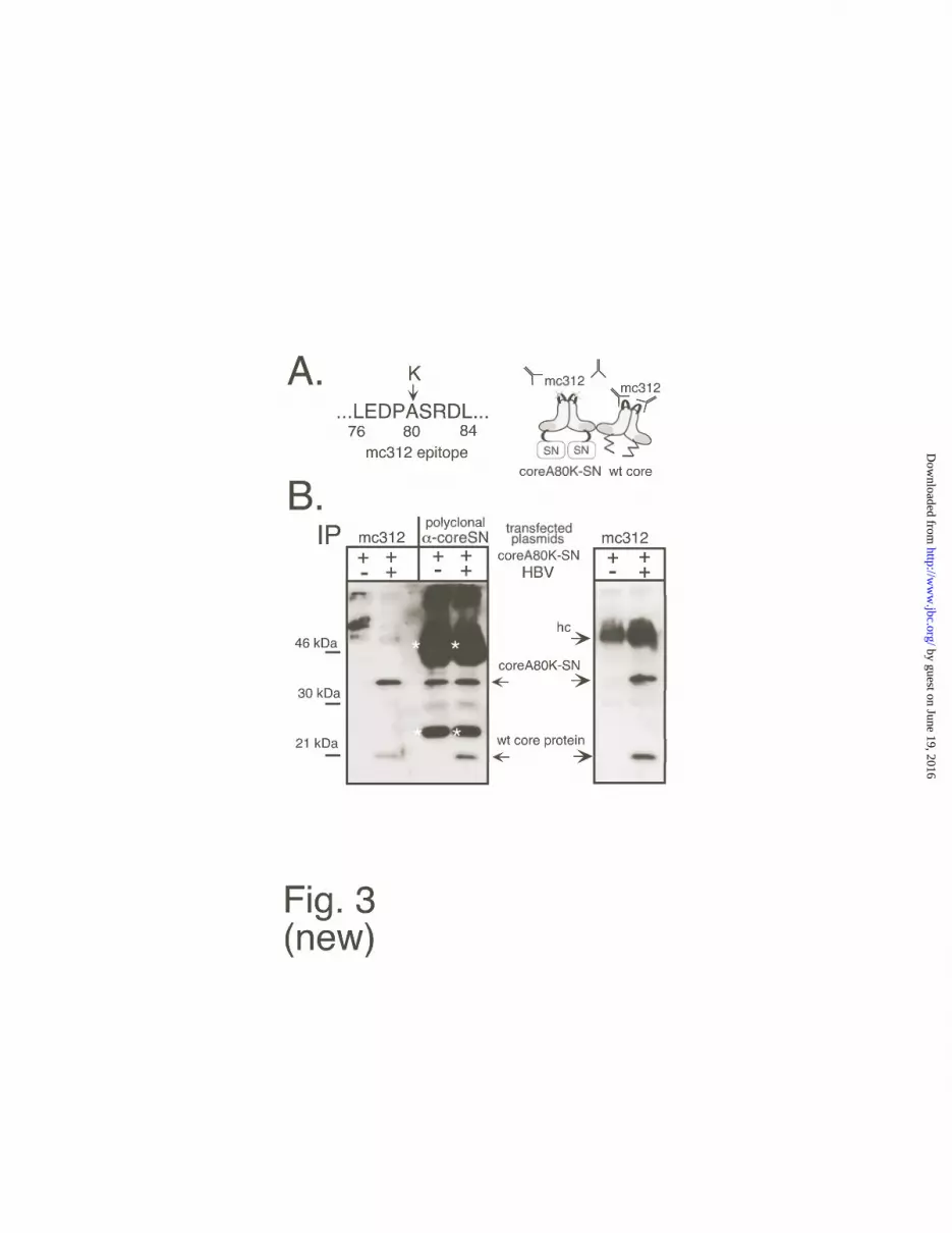
problem, we resorted to the monclonal anti-core antibody mc312 which recognizes a linear
epitope between aa positions 76 and 84 (41,43). This sequence overlaps with a surface
exposed loop located on the spikes of the core particles (Fig. 3A). Because an entire protein
such as GFP can be inserted into this loop without a major impact on protein folding (40), a
single aa exchange within the loop sequence would conceivably not affect the assembly
capability of a correspondingly altered core protein but inhibit binding of mc312. We
therefore generated a mutant core protein, coreA80K, in which the authentic alanine at
position 80 is replaced by lysine. This protein formed particles in E. coli, and it reacted very
poorly with mc312 on Western blots as well as in immunoprecipitation; a corresponding
coreA80K-SN fusion protein (Fig. A) was also assembly-competent (J. Vorreiter and M.
by guest on June 19, 2016http://w
ww
.jbc.org/D
ownloaded from

Nassal, unpublished data). This fusion protein should therefore not be precipitated by mc312
unless it is associated with wild-type core protein. This assumption was first confirmed using
mixtures of recombinant wild-type core and coreA80K-SN protein (data not shown). Next, a
eukaryotic expression vector for the mutant fusion protein, pCS1-coreA80K-SN, was
generated which, except for the single codon exchange, is identical to the coreSN vector. This
plasmid was transfected in Huh7 cells, either alone or together with the HBV expression
plasmid. Subsequently, equal aliquots of cytoplasmic lysates from both transfections were
subjected to immunoprecipitation with mc312 and, for control, with the polyclonal antiserum
against native coreSN. The precipitates were then analyzed by Western blotting using the
polyclonal antiserum against denatured coreSN. As shown in Fig. 3B, no antigen specific
signal was seen in the single transfection when mc312 was used for immunoprecipitation
while the protein was well detectable in the precipitate obtained with the polyclonal
antiserum. By contrast, coreA80K-SN was precipitated by mc312 when coexpressed with
wild-type core protein. This result indicated that wild-type and fusion protein did indeed
interact. To further prove that this interaction occured in the context of complete capsids,
aliquots of the same two lysates were subjected to sedimentation in sucrose gradients. Under
the conditions chosen, wild-type core particles are typically found in the center of the gradient
(37). The corresponding fractions were again used for immunoprecipitation with mc312. As
before, mc312 did not precipitate coreA80K-SN alone but did so when it was co-expressed
with wild-type protein (Fig. 3B, right panel). Together these data indicated that the mutant
coreSN fusion protein was able to form mixed particles with the wild-type core protein in
eukaryotic cells. The suitability of coreA80K-SN as a model compound for coreSN was
confirmed by its similar activity against particle-borne HBV DNA (see below).
coreSN protein drastically reduces HBV DNA in extracellular virions
The foremost aim of any antiviral strategy is to reduce the number of infectious virus
particles. Therefore we first compared the amounts of genome-containing enveloped virions
in supernatants from cells transfected with only HBV, or cotransfected with HBV and equal
amounts of the coreSN and core SNmut plasmids. A peculiarity of efficient HBV constructs
by guest on June 19, 2016http://w
ww
.jbc.org/D
ownloaded from

such as pCHT-9/3091 is the release, by an unknown mechanism, of nonenveloped cores (38).
We therefore used CsCl density gradients to separate enveloped and naked particles. Aliquots
from the gradient fractions were analyzed by dot blot (Fig. 4A) for HBV specific nucleic
acids, for core protein, and for HBsAg which is produced in large excess over virions. HBV-
only transfected cells showed two peaks of HBV nucleic acid: a weaker one in fractions 10 to
13 at a density around 1.25 g/ml (expected for enveloped virions), and a stronger one in
fractions 16 to 19 at around 1.35 g/ml (expected for naked cores). The presence of core
protein in these fractions was shown by anti-core immunoblotting. HBsAg appeared in
fractions 8 to 12 around the expected density of 1.20 g/ml.
The presence of active coreSN protein, but not of the inactive variant coreSNmut,
dramatically changed the DNA pattern: the virion peak was virtually absent, and the DNA
peak from naked cores was substantially reduced. This reduction was not due to global effects
on viral gene expression because the signal intensities and distributions of core protein and
HBsAg were similar in all three samples.
Next we analyzed the DNAs present in the corresponding fractions by Southern blotting. For
better comparison, all samples were run on one gel and transferred to one membrane for
hybridization and detection (Fig. 4B). For the HBV-only transfected cells, the virion peak
contained mostly mature DNAs at about 3.2 kb, and a weak band at the position of single-
stranded DNA (fractions 10 to 13). Naked cores (fractions 16 to 19) contained ssDNA plus
additional immature DNA species extending up to the 3.2 kb position. The samples from
cotransfection with coreSN, on the same exposure, gave no visible signals in the virion
fractions and only weak signals in the naked core fractions; these consisted of ssDNA and a
smear of slower, and of faster migrating material that was more pronounced than in the
absence of coreSN (panel marked 4x). Further overexposure (panel marked 10x) revealed
small amounts of DNA in the virion fractions, broadly distributed with no accumulation at the
position of full-length genomes. By contrast, cotransfection with coreSNmut gave, in general,
signals similar to those from HBV-only transfected cells. Virion DNA was somewhat reduced
by guest on June 19, 2016http://w
ww
.jbc.org/D
ownloaded from

and contained a higher proportion of ssDNA. In the naked core fractions, signals were
similarly strong but the smear above the ssDNA did not extend as far up.
Quantitation of the overall signal intensities using a phosphor imager revealed that coreSN
diminished the DNA in enveloped particles to 2.5%, or less, of that observed in its absence.
Co-expressed coreSNmut reduced the virion signal only to about 70%. For the naked capsids,
the signals were lowered to about 10% by coreSN, while coreSNmut had little effect
(reduction to 94%). These data confirmed that coreSN efficiently interferes with production of
complete virions, and that most of this interference depends on an active nuclease domain.
Whether the number of physical virus particles was reduced could not be definitely answered
because the large excess of HBsAg precludes a complete separation from virions; similarly,
the core blot signals were not sharply separated between the virion and core particle fractions,
possibly due to the presence of empty capsids.
Antiviral activity of coreSN depends on the presence of the core protein domain
That coreSN dramatically reduced DNA containing virions but had no significant effect on
HBsAg production (and neither on core protein, packaged viral pgRNA, or cellular RNA; see
below) strongly suggested a virus specific action. However, to further prove this specificity
we also tested the consequences of expressing active SN, and its enzymatically disabled
counterpart SNmut, without the core protein domain. Two possible outcomes were envisaged:
either the nuclease would exert a generally toxic effect and nonspecifically inhibit gene
expression in, or lead to the death of, the subset of successfully transfected cells (roughly 10
percent of the cells with our procedure). Alternatively, the low intracellular Ca2+ concentration
may be insufficient for activation of the enzyme, and no discernable difference between cells
expressing active, inactive, or no SN would be expected. Especially the latter case would
imply a major difference between free SN, and coreSN incorporated into nucleocapsids.
Two types of expression plasmids for SN and its enzymatically disabled double mutant
SNmut were constructed. The first is derived from plasmid pCS1 and encodes the mature
nuclease, i.e. aa 1-149, preceded by the dipeptide MD and followed by a His-tag, resulting in
a product with a nominal molecular mass of 17.8 kDa. The second is derived from the
by guest on June 19, 2016http://w
ww
.jbc.org/D
ownloaded from

commercial vector pcDNA6/Myc-His and contains, in addition, the same six aa propeptide
sequence as present in coreSN; its nominal molecular mass is 18.4 kDa. The rationale was
that the slightly different products should be discernable by SDS-PAGE and thus aid in
identifying the corresponding proteins by Western blotting using the anti-coreSNdenat
antiserum, as was indeed the case (see Fig. 5).
As an initial test for general toxicity we monitored the effect of SNwt and SNmut on co-
transfected GFP. Over five days we did not observe significant differences in the number of
GFP positive cells, or the intensity of GFP-fluorescence transfected with active, inactive, or
no SN. This was confirmed biochemically using Western blotting against GFP which also did
not reveal significant differences in the amounts of GFP protein (see below). This strongly
suggested that even expression of enzymatically active SN was not generally deleterious to
the transfected cells.
Next we performed triple transfections, using combinations of plasmids encoding HBV
(pCHT-9/3091), GFP (pTR-UF5) and one each of the different SN expression plasmids. Cells
transfected with expression plasmids for coreA80K-SN plus either GFP or HBV were
analyzed in parallel. Western blots of equal aliquots from the individual transfections,
developed using the polyclonal anti-coreSN denat serum, showed the expected results (Fig.
5A). The antiserum detected the coreSN fusions with either wild-type or coreA80K core part
(Fig. 5A, upper panel; lanes 1,2,8), and the 21 kDa wild-type core protein, in similar amounts,
in all transfections containing the HBV expression plasmid (lanes 2-8). In addition, slightly
faster migrating bands with small but distinct mobility differences were seen in samples
transfected with the non-fused SN expression constructs pCS1-SN (Fig. 5A, upper panel;
lanes 3, 4) and pcDNA6-SN (lanes 5,6), strongly suggesting they represented SN and SNmut.
Equally sized aliquots of the same lysates were analyzed in parallel (Fig. 5B, lower panel)
with monoclonal antibodies to core protein and GFP. The intensities of the wild-type core
protein signals paralleled those seen with the polyclonal anti-coreSN serum, and the GFP
specific signals were of comparable intensity in all corresponding samples. The slight signal
reduction for wild-type core protein and GFP in the cells transfected with plasmid pcDNA6-
by guest on June 19, 2016http://w
ww
.jbc.org/D
ownloaded from

SN (lane 5) was due to a slightly lower transfection efficiency because the band
corresponding to SN itself as well as a nonspecific background band were also somewhat
decreased. Importantly, no difference was seen for SN vs. SNmut from the pCS1 vectors
although the detectable SN amounts were, if anything, higher than from the pcDNA6
derivatives (Fig. 5A, upper panel; lanes 3 and 5). These data suggested that free SN lacking a
core protein domain did neither inhibit wild-type core protein nor GFP expression, and they
confirmed the absence of a general inhibitory effect on host cell expression by the SN
proteins.
Next we compared the DNA signals from enveloped virions, and from naked cores obtained
without additive, or with the active and inactive non-fused SN proteins encoded by pCS1-SN
and pCS1-SNmut by subjecting the cell culture supernatants from selected transfections to
CsCl density gradient centrifugation. As controls, supernatants from cells co-transfected with
the original coreSN fusion protein and its variant coreA80K-SN were analyzed in parallel.
Fractions covering the density range of enveloped virions were pooled, and the DNA was
analyzed by Southern blotting (Fig. 5B, left panel). As before, coreSN led to a drastic signal
reduction, and a similarly pronounced decrease was seen with the coreA80K-SN variant (lane
cA80K-SN), corroborating its suitability as a model compound in the above described co-
immunoprecipitation experiments. By contrast, the non-fused nuclease (lane SN) had no
detectable effect when compared to the HBV only (lane 0) or inactivated free SN (lane
SNmut) samples. Very similar results were obtained when the DNAs from released naked
cores were analyzed (Fig. 5B, right panel). Together, these data further corroborated the
specific targeting to viral particles of coreSN, but not free SN, by means of its core protein
domain.
coreSN protein moderately reduces the nucleic acid content but not the amount of
cytoplasmic cores
To analyze at what stage of virion morphogenesis coreSN was acting we next compared the
intracellular nucleocapsids produced in the absence or presence of coreSN and coreSNmut.
by guest on June 19, 2016http://w
ww
.jbc.org/D
ownloaded from

Native agarose gel electrophoresis allows to analyze whether the core protein is present as
intact particles (46). After transfer to a membrane, both core protein and the nucleic acid
contained in the particles can be detected. Cytoplasmic lysates from singly and doubly
transfected cells all contained similar amounts of particulate core protein (Fig. 6A). By
contrast, the HBV specific nucleic acid signal from cotransfection with coreSN was
selectively decreased. Semiquantitation by densitometric scanning showed a reduction to
about 30% of that in the other samples (Fig. 6B) when normalized to the scanned protein
signals; similar values (between 15% and 30%) were obtained in several independent
experiments. In contrast to encapsidated DNA, RNA may not be stable during the procedure
and hence cannot reliably be determined (J. Beck and M. Nassal, unpublished data). Cores
with coreSN could therefore have contained a higher proportion of pgRNA that was not
properly reverse transcribed to DNA, or have a defect in pgRNA encapsidation.
To test for a packaging defect we performed Northern blots comparing total cytoplasmic and
encapsidated HBV RNAs (Fig. 6C); for normalization and proof of absence of
nonencapsidated RNA in the core-derived samples the blot was also probed for GAPDH. All
total RNA preparations gave similar signals, suggesting no significant intracellular activity of
coreSN against free viral or cellular RNAs. For the encapsidated RNAs, full-length pgRNA
was slightly weakened in the presence of coreSN; however, the smear of smaller products was
proportionally stronger. This indicated that coreSN did not influence RNA packaging but
possibly led to moderate RNA degradation.
Next we analyzed the DNAs contained in the core particles by Southern blotting. DNA from
pure wild-type cores, expectedly, produced a smear of RIs extending up to the position of 3.2
kb linear HBV DNA, with a prominent single-stranded DNA band (Fig. 7A). A similar
pattern was obtained with coreSNmut. In the samples containing coreSN, however, all signals
were reduced, with the strongest remaining band at the position of ssDNA (Fig. 7B). In
several experiments, quantitation by phosphorimaging showed a reduction to 15 to 30%
compared to the other samples. This reduction in DNA but not RNA content by coreSN was
compatible with either a failure in reverse transcription, or a preferential degradation of the
by guest on June 19, 2016http://w
ww
.jbc.org/D
ownloaded from

reverse transcribed DNA. Evidence favoring the first possiblity was obtained by analyzing the
same samples after incubation with AMV-RT and dNTPs (Fig. 5, lanes + AMV). Authentic
RIs isolated from cores can be extended into full-length products using an exogenously added
polymerase (47). However, in contrast to the HBV-only, and HBV plus coreSNmut samples,
only a small proportion of the material from coexpressed coreSN was extended to the 3.2 kb
position (Fig. 7B). A semiquantitative evaluation by densitometric scanning revealed this full-
length fraction to account for about 50 percent of the total signal intensity for the HBV-only
and coreSNmut samples but at most 10 percent for the coreSN sample. This suggests the
existence of breaks in the RNA and/or DNA templates that impede elongation.
by guest on June 19, 2016http://w
ww
.jbc.org/D
ownloaded from

DISCUSSION
In this report we show the successful application of CTVI to an important nonretroviral
human pathogen, HBV. A chimera of the HBV capsid protein with the S. aureus nuclease,
coreSN, drastically reduced the titers of DNA-containing enveloped HB virions in
supernatants from cells cotransfected with an efficient HBV expression plasmid. The antiviral
mechanism depends on nuclease activity because only minor effects were observed with the
enzymatically inactive chimera coreSNmut; likewise, the presence of the core protein domain
is essential as no significant effects were observed with non-fused SN. coreSN prevents
proper reverse transcription of the encapsidated RNA, or leads to rapid degradation of the
genomic DNA. This “destruction from within” (26) is the hallmark of CTVI; unexpectedly, it
appears to proceed, to a significant extent, intracellularly.
Antiviral mechanism of coreSN
coreSN protein, when recombinantly expressed in E. coli, fulfilled two fundamental criteria
for CTVI: it cointegrates into wild-type capsids, and the nuclease domains are internally
localized (37); all data in the present study fully confirm this notion for a eukaryotic setting.
However, it was not a priori clear whether this would translate into a detectable antiviral
effect against HBV. In particular, the Ca2+-dependence of SN should require the particles to
reach a Ca2+ rich milieu. If this was exclusively the extracellular space it would, in addition,
require that the HBV envelope be permeable to Ca2+ ions, a factor for which no information
exists.
In spite of these concerns, coreSN, at 5% to at most 10% the concentration of wild-type core
protein, led to an at least 20-fold, and probably higher, reduction in the titers of extracellular
DNA containing enveloped particles. By contrast, coreSNmut exerted only minor effects,
proving the dependence on nuclease activity of the antiviral mechanism. The apparent lack of
full-length DNA forms as well as the failure of non-fused SN to show an antiviral effect
corroborated a direct action of coreSN on the packaged genomes. Therefore, the particles had
been in contact with sufficient concentrations of Ca2+ for nuclease activation, either after
release from the cells, or during export, or inside the cells. Attempts to obtain evidence for
by guest on June 19, 2016http://w
ww
.jbc.org/D
ownloaded from

extracellular activation by keeping the cells in medium with FCS as the only Ca2+ source
(final concentration about 0.3 mM), or by deliberate addition of Ca2+ to the supernatants gave
no conclusive answer because the results were essentially identical in all cases (data not
shown). This suggested the nuclease had acted before we analyzed the particles. We therefore
also investigated intracellular cores. Whereas their concentration was neither influenced by
coreSN nor by coreSNmut protein, selectively the cores produced in the presence of coreSN
contained about 70% to 90% less DNA. This reduction was not due to interference with
encapsidation of the RNA template, hence coreSN either inhibited reverse transcription, or it
led to a preferential degradation of viral DNA. To further minimize degradation after cell
lysis, we performed controls with lysis buffers containing 10 mM, or even 100 mM EDTA to
chelate the Ca2+ ions released from intracellular Ca2+ stores during work-up (data not shown),
again with no significant impact. While post-lysis degradation remains formally possible, the
data are fully consistent with an intracellular activation of the nuclease. Direct evidence
against a post-lysis artifact is our ability to isolate RNA containing cores in similar amounts in
the absence and presence of coreSN although SN degrades both RNA and DNA. Second,
even a very low level activation, resulting in a single cut anywhere within the pgRNA, or the
(-)-strand DNA, would prevent formation of mature double-stranded DNA. The failure to
efficiently extend by an exogenously added polymerase the RIs produced in the presence of
coreSN, but not of coreSNmut, supports this view.
A 70 to 90% reduction in the DNA content of intracellular cores, and a greater than 95%
reduction in extracellular enveloped particles was reproduced in several independent
experiments with coreSN, and also with the assembly-competent variant coreA80K-SN. We
therefore propose the following step-wise interference model (compare Fig. 1A): in the
cytoplasm, coreSN is cointegrated into core particles; the nuclease is activated to a low but
sufficient extent to damage the encapsidated viral nucleic acid, resulting in fewer mature
nucleocapsids. A second important component is the hepadnavirus-specific coupling of
genome maturation and envelopment: of the fewer DNA containing nucleocapsids in the
cytoplasm, even fewer harbor genomes mature enough for export. Passive steric hindrance
by guest on June 19, 2016http://w
ww
.jbc.org/D
ownloaded from
probably contributes to both factors because the most pronounced of the generally modest
effects of coreSNmut were seen with enveloped virions. Finally, the genomes present in
secreted virions may be subject to further nuclease attack such that, in sum, genome damage
plus secretion inhibition amount to the observed greater than 20-fold overall inhibition.
While an intracellular nuclease activation seemingly contradicts previous reports (25,34),
including safety aspects, the two viewpoints may be reconciled considering the intracellular
assembly mechanism of HBV, and the very low level activation required, namely a single hit
in any of the strands used as template. Like for MoMLV Gag-SN fusions (32) we found no
evidence that coreSN is cytotoxic; similarly, non-fused SN had neither a significant negative
influence on cell viability, nor on GFP or HBV expression, suggesting SN itself had no, or
only a marginal intracellular activity. A low level activity cannot be excluded because it
would not be directed against one specific but against many different targets, and therefore
remain undetectable. By contrast, coreSN integrated into the intracellularly assembled HBV
core particles is kept in close spatial proximity to its specific nucleic acid substrate for
extended time periods; this situation differs from MoMLV for which, as a typical C-type
retrovirus, nucleocapsid assembly and budding occur simultaneously at the plasma
membrane. In addition, many signaling pathways involve the intermittent release of Ca2+ into
the cytoplasm and, given the inner volume of the HBV capsid (48), about 6 Ca2+-ions per
particle suffice to make up for a local 1 mM concentration. Hence all of the data are
consistent with the specific action of coreSN on the encapsidated genome but not on cellular,
and total viral RNAs and/or protein products. It should be borne in mind that in the
transfection system, in contrast to the commonly used retroviral model infection systems (see
below), HBV expression relies solely on the transfected plasmid. Because the cells cannot be
reinfected, this synthesis route would not measurably be affected even by the complete
interruption of infectious HBV progeny formation.
Antiviral efficacy of HBV coreSN protein
In our cotransfection system, we measured a nominal reduction to about 2.5 % in the DNA
content of virions by coreSN. Because the residual signals were weak and broadly distributed,
by guest on June 19, 2016http://w
ww
.jbc.org/D
ownloaded from

this number may be slightly higher but could be substantially lower. In addition, the lack of a
feasible infection system precluded determination of infectious HBV particles; given the
incompleteness of most of the remaining DNA genomes, their number could lag far behind
that of physical particles. Assuming a 20-fold reduction caused by coreSN is therefore a
conservative estimate. In the retroviral model systems infectious particle titers can be directly
measured. In prophylactic assays, cells are intracellularly pre-immunized (49) with an effector
gene, and then are challenged with replication-competent virus at a low multiplicity of
infection. In this setting, from thirty to up to a few thousand-fold reductions compared to
untreated cells have been reported (30,32,34). However, because the challenge virus can
replicate, the difference between treated and untreated cells is amplified during each growth
cycle (32). In therapeutic assays, the effector gene is transduced into persistently infected
cells. Except for a recently reported (31) 1000-fold inhibition, mostly reductions in the range
of 10- to 60-fold have been observed in this format (30,32,33), comparable to our results. In
one case, the Gag-nuclease protein was determined to account for about 25 percent of the total
particulate Gag protein (32). That coreSN exerted a similar inhibition at a several-fold lower
ratio to the wild-type protein indicates that its antiviral potency against HBV is as high as that
of the Gag-nuclease proteins against MoMLV.
coreSN also appears to be at least as effective as other gene-based anti-HBV inhibitors. Most
nucleic acid-based approaches led to a lesser reduction of HBV production (19). Relatively
high inhibition rates were described for dominant-negative core protein derivatives (21,23); in
particular, a core protein fusion to part of the envelope protein was reported to reduce viral
replication by more than 95% at an effector to target ratio of 1:15 (22). The major antiviral
mechanisms appear to be inhibition of capsid shell formation (22) and passive interference
with RNA packaging and/or reverse transcription(24). Considering the active mechanism of
coreSN it should be even more detrimental to HBV infectivity.
Application of CTVI to HBV may be a valuable new tool to combat this important viral
infection. However, several important problems, in addition to delivery efficiency, need to be
solved before any therapeutic application can be thought of. An essential intermediate step is
by guest on June 19, 2016http://w
ww
.jbc.org/D
ownloaded from

to prove the concept in an in vivo setting. Toward this end, we are currently generating
adenoviruses carrying the coreSN gene for tests in HBV-transgenic mice and other surrogat
systems (8). Even more important will be experiments in naturally woodchuck hepatitis B
virus (WHV) infected woodchucks. They should also clarify whether induction of cytotoxic T
cells against the transduced coreSN protein would soon abolish its antiviral efficacy or, by
contrast, would further contribute to virus elimination by concomitantly inducing a response
against wild-type core protein in the infected cells.
by guest on June 19, 2016http://w
ww
.jbc.org/D
ownloaded from

Acknowledgements
We thank Johanna Römmelt and Anja Wahl-Feuerstein for excellent technical assistance, and
Hubert E. Blum for providing a stimulating research environment. This work was supported
by grants from the Deutsche Forschungsgemeinschaft (DFG Na154/5-1), the
Bundesministerium für Bildung und Forschung BMBF (01KV9804), the Center for Clinical
Research I (ZKF-B7) and the Fonds der Chemischen Industrie.
FOOTNOTE: AbbreviationsCTVI, capsid-targeted viral inactivation
HBV, hepatitis B virus
HBcAg, hepatitis B core antigen
HBeAg, hepatitis B e antigen
HBsAg, hepatitis B surface antigen
MoMLV, Moloney murine leukemia virus
RI, replicative intermediate
SN, Staphylococcus aureus nuclease
by guest on June 19, 2016http://w
ww
.jbc.org/D
ownloaded from

References
1. Summers, J., and Mason, W. S. (1982) Cell 29, 403-15
2. Blumberg, B. S. (1997) Proc Natl Acad Sci U S A 94, 7121-5
3. Hoofnagle, J. H. (1998) Digestion 59, 563-78
4. Lok, A. S. (2000) J Hepatol 32, 89-97
5. Locarnini, S., and Birch, C. (1999) J Hepatol 30, 536-50
6. Nassal, M. (1997) Arch Virol 142, 611-28
7. Nassal, M. (1999) Intervirology 42, 100-16
8. Nassal, M. (2000) in Frontiers in Molecular Biology: DNA virus replication (Cann, A. J.,
ed) Vol. 26, pp. 1-40, Oxford University Press, Oxford
9. Kann, M., Sodeik, B., Vlachou, A., Gerlich, W. H., and Helenius, A. (1999) J Cell Biol
145, 45-55
10. Hu, J., Toft, D. O., and Seeger, C. (1997) EMBO J 16, 59-68
11. Pollack, J. R., and Ganem, D. (1993) J Virol 67, 3254-63
12. Knaus, T., and Nassal, M. (1993) Nucleic Acids Res 21, 3967-75
13. Bartenschlager, R., and Schaller, H. (1992) EMBO J 11, 3413-20
14. Beck, J., and Nassal, M. (1998) Mol Cell Biol 18, 6265-72
15. Nassal, M., and Rieger, A. (1996) J Virol 70, 2764-73
16. Tavis, J. E., Perri, S., and Ganem, D. (1994) J Virol 68, 3536-43
17. Wang, G. H., and Seeger, C. (1993) J Virol 67, 6507-12
18. Gerelsaikhan, T., Tavis, J. E., and Bruss, V. (1996) J Virol 70, 4269-74
19. Wands, J. R., Geissler, M., Putlitz, J. Z., Blum, H., Weizsäcker, F. V., Mohr, L., Yoon, S.
K., Melegari, M., and Scaglioni, P. P. (1997) J Gastroenterol Hepatol 12, S354-69
20. Beck, J., and Nassal, M. (1995) Nucleic Acids Res 23, 4954-62
21. Scaglioni, P. P., Melegari, M., and Wands, J. R. (1994) Virology 205, 112-20
22. Scaglioni, P., Melegari, M., Takahashi, M., Chowdhury, J. R., and Wands, J. (1996)
Hepatology 24, 1010-7
23. von Weizsäcker, F., Wieland, S., and Blum, H. E. (1996) Hepatology 24, 294-9
by guest on June 19, 2016http://w
ww
.jbc.org/D
ownloaded from

24. von Weizsäcker, F., Köck, J., Wieland, S., Offensperger, W. B., and Blum, H. E. (1999)
Hepatology 30, 308-15
25. Natsoulis, G., and Boeke, J. D. (1991) Nature 352, 632-5
26. Boeke, J. D., and Hahn, B. (1996) Trends Microbiol 4, 421-6
27. Giordano, V., Jin, D. Y., Rekosh, D., and Jeang, K. T. (2000) Virology 267, 174-84
28. Sullenger, B. A., and Cech, T. R. (1993) Science 262, 1566-9
29. Tucker, P. W., Hazen, E. E., Jr., and Cotton, F. A. (1979) Mol Cell Biochem 23, 67-86
30. VanBrocklin, M., Ferris, A. L., Hughes, S. H., and Federspiel, M. J. (1997) J Virol 71,
3312-8
31. VanBrocklin, M., and Federspiel, M. J. (2000) Virology 267, 111-23
32. Schumann, G., Qin, L., Rein, A., Natsoulis, G., and Boeke, J. D. (1996) J Virol 70, 4329-
37
33. Schumann, G., Cannon, K., Ma, W. P., Crouch, R. J., and Boeke, J. D. (1997) Gene Ther
4, 593-9
34. Natsoulis, G., Seshaiah, P., Federspiel, M. J., Rein, A., Hughes, S. H., and Boeke, J. D.
(1995) Proc Natl Acad Sci U S A 92, 364-8
35. Okui, N., Sakuma, R., Kobayashi, N., Yoshikura, H., Kitamura, T., Chiba, J., and
Kitamura, Y. (2000) Hum Gene Ther 11, 537-46
36. Wu, X., Liu, H., Xiao, H., Kim, J., Seshaiah, P., Natsoulis, G., Boeke, J. D., Hahn, B. H.,
and Kappes, J. C. (1995) J Virol 69, 3389-98
37. Beterams, G., Böttcher, B., and Nassal, M. (2000) FEBS Lett 481, 169-76
38. Nassal, M. (1992) J Virol 66, 4107-16
39. Zolotukhin, S., Potter, M., Hauswirth, W. W., Guy, J., and Muzyczka, N. (1996) J Virol
70, 4646-54
40. Kratz, P. A., Böttcher, B., and Nassal, M. (1999) Proc Natl Acad Sci U S A 96, 1915-20
41. Pushko, P., Sällberg, M., Borisova, G., Ruden, U., Bichko, V., Wahren, B., Pumpens, P.,
and Magnius, L. (1994) Virology 202, 912-20
by guest on June 19, 2016http://w
ww
.jbc.org/D
ownloaded from

42. Paulij, W. P., de Wit, P. L., Sunnen, C. M., van Roosmalen, M. H., Petersen-van
Ettekoven, A., Cooreman, M. P., and Heijtink, R. A. (1999) J Gen Virol 80, 2121-6
43. Conway, J. F., Cheng, N., Zlotnick, A., Stahl, S. J., Wingfield, P. T., Belnap, D. M.,
Kanngiesser, U., Noah, M., and Steven, A. C. (1998) J Mol Biol 279, 1111-21
44. Protzer, U., Nassal, M., Chiang, P. W., Kirschfink, M., and Schaller, H. (1999) Proc Natl
Acad Sci U S A 96, 10818-23
45. Weber, D. J., Meeker, A. K., and Mildvan, A. S. (1991) Biochemistry 30, 6103-14
46. Birnbaum, F., and Nassal, M. (1990) J Virol 64, 3319-30
47. Radziwill, G., Tucker, W., and Schaller, H. (1990) J Virol 64, 613-20
48. Wynne, S. A., Crowther, R. A., and Leslie, A. G. (1999) Mol Cell 3, 771-80
49. Trono, D., Feinberg, M. B., and Baltimore, D. (1989) Cell 59, 113-20
by guest on June 19, 2016http://w
ww
.jbc.org/D
ownloaded from

Legends to Figures
Fig. 1. Essential intracellular steps of HBV replication and potential intervention points
for coreSN protein. A. Simplified view of progeny virion formation. Nuclear cccDNA is
the template for viral RNA synthesis; besides other transcripts (not shown) pgRNA (wavy
line) is translated into core (C) and P protein (P); TP denotes the Terminal Protein domain of
P which becomes covalently fixed to the (-)-strand DNA. P protein binds to the ε stem-loop,
initiating replication and association of core protein subunits. Inside the nucleocapsid, P
protein reverse transcribes the RNA via replicative intermediates (RI) into relaxed circular
(RC) DNA. Mature, DNA containing nucleocapsids bud, via interaction with the envelope
proteins (not shown), into a post-ER/pre-Golgi compartment and are secreted as enveloped
virions. Alternatively, they can reescort the DNA genome to nucleus. Apart from passively
interfering with capsid shell assembly, pgRNA encapsidation, or reverse transcription, coreSN
could actively degrade any of the packaged nucleic acids; even a single hit in one of the
intermediates would prevent formation of mature capsids and, possibly, their export as
enveloped virion. Note that in the transfection system used here, there is neither reinfection
nor accumulation of nuclear cccDNA; hence essentially all progeny virions are plasmid-
derived. B. Structure of coreSN protein. Bars on the left indicate the primary sequences of
the core and SN protein; numbers represent amino acid positions. The junction is shown in
detail. E43S and R87G denote aa exchanges present in coreSNmut that inhibit nuclease
activity. The 3D model on the right is derived from EM and x-ray studies. Only the fold in
front of aa P144 is known in detail. SN has similar dimensions as a core protein subunit but is
only symbolized by a box.
Fig. 2. Co-expression of coreSN and wild-type HBV core protein in HuH7 cells. A. Dose-
dependence. HuH7 cells were transfected with HBV and coreSN encoding plasmids at the
indicated ratios. Core proteins in cytoplasmic lysates were immunoprecipitated and detected
by Western blotting using an anti-coreSN antiserum that preferentially recognizes the fusion
by guest on June 19, 2016http://w
ww
.jbc.org/D
ownloaded from

protein. Immunoglobulin heavy chains of the precipitating antibodies are denoted by hc. B.
Semiquantitative determination of the relative ratio of coreSN to wild-type core protein.
For calibration, E. coli derived coreSN and wild-type core protein at the same molar
concentration, alone or in combination (lanes “mix”), were either directly loaded, or first
immunoprecipitated, and then detected using monoclonal antibody 10E11. Lysates from
HuH7 cells transfected with HBV alone (lane 0), or cotransfected with equal amounts of the
coreSN plasmids as indicated were analyzed on the same blot.
Fig. 3. Coassembly of coreSN with wild-type core protein. A. Outline of the co-
precipitation assay. Monoclonal antibody mc312 recognizes an epitope between aa 76-84 of
wild-type core protein. This sequence (left) overlaps with a surface exposed loop at the tips of
core particle spikes, schematically outlined for core protein dimers on the right. In variant
coreA80K-SN the central alanine residue at position 80 was mutated to lysine (symbolized by
the grey crosses). The variant should not be precipitated by mc312 unless it is physically
associated with wild-type core protein as in mixed particles. The Y-shaped objects symbolize
antibody mc312 (not drawn to scale), the zigzag lines on the wild-type core dimer denote the
basic C terminal region not required for assembly but for nucleic acid binding. B .
Immunoprecipitation. Plasmid pCS1-coreA80K-SN was transfected into Huh7 cells, either
without or with HBV plasmid pCHT-9/3091. Equal aliquots from cytoplasmic lysates were
used for direct immunoprecipitation with mc312, or with a polyclonal serum directed against
native recombinant coreSN particles (left panel). In addition, an aliquot from the co-
transfection lysate was subjected to sucrose gradient sedimentation, and immunoprecipitation
was performed from the pooled fractions that typically contain core particles (right panel).
Precipitates were resolved by SDS-PAGE and detected with a polyclonal rabbit serum against
denatured coreSN protein, anti-rabbit antibody-peroxidase conjugate and a chemiluminescent
substrate. The bands marked with an asterisk are derived from the polyclonal rabbit
immunoprecipitation antiserum; hc, heavy chain.
by guest on June 19, 2016http://w
ww
.jbc.org/D
ownloaded from

Fig. 4. Inhibition of HB virion production by coreSN protein. A. Dot blot of density
fractionated viral particles from supernatants of transfected HuH7 cells. CsCl gradient
fractions were analyzed for HBV specific DNA, core protein and HBsAg as indicated.
Fraction densities are given at the bottom. The DNA peak at around 1.25 g/ml corresponds to
enveloped virions, that around 1.35 g/ml to naked cores. The presence of core protein in the
intermediate fractions is probably due to the presence of empty cores. HBsAg appeared in
fractions 8 to 13. B. Southern blot of DNA in the virion and naked core fractions. DNA
present in the indicated fractions was isolated, separated by agarose gel electrophoresis and
detected using an HBV-specific 32P labeled probe. The positions of full-length double-
stranded 3.2 kb and single-stranded (ss) HBV DNA, and of marker DNAs of the indicated
sizes in kb are shown. The panels marked 10x and 4x are 10-fold and 4-fold longer exposures
to visualize the small amounts of DNA remaining upon cotransfection with coreSN.
Fig. 5. The core domain in coreSN is essential for antiviral activity. A. Lack of effect on
nonviral (GFP) and viral (core protein) gene expression by free SN. Huh7 cells were
transfected with combinations of plasmids encoding the indicated products. Equal aliquots
from cytoplasmic lysates were analyzed by Western blotting using a polyclonal serum against
denatured coreSN (upper panel), or a mixture of monoclonal antibodies against core protein
and GFP (lower panel). Specific bands were visualized using appropriate secondary antibody-
peroxidase conjugates and a chemiluminescent substrate. Wild-type (wt) and inactive (mut)
SN proteins from vector pcDNA6-SN contain the DPTVYS propeptide and are slightly larger
than than those from pCS1-SN; the specific products are marked with white arrowheads. The
weak bands marked with an asterisk were present irrespective of which plasmid was
transfected and are therefore non-specific. M2, linear 3.2 kb HBV fragment (ds); M1, same
but heat-denatured (ss). Numbers on the right indicate the size in kb of DNA marker
fragments. B. Lack of effect on DNA containing enveloped HB virions by free SN.
Particles contained in cell culture supernatants from cells transfected with the HBV vector
plus effector plasmids encoding the indicated products were separated on CsCl density
by guest on June 19, 2016http://w
ww
.jbc.org/D
ownloaded from

gradients. DNA was analyzed as in Fig. 4, except that fractions covering the density range for
enveloped virions (left), and from naked cores (right) were pooled, and that a DIG labeled
HBV probe was used for detection. coreA80K-SN led to a similar reduction as coreSN
whereas no significant effect was observed with free SN, or SNmut.
Fig. 6. coreSN does not affect intracellular nucleocapsid formation and RNA packaging
but reduces DNA content. A. and B. Native agarose gel electrophoresis. Lysates from
cells transfected with HBV DNA only (lanes 0), or cotransfected with coreSN (lanes cSN) or
coreSNmut (cSNmut) were subjected to electrophoresis, blotted, and HBV core protein (A.)
and DNA (B.) were sequentially detected on the same membrane. Densitometric scanning of
the DNA vs. core protein signals revealed a reduction in nucleic acid content by coreSN to
about 30%. C. Northern blot. Total and encapsidated RNAs from cells transfected as
indicated were separated by agarose gel electrophoresis and probed for HBV and cellular
GAPDH. Lanes marked “mock” are derived from non-transfected cells.
Fig. 7. coreSN reduces the template quality of encapsidated HBV nucleic acids. A. and
B. DNAs from intracellular cores were separated by agarose gel electrophoresis either without
(-AMV), or with prior fill-in reaction (+AMV) with exogenous AMV-RT and detected using
a DIG-labeled HBV probe. M denotes DIG-labeled marker fragments of the indicated sizes;
ds refers to a double-stranded 3.2 kb HBV fragment present at 100 pg (lane 4) and 400 pg
(lane 7); lane ss contains a heat-denatured, single-stranded aliquot of the same fragment. To
visualize the weak bands obtained in the presence of coreSN, overexposures of lanes 1 plus 2,
and 8 plus 9 are shown in B.. Note the only partial shift toward longer products by AMV-RT.
by guest on June 19, 2016http://w
ww
.jbc.org/D
ownloaded from
Gertrud Beterams and Michael Nassalprotein
Significant interference with hepatitis B virus replication by a core-nuclease fusion
published online December 21, 2000J. Biol. Chem.
10.1074/jbc.M006335200Access the most updated version of this article at doi:
Alerts:
When a correction for this article is posted•
When this article is cited•
to choose from all of JBC's e-mail alertsClick here
http://www.jbc.org/content/early/2000/12/21/jbc.M006335200.citation.full.html#ref-list-1
This article cites 0 references, 0 of which can be accessed free at
by guest on June 19, 2016http://w
ww
.jbc.org/D
ownloaded from