Embed Size (px)
Citation preview
RESEARCH PAPER
Silver, gold and the corresponding core shell nanoparticles:synthesis and characterization
Fraser Douglas Æ Ramon Yanez Æ Josep Ros ÆSergio Marın Æ Alfredo de la Escosura-Muniz ÆSalvador Alegret Æ Arben Merkoci
Received: 1 October 2007 / Accepted: 6 February 2008
� Springer Science+Business Media B.V. 2008
Abstract Simple strategies for producing silver and
gold nanoparticles (AgNP and AuNP) along with the
corresponding core shell nanoparticles (Au–Ag and
Ag–Au) by reduction of the metal salts AgBF4 and
HAuCl4 by NaBH4 in water will be presented. The
morphologies of the obtained nanoparticles are
determined by the order of addition of reactants.
The obtained NPs, with sizes in the range 3–40 nm,
are characterized by transmission electronic micros-
copy (TEM) and UV–Vis absorption spectroscopy, so
as to evaluate their qualities. Moreover, a direct
electrochemical detection protocol based on a cyclic
voltammetry in water solution that involves the use of
glassy carbon electrode is also applied to characterize
the prepared NPs. The developed NPs and the related
electroanalytical method seem to be with interest for
future sensing and biosensing applications including
DNA sensors and immunosensors.
Keywords Gold nanoparticles � Silver
nanoparticles � Au–Ag nanoparticles � Ag–Au
nanoparticles � Cyclic voltammetry � Colloids
Introduction
There is currently immense interest surrounding the
synthesis and characterisation of mono- and bime-
tallic nanoparticles. This field presents a broad scope
of research, as nanoparticles (NPs) can exhibit
unusual chemical, physical and electrical properties
that are not apparent in bulk materials (Schmid
1992; Mulvaney 1996), as well as possessing size-
dependent properties and the potential for construct-
ing nano- and micro assemblies (Ozin 1992). The
field of bimetallic nanoparticle synthesis is currently
an area of considerable interest, especially in the
area of catalysis, where the bimetallic nanoparticles
often posses greater catalytic activity than that of
their monometallic counterparts. Much work has
been carried out in creating Au–Pd and Au–Pt
The author Fraser Douglas is on leave from: Chemistry
Department, University of Glasgow, Glasgow G12 8QQ, UK.
Electronic supplementary material The online versionof this article (doi:10.1007/s11051-008-9374-3) containssupplementary material, which is available to authorized users.
F. Douglas � R. Yanez � J. Ros
Unitat de Quımica Inorganica, Departament de Quımica,
Universitat Autonoma de Barcelona, 08193 Bellaterra,
Catalonia, Spain
S. Marın � A. de la Escosura-Muniz � A. Merkoci
Nanobioelectronics & Biosensors Group, Institut Catala
de Nanotecnologia, Bellaterra, Catalonia, Spain
S. Marın � S. Alegret � A. Merkoci (&)
Group of Sensors & Biosensors, Departament de Quımica,
Universitat Autonoma de Barcelona, 08193 Bellaterra,
Catalonia, Spain
e-mail: [email protected]
123
J Nanopart Res
DOI 10.1007/s11051-008-9374-3
alloys, given the relatively low reactivity of gold
and the high selectivity that these catalysts posses in
a wide variety of reactions, including hydrogenation
(Toshima et al. 1992) and acetylene cyclisation
(Schmid et al. 1991). Colloidal gold particles did
not become the subject of scientific study until the
work of Michael Faraday in the 1850s (Faraday
1857) but have been reported as far back as 1612 as
a means of staining glass (Neri 1629). Current areas
of interest as to potential uses of gold nanoparticles
include nanoelectronic devices (using bacteria)
(Berry and Saraf 2005), self-assembled thin films
(Chumanov et al. 1996) and as a potential tool for
DNA labelling (Elghanian et al. 1997; Merkoci
et al. 2005; Merkoci 2007), an area of much interest
as NP labels present none of the associated health or
waste disposal issues as their radioactive counter-
parts. Several applications of gold nanoparticles for
DNA (Pumera et al. 2005; Castaneda et al. 2007)
and protein (Ambrosi et al. 2007) sensing have been
reported by our group. For the case of silver, a
number of interesting optical properties arise from
the size and shape dependence of the position and
shape of the Plasmon absorption band, in addition to
the absence of d-band transitions, which can overlap
with the free-electron contribution in the case of
gold (Brust and Kiely 2004). Silver particles are
also reported to give a greater Raman enhancement
(Vlckova et al. 1996). The well known anti-bacterial
properties of silver have directed research towards
the development of new processes to make new
antibacterial fabrics (Tarimala et al. 2006) and
engineering new methods to inhibit microbial activ-
ity (Nomiya et al. 2004).
There are many known methods of NP prepara-
tion. Most reported methods detail the reduction of a
metal salt by a suitable reducing agent, and by
carefully controlling the reagents and reaction con-
ditions, it is possible to direct NP formation to give a
specific final size and/or shape. Novel methods,
which are recently published use biosynthetic path-
ways and plant extracts to direct size and shape
(Huang et al. 2007). For Au–Ag alloy clusters, two
main preparation methods exist, an evaporation and
condensation procedure reported by Papavassiliou
(1976) or via a method whereby metal ions are
reduced under specific conditions. Recently, Au
particles and nano-rods have been synthesized from
Ag seeds (Xu et al. 2007).
In this report, we aim to examine a simple synthetic
strategy for producing Ag and AuNPs by reduction of
the metal salts AgBF4 and HAuCl4 by NaBH4 in water.
Methods for producing Au–Ag and Ag–Au alloy
clusters are also examined, where the morphology is
determined by the order of reactant addition.
The obtained NPs are characterized by transmis-
sion electronic microscopy (TEM) and UV–Vis
absorption spectroscopy, so as to evaluate their
qualities. Moreover, a direct electrochemical detec-
tion protocol that involves the use of glassy carbon
electrode is also applied to characterize the prepared
NPs. Cyclic voltamperometry that allows a simple
and fast detection and quantification of either silver
or gold NPs is used. The developed NPs and the
related electroanalytical method seem to be with
interest for future sensing and biosensing applications
including DNA sensors and immunosensors.
Experimental
Reagents and materials
Silver tetraflouroborate 98%, was purchased from
Aldrich. Hydrogen tetrachloroaurate (III) hydrate
99.9%, was purchased from Strem Chemicals and
sodium borohydride powder [98.5% was purchased
from Sigma-Aldrich. Milli-Q water from ELGA Lab-
water system was used. Analytical grade (Merck) HCl
and NH3 and ultra-pure water have been used for the
electrochemical measurements. All reagents were used
as received without any further purification. All glass-
ware and magnetic stirrers were cleaned with aqua regia
followed by copious rinsing with de-ionized water.
Electrochemical measurements were carried out at
room temperature in a 900 ll cell with a three electrode
configuration. A glassy carbon electrode (GCE, (CH
Instruments, Austin, TX, USA) with 2 mm of surface
diameter (area: 3.14 9 10-2 cm2), was used as work-
ing electrode. Prior to use, the surface of the electrode
was polished with alumina paper (polishing strips
301044-001, Orion, Spain) and rinsed carefully with
bi-distilled water. A Selecta ultrasonic bath was used to
clean the GCE surface. A platinum wire as counter
electrode and an Ag/AgCl double junction electrode,
with an internal solution of potassium chloride satu-
rated, reference electrode were used in the three
electrodes configuration. A magnetic stirrer from
J Nanopart Res
123
Science Basic Solutions (Spain) was used for the
deposition/conditioning steps.
Instruments
UV-VIS Spectroscopy measurements were performed
using a UV-Unicam 5625 UV-VIS spectrometer and
10 mm quartz cuvettes.
Transmission electron Microscopy (TEM) was
performed using a JEOL JEM 2011 (Jeol Ltd Tokyo,
Japan) operating at an accelerating voltage of
200 kV. Samples were prepared by placing a drop
of the nanoparticle solution on a 3 mm copper grid
covered with carbon film. Excess film was removed
with absorbent paper.
A centrifugal from Fisher Bioblock Scientific-Sigma
(Germany) was used for the nanoparticles purification.
Electrochemical measurements were performed
with a Compactstat potentiostat (Ivium Technolo-
gies-The Netherlands) interfaced to a personal
computer.
Synthesis of nanoparticles
Synthesis of silver nanoparticles (AgNP)
Water-dispersible unprotected AgNPs have been
prepared based on a method reported earlier by Dirk
et al. (1998), which involved the reduction of silver
perchlorate by sodium borohydride. The procedure
outlined below is adapted from the above but using
the metal salt AgBF4 in place of AgClO4, to see what
effect (if any) changing of the anion has on NP
characteristics such as size and shape.
Briefly, a typical experiment involved the required
amount (usually from 1 lL to 5.08 mL, 0.295 M) of
freshly prepared ice-cold aqueous NaBH4 being
added, under vigorous stirring, to 100 ml of Milli-Q
water in an ice bath. To this, the required amount of
AgBF4 solution (from 1 lL to 5 mL, 0.1 M) was then
added as quickly as possible via a syringe. The
reaction was left under vigorous stirring for 40 min,
to ensure that it continued to completion. The AgNPs
were then stored in darkness to prevent precipitation.
Synthesis of Ag–Au nanoparticles (Ag–AuNPs)
The Ag–AuNP solution was prepared by reaction of
a previously prepared AgNP solution (a 50 mL
aliquot) with a 0.01 M HAuCl4 stock solution, so
as to ensure concentration ratios (Ag/Au ions): 2:1;
1:1 and 1:2.
The required volumes of HAuCl4 solution (0.25,
0.5 and 1 mL from 0.01 M) were added drop wise to
the 50 mL AgNP aliquots in an ice bath. The
solutions were allowed to stir for 30 min to ensure
complete reaction. The Ag–AuNPs were then stored
in darkness to prevent precipitation.
Synthesis of gold nanoparticles
The AuNP solution was prepared by adapting the
previous report for the synthesis of AgNPs (sect.
‘‘Synthesis of Silver Nanoparticles (AgNP)’’).
Briefly, a red solution of AuNPs (representing a
HAuCl4 concentration of 1 9 10-4 M) was pre-
pared first by rapid injection of 1 mL 0.01 M
HAuCl4 solution into 100 mL of ice-cold milli-Q
water containing 3 9 10-4 L of 0.295 M NaBH4
solution, then allowed to stir for 40 minutes to
ensure the reaction had continued to completion.
The AuNPs were then stored in darkness to prevent
precipitation.
Synthesis of Au–Ag nanoparticles (Au–AgNPs)
To a 100 mL AuNP solution as prepared above (sect.
‘‘Synthesis of gold nanoparticles’’), 50 lL of 0.1 M
AgBF4 solution was added rapidly and the solution
was left to stir in an ice bath for 30 min. The Au–
AgNPs were then stored in darkness to prevent
precipitation.
Electrochemical detection of NPs
Cyclic voltammetry was the electrochemical tech-
nique used for the electrochemical detection of
NPs. Before the electrochemical measurements,
NPs solutions in appropriate electrolytes media
were prepared. The electrolytes used were NH3
1 M in the case of the AgNPs and HCl 0.1 M for
the AuNPs detection.
AgNPs electrochemical detection was performed
by immersing the three electrodes system in a stirred
solution of AgNPs in NH3 1 M, and applying a
potential of -0.80 V for 60 s. After that, cyclic
voltammograms were scanned from -0.80 V to +
0.30 V at a scan rate of 50 mV s-1.
J Nanopart Res
123
In the case of AuNPs, the electrodes (the same as
for AgNPs) were immersed in a stirred solution of
AuNPs in HCl 0.1 M and held at a potential of -
0.80 V for 240 s. Then, an anodic scan was per-
formed from -0.80 V to +1.30 V, at a scan rate of
50 mV/s. The silver and gold oxidations are observed
at +0.10 and +0.95 V, respectively and constitute the
analytical signals.
The negative conditioning potential is necessary in
both cases to reduce the possible metal oxides on the
surface of the nanoparticles and assure the maximum
re-oxidation signal—the measuring signal—during
the stripping step.
To prove that in all cases the analytical signal
corresponds to nanoparticles and not to possible
excess of metallic ions from the synthesis solutions,
aliquots of 2 ml of these solutions were centrifuged at
14,000 rpm for 30 min and then the supernatant was
separated from the precipitated nanoparticles. The
supernatant solutions were mixed with the appropri-
ate electrolyte and measured in the same way as the
NPs solutions.
Results and discussion
Nanoparticle generation
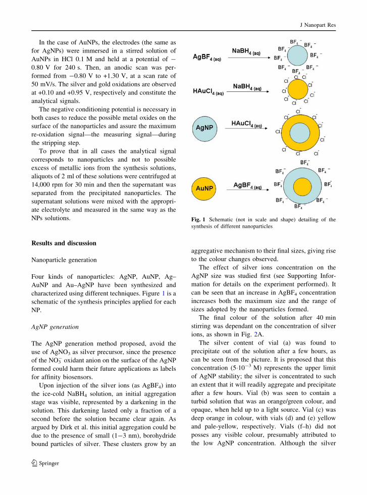
Four kinds of nanoparticles: AgNP, AuNP, Ag–
AuNP and Au–AgNP have been synthesized and
characterized using different techniques. Figure 1 is a
schematic of the synthesis principles applied for each
NP.
AgNP generation
The AgNP generation method proposed, avoid the
use of AgNO3 as silver precursor, since the presence
of the NO3- oxidant anion on the surface of the AgNP
formed could harm their future applications as labels
for affinity biosensors.
Upon injection of the silver ions (as AgBF4) into
the ice-cold NaBH4 solution, an initial aggregation
stage was visible, represented by a darkening in the
solution. This darkening lasted only a fraction of a
second before the solution became clear again. As
argued by Dirk et al. this initial aggregation could be
due to the presence of small (1-3 nm), borohydride
bound particles of silver. These clusters grow by an
aggregative mechanism to their final sizes, giving rise
to the colour changes observed.
The effect of silver ions concentration on the
AgNP size was studied first (see Supporting Infor-
mation for details on the experiment performed). It
can be seen that an increase in AgBF4 concentration
increases both the maximum size and the range of
sizes adopted by the nanoparticles formed.
The final colour of the solution after 40 min
stirring was dependant on the concentration of silver
ions, as shown in Fig. 2A.
The silver content of vial (a) was found to
precipitate out of the solution after a few hours, as
can be seen from the picture. It is proposed that this
concentration (5�10-3 M) represents the upper limit
of AgNP stability; the silver is concentrated to such
an extent that it will readily aggregate and precipitate
after a few hours. Vial (b) was seen to contain a
turbid solution that was an orange/green colour, and
opaque, when held up to a light source. Vial (c) was
deep orange in colour, with vials (d) and (e) yellow
and pale-yellow, respectively. Vials (f–h) did not
posses any visible colour, presumably attributed to
the low AgNP concentration. Although the silver
Fig. 1 Schematic (not in scale and shape) detailing of the
synthesis of different nanoparticles
J Nanopart Res
123
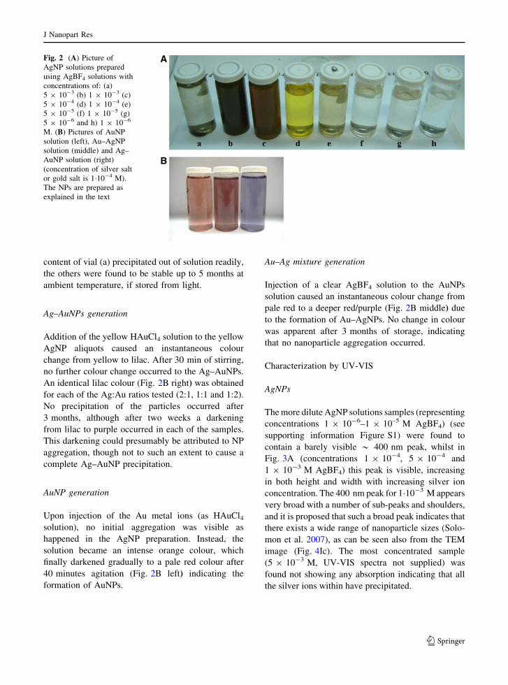
content of vial (a) precipitated out of solution readily,
the others were found to be stable up to 5 months at
ambient temperature, if stored from light.
Ag–AuNPs generation
Addition of the yellow HAuCl4 solution to the yellow
AgNP aliquots caused an instantaneous colour
change from yellow to lilac. After 30 min of stirring,
no further colour change occurred to the Ag–AuNPs.
An identical lilac colour (Fig. 2B right) was obtained
for each of the Ag:Au ratios tested (2:1, 1:1 and 1:2).
No precipitation of the particles occurred after
3 months, although after two weeks a darkening
from lilac to purple occurred in each of the samples.
This darkening could presumably be attributed to NP
aggregation, though not to such an extent to cause a
complete Ag–AuNP precipitation.
AuNP generation
Upon injection of the Au metal ions (as HAuCl4solution), no initial aggregation was visible as
happened in the AgNP preparation. Instead, the
solution became an intense orange colour, which
finally darkened gradually to a pale red colour after
40 minutes agitation (Fig. 2B left) indicating the
formation of AuNPs.
Au–Ag mixture generation
Injection of a clear AgBF4 solution to the AuNPs
solution caused an instantaneous colour change from
pale red to a deeper red/purple (Fig. 2B middle) due
to the formation of Au–AgNPs. No change in colour
was apparent after 3 months of storage, indicating
that no nanoparticle aggregation occurred.
Characterization by UV-VIS
AgNPs
The more dilute AgNP solutions samples (representing
concentrations 1 9 10-6–1 9 10–5 M AgBF4) (see
supporting information Figure S1) were found to
contain a barely visible * 400 nm peak, whilst in
Fig. 3A (concentrations 1 9 10-4, 5 9 10-4 and
1 9 10-3 M AgBF4) this peak is visible, increasing
in both height and width with increasing silver ion
concentration. The 400 nm peak for 1�10-3 M appears
very broad with a number of sub-peaks and shoulders,
and it is proposed that such a broad peak indicates that
there exists a wide range of nanoparticle sizes (Solo-
mon et al. 2007), as can be seen also from the TEM
image (Fig. 4Ic). The most concentrated sample
(5 9 10-3 M, UV-VIS spectra not supplied) was
found not showing any absorption indicating that all
the silver ions within have precipitated.
Fig. 2 (A) Picture of
AgNP solutions prepared
using AgBF4 solutions with
concentrations of: (a)
5 9 10-3 (b) 1 9 10-3 (c)
5 9 10-4 (d) 1 9 10-4 (e)
5 9 10-5 (f) 1 9 10-5 (g)
5 9 10-6 and h) 1 9 10-6
M. (B) Pictures of AuNP
solution (left), Au–AgNP
solution (middle) and Ag–
AuNP solution (right)
(concentration of silver salt
or gold salt is 1�10-4 M).
The NPs are prepared as
explained in the text
J Nanopart Res
123
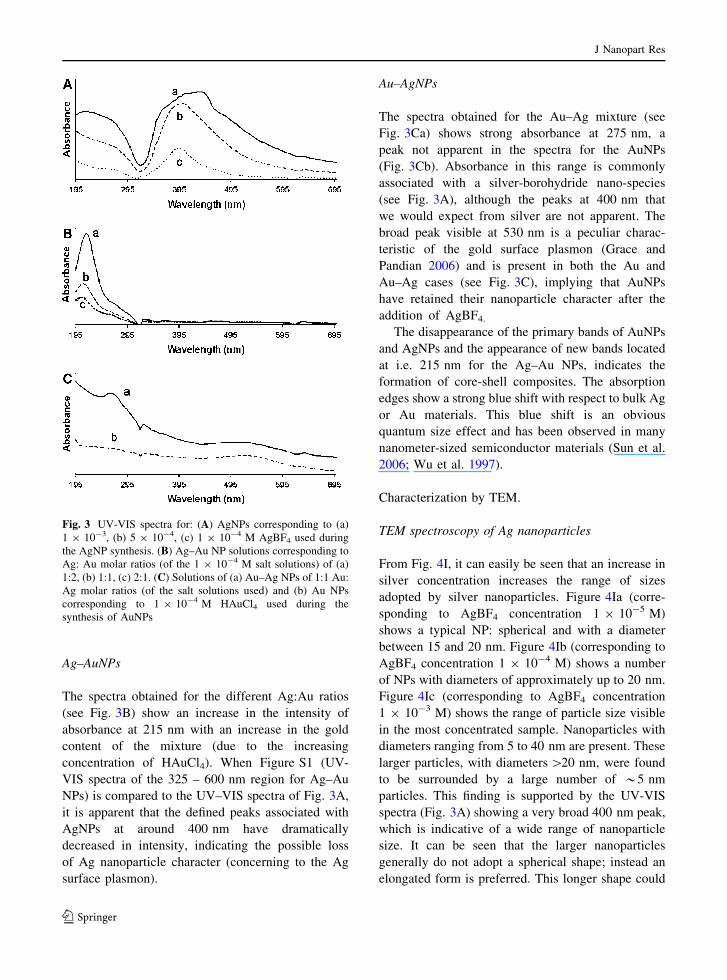
Ag–AuNPs
The spectra obtained for the different Ag:Au ratios
(see Fig. 3B) show an increase in the intensity of
absorbance at 215 nm with an increase in the gold
content of the mixture (due to the increasing
concentration of HAuCl4). When Figure S1 (UV-
VIS spectra of the 325 – 600 nm region for Ag–Au
NPs) is compared to the UV–VIS spectra of Fig. 3A,
it is apparent that the defined peaks associated with
AgNPs at around 400 nm have dramatically
decreased in intensity, indicating the possible loss
of Ag nanoparticle character (concerning to the Ag
surface plasmon).
Au–AgNPs
The spectra obtained for the Au–Ag mixture (see
Fig. 3Ca) shows strong absorbance at 275 nm, a
peak not apparent in the spectra for the AuNPs
(Fig. 3Cb). Absorbance in this range is commonly
associated with a silver-borohydride nano-species
(see Fig. 3A), although the peaks at 400 nm that
we would expect from silver are not apparent. The
broad peak visible at 530 nm is a peculiar charac-
teristic of the gold surface plasmon (Grace and
Pandian 2006) and is present in both the Au and
Au–Ag cases (see Fig. 3C), implying that AuNPs
have retained their nanoparticle character after the
addition of AgBF4.
The disappearance of the primary bands of AuNPs
and AgNPs and the appearance of new bands located
at i.e. 215 nm for the Ag–Au NPs, indicates the
formation of core-shell composites. The absorption
edges show a strong blue shift with respect to bulk Ag
or Au materials. This blue shift is an obvious
quantum size effect and has been observed in many
nanometer-sized semiconductor materials (Sun et al.
2006; Wu et al. 1997).
Characterization by TEM.
TEM spectroscopy of Ag nanoparticles
From Fig. 4I, it can easily be seen that an increase in
silver concentration increases the range of sizes
adopted by silver nanoparticles. Figure 4Ia (corre-
sponding to AgBF4 concentration 1 9 10-5 M)
shows a typical NP: spherical and with a diameter
between 15 and 20 nm. Figure 4Ib (corresponding to
AgBF4 concentration 1 9 10-4 M) shows a number
of NPs with diameters of approximately up to 20 nm.
Figure 4Ic (corresponding to AgBF4 concentration
1 9 10-3 M) shows the range of particle size visible
in the most concentrated sample. Nanoparticles with
diameters ranging from 5 to 40 nm are present. These
larger particles, with diameters [20 nm, were found
to be surrounded by a large number of *5 nm
particles. This finding is supported by the UV-VIS
spectra (Fig. 3A) showing a very broad 400 nm peak,
which is indicative of a wide range of nanoparticle
size. It can be seen that the larger nanoparticles
generally do not adopt a spherical shape; instead an
elongated form is preferred. This longer shape could
Fig. 3 UV-VIS spectra for: (A) AgNPs corresponding to (a)
1 9 10-3, (b) 5 9 10-4, (c) 1 9 10-4 M AgBF4 used during
the AgNP synthesis. (B) Ag–Au NP solutions corresponding to
Ag: Au molar ratios (of the 1 9 10-4 M salt solutions) of (a)
1:2, (b) 1:1, (c) 2:1. (C) Solutions of (a) Au–Ag NPs of 1:1 Au:
Ag molar ratios (of the salt solutions used) and (b) Au NPs
corresponding to 1 9 10-4 M HAuCl4 used during the
synthesis of AuNPs
J Nanopart Res
123
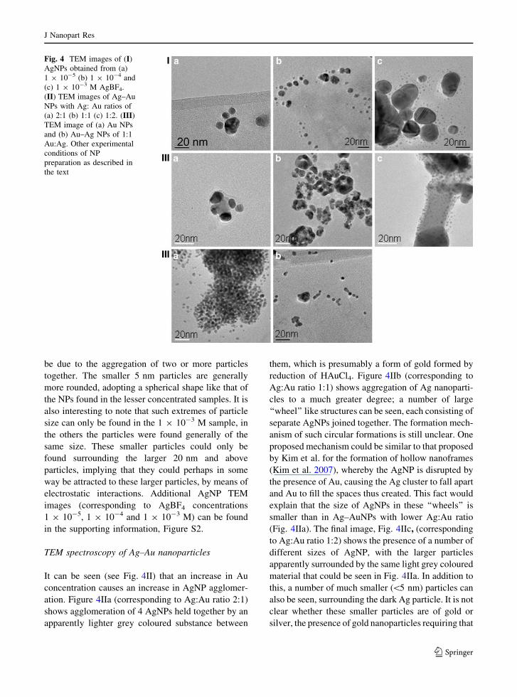
be due to the aggregation of two or more particles
together. The smaller 5 nm particles are generally
more rounded, adopting a spherical shape like that of
the NPs found in the lesser concentrated samples. It is
also interesting to note that such extremes of particle
size can only be found in the 1 9 10-3 M sample, in
the others the particles were found generally of the
same size. These smaller particles could only be
found surrounding the larger 20 nm and above
particles, implying that they could perhaps in some
way be attracted to these larger particles, by means of
electrostatic interactions. Additional AgNP TEM
images (corresponding to AgBF4 concentrations
1 9 10-5, 1 9 10-4 and 1 9 10-3 M) can be found
in the supporting information, Figure S2.
TEM spectroscopy of Ag–Au nanoparticles
It can be seen (see Fig. 4II) that an increase in Au
concentration causes an increase in AgNP agglomer-
ation. Figure 4IIa (corresponding to Ag:Au ratio 2:1)
shows agglomeration of 4 AgNPs held together by an
apparently lighter grey coloured substance between
them, which is presumably a form of gold formed by
reduction of HAuCl4. Figure 4IIb (corresponding to
Ag:Au ratio 1:1) shows aggregation of Ag nanoparti-
cles to a much greater degree; a number of large
‘‘wheel’’ like structures can be seen, each consisting of
separate AgNPs joined together. The formation mech-
anism of such circular formations is still unclear. One
proposed mechanism could be similar to that proposed
by Kim et al. for the formation of hollow nanoframes
(Kim et al. 2007), whereby the AgNP is disrupted by
the presence of Au, causing the Ag cluster to fall apart
and Au to fill the spaces thus created. This fact would
explain that the size of AgNPs in these ‘‘wheels’’ is
smaller than in Ag–AuNPs with lower Ag:Au ratio
(Fig. 4IIa). The final image, Fig. 4IIc, (corresponding
to Ag:Au ratio 1:2) shows the presence of a number of
different sizes of AgNP, with the larger particles
apparently surrounded by the same light grey coloured
material that could be seen in Fig. 4IIa. In addition to
this, a number of much smaller (\5 nm) particles can
also be seen, surrounding the dark Ag particle. It is not
clear whether these smaller particles are of gold or
silver, the presence of gold nanoparticles requiring that
Fig. 4 TEM images of (I)
AgNPs obtained from (a)
1 9 10-5 (b) 1 9 10-4 and
(c) 1 9 10-3 M AgBF4.
(II) TEM images of Ag–Au
NPs with Ag: Au ratios of
(a) 2:1 (b) 1:1 (c) 1:2. (III)
TEM image of (a) Au NPs
and (b) Au–Ag NPs of 1:1
Au:Ag. Other experimental
conditions of NP
preparation as described in
the text
J Nanopart Res
123
the HAuCl4 ions are reduced, although no further
NaBH4 was added after the injection of HAuCl4. This
reduction could only be possible by those BH4- ions
remaining in solution or by those bound to the surface
of the AgNP. To test this, an Ag–Au mixture was
prepared again, but this time the AgNPs were left for
24 h before HAuCl4 was added, as several studies
(Davis and Swain 1960; Davis et al. 1962) have shown
that BH4- ions in water will decompose into non
reductive species, as indicated in the equation below:
BH�4 + Hþ + 2H2O! intermediate! HBO2 + 4H2
Upon HAuCl4 injection, the same colour change as
before occurred, indicating the formation of a similar
chemical species as before. These results imply that
HAuCl4 is reduced by the BH4- ions found on the
AgNP surface, rather than those free in solution as we
expect these free ions to have decomposed in the time
between NaBH4 addition and the addition of HAuCl4.
A galvanic replacement reaction similar to the
described one by Lu et al. (2007) between the added
Au(III) and Ag(0) of the NPs surface is also possible:
Au(III) + 3Ag(0)! 3Agþ + Au(0)
In a similar mode, nearly monodisperse hollow
gold nanospheres have been reported to be synthe-
sized by sacrificial galvanic replacement of cobalt
nanoparticles (Schwartzberg et al. 2006).
The additional images (Figure S3) present in the
supporting information reveal the presence of a
number of triangular NP formations, consisting of a
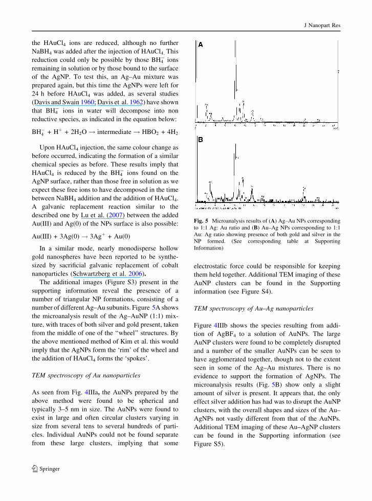
number of different Ag–Au subunits. Figure 5A shows
the microanalysis result of the Ag–AuNP (1:1) mix-
ture, with traces of both silver and gold present, taken
from the middle of one of the ‘‘wheel’’ structures. By
the above mentioned method of Kim et al. this would
imply that the AgNPs form the ‘rim’ of the wheel and
the addition of HAuCl4 forms the ‘spokes’.
TEM spectroscopy of Au nanoparticles
As seen from Fig. 4IIIa, the AuNPs prepared by the
above method were found to be spherical and
typically 3–5 nm in size. The AuNPs were found to
exist in large and often circular clusters varying in
size from several tens to several hundreds of parti-
cles. Individual AuNPs could not be found separate
from these large clusters, implying that some
electrostatic force could be responsible for keeping
them held together. Additional TEM imaging of these
AuNP clusters can be found in the Supporting
information (see Figure S4).
TEM spectroscopy of Au–Ag nanoparticles
Figure 4IIIb shows the species resulting from addi-
tion of AgBF4 to a solution of AuNPs. The large
AuNP clusters were found to be completely disrupted
and a number of the smaller AuNPs can be seen to
have agglomerated together, though not to the extent
seen in some of the Ag–Au mixtures. There is no
evidence to support the formation of AgNPs. The
microanalysis results (Fig. 5B) show only a slight
amount of silver is present. It appears that, the only
effect silver addition has had was to disrupt the AuNP
clusters, with the overall shapes and sizes of the Au–
AgNPs not vastly different from that of the AuNPs.
Additional TEM imaging of these Au–AgNP clusters
can be found in the Supporting information (see
Figure S5).
Fig. 5 Microanalysis results of (A) Ag–Au NPs corresponding
to 1:1 Ag: Au ratio and (B) Au–Ag NPs corresponding to 1:1
Au: Ag ratio showing presence of both gold and silver in the
NP formed. (See corresponding table at Supporting
Information)
J Nanopart Res
123
Electrochemical characterizations
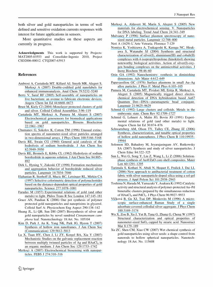
To confirm the possibility for future applications of the
developed nanoparticles for sensors, their electro-
chemical characterization was performed. Figure 6
shows cyclic voltammograms in 1 M NH3 obtained for
a 25 lM AgNPs solution (black curve), the supernatant
solution after centrifugation (grey curve) and for the
electrolyte (blank-dot line). It can be observed that the
current peak corresponds to the oxidation of the silver
at approximately +0.10 V for the AgNPs solution. The
supernatant solution does not exhibit any peak, so can
be assured that the entire signal observed corresponds
to NPs and not to possible excess of silver ions from the
synthesis solution.
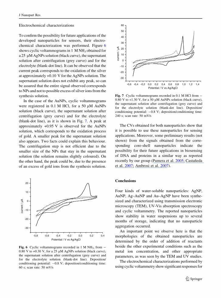
In the case of the AuNPs, cyclic voltammograms
were registered in 0.1 M HCl, for a 50 lM AuNPs
solution (black curve), the supernatant solution after
centrifugation (grey curve) and for the electrolyte
(blank-dot line), as it is shown in Fig. 7. A peak at
approximately +0.95 V is observed for the AuNPs
solution, which corresponds to the oxidation process
of gold. A smaller peak for the supernatant solution
also appears. Two facts could explain this behaviour.
The centrifugation step is not efficient due to the
smaller size of the NPs that stay in the supernatant
solution (the solution remains slightly coloured). On
the other hand, the peak could be, due to the presence
of an excess of gold ions from the synthesis solution.
The CVs obtained for both nanoparticles show that
it is possible to use these nanoparticles for sensing
applications. Moreover, some preliminary results (not
shown) from the signals obtained from the corre-
sponding core-shell nanoparticles indicate the
possibility for their future applications in biosensing
of DNA and proteins in a similar way as reported
recently by our group (Pumera et al. 2005; Castaneda
et al. 2007; Ambrosi et al. 2007).
Conclusions
Four kinds of water-soluble nanoparticles: AgNP,
AuNP, Ag–AuNP and Au–AgNP have been synthe-
sized and characterized using transmission electronic
microscopy (TEM), UV-Vis absorption spectroscopy
and cyclic voltammetry. The reported nanoparticles
show stability in water suspensions up to several
months of storage, indicating that no nanoparticle
aggregation occurred.
An important point we observe here is that the
morphologies of the obtained nanoparticles are
determined by the order of addition of reactants
beside the other experimental conditions such as the
metal ion concentration, and other appropriate
parameters, as was seen by the TEM and UV studies.
The electrochemical characterizations performed by
using cyclic voltammetry show significant responses for
Potential / V vs Ag/AgCl
-0,8
-30
-20
-10
0
10
20
curr
ent /
µA
-0,6 -0,4 -0,2 0,0 0,2 0,4
Fig. 6 Cyclic voltammograms recorded in 1 M NH3, from -
0.80 V to +0.30 V, for a 25 lM AgNPs solution (black curve),
the supernatant solution after centrifugation (grey curve) and
for the electrolyte solution (blank-dot line). Deposition/
conditioning potential: -0.8 V; deposition/conditioning time:
60 s; scan rate: 50 mV/s
-0,6
-30
-20
-10
0
10
20
30
40
50
60
Potential / V vs Ag/AgCl
curr
ent /
µA
-0,4 -0,2 1,41,21,00,80,60,40,20,0
Fig. 7 Cyclic voltammograms recorded in 0.1 M HCl from -
0.80 V to +1.30 V, for a 50 lM AuNPs solution (black curve),
the supernatant solution after centrifugation (grey curve) and
for the electrolyte solution (blank-dot line). Deposition/
conditioning potential: -0.8 V; deposition/conditioning time:
240 s; scan rate: 50 mV/s
J Nanopart Res
123
both silver and gold nanoparticles in terms of well
defined and sensitive oxidation currents responses with
interest for future applications in sensors.
More quantitative studies on these aspects are
currently in progress.
Acknowledgments This work is supported by Projects:
MAT2005-03553 and Consolider-Ingenio 2010, Project
CSD2006-00012. CTQ2007-63913
References
Ambrosi A, Castaneda MT, Killard AJ, Smyth MR, Alegret S,
Merkoci A (2007) Double-codified gold nanolabels for
enhanced immunoanalysis. Anal Chem 79:5232–5240
Berry V, Saraf RF (2005) Self-assembly of nanoparticles on
live bacterium: an avenue to fabricate electronic devices.
Angew Chem Int Ed 44:6668–6673
Brust M, Kiely CJ (2004) Monolayer protected clusters of gold
and silver. Colloid Colloid Assemblies 3:96–119
Castaneda MT, Merkoci A, Pumera M, Alegret S (2007)
Electrochemical genosensors for biomedical applications
based on gold nanoparticles. Biosens Bioelectron
22:1961–1967
Chumanov G, Sokolov K, Cotton TM (1996) Unusual extinc-
tion spectra of nanometer-sized silver patricles arranged
in two-dimensional arrays. J Phys Chem 100:5166–5168
Davis RE, Swain CG (1960) General acid catalysis of the
hydrolysis of sodium borohydride. J Am Chem Soc
82:5949–5950
Davis RE, Bromels E, Kibby CL (1962) Hydrolysis of sodium
borohydride in aqueous solution. J Am Chem Soc 84:885–
892
Dirk L, Hyning V, Zukoski CF (1998) Formation mechanisms
and aggregation behavior of borohydride reduced silver
particles. Langmuir 14:7034–7046
Elghanian R, Storhoff JJ, Mucic RC, Letsinger RL, Mirkin CA
(1997) Selective colorimetric detection of polynucleotides
based on the distance-dependent optical properties of gold
nanoparticles. Science 277:1078–1081
Faraday M (1857) Experimental relations of gold (and other
metals) to light. Philos Trans R Soc London 147:145–181
Grace AN, Pandian K (2006) One pot synthesis of polymer
protected gold nanoparticles and nanoprisims in glycerol.
Colloid Surf A: Physicochem Eng Aspect 290:138–142
Huang JL, Li QB, Sun DH (2007) Biosynthesis of silver and
gold nanoparticles by novel sundried Cinnamomum cam-phora leaf. Nanotechnology 18:Art. No. 105104
Kim D, Park J, An K, Yang NK, Park JG, Hyeon T (2007)
Synthesis of hollow iron nanoframes. J Am Chem Soc
(Communication) 129:5812–5813
Lu X, Tuan HY, Chen J, Li ZY, Korgel BA, Xia Y (2007)
Mechanistic Studies on the galvanic replacement reaction
between multiply twinned particles of Ag and HAuCl4 in
an organic medium. J Am Chem Soc 129:1733–1742
Merkoci A (2007) Electrochemical biosensing with nanopar-
ticles. FEBS J 274:310–316
Merkoci A, Aldavert M, Marın S, Alegret S (2005) New
materials for electrochemical sensing. V. Nanoparticles
for DNA labeling. Trend Anal Chem 24:341–349
Mulvaney P (1996) Surface plasmon spectroscopy of nano-
sized metal particles. Langmuir 12:788–800
Neri A (1629) L’Arte Vetraria. Florence 7:129
Nomiya K, Yoshizawa A, Tsukagoshi K, Kasuga NC, Hirak-
awa S, Watanabe SJ (2004) Synthesis and structural
characterization of silver(I), aluminium(III) and cobalt(II)
complexes with 4-isopropyltropolone (hinokitiol) showing
noteworthy biological activities. Action of silver(I)-oxy-
gen bonding complexes on the antimicrobial activities. J
Inorg Biochem 98:46–60
Ozin GA (1992) Nanochemistry: synthesis in diminishing
dimensions. Adv Mater 4:612–649
Papavassiliou GC (1976) Surface plasmons in small Au–Ag
alloy particles. J Phys F: Metal Phys 6:103–105
Pumera M, Castaneda MT, Pividori MI, Eritja R, Merkoci A,
Alegret S (2005) Magnetically trigged direct electro-
chemical detection of DNA hybridization based Au67
Quantum Dot—DNA—paramagnetic bead conjugate.
Langmuir 21:9625–9629
Schmid G (1992) Large clusters and colloids. Metals in the
embryonic state. Chem Rev 92:1709–1727
Schmid G, Lehnert A, Malm JO, Bovin JO (1991) Experi-
mental relations of gold (and other metals) to light.
Angew Chem Int Ed 30:874–876
Schwartzberg AM, Olson TY, Talley CE, Zhang JZ (2006)
Synthesis, characterization, and tunable optical properties
of hollow gold nanospheres. J Phys Chem B 110:19935–
19944
Solomon SD, Bahadory M, Jeyarajasingam AV, Rutkowsky
SA (2007) Synthesis and study of silver nanoparticles. J
Chem Educ 84:322–325
Sun L, Wei G, Song Y, Liu Z, Wang L, Li Z (2006) Solution-
phase synthesis of Au@ZnO core-shell composites. Mater
Lett 60:1291–1295
Tarimala S, Kothari N, Abidi N, Hequet E, Fralick J, Dai LL
(2006) New approach to antibacterial treatment of cotton
fabric with silver nanoparticle-doped silica using a sol-gel
process. J Appl Polym Sci 101:2938–2943
Toshima N, Harada M, Yamazaki Y, Asakura K (1992) Catalytic
activity and structural analysis of polymer-protected Au–Pd
bimetallic clusters prepared by the simultaneous reduction
of HAuCl4 and PdCl2. J Phys Chem 96:9927–9933
Vlckova B, Gu XJ, Tsai DP, Moskovits M (1996) A micro-
scopic surface-enhanced Raman Study of a single
adsorbate-covered collodial silver aggregate. J Phys Chem
100:3169–3174
Wu X, Zou B, Xu J, Yut B, Tang G, Zhang G, Cheng W (1997)
Structural characterization and optical properties of
nanometer-sized SnO2 capped by stearic acid. Nanostruct
Mat 8:179–189
Xu ZC, Shen CM, Xiao CW (2007) Wet chemical synthesis of
gold nanoparticles using silver seeds: a shape control from
nanorods to hollow spherical nanoparticles. Nanotech-
nology 18:Art. No. 115608
J Nanopart Res
123


