Embed Size (px)
Citation preview
LETTERS AND VIEWPOINTS
GUIDELINESLetters to the Editor and View-points are welcome. Letters tothe Editor discuss material re-cently published in the Journal.Letters will have the best chanceof acceptance if they are receivedwithin 8 weeks of an article’s pub-lication. Letters to the Editor
may be published with a response from the authors of thearticle being discussed. Discussions beyond the initial let-ter and response will not be published. Letters submittedpertaining to published Discussions of articles will not beprinted. Letters to the Editor are not usually peer re-viewed, but the Journal may invite replies from the authorsof the original publication. All Letters and Viewpoints arepublished at the discretion of the Editor.
Viewpoints pertain to issues of general interest, even ifthey are not related to items previously published (such asunique techniques, brief technology updates, technicalnotes, and so on). Viewpoints will be published on a space-available basis because they are typically less time-sensitivethan Letters and other types of articles. Please note thefollowing criteria for Letters and Viewpoints:• Text–maximum of 500 words (not including
references)• References–maximum of five• Authors–no more than five• Figures/Tables–no more than two figures and/or one
tableAuthors will be listed in the order in which they appear inthe submission. Letters and Viewpoints should be submit-ted electronically via PRS’ enkwell, at www.editorialman-ager.com/prs/. We strongly encourage authors to submitfigures in color.
We reserve the right to edit letters and viewpoints tomeet requirements of space and format. Any financialinterests relevant to the content of the correspondencemust be disclosed. Submission of a letter and/or viewpointconstitutes permission for the American Society of PlasticSurgeons and its licensees and assignees to publish it in theJournal and in any other form or medium.
The views, opinions, and conclusions expressed in theletters to the Editor and viewpoints represent the personalopinions of the individual writers and not those of thepublisher, the Editorial Board, or the sponsors of theJournal. Any stated views, opinions, and conclusions do notreflect the policy of any of the sponsoring organizations orof the institutions with which the writer is affiliated, andthe publisher, the Editorial Board, and the sponsoringorganizations assume no responsibility for the content ofsuch correspondence.
Letters
Soft-Tissue Response: Is It a Standing Obstaclein Distraction Osteogenesis?Sir:
We read with great interest the article by Hollier etal. entitled “Distraction Rate and Latency: Fac-
tors in the Outcome of Pediatric Mandibular Distrac-
tion” (Plast. Reconstr. Surg. 117: 2333, 2006). Accelera-tion of distraction osteogenesis is a controversial areaof new bone formation. Numerous disadvantages ofacceleration attempts, such as malunion, nonunion,and soft-tissue complications, keep clinicians away fromrapid distraction. Successful results have been reportedwith a distraction rate of 2 mm per day with no latencyperiod. This is a fascinating clinical study on the rate ofdistraction osteogenesis.
The soft-tissue response to rapid distraction is an im-portant factor and affects the success of new bone for-mation. Clinical studies in long bones have suggested lotsof complications related to rapid distraction, includingrelapse, muscle fibrosis, neurovascular injury, joint sub-luxation, and stiffness.1–4 In the sheep mandibular dis-traction model, it has been reported that distraction os-teogenesis in the facial skeleton produces an adaptiveresponse in muscle distracted at 1 mm per day.5 If thedistraction rate exceeds the adaptive response rate, thereis a muscle fiber force deficit and susceptibility to stretch-induced injury increases.5 We did not see any commenton soft-tissue response in the article. It seems that noneof the patients exhibited soft-tissue complications. Is itbecause of the great elastic potential or rapid regenera-tion capability of soft tissues in the pediatric population?We think the cause of the effective soft-tissue complianceshould be investigated and revealed.
Another extraordinary response of soft tissue torapid distraction, especially to rapid distraction withouta latency period, is prolapse of the soft tissue surround-ing the distraction zone into the distraction gap. In ourexperimental studies, rapid distraction groups demon-strated greater soft-tissue and muscle herniation intothe distraction gap (unpublished data). Maybe the mat-uration of the hematoma between distraction segmentsduring the latency period contributes to keeping thesurrounding soft tissue away from the distraction gap.
Soft-tissue reactions to rapid distraction in distractionosteogenesis are still controversial. In our view, there is anobvious need for more clinical research in this area. Wecompliment the authors on their successful results in theacceleration of distraction osteogenesis.DOI: 10.1097/01.prs.0000261061.95413.82
Ersoy Konas, M.D.
Mehmet Emin Mavili, M.D.Department of Plastic and Reconstructive Surgery
Hacettepe University School of MedicineAnkara, Turkey
Correspondence to Dr. KonasHacettepe University Faculty of Medicine
Department of Plastic and Reconstructive Surgery06100 Samanpazarı, Ankara, Turkey
[email protected]@gmail.com
REFERENCES1. Mackool, R. J., Hopper, R. A., Grayson, B. H., Holliday, R., and
McCarthy, J. G. Volumetric change of the medial pterygoidfollowing distraction osteogenesis of the mandible: An exam-
Copyright ©2007 by the American Society of Plastic Surgeons
DOI: 10.1097/01.prs.0000261078.27085.a0
www.PRSJournal.com2314
ple of the associated soft-tissue changes. Plast. Reconstr. Surg.111: 1804, 2003.
2. Day, C. S., Moreland, M. S., Floyd, S. S., Jr., and Huard,J. Limb lengthening promotes muscle growth. J. Orthop. Res.15: 227, 1997.
3. Eldridge, J. C., and Bell, D. F. Problems with substantial limblengthening. Orthop. Clin. North Am. 22: 625, 1991.
4. Paley, D. Problems, obstacles, and complications of limblengthening by the Ilizarov technique. Clin. Orthop. Relat. Res.250: 81, 1990.
5. van der Meulen, J. H., Borschel, G. H., Lynch, J. B., et al. Theeffect of rate of distraction osteogenesis on structure andfunction of anterior digastric muscle fibers. Plast. Reconstr.Surg. 115: 831, 2005.
ReplySir:
Drs. Konas and Mavili put forth a very valid criti-cism of our article, “Distraction Rate and Latency:Factors in the Outcome of Pediatric Mandibular Dis-traction” (Plast. Reconstr. Surg. 117: 2333, 2006). Verylittle is discussed regarding soft-tissue response to theaccelerated distraction. In part, however, this is be-cause very little difference was noted in the mannerin which the soft tissue responded in these cases.Subjectively, it was felt that the scars were worse inthis group of patients, although this could not bequantified. No differences in relapse or soft-tissueprolapse into the distraction gap were noted. Al-though, as the authors point out, clinical studies inlong bones have demonstrated soft-tissue problemswith rapid distraction, this cannot be directly corre-lated with pediatric mandibular distraction. The dis-traction distances are much greater in the lower ex-tremities, and the blood supply is not as robust as itis in the pediatric facial skeleton.
It should also be pointed out that rapid distractionwas not attempted in these patients to decrease theoverall treatment time. The bulk of the patients’ treat-ment time was spent in the consolidation phase.Rather, the accelerated rate of distraction was utilizedprimarily in an attempt to prevent premature consol-idation. We have found that when patients are dis-tracted at the standard rate of 1 mm per day, prematurehealing of the osteotomy site is seen far too often. Thisis most common with external devices, as the amountof device movement translated to the osteotomy sitevaries. Some of the device distraction is translated intothe bending of pins, particularly when the device ispositioned further from the face. Unpublished datawould seem to suggest that on the order of half of thedistraction is translated into bone movement.
In summary, soft-tissue response to accelerated man-dibular distraction using external devices was not sub-stantially different from that in cases of slower distrac-tion, with the exception of subjectively poorer scarring.Real progress in distraction will be accomplished whenthe period of time for bone consolidation is shortened.DOI: 10.1097/01.prs.0000233457.61615.c9
Patrick Cole, M.D.
Larry Hollier, Jr., M.D.Clinical Care Center
Houston, Texas
Correspondence to Dr. HollierClinical Care Center
6621 Fannin Street, Suite 620.10Houston, Texas 77030
[email protected]@bcm.edu
Nonresective Shrinkage of the Septum and FatCompartments of the Upper and Lower Eyelids:A Comparative Study with Carbon DioxideLaser and Colorado NeedleSir:
We read with great interest the article by Prado etal. entitled “Nonresective Shrinkage of the Sep-
tum and Fat Compartments of the Upper and LowerEyelids: A Comparative Study with Carbon Dioxide La-ser and Colorado Needle” (Plast. Reconstr. Surg. 117:1725, 2006). The authors nicely describe and comparetwo new nonresective methods for managing the her-niated fat pads of the lower and upper lids.
The conservative approach (septal plication instead offat excision) for correcting septal herniation is not a newconcept. In 1988, de la Plaza and Arroyo introduced theirown blepharoplasty technique, which consisted of return-ing fat to the orbital cavity and retaining it by means of acontinuous suture of the capsulopalpebral fascia to theperiosteum of the lower orbital rim.1 However, they ap-plied their technique only to the lower eyelids. The sep-torrhaphy technique of Dr. Sensoz was published in thisJournal in 1998, and this technique is applicable to boththe lower and upper eyelids.2 The method described byPrado et al. is also similar, plicating the septum with thelaser or electrocoagulation instead of suture placement.Some authors claim that septal plication may lead to ec-tropion, as is also discussed by Dr. Codner (Plast. Reconstr.Surg. 117: 1736, 2006). In our opinion, an experiencedsurgeon with a good knowledge of eyelid anatomy will notface such a problem while performing septal plication.The alternative transconjunctival approach avoids inci-sions on the septum, allowing only fat excision; additionalincisions are required to manipulate the anterior lamellastructures, which are more important in giving a youthfulappearance to the patient. Fat herniation usually occursat the junction of the septum orbitale and orbital rim. Theseptorrhaphy of the septum is performed by either ex-cising the weakened septal tissue, reinserting the septumto the orbital rim, or simply plicating the healthy septaltissue to the orbital rim. The formation of ectropion canusually be assessed during the operation. The surgeonshould carefully check the septum for retraction begin-ning from the first suture to the last suture placed on theseptum.2 We also agree with the authors that “the mainpoint seems to be the amount of manipulation ratherthan the elements manipulated,” but since Prado et al.
Volume 119, Number 7 • Letters and Viewpoints
2315
used indirect methods for plication (depending on scarcontracture formed after the intervention), the lack ofabsolute control on the amount of septum plicationseems to be the major drawback of their technique. Over-correction may lead to ectropion (as seen in two cases),and in contrast, undercorrection may lead to persistenceof the bulging.
In conclusion, the authors should be congratulatedon emphasizing the importance of the conservativeapproach in eyelid surgery and describing two new,easily applied methods for correcting fat herniationthrough the septum. We believe their study is a signif-icant contribution to the literature.DOI: 10.1097/01.prs.0000261063.17647.b6
Omer Sensoz, M.D.
Hakan Orbay, M.D.Department of Plastic and Reconstructive SurgeryAnkara Numune Training and Research Hospital
Ankara, Turkey
Correspondence to Dr. OrbayEryaman Evleri. Dil Devrimi Cd. B7/16
06770 Eryaman, Ankara, [email protected]
REFERENCES1. de la Plaza, R., and Arroyo, J. M. A new technique for the
treatment of palpebral bags. Plast. Reconstr. Surg. 81: 677, 1988.2. Sensoz, O., Unlu, R. E., Percin, A., et al. Septo-orbitoperios-
toplasty for the treatment of palpebral bags: A 10-year expe-rience. Plast. Reconstr. Surg. 101: 1657; discussion 1664, 1998.
ReplySir:
We truly appreciate the comments of Drs. Sensozand Orbay from Turkey. The purposes of our studywere to describe an alternative nonresective treatmentof the fat-septum elements of the eyelids using lowenergy delivered with a carbon dioxide laser and a gridof electrocautery to partially desiccate and shrink theseptum and underlying fat in a setback manner, and tocompare its clinical outcomes.1
It is a fact that resection of the fat bags of the eyelidsleads to a sunken and gaunt appearance of the eyes andaggravates a previous tear trough deformity.2 This studywas thought to avoid the standard plastic surgery res-idency teaching of blepharoplasty (surgical excision ofskin, muscle, and fat) and consolidate a conservativeapproach, as most plastic surgeons continue to removefat during blepharoplasty. Our residents are not expe-rienced surgeons and sometimes find the eyelid anat-omy disturbing and difficult to understand. As Dr. Cod-ner stated, mobilizing the septum with the aid ofsutures, especially after its resection, could lead to ec-tropion, and for inexperienced surgeons, this fine ma-nipulation and its amount cannot be assessed duringthe operation. So teaching our residents to preserve fatduring blepharoplasty with a simple and alternative
shrinking technique as opposed to surgical tighteningof the septum seemed sound, especially when electro-cautery appeared to be always available, with less ex-pense and theoretically less risk compared with lasertreatment. As we stress in the article, the correction ofshrinkage was stopped when the fat bag compartmentswere leveled and had a symmetrical contour, as deter-mined by gentle compression of the globe; this coun-teracted the “absolute lack of control” of the amount ofseptal shrinkage.
The best control of this manipulation to avoid ec-tropion is with a well-performed canthopexy. (Can-thopexy was never used in the cases presented in ourarticle so that we could evaluate the pure effect of heatapplication over the septum-fat components of the eye-lids alone.) We have modified our shrinkage techniqueso as to always finish the procedure with a canthopexy,making clear that this is indispensable in the presenceof lid laxity.
In summary, if anyone is interested in using thistechnique, the following are some key points:
1. Never use the technique where there is extremelid laxity.
2. The technique is suitable for treating young pa-tients with moderate skin excess, adequate mus-cle tone, moderate to severe bulging of the fat-septal component, and no supportingalterations.
3. Allow residents to start with this simple and safeprocedure, which should always be completedwith a lateral retinacular suspension used as asimplified suture canthopexy3; this will provide asubtle but long-lasting, adequate result.
As this clinical study was a team study, all of theauthors contributed to this reply.DOI: 10.1097/01.prs.0000261065.04552.8f
Arturo Prado, M.D.
Patricio Andrades, M.D.
Stefan Danilla, M.D.
Paulo Castillo, M.D.
Susana Benitez, M.D.Division of Plastic Surgery
School of MedicineClinical Hospital J. J. Aguirre
University of ChileSantiago, Chile
Correspondence to Dr. PradoManquehue Norte 1701 ofic 210
VitacuraSantiago, Metropolitana, Chile
REFERENCES1. Prado, A., Andrades, P., Danilla, S., Castillo, P., and Benitez,
S. Nonresective shrinkage of the septum and fat compart-ments of the upper and lower eyelids: A comparative study
Plastic and Reconstructive Surgery • June 2007
2316
with carbon dioxide laser and Colorado needle. Plast. Reconstr.Surg. 117: 1725, 2006.
2. Klatsky, S. A., Iliff, N., and Manson, P. N. Blepharoplasty. InR. M. Goldwyn and M. N. Cohen (Eds.), The Unfavorable Resultin Plastic Surgery. Philadelphia: Lippincott, Williams & Wilkins,2001.
3. Fagien, S. Algorithm for canthoplasty: The lateral retinacu-lar suspension: A simplified suture canthopexy. Plast. Reconstr.Surg. 103: 2042, 1999.
Investigation of Risk Acceptance in FacialTransplantationSir:
In their “Investigation of Risk Acceptance in FacialTransplantation,”1 Barker et al. refer to the praise
and criticism of Joseph Murray after the first successfulrenal transplant. In studying the history of clinicaltransplantation outlined by Starzl,2 it is clear that it isa field with its fair share of controversy. Indeed, at thebeginning of the twentieth century, the very first hu-man renal transplantations utilized kidneys from pigs,sheep, goats, and subhuman primate donors.
This research by Barker and colleagues providessome interesting data. It has the inherent flaw, how-ever, of participants speculating on how they might feelin the event of severe medical or aesthetic adverseevents. Such speculation is not necessarily predictive ofhow an individual would actually feel if the event oc-curred. It is quite revealing that the disfigured groupwas the least accepting of facial transplantation with itsimmunosuppressive risks. I agree that this is most likelydue to adjustment processes leading to a different“frame of reference.”
It is correct that the unique viewpoint of disfiguredpatients should be respected, as should their freedomto choose. It should be remembered, however, thatpatients tend to minimize risks in their eagerness toproceed with innovative surgery.3 Certainly the opinionof a well-informed patient is as important as that of acritic or proponent of the procedure, but when theprocedure excites a polarized debate, a potential pa-tient should perhaps be informed by both.DOI: 10.1097/01.prs.0000261067.90974.4b
Andrew J. Diver, M.R.C.S.Ed.Northern Ireland Plastic and Maxillofacial Service
The Ulster HospitalBelfast, Northern Ireland
REFERENCES1. Barker, J. H., Furr, A., Cunningham, M., et al. Investigation
of risk acceptance in facial transplantation. Plast. Reconstr.Surg. 118: 663, 2006.
2. Starzl, T. E. History of clinical transplantation. World J. Surg.24: 759, 2000.
3. Jones, N. F. Concerns about human hand transplantation inthe 21st century. Hand Surg. 27: 771, 2002.
ReplySir:
We appreciate Dr. Diver’s thoughtful comments con-cerning our article, “Investigation of Risk Acceptancein Facial Transplantation.” Our goal in the article wasto document the responses of a range of subject pop-ulations to the risks and benefits of a variety of trans-plantation opportunities. We agree with Dr. Diver thatthere are methodological ambiguities associated with“participants speculating on how they might feel in theevent of severe medical or aesthetic adverse events.” Forthat reason, the larger research project solicited theresponses of individuals who were uniquely qualified tocomment on the issues under consideration. Not onlydid we ask facially disfigured individuals about theirviews concerning the risks and benefits of face trans-plants, we also asked hand amputees about their per-ceptions of the risks and benefits of a hand transplant,and kidney transplant recipients about the risks andbenefits of a kidney transplant. The facially disfiguredand hand amputees were well positioned to describethe risks that they would take to derive the benefits ofa transplant. Conversely, the kidney transplant recipi-ents had doubtless spent considerable time weighingthe risks and benefits of the procedure that they hadundergone. Our finding that kidney transplant recip-ients, who were intimately familiar with the dangers ofimmunosuppression, would undergo even more risk toreceive a face transplant than a new kidney testifies tothe importance of the face in human life.
We conclude by agreeing with Dr. Diver that patientsare in the best position when they are informed aboutboth sides of a polarized debate. The views of knowl-edgeable patients simply contribute one piece of suchinformation.DOI: 10.1097/01.prs.0000261069.75644.77
John H. Barker, M.D., Ph.D.Plastic Surgery Research
Michael Cunningham, Ph.D.Department of Communication
University of LouisvilleLouisville, Ky.
Correspondence to Dr. BarkerPlastic Surgery Research
University of Louisville511 South Floyd Street
320 MDR BuildingLouisville, Ky. 40202
Mini Face Lift with Suspension Sutures:Historical Analysis of Development andMorphic ResonanceSir:
Tonnard and Verpaele are to be credited for popu-larizing the mini face lift with suspension sutures,
a technique today known worldwide as the minimalaccess craniofacial suspension suture lift, as they
Volume 119, Number 7 • Letters and Viewpoints
2317
termed it.1–3 We also use a modification of this tech-nique, which we probably would not have used untilnow without reading their publications and hearingtheir presentations at international meetings. Theircomprehensive book, published by Quality MedicalPublishing in 2004, is well worth buying and reading togain a close understanding of the basic ideas behindthis technique and the fine details for this type of miniface lifting. However, from a close review of their bookand publications as well as a further review and searchof the literature, it is very unfortunate that they failedto mention their predecessors with this suture suspen-sion technique.
A search of the literature readily shows us that, in1997, Duminy and Hudson,4 from Cape Town, SouthAfrica, were actually the first authors to report the useof a minimal access craniofacial suspension lift with twosuspension sutures; they described a series of 35 pa-tients with an average follow-up of more than 1 year.Almost 4 years later, 1 year before Tonnard et al.’spublication,1 Fulton et al.5 also reported on the use ofa minimal access craniofacial suspension lift with theuse of two craniofacial suspension sutures. They calledthese suspension sutures the U and O sutures, a termthat was later also advocated by Tonnard and Verpaele.2
As Hamra6 wrote recently in his letter to the editor,“We all know the familiar writing style in scientificjournal articles whereby an author conveniently omitspublished work when that work may not support histhesis or may predate his observations. Often this isinnocent and inadvertent, as one naturally cannot listevery reference. . . . However, with the topic on the useof craniofacial suspension sutures, which is so new andhas so few papers published, one has cause to wonder.”Why were these predecessors not mentioned in thewritings and presentations of Tonnard and Verpaele?
A thorough review of their literature shows that it isquite obvious that at least Fulton et al.’s publication5
was not missed in Tonnard et al.’s literature search.1They very briefly and hardly recognizably mention thispublication in the discussion section of their initialarticle in Plastic and Reconstructive Surgery, stating that“[a]lso the combination of centrofacial laser resurfac-ing and a [minimal access craniofacial suspension su-ture] lift can be performed with great safety.” In notproviding further details, they did not credit Fulton etal.5 for their earlier, probably inspiring, work on a min-imal access craniofacial suspension lift with suspensionO and U sutures, as Tonnard and Verpaele have alsonamed their craniofacial suspension sutures.2 They didnot mention these publications and facts in their sub-sequent textbook. Their textbook contains a section on“the history and evolution of rhytidectomy,” in whichthey review what is in their opinion the most essentialwork in the history of face lifting techniques in relationto their work. Tonnard and Verpaele even cited a Chi-nese proverb: “When you drink water, remember thespring.”
By means of this letter, we would like to give creditto those whom others have forgotten to mention in the
success story of the minimal access craniofacial suspen-sion suture lift. Duminy and Hudson4 were the first topublish in the English-language literature on a minimalaccess craniofacial suspension lift with suspension su-tures, and Fulton et al.5 followed with their work.
So what should we learn from this letter and actualsituation? That one should review the literature thor-oughly when publishing new ideas and thoughts, andthat one should give credit to one’s predecessors, evenif one has never heard of them, when describing abrilliant new idea. It is known that although similarideas and thoughts arise in more than one place in theworld seemingly independent of one another, theymight actually be more closely associated because of themysterious phenomenon of morphic resonance.7 Let-terpress printing was discovered in more than oneplace in the world, and it would be a shame not tomention all who have contributed. We all stand on theshoulders of giants,7 and we should credit these giants.We, the authors of this letter, credit Tonnard et al. fortheir excellent work on further developing and popu-larizing the minimal access craniofacial suspension su-ture lift, but credit should also go to the originalinventors.5,6
DOI: 10.1097/01.prs.0000261071.31701.5f
Berend van der Lei, M.D., Ph.D.Department of Plastic, Reconstructive, Aesthetic, and
Hand SurgeryMedical Center of Leeuwarden
Leeuwarden, the NetherlandsPrivate Clinic Heerenveen
Heerenveen, The NetherlandsDepartment of Plastic, Reconstructive, Aesthetic, and
Hand Surgery, University Hospital of GroningenGroningen, The Netherlands
Michel Cromheecke, M.D., Ph.D.Department of Plastic, Reconstructive, Aesthetic, and
Hand SurgeryMedical Center of LeeuwardenLeeuwarden, The Netherlands
Private Clinic HeerenveenHeerenveen, The Netherlands
Stefan O. P. Hofer, M.D., Ph.D.Department of Plastic and Reconstructive Surgery
Erasmus Medical CenterRotterdam, The Netherlands
Correspondence to Dr. van der LeiDepartment of Plastic, Reconstructive, Aesthetic, and
Hand SurgeryMedical Centre of Leeuwarden
Leeuwarden, Friesland 8934 AD, The [email protected]
REFERENCES1. Tonnard, P., Verpaele, A., Monstrey, S., et al. Minimal access
cranial suspension lift: A modified S-lift. Plast. Reconstr. Surg.109: 2074, 2002.
2. Tonnard, P. L., and Verpaele, A. M. The MACS-lift Short-Scar Rhytidectomy. St. Louis, Mo.: Quality Medical Publish-ing, Inc., 2004.
Plastic and Reconstructive Surgery • June 2007
2318
3. Tonnard, P., Verpaele, A., and Gaia, S. Optimising resultsfrom minimal access cranial suspension lifting (MACS lift).Aesthetic Plast. Surg. 29: 213, 2005.
4. Duminy, C. M., and Hudson, D. A. The mini rhytidectomy.Aesthetic Plast. Surg. 21: 280, 1997.
5. Fulton, J. E., Saylan, Z., Helton, P., Rahimi, A. D., and Gols-hani, M. The S-lift facelift featuring the U-suture and O-suturecombined with skin resurfacing. Dermatol. Surg. 27: 18, 2001.
6. Hamra, S. T. 25,000 dollars and still counting (Letter). Plast.Reconstr. Surg. 118: 801, 2006.
7. Sheldrake, R. An experimental test of the hypothesis of for-mative causation. Riv. Biol. 85: 431, 1992.
ReplySir:
It is with interest that we have read the letter by vander Lei et al. They point out that the bibliography inour book, The MACS-lift Short Scar Rhytidectomy, is notcomprehensive. We never pretended so and are con-vinced that a bibliography is a list of the scientific worksthat inspired the author to produce his or her publi-cation. Dr. van der Lei and coworkers need to be con-gratulated for the thoroughness of their literaturesearch. It is virtually impossible to read every literatureitem that could be related to the work one intends topublish. Sometimes one comes across a related articlepurely by coincidence. Indeed, there is a myriad ofpublications about “mini face lift,” “mini rhytidec-tomy,” “minimal face lift,” “minilift,” “minimal incisionrhytidectomy,” and so on, and it is only by reading thearticle in extenso that one can determine whether itreally refers to one’s own work. In MEDLINE, thesearch term “minimal incision AND face lift” alonedelivers 32 references.1–32 Most of us select relevantreferences on the basis of the published abstract. Nei-ther the abstract nor the title of Duminy and Hudson’sarticle33 mentions the use of purse-string sutures, andthis is probably the reason why we overlooked it at thetime of our publication. This article indeed shows in-teresting similarities as to the use of purse-string sutureson the subcutaneous tissues, as described by Saylan.34 Itwas Saylan’s 1999 article in the Aesthetic Surgery Journalthat drew our attention to the possibilities of usingpurse-string sutures in the face.
In their article, Fulton et al.35 describe their ex-perience with Saylan’s S-lift, without adding anythingnew. Their article was published after we submittedour article “Minimal Access Cranial Suspension Lift:A Modified S-lift”, which appeared in Plastic and Re-constructive Surgery in 2002. We later included it in thebibliography of our book, The MACS-Lift Short-ScarRhytidectomy.
It is regrettable that we overlooked Duminy andHudson’s article, but this was certainly because of un-awareness rather than negligence or, even worse, in-tentional omission.
One has to wonder though whether “being the first”is the essence of publishing one’s work. Was Illouz thefirst to aspirate subcutaneous fat? Was Coleman the first
to put it back into the body? Was Lejour the “inventor”of the vertical scar mammaplasty? Probably not. Butthey made these procedures work and popularizedthem. Without their efforts, we would probably still bereluctant to transplant or even suction fat, and thebreast reduction scars would still be popping out ofwomen’s brassieres on the lateral and medial sides be-cause “that’s the way it is.”
There is no such thing as an “original inventor” of asurgical procedure. Any “new” surgical technique isalways the result of a combination of several ideas thathave been tried out sometimes many years before. Weare the first to acknowledge that. Duminy and Hudsoncan indeed be considered the predecessors of a wholegeneration of facial rejuvenation procedures of whichthe minimal access craniofacial suspension suture lift isa part. We will definitely include their work in the newedition of our book and in any related publications inthe future.DOI: 10.1097/01.prs.0000261099.40156.4a
Patrick L. Tonnard, M.D.
Alexis M. Verpaele, M.D.
Koenraad Van Landuyt, M.D.
Moustapha Hamdi, M.D.Coupure Center for Plastic Surgery
Gent, Belgium
Correspondence to Dr. TonnardCoupure Center for Plastic Surgery
Coupure Rechts 164 C-DGent B-9000, Belgium
REFERENCES1. Krishna, S., and Williams, E. F. Lipocontouring in conjunc-
tion with the minimal incision brow and subperiosteal mid-face lift: The next dimension in midface rejuvenation. FacialPlast. Surg. Clin. North Am. 14: 221, 2006.
2. Prado, A., Andrades, P., Danilla, S., Castillo, P., and Leniz,P. A clinical retrospective study comparing two short-scarface lifts: Minimal access cranial suspension versus lateralSMASectomy. Plast. Reconstr. Surg. 117: 1413, 2006.
3. Eremia, S., and Willoughby, M. A. Novel face-lift suspen-sion suture and inserting instrument: Use of large anchorsknotted into a suture with attached needle and insertingdevice allowing for single entry point placement of suspen-sion suture: Preliminary report of 20 cases with 6- to 12-month follow-up. Dermatol. Surg. 32: 335, 2006.
4. Pontius, A. T., and Williams, E. F. The extended minimalincision approach to midface rejuvenation. Facial Plast. Surg.Clin. North Am. 13: 411, 2005.
5. Zager, W. H., and Dyer, W. K. Minimal incision faceliftFacial Plast. Surg 21: 21, 2005.
6. Agarwal, A., Dejoseph, L., and Silver, W. Anatomy of thejawline, neck, and perioral area with clinical correlations.Facial Plast. Surg. 21: 3, 2005.
7. McCarty, M. L., and Brackup, A. B. Minimal incision faceliftsurgery. Ophthalmol. Clin. North Am. 18: 305, 2005.
8. Seify, H., Jones, G., Bostwick, J., and Hester, T. R. Endo-scopic-assisted face lift: Review of 200 cases. Ann. Plast. Surg.52: 234, 2004.
Volume 119, Number 7 • Letters and Viewpoints
2319
9. Viksraitis, S., Astrauskas, T., Karbonskiene, A., and Budnikas,G. Endoscopic aesthetic facial surgery: Technique and re-sults. Medicina (Kaunas) 40: 149, 2004.
10. Yang, M. Y., Li, S. K., and Li, Q. Correction of nasolabialfold (in Chinese). Zhongguo Xiu Fu Chong Jian Wai Ke Za Zhi18: 40, 2004.
11. Williams, E. F., Vargas, H., Dahiya, R., Hove, C. R., Rodg-ers, B. J., and Lam, S. M. Midfacial rejuvenation viaa minimal-incision brow-lift approach: Critical evalua-tion of a 5-year experience. Arch. Facial Plast. Surg. 5:470, 2003.
12. Ruiz-Esparza, J., and Gomez, J. B. The medical face lift: Anoninvasive, nonsurgical approach to tissue tightening infacial skin using nonablative radiofrequency. Dermatol. Surg.29: 325; discussion 332, 2003.
13. De Cordier, B. C., de la Torre, J. I., Al-Hakeem, M. S., etal. Endoscopic forehead lift: Review of technique, cases,and complications. Plast. Reconstr. Surg 110: 1558; discussion1569, 2002.
14. Tonnard, P., Verpaele, A., Monstrey, S., et al. Minimal ac-cess cranial suspension lift: A modified S-lift. Plast. Reconstr.Surg. 109: 2074, 2002.
15. Patel, B. C. Midface rejuvenation. Facial Plast. Surg. 15: 231,1999.
16. Ramirez, O. M. Three-dimensional endoscopic midfaceenhancement: A personal quest for the ideal cheek reju-venation. Plast. Reconstr. Surg 109: 329; discussion 341,2002.
17. Paul, M. D. The evolution of the brow lift in aesthetic plasticsurgery. Plast. Reconstr. Surg. 108: 1409, 2001.
18. Finger, E. R. A 5-year study of the transmalar subperiostealmidface lift with minimal skin and superficial musculoapo-neurotic system dissection: A durable, natural-appearing liftwith less surgery and recovery time. Plast. Reconstr. Surg 107:1273; discussion 1284, 2001.
19. Maloney, B. P., and Schiebelhoffer, J. Minimal-incision en-doscopic face-lift. Arch. Facial Plast. Surg. 2: 274, 2000.
20. Schaeffer, B. T. Endoscopic liposhaving for neck recon-touring. Arch. Facial Plast. Surg. 2: 264, 2000.
21. Aly, A., Avila, E., and Cram, A. E. Endoscopic plastic sur-gery. Surg. Clin. North Am. 80: 1373, 2000.
22. Ramirez, O. M. High-tech facelift. Aesthetic Plast. Surg. 22:318, 1998.
23. Ramirez, O. M. Cervicoplasty: Nonexcisional anterior ap-proach. Plast. Reconstr. Surg. 99: 1576, 1997.
24. Burnett, C. D., Rabinowitz, S., and Rauscher, G. E. Endo-scopic-assisted midface lift utilizing retrograde dissection.Ann. Plast. Surg. 36: 449, 1996.
25. Abramo, A. C. Forehead rhytidoplasty: Endoscopic ap-proach. Aesthetic Plast. Surg. 19: 463, 1995.
26. Ramirez, O. M. Endoscopically assisted biplanar foreheadlift. Plast. Reconstr. Surg. 96: 323, 1995.
27. Ramirez, O. M. The anchor subperiosteal forehead lift.Plast. Reconstr. Surg. 95: 993; discussion 1004, 1995.
28. Guyuron, B. Subcutaneous approach to forehead, brow,and modified temple incision. Clin. Plast. Surg. 19: 461,1992.
29. McKinney, P., Mossie, R. D., and Zukowski, M. L. Criteriafor the forehead lift. Aesthetic Plast. Surg. 15: 141, 1991.
30. Man, D. Stretching and tissue expansion for rhytidectomy:An improved approach. Plast. Reconstr. Surg. 84: 561; discus-sion 570, 1989.
31. Guyuron, B. Modified temple incision for facial rhytidec-tomy. Ann. Plast. Surg. 21: 439, 1988.
32. Johnson, C. M., Adamson, P. A., and Anderson, J. R. Theface-lift incision. Arch. Otolaryngol. 110: 371, 1984.
33. Duminy, F., and Hudson, D. A. The mini rhytidectomy.Aesthetic Plast. Surg. 21: 280, 1997.
34. Saylan, Z. The S-lift: Less is more. Aesthetic Surg. J. 19: 406,1999.
35. Fulton, J. E., Saylan, Z., Helton, P. Rahimi, A. D., andGolshani, M. The S-lift facelift featuring the U-suture andO-suture combined with skin resurfacing. Dermatol. Surg.27: 18, 2001.
Use of an Expanded Temporoparietal FascialFlap Technique for Total AuricularReconstruction
Sir:
We read with great interest the article by Park andMun entitled “Use of an Expanded Tem-
poroparietal Fascial Flap Technique for Total Auric-ular Reconstruction.” We congratulate the authorson their useful effort. Although the postoperativephotographs of their patients are quite impressive,there are a few points which we would like to discussfurther.
As a sort of silicone implant, tissue expanders placedanywhere within the body do certainly lead to the for-mation of a thick, dense fibrotic capsule around theexpander.1 The authors state that they completely re-move this capsule in the second stage of their proce-dure. In our opinion, however, complete removal ofsuch a fibrotic capsule from thin fascial tissue, which isthinner after tissue expansion, is a difficult surgicalintervention that may damage the vascularity of theunderlying temporoparietal fascia tissue.
Another point to be questioned is the limited ex-pandability of a 1-month-old skin graft. The well-knowntendency of a split-thickness skin graft to contract andget thicker after graft take is an additional obstacleagainst the expansion procedure, which is performedunder temporal fascia tissue that is not quite an ex-pandable structure itself.
The authors prefer the scalp for the thick split-thick-ness donor site. The potential risks and complicationsof the scalp as a thick split-thickness skin graft donorsite have been discussed in the literature.2 The rapidhealing potential of the scalp as a donor site for split-thickness skin grafts is actually valid for thin grafts (i.e.,thinner than 0.25 mm).3 When one attempts to harvesta thick split-thickness skin graft, one should considerthe risk of alopecia, which is not acceptable for a patientwho is seeking a better aesthetic appearance for anabsent ear. Moreover, the need to shave hair makes thepatient look like he or she has undergone some kindof psychological trauma, especially in female patients.
We think that illumination of above-mentionedpoints will contribute to the scientific value of thissignificant study. We thank Dr. Park and Dr. Mun fortheir interesting work.DOI: 10.1097/01.prs.0000261075.10453.1d
Plastic and Reconstructive Surgery • June 2007
2320
Metin Kerem, M.D.
Hakan Orbay, M.D.
Omer Sensoz, M.D.Second Department of Plastic and Reconstructive Surgery
Ankara Numune Training and Research HospitalAnkara, Turkey
Correspondence to Dr. KeremBeril Sitesi 449. Sokak No. 27
06530 Umitkoy, Ankara, [email protected]
REFERENCES1. Breitbard, A. S., and Ablaza, V. J. Implant materials. In S. J.
Aston, R. W. Beasley, and H. M. Thorne (Eds.), Grabb andSmith’s Plastic Surgery, 5th Ed. Philadelphia: Lippincott-Raven,1997. Pp. 39-45.
2. Zingaro, E. A., Capozzi, A., and Penissi, V. R. The scalp as adonor site in burns. Arch. Surg. 123: 652, 1988.
3. Dardour, J. C., Noury-Duperrat, G., and Dufourmontel,C. Le cuir chevelu, zone donneuse des greffes minces. Ann.Chir. Plast. 22: 169, 1977.
ReplySir:
I thank Dr. Kerem and colleagues for their interestin our article, “Use of an Expanded TemporoparietalFascial Flap Technique for Total Auricular Reconstruc-tion.” With regard to their three points of comment–-the removal procedure for the capsule, the thickness ofthe scalp graft, and the expansibility of grafted skin–-Iwould like to present further clarification.
By using the expression “complete removal of thecapsule,” my coauthor and I meant the removal of theentire capsule area rather than removal of the full-layered capsule. However, I would like to commentmore on the capsule removal. We encountered twosituations during capsule removal. First, when the tem-poroparietal fascia was used as a wraparound regionalflap to cover the expander, the capsule became presenton the superficial side of the fascia. The vessels werelocated on the superficial side of the fascia, so thecapsule had to be carefully removed in order not todamage the vessels. (When the capsule consists of mul-tiple layers, it is not possible to remove full layers of thecapsule.) Meanwhile, when the expander was wrappedwith a free flap harvested from the opposite temporopa-rietal side, we attached the deeper side of the fascia tothe expander; therefore, when the capsule formed onthe deeper side of the fascia, most of the capsule layerscould be removed safely.
My coauthor and I prefer to harvest the ratherthicker skin from the scalp using a no. 15 scalpel. Toprevent postoperative complications at the donor site,such as delayed epidermization, folliculitis, concretescalp deformity, or focal alopecia, we regraft pieces ofultrathin scalp skin to the donor site.1–4 The ultrathinskin is harvested using a razor. Thicker scalp skin pro-
vides less secondary contracture and better skin quality.In addition, we did not experience limited expansibilityof the grafted skin in any of the presented cases. Thismight have been due to the thicker grafted skin.
We hope that these clarifications will prove satisfac-tory to Dr. Kerem and colleagues.DOI: 10.1097/01.prs.0000261077.81483.ec
Chul Park, M.D.The Seoul Center for Developmental Ear Anomalies
Department of Plastic SurgeryKorea University College of Medicine and Anam Hospital
126-1, 5th, Anam-Dong, Seongbuk-GuSeoul 136-705, South Korea
REFERENCES1. Engrav, L. V., Grube, B. J., and Bubak, P. J. Treatment of the
concrete scalp donor site. Ann. Plast. Surg. 24: 162, 1990.2. Carter, Y. M., Summer, G. J., Engrav, L. H., Hansen, F. L.,
Costa, B. A., and Matsumura, H. Incidence of the concretescalp deformity associated with deep scalp donor sites andmanagement with the Unna cap. J. Burn Care Rehabil. 20: 141,1999.
3. Hockel, M., Menke, H., and Germann, G. Vaginoplasty withsplit skin grafts from the scalp: Optimization of the surgicaltreatment for vaginal agenesis. Am. J. Obstet. Gynecol. 188: 1100,2003.
4. Mimoun, M., Chaouat, M., Picovski, D., Serroussi, D., andSmarrito, S. The scalp is an advantageous donor site forthin-skin grafts: A report on 945 harvested samples. Plast.Reconstr. Surg. 118: 369, 2006.
Preservation of Digital Palmar Veins to AvoidVenous Congestion in Heterodigital ArterializedFlapsSir:
We read with interest three articles recently pub-lished in Plastic and Reconstructive Surgery about
heterodigital arterialized flaps for finger and handreconstruction.1–3 It is generally agreed that the mainproblem with these flaps is venous congestion.1–4 Theauthors introduced the use of a dorsal vein from thedonor finger to reduce postoperative venous conges-tion. Although it was successful, this method had thedisadvantage of decreasing the reach of the flap, par-ticularly when it was used to reconstruct nonadjacentfinger defects.1 To extend the reach of the flap, theysuccessively explored the possibility of vein division andsecondary anastomosis2 as well as cross-finger transfer,3with obvious pros and cons.
Venous drainage of the fingers, studied by Moss etal.,5 involves the palmar and dorsal systems. The dorsalsystem corresponds to the veins used by the authors,while palmar system is subdivided into a superficialsystem and a deep system. The deep system is composedof the venae comitantes of the proper and commondigital arteries, while the superficial system, which islocated subdermally, drains either dorsally or into thedeep system.
Volume 119, Number 7 • Letters and Viewpoints
2321
The causes of postoperative congestion have beenimputed to injury to the venae comitantes during dis-section of the digital artery from the digital nerve,despite the use of loupes and meticulous technique. Intheir first article, Teoh et al. reported in the descriptionof the flap dissection the division and cauterization ofthe abundant palmar veins.1
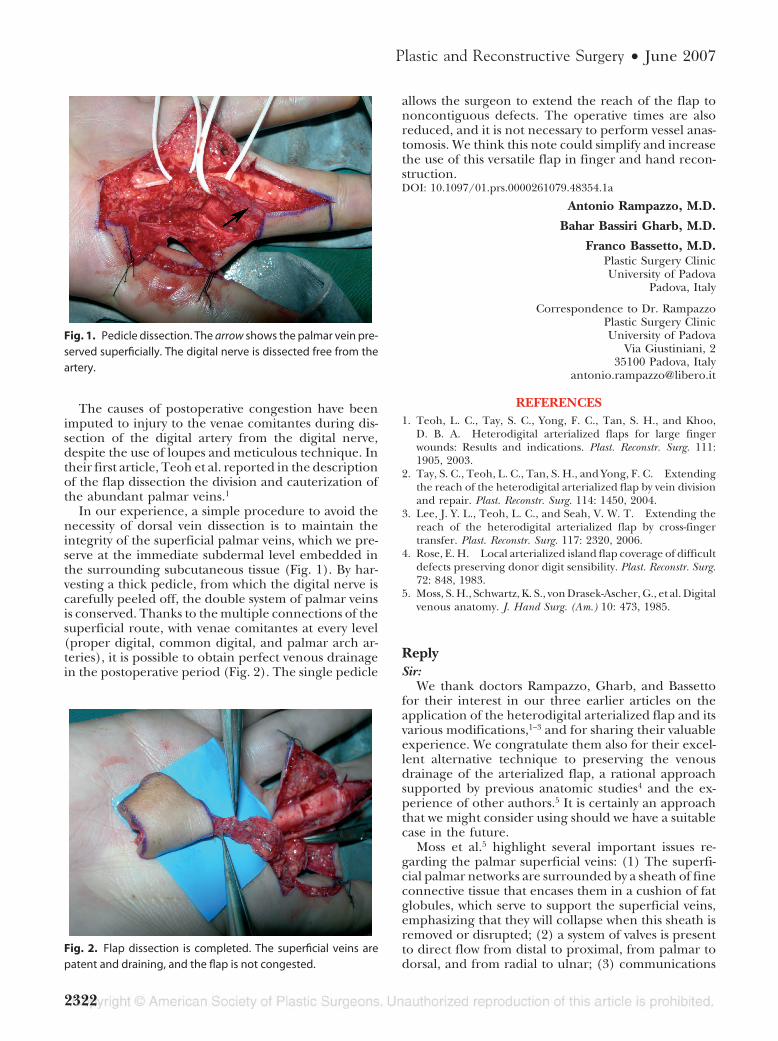
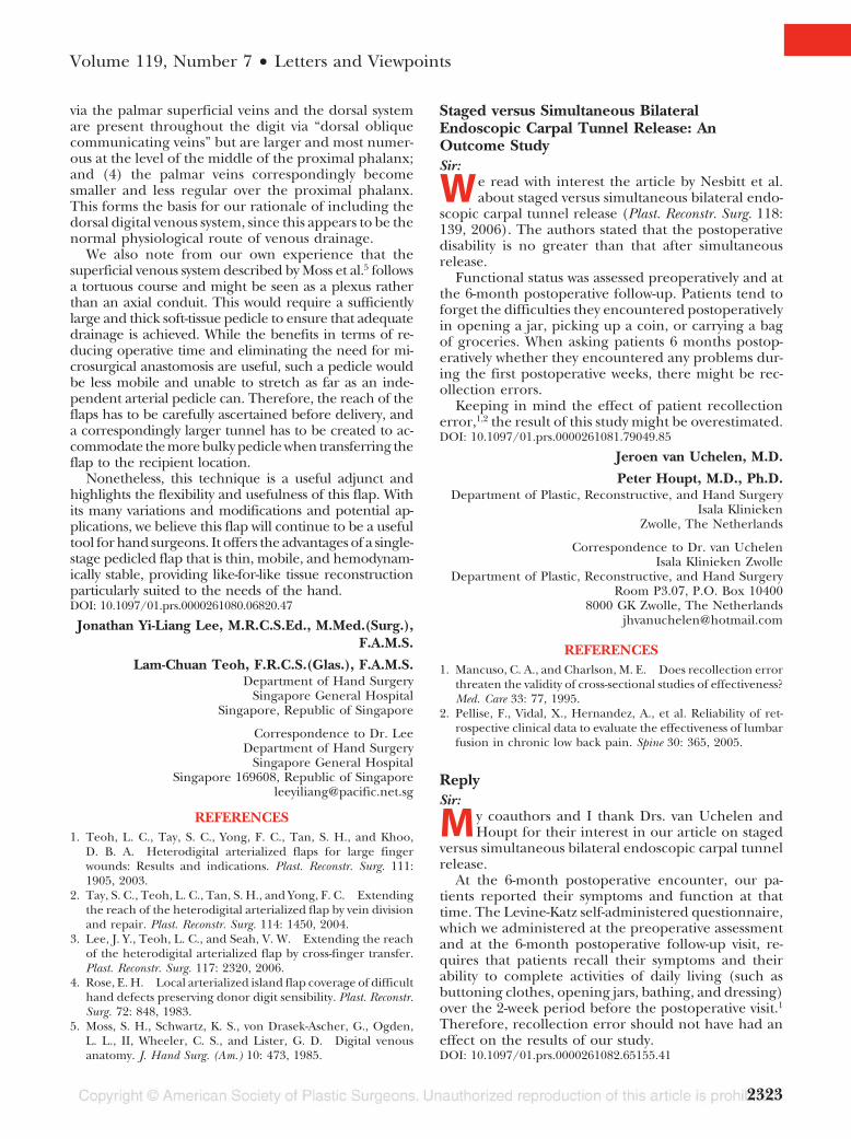
In our experience, a simple procedure to avoid thenecessity of dorsal vein dissection is to maintain theintegrity of the superficial palmar veins, which we pre-serve at the immediate subdermal level embedded inthe surrounding subcutaneous tissue (Fig. 1). By har-vesting a thick pedicle, from which the digital nerve iscarefully peeled off, the double system of palmar veinsis conserved. Thanks to the multiple connections of thesuperficial route, with venae comitantes at every level(proper digital, common digital, and palmar arch ar-teries), it is possible to obtain perfect venous drainagein the postoperative period (Fig. 2). The single pedicle
allows the surgeon to extend the reach of the flap tononcontiguous defects. The operative times are alsoreduced, and it is not necessary to perform vessel anas-tomosis. We think this note could simplify and increasethe use of this versatile flap in finger and hand recon-struction.DOI: 10.1097/01.prs.0000261079.48354.1a
Antonio Rampazzo, M.D.
Bahar Bassiri Gharb, M.D.
Franco Bassetto, M.D.Plastic Surgery ClinicUniversity of Padova
Padova, Italy
Correspondence to Dr. RampazzoPlastic Surgery ClinicUniversity of Padova
Via Giustiniani, 235100 Padova, Italy
REFERENCES1. Teoh, L. C., Tay, S. C., Yong, F. C., Tan, S. H., and Khoo,
D. B. A. Heterodigital arterialized flaps for large fingerwounds: Results and indications. Plast. Reconstr. Surg. 111:1905, 2003.
2. Tay, S. C., Teoh, L. C., Tan, S. H., and Yong, F. C. Extendingthe reach of the heterodigital arterialized flap by vein divisionand repair. Plast. Reconstr. Surg. 114: 1450, 2004.
3. Lee, J. Y. L., Teoh, L. C., and Seah, V. W. T. Extending thereach of the heterodigital arterialized flap by cross-fingertransfer. Plast. Reconstr. Surg. 117: 2320, 2006.
4. Rose, E. H. Local arterialized island flap coverage of difficultdefects preserving donor digit sensibility. Plast. Reconstr. Surg.72: 848, 1983.
5. Moss, S. H., Schwartz, K. S., von Drasek-Ascher, G., et al. Digitalvenous anatomy. J. Hand Surg. (Am.) 10: 473, 1985.
ReplySir:
We thank doctors Rampazzo, Gharb, and Bassettofor their interest in our three earlier articles on theapplication of the heterodigital arterialized flap and itsvarious modifications,1–3 and for sharing their valuableexperience. We congratulate them also for their excel-lent alternative technique to preserving the venousdrainage of the arterialized flap, a rational approachsupported by previous anatomic studies4 and the ex-perience of other authors.5 It is certainly an approachthat we might consider using should we have a suitablecase in the future.
Moss et al.5 highlight several important issues re-garding the palmar superficial veins: (1) The superfi-cial palmar networks are surrounded by a sheath of fineconnective tissue that encases them in a cushion of fatglobules, which serve to support the superficial veins,emphasizing that they will collapse when this sheath isremoved or disrupted; (2) a system of valves is presentto direct flow from distal to proximal, from palmar todorsal, and from radial to ulnar; (3) communications
Fig. 1. Pedicle dissection. The arrow shows the palmar vein pre-served superficially. The digital nerve is dissected free from theartery.
Fig. 2. Flap dissection is completed. The superficial veins arepatent and draining, and the flap is not congested.
Plastic and Reconstructive Surgery • June 2007
2322
via the palmar superficial veins and the dorsal systemare present throughout the digit via “dorsal obliquecommunicating veins” but are larger and most numer-ous at the level of the middle of the proximal phalanx;and (4) the palmar veins correspondingly becomesmaller and less regular over the proximal phalanx.This forms the basis for our rationale of including thedorsal digital venous system, since this appears to be thenormal physiological route of venous drainage.
We also note from our own experience that thesuperficial venous system described by Moss et al.5 followsa tortuous course and might be seen as a plexus ratherthan an axial conduit. This would require a sufficientlylarge and thick soft-tissue pedicle to ensure that adequatedrainage is achieved. While the benefits in terms of re-ducing operative time and eliminating the need for mi-crosurgical anastomosis are useful, such a pedicle wouldbe less mobile and unable to stretch as far as an inde-pendent arterial pedicle can. Therefore, the reach of theflaps has to be carefully ascertained before delivery, anda correspondingly larger tunnel has to be created to ac-commodate the more bulky pedicle when transferring theflap to the recipient location.
Nonetheless, this technique is a useful adjunct andhighlights the flexibility and usefulness of this flap. Withits many variations and modifications and potential ap-plications, we believe this flap will continue to be a usefultool for hand surgeons. It offers the advantages of a single-stage pedicled flap that is thin, mobile, and hemodynam-ically stable, providing like-for-like tissue reconstructionparticularly suited to the needs of the hand.DOI: 10.1097/01.prs.0000261080.06820.47
Jonathan Yi-Liang Lee, M.R.C.S.Ed., M.Med.(Surg.),F.A.M.S.
Lam-Chuan Teoh, F.R.C.S.(Glas.), F.A.M.S.Department of Hand Surgery
Singapore General HospitalSingapore, Republic of Singapore
Correspondence to Dr. LeeDepartment of Hand Surgery
Singapore General HospitalSingapore 169608, Republic of Singapore
REFERENCES1. Teoh, L. C., Tay, S. C., Yong, F. C., Tan, S. H., and Khoo,
D. B. A. Heterodigital arterialized flaps for large fingerwounds: Results and indications. Plast. Reconstr. Surg. 111:1905, 2003.
2. Tay, S. C., Teoh, L. C., Tan, S. H., and Yong, F. C. Extendingthe reach of the heterodigital arterialized flap by vein divisionand repair. Plast. Reconstr. Surg. 114: 1450, 2004.
3. Lee, J. Y., Teoh, L. C., and Seah, V. W. Extending the reachof the heterodigital arterialized flap by cross-finger transfer.Plast. Reconstr. Surg. 117: 2320, 2006.
4. Rose, E. H. Local arterialized island flap coverage of difficulthand defects preserving donor digit sensibility. Plast. Reconstr.Surg. 72: 848, 1983.
5. Moss, S. H., Schwartz, K. S., von Drasek-Ascher, G., Ogden,L. L., II, Wheeler, C. S., and Lister, G. D. Digital venousanatomy. J. Hand Surg. (Am.) 10: 473, 1985.
Staged versus Simultaneous BilateralEndoscopic Carpal Tunnel Release: AnOutcome StudySir:
We read with interest the article by Nesbitt et al.about staged versus simultaneous bilateral endo-
scopic carpal tunnel release (Plast. Reconstr. Surg. 118:139, 2006). The authors stated that the postoperativedisability is no greater than that after simultaneousrelease.
Functional status was assessed preoperatively and atthe 6-month postoperative follow-up. Patients tend toforget the difficulties they encountered postoperativelyin opening a jar, picking up a coin, or carrying a bagof groceries. When asking patients 6 months postop-eratively whether they encountered any problems dur-ing the first postoperative weeks, there might be rec-ollection errors.
Keeping in mind the effect of patient recollectionerror,1,2 the result of this study might be overestimated.DOI: 10.1097/01.prs.0000261081.79049.85
Jeroen van Uchelen, M.D.
Peter Houpt, M.D., Ph.D.Department of Plastic, Reconstructive, and Hand Surgery
Isala KliniekenZwolle, The Netherlands
Correspondence to Dr. van UchelenIsala Klinieken Zwolle
Department of Plastic, Reconstructive, and Hand SurgeryRoom P3.07, P.O. Box 10400
8000 GK Zwolle, The [email protected]
REFERENCES1. Mancuso, C. A., and Charlson, M. E. Does recollection error
threaten the validity of cross-sectional studies of effectiveness?Med. Care 33: 77, 1995.
2. Pellise, F., Vidal, X., Hernandez, A., et al. Reliability of ret-rospective clinical data to evaluate the effectiveness of lumbarfusion in chronic low back pain. Spine 30: 365, 2005.
ReplySir:
My coauthors and I thank Drs. van Uchelen andHoupt for their interest in our article on staged
versus simultaneous bilateral endoscopic carpal tunnelrelease.
At the 6-month postoperative encounter, our pa-tients reported their symptoms and function at thattime. The Levine-Katz self-administered questionnaire,which we administered at the preoperative assessmentand at the 6-month postoperative follow-up visit, re-quires that patients recall their symptoms and theirability to complete activities of daily living (such asbuttoning clothes, opening jars, bathing, and dressing)over the 2-week period before the postoperative visit.1Therefore, recollection error should not have had aneffect on the results of our study.DOI: 10.1097/01.prs.0000261082.65155.41
Volume 119, Number 7 • Letters and Viewpoints
2323
E. F. Shaw Wilgis, M.D.The Curtis National Hand Center
Union Memorial Hospital3333 North Calvert Street
JPB-mezzanineBaltimore, Md. 21218
REFERENCE1. Levine, D. W., Simmons, B. P., Koris, M. J., et al. A self-
administered questionnaire for the assessment of severity ofsymptoms and functional status in carpal tunnel syndrome.J. Bone Joint Surg. (Am.) 75: 1585, 1993.
Hernia Repairs in Postbariatric PatientsSir:
I read with interest Dr. Michele Shermak’s articleentitled “Hernia Repair and Abdominoplasty in
Gastric Bypass Patients” (Plast. Reconstr. Surg. 117:1145, 2006). Dr. Shermak retrospectively reviewed 40patients who had undergone open incisional herniarepair combined with panniculectomy after massiveweight loss. She describes opening the abdominalcavity and lysing adhesions and then repairing thehernia. In his discussion of her article, Dr. Aly men-tions his concerns about plastic surgeons operatingin the abdominal cavity unless they have been per-forming intra-abdominal procedures in the recentpast. The risks associated with opening the abdom-inal cavity, whether it is done by a plastic surgeon ora general surgeon, are certainly well known, and ifthey can be avoided, they should be. These risks,which can be as significant as bowel perforation, canaffect quality of life, hospital stays, and the need forfurther surgical intervention. As Dr. Shermak states,this patient population is unique and often presentswith attenuated fascia, which allows for a primaryrepair that is not otherwise possible in the non–massive weight loss population. This ability to closethe fascia primarily has allowed my group, with Dr.Gary Anthone, bariatric surgeon, to develop a tech-nique for hernia repair that avoids opening the ab-dominal cavity and therefore avoids all possibility ofintra-abdominal complications. However, due to thenature of these patients’ attenuated fascia, we haveutilized onlay fascia to minimize the risk of herniarecurrence in our series of 50 patients. The increasedcomplication rates predicted by Dr. Shermak havenot been seen, and unlike in her series, we have notseen a recurrence of the hernia. The results of our50-consecutive-patient series were presented at the21st Annual Meeting of the American Society forBariatric Surgery, in San Diego, California, June16, 2004, and published in Surgery for Obesity andRelated Diseases (1: 458, 2005). I believe that this tech-nique is a safer procedure, with less risk for thepatient and the plastic surgeon as well as betterresults.DOI: 10.1097/01.prs.0000261083.95211.70
Susan E. Downey, M.D.University of Southern California1301 Twentieth Street, Suite 470
Santa Monica, Calif. [email protected]
ReplySir:
I thank Dr. Downey for her interest in the article. Iappreciate the opportunity to reply to the issues sheraises.
First is her question with regard to the role of plasticsurgeons in hernia repair. At the Bariatric Center ofExcellence at Johns Hopkins, patients achieving mas-sive weight loss after gastric bypass who require pan-niculectomy and incisional hernia repair are referredto plastic surgeons on the team. Throughout theUnited States, most plastic surgeons have participatedin general surgery training programs, where there is alarge amount of experience to be had in hernia repair.At the annual meeting of the American College ofSurgeons in Chicago this year, most of the surgeons onthe hernia panel were plastic surgeons. Traditionalteaching prescribes clear visualization of intra-abdom-inal structures, and this is more favorable to blind clo-sure of the hernia sac due to the risk of bowel perfo-ration that could occur with placing a large needlethrough attenuated fascia around an incisional hernia.Other concerns include incarceration of bowel or fatwithin the closure if these structures are not visualizedduring approximation of fascia. In most cases, theomentum is adhered up to the hernia sac and beyondthe edges of the hernia sac. The omentum comes downeasily with careful dissection. In cases where bowel ad-hesions are stuck to or around the hernia sac, my gen-eral surgery colleagues assist in taking adhesions down;this has occurred five times in my current experienceof 96 cases. By gaining access to the peritoneal cavity,we are also better able to explore for other distinct,smaller, satellite hernias that frequently occur and maybe less obvious in a supine patient under anesthesiawith a relaxed abdominal wall. The Bariatric Center’sdivision of labor has resulted in streamlined patientcare and concentrated, specialized experience on thepart of plastic surgeons, who competently and success-fully perform hernia repair and teach it to the surgicalresidents.
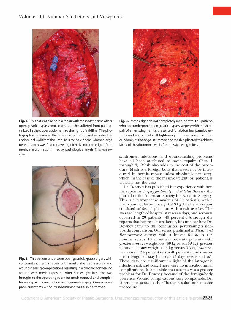
Dr. Downey advocates for the value of mesh in herniarepair in the massive weight loss patient. Massive weightloss patients with hernias are a distinct population withtissue excess, not tissue deficiency. This redundancy offascial tissue not only affords primary repair but alsoallows plication over top of the hernia repair, in effect,an autologous fascial reinforcement. My practice in-cludes complex abdominal wall reconstruction forother patient populations as well, and patients present-ing with problems after prior hernia repairs often haveproblems secondary to mesh utilization. Seromas, pain
Plastic and Reconstructive Surgery • June 2007
2324
syndromes, infections, and wound-healing problemshave all been attributed to mesh repairs (Figs. 1through 3). Mesh also adds to the cost of the proce-dure. Mesh is a foreign body that need not be intro-duced in hernia repair unless absolutely necessary,which, in the case of the massive weight loss patient, istypically not the case.
Dr. Downey has published her experience with her-nia repair in Surgery for Obesity and Related Diseases, thejournal of the American Society for Bariatric Surgery.This is a retrospective analysis of 50 patients, with amean panniculectomy weight of 3 kg. The hernia repairconsisted of fascial plication with mesh overlay. Theaverage length of hospital stay was 4 days, and seromasoccurred in 20 patients (40 percent). Although shereports that her results are better, it is unclear how Dr.Downey came to this conclusion, performing a side-by-side comparison. Our series, published in Plastic andReconstructive Surgery, with a longer follow-up (23months versus 18 months), presents patients withgreater average weight loss (69 kg versus 59 kg), greaterpanniculectomy weight (4.5 kg versus 3 kg), lower se-roma risk (12.5 percent versus 40 percent), and shortermean length of stay by a day (3 days versus 4 days).These data are significant in light of the iatrogenicinfection risk and cost. There were no intra-abdominalcomplications. It is possible that seroma was a greaterproblem for Dr. Downey because of the foreign-bodypresence. Wound complications were comparable. Dr.Downey presents neither “better results” nor a “saferprocedure.”
Fig. 1. This patient had hernia repair with mesh at the time of heropen gastric bypass procedure, and she suffered from pain lo-calized in the upper abdomen, to the right of midline. The pho-tograph was taken at the time of exploration and includes theabdominal wall from the umbilicus to the xiphoid, where a largenerve branch was found traveling directly into the edge of themesh, a neuroma confirmed by pathologic analysis. This was ex-cised.
Fig. 2. This patient underwent open gastric bypass surgery withconcomitant hernia repair with mesh. She had seroma andwound-healing complications resulting in a chronic nonhealingwound with mesh exposure. After her weight loss, she wasbrought to the operating room for mesh removal and complexhernia repair in conjunction with general surgery. Conservativepanniculectomy without undermining was also performed.
Fig. 3. Mesh edges do not completely incorporate. This patient,who had undergone open gastric bypass surgery with mesh re-pair of an existing hernia, presented for abdominal panniculec-tomy and abdominal wall tightening. In these cases, mesh re-dundancy at the edge is trimmed and mesh is plicated to addresslaxity of the abdominal wall after massive weight loss.
Volume 119, Number 7 • Letters and Viewpoints
2325
I have really enjoyed both the reconstructive andaesthetic aspects of body contouring for massiveweight loss patients, a perfect marriage of cosmeticbody contouring guided by reconstructive principlesfrom both general and plastic surgery. I also enjoy themultidisciplinary approach to the massive weight losspatient and the manner in which specialists withdifferent yet complementary areas of expertise cometogether to create a satisfied, healthy, functional pa-tient. Mostly, I have enjoyed learning from the dia-logue I share with my plastic surgery colleagues, whoare evolving and improving their techniques, takingresults to the highest possible level while holdingpatient safety as a priority. I look forward to seeingmore in the literature about this unique and growingpopulation.DOI: 10.1097/01.prs.0000261102.04077.88
Michele Arlene Shermak, M.D.Department of Plastic Surgery
Johns Hopkins Medical Institutions4940 Eastern Avenue, Suite A640
Baltimore, Md. [email protected]
The “Reverse” Latissimus Dorsi MyocutaneousFlap for Reconstruction of the Gluteal RegionSir:
We read with interest the article by Muramatsu etal. entitled “Experiences with the ‘Reverse’ La-
tissimus Dorsi Myocutaneous Flap” (Plast. Reconstr.Surg. 117: 2456, 2006) and would like to commend the
authors for an interesting article. It is refreshing to see,in this age of ultramicrosurgery, that basic plastic sur-gery techniques and time-proven flaps still have theiruse, even if only as “lifeboats,” as Sir Harold Gillieswrote in The Principles and Art of Plastic Surgery.1
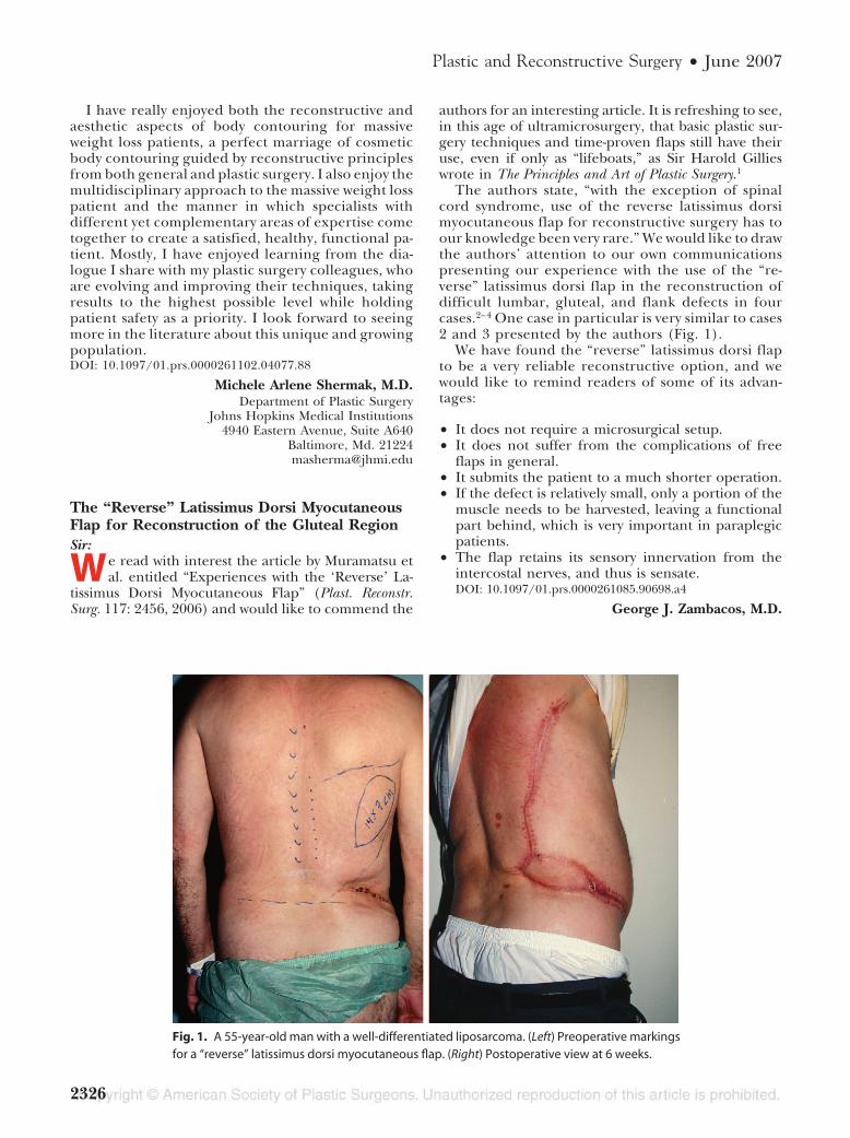
The authors state, “with the exception of spinalcord syndrome, use of the reverse latissimus dorsimyocutaneous flap for reconstructive surgery has toour knowledge been very rare.” We would like to drawthe authors’ attention to our own communicationspresenting our experience with the use of the “re-verse” latissimus dorsi flap in the reconstruction ofdifficult lumbar, gluteal, and flank defects in fourcases.2– 4 One case in particular is very similar to cases2 and 3 presented by the authors (Fig. 1).
We have found the “reverse” latissimus dorsi flapto be a very reliable reconstructive option, and wewould like to remind readers of some of its advan-tages:
• It does not require a microsurgical setup.• It does not suffer from the complications of free
flaps in general.• It submits the patient to a much shorter operation.• If the defect is relatively small, only a portion of the
muscle needs to be harvested, leaving a functionalpart behind, which is very important in paraplegicpatients.
• The flap retains its sensory innervation from theintercostal nerves, and thus is sensate.DOI: 10.1097/01.prs.0000261085.90698.a4
George J. Zambacos, M.D.
Fig. 1. A 55-year-old man with a well-differentiated liposarcoma. (Left) Preoperative markingsfor a “reverse” latissimus dorsi myocutaneous flap. (Right) Postoperative view at 6 weeks.
Plastic and Reconstructive Surgery • June 2007
2326
Apostolos D. Mandrekas, M.D.Artion Plastic Surgery Center
Athens, Greece
Correspondence to Dr. Zambacos27 Skoufa Street
Athens 10673, [email protected]
REFERENCES1. Gillies, H., and Millard, D. R. The Principles and Art of Plastic
Surgery, 1st Ed. Philadelphia: Lippincott Williams & Wilkins,1957. Pp 49-54.
2. Mandrekas, A. D., and Gilbert, P. M. The reverse latissimusdorsi flap for the closure of full thickness defects of the back.Eur J. Plast. Surg. 9: 57, 1986.
3. Zambacos, G. J., and Mandrekas, A. D. The reverse myocu-taneous flap of the latissimus dorsi: An alternative in theflank’s large defect reconstruction (Abstract.) Plast. Reconstr.Surg. 101: 1425, 1998.
4. Zambacos, G. J., and Mandrekas, A. D. The reverse latissimusdorsi flap for lumbar defects. Plast. Reconstr. Surg. 111: 1576,2003.
Free Microdissected Thin Groin Flap Designwith an Extended Vascular Pedicle; ThinAnterolateral Thigh Perforator Flap Using aModified Microdissection TechniqueSir:
We read with great interest two articles recentlypublished in Plastic and Reconstructive Surgery,
namely “Free Microdissected Thin Groin Flap Designwith an Extended Vascular Pedicle”1 and “Thin An-terolateral Thigh Perforator Flap Using a Modified Mi-crodissection Technique and Its Clinical Applicationfor Foot Resurfacing.”2
If microsurgical reconstruction is to be advanced,then it is essential to further refine flap reconstruc-tion procedures in three main ways: to improve thequality of the outcome (both aesthetically and func-tionally) at the recipient site, to reduce overall mor-bidity, and, finally, to simplify procedures to reduceoperative time and broaden applicability. These fac-tors are clearly interdependent in many ways, and thetwo articles beautifully demonstrate elegant techni-cal modifications to permit uniform, radical, primarythinning of skin flaps with low donor-site morbidity.Kimura and Saitoh1 should be particularly com-mended in this regard for their refinement of thegroin flap. In so doing, they minimized secondaryrevisionary procedures, and by enhancing outcomes,they broadened the flap’s indications to encroachinto the traditional territory of the skin graft as thefirst choice for reconstruction.
To facilitate hemostasis, thinning is appropriatelycarried out in both articles before flap transfer, usingthe differing morphologies of the superficial and deepfat layers as a guide to preserving the subdermal vas-
cular plexus by which the thin perforator flap perfuses.For both techniques, the thinning procedure is carriedout using microscope magnification. Kimura andSaitoh1 first confirm the pedicle anatomy, microdis-secting it into the superficial fat before completingelevation of the flap in the superficial fat layer, whereasYang et al.2 raise the flap subfascially and only thin itafter completing proximal pedicle dissection. Bothtechniques have logic, and demonstrable efficacy, butit is our belief that neither technique optimally ad-dresses the need to reduce operative time and simplifyflap procedures to enhance uptake within the recon-structive community, while retaining safety and qualityof outcome.
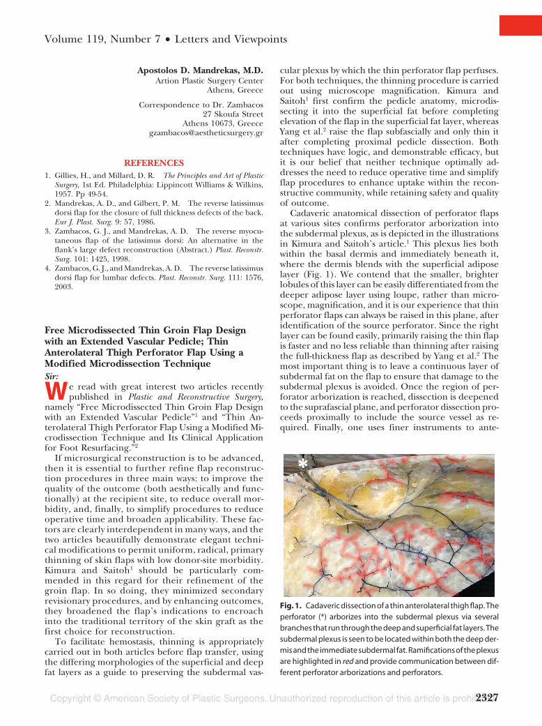
Cadaveric anatomical dissection of perforator flapsat various sites confirms perforator arborization intothe subdermal plexus, as is depicted in the illustrationsin Kimura and Saitoh’s article.1 This plexus lies bothwithin the basal dermis and immediately beneath it,where the dermis blends with the superficial adiposelayer (Fig. 1). We contend that the smaller, brighterlobules of this layer can be easily differentiated from thedeeper adipose layer using loupe, rather than micro-scope, magnification, and it is our experience that thinperforator flaps can always be raised in this plane, afteridentification of the source perforator. Since the rightlayer can be found easily, primarily raising the thin flapis faster and no less reliable than thinning after raisingthe full-thickness flap as described by Yang et al.2 Themost important thing is to leave a continuous layer ofsubdermal fat on the flap to ensure that damage to thesubdermal plexus is avoided. Once the region of per-forator arborization is reached, dissection is deepenedto the suprafascial plane, and perforator dissection pro-ceeds proximally to include the source vessel as re-quired. Finally, one uses finer instruments to ante-
Fig. 1. Cadaveric dissection of a thin anterolateral thigh flap. Theperforator (*) arborizes into the subdermal plexus via severalbranches that run through the deep and superficial fat layers. Thesubdermal plexus is seen to be located within both the deep der-mis and the immediate subdermal fat. Ramifications of the plexusare highlighted in red and provide communication between dif-ferent perforator arborizations and perforators.
Volume 119, Number 7 • Letters and Viewpoints
2327
gradely dissect the perforator out of the residual deepadipose layer before tissue transfer.
Although the removal of fat lobules from betweenthe perforator’s terminal arborizations, as described byYang et al.,2 is technically elegant, we do not believe thatthis is routinely necessary to the application of a thinflap, and by making the procedure excessively exacting,it may compromise widespread uptake of the tech-nique.
Our practice has shown that we are able to raise fromvarious donor sites thin perforator flaps of the samedimensions mentioned by the authors of both articles,but by avoiding the need to use the microscope duringthe raising and thinning of the flap, the procedure ismade shorter and simpler and is more easily learned bythe new surgeon.DOI: 10.1097/01.prs.0000261086.77699.c9
Jorg Dabernig, M.D.
Stuart Watson, F.R.C.S.(Plast.)
Andrew Hart, M.D., Ph.D.Canniesburn Plastic Surgery Unit
Glasgow, United Kingdom
Correspondence to Dr. DabernigCanniesburn Plastic Surgery UnitRoyal Infirmary, Jubilee Building
84 Castle StreetGlasgow G4 0SF, United Kingdom
REFERENCES1. Kimura, N., and Saitoh, M. Free microdissected thin groin
flap design with an extended vascular pedicle. Plast. Reconstr.Surg. 117: 986, 2006.
2. Yang, W. G., Chiang, Y. C., Wei, F. C., Feng, G. M., and Chen,K. T. Thin anterolateral thigh perforator flap using a mod-ified perforator microdissection technique and its clinical ap-plication for foot resurfacing. Plast. Reconstr. Surg. 117: 1004,2006.
ReplySir:
I would like to thank Drs. Dabernig, Watson, andHart for their comments on our article. I certainlyappreciate and am willing to adopt simpler ways to thinthe flap primarily. However, as we did not want to takerisks that might lead to compromise of the flap’s cir-culation, we developed this thinning technique. Oncewe accumulate enough experience, we may modify thistechnique.
While I congratulate their success and agree withmost of the points raised by Drs. Dabernig, Watson, andHart, I strongly believe that loupes and the microscopemake dissection of the small branches of the perfora-tors safer and easier.DOI: 10.1097/01.prs.0000261087.04002.57
Fu-Chan Wei, M.D.Department of Plastic Surgery
Chang Gung Memorial Hospital199 Tung Hwa North Road
Taipei 105, [email protected]@adm.cgmh.org.tw
Is There Any Need to Localize the Perforatorof the Anterolateral Thigh Flap?Sir:
We read with interest the article entitled “Efficacyof the Handheld Doppler in Preoperative Iden-
tification of the Cutaneous Perforators in the An-terolateral Thigh Flap” by Yu and Youssef.1 The per-forator-based free anterolateral thigh flap hasbecome the workhorse of reconstruction, the uncer-tainty of perforator localization notwithstanding. Al-though many methods, including selective angiog-raphy to handheld color Doppler ultrasonography,have been suggested,2– 6 the final word about theirutility is still elusive. The efficacy of handheld Dopp-ler imaging, which provides a “semiquantitative” eval-uation, has been reported differently by differentauthors in their series. Color Doppler has a highsensitivity and specificity but involves expertise andcost and is time-consuming. The big question is,“How important is this preoperative localization?” Areview of the literature clearly shows that the positionof the perforator is more or less constant. In 92percent of cases, the perforator is located within acircle of 3-cm radius drawn around the midpoint ofthe line joining the anterosuperior iliac spine andthe superolateral border of the patella, and nearly allperforators will lie in a circle of 5-cm radius drawnaround the same aforementioned point.2–5 Yu andYoussef agree with this and have designated the ABCsystem based on this principle. The authors also re-port that in 20 cases with no preoperative localizationthere was no flap failure.
One would agree that the anterolateral thigh flapis utilized mainly for defects larger than 10 � 10 cm.A skin island of this dimension will undoubtedly havethe perforator included if the ABC system describedby the authors is followed. It is obvious, therefore,that routine mapping of the perforator with any de-vice is not required. However, we may need to local-ize the perforator preoperatively in the followingsituations:
1. When a flap less than 10 � 10 cm is needed.2. When a thin or an adiposofascial anterolateral
thigh flap is needed.3. When a chimeric anterolateral thigh flap is de-
signed such that the position of skin paddle isprefixed.
DOI: 10.1097/01.prs.0000261088.08288.29
Ramesh Kumar Sharma, M.Ch.(Plast.Surg.)
Plastic and Reconstructive Surgery • June 2007
2328
Puneet Tuli, M.Ch.(Plast.Surg.)Department of Plastic Surgery
Postgraduate Institute of Medical Education and ResearchChandigarh, India
Correspondence to Dr. SharmaDepartment of Plastic Surgery
Postgraduate Institute of Medical Education and ResearchSector 12
Chandigarh, Pin 160012, [email protected]
REFERENCES1. Yu, P., and Youssef, A. Efficacy of the handheld Doppler
in preoperative identification of the cutaneous perforatorsin the anterolateral thigh flap. Plast. Reconstr. Surg 118: 928,2006.
2. Xu, D.-C., Zhong, S. Z., Kong, J. M., et al. Applied anatomy ofthe anterolateral femoral flap. Plast. Reconstr. Surg. 82: 305,1988.
3. Wei, F.-C., Jain, V., Celik, N., Chen, H. C., Chuang, D. C., andLin, C. H. Have we found and ideal soft tissue flap? An ex-perience with 672 anterolateral thigh flaps. Plast. Reconstr.Surg. 109: 2219, 2002.
4. Kuo, Y. R., Seng-Feng, J., Kuo, F. M., et al. Versatility of thefree anterolateral thigh flap for reconstruction of soft tis-sue defects: Review of 140 cases. Ann. Plast. Surg. 48:161, 2002.
5. Hallock, G. G. Doppler sonography and color duplex im-aging for planning a perforators flap. Clin. Plast. Surg. 30: 347,2003.
6. Yildirim, S., Avci, G., and Akoz, T. Soft-tissue reconstructionusing a free anterolateral thigh flap: Experience with 28 pa-tients. Ann. Plast. Surg. 51: 37, 2003.
ReplySir:
In our article titled “Efficacy of the HandheldDoppler in Preoperative Identification of the Cuta-neous Perforators in the Anterolateral Thigh Flap,”1
my coauthor and I concluded that the handheldDoppler devices were not very accurate in localizingthe cutaneous perforators in an American popula-tion and that the anterolateral thigh flap could beraised safely based on the “ABC” system without pre-operative Doppler examination. Since that study, Ihave performed another 100 anterolateral thigh freeflap procedures successfully without preoperativeDoppler examination and feel very comfortable withthe flap. Drs. Sharma and Tuli seem to agree with thisin their letter and suggest that preoperative local-ization may be needed in the following three cir-cumstances: a flap less than 10 � 10 cm, a thin oradipofascial flap, and a multi-island (chimeric) an-terolateral thigh flap.
A flap less than 10 � 10 cm. The overwhelmingmajority of anterolateral thigh flap procedures I haveperformed, including the ones in the study, involvedflaps that were less than 10 cm wide (usually between6 and 9.5 cm). The length of the flap is less impor-tant, since a longer incision toward the groin will be
needed to dissect out the main vascular pedicle (un-less the perforator itself is used as the main pedicle)and the dog-ear will need to be removed to facilitateprimary closure. If I have to suggest a cutoff for theflap width when preoperative Doppler localization isneeded, I would say it is 6 cm. I usually position theanterior (medial) incision 1 or 2 cm medial to theanteroposterior line depending on the width of theflap needed and explore the cutaneous perforatorsfirst before making the posterior incision. I shouldhave a 97 percent or higher success rate this way,since 97 percent of the perforators are located lateralto the anteroposterior line. For example, if the per-forator is located 3 cm lateral to the anteroposteriorline (an extreme case) and the anterior incision is 1cm medial to the anteroposterior line, I can stillsafely raise a 5-cm-wide flap with the posterior inci-sion 1 cm outside the perforator, which is safe for asmall flap. To be safer, one can make a 6-cm-wide flap(with the posterior incision 2 cm outside the perfo-rator) and trim the anterior edge of the flap to thedesired size. Primary closure should be possible fora 6-cm-wide defect. I always place a 5-0 Prolene sutureon the skin where the actual perforator is located sothat I know exactly where to trim during flap inset-ting. An ink mark may disappear easily. I would notmake the anterior incision lateral to the anteropos-terior line even if I have a good Doppler signal,because it may not be accurate. Making an incisionmedial to the anteroposterior line, as mentionedabove, is much safer and has nothing to lose. Thatway, I really do not need preoperative localization.However, the accuracy of Doppler examination in-creases with the decrease in body mass index and thethickness of the anterolateral flap. The flap thicknessin my series doubles that of an Asian population.2
Therefore, Doppler examination may be much moreaccurate in Asian patients with a thin thigh, where afreestyle free flap can easily be designed. Also be-ginners might feel more comfortable getting a Dopp-ler signal preoperatively before raising the flap.
A thin or an adipofascial flap. I think the authors meanta “thinned” flap. When flap thinning is required, it iscrucial to know the exact location of the cutaneousperforators. Unfortunately, preoperative localization iseven less accurate in a thick thigh. Therefore, I wouldnot trust the Doppler in planning a flap thinning. Usingthe aforementioned method (placing the anterior in-cision medial to the anteroposterior line) to surgicallylocate the perforators is a safer approach, in my opin-ion. If only an adipofascial flap is needed, it is unnec-essary to preoperatively locate the perforators since noskin is taken. I use the same approach to identify theperforators and then dissect the adipofascial tissue offthe skin starting medially.
A chimeric (multi-island) flap. Use of a chimeric flapreally depends on the accuracy of the localizationmethod. If you feel confident about the preoperativelocalization, you can design the flap accordingly. SinceI have not found a reliable and practical way to locate
Volume 119, Number 7 • Letters and Viewpoints
2329
the perforators preoperatively in our patient popula-tion, I rely more on surgical exploration, as mentionedabove. Once the perforators are directly visualized, youcan design multi-islands in a free style to meet yourneeds. I would try to be more flexible in flap design (not“prefixed”) and be prepared for any possible variations.DOI: 10.1097/01.prs.0000261089.60877.ae
Peirong Yu, M.D.Department of Plastic Surgery
University of Texas M. D. Anderson Cancer Center1400 Holcombe Boulevard, FC8.2000
Houston, Texas [email protected]
REFERENCES1. Yu, P., and Youssef, A. Efficacy of the handheld Doppler
in preoperative identification of the cutaneous perforatorsin the anterolateral thigh flap. Plast. Reconstr. Surg. 118: 928,2006.
2. Yu, P. Characteristics of the anterolateral thigh flap in aWestern population and its application in head and neckreconstruction. Head Neck 26: 759, 2004.
Full-Thickness Skin Graft as a One-StageDebulking Procedure after Free FlapReconstruction for the Lower LegSir:
We read with interest the article entitled “Full-Thickness Skin Graft as a One-Stage Debulking
Procedure after Free Flap Reconstruction for theLower Leg,” by Lin and Jeng.1 The authors have sug-gested that all of the skin should be taken off and thenre-applied as a full-thickness skin graft. We have beenthinning these flaps by raising the skin flap at the sub-dermal level as with a full-thickness skin graft, but main-taining the flap attachment along one suture line so asto make it a “pedicled full-thickness skin graft.” Thistechnique allows the desired contouring and also helpsmaintain the blood supply to the superthin “pedicledfull-thickness skin graft flap” through the attachededge, thereby improving the survival/take of the “flap.”Second, this permits better draping of the skin as theattached end provides a fixed reference point. Third,flap thinning is much easier as the attached edge actslike a hinge and the raised “flap” can be spread over asponge.
The authors have also suggested that the secondoperation (i.e., thinning) can be done even at an earlypoint in time [two cases at 1 month, three cases at 2months, and eight cases at 3 months (Table 1 in arti-cle)]. We also believe that a waiting period of about 3months before flap thinning is worthwhile, as massageduring this period significantly decreases edema andthe flap becomes supple.
Finally, the authors’ apprehension regarding an-other operation after liposuction of the flap for ex-cision of redundant skin seems to be an undue con-cern. We think that excision of the redundant skin
can be combined with the primary liposuction pro-cedure, and this has also been reported in literature.2
DOI: 10.1097/01.prs.0000261090.58038.13
Ramesh Kumar Sharma, M.Ch.(Plast.Surg.)
Puneet Tuli, M.Ch.(Plast.Surg.)
Surinder Singh Makkar, M.Ch.(Plast.Surg.)Department of Plastic Surgery
Postgraduate Institute of Medical Education and ResearchChandigarh India
Correspondence to Dr. SharmaDepartment of Plastic Surgery
Postgraduate Institute of Medical Education and ResearchChandigarh, Pin 160012, India
REFERENCES1. Lin, T. S., and Jeng, S. F. Full-thickness skin graft as a one-
stage debulking procedure after free flap reconstruction forthe lower leg. Plast. Reconstr. Surg. 118: 408, 2006.
2. Hallock, G. G. Defatting of flaps by means of suction-assistedlipectomy. Plast. Reconstr. Surg 76: 948, 1985.
ReplySir:
We acknowledge Sharma et al.’s procedure of flapdebulking, as well as their comments regarding ourpublished article, “Full-Thickness Skin Graft as a One-Stage Debulking Procedure after Free Flap Reconstruc-tion for the Lower Leg” (Plast. Reconstr. Surg. 118: 408,2006). We think Sharma et al.’s debulking procedureas described is similar to the conventional method witha more aggressive intent in thinning the flaps. Althoughmaintaining the thinned skin flap along one suture linemakes the flap easier to handle, because the attachededge acts like a hinge, this procedure is dangerous forthe flap’s distal circulation, where manipulation maylead to flap necrosis, especially in larger skin flaps. Atrue full-thickness skin graft following a flap debulkingprocedure makes reshaping and contouring of the flapeasier in the ankle and foot areas, because these arethree-dimensional structures.
A longer waiting period before a debulking proce-dure is not a prerequisite in our method. The mainblood supply to the full-thickness skin graft comes fromthe fascia layer and not from the wound edges.
Liposuction as a debulking procedure may be help-ful in other areas, but it cannot provide a very thin skinenvelope for the ankle and foot. Although the redun-dant skin after liposuction can be partially removedduring the same primary liposuction procedure, thecircumferential scar should be revised in another pro-cedure.DOI: 10.1097/01.prs.0000261091.19290.0a
Tsan-Shiun Lin, M.D.
Plastic and Reconstructive Surgery • June 2007
2330
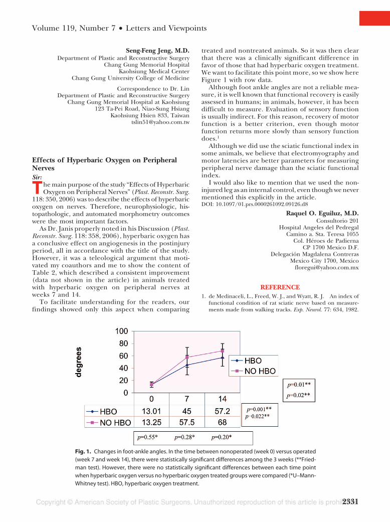
Seng-Feng Jeng, M.D.Department of Plastic and Reconstructive Surgery
Chang Gung Memorial HospitalKaohsiung Medical Center
Chang Gung University College of Medicine
Correspondence to Dr. LinDepartment of Plastic and Reconstructive Surgery
Chang Gung Memorial Hospital at Kaohsiung123 Ta-Pei Road, Niao-Sung Hsiang
Kaohsiung Hsien 833, [email protected]
Effects of Hyperbaric Oxygen on PeripheralNervesSir:
The main purpose of the study “Effects of HyperbaricOxygen on Peripheral Nerves” (Plast. Reconstr. Surg.
118: 350, 2006) was to describe the effects of hyperbaricoxygen on nerves. Therefore, neurophysiologic, his-topathologic, and automated morphometry outcomeswere the most important factors.
As Dr. Janis properly noted in his Discussion (Plast.Reconstr. Surg. 118: 358, 2006), hyperbaric oxygen hasa conclusive effect on angiogenesis in the postinjuryperiod, all in accordance with the title of the study.However, it was a teleological argument that moti-vated my coauthors and me to show the content ofTable 2, which described a consistent improvement(data not shown in the article) in animals treatedwith hyperbaric oxygen on peripheral nerves atweeks 7 and 14.
To facilitate understanding for the readers, ourfindings showed only this aspect when comparing
treated and nontreated animals. So it was then clearthat there was a clinically significant difference infavor of those that had hyperbaric oxygen treatment.We want to facilitate this point more, so we show hereFigure 1 with row data.
Although foot ankle angles are not a reliable mea-sure, it is well known that functional recovery is easilyassessed in humans; in animals, however, it has beendifficult to measure. Evaluation of sensory functionis usually indirect. For this reason, recovery of motorfunction is a better criterion, even though motorfunction returns more slowly than sensory functiondoes.1
Although we did use the sciatic functional index insome animals, we believe that electromyography andmotor latencies are better parameters for measuringperipheral nerve damage than the sciatic functionalindex.
I would also like to mention that we used the non-injured leg as an internal control, even though we nevermentioned this explicitly in the article.DOI: 10.1097/01.prs.0000261092.09126.d8
Raquel O. Eguiluz, M.D.Consultorio 201
Hospital Angeles del PedregalCamino a. Sta. Teresa 1055
Col. Heroes de PadiernaCP 1700 Mexico D.F.
Delegacion Magdalena ContrerasMexico City 1700, Mexico
REFERENCE1. de Medinaceli, L., Freed, W. J., and Wyatt, R. J. An index of
functional condition of rat sciatic nerve based on measure-ments made from walking tracks. Exp. Neurol. 77: 634, 1982.
Fig. 1. Changes in foot-ankle angles. In the time between nonoperated (week 0) versus operated(week 7 and week 14), there were statistically significant differences among the 3 weeks (**Fried-man test). However, there were no statistically significant differences between each time pointwhen hyperbaric oxygen versus no hyperbaric oxygen treated groups were compared (*U–Mann-Whitney test). HBO, hyperbaric oxygen treatment.
Volume 119, Number 7 • Letters and Viewpoints
2331
Update on Office-Based Surgery Regulations inNew York StateSir:
A fter reading “Office Surgery Safety: The Myths andTruths behind the Florida Moratoria---Six Years of
Florida Data,” by Clayman and Seagle1 and the Discus-sion by Guy2 in the September 1, 2006, issue of theJournal, I felt that the Journal’s readers should knowabout New York’s progress toward regulation of office-based surgery.
In the fall of 2005, the commissioner of health, An-tonia C. Novello, M.D., M.P.H., Dr.P.H., reconvenedthe Committee on Quality Assurance in Office-BasedSurgery. The committee was reconstituted with addi-tional members, because the number of office-basedsurgery procedures had increased and New York Statehad experienced several high-profile cases of medical mis-conduct in the office-based surgical setting in the previousfew years, raising increasing concern for public safety. Thecommittee was originally set up in late 1997 under theauspices of the New York State Public Health Council. Itwas composed of individuals representing various special-ties, including anesthesiology, internal medicine, oph-thalmology, general surgery, orthopedics, and dentistry,and professional organizations and consumers. The com-mittee developed clinical guidelines that physicians, den-tists, and podiatrists could use in establishing and oper-ating office-based surgery practices. The guidelines werefinalized in December of 2000 and included recommen-dations for the environment of care and safe provision ofanesthesia services by trained and qualified personnel.The guidelines also included recommendations for thehiring and privileging of staff, office procedures, medicalrecord keeping, infection control techniques, informedconsent, and the maintenance of equipment and oper-ating rooms. These guidelines can be found at http://www.health.state.ny.us/nysdoh/obs/toc.htm.
Distribution of the guidelines was suspended in 2001due to a legal challenge by the New York State Asso-ciation of Nurse Anesthetists. During this time, theDepartment of Health was enjoined from publishingand distributing the guidelines. In March of 2004, theNew York Court of Appeals ruled in favor of the NewYork State Department of Health and the departmentreissued the guidelines.
At the first meeting of the reconstituted committeeon October 11, 2005, Commissioner Novello outlinedseveral areas for which she requested recommenda-tions be developed:
1. Identify and track adverse events in the office-based setting;
2. Review what additional data are required to dis-cover, evaluate, and prevent adverse events, suchas transfer to the emergency department of pa-tients following office-based surgical care;
3. Study the potential risks and complications associatedwith multiple surgical procedures being performedon a single patient during the same day; and
4. Make any additional recommendations the com-mittee deems necessary to improve the quality ofcare in office-based surgical practices, includingrecommendations to increase consumer aware-ness in this area.3
This updated committee added two practicing plasticsurgeons besides myself as members, as well as a derma-tologist. There were many meetings, much heated dis-cussion, and reports from all three nationally accreditedcertifying agencies–-the Accreditation Association for Am-bulatory Health Care, the Joint Commission on Accred-itation of Healthcare Organizations, and the AmericanAssociation for Accreditation of Ambulatory Surgery Fa-cilities, Inc.–-as well as reports from representatives ofboth Florida and California regarding their office-basedsurgery issues. In addition, there were comments fromsuch groups as the American College of Surgeons and theMedical Liability Mutual Insurance Company (the majormalpractice carrier in New York). After these presenta-tions, the committee reached consensus and presented afinal report to the Public Health Council at their meetingin July of 2006.
The report contained the following suggestions andlegislative wording to accomplish the following:
• Physicians operating in office-based surgery settingswhere anything other than local anesthesia is usedneed to have their facility certified by a Department ofHealth–approved, nationally accredited certifyingagency. At the moment, the American Association forAccreditation of Ambulatory Surgery Facilities, Inc.,Accreditation Association for Ambulatory Health Care,and Joint Commission on Accreditation of HealthcareOrganizations are all approved certifying agencies.
• Surgery under local anesthesia only and liposuc-tion, only if the total volume is less than 500 cc, canbe done in a nonaccredited facility.
• Adverse events (deaths, hospital transfers and ad-missions, and other serious occurrences) need to bereported directly to the Department of Health.
• Physicians performing surgery in an office settingneed to have hospital privileges in the specialtypracticed in the office-based surgery setting. Theprocedures the physician performs should be onesgenerally recognized by the American Board ofMedical Specialties or its American Osteopathic As-sociation counterpart as falling within the scope ofpractice of the physician providing the care.
• Physicians performing office-based surgery shouldbe board certified by the American Board of Medi-cal Specialties or the American Osteopathic Associ-ation counterpart.
• Physicians performing office-based surgery need tohave a transfer agreement with a hospital or an-other physician with admitting privileges at a nearbyhospital.
• When performing multiple procedures, guidelinesand/or recommendations from the appropriate spe-cialty/subspecialty should be followed.
Plastic and Reconstructive Surgery • June 2007
2332
The teeth in these changes will be the enactment ofthe regulations by the state legislature. The wording hasbeen set and we are awaiting enactment of these reg-ulations. Here, it will be a violation of the public healthlaw not to follow the above changes and the physician’slicense can be summarily suspended. For plastic sur-geons who are members of the American Society ofPlastic Surgeons or the American Society for AestheticPlastic Surgery, these regulations should not pose aproblem; however, for many of our physician col-leagues, these regulations will require them to havetheir surgical facilities approved.
The committee did an excellent job of simplifyingrules for office-based surgery that are meant to helpensure patient safety without overburdening the phy-sician with duplicative rules. Competent, caring doctorsshould have no problem following the rules. Will thishelp the public? I hope so; however, we need to educatepatients so that when they come in for a consultationand ask “How much does it cost,” they should also ask,“Will this be done in an accredited facility.” We arecurrently awaiting the final enactment of legislation tobring this work to fruition.DOI: 10.1097/01.prs.0000261118.49909.13
William B. Rosenblatt, M.D.Lenox Hill Plastic Surgery Center
308 East 79th StreetNew York, N.Y. 10021
REFERENCES1. Clayman, M. A., and Seagle, B. M Office surgery safety: The
myths and truths behind the Florida moratoria–-Six years ofFlorida data. Plast. Reconstr. Surg. 118: 777, 2006.
2. Guy, R. J. Office surgery safety: The myths and truths behindthe Florida moratoria–-Six years of Florida data (Discussion).Plast. Reconstr. Surg. 118: 786, 2006.
3. Clinical guidelines for office-based surgery issued December2000. Available at http://www.health.state.ny.us/nysdoh/obs/toc.htm
ReplySir:
Dr. Rosenblatt should be congratulated for bringingto light what is currently happening in New York Statein the realm of office surgery. His letter is well received,and gives us the opportunity to provide an even deeperunderstanding of how important patient safety, medi-cal error reduction, and patient education are withinthe office surgery setting.
Although there has been considerable media con-troversy over the years regarding reports of decreasedpatient safety and errors that have occurred in theoffice surgery setting, the majority of data across variousspecialties demonstrate a very low incidence of adverseevents resulting from office-based surgery. The regu-latory changes stated within Dr. Rosenblatt’s letter ap-pear to have simplified the rules for office-based sur-gery in his state, but there are still some deficienciesthat may hinder patient safety.
I agree with the majority of the regulations noted inthe letter, but we should look closer at certain situa-tions. For instance, allowing surgery under local anes-thesia with the use of oral sedation and/or the removalof less than 500 cc of lipoaspirate in a nonaccreditedfacility will allow physicians and nonphysicians to per-form procedures outside the scope of their training.Not regulating this aspect of office procedures putspatient safety at risk when procedures are performed bya physician who advertises certain procedures andclaims to be trained by a non—Accreditation Councilfor Graduate Medical Education–approved cosmetic oraesthetic board. The uneducated or uninformed pa-tient might undergo surgery performed by a physicianwith no surgical training whatsoever or by someonewho is trained only to operate on one aspect of a pa-tient’s body and is only familiar with taking care ofcomplications located to that specific area.
Also, if the Department of Health is to allow physiciansto perform procedures only within their scope of trainingand make it necessary for the doctor to have hospitalprivileges in the same specialty practiced within the officesetting, the question remains of what the Department ofHealth does when a dentist, oral surgeon, family practi-tioner, endocrinologist, or other practitioner performssurgery of the breast, body contouring, or other proce-dures outside their scope of training in an accreditedfacility? In the state of Florida, all procedures performedin an accredited office facility are reported to the boardof medicine. This allows the state to identify which sur-geons are performing which type of procedures, but itdoes not regulate them to the scope of training of thephysician at this time. For example, oral surgeons in thestate of Florida are performing transumbilical breast aug-mentation and abdominoplasty without regulation at thistime, but it remains unclear as to whether or not sedationunder dental accreditation is regulated in a similar fash-ion or whether some other loophole exists. It also remainsunclear whether the decision in California, allowing theAmerican Board of Cosmetic Surgery to be consideredequivalent to boards approved by the Accreditation Coun-cil for Graduate Medical Education, will change the de-cisions of hospitals in the future, allowing privileges basedon this non–-Accreditation Council for Graduate MedicalEducation–approved board. Will this take effect in otherstates?
What we do know is that we must continue to create aculture of patient safety in our specialty, with patient edu-cation and surgeon vigilance. The ultimate judgment re-garding the care of a patient lies with the physician. Physi-cians must consider the risk factors associated with certainprocedures, such as patient selection, length of the proce-dure, comorbidities, deep vein thrombosis prophylaxis, andso on, when deciding whether such procedures should beperformed in an accredited office-based facility, outpatientsurgery center, or hospital-based facility.
One of the remaining challenges is the need to con-tinually improve this culture of safety by emphasizingthe need for continued patient education specificallyrelated to aesthetic procedures administered in non-
Volume 119, Number 7 • Letters and Viewpoints
2333
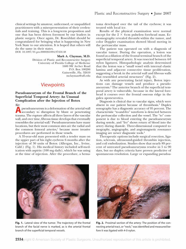
clinical settings by amateur, unlicensed, or unqualifiedpractitioners with a misrepresentation of their creden-tials and training. This is a long-term proposition andone that has been driven foremost by our leaders inplastic surgery. Once again, Dr. Rosenblatt should becongratulated for bringing what is happening in NewYork State to our attention. It is hoped that others willdo the same in their states.DOI: 10.1097/01.prs.0000261094.97450.48
Mark A. Clayman, M.D.Division of Plastic and Reconstructive Surgery
University of Florida College of Medicine1600 SW Archer Road
P.O. Box 100286Gainesville, Fla. 32610
Viewpoints
Pseudoaneurysm of the Frontal Branch of theSuperficial Temporal Artery: An UnusualComplication after the Injection of BotoxSir:
Apseudoaneurysm is a deformation of the arterial wallsecondary to disruption by blunt or penetrating
trauma. The rupture affects all three layers of the vascularwall, and over time, fibrous tissue develops that eventuallyresembles the arterial wall.1 Pseudoaneurysms have manylocations, but their most common sites are the radial andthe common femoral arteries,1 because more invasiveprocedures are performed in those vessels.
A 35-year-old man presented with a tender mass onthe upper part of his right eyebrow 6 months after theinjection of 30 units of Botox (Allergan, Inc., Irvine,Calif.) (Fig. 1). His medical history included self-med-ication with aspirin (100 mg daily), which he was usingat the time of injection. After the procedure, a hema-
toma developed over the tail of the eyebrow; it wastreated with local ice.
Results of the physical examination were normalexcept for the 2 � 4-cm pulseless forehead mass. Ec-otomography revealed thrombi within the tumor, andcolor Doppler examination showed blood moving inthe perivascular mass.
The patient was operated on with a diagnosis ofvascular tumor. During the operation, a lesion wasfound as a dilation of the frontal terminal branch of thesuperficial temporal artery. It was resected between 4-0nylon ligatures. Histopathologic analysis determinedthat the lesion was a “small vessel with internal elasticlamina and adjacent connective tissue proliferation,suggesting a break in the arterial wall and fibrous wallsthat resembled arterial structures” (Fig. 2).
As with any penetrating facial injury, Botox injec-tions can damage vessels and produce a pseudo-aneurysm.2 The anterior branch of the superficial tem-poral artery is vulnerable, because in the lateral fore-head it courses over the frontal osseous ridge in thegalea aponeurotica.
Diagnosis is clinical due to vascular signs, which wereabsent in our patient because of thrombosis.3 Duplexsonography has a diagnostic accuracy of 95 percent. Thecharacteristic “to-and-fro” waveform is detected betweenthe perivascular collection and the vessel. The “to” com-ponent is due to blood entering the pseudoaneurysmduring systole, and “fro” shows return of blood into theartery during diastole. Three-dimensional computed to-mography, angiography, and angiomagnetic resonanceimaging are newer diagnostic tools.4
Therapeutic options include surgical resection, liga-tion, sclerosis, ultrasound-guided thrombin injection,and coil embolization. Studies show that nearly 89 per-cent of untreated pseudoaneurysms resolve in 5 to 90days, but no duplex criteria have proven predictive ofspontaneous resolution. Large or expanding pseudoa-
Fig. 1. Lateral view of the tumor. The trajectory of the frontalbranch of the facial nerve is marked, as is the arterial frontalbranch of the superficial temporal vessels.
Fig. 2. Proximal section of the artery: The position of the con-necting arterial tract, or “neck,” was identified and measured be-fore it was ligated with 4-0 nylon.
Plastic and Reconstructive Surgery • June 2007
2334
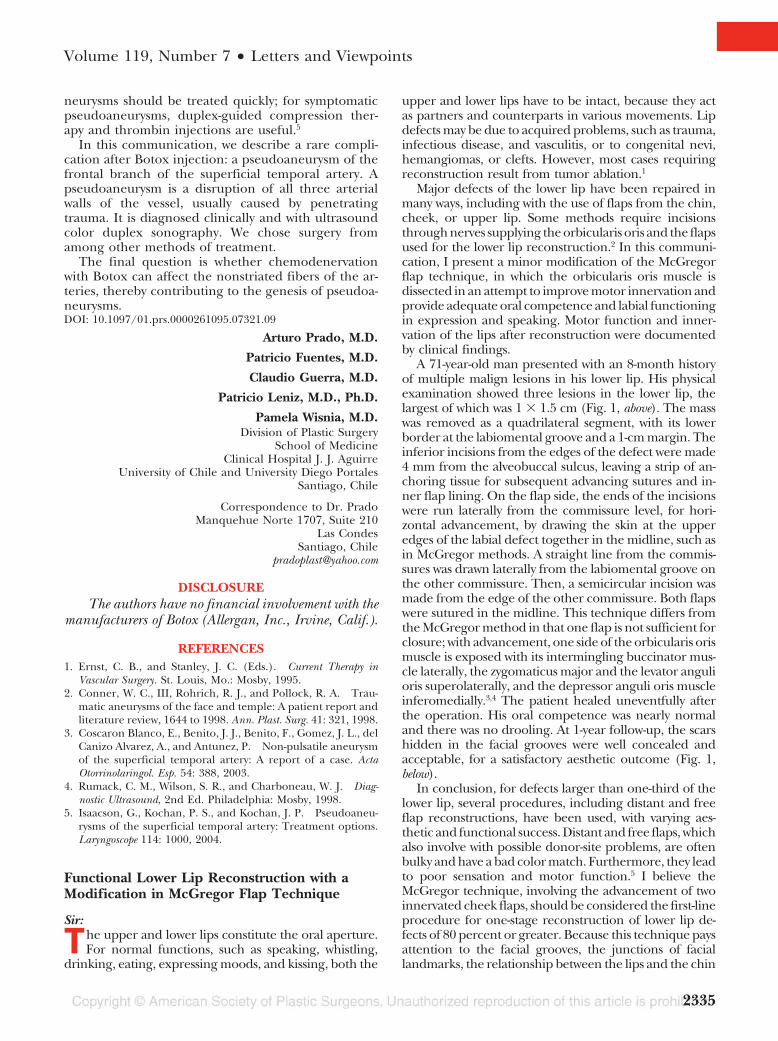
neurysms should be treated quickly; for symptomaticpseudoaneurysms, duplex-guided compression ther-apy and thrombin injections are useful.5
In this communication, we describe a rare compli-cation after Botox injection: a pseudoaneurysm of thefrontal branch of the superficial temporal artery. Apseudoaneurysm is a disruption of all three arterialwalls of the vessel, usually caused by penetratingtrauma. It is diagnosed clinically and with ultrasoundcolor duplex sonography. We chose surgery fromamong other methods of treatment.
The final question is whether chemodenervationwith Botox can affect the nonstriated fibers of the ar-teries, thereby contributing to the genesis of pseudoa-neurysms.DOI: 10.1097/01.prs.0000261095.07321.09
Arturo Prado, M.D.
Patricio Fuentes, M.D.
Claudio Guerra, M.D.
Patricio Leniz, M.D., Ph.D.
Pamela Wisnia, M.D.Division of Plastic Surgery
School of MedicineClinical Hospital J. J. Aguirre
University of Chile and University Diego PortalesSantiago, Chile
Correspondence to Dr. PradoManquehue Norte 1707, Suite 210
Las CondesSantiago, Chile
DISCLOSUREThe authors have no financial involvement with the
manufacturers of Botox (Allergan, Inc., Irvine, Calif.).
REFERENCES1. Ernst, C. B., and Stanley, J. C. (Eds.). Current Therapy in
Vascular Surgery. St. Louis, Mo.: Mosby, 1995.2. Conner, W. C., III, Rohrich, R. J., and Pollock, R. A. Trau-
matic aneurysms of the face and temple: A patient report andliterature review, 1644 to 1998. Ann. Plast. Surg. 41: 321, 1998.
3. Coscaron Blanco, E., Benito, J. J., Benito, F., Gomez, J. L., delCanizo Alvarez, A., and Antunez, P. Non-pulsatile aneurysmof the superficial temporal artery: A report of a case. ActaOtorrinolaringol. Esp. 54: 388, 2003.
4. Rumack, C. M., Wilson, S. R., and Charboneau, W. J. Diag-nostic Ultrasound, 2nd Ed. Philadelphia: Mosby, 1998.
5. Isaacson, G., Kochan, P. S., and Kochan, J. P. Pseudoaneu-rysms of the superficial temporal artery: Treatment options.Laryngoscope 114: 1000, 2004.
Functional Lower Lip Reconstruction with aModification in McGregor Flap Technique
Sir:
The upper and lower lips constitute the oral aperture.For normal functions, such as speaking, whistling,
drinking, eating, expressing moods, and kissing, both the
upper and lower lips have to be intact, because they actas partners and counterparts in various movements. Lipdefects may be due to acquired problems, such as trauma,infectious disease, and vasculitis, or to congenital nevi,hemangiomas, or clefts. However, most cases requiringreconstruction result from tumor ablation.1
Major defects of the lower lip have been repaired inmany ways, including with the use of flaps from the chin,cheek, or upper lip. Some methods require incisionsthrough nerves supplying the orbicularis oris and the flapsused for the lower lip reconstruction.2 In this communi-cation, I present a minor modification of the McGregorflap technique, in which the orbicularis oris muscle isdissected in an attempt to improve motor innervation andprovide adequate oral competence and labial functioningin expression and speaking. Motor function and inner-vation of the lips after reconstruction were documentedby clinical findings.
A 71-year-old man presented with an 8-month historyof multiple malign lesions in his lower lip. His physicalexamination showed three lesions in the lower lip, thelargest of which was 1 � 1.5 cm (Fig. 1, above). The masswas removed as a quadrilateral segment, with its lowerborder at the labiomental groove and a 1-cm margin. Theinferior incisions from the edges of the defect were made4 mm from the alveobuccal sulcus, leaving a strip of an-choring tissue for subsequent advancing sutures and in-ner flap lining. On the flap side, the ends of the incisionswere run laterally from the commissure level, for hori-zontal advancement, by drawing the skin at the upperedges of the labial defect together in the midline, such asin McGregor methods. A straight line from the commis-sures was drawn laterally from the labiomental groove onthe other commissure. Then, a semicircular incision wasmade from the edge of the other commissure. Both flapswere sutured in the midline. This technique differs fromthe McGregor method in that one flap is not sufficient forclosure; with advancement, one side of the orbicularis orismuscle is exposed with its intermingling buccinator mus-cle laterally, the zygomaticus major and the levator angulioris superolaterally, and the depressor anguli oris muscleinferomedially.3,4 The patient healed uneventfully afterthe operation. His oral competence was nearly normaland there was no drooling. At 1-year follow-up, the scarshidden in the facial grooves were well concealed andacceptable, for a satisfactory aesthetic outcome (Fig. 1,below).
In conclusion, for defects larger than one-third of thelower lip, several procedures, including distant and freeflap reconstructions, have been used, with varying aes-thetic and functional success. Distant and free flaps, whichalso involve with possible donor-site problems, are oftenbulky and have a bad color match. Furthermore, they leadto poor sensation and motor function.5 I believe theMcGregor technique, involving the advancement of twoinnervated cheek flaps, should be considered the first-lineprocedure for one-stage reconstruction of lower lip de-fects of 80 percent or greater. Because this technique paysattention to the facial grooves, the junctions of faciallandmarks, the relationship between the lips and the chin
Volume 119, Number 7 • Letters and Viewpoints
2335
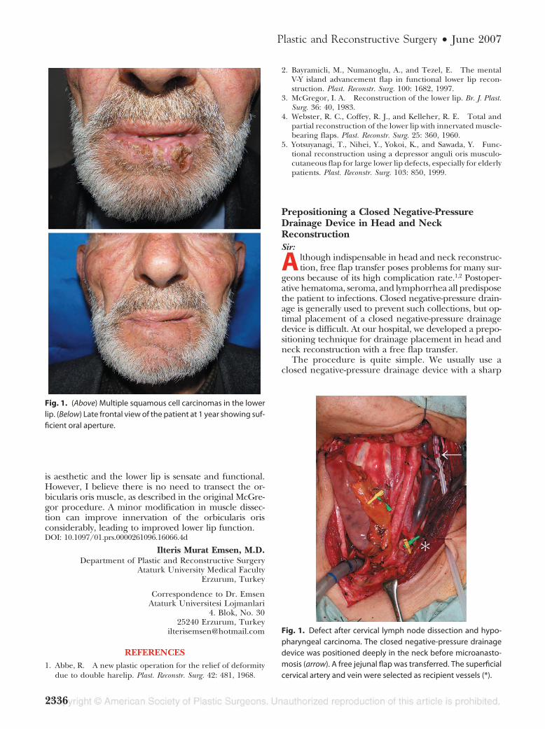
is aesthetic and the lower lip is sensate and functional.However, I believe there is no need to transect the or-bicularis oris muscle, as described in the original McGre-gor procedure. A minor modification in muscle dissec-tion can improve innervation of the orbicularis orisconsiderably, leading to improved lower lip function.DOI: 10.1097/01.prs.0000261096.16066.4d
Ilteris Murat Emsen, M.D.Department of Plastic and Reconstructive Surgery
Ataturk University Medical FacultyErzurum, Turkey
Correspondence to Dr. EmsenAtaturk Universitesi Lojmanlari
4. Blok, No. 3025240 Erzurum, Turkey
REFERENCES1. Abbe, R. A new plastic operation for the relief of deformity
due to double harelip. Plast. Reconstr. Surg. 42: 481, 1968.
2. Bayramicli, M., Numanoglu, A., and Tezel, E. The mentalV-Y island advancement flap in functional lower lip recon-struction. Plast. Reconstr. Surg. 100: 1682, 1997.
3. McGregor, I. A. Reconstruction of the lower lip. Br. J. Plast.Surg. 36: 40, 1983.
4. Webster, R. C., Coffey, R. J., and Kelleher, R. E. Total andpartial reconstruction of the lower lip with innervated muscle-bearing flaps. Plast. Reconstr. Surg. 25: 360, 1960.
5. Yotsuyanagi, T., Nihei, Y., Yokoi, K., and Sawada, Y. Func-tional reconstruction using a depressor anguli oris musculo-cutaneous flap for large lower lip defects, especially for elderlypatients. Plast. Reconstr. Surg. 103: 850, 1999.
Prepositioning a Closed Negative-PressureDrainage Device in Head and NeckReconstructionSir:
A lthough indispensable in head and neck reconstruc-tion, free flap transfer poses problems for many sur-
geons because of its high complication rate.1,2 Postoper-ative hematoma, seroma, and lymphorrhea all predisposethe patient to infections. Closed negative-pressure drain-age is generally used to prevent such collections, but op-timal placement of a closed negative-pressure drainagedevice is difficult. At our hospital, we developed a prepo-sitioning technique for drainage placement in head andneck reconstruction with a free flap transfer.
The procedure is quite simple. We usually use aclosed negative-pressure drainage device with a sharp
Fig. 1. (Above) Multiple squamous cell carcinomas in the lowerlip. (Below) Late frontal view of the patient at 1 year showing suf-ficient oral aperture.
Fig. 1. Defect after cervical lymph node dissection and hypo-pharyngeal carcinoma. The closed negative-pressure drainagedevice was positioned deeply in the neck before microanasto-mosis (arrow). A free jejunal flap was transferred. The superficialcervical artery and vein were selected as recipient vessels (*).
Plastic and Reconstructive Surgery • June 2007
2336
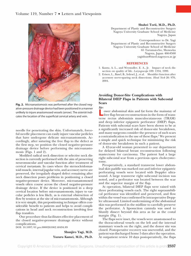
needle for penetrating the skin. Unfortunately, force-ful needle placement can easily injure vascular pediclesthat have undergone delicate microanastomosis. Ac-cordingly, after suturing the free flap to the defect asthe first step, we position the closed negative-pressuredrainage device before performing the microanasto-mosis (Figs. 1 and 2).
Modified radical neck dissection or selective neck dis-section is currently performed with the aim of preservingneuromuscular and vascular function after treatment ofcervical metastasis. In cases where the sternocleidomas-toid muscle, internal jugular vein, and accessory nerve arepreserved, the irregularly shaped defect remaining afterneck dissection poses problems in positioning a closednegative-pressure device. Moreover, microanastomosedvessels often course across the closed negative-pressuredrainage device. If the device is positioned in a deepcervical location before microanastomosis, injury to vas-cular pedicles is less likely, as is external obstruction offlow by tension at the site of microanastomosis. Althoughit is very simple, this prepositioning technique offers con-siderable benefit to patients and helps to avoid compli-cations in head and neck reconstruction involving freeflap transfers.
Our procedure thus facilitates effective placement ofthe closed negative-pressure drainage device withoutcomplications.DOI: 10.1097/01.prs.0000261062.40494.89
Shunjiro Yagi, M.D.
Yuzuru Kamei, M.D., Ph.D.
Shuhei Torii, M.D., Ph.D.Department of Plastic and Reconstructive Surgery
Nagoya University Graduate School of MedicineNagoya, Japan
Correspondence to Dr. YagiDepartment of Plastic and Reconstructive Surgery
Nagoya University Graduate School of Medicine65 Turumai-cho, Showa-ku
Nagoya, Japan [email protected]
REFERENCES1. Kuntz, A. L., and Weymuller, E. A., Jr. Impact of neck dis-
section on quality of life. Laryngoscope 109: 1334, 1999.2. Erisen, L., Basel, B., Irdesel, J., et al. Shoulder function after
accessory nerve-sparing neck dissections. Head Neck 26: 976,2004.
Avoiding Donor-Site Complications withBilateral DIEP Flaps in Patients with SubcostalScarsSir:
Lower abdominal skin and fat form the mainstay offree flap breast reconstructions in the form of trans-
verse rectus abdominis musculocutaneous (TRAM)and deep inferior epigastric perforator (DIEP) flaps.Patients with subcostal scars have been shown to be ata significantly increased risk of donor-site breakdown,and many surgeons consider the presence of such scarsa contraindication to the use of these flaps.1 We presenta simple and quick technique for minimizing the riskof donor-site breakdown in such a patient.
A 42-year-old woman presented to our departmentfor delayed bilateral breast reconstruction followingmastectomy for carcinoma. The patient had a largeright subcostal scar from a previous open cholecystec-tomy.
Preoperatively, a standard transverse lower abdom-inal skin paddle was marked out and inferior epigastricperforating vessels were located with Doppler ultra-sound. A large transverse right subcostal incision wasnoted, and a perforator was located between the scarand the superior margin of the flap.
At operation, bilateral DIEP flaps were raised withthree perforating vessels each. The right supraumbili-cal perforator was identified and preserved, and flowwithin the vessel was confirmed by intraoperative Dopp-ler ultrasound. Limited undermining of the abdominalskin was performed in the midline to carefully preservethe perforator. A liposuction cannula was used tobluntly dissect beyond this area as far as the costalmargin (Fig. 1).
The flaps were inset, the vessels were anastomosed tothe thoracodorsal vessels on the left and the internalmammary vessels on the right, and the donor site wasclosed. Postoperative recovery was uneventful, and thepatient was discharged home 5 days after the operation.At outpatient review 10 days postoperatively, the flaps
Fig. 2. Microanastomosis was performed after the closed neg-ative-pressure drainage device had been positioned in a mannerunlikely to injure anastomosed vessels (arrow). The asterisk indi-cates the location of the superficial cervical artery and vein.
Volume 119, Number 7 • Letters and Viewpoints
2337
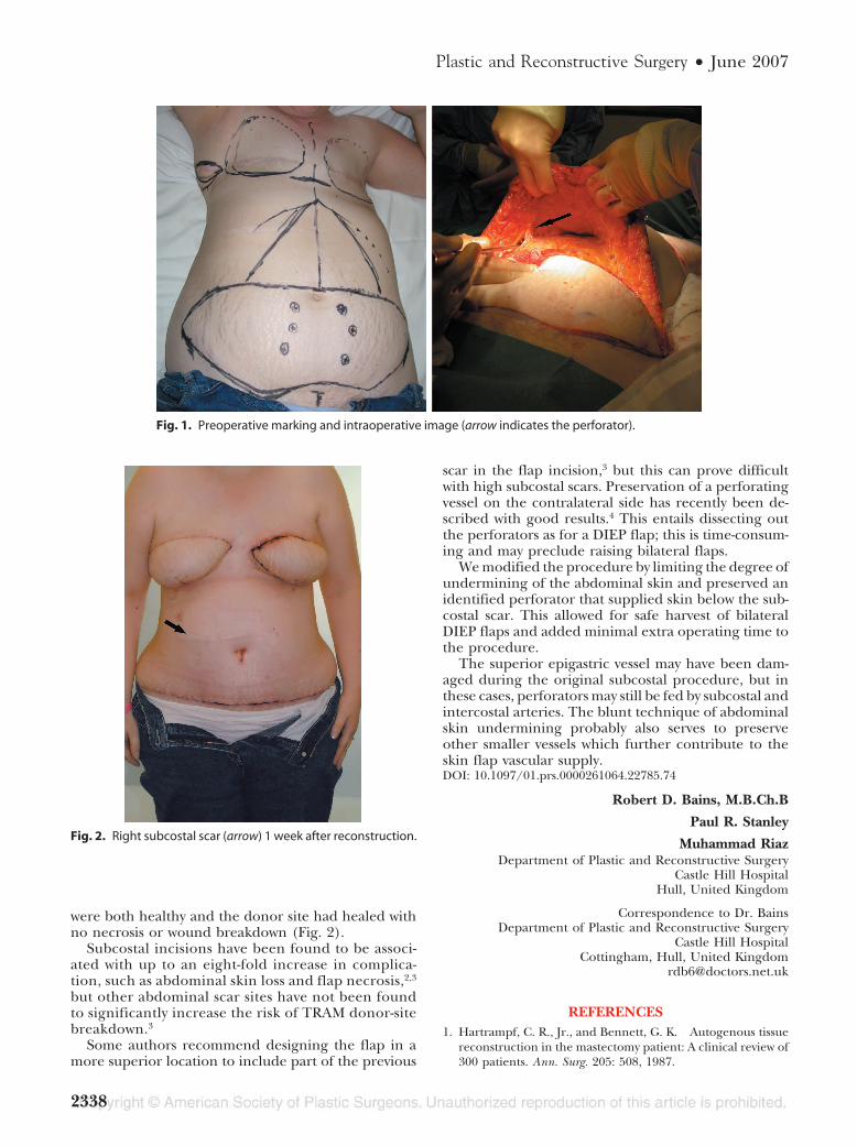
were both healthy and the donor site had healed withno necrosis or wound breakdown (Fig. 2).
Subcostal incisions have been found to be associ-ated with up to an eight-fold increase in complica-tion, such as abdominal skin loss and flap necrosis,2,3
but other abdominal scar sites have not been foundto significantly increase the risk of TRAM donor-sitebreakdown.3
Some authors recommend designing the flap in amore superior location to include part of the previous
scar in the flap incision,3 but this can prove difficultwith high subcostal scars. Preservation of a perforatingvessel on the contralateral side has recently been de-scribed with good results.4 This entails dissecting outthe perforators as for a DIEP flap; this is time-consum-ing and may preclude raising bilateral flaps.
We modified the procedure by limiting the degree ofundermining of the abdominal skin and preserved anidentified perforator that supplied skin below the sub-costal scar. This allowed for safe harvest of bilateralDIEP flaps and added minimal extra operating time tothe procedure.
The superior epigastric vessel may have been dam-aged during the original subcostal procedure, but inthese cases, perforators may still be fed by subcostal andintercostal arteries. The blunt technique of abdominalskin undermining probably also serves to preserveother smaller vessels which further contribute to theskin flap vascular supply.DOI: 10.1097/01.prs.0000261064.22785.74
Robert D. Bains, M.B.Ch.B
Paul R. Stanley
Muhammad RiazDepartment of Plastic and Reconstructive Surgery
Castle Hill HospitalHull, United Kingdom
Correspondence to Dr. BainsDepartment of Plastic and Reconstructive Surgery
Castle Hill HospitalCottingham, Hull, United Kingdom
REFERENCES1. Hartrampf, C. R., Jr., and Bennett, G. K. Autogenous tissue
reconstruction in the mastectomy patient: A clinical review of300 patients. Ann. Surg. 205: 508, 1987.
Fig. 1. Preoperative marking and intraoperative image (arrow indicates the perforator).
Fig. 2. Right subcostal scar (arrow) 1 week after reconstruction.
Plastic and Reconstructive Surgery • June 2007
2338
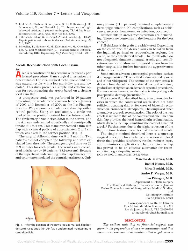
2. Losken, A., Carlson, G. W., Jones, G. E., Culbertson, J. H.,Schoemann, M., and Bostwick, J., III. Importance of rightsubcostal incisions in patients undergoing TRAM flap breastreconstruction. Ann. Plast. Surg. 49: 115, 2002.
3. Takeishi, M., Shaw, W. W., Ahn, C. Y., and Borud, L. J. TRAMflaps in patients with abdominal scars. Plast. Reconstr. Surg. 99:713, 1997.
4. Schoeller, T., Huemer, G. M., Kolehmainen, M., Otto-Schoe-ller, A., and Wechselberger, G. Management of subcostalscars during DIEP flap raising. Br. J. Plast. Surg. 57: 511, 2004.
Areola Reconstruction with Local TissueSir:
Areola reconstruction has become a frequently per-formed procedure. Many surgical alternatives are
now available. The ideal surgical technique should pro-vide natural results with a low morbidity rate and lowcosts.1,2 This study presents a simple and effective op-tion for reconstructing the areola based on a circularlocal skin flap.
A prospective study was performed in 20 patientspresenting for areola reconstruction between Januaryof 2000 and December of 2004 at the Ivo PitanguyInstitute. We proposed a circular local skin flap with acentral pedicle. Using an areolatome, a circle wasmarked in the position desired for the future areola.The circle margin was incised down to the dermis, andthe skin was undermined superficially and centripetallyfor about 1 to 2 cm. This maneuver created a thin skinflap with a central pedicle of approximately 2 to 3 cmwhich was fixed in the former position (Fig. 1).
The surgical follow-up time was 8 � 3 months. Twopatients did not complete the follow-up and were ex-cluded from the study. The average surgical time was 20� 9 minutes for each areola. The results were consid-ered satisfactory by 16 patients (88.9 percent). Becauseof the superficial undermining of the flap, final textureand color tone simulated the contralateral areola. Only
two patients (11.1 percent) required complementarydermopigmentation. No complications, such as dehis-cence, necrosis, hematoma, or infection, occurred.
Refinements in areola reconstruction are demand-ing. There is no consensus in the literature for the bestchoice.3
Full-thickness skin grafts are widely used. Dependingon the color tone, the desired skin can be taken fromthe inguinal, perineal or retroauricular regions, theeyelid, or the contralateral areola. The results often donot adequately simulate a natural areola, and compli-cations can occur. Moreover, removal of skin from an-other surgical site further increases the aggressivenessof the procedure.4
Some authors advocate a nonsurgical procedure, such asdermopigmentation.3 This method is also criticized by someand is not widespread. The texture of the new areola isdifferent from that of the contralateral one, and over time,gradual lossofpigmentationdemandsrepeatedprocedures.For more natural results, an alternative is skin grafting withpostoperative dermopigmentation.3,4
The circular flap, described here, is an alternative forcases in which the contralateral areola does not havesufficient donating skin or for cases of bilateral recon-struction. From several standpoints, it has surpassed otheralternatives to areola reconstruction. The color of the newareola is similar to that of the contralateral one. The thinskin flap provides the local hemosiderin sedimentation,which darkens the flap, simulating the color of a naturalareola.5 Furthermore, due to the slight retraction of theflap, the tissue texture resembles that of a natural areola.
The simple method described here is a one-stepsurgical procedure for areola reconstruction with localtissue that provides satisfactory results, reduces costs,and minimizes complications. The local circular flaphas proved to be an effective alternative for recon-structing a good-quality areola.DOI: 10.1097/01.prs.0000261066.12736.aa
Marcelo de Oliveira, M.D.
Daniel Nunes, M.D.
Mirta Beolchi, M.D.
Andre F. Vargas, M.D.
Ivo Pitanguy, M.D.Department of Plastic Surgery
The Pontifical Catholic University of Rio de JaneiroCarlos Chagas Institute of Postgraduate Medical Studies,
andIvo Pitanguy InstituteRio de Janeiro, Brazil
Correspondence to Dr. de OliveiraRua Afranio de Melo Franco, 141/311Rio de Janeiro, Brazil, CEP 22430-060
DISCLOSUREThe authors state that no financial support was
given in the preparation of the communication and thatthere are no commercial associations that might create a
Fig. 1. After the position of the new areola is marked, flap bor-ders are incised and a thin skin flap is undermined, maintaining itscentral pedicle.
Volume 119, Number 7 • Letters and Viewpoints
2339
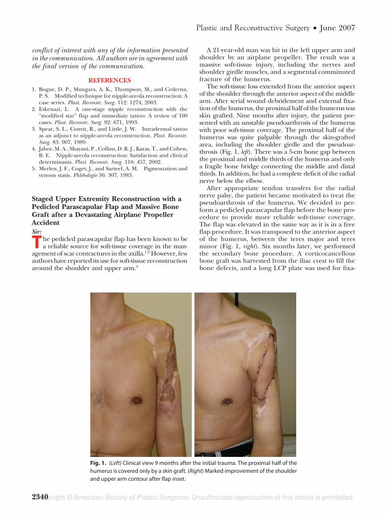
conflict of interest with any of the information presentedin the communication. All authors are in agreement withthe final version of the communication.
REFERENCES1. Bogue, D. P., Mungara, A. K., Thompson, M., and Cederna,
P. S. Modified technique for nipple-areola reconstruction: Acase series. Plast. Reconstr. Surg. 112: 1274, 2003.
2. Eskenazi, L. A one-stage nipple reconstruction with the“modified star” flap and immediate tattoo: A review of 100cases. Plast. Reconstr. Surg. 92: 671, 1993.
3. Spear, S. L., Convit, R., and Little, J. W. Intradermal tattooas an adjunct to nipple-areola reconstruction. Plast. Reconstr.Surg. 83: 907, 1989.
4. Jabor, M. A., Shayani, P., Collins, D. R. J., Karas, T., and Cohen,B. E. Nipple-areola reconstruction: Satisfaction and clinicaldeterminants. Plast. Reconstr. Surg. 110: 457, 2002.
5. Merlen, J. F., Coget, J., and Sarteel, A. M. Pigmentation andvenous stasis. Phlebologie 36: 307, 1983.
Staged Upper Extremity Reconstruction with aPedicled Parascapular Flap and Massive BoneGraft after a Devastating Airplane PropellerAccidentSir:
The pedicled parascapular flap has been known to bea reliable source for soft-tissue coverage in the man-
agement of scar contractures in the axilla.1,2 However, fewauthors have reported its use for soft-tissue reconstructionaround the shoulder and upper arm.3
A 21-year-old man was hit in the left upper arm andshoulder by an airplane propeller. The result was amassive soft-tissue injury, including the nerves andshoulder girdle muscles, and a segmental comminutedfracture of the humerus.
The soft-tissue loss extended from the anterior aspectof the shoulder through the anterior aspect of the middlearm. After serial wound debridement and external fixa-tion of the humerus, the proximal half of the humerus wasskin grafted. Nine months after injury, the patient pre-sented with an unstable pseudoarthrosis of the humeruswith poor soft-tissue coverage. The proximal half of thehumerus was quite palpable through the skin-graftedarea, including the shoulder girdle and the pseudoar-throsis (Fig. 1, left). There was a 5-cm bone gap betweenthe proximal and middle thirds of the humerus and onlya fragile bone bridge connecting the middle and distalthirds. In addition, he had a complete deficit of the radialnerve below the elbow.
After appropriate tendon transfers for the radialnerve palsy, the patient became motivated to treat thepseudoarthrosis of the humerus. We decided to per-form a pedicled parascapular flap before the bone pro-cedure to provide more reliable soft-tissue coverage.The flap was elevated in the same way as it is in a freeflap procedure. It was transposed to the anterior aspectof the humerus, between the teres major and teresminor (Fig. 1, right). Six months later, we performedthe secondary bone procedure. A corticocancellousbone graft was harvested from the iliac crest to fill thebone defects, and a long LCP plate was used for fixa-
Fig. 1. (Left) Clinical view 9 months after the initial trauma. The proximal half of thehumerus is covered only by a skin graft. (Right) Marked improvement of the shoulderand upper arm contour after flap inset.
Plastic and Reconstructive Surgery • June 2007
2340
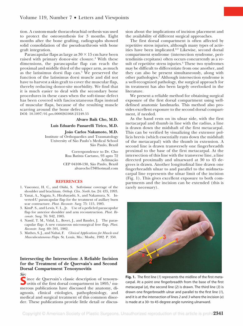
tion. A custom-made thoracobrachial orthesis was usedto protect the osteosynthesis for 3 months. Eightmonths after the bone grafting, radiographs showedsolid consolidation of the pseudoarthrosis with bonegraft integration.
Parascapular flaps as large as 30 � 15 cm have beenraised with primary donor-site closure.4 With thesedimensions, the parascapular flap can reach theproximal and middle third of the upper arm, as muchas the latissimus dorsi flap can.5 We preserved thefunction of the latissimus dorsi muscle and did nothave to harvest a skin graft to cover the muscular flap,thereby reducing donor-site morbidity. We find thatit is much easier to deal with the secondary boneprocedures in these cases when the soft-tissue defecthas been covered with fasciocutaneous flaps insteadof muscular flaps, because of the resulting musclescarring around the bone defect.DOI: 10.1097/01.prs.0000261068.21249.55
Alvaro Baik Cho, M.D.
Luıs Eduardo Passarelli Tırico, M.D.
Joao Carlos Nakamoto, M.D.Institute of Orthopaedics and Traumatology
University of Sao Paulo’s Medical SchoolSao Paulo, Brazil
Correspondence to Dr. ChoRua Batista Caetano, 93 apto 72
AclimacaoCEP 04108-130, Sao Paulo, Brazil
REFERENCES1. Vasconez, H. C., and Oishi, S. Soft-tissue coverage of the
shoulder and brachium. Orthop. Clin. North Am. 24: 435, 1993.2. Yanai, A., Nagata, S., Hirabayashi, S., and Nakamura, N. In-
verted-U parascapular flap for the treatment of axillary burnscar contracture. Plast. Reconstr. Surg. 75: 115, 1985.
3. KimP. S., and Lewis, V. L., Jr. Use of a pedicled parascapularflap for anterior shoulder and arm reconstruction. Plast. Re-constr. Surg. 76: 942, 1985.
4. Nassif, T. M., Vidal, L., Bovet, J., and Baudet, J. The paras-capular flap: A new cutaneous microsurgical free flap. Plast.Reconstr. Surg. 69: 591, 1982.
5. Mathes, S. J., and Nahai, F. Clinical Applications for Muscle andMusculocutaneous Flaps. St. Louis, Mo.: Mosby, 1982. P. 350.
Intersecting the Intersection: A Reliable Incisionfor the Treatment of de Quervain’s and SecondDorsal Compartment TenosynovitisSir:
S ince de Quervain’s classic description of tenosyn-ovitis of the first dorsal compartment in 1895,1 nu-
merous publications have discussed the anatomy, di-agnosis, clinical etiologies, pathophysiology, andmedical and surgical treatment of this common disor-der. These publications provide little detail or discus-
sion about the implications of incision placement andthe availability of different surgical approaches.
The first dorsal compartment is often affected byrepetitive stress injuries, although many types of activ-ities have been implicated.2,3 Likewise, second dorsalcompartment syndrome (intersection syndrome, peri-tendinitis crepitans) often occurs concurrently as a re-sult of repetitive stress injuries.4 These two syndromesmay be difficult to differentiate from one another, andthey can also be present simultaneously, along withother pathologies.5 Although intersection syndrome isa well-recognized pathology, the surgical approach forits treatment has also been largely overlooked in theliterature.
We present a reliable method for obtaining surgicalexposure of the first dorsal compartment using well-defined anatomic landmarks. This method also pro-vides excellent exposure to the second dorsal compart-ment, if needed.
As the hand rests on its ulnar side, with the firstmetacarpal and thumb in line with the radius, a lineis drawn down the midshaft of the first metacarpal.This can be verified by visualizing the extensor pol-licis brevis (which essentially runs down the midshaftof the metacarpal) with the thumb in extension. Asecond line is drawn transversely one fingerbreadthproximal to the base of the first metacarpal. At theintersection of this line with the transverse line, a linedirected proximally and ulnarward at 30 to 45 de-grees is drawn. Another longitudinal line drawn onefingerbreadth ulnar to and parallel to the midmeta-carpal line represents the ulnar limit of the incision(Fig. 1). This gives excellent exposure to both com-partments and the incision can be extended (this israrely necessary).
Fig. 1. The first line (1) represents the midline of the first meta-carpal. At a point one fingerbreadth from the base of the firstmetacarpal (a), the second line (2) is drawn. The third line (3) isdrawn one fingerbreadth ulnar and parallel to the first line (1),and it is at the intersection of lines 2 and 3 where the incision (x)is made at a 30- to 45-degree angle running ulnarward.
Volume 119, Number 7 • Letters and Viewpoints
2341
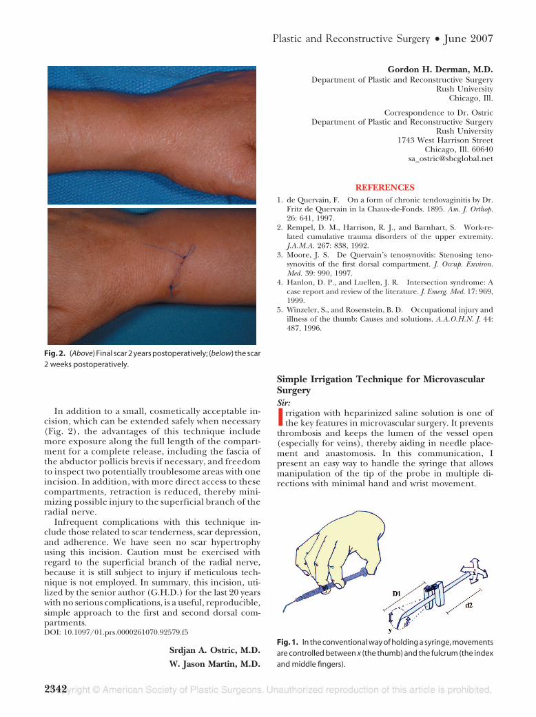
In addition to a small, cosmetically acceptable in-cision, which can be extended safely when necessary(Fig. 2), the advantages of this technique includemore exposure along the full length of the compart-ment for a complete release, including the fascia ofthe abductor pollicis brevis if necessary, and freedomto inspect two potentially troublesome areas with oneincision. In addition, with more direct access to thesecompartments, retraction is reduced, thereby mini-mizing possible injury to the superficial branch of theradial nerve.
Infrequent complications with this technique in-clude those related to scar tenderness, scar depression,and adherence. We have seen no scar hypertrophyusing this incision. Caution must be exercised withregard to the superficial branch of the radial nerve,because it is still subject to injury if meticulous tech-nique is not employed. In summary, this incision, uti-lized by the senior author (G.H.D.) for the last 20 yearswith no serious complications, is a useful, reproducible,simple approach to the first and second dorsal com-partments.DOI: 10.1097/01.prs.0000261070.92579.f5
Srdjan A. Ostric, M.D.
W. Jason Martin, M.D.
Gordon H. Derman, M.D.Department of Plastic and Reconstructive Surgery
Rush UniversityChicago, Ill.
Correspondence to Dr. OstricDepartment of Plastic and Reconstructive Surgery
Rush University1743 West Harrison Street
Chicago, Ill. [email protected]
REFERENCES1. de Quervain, F. On a form of chronic tendovaginitis by Dr.
Fritz de Quervain in la Chaux-de-Fonds. 1895. Am. J. Orthop.26: 641, 1997.
2. Rempel, D. M., Harrison, R. J., and Barnhart, S. Work-re-lated cumulative trauma disorders of the upper extremity.J.A.M.A. 267: 838, 1992.
3. Moore, J. S. De Quervain’s tenosynovitis: Stenosing teno-synovitis of the first dorsal compartment. J. Occup. Environ.Med. 39: 990, 1997.
4. Hanlon, D. P., and Luellen, J. R. Intersection syndrome: Acase report and review of the literature. J. Emerg. Med. 17: 969,1999.
5. Winzeler, S., and Rosenstein, B. D. Occupational injury andillness of the thumb: Causes and solutions. A.A.O.H.N. J. 44:487, 1996.
Simple Irrigation Technique for MicrovascularSurgerySir:
Irrigation with heparinized saline solution is one ofthe key features in microvascular surgery. It prevents
thrombosis and keeps the lumen of the vessel open(especially for veins), thereby aiding in needle place-ment and anastomosis. In this communication, Ipresent an easy way to handle the syringe that allowsmanipulation of the tip of the probe in multiple di-rections with minimal hand and wrist movement.
Fig. 2. (Above) Final scar 2 years postoperatively; (below) the scar2 weeks postoperatively.
Fig. 1. In the conventional way of holding a syringe, movementsare controlled between x (the thumb) and the fulcrum (the indexand middle fingers).
Plastic and Reconstructive Surgery • June 2007
2342
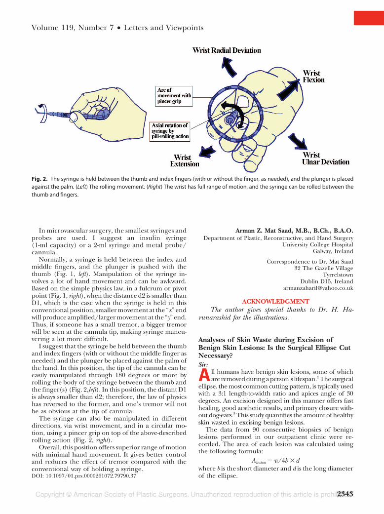
In microvascular surgery, the smallest syringes andprobes are used. I suggest an insulin syringe(1-ml capacity) or a 2-ml syringe and metal probe/cannula.
Normally, a syringe is held between the index andmiddle fingers, and the plunger is pushed with thethumb (Fig. 1, left). Manipulation of the syringe in-volves a lot of hand movement and can be awkward.Based on the simple physics law, in a fulcrum or pivotpoint (Fig. 1, right), when the distance d2 is smaller thanD1, which is the case when the syringe is held in thisconventional position, smaller movement at the “x” endwill produce amplified/larger movement at the “y” end.Thus, if someone has a small tremor, a bigger tremorwill be seen at the cannula tip, making syringe maneu-vering a lot more difficult.
I suggest that the syringe be held between the thumband index fingers (with or without the middle finger asneeded) and the plunger be placed against the palm ofthe hand. In this position, the tip of the cannula can beeasily manipulated through 180 degrees or more byrolling the body of the syringe between the thumb andthe finger(s) (Fig. 2,left). In this position, the distant D1is always smaller than d2; therefore, the law of physicshas reversed to the former, and one’s tremor will notbe as obvious at the tip of cannula.
The syringe can also be manipulated in differentdirections, via wrist movement, and in a circular mo-tion, using a pincer grip on top of the above-describedrolling action (Fig. 2, right).
Overall, this position offers superior range of motionwith minimal hand movement. It gives better controland reduces the effect of tremor compared with theconventional way of holding a syringe.DOI: 10.1097/01.prs.0000261072.79790.37
Arman Z. Mat Saad, M.B., B.Ch., B.A.O.Department of Plastic, Reconstructive, and Hand Surgery
University College HospitalGalway, Ireland
Correspondence to Dr. Mat Saad32 The Gazelle Village
TyrrelstownDublin D15, Ireland
ACKNOWLEDGMENTThe author gives special thanks to Dr. H. Ha-
runarashid for the illustrations.
Analyses of Skin Waste during Excision ofBenign Skin Lesions: Is the Surgical Ellipse CutNecessary?Sir:
A ll humans have benign skin lesions, some of whichare removed during a person’s lifespan.1 The surgical
ellipse, the most common cutting pattern, is typically usedwith a 3:1 length-to-width ratio and apices angle of 30degrees. An excision designed in this manner offers fasthealing, good aesthetic results, and primary closure with-out dog-ears.2 This study quantifies the amount of healthyskin wasted in excising benign lesions.
The data from 90 consecutive biopsies of benignlesions performed in our outpatient clinic were re-corded. The area of each lesion was calculated usingthe following formula:
Alesion � � ⁄ 4b � dwhere b is the short diameter and d is the long diameterof the ellipse.
Fig. 2. The syringe is held between the thumb and index fingers (with or without the finger, as needed), and the plunger is placedagainst the palm. (Left) The rolling movement. (Right) The wrist has full range of motion, and the syringe can be rolled between thethumb and fingers.
Volume 119, Number 7 • Letters and Viewpoints
2343
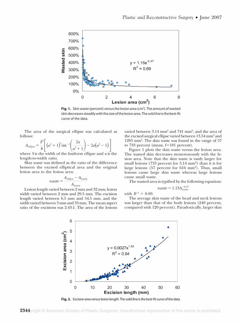
The area of the surgical ellipse was calculated asfollows:
Aellipse �S2
8 ��a2 � 1�2sin�1� 2a
a2 � 1�� 2a�a2 � 1��where S is the width of the fusiform ellipse and a is thelength-to-width ratio.
Skin waste was defined as the ratio of the differencebetween the excised elliptical area and the originallesion area to the lesion area:
waste �Aellipse � Alesion
Alesion
Lesion length varied between 2 mm and 32 mm; lesionwidth varied between 2 mm and 29.5 mm. The excisionlength varied between 6.5 mm and 54.5 mm, and thewidth varied between 3 mm and 33 mm. The mean aspectratio of the excisions was 2.43:1. The area of the lesions
varied between 3.14 mm2 and 741 mm2, and the area ofthe excised surgical ellipse varied between 13.54 mm2 and1283 mm2. The skin waste was found in the range of 57to 733 percent (mean, f�181 percent).
Figure 1 plots the skin waste versus the lesion area.The wasted skin decreases monotonously with the le-sion area. Note that the skin waste is vastly larger forsmall lesions (733 percent for 3.14 mm2) than it is forlarge lesions (57 percent for 616 mm2). Thus, smalllesions cause large skin waste whereas large lesionscause small waste.
The wasted area is typified by the following equation:
waste � 1.15Alesion�0.37
with R 2 � 0.69.The average skin waste of the head and neck lesions
was larger than that of the body lesions (240 percent,compared with 120 percent). Paradoxically, larger skin
Fig. 1. Skin waste (percent) versus the lesion area (cm2). The amount of wastedskin decreases steadily with the size of the lesion area. The solid line is the best-fitcurve of the data.
Fig. 2. Excisionareaversuslesionlength.Thesolidlineisthebest-fitcurveofthedata.
Plastic and Reconstructive Surgery • June 2007
2344
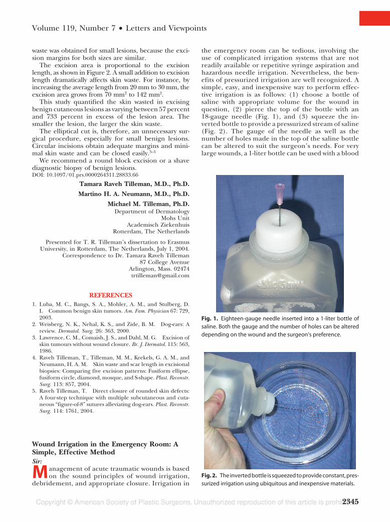
waste was obtained for small lesions, because the exci-sion margins for both sizes are similar.
The excision area is proportional to the excisionlength, as shown in Figure 2. A small addition to excisionlength dramatically affects skin waste. For instance, byincreasing the average length from 20 mm to 30 mm, theexcision area grows from 70 mm2 to 142 mm2.
This study quantified the skin wasted in excisingbenign cutaneous lesions as varying between 57 percentand 733 percent in excess of the lesion area. Thesmaller the lesion, the larger the skin waste.
The elliptical cut is, therefore, an unnecessary sur-gical procedure, especially for small benign lesions.Circular incisions obtain adequate margins and mini-mal skin waste and can be closed easily.3–5
We recommend a round block excision or a shavediagnostic biopsy of benign lesions.DOI: 10.1097/01.prs.0000264311.28833.66
Tamara Raveh Tilleman, M.D., Ph.D.
Martino H. A. Neumann, M.D., Ph.D.
Michael M. Tilleman, Ph.D.Department of Dermatology
Mohs UnitAcademisch Ziekenhuis
Rotterdam, The Netherlands
Presented for T. R. Tilleman’s dissertation to ErasmusUniversity, in Rotterdam, The Netherlands, July 1, 2004.
Correspondence to Dr. Tamara Raveh Tilleman87 College Avenue
Arlington, Mass. [email protected]
REFERENCES1. Luba, M. C., Bangs, S. A., Mohler, A. M., and Stulberg, D.
L Common benign skin tumors. Am. Fam. Physician 67: 729,2003.
2. Weisberg, N. K., Nehal, K. S., and Zide, B. M. Dog-ears: Areview. Dermatol. Surg. 26: 363, 2000.
3. Lawrence, C. M., Comaish, J. S., and Dahl, M. G. Excision ofskin tumours without wound closure. Br. J. Dermatol. 115: 563,1986.
4. Raveh Tilleman, T., Tilleman, M. M., Krekels, G. A. M., andNeumann, H. A. M. Skin waste and scar length in excisionalbiopsies: Comparing five excision patterns: Fusiform ellipse,fusiform circle, diamond, mosque, and S-shape. Plast. Reconstr.Surg. 113: 857, 2004.
5. Raveh Tilleman, T. Direct closure of rounded skin defects:A four-step technique with multiple subcutaneous and cuta-neous “figure-of-8” sutures alleviating dog-ears. Plast. Reconstr.Surg. 114: 1761, 2004.
Wound Irrigation in the Emergency Room: ASimple, Effective MethodSir:
Management of acute traumatic wounds is basedon the sound principles of wound irrigation,
debridement, and appropriate closure. Irrigation in
the emergency room can be tedious, involving theuse of complicated irrigation systems that are notreadily available or repetitive syringe aspiration andhazardous needle irrigation. Nevertheless, the ben-efits of pressurized irrigation are well recognized. Asimple, easy, and inexpensive way to perform effec-tive irrigation is as follows: (1) choose a bottle ofsaline with appropriate volume for the wound inquestion, (2) pierce the top of the bottle with an18-gauge needle (Fig. 1), and (3) squeeze the in-verted bottle to provide a pressurized stream of saline(Fig. 2). The gauge of the needle as well as thenumber of holes made in the top of the saline bottlecan be altered to suit the surgeon’s needs. For verylarge wounds, a 1-liter bottle can be used with a blood
Fig. 1. Eighteen-gauge needle inserted into a 1-liter bottle ofsaline. Both the gauge and the number of holes can be altereddepending on the wound and the surgeon’s preference.
Fig. 2. The inverted bottle is squeezed to provide constant, pres-surized irrigation using ubiquitous and inexpensive materials.
Volume 119, Number 7 • Letters and Viewpoints
2345

pressure cuff (another ubiquitous emergency roomitem) inflated around it to provide constant, pres-surized irrigation. The lack of a protective shieldallows for better visualization, but proper eye pro-tection must be worn. This method has provided veryeffective irrigation using readily available and inex-pensive materials.DOI: 10.1097/01.prs.0000261074.32202.ff
Eric G. Halvorson, M.D.Division of Plastic Surgery
University of North CarolinaChapel Hill, N.C. [email protected]
Use of Hyaluronan Dressings followingDermabrasion Avoids Escharectomy andFacilitates Healing in Pediatric Burn PatientsSir:
Deep partial-thickness burns are the most frequenttype of burn in pediatric patients and pose difficult
dilemmas for caregivers. First, the surgeon is expectedto obtain good cosmetic results while at the same timeavoiding hypertrophic healing processes. Second, in-dications and timing to early escharectomy are not asstandardized as protocols for other lesions, because ofthe possible evolution of the burn into full-thicknessand superficial burns. Third, if escharectomy is deemednecessary, the dermal plane must be respected andcovered as soon as possible with skin grafts to preventthe hypertrophic healing process from occurring. Forall of these reasons, deep partial-thickness burns seri-ously challenge the surgeon’s abilities.
To solve these problems, since 2001, we have com-bined rotating-head dermabrasion with hyaluronan-based dressings. We previously described the positiveresults we obtained with dermabrasion, which pre-vented further necrosis and blocked the bad evolutionof deep burns into full-thickness lesions.1 Other au-thors have outlined the important wound-healing ac-tivity shown by hyaluronan-based matrixes on deep par-tial-thickness burns, in their ability to stimulateregeneration and avoid hypertrophy.2 We have adoptedthe following protocol. Escharectomy and skin graftingare avoided, at least initially. All patients have theirdeep partial-thickness burns covered with silver anti-microbial dressings (Acticoat); superficial partial-thick-ness burns are covered with a transparent film of hy-aluronic acid medication (Jaloskin) at recovery. On thefifth day, dermabrasion is performed on deeply burnedareas to remove the superficial ischemic dermis andleave a mildly bloody surface. We cover these areas witha particular variant of hyaluronic matrix (HyalomatrixPA), composed of silicone and hyaluronic acid, that hasa slower degradation time. Serial medications are fol-lowed by hyaluronan matrix changes every 7 days. Onday 21, those areas where deep partial-thickness burnspersist with no sign of recovery are removed by eschar-ectomy and covered with thin skin grafts.
Three hundred pediatric patients with deep partial-thickness burns were treated from February of 2001 toSeptember of 2005 (78 percent of all patients). Sixty-one percent (183 patients) needed only one dermabra-sion, 22.3 percent (67 patients) required more thanone, and only 16.7 percent (50 patients) required clas-sic escharectomy. Eighty-three percent of patients withdeep partial-thickness burns (250 patients) avoided es-charectomy and had their dermal plane shifted towarda superficial partial-thickness burn with spontaneous
Fig. 1. Four-year-old patient with deep partial-thickness burnson the head.
Fig. 2. At postoperative day 20, there is spontaneous re-epithe-lization after dermabrasion and three hyaluronan-based occlu-sive dressings.
Plastic and Reconstructive Surgery • June 2007
2346
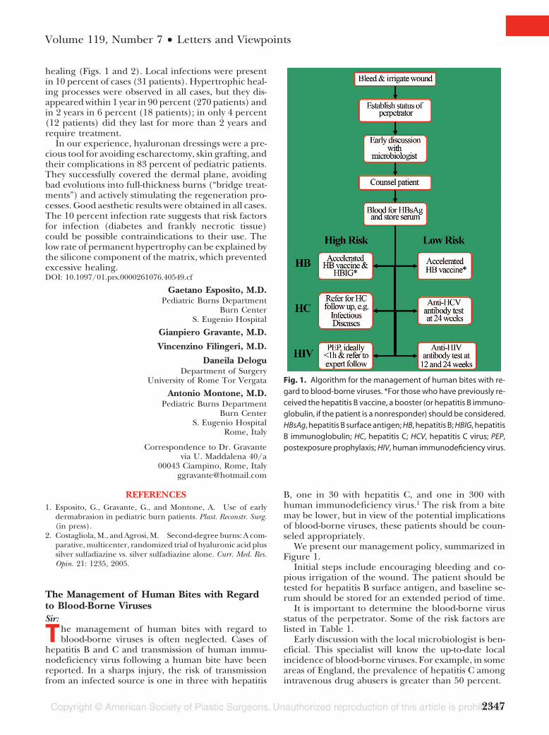
healing (Figs. 1 and 2). Local infections were presentin 10 percent of cases (31 patients). Hypertrophic heal-ing processes were observed in all cases, but they dis-appeared within 1 year in 90 percent (270 patients) andin 2 years in 6 percent (18 patients); in only 4 percent(12 patients) did they last for more than 2 years andrequire treatment.
In our experience, hyaluronan dressings were a pre-cious tool for avoiding escharectomy, skin grafting, andtheir complications in 83 percent of pediatric patients.They successfully covered the dermal plane, avoidingbad evolutions into full-thickness burns (“bridge treat-ments”) and actively stimulating the regeneration pro-cesses. Good aesthetic results were obtained in all cases.The 10 percent infection rate suggests that risk factorsfor infection (diabetes and frankly necrotic tissue)could be possible contraindications to their use. Thelow rate of permanent hypertrophy can be explained bythe silicone component of the matrix, which preventedexcessive healing.DOI: 10.1097/01.prs.0000261076.40549.cf
Gaetano Esposito, M.D.Pediatric Burns Department
Burn CenterS. Eugenio Hospital
Gianpiero Gravante, M.D.
Vincenzino Filingeri, M.D.
Daneila DeloguDepartment of Surgery
University of Rome Tor Vergata
Antonio Montone, M.D.Pediatric Burns Department
Burn CenterS. Eugenio Hospital
Rome, Italy
Correspondence to Dr. Gravantevia U. Maddalena 40/a
00043 Ciampino, Rome, [email protected]
REFERENCES1. Esposito, G., Gravante, G., and Montone, A. Use of early
dermabrasion in pediatric burn patients. Plast. Reconstr. Surg.(in press).
2. Costagliola, M., and Agrosi, M. Second-degree burns: A com-parative, multicenter, randomized trial of hyaluronic acid plussilver sulfadiazine vs. silver sulfadiazine alone. Curr. Med. Res.Opin. 21: 1235, 2005.
The Management of Human Bites with Regardto Blood-Borne VirusesSir:
The management of human bites with regard toblood-borne viruses is often neglected. Cases of
hepatitis B and C and transmission of human immu-nodeficiency virus following a human bite have beenreported. In a sharps injury, the risk of transmissionfrom an infected source is one in three with hepatitis
B, one in 30 with hepatitis C, and one in 300 withhuman immunodeficiency virus.1 The risk from a bitemay be lower, but in view of the potential implicationsof blood-borne viruses, these patients should be coun-seled appropriately.
We present our management policy, summarized inFigure 1.
Initial steps include encouraging bleeding and co-pious irrigation of the wound. The patient should betested for hepatitis B surface antigen, and baseline se-rum should be stored for an extended period of time.
It is important to determine the blood-borne virusstatus of the perpetrator. Some of the risk factors arelisted in Table 1.
Early discussion with the local microbiologist is ben-eficial. This specialist will know the up-to-date localincidence of blood-borne viruses. For example, in someareas of England, the prevalence of hepatitis C amongintravenous drug abusers is greater than 50 percent.
Fig. 1. Algorithm for the management of human bites with re-gard to blood-borne viruses. *For those who have previously re-ceived the hepatitis B vaccine, a booster (or hepatitis B immuno-globulin, if the patient is a nonresponder) should be considered.HBsAg, hepatitis B surface antigen; HB, hepatitis B; HBIG, hepatitisB immunoglobulin; HC, hepatitis C; HCV, hepatitis C virus; PEP,postexposure prophylaxis; HIV, human immunodeficiency virus.
Volume 119, Number 7 • Letters and Viewpoints
2347
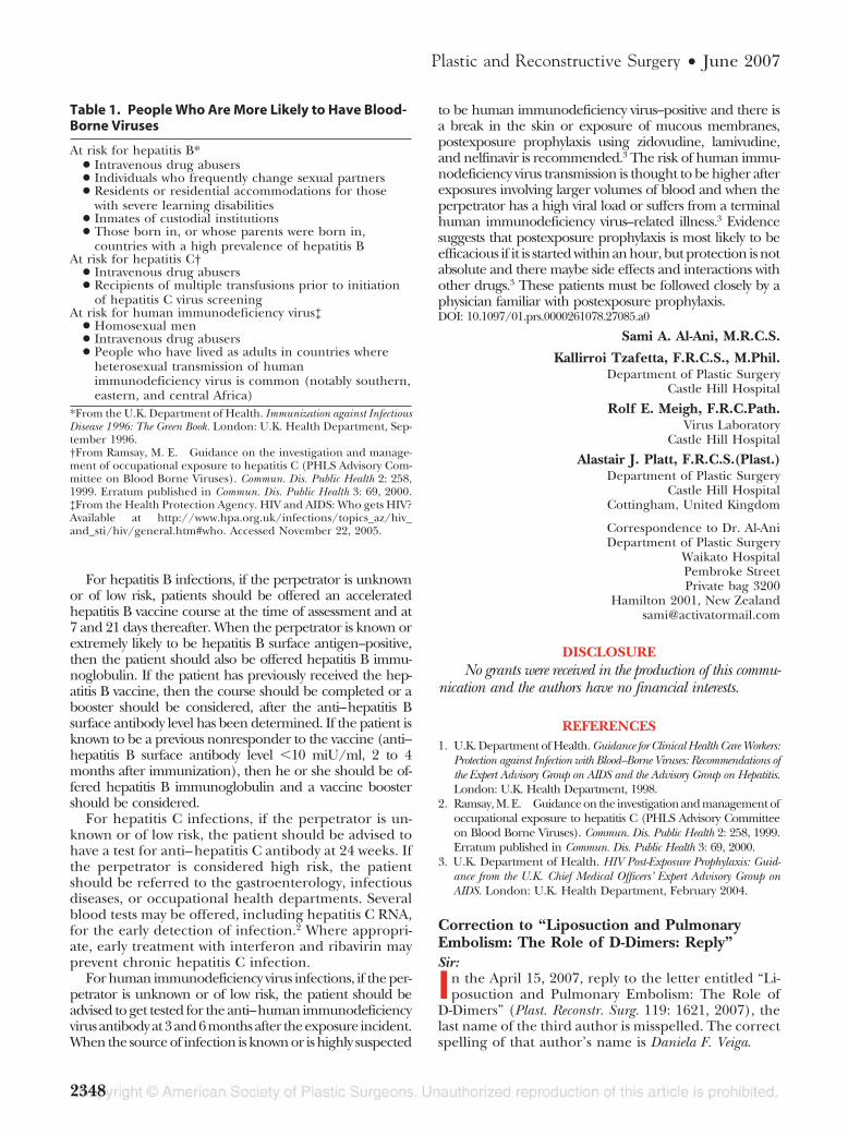
For hepatitis B infections, if the perpetrator is unknownor of low risk, patients should be offered an acceleratedhepatitis B vaccine course at the time of assessment and at7 and 21 days thereafter. When the perpetrator is known orextremely likely to be hepatitis B surface antigen–positive,then the patient should also be offered hepatitis B immu-noglobulin. If the patient has previously received the hep-atitis B vaccine, then the course should be completed or abooster should be considered, after the anti–hepatitis Bsurface antibody level has been determined. If the patient isknown to be a previous nonresponder to the vaccine (anti–hepatitis B surface antibody level �10 miU/ml, 2 to 4months after immunization), then he or she should be of-fered hepatitis B immunoglobulin and a vaccine boostershould be considered.
For hepatitis C infections, if the perpetrator is un-known or of low risk, the patient should be advised tohave a test for anti–hepatitis C antibody at 24 weeks. Ifthe perpetrator is considered high risk, the patientshould be referred to the gastroenterology, infectiousdiseases, or occupational health departments. Severalblood tests may be offered, including hepatitis C RNA,for the early detection of infection.2 Where appropri-ate, early treatment with interferon and ribavirin mayprevent chronic hepatitis C infection.
For human immunodeficiency virus infections, if the per-petrator is unknown or of low risk, the patient should beadvised to get tested for the anti–human immunodeficiencyvirusantibodyat3and6monthsafter theexposure incident.When the source of infection is known or is highly suspected
to be human immunodeficiency virus–positive and there isa break in the skin or exposure of mucous membranes,postexposure prophylaxis using zidovudine, lamivudine,and nelfinavir is recommended.3 The risk of human immu-nodeficiency virus transmission is thought to be higher afterexposures involving larger volumes of blood and when theperpetrator has a high viral load or suffers from a terminalhuman immunodeficiency virus–related illness.3 Evidencesuggests that postexposure prophylaxis is most likely to beefficacious if it is started within an hour, but protection is notabsolute and there maybe side effects and interactions withother drugs.3 These patients must be followed closely by aphysician familiar with postexposure prophylaxis.DOI: 10.1097/01.prs.0000261078.27085.a0
Sami A. Al-Ani, M.R.C.S.
Kallirroi Tzafetta, F.R.C.S., M.Phil.Department of Plastic Surgery
Castle Hill Hospital
Rolf E. Meigh, F.R.C.Path.Virus Laboratory
Castle Hill Hospital
Alastair J. Platt, F.R.C.S.(Plast.)Department of Plastic Surgery
Castle Hill HospitalCottingham, United Kingdom
Correspondence to Dr. Al-AniDepartment of Plastic Surgery
Waikato HospitalPembroke StreetPrivate bag 3200
Hamilton 2001, New [email protected]
DISCLOSURENo grants were received in the production of this commu-
nication and the authors have no financial interests.
REFERENCES1. U.K. Department of Health. Guidance for Clinical Health Care Workers:
Protection against Infection with Blood–Borne Viruses: Recommendations ofthe Expert Advisory Group on AIDS and the Advisory Group on Hepatitis.London: U.K. Health Department, 1998.
2. Ramsay, M. E. Guidance on the investigation and management ofoccupational exposure to hepatitis C (PHLS Advisory Committeeon Blood Borne Viruses). Commun. Dis. Public Health 2: 258, 1999.Erratum published in Commun. Dis. Public Health 3: 69, 2000.
3. U.K. Department of Health. HIV Post-Exposure Prophylaxis: Guid-ance from the U.K. Chief Medical Officers’ Expert Advisory Group onAIDS. London: U.K. Health Department, February 2004.
Correction to “Liposuction and PulmonaryEmbolism: The Role of D-Dimers: Reply”Sir:
In the April 15, 2007, reply to the letter entitled “Li-posuction and Pulmonary Embolism: The Role of
D-Dimers” (Plast. Reconstr. Surg. 119: 1621, 2007), thelast name of the third author is misspelled. The correctspelling of that author’s name is Daniela F. Veiga.
Table 1. People Who Are More Likely to Have Blood-Borne Viruses
At risk for hepatitis B*● Intravenous drug abusers● Individuals who frequently change sexual partners● Residents or residential accommodations for those
with severe learning disabilities● Inmates of custodial institutions● Those born in, or whose parents were born in,
countries with a high prevalence of hepatitis BAt risk for hepatitis C†
● Intravenous drug abusers● Recipients of multiple transfusions prior to initiation
of hepatitis C virus screeningAt risk for human immunodeficiency virus‡
● Homosexual men● Intravenous drug abusers● People who have lived as adults in countries where
heterosexual transmission of humanimmunodeficiency virus is common (notably southern,eastern, and central Africa)
*From the U.K. Department of Health. Immunization against InfectiousDisease 1996: The Green Book. London: U.K. Health Department, Sep-tember 1996.†From Ramsay, M. E. Guidance on the investigation and manage-ment of occupational exposure to hepatitis C (PHLS Advisory Com-mittee on Blood Borne Viruses). Commun. Dis. Public Health 2: 258,1999. Erratum published in Commun. Dis. Public Health 3: 69, 2000.‡From the Health Protection Agency. HIV and AIDS: Who gets HIV?Available at http://www.hpa.org.uk/infections/topics_az/hiv_and_sti/hiv/general.htm#who. Accessed November 22, 2005.
Plastic and Reconstructive Surgery • June 2007
2348


