Embed Size (px)
Citation preview

SirT1 enhances survival of human osteoarthritic chondrocytesby repressing PTP1B and activating the IGF receptor pathway
Viktoria Gagarina, MD/PhD,Cartilage Molecular Genetics Group, Cartilage Biology and Orthopedics Branch, National Instituteof Arthritis and Musculoskeletal and Skin Diseases, NIH, 50 South Drive, Rm 1524, Bethesda,MD 20892
Odile Gabay, PhD,Cartilage Molecular Genetics Group, Cartilage Biology and Orthopedics Branch, National Instituteof Arthritis and Musculoskeletal and Skin Diseases, NIH, 50 South Drive, Rm 1524, Bethesda,MD 20892
Mona Dvir-Ginzberg, PhD,Cartilage Molecular Genetics Group, Cartilage Biology and Orthopedics Branch, National Instituteof Arthritis and Musculoskeletal and Skin Diseases, NIH, 50 South Drive, Rm 1524, Bethesda,MD 20892
Eun-Jin Lee,Cartilage Molecular Genetics Group, Cartilage Biology and Orthopedics Branch, National Instituteof Arthritis and Musculoskeletal and Skin Diseases, NIH, 50 South Drive, Rm 1524, Bethesda,MD 20892
Jillian K. Brady, BA, PhD,Cartilage Molecular Genetics Group, Cartilage Biology and Orthopedics Branch, National Instituteof Arthritis and Musculoskeletal and Skin Diseases, NIH, 50 South Drive, Rm 1524, Bethesda,MD 20892
Michael J. Quon, PhD, andDiabetes Unit, Laboratory of Clinical Investigation, National Center for Complementary andAlternative Medicine, NIH, 50 South Drive, Rm 1524, Bethesda, MD 20892
David J. Hall, PhD#
Cartilage Molecular Genetics Group, Cartilage Biology and Orthopedics Branch, National Instituteof Arthritis and Musculoskeletal and Skin Diseases, NIH, 50 South Drive, Rm 1524, Bethesda,MD 20892
AbstractObjective—The protein deacetylase SirT1 inhibits apoptosis in a variety of cell systems bydistinct mechanisms, yet its role in chondrocyte death has not been explored. Here we assess therole of SirT1 in the survival of osteoarthritic human chondrocytes.
Methods—SirT1, PTP1B, PTP1Bmutant expression plasmids and SirT1siRNA andPTP1BsiRNA were transfected into primary human chondrocytes. Levels of apoptosis weredetermined by flow cytometry and activation of components of the IGFR/Akt pathway wasassessed by immunoblotting. Immunohistochemistry was performed on osteoarthritic (OA) andnormal knee cartilage samples.
#Corresponding Author: Phone: 301-451-6860, Fax: 301-480-2958, [email protected].
NIH Public AccessAuthor ManuscriptArthritis Rheum. Author manuscript; available in PMC 2011 May 1.
Published in final edited form as:Arthritis Rheum. 2010 May ; 62(5): 1383–1392. doi:10.1002/art.27369.
NIH
-PA Author Manuscript
NIH
-PA Author Manuscript
NIH
-PA Author Manuscript

Results—Expression of SirT1 in chondrocytes led to increased chondrocyte survival in either thepresence or absence of TNFα/Actinomycin D, while a reduction of SirT1 by siRNA led toincreased in chondrocyte apoptosis. Expression of SirT1 in chondrocytes led to activation of theIGF receptor (IGFR) and the downstream kinases PI3K, PDK1, mTOR and Akt which in turn ledto phosphorylation of MDM2, inhibition of p53 and a block to apoptosis. Activation of the IGFRoccurs at least in part via SirT1-mediated repression of the protein tyrosine phosphatase PTP1B.Expression of PTP1B in chondrocytes increased apoptosis and reduced IGFR phosphorylation,while downregulation of PTP1B by siRNA significantly decreased apoptosis. Examination ofcartilage from normal donors and osteoarthritic patients revealed that PTP1B levels are elevated inOA cartilage where SirT1 levels are decreased.
Conclusion—This is the first demonstration that SirT1 is a mediator of human chondrocytesurvival via downregulation of PTP1B a potent chondrocyte proapoptotic protein that is elevatedin OA cartilage.
IntroductionMaintenance of healthy adult articular cartilage is dependent on the synthetic capabilities ofthe chondrocytes residing within this tissue. Chondrocytes produce the unique extracellularmatrix (ECM) proteins that provide this tissue with the mechanical characteristics requiredfor resistance to cyclic loading. Osteoarthritis (OA), the predominant age-related arthriticdisease of the west, primarily affects this hyaline cartilage of the load bearing joints. Onekey feature of this disease is the elevation of chondrocyte death in cartilage, which leads to areduction in the number of cells able to produce the specialized ECM (1). It has beendemonstrated that a correlation exists between the level of chondrocyte death and both theage of individual and the severity of disease (2). Additional studies have revealed that celldeath is elevated in OA cartilage and that this death has features attributed to apoptosis(3-6). In total these data indicate that apoptosis of chondrocytes plays a role in the pathologyof OA (1).
Since OA is an age-associated disease it is relevant to explore the gene products that prolongaging in mammals, with regard to the mechanisms underlying chondrocyte apoptosis. Animportant mediator of aging in mammals is SirT1 an NAD-dependent histone deacetylase.SirT1 has been shown to be responsible for lifespan extension during caloric restriction in avariety of organisms (7-8). Additionally, SirT1 can enhance cell survival and affectdifferentiation and proliferation (7-8). While little is known of the function of SirT1 inhuman chondrocytes, it has been recently demonstrated that SirT1 can positively regulateexpression of a number of cartilage specific extracellular matrix genes, such as collagen typeII(α1), type IX(α1), aggrecan and COMP (9). It appears that SirT1 proteins levels arereduced in chondrocytes derived from OA cartilage compared to normal cartilage (9).Additionally it has been demonstrated that resveratrol, a natural product known to activate anumber of cellular proteins including SirT1, can enhance chondrocyte survival (10,11). Inother cell systems, SirT1 has been demonstrated to block apoptosis and that it accomplishesthis function through a variety of mechanisms (7). For example, SirT1 can directlydeacetylate and inactivate the p53 and p73 tumor suppressors (12-15), it can deacetylate theFOXO transcription factors (16-19) and it can deacetylate Ku70 thereby inhibiting Bax-induced apoptosis (20). Given that SirT1 appears to be downregulated in OA chondrocytes(9), it suggests that the increased chondrocyte death in OA cartilage is due in part to areduction in SirT1 levels.
Here we explored the role of SirT1 in the survival of chondrocytes derived from knee jointsof osteoarthritic patients. The results show that SirT1 is a potent prosurvival protein and thatit accomplishes this function by activating the IGF-receptor/Akt pathway via suppression of
Gagarina et al. Page 2
Arthritis Rheum. Author manuscript; available in PMC 2011 May 1.
NIH
-PA Author Manuscript
NIH
-PA Author Manuscript
NIH
-PA Author Manuscript

a potent proapoptotic factor, namely protein tyrosine phosphatase 1B (PTP1B). Consistentwith the increase in chondrocyte death in OA cartilage, PTP1B protein levels are enhancedin this diseased cartilage compared to normal cartilage.
Materials and MethodsCell culture, transfections and flow cytometry
Human chondrocytes were isolated from patients undergoing total knee arthroplasty(provided by NDRI, Philadelphia, PA). The average age was 62 years old (range 54-70).Chondrocyte isolation and culture was as previously described (9). Passage 0, 1 and 2 (P0,P1, P2) human articular chondrocytes were used for all experiments since cartilage markergenes are expressed optimally in these cells. Monolayer cultures were maintained in DMEM(4.5 g/l glucose with L-glutamine) supplemented with 10% FBS, 50 U/ml penicillin and 50ug/ml streptomycin. All transfection experiments were initiated on 50 % confluentmonolayer cultures. Plasmids (4 ug) were transfected by Amaxa nucleofector technology forhuman chondrocytes according to the manufacturers protocol (9). The murine SirT1expression plasmid was obtained from Upstate/Millipore (Boston, MA). The PTP1B andPTP1BMutant plasmids were as previously described (38,39). SiRNAs were purchased fromAmbion (Applied Biosystems).
Actinomycin D was purchased from Sigma-Aldrich Chemical Co (St Louis, MO) and TNFαwas purchased from R&D Systems, Inc (Mpls, MN).
Percent apoptotic cells were determined by flow cytometry as in Gagarina et al., 2008 (21)on a Coulter Profile 2, Flow Cytometer. Apoptosis is evident as a population of cells withless than 2N (diploid) DNA content (21). A TUNEL assay was used to determine thepercent apoptotic cells in cartilage tissue. SirT1 activity assays were performed aspreviously described (9).
RNA isolation and RT/PCR analysisTotal RNA was isolated from cells by the Trizol method (Gibco/BRL). Oligo dT was usedas the primer in the reverse transcription reaction. Real-time PCR reactions were carried outusing 10ng of cDNA and Syber Green mix (BioRad Laboratories, Hercules, CA), aspreviously described by (22). Quantitative analyses were performed using the BioRadiCycler software. Additionally, where indicated, semi-quantitative RT/PCR reactions wereperformed with the appropriate primers. For the RT/PCR reactions, one microgram of totalRNA from the cells was used in each reaction. All RNA samples were DNase I treated priorto the PCR reactions. Additionally, as a control, PCR done in the absence of RT wasnegative for any ethidium bromide stained bands (data not shown).
Protein analysis and immunoblottingCell lysis and the generation of protein extracts were carried out as previously described(9,21). The protein extracts were resolved by SDS-PAGE (5-10 ug protein/lane) andtransferred onto nitrocellulose membranes for immunoblotting. The blots were processed aspreviously described (9,21) and then probed with the antibodies indicated below. The blotswere developed by using secondary antibodies to either alkaline phosphatase (BCIP/NBT)as a color developer or horseradish peroxidase (Chemiluminescence) and exposed to X-rayfilm.
ImmunohistochemistryFor immunohistochemistry, cartilage samples were fixed in 4% paraformaldehyde for 24hours, dehydrated in a graded series of ethanol baths, embedded in parrafin and sectioned (4
Gagarina et al. Page 3
Arthritis Rheum. Author manuscript; available in PMC 2011 May 1.
NIH
-PA Author Manuscript
NIH
-PA Author Manuscript
NIH
-PA Author Manuscript

uM). The sections on slides were rehydrated and incubated with anti-SirT1, anti-PTP1B oranti-MMP13 specific antibodies and visualized using a broad-spectrumimmunohistochemistry kit (DAB) (Zymed, Invitrogen, Carlsbad, CA).
Statistical analysesStatistical analysis was obtained using one-way ANOVA, assuming confidence levels of95% or less (p≤0.05) to be statistically significant. Error bars indicate the standard deviationaround the mean value of each data point. An asterisk (*) was used to express a statisticallysignificant difference between controls and the experimental condition. The immunoblotsand co-immunoprecipitation assays represent three repetitions from two distinct cell sources(n=6). The SirT1 activity assays were carried out in triplicates from two differenttransfections (n=6). The qPCR were generated from 3 runs of two separate experiments(n=6)).
ResultsExpression of SirT1 in human chondrocytes reduces apoptosis
To determine if SirT1 has a prosurvival effect on human chondrocytes, it was transientlyoverexpressed in these cells by Amaxa nucleofector technology resulting in 90-95 %transfection efficiency using the solutions and electroporation program recommended by themanufacturer. As shown in Figure 1, SirT1 was efficiently overexpressed by day twoposttransfection, as assessed by both western blot (Figure 1A) and SirT1 activity assay(Figure 1B). The ectopically expressed SirT1 was also found to be targeted to the nucleus(data not shown). As shown in Figure 1C, the P1 cells used display relatively high levels ofcollagen 2(α1) and aggrecan gene expression, compared to that of freshly isolated cells. Thetransfected cells were then monitored for levels of apoptosis by flow cytometry. Figure 1Dshows that the level of apoptosis drops by 5.5-fold when SirT1 was expressed. Adding theSirT1 inhibitor nicotinamide (NAM) to the culture at the time of transfection abolishedSirT1's protective effect against apoptosis (Figure 1D). When an enzymatically inactiveSirT1 mutant (SirT1H355Y) was expressed in chondrocytes, it had no effect on chondrocytesurvival (data not shown).
Apoptosis was induced in chondrocytes by the addition of TNFα and Actinomycin D, whichelevated the level of cell death by over 3-fold (Figure 1E). Overexpression of SirT1 in thesecells significantly reduced this TNFα/ActD-mediated apoptosis (Figure 1E). Apoptosis wasalso induced by culture in low serum (0.5% FBS) (Figure 1F) or at low confluence (20%)(Figure 1G). Under both physiological apoptotic conditions, expression of SirT1 led todecreased apoptosis. A SirT1 specific siRNA was transfected into chondrocytes, whichlowered the levels of endogenous SirT1 in untransfected human chondrocytes by 3-fold(Figure 1H). This reduction in SirT1 resulted in a corresponding increase in the level ofapoptosis in either the presence or absence of TNFα/ActD (Figure 1I). In total, these dataindicate that SirT1 is an anti-apoptotic protein in human chondrocytes and that this functionrequires its enzymatic activity.
Expression of SirT1 in chondrocytes leads to activation of the IGF receptor/Akt pathwayTo address the mechanism by which SirT1 protects chondrocytes against apoptosis, the IGFreceptor (IGFR) pathway was examined (Figure 2A), since IGF1 is a well-known survivalfactor for chondrocytes (23,24) and SirT1 has been shown to affect components of thispathway (13, 25,26). Chondrocytes were transfected with the SirT1 expression plasmid andIGFR levels were assessed. As shown in Figure 2B, total IGFR levels were unchanged bySirT1, however levels of the activated/tyrosine phosphorylated form of the receptor,pIGFR(Tyr1135/1136), were significantly elevated in the presence of elevated SirT1 (Figure
Gagarina et al. Page 4
Arthritis Rheum. Author manuscript; available in PMC 2011 May 1.
NIH
-PA Author Manuscript
NIH
-PA Author Manuscript
NIH
-PA Author Manuscript

2B). Activation of the IGFR leads to phosphorylation of PI3kinase which in turn leads tophosphorylation of PDK1 (27), as outlined in Figure 2A. As shown in Figure 2C the levelsof the phosphorylated forms of both PI3kinase and PDK1 were significantly increased bySirT1, while there was no change in the total levels of PI3Kinase, PDK1 or GAPDH.Additionally SirT1 did not change the phosphorylation status of the phosphatase PTEN(Figure 2C) a negative regulator of this pathway. pPDK1 is known to phosphorylate theprosurvival kinase Akt on Thr308, resulting in Akt activation (27). As shown in Figure 2D,pAkt(Thr308) levels were significantly increased by SirT1.
In order to achieve optimal activation of Akt, phosphorylation on Ser473 is also required(Figure 2A). As shown in Figure 2D, pAkt(Ser473) levels were significantly elevated bySirT1. The protein kinase responsible for phosphorylation of Ser(473) is mTOR, which inturn needs to be phosphorylated in order to be activated. Assessment of the phosphorylationstatus of mTOR, on residues Ser2448 and Ser2481 show increased phosphorylation in thepresence of SirT1 (Figure 2E), indicating mTOR was activated.
To confirm that SirT1 mediates the activation of Akt by phosphorylation on Thr308 andSer473, the SirT1 inhibitor NAM was added to the cultures following the SirT1transfection.In the presence of NAM, phosphorylation of Akt on Ser473 and Thr308 was not elevated bySirT1 (Figure 2F). In additional control experiments the IGFR antagonist AG1024 or thePI3Kinase inhibitor LY294002 was added to the cells following the SirT1 transfection.These inhibitors significantly blocked the phosphorylation of Akt on Thr308 in the presenceof elevated SirT1 (data not shown), indicating that activation of Akt by SirT1 occurs via theIGFR and PI3Kinases. Taken together, these data show that elevated expression of SirT1 inhuman chondrocytes leads to the activation of the IGFR, which initiates a phosphorylationcascade culminating in phosphorylation of Akt on the two amino-acid residues needed foractivation.
As controls we examined the status of other tyrosine kinases potentially affected by SirT1.Figure 2G shows a modest increase in EGF receptor tyrosine phosphorylation in thepresence of SirT1, however no increases were detected in tyrosine phosphorylation of thePDGFalpha receptor, PDGFbeta receptor or Src.
Activation of Akt by SirT1 leads to phosphorylation of MDM2 and inhibition of theproapoptotic protein p53
Activated Akt has multiple cellular targets that participate in cell survival, which includesMDM2, a protein that functions in part by binding and blocking the pro-apoptotic proteinp53. It is known that activated Akt phosphorylates human MDM2 on Ser186 (Figure 3A),leading to increased affinity of MDM2 for p53 (28-30). As shown in Figure 3B,chondrocytes overexpressing SirT1 show a significantly increased level of pMDM2Ser186compared to the control transfected cells while the total levels of MDM2 did not vary. Thesedata are consistent with fact that SirT1 activates Akt, which in turns leads tophosphorylation of MDM2.
It has been demonstrated that phosphorylated MDM2 binds p53 more efficiently thanunphosphorylated MDM2 (28). To determine if this was the case in our chondrocyteextracts, MDM2 was immunoprecipitated (IP) from extracts of the control and SirT1transfected cells and these IPs were then immunoblotted for MDM2, pMDM2 and p53(Figure 3C). It is clear from the figure that in cells expressing SirT1, there is a significantincrease in the amount of pMDM2 in the MDM2 IP while the total amount of MDM2 doesnot change between the two conditions. Consistent with this increase in phosphorylatedMDM2 there is a large increase in the amount of p53 in the IP from the SirT1 expressing
Gagarina et al. Page 5
Arthritis Rheum. Author manuscript; available in PMC 2011 May 1.
NIH
-PA Author Manuscript
NIH
-PA Author Manuscript
NIH
-PA Author Manuscript

extracts which contain elevated pMDM2. These data indicate that p53 associates moreefficiently with phosphorylated MDM2 as has been reported (28).
If MDM2 mediates the survival effect of SirT1 in chondrocytes, then downregulation ofMDM2 by siRNA transfection should abolish the anti-apoptotic effect of SirT1.Chondrocytes were transfected with either a control siRNA or MDM2 siRNA and the levelsof MDM2 protein assessed. From Figure 3D, upper panel, it is clear that the MDM2 siRNAreduced the level of MDM2 protein by about 4-fold. When cells were cotransfected with theMDM2 siRNA and the SirT1 expression plasmid (Figure 3D, lower panel), SirT1 was notnearly as effective at reducing the level of apoptosis in the presence of the MDM2 siRNA,compared to the control siRNA (the number of apoptotic cells increased 2-fold in thepresence of the MDM2 siRNA). Thus, reducing MDM2 levels reduces the ability of SirT1to block apoptosis.
While p53 can be blocked by its association with pMDM2, it also has been demonstratedthat p53 can be inactivated by SirT1-mediated deacetylation (12,14). To determine if p53was affected by SirT1 in human chondrocytes, the acetylation status of p53 was assessed. Asshown in Figure 3E, SirT1 did not affect total levels of p53 but dramatically reduced thelevel of acetylated p53. Since p53 activity can be blocked by both pMDM2 binding and bydeacetylation it was expected that p53 target gene expression should be reduced by SirT1.One such gene is p21 and as shown in Figure 3E, p21 protein levels were down regulated bySirT1. Additionally, cells were transfected with a p53-responsive promoter/luciferase in thepresence or absence of SirT1. As shown in Figure 3F, left panel, this p53 responsivepromoter was significantly repressed in cells expressing SirT1, while a control promoter wasnot affected (Figure 3F right panel). In total these data indicate that activation of Akt leadsto phosphorylation of MDM2 and inactivation of the pro-apoptotic protein p53.
It is recognized that active Akt serves an anti-apoptotic function by phosphorylating anumber of proteins in addition to MDM2, which includes the FOXO family of proteins(40,41) and Bad (42,43). In our experiments we detected no increased phosphorylation ofBad, FOXO1, FOXO3a or FOXO4 following overexpression of SirT1 in chondrocytes (datanot shown), indicating that at least in chondrocytes Akt activation by SirT1 may not lead toefficient phosphorylation of these proteins.
SirT1 represses expression of protein tyrosine phosphatase 1B (PTP1B), a pro-apoptoticprotein that targets the IGF receptor
The data above show that SirT1 is able to activate the IGFR pathway. To determine if theIGFR was activated by autocrine production of IGF1 and IGF2, the levels of these growthfactors were assessed in chondrocytes expressing SirT1. The IGFs (IGF1 in particular), areknown to activate Akt (23), are a well-known component of cartilage and can be secreted bychondrocytes (34). SirT1 had no effect on IGF1 at either the RNA or protein level (data notshown). SirT1 was able to induce IGF2 in chondrocytes at both the RNA and protein level,however, pure IGF2 had no effect on reducing the level of chondrocyte apoptosis (data notshown) while IGF1 had an anti-apoptotic effect consistent with previous reports (23,24).These data suggested that activation of the IGFR by SirT1 may be due to factors in additionto IGF1. One factor that has been demonstrated to block IGFR activity is the proteintyrosine phosphatase PTP1B, which dephosphorylates the IGFR thereby inactivating it(31,32). Importantly, SirT1 can repress expression of PTP1B, thereby enhancing insulinsignaling (33). It was therefore thought that SirT1 activates the IGFR pathway inchondrocytes by repression of PTP1B. As shown in Figures 4A and 4B, SirT1 had asignificant effect on repression of PTP1B at both the RNA and protein levels. Whenadditional PTPs were assessed (Figure 4B), PTPalpha levels were not changed in cellsexpressing elevated Sirt1, indicating that SirT1 does not affect it. We could not detect
Gagarina et al. Page 6
Arthritis Rheum. Author manuscript; available in PMC 2011 May 1.
NIH
-PA Author Manuscript
NIH
-PA Author Manuscript
NIH
-PA Author Manuscript
PTPgamma, PTPkappa or LAR by immunoblot in these chondrocyte extracts (data notshown).
Since repression of PTP1B by SirT1 was associated with a decrease in apoptosis it would beexpected that increased expression of PTP1B would lead to increased apoptosis. PTP1B wastherefore overexpressed in chondrocytes (Figure 4C). When percent apoptosis was assayed(Figure 4D), it was clear that PTP1B was a potent pro-apoptotic protein, leading to asignificant increase in chondrocyte cell death. This proapoptotic effect of PTP1B wasdependent upon its enzyme activity since expression of an enzymatically inactive mutantPTP1B in chondrocytes (Figure 4C) had no effect on apoptosis (Figure 4D).
When IGFR was assessed in cells expressing PTP1B there was a clear down regulation inpIGFR(Tyr1135/1136) levels indicating that the phosphatase was effective indephosphorylating the receptor. In addition, pAkt and pMDM2 levels were significantlydecreased in cells expressing PTP1B (Figure 4C), indicating that tyrosine dephosphorylationmarkedly impaired the activation of Akt and MDM2. As controls the inactive PTP1B mutanthad no effect on the levels of pIGFR, pAkt or pMDM2 (Figure 4C).
To further assess the role of PTP1B in the IGFR pathway, cells were transfected with aPTP1B siRNA. From Figure 4E, it was clear that PTP1B levels dropped significantly incells expressing PTP1B siRNA. Correspondingly, phosphorylation of the IGFR and Aktincreased significantly in cells transfected with the PTP1B siRNA (Figure 4E).Measurement of the percent apoptotic cells and total viable cell number following PTP1BsiRNA transfection indicated that cell death dramatically declined while viable cell numbersincreased when PTP1B levels were reduced (Figures 4F & G). Additionally, survival wasassessed after PTP1B siRNA transfection and following induction by TNFα/ActD (Figure4H) treatment or culture in low serum (0.5%) (Figure 4I) and at low confluence (20%)(Figure 4J). The data show a reduction in apoptosis under all conditions followingPTP1BsiRNA transfection. These data indicate that PTP1B is a potent negative regulator ofchondrocyte survival.
As controls we also determined whether other PTP's were able to induce apoptosis. Asshown in Figure 4K, PTPalpha and PTPgamma were efficiently overexpressed (upper panel)in chondrocytes, however only PTPalpha was able to induce apoptosis. This data indicatesthat only some PTPs induce chondrocyte apoptosis and that of those tested, only PTP1B isregulated by SirT1.
SirT1 and PTP1B show inverse expression patterns in OA and normal cartilage samplesRecent data indicates that SirT1 protein levels are downregulated in the chondrocytes fromOA knee cartilage compared to normal knee cartilage as assessed by immunoblot (ref 9,Supplemental Data). Assessment of cartilage sections by immunohistochemistry confirmedthis finding. The staining intensity and number of stained cells of SirT1 is reduced in the OAcartilage sections compared to similar sections from normal cartilage (Figure 5A (left panel).The percent of SirT1-stained cells was quantitated in Figure 5A (right panel), and shows asignificant decrease in the percent of SirT1-postive cells in the OA cartilage sections.
Since SirT1 is a repressor of PTP1B, a decrease in SirT1 levels in OA cartilage should leadto an upregulation of PTP1B. This appears to be the case (Figure 5B (left panel). Thestaining intensity and number of stained PTP1B-positive cells was significantly elevated inthe OA cartilage samples compared to normal cartilage. This elevation in PTP1B levels wasconfirmed by the western blots in Figure 5B, demonstrating elevated PTP1B protein infreshly isolated chondrocyte extracts from OA patients. The percent of PTP1B-stained cellsin the cartilage sections was quantitated in Figure 5B (right panel) and revealed a significant
Gagarina et al. Page 7
Arthritis Rheum. Author manuscript; available in PMC 2011 May 1.
NIH
-PA Author Manuscript
NIH
-PA Author Manuscript
NIH
-PA Author Manuscript

elevation in the percent of SirT1-postive cells in the OA cartilage sections. When percentapoptotic cells were assessed in these sections, a relationship was evident between PTP1Blevels and percent apoptosis (Figure 1B). As a control, assessment of MMP13 showedsignificant expression within the extracellular matrix in the OA samples (Figure 5C, left &right panels), while very little is detected in the sections from normal cartilage samples,which consistent with its reported expression pattern in OA cartilage (36,37). Together thesedata indicate that when OA and normal knee articular cartilage was compared, there existsan inverse relationship in expression pattern between SirT1 and PTP1B; SirT1 levels arehigh in normal cartilage where PTP1B levels are lower, while the reverse appears to be thecase in OA cartilage where SirT1 levels are low and PTP1B levels are high.
DiscussionIt has been previously demonstrated that SirT1 blocks cell death in different cell systemsand that it does so through distinct mechanisms (12,14-20). In light of these findings, it washypothesized that SirT1 would play a positive role in human chondrocyte survival. Whenapoptosis was assayed in primary human chondrocytes overexpressing SirT1 (eithertransiently or by retroviral expression) it was clear that SirT1 reduced both the backgroundlevel of cell death and the apoptosis mediated by TNFα/ActD (Figure 1). In contrast,reduction of endogenous SirT1 levels by siRNA or inactivation of SirT1 by treatment ofcells with nicotinamide led to an increase in apoptosis. Thus, SirT1 appears to be amodulator of human chondrocyte survival.
The mechanism by which SirT1 mediates chondrocyte survival appears to involve, at leastin part, the activation of the IGF receptor (IGFR) pathway (outlined in Figure 6). Activationof the IGFR leads to activation via phosphorylation of a number of proteins known toparticipate in cell survival. These proteins include PI3Kinase, PDK1, mTOR, Akt andMDM2 (Figure 6). Phosphorylation of MDM2 culminates in the binding of pMDM2 to p53and inhibition of p53 activity. Elevation of SirT1 therefore leads to activation of a well-defined prosurvival pathway. The data presented here are consistent with those of othergroups showing that treating chondrocytes with resveratrol enhances chondrocyte survival(10,11). However it should be noted that while resveratrol is known to activate SirT1, it alsohas multiple additional protein targets within the cell so it cannot be concluded that its soleeffect is on SirT1.
Of critical interest is the mechanism by which SirT1 activates the IGFR. This appearsindependent of any effect on IGF1 since its levels are not affected by SirT1 (data notshown). While IGF2 levels were increased by SirT1 this growth factor does not appear toaffect chondrocyte survival in our system (data not shown). However, a powerful mediatorof IGFR activity is PTP1B (protein tyrosine phosphatase 1b). This phosphatase caninactivate both the insulin and IGF receptor tyrosine kinases through tyrosinedephosphorylation (31,32). It has been recently demonstrated that SirT1 can elevate insulinsensitivity in mice via repression of PTP1B, thereby increasing the activity of the insulinreceptor (33). In chondrocytes when SirT1 is overexpressed, we find significantly reducedlevels of PTP1B at both the protein and RNA levels. Correspondingly, in cellsoverexpressing PTP1B, the levels of active/phosphorylated IGFR were dramatically downregulated. Additionally, when PTP1B levels were reduced by siRNA, pIGFR levelsincreased and the number of apoptotic cells declined. These data strongly point to PTP1B asan intermediate in SirT1-mediated chondrocyte survival. It would then be predicted thatPTP1B would be pro-apoptotic when overexpressed in these chondrocytes and in fact wasfound to be a very potent inducer of apoptosis. This is the first demonstration that PTP1B isa pro-apoptotic protein for human chondrocytes.
Gagarina et al. Page 8
Arthritis Rheum. Author manuscript; available in PMC 2011 May 1.
NIH
-PA Author Manuscript
NIH
-PA Author Manuscript
NIH
-PA Author Manuscript

Recent data has indicated that SirT1 levels are reduced in the knee articular cartilage of OApatients relative to normal cartilage (9). A reduction in SirT1 levels in OA cartilage appearsto coincide with a derepression of PTP1B (Figure 5) resulting in an inverse relationshipbetween SirT1 and PTP1B levels in normal and OA cartilage samples. Given thatchondrocyte death is elevated in OA cartilage and that PTP1B is a potent inducer ofchondrocyte death, the data presented here would suggest that the elevation of PTP1B in OAin part plays an important role in chondrocyte death and could be a contributing factor in thepathology of this disease. In addition, since IGF signaling can affect expression of bothcartilage ECM proteins and matrix degrading enzymes (24,35) it is possible that PTP1Bplays additional roles in regulating these other important aspects of chondrocyte biology.SirT1 could aid in maintaining chondrocyte phenotype by directly regulating such factors asSox9 (9) and by modulating growth factor receptor activity through PTP1B.
In conclusion, the studies here show a link between SirT1 and chondrocyte survival. PTP1Bappears to be an intermediate in SirT1-mediated survival via regulation of the IGFRpathway. Finally, the inverse relationship between SirT1 and PTP1B levels in the samples ofOA and normal articular cartilage suggest that PTP1B is a critical SirT1 target gene, playinga role in OA pathology.
AcknowledgmentsWe thank NDRI, Philadelphia, PA, for providing the human cartilage tissue samples. We would like to thank Dr.James Simone (NIAMS) for assistance with flow cytometry. This work was supported by the Intramural ResearchProgram of the National Institute of Arthritis and Musculoskeletal and Skin Diseases (NIAMS) and the NationalCenter for Complementary and Alternative Medicine (NCCAM) within the National Institutes of Health (NIH),Bethesda, MD.
References1. Kuhn K, D'Lima DD, Hashimoto S, Lotz M. Cell death in cartilage. Osteoarth & Cart. 2004; 12:1–
16.2. Kim HA, Lee YJ, Seong SC, Choe KW, Song YW. Apoptotic chondrocyte death in human
osteoarthritis. J Rheumatol. 2000; 27:455–62. [PubMed: 10685814]3. Heraud F, Heraud A, Harmand MF. Apoptosis in normal and osteoarthritic human articular
cartilage. Ann Rheum Dis. 2000; 59:959–65. [PubMed: 11087699]4. Yatsugi N, Tsukazaki T, Osaki M, Koji T, Tamashita S, Shindo H. Apoptosis of articular
chondrocytes in rheumatoid arthritis and osteoarthritis: correlation of apoptosis with degree ofcartilage destruction and expression of apoptosis-related proteins of p53 and c-myc. J Orthop Sci.2000; 5:150–6. [PubMed: 10982649]
5. Kouri JB, Aguilera JB, Reyes J, Lozoya KA, Gonzalez S. Apoptotic chondrocytes from osteo-arthritic human articular cartilage and abnormal calcification of subchondroal bone. J Rheumatol.2000; 27:1005–9. [PubMed: 10782830]
6. Blanco FJ, Guitian R, Vazquez-Martul E, de Toro FJ, Galdo F. Osteoarthritis chondroyctes die byapoptosis. A possible pathway for osteoarthritis pathology. Arthritis Rheum. 1998; 41:284–9.[PubMed: 9485086]
7. Blander G, Guarente L. The sir2 family of protein deacetylases. Ann Rev Biochem. 2004; 73:417–435. [PubMed: 15189148]
8. Sauve AA, Wolberger C, Schramm VL, Boeke JD. The biochemistry of sirtuins. Ann Rev Biochem.2006; 75:435–465. [PubMed: 16756498]
9. Dvir-Ginzberg M, Gagarina V, Lee EJ, Hall DJ. Regulation of cartilage-specific gene expression inhuman chondrocytes by SirT1 and nicotinamide phosphoribisyltransferase. J Biol Chem. 2008;283:36300–10. [PubMed: 18957417]
10. Dave M, Attur M, Palmer G, Al-Mussawir HE, Kennish L, Patel J, Abramson SB. The antioxidantresveratrol protects against chondrocyte apoptosis via effects on mitochondrial polarization andATP production. Arth & Rheum. 2008; 58:2786–97. [PubMed: 18759268]
Gagarina et al. Page 9
Arthritis Rheum. Author manuscript; available in PMC 2011 May 1.
NIH
-PA Author Manuscript
NIH
-PA Author Manuscript
NIH
-PA Author Manuscript

11. Csaki C, Keshishzadeh N, Fischer K, Shakibaei M. Regulation of inflammation signaling byresveratrol in human chondrocytes in vitro. Biochem Pharmacol. 2008; 75:677–87. [PubMed:17959154]
12. Luo J, Nikolaev AY, Imai SI, Chen D, Su F, Shiloh A, Guarente L, Gu W. Negative control of p53by sir2alpha promotes cell survival under stress. Cell. 2001; 107:137–148. [PubMed: 11672522]
13. Tissenbaum HA, Guarente L. Increased dosage of a sir-2 gene extends lifespan in Caenorhabditiselegans. Nature. 2001; 410:227–30. [PubMed: 11242085]
14. Vaziri H, Dessain SK, Ng Eaton E, Imai SI, Frye RA, Pandita TK, Guarente L, Weinberg RA.hSIR2(SIRT1) functions as an NAD-dependent p53 deacetylase. Cell. 2001; 107:149–59.[PubMed: 11672523]
15. Dai JM, Wang ZY, Sun DC, Wang SQ. Sirt1 interacts with p73 and suppresses p73-dependenttranscriptional activity. J Cell Phys. 2007; 210:161–6.
16. Brunet A, Sweeney LB, Sturgill JF, Chua KF, Greer PL, Lin Y, Tran H, Ross SE, Mostoslavsky R,Cohen HY, Hu LS, Cheng HL, Jedrychowski MP, Gygi SP, Sinclair DA, Alt FW, Greenberg ME.Stress-dependent regulation of FOXO transcription factors by the SIRT1 deacetylase. Science.2004; 303:2011–5. [PubMed: 14976264]
17. Daitoku H, Hatta M, Matsuzaki H, Aratani S, Ohshima T, Miyagishi M, Nakajima T, Fukamizu A.Silent information regulator 2 potentiates Foxo1-mediated transcription through its deacetylaseactivity. Proc Natl Acad Sci U S A. 2004; 101(27):10042–7. [PubMed: 15220471]
18. Motta MC, Divecha N, Lemieux M, Kamel C, Chen D, Gu W, Bultsma Y, McBurney M, GuarenteL. Mammalian SIRT1 represses forkhead transcription factors. Cell. 2004; 116:551–63. [PubMed:14980222]
19. van der Horst A, Tertoolen LG, de Vries-Smits LM, Frye RA, Medema RH, Burgering BM.FOXO4 is acetylated upon peroxide stress and deacetylated by the longevity proteinhSir2(SIRT1). J Biol Chem. 2004; 279:28873–9. [PubMed: 15126506]
20. Cohen HY, Lavu S, Bitterman KJ, Hekking B, Imahiyerobo TA, Miller C, Frye R, Ploegh H,Kessler BM, Sinclair DA. Acetylation of the C terminus of Ku70 by CBP and PCAF controls Bax-mediated apoptosis. Mol Cell. 2004; 13:627–638. [PubMed: 15023334]
21. Gagarina V, Carlberg AL, Pereira-Mouries L, Hall DJ. Cartilage oligomeric matrix protein protectscells against death by elevating members of the IAP family of survival proteins. J Biol Chem.2008; 283:648–59. [PubMed: 17993464]
22. Derfoul A, Miyoshi AD, Freeman DE, Tuan RS. Glucosamine promotes chondrogenic phenotypein both chondrocytes and mesenchymal stem cells and inhibits MMP-13 expression and matrixdegradation. Osteoarthritis Cartilage. 2007; 15:646–55. [PubMed: 17337215]
23. Kurmasheva RT, Houghton PJ. IGF1 mediated survival pathways in normal and malignant cells.Biochem Biophys Acta. 2006; 1766:1–22. [PubMed: 16844299]
24. Oh CD, Chun JS. Signaling mechanisms leading to the regulation of differentiation and apoptosisof articular chondrocytes by insulin-like growth factor-1. J Biol Chem. 2003; 278:36563–71.[PubMed: 12853454]
25. Li Y, Xu W, McBurney MW, Longo VD. SirT1 inhibition reduces IGF-1/IRS-2/RAS/ERK1/2signaling and protects neurons. Cell. 2008:38–48. [PubMed: 18394988]
26. Lemieux ME, Yang X, Jardine K, He X, Jacobsen KX, Staines WA, Harper ME, McBurney MW.The SirT1 deacetylase modulates the insulin-like growth factor signaling pathway in mammals.Mech of Ageing Develop. 2005; 126:1097–1105.
27. Manning BD, Cantley LC. AKT/PKB signaling: navigating downstream. Cell. 2007; 29:1261–74.[PubMed: 17604717]
28. Zhou BP, Liao Y, Xia W, Zou Y, Spohn B, Hung MC. Her-2/neu induces p53 ubiquitination viaAkt-mediated MDM2 phosphorylation. Nat Cell Biol. 2001; 3:973–81. [PubMed: 11715018]
29. Ogawara Y, Kishishita S, Obata T, Isazawa Y, Suzuki T, Tanaka K, Masuyama N, Gotoh Y. Aktenhances MDM2-mediated ubiquitination and degradation of p53. J Biol Chem. 2002; 277:21843–50. [PubMed: 11923280]
30. Limesand KH, Schwertfeger KL, Anderson SM. MDM2 is required for suppression of apoptosisby activated Akt1 in salivary acinar cells. Mol Cell Biol. 2006; 26:8840–56. [PubMed: 16982679]
Gagarina et al. Page 10
Arthritis Rheum. Author manuscript; available in PMC 2011 May 1.
NIH
-PA Author Manuscript
NIH
-PA Author Manuscript
NIH
-PA Author Manuscript

31. Buckley DA, Cheng A, Kiely PA, Tremblay ML, O'Connor R. Regulation of insulin-like growthfactor type I (IGF-I) receptor kinase activity by protein tyrosine phosphatase 1B (PTP-1B) andenhanced IGF-I-mediated suppression of apoptosis and motility in PTP-1B-deficient fibroblasts.Mol Cell Biol. 2002; 22:1998–2010. [PubMed: 11884589]
32. Kenner KA, Anyanwu E, Olefsky J, Kusari J. Protein-tyrosine phosphatase 1B is a negativeregulator of insulin- and insulin-like growth factor-I-stimulated signaling. J Biol Chem. 1998;271:19810–16. [PubMed: 8702689]
33. Sun C, Zhang F, Ge X, Yan T, Chen X, Shi X, Zhai Q. SirT1 improves insulin sensitivity underinsulin-resistant conditions by repressing PTP1B. Mol Cell. 2007; 6:307–19.
34. Trippel SB. Growth factor actions on articular cartilage. J Rheumatol Suppl. 1995; 43:129–32.[PubMed: 7752116]
35. Zhang M, Zhou Q, Liang QQ, Li CG, Holz JD, Tang D, Sheu TJ, Li TF, Shi Q, Wang YJ. IGF-1regulation of type II collagen and MMP-13 expression in rat endplate chondrocytes via distinctsignaling pathways. Osteoarth Cart. 2009; 17:100–106.
36. Martel-Pelletier J, Welsch DJ, Pelletier JP. Metalloproteases and inhibitors in arthritic diseases.Best Pract Res Clin Rheumatol. 2001; 15:805–829. [PubMed: 11812023]
37. Burrage PS, Mix KS, Brinckerhoff CE. Matrix metalloproteinases: role in arthritis. Frontiers InBiosci. 2006; 11:529–543.
38. Chen H, Wertheimer SJ, Lin CH, Katz SL, Amrein KE, Burn P, Quon MJ. Protein-tyrosinephosphatases PTP1B and Syp are modulators of insulin-stimulated translocation of GLUT4 intransfected rat adipose cells. J Biol Chem. 1997; 272:8026–8031. [PubMed: 9065475]
39. Chen G, Cong LN, Li Y, Yao ZJ, Wu L, Zhang ZY, Burke TR, Quon MJ. A phosphotyrosylmimetic peptide reverses impairment of insulin-stimulated translocation of GLUT4 caused byoverexpression of PTP1B in rat adipose cells. Biochemistry. 1999; 38:384–389. [PubMed:9890920]
40. Burgering BM. A brief introduction to FOXOlogy. Oncogene. 2008; 27:2258–2262. [PubMed:18391968]
41. Greer EL, Brunet A. FOXO transcription factors in ageing and cancer. Acta Physiol. 2008;192:19–28.
42. Horbinski C, Chu CT. Kinase signaling cascades in the mitochondrion: a matter of life or death.Free Radic Biol Med. 2005; 38:2–11. [PubMed: 15589366]
43. Chong ZZ, Li F, Maiese K. Activating AKT and the brain's resources to drive cellular survival andprevent inflammatory injury. Histol Histopathol. 2005; 20:299–315. [PubMed: 15578447]
Gagarina et al. Page 11
Arthritis Rheum. Author manuscript; available in PMC 2011 May 1.
NIH
-PA Author Manuscript
NIH
-PA Author Manuscript
NIH
-PA Author Manuscript
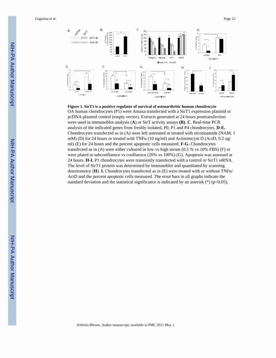
Figure 1. SirT1 is a positive regulator of survival of osteoarthritic human chondrocyteOA human chondrocytes (P1) were Amaxa transfected with a SirT1 expression plasmid orpcDNA plasmid control (empty vector). Extracts generated at 24 hours posttransfectionwere used in immunoblot analysis (A) or SirT activity assays (B). C. Real-time PCRanalysis of the indicated genes from freshly isolated, P0, P1 and P4 chondrocytes. D-E.Chondrocytes transfected as in (A) were left untreated or treated with nicotinamide (NAM, 1mM) (D) for 24 hours or treated with TNFα (10 ng/ml) and Actinomcyin D (ActD, 0.2 ug/ml) (E) for 24 hours and the percent apoptotic cells measured. F-G. Chondrocytestransfected as in (A) were either cultured in low vs high serum (0.5 % vs 10% FBS) (F) orwere plated at subconfluence vs confluence (20% vs 100%) (G). Apoptosis was assessed at24 hours. H-I. P1 chondrocytes were transiently transfected with a control or SirT1 siRNA.The level of SirT1 protein was determined by immunoblot and quantitated by scanningdensitometry (H). I. Chondrocytes transfected as in (E) were treated with or without TNFα/ActD and the percent apoptotic cells measured. The error bars in all graphs indicate thestandard deviation and the statistical significance is indicated by an asterisk (*) (p<0.05).
Gagarina et al. Page 12
Arthritis Rheum. Author manuscript; available in PMC 2011 May 1.
NIH
-PA Author Manuscript
NIH
-PA Author Manuscript
NIH
-PA Author Manuscript

Figure 2. Activation of the IGF receptor and Akt in human chondrocytes expressing SirT1A. Pathway outlining phosphorylation of Akt by IGF-activated protein kinases and mTOR.pAkt(Thr308) is a target of pPI3Kinase while pAkt(Ser473) is a target of pmTOR. B-E. P1human chondrocytes were transiently Amaxa transfected with a SirT1 expression plasmid ora pcDNA control (empty vector). Extracts were generated at 24 hours posttransfection andused in immunoblot analysis with the indicated antibodies. F. Chondrocytes transfected as in(B) were treated with NAM (1 mM) where indicated, extracts were generated andimmunoblotted with the indicated antibodies. G. Chondrocytes were transfected as in (B),extracts were generated and immunoblotted with the indicated antibodies.
Gagarina et al. Page 13
Arthritis Rheum. Author manuscript; available in PMC 2011 May 1.
NIH
-PA Author Manuscript
NIH
-PA Author Manuscript
NIH
-PA Author Manuscript

Figure 3. Phosphorylation of MDM2 and inactivation of p53 in chondrocytes expressing SirT1A. Pathway outlining MDM2 phosphorylation by Akt. B. P1 human chondrocytes weretransiently Amaxa transfected with a SirT1 expression plasmid or pcDNA control (emptyvector). Extracts generated at 24 hours posttransfection were used in immunoblot analysiswith the indicated antibodies. C. Extracts from (B) were immunoprecipitated with anMDM2 specific antibody were then immunoblotted and probed with the indicatedantibodies. D. Chondrocytes were transfected with a control siRNA or an MDM2 siRNA.The level of MDM2 protein was determined by immunoblot and quantitated by scanningdensitometry (upper panel). Lower Panel; Chondrocytes were transfected with a pcDNAcontrol, the SirT1 plasmid, a control siRNA and an MDM2 siRNA where indicated. Thepercent apoptotic cells was assessed by flow cytometry. E. Chondrocytes were transfectedwith a pcDNA control or the SirT1 plasmid. Extracts were generated and immunoblottedwith the indicated antibodies. F. Chondrocytes transfected with a pcDNA control, a SirT1expressing plasmid, a p53 responsive promoter/luciferase construct or a control promoter/luciferase construct were assayed for luciferase activity. The error bars in all graphs indicatethe standard deviation and the statistical significance is indicated by an asterisk (*) (p<0.05).
Gagarina et al. Page 14
Arthritis Rheum. Author manuscript; available in PMC 2011 May 1.
NIH
-PA Author Manuscript
NIH
-PA Author Manuscript
NIH
-PA Author Manuscript

Figure 4. PTP1B is a potent pro-apoptotic protein in human osteoarthritic chondrocytes, isrepressed by SirT1 and reduces the level of activated IGF ReceptorA-B. P1 human chondrocytes were transfected with a SirT1 expression plasmid or pcDNAcontrol. RNA and protein extracts were used in quantitative RT/PCR (A) or immunoblotanalysis (B) with the indicated antibodies. The blot in B (upper panel) was scanned andrelative protein levels determined (lower panel). C & D. Chondrocytes were transfectedwith a PTP1B or PTP1Bmutant expression plasmid or a pcDNA control. Protein extractswere used in immunoblot analysis with the indicated antibodies (C) or the cells wereprocessed for flow cytometry and the percent apoptotic cells determined (D). E & F.Chondrocytes were transfected with a control siRNA or a PTP1BsiRNA. Protein extractswere used in immunoblot analysis with the indicated antibodies (E) or the cells wereprocessed for flow cytometry and the percent apoptotic cells determined (F). G.Chondrocytes transfected with the PTP1B expression plasmid and the PTP1BsiRNA as in(C & E) were assayed for viable adherent cell numbers at 24 hours posttransfection. Theerror bars in all graphs indicate the standard deviation and the statistical significance isindicated by an asterisk (*) (p<0.05). H-J. Chondrocytes were transfected with a controlsiRNA or a PTP1BsiRNA. H. The cells were left untreated or treated with TNFα (10 ng/ml)and Actinomcyin D (ActD, 0.2 ug/ml) for 24 hours and the percent apoptotic cells measured.I, J. The cells were either cultured in low vs high serum (0.5 % vs 10% FBS) (F) or wereplated at subconfluence vs confluence (20% vs 100%) (G). Apoptosis was assessed at 24hours. K. Chondrocytes were transfected with the PTPalpha, PTPgamma and PTP1Bexpression plasmids. Upper panel; immunoblot of extracts. Lower Panel; assessment ofpercent apoptosis. The error bars in all graphs indicate the standard deviation and thestatistical significance is indicated by an asterisk (*) (p<0.025).
Gagarina et al. Page 15
Arthritis Rheum. Author manuscript; available in PMC 2011 May 1.
NIH
-PA Author Manuscript
NIH
-PA Author Manuscript
NIH
-PA Author Manuscript

Figure 5. SirT1 levels are reduced in OA cartilage where PTP1B levels are elevatedNormal and OA human cartilage samples were processed for immunohistochemistry foreither SirT1 (A), PTP1B (B) or MMP13 (C) (left panels). The percent positive stained cellsper field was determined and is shown in the graphs (right panels). Stained cells arereflective of greater than 10-fold intensity above the background (as determined by scanningdensitometry). An average of 10 fields from three sections of six separate OA and Normalcartilage samples were assessed. Each field was blindly read by two different individuals.The images in A, B and C represent the intermediate layer of cartilage. B. Immunoblot ofextracts from freshly isolated chondrocyte extracts from two normal and two OA cartilagesamples probed with the indicated antibodies. Percent apoptotic cells in the OA and Normalcartilage samples were assessed by TUNEL assay (B). The error bars in all graphs indicatethe standard deviation and the (*) indicates a statistically significant difference (p<0.05).
Gagarina et al. Page 16
Arthritis Rheum. Author manuscript; available in PMC 2011 May 1.
NIH
-PA Author Manuscript
NIH
-PA Author Manuscript
NIH
-PA Author Manuscript

Figure 6. SirT1-mediated activation of the IGFR pathway and suppression of apoptosis inhuman chondrocytesThe pathway outlines that the proteins are affected by increased expression of Sirt1 inchondrocytes. A reduction in PTP1B levels directly by SirT1 led to activation of the IGFRalong with the attendant kinases PI3K, PDK1 and Akt. Activation of mTOR is thought tooccur via receptor tyrosine kinases through unknown intermediates. pAkt also functions toactivate mTOR by phosphorylation, leading to increased phosphorylation of pAkt (onresidue Ser473). Sirt1 also functions to directly block p53 by deacetylation.
Gagarina et al. Page 17
Arthritis Rheum. Author manuscript; available in PMC 2011 May 1.
NIH
-PA Author Manuscript
NIH
-PA Author Manuscript
NIH
-PA Author Manuscript


