Embed Size (px)
Citation preview
Tumor and Stem Cell Biology
Sirtuin-1 Regulates Acinar-to-Ductal Metaplasia andSupports Cancer Cell Viability in Pancreatic Cancer
Elke Wauters2, Victor J. Sanchez-Ar�evalo Lobo4, Andreia V. Pinho1,4, Amanda Mawson1, Daniel Herranz5,Jianmin Wu1, Mark J. Cowley1, Emily K. Colvin1, Erna Ngwayi Njicop2, Rob L. Sutherland1, Tao Liu6,Manuel Serrano5, Luc Bouwens2, Francisco X. Real4,7, Andrew V. Biankin1, and Ilse Rooman1,2,3
AbstractThe exocrine pancreas can undergo acinar-to-ductal metaplasia (ADM), as in the case of pancreatitis where
precursor lesions of pancreatic ductal adenocarcinoma (PDAC) can arise. The NADþ-dependent proteindeacetylase Sirtuin-1 (Sirt1) has been implicated in carcinogenesis with dual roles depending on its subcellularlocalization. In this study, we examined the expression and the role of Sirt1 in different stages of pancreaticcarcinogenesis, i.e. ADM models and established PDAC. In addition, we analyzed the expression of KIAA1967, akey mediator of Sirt1 function, along with potential Sirt1 downstream targets. Sirt1 was co-expressed withKIAA1967 in the nuclei of normal pancreatic acinar cells. In ADM, Sirt1 underwent a transient nuclear-to-cytoplasmic shuttling. Experiments where during ADM, we enforced repression of Sirt1 shuttling, inhibitionof Sirt1 activity or modulation of its expression, all underscore that the temporary decrease of nuclear andincrease of cytoplasmic Sirt1 stimulate ADM. Our results further underscore that important transcriptionalregulators of acinar differentiation, that is, Pancreatic transcription factor-1a and b-catenin can bedeacetylated by Sirt1. Inhibition of Sirt1 is effective in suppression of ADM and in reducing cell viabilityin established PDAC tumors. KIAA1967 expression is differentially downregulated in PDAC and impacts onthe sensitivity of PDAC cells to the Sirt1/2 inhibitor Tenovin-6. In PDAC, acetylation of b-catenin isnot affected, unlike p53, a well-characterized Sirt1-regulated protein in tumor cells. Our results revealthat Sirt1 is an important regulator and potential therapeutic target in pancreatic carcinogenesis. Cancer Res;73(7); 2357–67. �2012 AACR.
IntroductionPancreatic ductal adenocarcinoma (PDAC) is the fourth
leading cause of cancer death in the United States, an outcomethat has not changed for 50 years (1). Understanding themolecular mechanisms of PDAC initiation and tumor mainte-nance is imperative to develop chemoprevention and therapeu-tic strategies. New insights in PDAC initiation show that adultexocrine acinar cells under stress can dedifferentiate and gain
metaplastic ductal characteristics (referred to as acinar-to-duc-tal metaplasia, ADM). There is compelling evidence frommousemodels that ADM is a precursor lesion of PDAC (2, 3). ADM alsooccurs in pancreatitis, which may explain why pancreatitis is amajor risk factor for PDAC (2, 3). Prevention of ADM andmaintenance of acinar cell differentiation could suppress pan-creatic carcinogenesis. Previously, we observed that nicotin-amide repressed acinar cell dedifferentiation and ADM in cul-ture (4). Nicotinamide is an end product and feedback inhibitorof Sirtuin-mediated protein deacetylation (5).
The repertoire of Sirtuin functions is broader than the role inlongevity for which they were originally identified (6) andSirtuins target a range of nuclear, mitochondrial, and cyto-plasmic proteins. Sirtuins havemultifaceted roles in cell death,differentiation, metabolism, and senescence. Sirtuin-1 (Sirt1),the best studied of the family, also plays roles in cancer and hasbeen reported to be an oncogene as well as a tumor suppressor(7). Related to pancreas, Sirt1 has been studied in islets anddiabetes (8, 9) (10), but only limited evidence exists that Sirt1 isimportant in PDAC (11).
One of the best-characterized regulators of Sirt1 is deletedin breast cancer 1 (Dbc1, KIAA1967; ref. 12). Dbc1 directlyinteracts with Sirt1 and inhibits Sirt1 activity (12, 13). Changesin either Dbc1 or Sirt1 expression result in altered Sirt1-driveneffects.
Authors' Affiliations: 1Cancer Research Program, Garvan Institute ofMedical Research, Sydney, Australia; 2Diabetes Research Center, VrijeUniversiteit Brussel, Brussels, Belgium; 3St Vincent's Clinical School,University New South Wales, Australia; 4Programa de Patología Molecularand 5Programa de Oncología Molecular, CNIO (Spanish National CancerResearch Center), Madrid, Spain; 6Children's Cancer Institute Australia forMedical Research, Randwick, Australia; and 7Departament de Ci�enciesExperimentals i de la Salut, Universitat Pompeu Fabra, Barcelona, Spain
Note: Supplementary data for this article are available at Cancer ResearchOnline (http://cancerres.aacrjournals.org/).
V.J. S.-A. Lobo and A.V. Pinho contributed equally to this work.
Corresponding Author: Ilse Rooman, Cancer Research Program, TheGarvan Institute of Medical Research, The Kinghorn Cancer Centre, 370,Victoria Street, Sydney NSW 2010, Australia. Phone: 612-93555806; Fax:612-93555868; E-mail: [email protected]
doi: 10.1158/0008-5472.CAN-12-3359
�2012 American Association for Cancer Research.
CancerResearch
www.aacrjournals.org 2357
on June 2, 2016. © 2013 American Association for Cancer Research. cancerres.aacrjournals.org Downloaded from
Published OnlineFirst January 31, 2013; DOI: 10.1158/0008-5472.CAN-12-3359
Because there was preliminary evidence that a Sirtuininhibitor impairs ADM and Sirt1 is currently seen as apromising target of therapeutic intervention in other can-cers (14), we aimed to study the expression of Sirt1 and Dbc1in normal exocrine pancreas and during ADM to define thecontext-specific target genes of Sirt1 and to reveal Sirt1effects in PDAC.
Materials and MethodsCell cultures
Primary acinar cell culture protocols were adapted fromprevious works (15, 16). The cell lines 266-6 and AR42J-B13,the PDAC cell lines, and HEK293 cells were obtained fromAmerican Type Culture Collection (ATCC) Cell Biology Col-lection and used within 6 months between resuscitation andexperimentation. The ATCC authentication protocols includetesting for mycoplasma, bacteria, fungi contamination, con-firmation of species identity, and detection of cellular con-tamination or misidentification using COI for interspeciesidentification and DNA profiling as well as cytogenetic anal-ysis, flow cytometry, and immunocytochemistry with consis-tent refinement of cell growth conditions as well as documen-tation systems, ensuring traceability. Cells were cultured withleptomycin B (LMB; 0.25mg/mL, Sigma-Aldrich), nicotinamide(20–40 mmol/L, Sigma), resveratrol (50 mmol/L, Sigma), andTenovin-6 (Cayman Chemical).
Animals and in vivo experimentationPancreatitis and pancreatic duct ligation were conducted as
described previously (17, 18) and approved by the ethicscommittee of the Vrije Universiteit Brussel (Brussels, Belgium)and the Garvan Animal Ethical Committee and were con-ducted in accordance with the Declaration of Helsinki asrevised in 2000.
StatisticsResults are presented as mean � SEM. Data were analyzed
by Prism 5.0 (Student t test or one-sample t test). Unless statedotherwise, the experiments were carried out at least 3 timesindependently. P values are indicated.
ResultsSirt1 and Dbc1 expression in normal exocrine tissue andADM
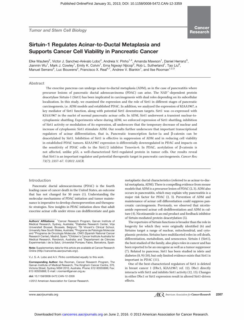
We assessed the expression of Sirt1 and Dbc1 in normalpancreas and mouse models of ADM. Staining of Sirt1 in cellswith the digestive enzyme amylase or with insulin illustratesthat Sirt1 is expressed in the exocrine and endocrine pancreas,with only the latter reported previously (Fig. 1A; refs. 8, 9). Sirt1and Dbc1 were co-expressed in the nuclei of exocrine pancreas(Fig. 1A). Expression was undetectable in about 30% of acinarcells (Supplementary Fig S1A).
In caerulein-induced acute pancreatitis (Cae-AP), aciniundergo ADM and regenerate within 1 week (18). The ADMin our in vivomodels corresponds to what has been describedby Strobel and colleagues (19), that is, tubular complexes and
mucinous metaplastic lesions (MML) and occurs isolated inthe absence of pancreatic intraepithelial neoplasias (PanIN;ref. 20). The transient ADM is characterized by cytoplasmicretention of acinar enzymes, such as carboxypeptidase A1(CpA1; Fig. 1B), increased expression and cytoplasmic accu-mulation of b-catenin and induction of ductal markers askeratin19 (Krt19) (SupplementaryFig. S1BandS1C; refs. 18, 21).Nuclear Dbc1 staining did not change (Fig. 1B). In contrast, theSirt1 staining observed in acini of untreated (Fig. 1A) andcontrol PBS-treated pancreata (Fig. 1B) shifted from the nucle-us to the cytoplasm within the first 2 days of Cae-AP (Fig. 1B).Nuclear Sirt1 expression was reestablished by day 8 (Fig. 1B).We noted that one third of the mice with Cae-AP showedlonger lasting ADM with more cytoplasmic b-catenin andKrt19þ complexes and more prominent cytoplasmic Sirt1(Supplementary Fig. S1B).
Pancreatic duct ligation (PDL) is another model of injurywith acinar tissue replaced byKrt19þ complexes resulting frompermanent ADM and ductal proliferation (ref. 17; Fig. 1C,Supplementary Fig. S2). Dbc1 expression remained unchanged(Supplementary Fig. S2B), but a nuclear-to-cytoplasmic shift ofSirt1 was again found (Fig. 1C). This shift was early andtransient with restoration of nuclear expression within 1 week.The percentage ofmice inwhichwe observed cytoplasmic Sirt1matched the success rate of PDL.
In conclusion, in the 2 in vivo models of ADM, the Dbc1expression pattern was unchanged but an obvious intracel-lular shift of Sirt1 was noted, confined to the exocrine cells,that is, islet cells retained nuclear Sirt1 and Dbc1 (notshown). This observation suggests that in ADM, Sirt1 canhave altered activity and effects on target proteins as a resultof changed interaction with Dbc1 and changed intracellularlocalization.
Functional significance of nuclear-to-cytoplasmicshuttling of Sirt1 in ADM
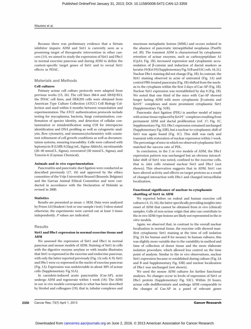
We reported before on rodent and human exocrine cellcultures (4, 15, 16), the latter specifically providing insights intoonset of ADM that cannot be obtained from in vivo (clinical)samples. Cells of non-acinar origin that also can contribute tothe in vivoADM-type lesions are likely not represented in the invitro models.
Again, we observed that, in contrast to the overall nuclearlocalization in normal tissue, the exocrine cells showed man-ifest cytoplasmic Sirt1 staining at the time of cell isolation(Fig. 2A for human and B for mouse). In human cultures, thiswas slightly more variable due to the variability in method andtime of collection of donor tissue and the more elaborateisolation procedure, which allowed less control on the timepoint of analysis. Similar to the in vivo observations, nuclearSirt1 expression became re-established during culture (Fig. 2Aand B and Supplementary Fig. S3B) and nuclear localizationof Dbc1 was unchanged (not shown).
We used the mouse ADM cultures for further functionalanalyses. No changes occur in levels of expression of Sirt1 orDbc1 protein (Supplementary Fig. S3C). Within 24 hours,acinar cells dedifferentiate and undergo ADM comparable tothe changes of Cae-AP in a panel of relevant genes
Wauters et al.
Cancer Res; 73(7) April 1, 2013 Cancer Research2358
on June 2, 2016. © 2013 American Association for Cancer Research. cancerres.aacrjournals.org Downloaded from
Published OnlineFirst January 31, 2013; DOI: 10.1158/0008-5472.CAN-12-3359
(Supplementary Fig. S3A), induction of Krt19 and changedb-catenin expression (Supplementary Fig. S3A and S3B).To address the impact of nuclear-to-cytoplasmic shuttling of
Sirt1, we treated the acinar cells with LMB, an antibiotic thatinhibits nuclear export of proteins with a nuclear export signal,including Sirt1 (22). This resulted in more cells presentingnuclear Sirt1 staining (co-localization with Mist1 was used tolabel the acinar cell nuclei; Fig. 2C). Western blotting con-firmed higher nuclear Sirt1 expression with LMB (Supple-mentary Fig. S3D). LMB restrained the induction of Krt19, aductal marker that is strongly induced under control condi-
tions, as seen in immunofluorescence (IF; Fig. 2C). Consistentwith this, a lesser induction of Krt19 mRNA was observedin LMB conditions (4.1 � 0.8-fold vs. 10.9 � 2.2-fold incontrols, P < 0.04; Fig. 2D). No profound effect on the expres-sion of acinar enzymes or transcription factors was found (notshown).
We explored further whether changes in Sirt1 expressionimpacted on acinar cell differentiation, starting with non-stress conditions. Pancreata from Pdx1-Cre;Sirt1ex4lox/lox miceshow a mutant Sirt1 protein band that migrates slightly fasterin Western blot analysis (Supplementary Fig. S4A), in line with
Figure 1. Sirt1 andDbc1 expressionin normal exocrine tissue and ADM.A, IF detection of Sirt1 and Dbc1 innormal mouse pancreas; co-localization of Dbc1 with Sirt1 (left),Sirt1 with the acinar cell markeramylase (middle), and Sirt1 with theendocrine hormone insulin (right). B,Sirt1 with Dbc1 or the acinar markercarboxypeptidase A (CpA) incaerulein-induced acute pancreatitis(Cae-AP 0–8 days). C, IF detection ofSirt1 and CpA in pancreatic ductligation (PDL, 0–15 days). Pictures ina sequence are taken with the sameexposure times. Inset, highermagnification and arrowpoints to thenuclear-to-cytoplasmic shuttling.
Sirtuin1 in Pancreatic Carcinogenesis
www.aacrjournals.org Cancer Res; 73(7) April 1, 2013 2359
on June 2, 2016. © 2013 American Association for Cancer Research. cancerres.aacrjournals.org Downloaded from
Published OnlineFirst January 31, 2013; DOI: 10.1158/0008-5472.CAN-12-3359
previous study (23). This Sirt1 is an inactive mutant form andSirt1 expression is also strongly suppressed (SupplementaryFig. S4A and S4B). Whereas there is less Pancreatic transcrip-tion factor-1a (Ptf1a), no significant changes occur in theexpression of the acinar genes Amylase 2 (Amy2), Elastase 1(Ela1), Carboxypeptidase A1 (CpA1), Chymotrypsin B1 (Ctrb1),or the recombinant signal binding protein for immunoglobulinkappa J-region-like (Rbpjl) (Fig. 3B, control), suggesting thatabsence of Sirt1 in unstressed conditions does not affect acinardifferentiation. However, upon acute caerulein pancreatitis(Fig. 3A), the role of Sirt1 becomes apparent, with the pan-creata from the Pdx1-Cre;Sirt1ex4lox/lox mice showing a more
profound ADM (Fig. 3A and B), characterized by a highersuppression of acinar enzymes and of the transcription factorsPtf1a and Rbpjl. Similar results were obtained using the in vitroADM model (Supplementary Fig. S4C). In the Pdx1-Cre;Sir-t1ex4lox/lox mice, the ADM was also more persistent withsuppression of acinar markers 8 days post-caerulein. At thistime point, the pancreatic tissue from control mice had almostcompletely recovered (with <10% of affected areas in tissuesections), whereas ADM lesionswere stillmanifest in Pdx1-Cre;Sirt1ex4lox/loxmice (3 of 5 animals with >50% of affected areas intissue sections). We also noticed a reduction in pancreasvolume in the Pdx1-Cre;Sirt1ex4lox/lox (not shown). These
Figure 2. Sirt1 nuclear-to-cytoplasmic shuttling in ADM. A, IFdetection of Sirt1 and the ductalmarker Krt19 in normal humanpancreas and in the in vitro modelof ADM. B, same as A here inmouse tissue and cells. The insetshows the nuclear-to-cytoplasmicshuttling of Sirt1 in detail. Thearrow points at a Krt19-expressingduct in normal pancreas. C, IFdetection of Sirt1 and Krt19 inacinar cells treatedwith the nuclearexport inhibitor LMB (c and d) or itssolvent methanol (a and b). Thearrow points to absence of Sirt1 inthe nucleus of cells under controlmethanol conditions. D, foldchange (FC) inmRNAexpressionofKrt19 in the conditions of C, that is,cultured acini with LMB or itssolvent relative to isolated cells(n ¼ 5–6; �, P < 0.05).
Wauters et al.
Cancer Res; 73(7) April 1, 2013 Cancer Research2360
on June 2, 2016. © 2013 American Association for Cancer Research. cancerres.aacrjournals.org Downloaded from
Published OnlineFirst January 31, 2013; DOI: 10.1158/0008-5472.CAN-12-3359
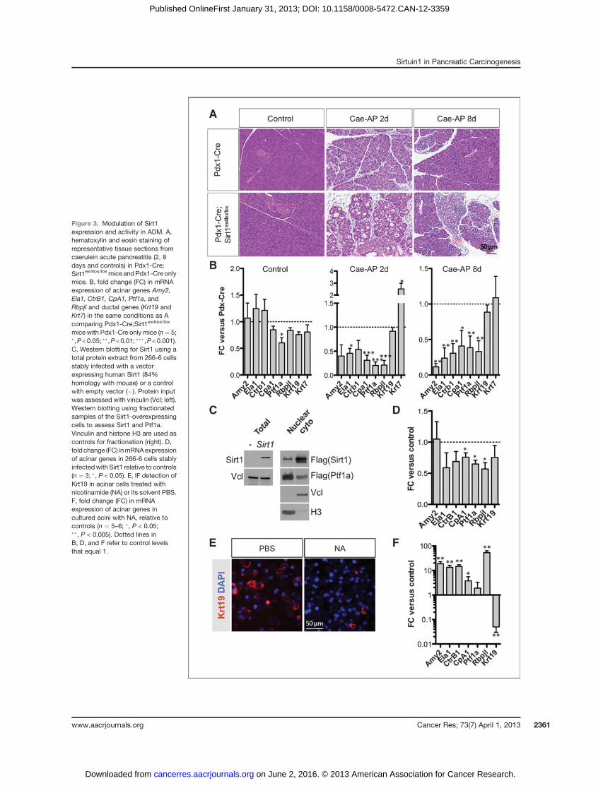
Figure 3. Modulation of Sirt1expression and activity in ADM. A,hematoxylin and eosin staining ofrepresentative tissue sections fromcaerulein acute pancreatitis (2, 8days and controls) in Pdx1-Cre;Sirt1ex4lox/loxmice andPdx1-Cre onlymice. B, fold change (FC) in mRNAexpression of acinar genes Amy2,Ela1, CtrB1, CpA1, Ptf1a, andRbpjl and ductal genes (Krt19 andKrt7) in the same conditions as Acomparing Pdx1-Cre;Sirt1ex4lox/lox
mice with Pdx1-Cre only mice (n¼ 5;�,P<0.05; ��,P<0.01; ���,P<0.001).C, Western blotting for Sirt1 using atotal protein extract from 266-6 cellsstably infected with a vectorexpressing human Sirt1 (84%homology with mouse) or a controlwith empty vector (�). Protein inputwas assessed with vinculin (Vcl; left).Western blotting using fractionatedsamples of the Sirt1-overexpressingcells to assess Sirt1 and Ptf1a.Vinculin and histone H3 are used ascontrols for fractionation (right). D,fold change (FC) inmRNAexpressionof acinar genes in 266-6 cells stablyinfectedwith Sirt1 relative to controls(n ¼ 3; �, P < 0.05). E, IF detection ofKrt19 in acinar cells treated withnicotinamide (NA) or its solvent PBS.F, fold change (FC) in mRNAexpression of acinar genes incultured acini with NA, relative tocontrols (n ¼ 5–6; �, P < 0.05;��, P < 0.005). Dotted lines inB, D, and F refer to control levelsthat equal 1.
Sirtuin1 in Pancreatic Carcinogenesis
www.aacrjournals.org Cancer Res; 73(7) April 1, 2013 2361
on June 2, 2016. © 2013 American Association for Cancer Research. cancerres.aacrjournals.org Downloaded from
Published OnlineFirst January 31, 2013; DOI: 10.1158/0008-5472.CAN-12-3359
results underscore that, under stress conditions, absence ofSirt1 enhances the ADM.
To evaluate whether increased Sirt1 expression also hadeffects on acinar gene expression and ADM, we analyzed aSirt1 transgenic (SirtTg) mouse strain (24) that expressedincreased Sirt1 levels from its endogenous promoter ubiqui-tously, including in acinar cells (Supplementary Fig. S5A), toevaluate whether increased Sirt1 expression had effects on theacinar gene expression. We did not detect significant differ-ences (Supplementary Fig. S5B) either in unstressed or uponpancreatitis/ADM conditions. We note that the Sirt1 over-expression in acinar cells was modest (�2-fold), in line withprevious study (24).
To further study Sirt1 overexpression and address the ques-tionwhether Sirt1 accumulation in the cytoplasm of acinar cellshas a contribution to ADM, we analyzed whether acinar 266-6cells with stable and robust overexpression of Sirt1 mostly con-fined to the cytoplasm (Fig. 3C) had different features.We foundconsistent suppression of CpA1, Ptf1a, and Rbpjl (Fig. 3D).
Finally, we wanted to explore whether pharmacologicinterference could affect ADM. We used a Sirt1 inhibitornicotinamide (NA) in the ADM in vitro model. Nicotinamidetreatment during the first 24 hours, when Sirt1 was pre-dominantly cytoplasmic, resulted in a suppression of Krt19induction, noted by IF and reverse transcription-quantita-tive PCR (RT-qPCR) (Fig. 3E and F). In addition, higherexpression of the acinar cell–specific markers Amy2, Ela1,Ctrb1, CpA1, and Rbpjl was maintained (Fig. 3F). SimilarADM repression was observed in preliminary experimentswith human acinar cells exposed to nicotinamide (Supple-mentary Fig. S6A and S6B).
In conclusion, loss of nuclear Sirt1 and cytoplasmic accu-mulation both contribute to ADM. Inhibition of the shuttlingactivity of Sirt1 during ADM restrains the process.
Sirt1 deacetylates b-catenin and Ptf1aSirt1 can deacetylate b-catenin, a posttranslational modifi-
cation that impairs its transcriptional activity (25), and mod-ulatesWnt/b-catenin signaling, as in colon cancer (26). Loss ofb-catenin signaling is decisive in installing persistent ADMandimpairs acinar cell regeneration (21).
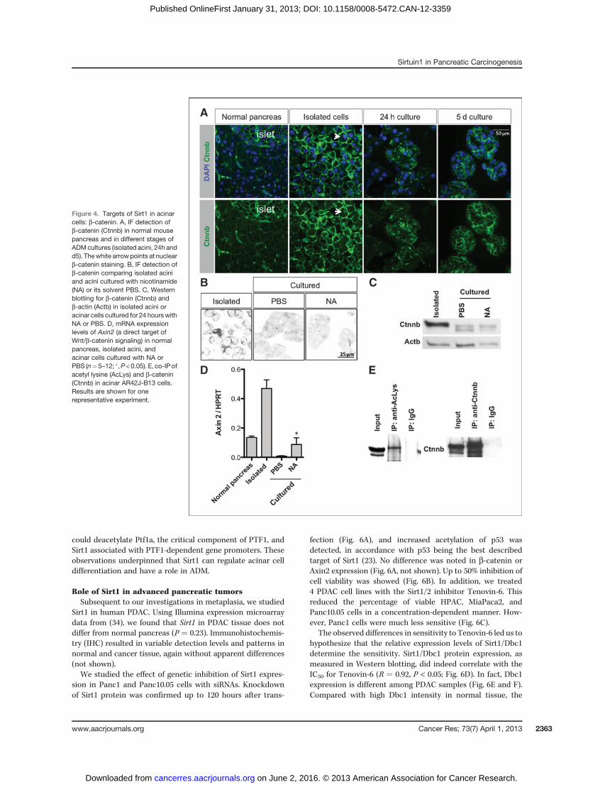
In acini, b-catenin is associated with the plasma membrane(Fig. 4A). A cytoplasmic localization of b-catenin has beenreported in ADM (21, 27, 28) and correlates well with thoseconditions in which we showed a predominant cytoplasmicSirt1. Therefore, we investigated a possible relation of b-cate-nin and Sirt1 in ADM.
First, we analyzed the cellular localization of b-catenin:in isolated acini, preferential membrane and occasional nucle-ar staining were seen. A cytoplasmic distribution predomi-nated at 24 hours with a redistribution to the membrane by5 days of culture (Fig. 4A). This transient cytoplasmic stainingwas not observed in presence of nicotinamide (Fig. 4B). InWestern blotting, 2 bands were detected: The higher bandlikely results from posttranslational modifications (see below)and persisted upon treatment with nicotinamide, whereas thelower band was induced in controls (Fig. 4C). In Cae-AP in vivo,we found a similar pattern of b-catenin in immunostaining
(Supplementary Fig. S1B and S1C) and inWestern blot analysis(not shown).
We then used AR42J-B13 cells to further investigate thefunctional interaction of Sirt1 with b-catenin in an acinarcell context. We chose these cells because Sirt1 was predom-inantly localized in the cytoplasm (Supplementary Fig. S7A).The 2 reported isoforms of Sirt1 were detected (not shown).Sirt1 co-localized and interacted with b-catenin, as shownby IF, the Duolink assay and co-immunopreciptitation (IP;Supplementary Fig. S7A and S7B). Using co-IP, we detectedthe Sirt1 isoform that has the highest deacetylase activity (29).As of b-catenin, 2 bands were detected (Fig. 4E), consistentwith findings by others (30). By co-IP, we showed that theupper b-catenin band corresponded to an acetylated form(Fig. 4E). Exposure to resveratrol to stimulate Sirt1 activity(31) resulted in a 3.2-fold increased density of the lowerdeacetylated b-catenin band (P < 0.05, n ¼ 4), specificallywithin the cytoplasm (Supplementary Fig. S7C). Of note, theupper band was maintained when nicotinamide was appliedto the primary culture model (Fig. 4C).
We then examined whether our observations are related toWnt/b-catenin signaling. Axin2, a Wnt/b-catenin target gene,was induced upon acinar cell isolation but was highly reducedat 24 hours in control (PBS) ADM cultures (Fig. 4D). Thisparalleled the IF data where membrane-associated b-cateninand accumulation of b-catenin in the nucleus were onlyobserved in isolated acini (Fig. 4A). With nicotinamide, theexpression of Axin2 was maintained at higher levels (Fig. 4D).We confirmed this effect of nicotinamide on Wnt/b-cateninsignaling by comparing a larger panel of targets and regulatorsof the pathway (Supplementary Fig. S8A and S8B).
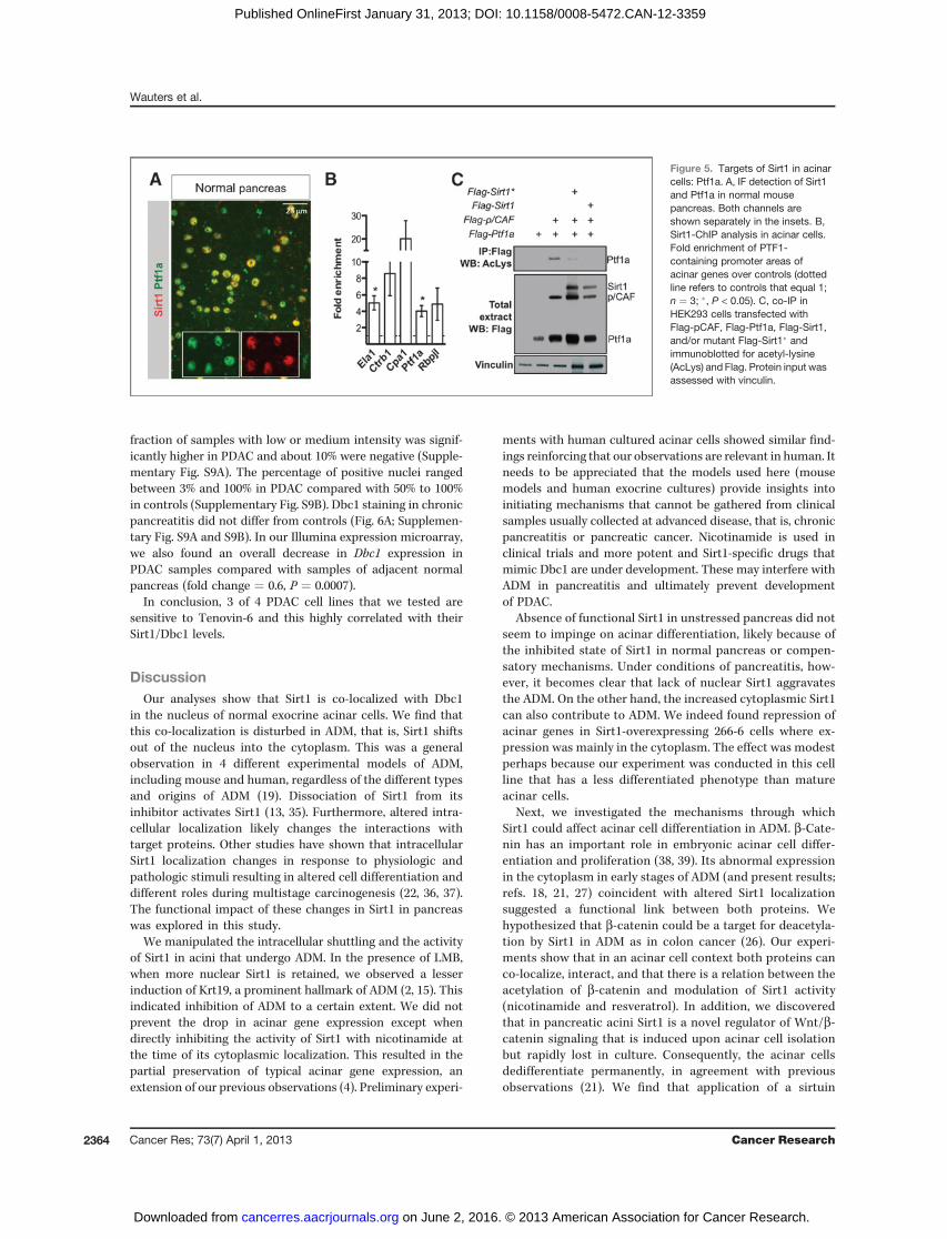
The nuclear co-localization of Sirt1 and Ptf1a in normalacini (Fig. 5A), the changes in subcellular localization ofSirt1, and the changes in digestive enzyme levels in ADMsuggested that Sirt1 might contribute to regulate the func-tion of PTF1, the master regulator of the acinar program.PTF1 is a transcription factor complex composed of Ptf1a,Rbpjl, and a class A bHLH protein (32). P300/CBP-associatedfactor (pCAF) is associated to it, acetylates Ptf1a, andensures high transcriptional activity of Ptf1a (33). We under-took chromatin immunopreciptitation (ChIP) experimentsto verify whether Sirt1 localized together with Ptf1a in thePTF1-binding sites of acinar gene promoters. CpA1, CtrB1,Ela1, Ptf1a, and Rbpjl promoter areas were indeed 3- to 30-fold enriched in anti-Sirt1 ChIPs (Fig. 5B). To assess whetherSirt1 and Ptf1a interact and impact on the acetylation state,Flag-pCAF, Flag-Ptf1a, Flag-Sirt1, and Flag-Sirt1� [a mutantSirt1 in which a critical histidine in the deacetylase domainhas been replaced by a tyrosine residue (H363Y)] weretransfected in HEK293 cells and assayed by co-IP. The resultsconfirmed that Ptf1a became acetylated upon co-transfec-tion of pCAF (as reported in ref. 33), and there is loss ofacetylation of Ptf1a when co-transfected with Sirt1, but notwhen co-transfected with the inactive Sirt1� (Fig. 5C). Therewere no effects on Rbpjl (not shown).
Together, our findings showed altered acetylation of theSirt1 target b-catenin in the context of ADM and therebyinterference with b-catenin/Wnt signaling. Furthermore, Sirt1
Wauters et al.
Cancer Res; 73(7) April 1, 2013 Cancer Research2362
on June 2, 2016. © 2013 American Association for Cancer Research. cancerres.aacrjournals.org Downloaded from
Published OnlineFirst January 31, 2013; DOI: 10.1158/0008-5472.CAN-12-3359
could deacetylate Ptf1a, the critical component of PTF1, andSirt1 associated with PTF1-dependent gene promoters. Theseobservations underpinned that Sirt1 can regulate acinar celldifferentiation and have a role in ADM.
Role of Sirt1 in advanced pancreatic tumorsSubsequent to our investigations in metaplasia, we studied
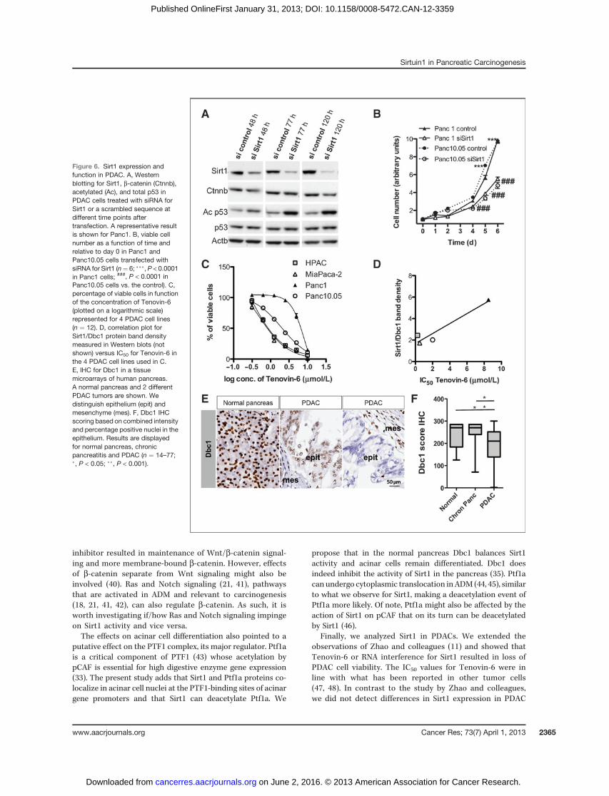
Sirt1 in human PDAC. Using Illumina expression microarraydata from (34), we found that Sirt1 in PDAC tissue does notdiffer from normal pancreas (P ¼ 0.23). Immunohistochemis-try (IHC) resulted in variable detection levels and patterns innormal and cancer tissue, again without apparent differences(not shown).We studied the effect of genetic inhibition of Sirt1 expres-
sion in Panc1 and Panc10.05 cells with siRNAs. Knockdownof Sirt1 protein was confirmed up to 120 hours after trans-
fection (Fig. 6A), and increased acetylation of p53 wasdetected, in accordance with p53 being the best describedtarget of Sirt1 (23). No difference was noted in b-catenin orAxin2 expression (Fig. 6A, not shown). Up to 50% inhibition ofcell viability was showed (Fig. 6B). In addition, we treated4 PDAC cell lines with the Sirt1/2 inhibitor Tenovin-6. Thisreduced the percentage of viable HPAC, MiaPaca2, andPanc10.05 cells in a concentration-dependent manner. How-ever, Panc1 cells were much less sensitive (Fig. 6C).
The observed differences in sensitivity to Tenovin-6 led us tohypothesize that the relative expression levels of Sirt1/Dbc1determine the sensitivity. Sirt1/Dbc1 protein expression, asmeasured in Western blotting, did indeed correlate with theIC50 for Tenovin-6 (R ¼ 0.92, P < 0.05; Fig. 6D). In fact, Dbc1expression is different among PDAC samples (Fig. 6E and F).Compared with high Dbc1 intensity in normal tissue, the
Figure 4. Targets of Sirt1 in acinarcells: b-catenin. A, IF detection ofb-catenin (Ctnnb) in normal mousepancreas and in different stages ofADMcultures (isolated acini, 24h andd5). Thewhite arrowpoints at nuclearb-catenin staining. B, IF detection ofb-catenin comparing isolated aciniand acini cultured with nicotinamide(NA) or its solvent PBS. C, Westernblotting for b-catenin (Ctnnb) andb-actin (Actb) in isolated acini oracinar cells cultured for 24 hours withNA or PBS. D, mRNA expressionlevels of Axin2 (a direct target ofWnt/b-catenin signaling) in normalpancreas, isolated acini, andacinar cells cultured with NA orPBS (n¼5–12; �,P<0.05). E, co-IP ofacetyl lysine (AcLys) and b-catenin(Ctnnb) in acinar AR42J-B13 cells.Results are shown for onerepresentative experiment.
Sirtuin1 in Pancreatic Carcinogenesis
www.aacrjournals.org Cancer Res; 73(7) April 1, 2013 2363
on June 2, 2016. © 2013 American Association for Cancer Research. cancerres.aacrjournals.org Downloaded from
Published OnlineFirst January 31, 2013; DOI: 10.1158/0008-5472.CAN-12-3359
fraction of samples with low or medium intensity was signif-icantly higher in PDAC and about 10% were negative (Supple-mentary Fig. S9A). The percentage of positive nuclei rangedbetween 3% and 100% in PDAC compared with 50% to 100%in controls (Supplementary Fig. S9B). Dbc1 staining in chronicpancreatitis did not differ from controls (Fig. 6A; Supplemen-tary Fig. S9A and S9B). In our Illumina expression microarray,we also found an overall decrease in Dbc1 expression inPDAC samples compared with samples of adjacent normalpancreas (fold change ¼ 0.6, P ¼ 0.0007).
In conclusion, 3 of 4 PDAC cell lines that we tested aresensitive to Tenovin-6 and this highly correlated with theirSirt1/Dbc1 levels.
DiscussionOur analyses show that Sirt1 is co-localized with Dbc1
in the nucleus of normal exocrine acinar cells. We find thatthis co-localization is disturbed in ADM, that is, Sirt1 shiftsout of the nucleus into the cytoplasm. This was a generalobservation in 4 different experimental models of ADM,including mouse and human, regardless of the different typesand origins of ADM (19). Dissociation of Sirt1 from itsinhibitor activates Sirt1 (13, 35). Furthermore, altered intra-cellular localization likely changes the interactions withtarget proteins. Other studies have shown that intracellularSirt1 localization changes in response to physiologic andpathologic stimuli resulting in altered cell differentiation anddifferent roles during multistage carcinogenesis (22, 36, 37).The functional impact of these changes in Sirt1 in pancreaswas explored in this study.
We manipulated the intracellular shuttling and the activityof Sirt1 in acini that undergo ADM. In the presence of LMB,when more nuclear Sirt1 is retained, we observed a lesserinduction of Krt19, a prominent hallmark of ADM (2, 15). Thisindicated inhibition of ADM to a certain extent. We did notprevent the drop in acinar gene expression except whendirectly inhibiting the activity of Sirt1 with nicotinamide atthe time of its cytoplasmic localization. This resulted in thepartial preservation of typical acinar gene expression, anextension of our previous observations (4). Preliminary experi-
ments with human cultured acinar cells showed similar find-ings reinforcing that our observations are relevant in human. Itneeds to be appreciated that the models used here (mousemodels and human exocrine cultures) provide insights intoinitiating mechanisms that cannot be gathered from clinicalsamples usually collected at advanced disease, that is, chronicpancreatitis or pancreatic cancer. Nicotinamide is used inclinical trials and more potent and Sirt1-specific drugs thatmimic Dbc1 are under development. These may interfere withADM in pancreatitis and ultimately prevent developmentof PDAC.
Absence of functional Sirt1 in unstressed pancreas did notseem to impinge on acinar differentiation, likely because ofthe inhibited state of Sirt1 in normal pancreas or compen-satory mechanisms. Under conditions of pancreatitis, how-ever, it becomes clear that lack of nuclear Sirt1 aggravatesthe ADM. On the other hand, the increased cytoplasmic Sirt1can also contribute to ADM. We indeed found repression ofacinar genes in Sirt1-overexpressing 266-6 cells where ex-pression was mainly in the cytoplasm. The effect was modestperhaps because our experiment was conducted in this cellline that has a less differentiated phenotype than matureacinar cells.
Next, we investigated the mechanisms through whichSirt1 could affect acinar cell differentiation in ADM. b-Cate-nin has an important role in embryonic acinar cell differ-entiation and proliferation (38, 39). Its abnormal expressionin the cytoplasm in early stages of ADM (and present results;refs. 18, 21, 27) coincident with altered Sirt1 localizationsuggested a functional link between both proteins. Wehypothesized that b-catenin could be a target for deacetyla-tion by Sirt1 in ADM as in colon cancer (26). Our experi-ments show that in an acinar cell context both proteins canco-localize, interact, and that there is a relation between theacetylation of b-catenin and modulation of Sirt1 activity(nicotinamide and resveratrol). In addition, we discoveredthat in pancreatic acini Sirt1 is a novel regulator of Wnt/b-catenin signaling that is induced upon acinar cell isolationbut rapidly lost in culture. Consequently, the acinar cellsdedifferentiate permanently, in agreement with previousobservations (21). We find that application of a sirtuin
Figure 5. Targets of Sirt1 in acinarcells: Ptf1a. A, IF detection of Sirt1and Ptf1a in normal mousepancreas. Both channels areshown separately in the insets. B,Sirt1-ChIP analysis in acinar cells.Fold enrichment of PTF1-containing promoter areas ofacinar genes over controls (dottedline refers to controls that equal 1;n ¼ 3; �, P < 0.05). C, co-IP inHEK293 cells transfected withFlag-pCAF, Flag-Ptf1a, Flag-Sirt1,and/or mutant Flag-Sirt1� andimmunoblotted for acetyl-lysine(AcLys) and Flag. Protein input wasassessed with vinculin.
Wauters et al.
Cancer Res; 73(7) April 1, 2013 Cancer Research2364
on June 2, 2016. © 2013 American Association for Cancer Research. cancerres.aacrjournals.org Downloaded from
Published OnlineFirst January 31, 2013; DOI: 10.1158/0008-5472.CAN-12-3359
inhibitor resulted in maintenance of Wnt/b-catenin signal-ing and more membrane-bound b-catenin. However, effectsof b-catenin separate from Wnt signaling might also beinvolved (40). Ras and Notch signaling (21, 41), pathwaysthat are activated in ADM and relevant to carcinogenesis(18, 21, 41, 42), can also regulate b-catenin. As such, it isworth investigating if/how Ras and Notch signaling impingeon Sirt1 activity and vice versa.The effects on acinar cell differentiation also pointed to a
putative effect on the PTF1 complex, its major regulator. Ptf1ais a critical component of PTF1 (43) whose acetylation bypCAF is essential for high digestive enzyme gene expression(33). The present study adds that Sirt1 and Ptf1a proteins co-localize in acinar cell nuclei at the PTF1-binding sites of acinargene promoters and that Sirt1 can deacetylate Ptf1a. We
propose that in the normal pancreas Dbc1 balances Sirt1activity and acinar cells remain differentiated. Dbc1 doesindeed inhibit the activity of Sirt1 in the pancreas (35). Ptf1acan undergo cytoplasmic translocation inADM (44, 45), similarto what we observe for Sirt1, making a deacetylation event ofPtf1a more likely. Of note, Ptf1a might also be affected by theaction of Sirt1 on pCAF that on its turn can be deacetylatedby Sirt1 (46).
Finally, we analyzed Sirt1 in PDACs. We extended theobservations of Zhao and colleagues (11) and showed thatTenovin-6 or RNA interference for Sirt1 resulted in loss ofPDAC cell viability. The IC50 values for Tenovin-6 were inline with what has been reported in other tumor cells(47, 48). In contrast to the study by Zhao and colleagues,we did not detect differences in Sirt1 expression in PDAC
Figure 6. Sirt1 expression andfunction in PDAC. A, Westernblotting for Sirt1, b-catenin (Ctnnb),acetylated (Ac), and total p53 inPDAC cells treated with siRNA forSirt1 or a scrambled sequence atdifferent time points aftertransfection. A representative resultis shown for Panc1. B, viable cellnumber as a function of time andrelative to day 0 in Panc1 andPanc10.05 cells transfected withsiRNA for Sirt1 (n¼ 6; ���, P < 0.0001in Panc1 cells; ###, P < 0.0001 inPanc10.05 cells vs. the control). C,percentage of viable cells in functionof the concentration of Tenovin-6(plotted on a logarithmic scale)represented for 4 PDAC cell lines(n ¼ 12). D, correlation plot forSirt1/Dbc1 protein band densitymeasured in Western blots (notshown) versus IC50 for Tenovin-6 inthe 4 PDAC cell lines used in C.E, IHC for Dbc1 in a tissuemicroarrays of human pancreas.A normal pancreas and 2 differentPDAC tumors are shown. Wedistinguish epithelium (epit) andmesenchyme (mes). F, Dbc1 IHCscoring based on combined intensityand percentage positive nuclei in theepithelium. Results are displayedfor normal pancreas, chronicpancreatitis and PDAC (n ¼ 14–77;�, P < 0.05; ��, P < 0.001).
Sirtuin1 in Pancreatic Carcinogenesis
www.aacrjournals.org Cancer Res; 73(7) April 1, 2013 2365
on June 2, 2016. © 2013 American Association for Cancer Research. cancerres.aacrjournals.org Downloaded from
Published OnlineFirst January 31, 2013; DOI: 10.1158/0008-5472.CAN-12-3359
compared with normal tissue. However, we found a decreaseof Dbc1 in PDAC tissues, including a subset with undetect-able expression. Loss of Dbc1 has been reported in breast,colon, and lung cancer (12). We propose that variation inDbc1 is relevant in PDAC as Dbc1 directly determines Sirt1activity (12, 13, 49). The recent finding that c-myc activatesSirt1, and as such favors tumor growth, by sequesteringDbc1 from Sirt1 supports this hypothesis (50). We appliedan assay for quantifying Sirt1 activity (13) but failed to getSirt1 specific results for our pancreatic tissue and cellextracts (not shown). In addition, we found that PDAC celllines respond variably to Tenovin-6 and that this correlateswith the levels of Sirt1/Dbc1. Our data therefore suggestthat Dbc1 can be a biomarker for those pancreatic tumorsthat benefit from Sirt1-inhibitory drugs.
We did not investigate Sirt1 target genes in PDAC cells, butpreliminary data point out that there is no effect on b-catenin(Fig. 6A). A role for p53 (Fig. 6A), the best-characterized targetof Sirt1, was beyond the scope of our study.
In conclusion, this is the first study that examines the roleof Sirt1 on differentiation and tumor maintenance in the exo-crine pancreas, contributing to our understanding of thosemechanisms that could be harnessed in therapeutic control.
Disclosure of Potential Conflicts of InterestNo potential conflicts of interest were disclosed.
Authors' ContributionsConception and design: V.J.S.-A. Lobo, E.N. Njicop, L. Bouwens, A.V. Biankin,I. Rooman, E. Wauters
Development of methodology: V.J.S.-A. Lobo, A. Mawson, E.N. Njicop,L. Bouwens, I. Rooman, E. Wauters, A.V. PinhoAcquisition of data (provided animals, acquired and managed patients,provided facilities, etc.): E. Wauters, A.V. Pinho, A. Mawson, E.K. Colvin, E.N.Njicop, T. Liu,M. Serrano, L. Bouwens, F.X. Real, A.V.Biankin, I. Rooman,D.HerranzAnalysis and interpretation of data (e.g., statistical analysis, biostatistics,computational analysis): E. Wauters, A.V. Pinho, A. Mawson, J. Wu, M.J.Cowley, T. Liu, F.X. Real, I. RoomanWriting, review, and/or revision of the manuscript: V.J.S.-A. Lobo, A.V.Pinho, A. Mawson, T. Liu, L. Bouwens, F.X. Real, A.V. Biankin, I. Rooman,E. Wauters, A.V. PinhoAdministrative, technical, or material support (i.e., reporting or organiz-ing data, constructing databases): R.L. Sutherland, A.V. Biankin, I. RoomanStudy supervision: R.L. Sutherland, L. Bouwens, I. Rooman
AcknowledgmentsThe authors thank E. De Blay, W. Rabiot, C. Mehiri, I. Mathijs, J. Lardon, G.
Stang�e, L. Baeyens, G. Martens, C. Hang Ho, M. Flandez, N. del Pozo, Y. C�ecilia,and S. Velasco for assisting in experiments and sharing of tools; M. Dufresne forproviding the anti-Ptf1a antibody; and P. Croucher and C. Ormandy for criticalreading of the manuscript.
Grant SupportThis work was supported by funding fromCancer Institute New SouthWales-
Australia-10FRL203 fellowship (I. Rooman), Vrije Universiteit Brussel-OZR (I.Rooman), Francqui Foundation (I. Rooman), grants SAF2007-60860, SAF2011-29530, and ONCOBIO Consolider from Ministerio de Ciencia e Innovaci�on(Madrid, Spain) to F.X. Real. E. Wauters is a research fellow of the Fund forScientific Research Flanders.
The costs of publication of this article were defrayed in part by the payment ofpage charges. This article must therefore be hereby marked advertisement inaccordance with 18 U.S.C. Section 1734 solely to indicate this fact.
Received August 24, 2012; revised November 20, 2012; accepted December 4,2012; published OnlineFirst January 31, 2013.
References1. Siegel R, Naishadham D, Jemal A. Cancer statistics, 2012. CA Cancer
J Clin 2012;62:10–29.2. Rooman I, Real FX. Pancreatic ductal adenocarcinoma and
acinar cells: amatter of differentiation and development?Gut 2012;61:449–58.
3. Perez-Mancera PA, Guerra C, BarbacidM, TuvesonDA.What we havelearned about pancreatic cancer from mouse models. Gastroenterol-ogy 2012;142:1079–92.
4. Rooman I, Heremans Y, Heimberg H, Bouwens L. Modulation of ratpancreatic acinoductal transdifferentiation and expression of PDX-1in vitro. Diabetologia 2000;43:907–14.
5. Michan S, Sinclair D. Sirtuins in mammals: insights into their biologicalfunction. Biochem J 2007;404:1–13.
6. Herranz D, Serrano M. SIRT1: recent lessons frommousemodels. NatRev Cancer 2010;10:819–23.
7. Bosch-Presegue L, Vaquero A. The dual role of sirtuins in cancer.Genes Cancer 2011;2:648–62.
8. Lee JH, Song MY, Song EK, Kim EK, Moon WS, Han MK, et al.Overexpression of SIRT1 protects pancreatic beta-cells against cyto-kine toxicity by suppressing the nuclear factor-kappaB signalingpathway. Diabetes 2009;58:344–51.
9. Bordone L, Motta MC, Picard F, Robinson A, Jhala US, Apfeld J, et al.Sirt1 regulates insulin secretion by repressingUCP2 in pancreatic betacells. PLoS Biol 2006;4:e31.
10. Bastien-Dionne PO, Valenti L, Kon N, Gu W, Buteau J. Glucagon-likepeptide 1 inhibits the sirtuin deacetylase SirT1 to stimulate pancreaticbeta-cell mass expansion. Diabetes 2011;60:3217–22.
11. Zhao G, Cui J, Zhang JG, Qin Q, Chen Q, Yin T, et al. SIRT1 RNAiknockdown induces apoptosis and senescence, inhibits invasion andenhances chemosensitivity in pancreatic cancer cells. Gene Ther2011;18:920–8.
12. Kim JE, Chen J, Lou Z. p30 DBC is a potential regulator of tumori-genesis. Cell Cycle 2009;8:2932–5.
13. Nin V, Escande C, Chini CC, Giri S, Camacho-Pereira J, Matalonga J,et al. Role of Deleted in Breast Cancer 1 (DBC1) protein in SIRT1deacetylase activation inducedbyprotein kinaseAandAMP-activatedprotein kinase. J Biol Chem 2012;287:23489–501.
14. Li L,Wang L,Wang Z, HoY,McDonald T, Holyoake TL, et al. Activationof p53 by SIRT1 inhibition enhances elimination of CML leukemia stemcells in combination with imatinib. Cancer Cell 2012;21:266–81.
15. Pinho AV, Rooman I, Reichert M, De Medts N, Bouwens L, RustgiAK, et al. Adult pancreatic acinar cells dedifferentiate to an embry-onic progenitor phenotype with concomitant activation of a senes-cence programme that is present in chronic pancreatitis. Gut 2011;60:958–66.
16. Houbracken I, De Waele E, Lardon J, Ling Z, Heimberg H, Rooman I,et al. Lineage tracing evidence for transdifferentiation of acinar to ductcells and plasticity of human pancreas. Gastroenterology 2011;141:731–41.
17. Xu X, D'Hoker J, Stange G, Bonne S, De Leu N, Xiao X, et al. Beta cellscan be generated fromendogenous progenitors in injured adultmousepancreas. Cell 2008;132:197–207.
18. Jensen JN, Cameron E, Garay MV, Starkey TW, Gianani R, JensenJ. Recapitulation of elements of embryonic development inadult mouse pancreatic regeneration. Gastroenterology 2005;128:728–41.
19. Strobel O, Dor Y, Alsina J, StirmanA, LauwersG, Trainor A, et al. In vivolineage tracing defines the role of acinar-to-ductal transdifferentia-tion in inflammatory ductal metaplasia. Gastroenterology 2007;133:1999–2009.
20. Hong SM, Heaphy CM, Shi C, Eo SH, Cho H, Meeker AK, et al.Telomeres are shortened in acinar-to-ductal metaplasia lesions
Wauters et al.
Cancer Res; 73(7) April 1, 2013 Cancer Research2366
on June 2, 2016. © 2013 American Association for Cancer Research. cancerres.aacrjournals.org Downloaded from
Published OnlineFirst January 31, 2013; DOI: 10.1158/0008-5472.CAN-12-3359
associated with pancreatic intraepithelial neoplasia but not in isolatedacinar-to-ductal metaplasias. Mod Pathol 2011;24:256–66.
21. Morris JPt, Cano DA, Sekine S, Wang SC, Hebrok M. Beta-cateninblocks Kras-dependent reprogramming of acini into pancreatic cancerprecursor lesions in mice. J Clin Invest 2010;120:508–20.
22. Tanno M, Sakamoto J, Miura T, Shimamoto K, Horio Y. Nucleocyto-plasmic shuttling of the NADþ-dependent histone deacetylase SIRT1.J Biol Chem 2007;282:6823–32.
23. Cheng H-L, Mostoslavsky R, Saito Si, Manis JP, Gu Y, Patel P, et al.Developmental defects and p53 hyperacetylation in Sir2 homo-log (SIRT1)-deficient mice. Proc Natl Acad Sci U S A 2003;100:10794–9.
24. Pfluger PT, Herranz D, Velasco-Miguel S, SerranoM, TschopMH. Sirt1protects against high-fat diet-induced metabolic damage. Proc NatlAcad Sci U S A 2008;105:9793–8.
25. Ge X, Jin Q, Zhang F, Yan T, Zhai Q. PCAF acetylates {beta}-cateninand improves its stability. Mol Biol Cell 2009;20:419–27.
26. FiresteinR, Blander G,MichanS,Oberdoerffer P, OginoS, Campbell J,et al. The SIRT1 deacetylase suppresses intestinal tumorigenesis andcolon cancer growth. PLoS One 2008;3:e2020.
27. Minami K, Okano H, Okumachi A, Seino S. Role of cadherin-mediatedcell-cell adhesion in pancreatic exocrine-to-endocrine transdifferen-tiation. J Biol Chem 2008;283:13753–61.
28. Lerch MM, Lutz MP, Weidenbach H, Muller-Pillasch F, Gress TM,Leser J, et al. Dissociation and reassembly of adherens junctionsduring experimental acute pancreatitis. Gastroenterology 1997;113:1355–66.
29. Lynch CJ, Shah ZH, Allison SJ, Ahmed SU, Ford J, Warnock LJ, et al.SIRT1 undergoes alternative splicing in a novel auto-regulatory loopwith p53. PLoS One 2010;5:e13502.
30. Wallace K, Marek CJ, Hoppler S, Wright MC. Glucocorticoid-depen-dent transdifferentiation of pancreatic progenitor cells into hepato-cytes is dependent on transient suppression of WNT signalling. J CellSci 2010;123:2103–10.
31. Alcain FJ, Villalba JM. Sirtuin activators. Expert Opin Ther Pat 2009;19:403–14.
32. Masui T, Swift GH, Deering T, Shen C, Coats WS, Long Q, et al.Replacement of Rbpj with Rbpjl in the PTF1 complex controls the finalmaturation of pancreatic acinar cells. Gastroenterology 2010;139:270–80.
33. Rodolosse A, CamposM-L, Rooman I, LichtensteinM, Real FX. p/CAFmodulates the activity of the transcription factor p48/Ptf1a involved inpancreatic acinar differentiation. Biochem J 2009;418:463–73.
34. Biankin AV, Waddell N, Kassahn KS, Gingras MC, Muthuswamy LB,Johns AL, et al. Pancreatic cancer genomes reveal aberrations in axonguidance pathway genes. Nature 2012;491:399–405.
35. Escande C, Chini CC, Nin V, Dykhouse KM, Novak CM, Levine J, et al.Deleted in breast cancer-1 regulates SIRT1 activity and contributes to
high-fat diet-induced liver steatosis in mice. J Clin Invest 2010;120:545–58.
36. North BJ, Verdin E. Interphase nucleo-cytoplasmic shuttling andlocalization of SIRT2 during mitosis. PLoS One 2007;2:e784.
37. Song NY, Surh YJ. Janus-faced role of SIRT1 in tumorigenesis. AnnN Y Acad Sci 2012;1271:10–9.
38. Dessimoz J, Grapin-Botton A. Pancreas development and cancer:Wnt/beta-catenin at issue. Cell Cycle 2006;5:7–10.
39. Murtaugh LC, Law AC, Dor Y, Melton DA. Beta-catenin is essential forpancreatic acinar but not islet development. Development 2005;132:4663–74.
40. Dessimoz J, Bonnard C, Huelsken J, Grapin-Botton A. Pancreas-specific deletion of beta-catenin reveals Wnt-dependent andWnt-independent functions during development. Curr Biol 2005;15:1677–83.
41. Siveke JT, Lubeseder-Martellato C, Lee M, Mazur PK, Nakhai H,Radtke F, et al. Notch signaling is required for exocrine regenerationafter acute pancreatitis. Gastroenterology 2008;134:544–55.
42. Rooman I, De Medts N, Baeyens L, Lardon J, De Breuck S, HeimbergH, et al. Expression of the Notch signaling pathway and effect onexocrine cell proliferation in adult rat pancreas. Am J Pathol 2006;169:1206–14.
43. MacDonald RJ, Swift GH, Real FX. Transcriptional control of acinardevelopment and homeostasis. Pro Mol Biol Transl Sci 2010;97:1–40.
44. DufresneM,ClercP,DiengM,Edir A,CouvelardA,DelisleMB, et al. Id3modulates cellular localization of bHLH Ptf1-p48 protein. Int J Cancer2011;129:295–306.
45. Adell T, Gomez-CuadradoA, SkoudyA, Pettengill OS, Longnecker DS,Real FX. Role of the basic helix-loop-helix transcription factor p48 inthe differentiation phenotype of exocrine pancreas cancer cells. CellGrowth Differ 2000;11:137–47.
46. Pediconi N, Guerrieri F, Vossio S, Bruno T, Belloni L, Schinzari V, et al.hSirT1-dependent regulation of the PCAF-E2F1-p73 apoptotic path-way in response to DNA damage. Mol Cell Biol 2009;29:1989–98.
47. Yuan H, Wang Z, Li L, Zhang H, Modi H, Horne D, et al. Activation ofstress response gene SIRT1 by BCR-ABL promotes leukemogenesis.Blood 2012;119:1904–14.
48. Marshall GM, Liu PY, Gherardi S, Scarlett CJ, Bedalov A, Xu N, et al.SIRT1promotesN-Myconcogenesis through apositive feedback loopinvolving the effects of MKP3 and ERK on N-Myc protein stability.PLoS Genet 2011;7:e1002135.
49. Yuan J, Luo K, Liu T, Lou Z. Regulation of SIRT1 activity by genotoxicstress. Genes Dev 2012;26:791–6.
50. Menssen A, Hydbring P, Kapelle K, Vervoorts J, Diebold J, Luscher B,et al. The c-MYConcoprotein, theNAMPTenzyme, the SIRT1-inhibitorDBC1, and the SIRT1 deacetylase form a positive feedback loop. ProcNatl Acad Sci U S A 2012;109:E187–96.
Sirtuin1 in Pancreatic Carcinogenesis
www.aacrjournals.org Cancer Res; 73(7) April 1, 2013 2367
on June 2, 2016. © 2013 American Association for Cancer Research. cancerres.aacrjournals.org Downloaded from
Published OnlineFirst January 31, 2013; DOI: 10.1158/0008-5472.CAN-12-3359
2013;73:2357-2367. Published OnlineFirst January 31, 2013.Cancer Res Elke Wauters, Victor J. Sanchez-Arévalo Lobo, Andreia V. Pinho, et al. Cancer Cell Viability in Pancreatic CancerSirtuin-1 Regulates Acinar-to-Ductal Metaplasia and Supports
Updated version
10.1158/0008-5472.CAN-12-3359doi:
Access the most recent version of this article at:
Material
Supplementary
http://cancerres.aacrjournals.org/content/suppl/2013/01/31/0008-5472.CAN-12-3359.DC1.html
Access the most recent supplemental material at:
Cited articles
http://cancerres.aacrjournals.org/content/73/7/2357.full.html#ref-list-1
This article cites 50 articles, 19 of which you can access for free at:
Citing articles
http://cancerres.aacrjournals.org/content/73/7/2357.full.html#related-urls
This article has been cited by 2 HighWire-hosted articles. Access the articles at:
E-mail alerts related to this article or journal.Sign up to receive free email-alerts
Subscriptions
Reprints and
To order reprints of this article or to subscribe to the journal, contact the AACR Publications Department at
Permissions
To request permission to re-use all or part of this article, contact the AACR Publications Department at
on June 2, 2016. © 2013 American Association for Cancer Research. cancerres.aacrjournals.org Downloaded from
Published OnlineFirst January 31, 2013; DOI: 10.1158/0008-5472.CAN-12-3359


![[Cancer Genome Atlas Pan-cancer Analysis Project]](https://img.pdfslide.net/doc/110x75/63338fb2ce61be0ae50eb590/cancer-genome-atlas-pan-cancer-analysis-project.jpg)


