Embed Size (px)
Citation preview

Size dependent elastic modulus and mechanical resilienceof dental enamel
Simona O'Brien a,b, Jeremy Shaw c, Xiaoli Zhao a, Paul V. Abbott d, Paul Munroe e,Jiang Xu f,g, Daryoush Habibi a, Zonghan Xie a,h,i,n
a School of Engineering, Edith Cowan University, Perth, WA, Australiab Perth Institute of Business and Technology, Perth, WA, Australiac Centre for Microscopy, Characterisation and Analysis, The University of Western Australia, Perth, WA, Australiad School of Dentistry, The University of Western Australia, Perth, WA, Australiae School of Materials Science and Engineering, University of New South Wales, Sydney, NSW, Australiaf Department of Materials Science and Engineering, Nanjing University of Aeronautics and Astronautics, Nanjing 210016, People's Republic of Chinag School of Mechanical and Electrical Engineering, Wuhan Institute of Technology, Wuhan 430073, People's Republic of Chinah School of Mechanical Engineering, University of Adelaide, Adelaide, SA, Australiai School of Materials Science and Engineering, Tianjin Polytechnic University, Tianjin 300387, People's Republic of China
a r t i c l e i n f o
Article history:Accepted 22 December 2013
Keywords:Tooth enamelYoung's modulusNanoindentationDeformationFinite element analysis
a b s t r a c t
Human tooth enamel exhibits a unique microstructure able to sustain repeated mechanical loadingduring dental function. Although notable advances have been made towards understanding themechanical characteristics of enamel, challenges remain in the testing and interpretation of itsmechanical properties. For example, enamel was often tested under dry conditions, significantly differentfrom its native environment. In addition, constant load, rather than indentation depth, has been usedwhen mapping the mechanical properties of enamel. In this work, tooth specimens are prepared underhydrated conditions and their stiffnesses are measured by depth control across the thickness of enamel.Crystal arrangement is postulated, among other factors, to be responsible for the size dependentindentation modulus of enamel. Supported by a simple structure model, effective crystal orientationangle is calculated and found to facilitate shear sliding in enamel under mechanical contact. In doing so,the stress build-up is eased and structural integrity is maintained.
& 2014 Elsevier Ltd. All rights reserved.
1. Introduction
Being the hardest tissue in the human body (Fig. 1(a)), dentalenamel is built to last (Lawn et al., 2009). The remarkableresilience of enamel stems from its unique microstructure thatcomprises highly organised hydroxyapatite (HAP) platelets(Fig. 1b, c); Nanci, 2008; Xie et al., 2009a, 2008). Electron andatomic force microscopy investigations also reveal that a thinprotein layer is present between the HAP crystallites in matureenamel (Habelitz et al., 2001; Warshawsky, 1989) and this, thoughsmall in quantity, plays an important role in regulating themechanical properties of enamel to uniquely suit its functions(Gao et al., 2003; Lawn et al., 2010). Young's modulus is commonlyused to quantify a material's stiffness. Enamel has a hybridlaminate structure at nanoscale (He and Swain, 2007; Xie et al.,2009b), which means its modulus is determined not only by the
volume fraction of its constituents but also by the spatial arrange-ment of the mineral crystals (Xie et al., 2009a).
Young's modulus of enamel is routinely measured using thedepth-sensing indentation (DSI, also called nanoindentation)method (Oliver and Pharr, 1992, 2004). Values reported in theliterature vary by over 100% (Cuy et al., 2002). Such a wide rangeresults from multiple factors, such as variations in location, testingconditions (i.e., dry or wet) or age (Cuy et al., 2002; Habelitz et al.,2001; Lewis and Nyman, 2008; Park et al., 2008; Staines et al.,1981). Moreover, the Young's modulus of enamel was found todecrease with increasing indentation depth (Zhou and Hsiung,2007). Given the dependence of Young's modulus of enamel onindentation depth, it is important to conduct measurements underdepth control when interrogating the stiffness over the thicknessof enamel. Unfortunately, most nanoindentation tests have beenperformed under load control (i.e., at constant load), making itdifficult to compare the modulus values obtained from differentregions, where indentation depths may differ (He et al., 2006; Xieet al., 2007b; Zhou and Hsiung, 2007). Moreover, although thesample preparation and testing conditions are known to influencethe measured mechanical properties of enamel, (Guidoni et al.,
Contents lists available at ScienceDirect
journal homepage: www.elsevier.com/locate/jbiomechwww.JBiomech.com
Journal of Biomechanics
0021-9290/$ - see front matter & 2014 Elsevier Ltd. All rights reserved.http://dx.doi.org/10.1016/j.jbiomech.2013.12.030
n Corresponding author at: School of Mechanical Engineering, University ofAdelaide, SA 5005, Australia. Tel.: þ61 8 8313 3890; fax: þ61 8 8313 4367.
E-mail address: [email protected] (Z. Xie).
Journal of Biomechanics 47 (2014) 1060–1066
2006; Staines et al., 1981) the majority of mechanical tests werecarried out under dry condition or on ‘wet’ specimens that hadpreviously been dried during preparation (Cuy et al., 2002; Xuet al., 1998).
In this study, we conducted an in-depth investigation of theelastic behaviour of enamel under the influence of mechanicalloads. Enamel samples were prepared and tested in a hydratedstate. Young's modulus of enamel was determined using a linescan obtained from the occlusal surface to the enamel–dentinjunction (EDJ) at various depths. A mechanistic model was used tolink crystal orientation angle with the depth dependent elasticbehaviour of enamel. Furthermore, the implications of crystalorientation in promoting shear deformation and dissipating inden-tation energy were examined and clarified.
2. Experimental
2.1. Sample collection and preparation
Ethics approval was given by the Ethics Review Committee of Edith CowanUniversity prior to the collection of teeth. Nine third molars extracted from patientsaged 20–30 years were used in this study. The teeth were intact without any decayor other disease and were extracted for clinical reasons and on the treating dentists'advice because of crowding in the jaw and impaction of the teeth preventing themfrom eruption. Informed consents were obtained from patients involved. Uponextraction, teeth were stored at 4 1C in Hanks' Balanced Salt Solution (HBSS)(Sigma-Aldrich Co., St. Louis, USA) with the addition of 0.02% thymol crystals toprevent demineralisation and inhibit bacterial growth (Habelitz et al., 2002; Whiteet al., 1994).
Struers resin and hardener were mixed to form 30 mm diameter by 10 mmhigh cylindrical blocks inside Struers plastic moulding cups. A �15 mm diameterhole was then drilled through the centreline of the block, which was later filled
from one end with aquatic putty (Selleys Knead-It Aqua, Selleys, Australia). Thetooth was inserted root first into the cylinder and pressed into the putty, leavingonly the occlusal surface exposed. A precision saw (Isomet 1000, Buehler Ltd., LakeBluff, IL, USA) was used to section the partially embedded tooth at a distance of5 mm from its edge in a direction parallel to mesial–distal plane. HBSS was used asa coolant during sectioning. The cut surface was then ground and polished withseries of Struers silicon carbide papers up to 4000 grit. The final polishing of thespecimens was done on a soft polishing cloth soaked in water (LaboPol 25, Struers,Copenhagen, Denmark). The c-axis of prisms was organised roughly parallel to thepolished surface. Once prepared, the specimen was stored in HBSS that contains0.02% thymol crystals.
2.2. Nanoindentation testing
A depth-sensing indentation system (Ultra-Micro Indentation System, UMIS-2000, CSIRO, Australia) equipped with a Berkovich indenter was used to measurethe Young's modulus of enamel (Oliver and Pharr, 1992, 2004). The indenter tip wascalibrated by conducting single-cycling indentation tests on fused silica (a standardmaterial having a known modulus of 72 GPa) at different loads. To conductmeasurements in a wet condition, a specially designed stainless steel holder wasused, and the specimen was clamped onto the bottom using a slide-screwassembly. A stainless steel plate of 1 mm in thickness was placed on the specimento distribute the clamping force evenly. The specimen was submerged in HBSSbefore testing. The polished surface of enamel was indented from the occlusalsurface to the enamel–dentin junction (EDJ). Each row was generated along a lineparallel to the surface and contains 5 indents with an interval of 50 mm. The spacingbetween rows is also 50 mm. Load–partial unload tests were run in a closed-loopunder load control to determine the elastic modulus of enamel at different depths.A maximum load of 400 mN was applied in eight increments. The loading rate wasset to 2.5 mN/s, which represents the static response of the material. Followingeach increment were 10 decrements, fromwhich the average Young's modulus andstandard errors were calculated with the load applied roughly perpendicular to theprism direction (Oliver and Pharr, 1992, 2004) and plotted as a function ofindentation depth. Recent nanoindentation studies have shown that prism orienta-tion has little impact on the hardness and modulus of enamel. (Braly et al., 2007;
Enamel Dentin
Aqueous putty
1mm5 μm
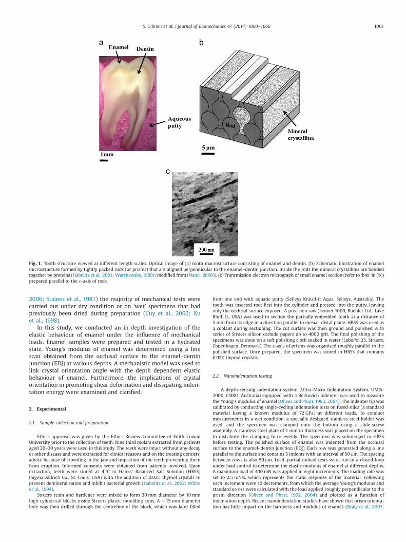
Fig. 1. Tooth structure viewed at different length scales. Optical image of (a) tooth macrostructure consisting of enamel and dentin. (b) Schematic illustration of enamelmicrostructure formed by tightly packed rods (or prisms) that are aligned perpendicular to the enamel–dentin junction. Inside the rods the mineral crystallites are bondedtogether by proteins (Habelitz et al., 2001; Warshawsky, 1989) (modified from (Nanci, 2008)). (c) Transmission electron micrograph of small enamel section (refer to ‘box’ in (b))prepared parallel to the c-axis of rods.
S. O'Brien et al. / Journal of Biomechanics 47 (2014) 1060–1066 1061

Xie et al., 2009a). To ensure that the multi-cycling nanoindentation is a suitabletechnique for the modulus measurement of materials at different depths, fusedsilica was tested by following the above procedure. It was found that the measuredmodulus of fused silica is independent of depth and within 1% of thetheoretical value.
2.3. Microstructure observation
Ultra-thin sections through the central head region in a direction parallel toenamel rods were prepared for transmission electron microscopy (TEM). To do this,a dual-electron/focused ion beam (FIB) workstation (Nova Nanolab 200, FEICompany, Hillsboro, OR, USA) was used. The detailed procedure can be foundelsewhere (Xie et al., 2007a). Briefly, prior to the milling a �1 μm thick platinumlayer was deposited to protect the surface area of interest from ion beam damage.A ‘rough’ sectioning was then performed with a current of 10 nA, in which trencheswere created at both sides of the platinum strip to obtain a cross section of �3 μmthickness. A number of ‘fine’ mills were taken at reduced currents (5–1 nA) to thinthe section to �1 μm. Final mills were carried out at further reduced currents(300–100 pA) to reduce the thickness down to �100 nm suitable for electrontransparency. The TEM specimens were transferred from the FIB sample holder tocarbon coated copper TEM grids using a high-precision micromanipulator (KleindiekNanotechnik GmbH, Reutlingen, Germany). TEM imaging was performed using afield emission gun-TEM (Philips CM200, Eindhoven, Netherlands).
2.4. Finite element analysis
Mineral platelets of enamel start to slide against each other, when the shearstress in the protein matrix exceeds a critical value (He and Swain, 2007). As aresult, there were no obvious cracks near the indentation site (Xie et al., 2008).According to this, finite element models were constructed in this work to clarify theeffect of the shear deformation of the protein layer on the magnitude anddistribution of contact-induced stress in enamel. The simulations were performedusing COMSOL software. Two-dimensional axisymmetric models, with the axialcoordinate z along the loading direction of the indenter, were constructed. Themodels consisted of an enamel block measuring 50�50 mm2, loaded by a sphericalindenter with a radius of 5 mm. A refined mesh was created around the contactzone within an area of 10�10 mm2 directly underneath the indenter where thestress concentration was expected to rise during indentation. The total number ofmesh elements is 17631, which includes the elements within the indenter. Furthermesh refinement does not improve the simulation accuracy significantly. Thecontact between the indenter and the sample was assumed to be frictionless. Theboundary conditions are similar to those used in the previous works (Wo et al.,2013; Zhao et al., 2011) and are briefly described as follows: The bottom of theblock (z¼50 mm) is fixed in the z direction, while its right edge (r¼50 mm) is fixedin the x direction. The axisymmetric axis coincides with the left edge of the block(r¼0) for obtaining 3D simulation results. The tip of the indenter was positioned atz¼0 mm (0 displacement) before the simulation started. The indentation process ismodelled as a downward displacement of the indenter tip from 0 to 0.4 mm at astep of 0.04 mm.
As mentioned above, the main objective for the finite element analysis (FEA) isto clarify the difference between the mechanical response of enamel with andwithout shear sliding. Hence, the shear sliding effect was treated in a parametricway without introducing structural complexity into the modelling. Specifically thestructural mechanics module was applied, using Hook's law as the constitutiverelation. As indicated in Fig. 1(c), on a sub-micrometre scale enamel exhibits a‘bricks-and-mortar’ structure, with the ‘bricks’ being made from mineral crystalswhich are embedded in a protein matrix. The volume fraction of protein is below4%, which can be derived from the dimensional parameters listed in Table 1. In theFEA model Young's modulus (129 GPa) and Poisson's ratio (0.25) values were usedfor the mineral phase (refer to Table 1). First, a perfect elastic response was
simulated for comparison purpose. Then, shear sliding effect was enabled andimplemented by defining the material to be elasto-plastic, with the shearing takingplace along the vertical direction and the yield strength being that of protein(1.09 GPa) (Xie et al., 2008).
3. Results and discussion
We first investigated how Young's modulus of enamel changedacross its thickness at various indentation depths. A typical load–partial unload curve obtained from the middle region of enamel isshown in Fig. 2(a), from which Young's modulus of enamel iscalculated (Oliver and Pharr, 1992) and found to decrease withincreasing depth (Fig. 2(b)). Given the depth-dependent elasticproperty of enamel, the modulus, measured as a function ofindentation depth, should then be applied to simulations, in orderto better understand the mechanical design of enamel. Followingthe same approach, we determined Young's modulus of enamelover its thickness from the occlusal surface to the EDJ and plottedfor three indentation depths, namely 0 mm, 0.5 mm and 2 mm(Fig. 2(c)). For each depth, Young's modulus decreased withincreasing distance from the occlusal surface, confirming thatenamel is indeed a functionally graded material (He and Swain,2009). The change of the enamel elasticity over its thickness isattributed to the variation in the microstructure and compositionfrom the surface towards the EDJ (Cuy et al., 2002; Low et al.,2008). Moreover, as the indenter tip approaches the enamelspecimen, the contact point position, which could be at eithermineral platelet or protein, has its strong influence onthe load–displacement curve, and thus the value of Young'smodulus. Consequently, modulus values obtained for shallowerindentation depths, for example at ht¼0.5 mm, show a morecomplex undulating pattern than those obtained at a depth of2 mm, presumably due to the inhomogeneous nature of the enamelstructure.
A plastic-damage model was developed for enamel and,according to it, the shear deformation of protein (i.e., the extensionand break of peptide chains) can cause the reduction in stiffnessunder indentation loading (An et al., 2012a). Recently, the non-uniform arrangement of the HAP crystallites was also identified tobe a key factor responsible for unique mechanical behaviour ofenamel (An et al., 2012b). It is, therefore, natural to inquire whatroles the crystal arrangement or orientation plays in the depth-dependent elastic response of enamel. For this purpose, a mechan-istic model was built from the experimental observation thatenamel exhibits a staggered hybrid structure (as seen in Fig. 1(c)),in which the mineral platelets can rotate to accommodate thedeformation (Zhou and Hsiung, 2007). Notably, such a structuralconfiguration has been successfully adopted by others to under-stand unique mechanical characteristics of enamel, for instance,rate-dependent deformation (Zhou and Hsiung, 2006) and creep(He and Swain, 2008; Schneider et al., 2008). According to the
Table 1Structural and mechanical parameters of enamel constituents used in the theoretical analysis and modeling.
Parameter Mineral crystal Protein
Symbol Value Reference Symbol Value Reference
Thickness (nm) hm 50 Kerebel et al. (1979) hp 1.5a
Length (nm) l 500 Driessens and Verbeeck (1990)Poisson's ratio νm 0.25 Liu et al. (2006) νp 0.45 Liu et al. (2006)Young's modulus (GPa) Em 129 Ang et al. (2010) Ep 2 Ji and Gao (2004), Spears (1997)Spacing of mineral crystals (nm) d 1.5
a The thickness of protein, hp is calculated from hp/hm¼0.03 which corresponds to a volume fraction of mineral E97% in the enamel.
S. O'Brien et al. / Journal of Biomechanics 47 (2014) 1060–10661062

model (inset in Fig. 3), Young's modulus of enamel can be expressedas (Xie et al., 2008)
Ee ¼ð cos 2θ� sin 2θÞ2
sin 4θE2þ4
cos 2θ
sin 2θG ð1Þ
where θ is the angle between the c-axis of the mineral platelet andthe loading direction (also termed as the crystal orientation angle).The compression modulus, E2, and the shear modulus, G, of thestaggered composite are given as (Liu et al., 2006):
E2 ¼1
ð12KphpðhmþhpÞ=αl2Gpð4Gpþ3KpÞÞþð1=EzÞð2Þ
G � hpþhm
hpGp ð3Þ
where hm and l are the thickness and length of the mineral crystal,respectively, hp and Gp are the thickness and shear modulus ofprotein layers between mineral platelets, α is the non-uniformedshear strain factor of the composite, Ez the modulus of thecomposite perpendicular to the c axis of the mineral platelet, andKp the bulk modulus of protein. Note that Gp, α, Ez and Kp can bederived from (Liu et al., 2006):
Gp ¼Ep
2ð1þνpÞð4Þ
α¼ 1þ 43
ðhpþhmÞlhpð1þdÞ
� �2( )
1�d1þdÞ
� �ð5Þ
Fig. 3. Understanding the depth-dependent Young's modulus of enamel duringdeformation. (a) Young's modulus of enamel in relation to effective crystalorientation angle. Inset shows the mechanistic model used. (b) Schematic illustra-tions showing the local responses of mineral crystals to increasing indentationloads (or depths) from P1 to P3 by rotation from θ1 to θ3, resulting in a reduction inYoung's modulus of enamel.
400
200
0
Load
, P (m
N)
0.0 0.5 1.0 1.5 2.0 2.5Indentation depth, h [μm]
Indentation depth, h [μm]
93
85
770.7 1.7 2.7
120ht = 0.0h =tht = 2.0
90
600.0 0.5 1.0
You
ng’s
mod
ulus
, E [G
Pa]
You
ng’s
mod
ulus
, E [G
Pa]
Normalised distance
05
Fig. 2. Young's modulus of dental enamel measured by depth-sensing indentationtests at different depths and locations. (a) Representative indentation load, P,versus displacement (i.e., penetration depth), h, curve produced by a Berkovichindenter in the middle region of the enamel. (b) The average Young's modulus andstandard deviations of enamel obtained from the middle region at differentpenetration depths. (c) The average Young's modulus and standard deviations ofenamel corresponding to three indentation depths of 0, 0.5 and 2 μm plotted fromthe occlusal surface to the EDJ. The normalised distance is calculated as the distancefrom the surface to the indentation divided by the enamel thickness. The values inthe shaded region in (c) are used in plotting (b).
Ez ¼ 1
ðð3hp=ðhpþhmÞð4Gpþ3KpÞÞþð1=EmÞð3Kp=ð4Gpþ3KpÞÞððh2mþ4νmhmhpþ2h2p�2νmh2pÞ=hmðhpþhmÞÞÞ
ð6Þ
S. O'Brien et al. / Journal of Biomechanics 47 (2014) 1060–1066 1063

Kp ¼Ep
3ð1�2νpÞð7Þ
The structural and mechanical properties of the main constituentsof enamel, i.e., the mineral and protein, are given in Table 1. Young'smodulus of enamel, measured under different indentation depthswithin a narrow region halfway between the occlusal surface andEDJ, is shown in Fig. 3. It is worth noting that the effective crystalorientation angle of enamel changes from 451 to 481 when theindentation depth increases from 0.75 mm to 2.42 mm. Also note thatthe length of the crystallites used in the modelling is 500 nm(Driessens and Verbeeck, 1990). However, there is no consensuson the crystal length in the literature. The possibility that they areconsiderably longer cannot be ruled out (Nanci, 2008; Waters,1980). To appreciate the effect that the crystal length may haveupon the angle calculation, a wide range of values from 150 nm to500 mmwas modelled. Surprisingly, the results show that the crystallength has little impact on both compression modulus and shearmodulus, consequently exerting minimal effect on the anglesderived.
The size dependent indentation modulus has also been observedfor cortical bone. The plastic-damage model was proposed andsuccessfully captured the stiffness loss during unloading (Lucchiniet al., 2011; Zhang et al., 2010). However, the damage mechanismremains unclear, which renders the model phenomenological andlacking in a physical interpretation of its parameters. To overcomethis problem, a physically sound approach was adopted for simulat-ing the modulus reduction of dental enamel, (An et al., 2012a) basedupon the experimental observation (Xie et al., 2008). It suggeststhat the shear deformation of protein contributes to the degrada-tion of stiffness in the unloading curve. In another study, Zhou andHsiung postulated that the rotation of mineral crystallites mightalso be responsible for the change of elastic modulus of enamelunder indentation loading (Zhou and Hsiung, 2007). Recently,the crystal arrangement or orientation has been identified, amongothers, as a critical parameter that regulates the mechanicalbehaviour of enamel (An et al., 2012b; Xie et al., 2008). From thisperspective, the structural basis that underpins the elastic beha-viour of enamel was examined in this work; that is, the decrease ofthe Young's modulus of enamel with increasing indentation depthmay also be governed by bending and/or tilting of the mineralcrystals. As such, multiple origins of the depth-dependent elasticbehaviour were possible. Note that the bending deformation ofslender crystals is not explicitly considered during modelling.However, the bending would result in the (partial) tilting of thecrystals; further the crystal length was found to have minimalimpact on the angles derived. The crystal bending can thus beaccommodated by the proposed mechanical model. More interest-ingly such an adaptive response is able to increase the contact areaand, consequently, lower the stress concentration in enamel. Forexample, with the increase of indentation load from 100 to 250 mN,the contact pressure decreases by about 13%. By doing so, the load-bearing ability of enamel would be enhanced. It is noteworthy thatfatigue damage may occur during multi-cycling nanoindentation(Jia and Xuan, 2012), and if so, that would affect the measurementaccuracy of the indentation modulus. On the other hand, therelative orientations of the indenter to the enamel rods' directionwere found to affect the measured elastic modulus (Cheng et al.,2010). To minimise its impact in the study of depth-dependentmodulus in enamel, the multi-cycling indentations were conductedin the present work. This means the depth-dependent elasticproperty was measured at fixed locations, where the relativeorientation of the indenter always remains constant during amulti-cycling test cycle. While the direction of indentation on theenamel may affect the absolute value of modulus, it would have
little impact on its depth-dependent nature and the underlyingmechanism discovered here.
As mentioned earlier, shear deformation between mineralcrystals has been observed in human enamel by using transmis-sion electron microscopy (Xie et al., 2007b). This deformationmode is considered to be essential for the remarkable damagetolerance of enamel (He and Swain, 2007; Xie et al., 2009b). Tofacilitate such a shear process, the shear stress developed betweenthe mineral crystals under indentation loading should be max-imised from the viewpoint of mechanical design. According to themodel shown in Fig. 3, the shear stress, τ, can be written as
τ¼ 12s sin 2θ ð8Þ
where s is the normal stress. Therefore, to meet this designrequirement, the effective crystal orientation angle would be 451,which is consistent with the modelling result (Fig. 3). Although theshearing is considered to be the predominant deformation mechan-ism of enamel (He and Swain, 2007; Xie et al., 2009b) a quantitativeunderstanding of its role in mitigating fracture and preventingcatastrophic failure remains lacking. To redress this gap, finiteelemental analysis (FEA) simulations was performed here to eluci-date the benefit of shear deformation for the stress management inenamel. Compared to the ‘purely elastic’ deformation (i.e., notconsidering the plastic shear process); Fig. 4(a)), a reduction in themaximum shear stress by 9% (i.e., from 5.5 GPa to 5.0 GPa) for anindentation depth of 0.28 mm is observed when the shearing effect is
τ (GPa)5.50
2.75
0.00
Mat
eria
l vol
ume,
(μm
3 )
s1s2s3s4
Fig. 4. The shear stress distribution generated in enamel by indentation at a depthof 0.28 μm: (a) perfect elastic model, (b) shear sliding enabled. (c) the size ofregions under the influence of shear stresses greater than (s1) 3.0 GPa, (s2) 3.5 GPa,(s3) 4.0 GPa and (s4) 4.5 GPa, calculated from the FEA simulations; note that ‘PE’stands for the perfect elastic model and ‘SS’ for the shear sliding model.
S. O'Brien et al. / Journal of Biomechanics 47 (2014) 1060–10661064

taken into account (Fig. 4(b)). The stress distribution pattern is alsomodified under plastic shear and the volume populated by largerstress is reduced significantly (Fig. 4(c)). For example, the volumeinfluenced by the stress level 43.5 GPa is reduced by more than 70%when the plastic shearing is enabled, and the volume influenced bythe stress level 44.5 GPa almost completely diminishes when theshear deformation is considered. Consequently, the decrease of stresslevel and reduction of material volumes subjected to higher stresses,resulting from the shear sliding between mineral crystals, couldfurther enhance the load-bearing ability of enamel.
4. Conclusion
By combining depth-sensing indentation tests and modelling,multiple origins of the depth-dependent indentation stiffness wereestablished in this study with a special focus on the crystalarrangement. The effective crystal orientation angle was calculatedand found to facilitate the shear sliding of mineral crystals duringindentation loading. Finite element analysis reveals that the sheardeformation between crystal platelets serves to (a) lower the stresslevel and (b) reduce the material volume populated by largerstresses. This leads to a critical appreciation of the stress manage-ment ability of enamel during loading. The present work willrekindle interest towards understanding the mechanical responsesof mineralised tissues to changing loads. From that, the mechanicaldesign principles of these materials may be better understood.
Conflict of interest statement
The authors certify that there is no conflict of interest with anyfinancial organisation regarding the material discussed in themanuscript.
Acknowledgements
We would like to thank Professor Mark Hoffman of Universityof New South Wales for providing constructive comments on themanuscript. The authors also thank Sparkle Dental in Joondalup,Western Australia, for providing fresh human molars. This workwas partially supported by an Australian Dental Research Founda-tion (ADRF) grant (6/2010). The authors acknowledge the facilities,scientific and technical assistance of the Australian Microscopyand Microanalysis Research Facility at the Centre for Microscopy,Characterisation and Analysis, the University of Western Australia,a facility funded by the University as well as State and Common-wealth Governments. S. O'Brien also acknowledges an ECU Post-graduate Scholarship for her Ph.D. study.
References
An, B.B., Wang, R.R., Zhang, D.S., 2012a. Region-dependent micro damage of enamelunder indentation. Acta Mech. Sin. 28, 1651–1658.
An, B., Wang, R., Zhang, D., 2012b. Role of crystal arrangement on the mechanicalperformance of enamel. Acta Biomater. 8, 3784–3793.
Ang, S.F., Bortel, E.L., Swain, M.V., Klocke, A., Schneider, G.A., 2010. Size-dependentelastic/inelastic behaviour of enamel over millimeter and nanometer lengthscales. Biomaterials 31, 1955–1963.
Braly, A., Darnell, L., Mann, A., Teaford, M., Weihs, T., 2007. The effect of prismorientation on the indentation testing of human molar enamel. Arch. Oral Biol.52, 856–860.
Cheng, Z.J., Wang, X.M., Ge, J., Yan, J.X., Ji, N., Tian, L.L., Cui, F.Z., 2010. Themechanical anisotropy on a longitudinal section of human enamel studied bynanoindentation. J. Mater. Sci: Mater. Med. 21, 1811–1816.
Cuy, J.L., Mann, A.B., Livi, K.J., Teaford, M.F., Weihs, T.P., 2002. Nanoindentationmapping of the mechanical properties of human molar tooth enamel. Arch. OralBiol. 47, 281–291.
Driessens, F.C.M., Verbeeck, R.M.H., 1990. The mineral in tooth enamel and dentalcaries. In: Driessens, F.C.M., Verbeeck, R.M.H. (Eds.), Biominerals. CRC Press Inc.,Boca Raton, Florida
Gao, H.J., Ji, B.H., Jager, I.L., Arzt, E., Fratzl, P., 2003. Materials become insensitive toflaws at nanoscale: lessons from nature. Proc. Natl. Acad. Sci. USA 100, 5597–5600.
Guidoni, G., Denkmayr, J., Schoberl, T., Jager, I., 2006. Nanoindentation in teeth:influence of experimental conditions on local mechanical properties. Philos.Mag. 86, 5705–5714.
Habelitz, S., Marshall, G.W., Balooch, M., Marshall, S.J., 2002. Nanoindentation andstorage of teeth. J. Biomech. 35, 995–998.
Habelitz, S., Marshall, S.J., Marshall, G.W., Balooch, M., 2001. Mechanical propertiesof human dental enamel on the nanometre scale. Arch. Oral Biol. 46, 173–183.
He, L.H., Fujisawa, N., Swain, M.V., 2006. Elastic modulus and stress–strain responseof human enamel by nanoindentation. Biomaterials 27, 4388–4398.
He, L.H., Swain, M.V., 2007. Contact induced deformation of enamel. Appl. Phys.Lett. 90, 171916.
He, L.H., Swain, M.V., 2008. Understanding the mechanical behaviour of humanenamel from its structural and compositional characteristics. J. Mech. Behav.Biomed. Mater. 1, 18–29.
He, L.H., Swain, M.V., 2009. Enamel – a functionally graded natural coating. J. Dent.37, 596–603.
Ji, B., Gao, H., 2004. Mechanical properties of nanostructure of biological materials.J. Mech. Phys. Solids 52, 1963–1990.
Jia, Y.F., Xuan, F.Z., 2012. Anisotropic fatigue behaviour of human enamel character-ized by multi-cycling nanoindentation. J. Mech. Behav. Biomed. Mater. 16,163–168.
Kerebel, B., Daculsi, G., Kerebel, L.M., 1979. Ultrastructural studies of enamelcrystallites. J. Dent. Res. 58, 844–851.
Lawn, B.R., Lee, J.J.W., Constantino, P.J., Lucas, P.W., 2009. Predicting failure inmammalian enamel. J. Mech. Behav. Biomed. Mater. 2, 33–42.
Lawn, B.R., Lee, J.J.W., Chai, H., 2010. Teeth: among nature’s most durablebiocomposites. Annu. Rev. Mater. Res. 40, 55–75.
Lewis, G., Nyman, J.S., 2008. The use of nanoindentation for characterizing theproperties of mineralized hard tissues: state-of-the art review. J. Biomed.Mater. Res. Part B 87B, 286–301.
Liu, B., Zhang, L., Gao, H., 2006. Poisson ratio can play a crucial role in mechanicalproperties of biocomposites. Mech. Mater. 38, 1128–1142.
Low, I.M., Duraman, N., Mahmood, U., 2008. Mapping the structure, compositionand mechanical properties of human teeth. Mater. Sci. Eng. C 28, 243–247.
Lucchini, R., Carnelli, D., Ponzoni, M., Bertarelli, E., Gastaldi, D., Vena, P., 2011. Roleof damage mechanics in nanoindentation of lamellar bone at multiple sizes:experiments and numerical modeling. J. Mech. Behav. Biomed. Mater 4,1852–1863.
Nanci, A., 2008. Ten Cate’s Oral Histology: Development, Structure, and Function,7th ed. Mosby Elsevier, St. Louis, Missouri, USA
Oliver, W.C., Pharr, G.M., 1992. An improved technique for determining hardnessand elastic modulus using load and displacement sensing indentation experi-ments. J. Mater. Res 7, 1564–1583.
Oliver, W.C., Pharr, G.M., 2004. Measurement of hardness and elastic modulus byinstrumented indentation: advances in understanding and refinements tomethodology. J. Mater. Res. 19, 3–20.
Park, S., Wang, D.H., Dongsheng, Z., Romberg, E., Arola, D., 2008. Mechanicalproperties of human enamel as a function of age and location in the tooth.J. Mater. Sci. - Mater. Med 19, 2317–2324.
Schneider, G.A., He, L.H., Swain, M.V., 2008. Viscous flow model of creep in enamel.J. Appl. Phys., 103
Spears, I.R., 1997. A three-dimensional finite element model of prismatic enamel: are-appraisal of the data on the Young's modulus of enamel. J. Dent. Res 76,1690–1697.
Staines, M., Robinson, W.H., Hood, J.A.A., 1981. Spherical indentation of toothenamel. J. Mater. Sci. 16, 2551–2556.
Warshawsky, H., 1989. Organization of crystals in enamel. Anat. Rec. 224, 242–262.Waters, N.E., 1980. Some mechanical and physical properties of teeth. In: Vincent, J.
F.V., Currey, J.D. (Eds.), The Mechanical Properties of Biological Materials.Cambridge University Press, Cambridge, UK
White, J.M., Goodis, H.E., Marshall, S.J., Marshall, G.W., 1994. Sterilization of teeth bygamma radiation. J. Dent. Res 73, 1560–1567.
Wo, P.C., Zhao, X.L., Munroe, P.R., Zhou, Z., Li, K., Habibi, D., Xie, Z., 2013. Extremelyhard, damage-tolerant ceramic coatings with functionally graded, periodicallyvarying architecture. Acta Mater. 61, 193–204.
Xie, Z.H., Swain, M.V., Swadener, G., Munroe, P., Hoffman, M., 2009a. Effect ofmicrostructure upon elastic behaviour of human tooth enamel. J. Biomech. 42,1075–1080.
Xie, Z., Swain, M.V., Hoffman, M.J., 2009b. Structural integrity of enamel: experi-mental and modeling. J. Dent. Res. 88, 529–533.
Xie, Z.H., Hoffman, M., Munroe, P., Singh, R., Bendavid, A., Martin, P.J., 2007a.Microstructural response of TiN monolithic and multilayer coatings duringmicroscratch testing. J. Mater. Res. 22, 2312–2318.
Xie, Z.H., Mahoney, E.K., Kilpatrick, N.M., Swain, M.V., Hoffman, M., 2007b. On thestructure–property relationship of sound and hypomineralized enamel. ActaBiomater. 3, 865–872.
Xie, Z.H., Swain, M., Munroe, P., Hoffman, M., 2008. On the critical parameters thatregulate the deformation behaviour of tooth enamel. Biomaterials 29, 2697–2703.
Xu, H.H.K., Smith, D.T., Jahanmir, S., Romberg, E., Kelly, J.R., Thompson, V.P., Rekow,E.D., 1998. Indentation damage and mechanical properties of human enameland dentin. J. Dent. Res. 77, 472–480.
S. O'Brien et al. / Journal of Biomechanics 47 (2014) 1060–1066 1065

Zhang, J., Michalenko, M.M., Kuhl, E., Ovaert, T.C., 2010. Characterization ofindentation response and stiffness reduction of bone using a continuumdamage model. J. Mech. Behav. Biomed. Mater. 3, 189–202.
Zhao, X., Xie, Z., Munroe, P., 2011. Nanoindentation of hard multilayer coatings:finite element modelling. Mater. Sci. Eng. A 528, 1111–1116.
Zhou, J., Hsiung, L.L., 2007. Depth-dependent mechanical properties of enamel bynanoindentation. J. Biomed. Mater. Res. Part A 81A, 66–74.
Zhou, J.K., Hsiung, L.L., 2006. Biomolecular origin of the rate-dependent deforma-tion of prismatic enamel. Appl. Phys. Lett. 89, 3.
S. O'Brien et al. / Journal of Biomechanics 47 (2014) 1060–10661066


