Embed Size (px)
Citation preview
ORIGINAL ARTICLE
doi:10.1111/evo.12059
SKULL SHAPE EVOLUTION IN DUROPHAGOUSCARNIVORANSBorja Figueirido,1,2 Zhijie Jack Tseng,3 and Alberto Martın-Serra1
1Departamento de Ecologıa y Geologıa de la Facultad de Ciencias, Universidad de Malaga, 29071-Malaga, Spain2E-Mail: [email protected]
3Department of Vertebrate Paleontology, Natural History Museum of Los Angeles County, 900 Exposition Boulevard, Los
Angeles, California 90007
Received September 24, 2012
Accepted January 9, 2013
In this article, we investigate convergent evolution toward durophagy in carnivoran skull shape using geometric morphometrics in a
sample of living and extinct species. Principal components analysis indicate that, in spite of the different dietary resources consumed
by durophages—that is, bone-crackers and bamboo-feeders—both groups of carnivorans share portions of skull phenotypic spaces.
We identify by discriminant analyses a shared set of adaptations toward durophagy in the skull of carnivores. However, ancestral
states indicate that although durophages reached similar phenotypes, the evolutionary pathways that they followed are different
depending upon the family to which they belong. Furthermore, while the carnivoran cranium more closely reflects the nature
of the resources consumed—that is, soft or hard and tough items—the mandible shows particular feeding adaptations—that is,
bamboo or bone. This finding supports the interpretation that the mandible has more evolutionary plasticity than the cranium,
which is more limited to evolve toward a particular feeding adaptation. However, we find that the shapes of the cranium and the
mandible are highly integrated for the whole order Carnivora. Published studies of teratological cats and dogs indicate that the
role of internal constraints in shaping this pattern of integration is absent or weak and malleable by selection.
KEY WORDS: Carnivore, durophagy, evolutionary convergence, morphometrics, phenotype.
Convergent evolution is a central topic in evolutionary biology
(Winter and Oxnard 2001; Madsen et al. 2001; Nevo 2001; Wroe
and Milne 2007; Futuyma 2010). Despite this, interpretations on
its potential causes are not clear-cut (e.g., Stayton 2008; Losos
2011; Olson 2012) and most contemporary surveys recognize two
different points of view for explaining the evolutionary conver-
gence of traits: internalist and externalist explanations (e.g., Wake
1991). From an externalist point of view, convergent evolution is
exclusively an evidence of adaptation due to the powerful action
of natural selection (e.g., Schlueter 2000; Conway-Morris 2003;
Blackledge and Gillespie 2004; Grenier and Greenberg 2005;
Langerhans et al. 2006; Stayton 2008). Under this perspective of
understanding phenotypic evolution, convergent traits are opti-
mal solutions to repeated environmental problems (Losos 2011).
In contrast, the internalist point of view argues that convergence
is the result of internal constraints, which hamper the production
of phenotypic variability upon which natural selection can op-
erate (e.g., Alberch and Gale 1985; Maynard-Smith et al. 1985;
Alberch 1989; Wake 1991; Schwenk 1995; Schwenk and Wagner
2003, 2004; Jaekel and Wake 2007; Figueirido et al. 2011a).
Although both adaptation and constraint give account of or-
der and discreteness in phenotypic spaces (Gould 2002), the two
concepts have become divorced due to a dichotomous attitude
about them (Schwenk and Wagner 2004). Good examples of these
“rival traditions” in evolutionary morphology are the seminal ar-
ticles on this topic published recently by Losos (2011) and Olson
(2012). Both reviews indicate the need for integrative studies of
the effects of natural selection and constraints using different
kinds of approaches for understanding phenotypic evolution.
In this article, we investigate the evolutionary convergence
toward durophagy in the skull of mammalian carnivores and dis-
cuss the role of natural selection and different kinds of constraints
1 9 7 5C© 2013 The Author(s). Evolution C© 2013 The Society for the Study of Evolution.Evolution 67-7: 1975–1993
BORJA FIGUEIRIDO ET AL.
in shaping this pattern. The order Carnivora is an excellent choice
for this study because its evolution represents one of the most
spectacular cases of repeated and independent evolution of similar
morphologies on a limited range of ecologies (Van Valkenburgh
2007; Wroe and Milne 2007; Wroe et al. 2007; Wroe 2008). In
fact, convergent patterns in the evolution of carnivoran skull shape
have been reported by a number of researchers, either toward hy-
percarnivory (e.g., Van Valkenburgh 1991; Holliday and Steppan
2001; Van Valkenburgh 2007; Wroe and Milne 2007; Figueirido
et al. 2011a; Goswami et al. 2011), bone cracking (Van Valken-
burgh et al. 2003, 2007; Palmqvist et al. 2011; Tseng and Wang
2011), or even herbivory (Figueirido et al. 2010b). However, it
is worth noting that convergent trends toward durophagy are still
unexplored. Furthermore, durophagous carnivorans open the pos-
sibility of testing if two distantly related groups of carnivorans—
for example, living pandas and hyenas, which belong to subor-
ders Caniformia and Feliformia, respectively—adapted to feed
on extremely different resources—that is, bamboo and bones,
respectively—converge in skull biomechanics as a consequence
of the required performance to feed on hard and tough foods. To
test this hypothesis, we applied landmark-based methods of geo-
metric morphometrics in a large sample of living and extinct repre-
sentatives of the order Carnivora. We define convergent evolution
as the repeated and independent evolution of similar traits toward
the same environmental regimes (Futuyma 2010). Therefore, we
include under evolutionary convergence both parallel and conver-
gent evolution, because many authors cast doubts on whether there
is a clear-cut theoretical distinction between these two evolution-
ary phenomena, which are frequently envisaged as extremes of a
continuum (e.g., Meyer 1999; Gould 2002; Desutter-Grandcolas
et al. 2005; Hall 2007; Abouheif 2008; Arendt and Reznick
2008).
HARD AND TOUGH FOOD
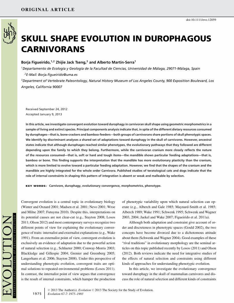
We classify those species as durophagous carnivorans that
usually feed on bones and bamboo. Bone and bamboo are
hard materials because they have high values of yield strength
(σ)—that is, a large force is required to produce a material failure.
Furthermore, they are also tough because they have high values of
toughness (Jc)—that is, a high capacity to absorb a large amount
of energy before breaking (e.g., Wegst and Ashby 2004). The
combination of these parameters relative to the Young’s modulus
(i.e., elasticity) unequivocally indicates that both biomaterials
are almost identical in hardness and toughness (see Fig. 1A,B).
As a consequence, osseous tissue—that is, calcium phosphate
in the hydroxylapatite chemical arrangement—is a hard organic
material (Schmidt-Nielsen 1984) and the physical and mechanical
properties of bamboo are comparable to those of hard materials,
such as low-carbon steel and glass-reinforced plastics, which
leads to its frequent use in industry for constructing scaffolds and
Figure 1. Material properties of bone and bamboo and bite
force in bone-cracking and bamboo-feeding carnivorans. (A) Bi-
variate plot showing Young’s modulus (E; measured in GPa) on the
strength (σ; measured in MPa) of bone (black) and bamboo (white)
relative to other natural materials (grey) (redrawn from Wegst and
Ashby [2004]). (B) Bivariate plot depicting the toughness of bone
and bamboo (Jc; measured in kJ/m2) on Young’s modulus (E; mea-
sured in GPa) relative to other natural materials (redrawn from
Wegst and Ashby [2004]). (C) Box plot showing the bite force quo-
tient (bite force for each species in relation to its body mass) at the
carnassials (BFQcarnassial) estimated by the dry skull method in
the living bone-crackers, bamboo-feeders, and other taxa included
in our sample. The vertical line inside each box is the median. Box
length is the interquartile range (IQR) and shows the difference
between the 75th and 25th percentiles (data from Christiansen
and Wroe 2007). Horizontal bars enclose values of 5–95%.
1 9 7 6 EVOLUTION JULY 2013
EVOLUTION IN DUROPHAGES
as a reinforcement for cement, rubber, thermoplastic, and even
aluminum (Low et al. 2006).
BONE-CRACKERS AND BAMBOO-FEEDERS
AS DUROPHAGOUS CARNIVORANS
Bone-crackers are considered strict hypercarnivores because they
feed primarily on limb bones with high nutritional content of
blood and fat-rich marrow tissues (e.g., Kruk 1972; Palmqvist
et al. 1999, 2011; Van Valkenburgh 2007). As indicated by fossil
data, the bone-cracking ecomorph evolved independently at least
four times during the Cenozoic: in Hyaenidae, Percrocutidae†(Daggers denote extinct taxa), and twice within Canidae—
in the Aelurodontina and Borophagina subtribes of subfamily
Borophaginae†—(e.g., Van Valkenburgh 1999; Wang et al. 1999;
Van Valkenburgh 2007; Tseng and Wang 2011). In contrast,
bamboo-feeders are considered as strict herbivores feeding al-
most entirely on bamboo (e.g., Gittleman 1994; Figueirido et al.
2010b). Similarly, data from the fossil record suggest that the
bamboo-feeding ecomorph evolved independently in two living
species of carnivorans: the giant panda (Ailuropoda melanoleuca)
within Ursidae and the red or lesser panda (Ailurus fulgens) within
Ailuridae (e.g., Flynn et al. 2000, 2005). However, despite that
both species feed entirely on bamboo they also differ in their
feeding behavior. The giant panda usually eat on bamboo leaves
and trunks depending upon the season (from March to July they
feed almost entirely on trunks in the wild; Schaller et al. 1985).
However, they remove the outer, green, smooth, and waxy layer
of the trunks before chewing or biting because this is their less
nutritive part (Hansen et al. 2010). Although the red panda feed
almost entirely on leaves supplemented by arboreal fruits and
bamboo shoots (Reid et al. 1991; Wei et al. 1999), sometimes
they also feed on peeled trunks. Furthermore, it is worth noting
that both pandas are the only carnivorans adapted to feed regularly
on leaves—that is, a fibrous material with high contents in lignin,
cellulose, and hemicellulose. Other herbivorous carnivorans, such
as the kinakajou (Potos flavus) or the Andean bear (Tremarctos
ornatus), mostly feed regularly on fruits and shoots but not on
leaves (Figueirido et al. 2010b, 2011b, 2012).
Therefore, we considered both bamboo-feeders and bone-
crackers as durophages because they feed regularly on hard and
tough materials such are bamboo and bones, respectively.
ANATOMICAL AND FUNCTIONAL TRAITS SHARED
BY BONE-CRACKERS AND BAMBOO-FEEDERS
Previous researchers have noted qualitatively an external mor-
phological resemblance between the skulls of bamboo-feeders
and bone-crackers (e.g., Davis 1964) which led to analyze
both groups of carnivorans jointly in functional studies (e.g.,
Christiansen and Wroe 2007). In fact, a brief review of the liter-
ature indicates that both types of durophages share a number
of morphological traits in the skull that could be considered
potential adaptations to feed on hard and tough foods. These
traits include a robust craniodental morphology with a raised
and dome-like frontal region of the cranium, enlarged areas for
the attachment of masticatory muscles, well-developed frontal si-
nuses, enlarged premolars, and microstructurally reinforced tooth
enamel (Joeckel 1998; Stefen 1999, 2001; Stefen and Rensberger
2002; Dong 2008; Tanner et al. 2008; Tseng 2009; Figueirido
et al. 2012; Tseng 2012). All of these morphological traits have
been interpreted as adaptations to exert extremely high bite forces
for chewing and biting on hard materials (Fig. 1C) and to dis-
sipate the stresses generated (Sacco and Van Valkenburgh 2004;
Christiansen and Adolfssen 2005; Christiansen and Wroe 2007;
Figueirido and Soibelzon 2010; Figueirido et al. 2010a,b, 2011a,b,
2012; Oldfield et al. 2012). However, several functional differ-
ences between bamboo feeders and bone-crackers also exist. For
example, bamboo-feeders have a higher position of the condy-
lar process of the mandible (Fig. 2) relative to the tooth row
than bone-crackers which allow the former to have a slight trans-
lation movement of the lower jaw. However, although this ac-
tion is an important movement for chewing and processing the
fibrous bamboo leaves, the translation movement in the giant
panda is very limited (only 5.4 mm) as a consequence of hav-
ing a transverse cylindrical mandibular articulation forcing the
jaw action mainly to a simple hinge movement vertically (Davis
1964).
Another morpho-functional difference between both groups
of canivorans is that although both have an exceptional ability
for exerting large bite forces (Fig. 1C), they could be using this
ability in a different way. In fact, while bone-crackers are partic-
ularly adapted to exert high-peak forces to cracking bones dur-
ing punctual periods of time and to resist the stresses generated,
bamboo-feeders could be adapted to resist fatigue as a result of
constant chewing applying submaximal forces over protracted pe-
riods. However, the fact that the giant panda torn off the stripped
outer layer of bamboo trunks by means of a twisting movement of
the fore foot coupled with a lateral turning of the head by an active
bite (Davis 1964) indicates that most probably giant pandas are
adapted to resist both: high peak forces during punctual periods of
time and fatigue during constant chewing. In fact, although both
pandas invest more time chewing on bamboo leaves and stalks
than cracking bamboo trunks (at least the red panda), hyenas also
feed on flesh on a regular basis and fracture limb bones less fre-
quently for accessing their medullary contents (e.g., Kruk 1972;
Ewer 1973; Palmqvist et al. 1999, 2011). From a biomechanical
point of view, in both cases what matters is the force that is ex-
erted during those activities that demand a greater resistance of
the skull against the stresses generated by elevated loads (i.e., as
those produced during bamboo/bone-cracking). In other words,
although these loads are only exerted occasionally, there is safety
EVOLUTION JULY 2013 1 9 7 7
BORJA FIGUEIRIDO ET AL.
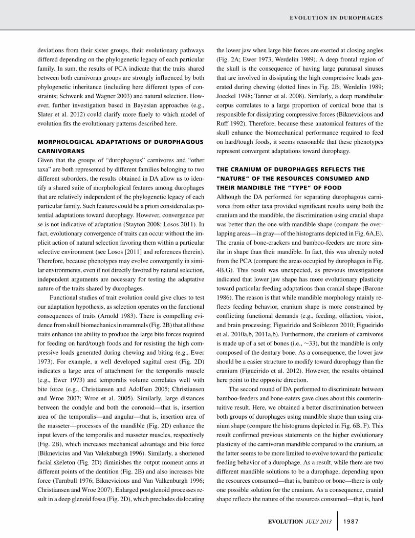
Figure 2. Skull biomechanics in durophagous carnivorans. (A) Main muscles and dentition involved in the mastication practice, exempli-
fied in a skull of spotted hyena (Crocuta crocuta) and in a skull of giant panda (Ailuropoda melanoleuca) with an hypothetical cross-section
of a bone and bamboo stem, respectively. (B) Masticatory apparatus modeled as a third-class lever system showing the combined effort
of the temporalis and masseter muscles as a function of their input arms, applied in a point between the fulcrum (temporomandibular
joint) and the resistance exerted at the main teeth involved in processing food. The line of stress dissipation is a circle drawn with its
center at the fulcrum and with a radius of length from the fulcrum to P3 in the case of spotted hyaena and to M1 in the case of the giant
panda (Werdelin 1989). (C) Landmarks used in the morphometric analysis of skull function. (D) Functional key aspects in recovered in the
morphometric analyses. Abbreviations: C, upper canine; M, upper molar; P, upper premolar; c, lower canine; m, lower molar; p, lower
premolar. The numbers indicate the position of each type of teeth. Skulls not to scale.
factor which implies that the skulls of pandas and hyenas must
be adapted for withstanding the maximal loads exerted during
chewing and biting.
In sum, bone-crackers and bamboo-feeders feed regularly
on extremely hard and tough materials (Fig. 1A,B). Accordingly,
both groups of carnivorans seem to share a set of skull traits re-
lated with the ability for exerting exceptionally large bite forces
(Fig. 1C) and to dissipate the stresses generated. As a conse-
quence, these shared traits between both groups of carnivorans
could be interpreted as potential adaptations toward durophagy.
However, in spite of this morphological resemblance and
presumed functional similarity between bone-crackers and
1 9 7 8 EVOLUTION JULY 2013
EVOLUTION IN DUROPHAGES
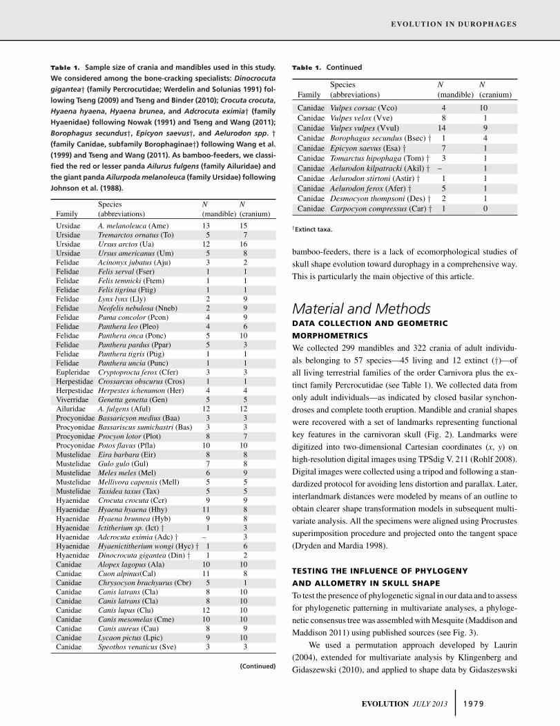
Table 1. Sample size of crania and mandibles used in this study.
We considered among the bone-cracking specialists: Dinocrocuta
gigantea† (family Percrocutidae; Werdelin and Solunias 1991) fol-
lowing Tseng (2009) and Tseng and Binder (2010); Crocuta crocuta,
Hyaena hyaena, Hyaena brunea, and Adcrocuta eximia† (family
Hyaenidae) following Nowak (1991) and Tseng and Wang (2011);
Borophagus secundus†, Epicyon saevus†, and Aelurodon spp. †(family Canidae, subfamily Borophaginae†) following Wang et al.
(1999) and Tseng and Wang (2011). As bamboo-feeders, we classi-
fied the red or lesser panda Ailurus fulgens (family Ailuridae) and
the giant panda Ailurpoda melanoleuca (family Ursidae) following
Johnson et al. (1988).
Species N NFamily (abbreviations) (mandible) (cranium)
Ursidae A. melanoleuca (Ame) 13 15Ursidae Tremarctos ornatus (To) 5 7Ursidae Ursus arctos (Ua) 12 16Ursidae Ursus americanus (Um) 5 8Felidae Acinonyx jubatus (Aju) 3 2Felidae Felis serval (Fser) 1 1Felidae Felis temnicki (Ftem) 1 1Felidae Felis tigrina (Ftig) 1 1Felidae Lynx lynx (Lly) 2 9Felidae Neofelis nebulosa (Nneb) 2 9Felidae Puma concolor (Pcon) 4 9Felidae Panthera leo (Pleo) 4 6Felidae Panthera onca (Ponc) 5 10Felidae Panthera pardus (Ppar) 5 3Felidae Panthera tigris (Ptig) 1 1Felidae Panthera uncia (Punc) 1 1Eupleridae Cryptoprocta ferox (Cfer) 3 3Herpestidae Crossarcus obscurus (Cros) 1 1Herpestidae Herpestes ichenumon (Her) 4 4Viverridae Genetta genetta (Gen) 5 5Ailuridae A. fulgens (Aful) 12 12Procyonidae Bassaricyon medius (Baa) 3 3Procyonidae Bassariscus sumichastri (Bas) 3 3Procyonidae Procyon lotor (Plot) 8 7Procyonidae Potos flavus (Pfla) 10 10Mustelidae Eira barbara (Eir) 8 8Mustelidae Gulo gulo (Gul) 7 8Mustelidae Meles meles (Mel) 6 9Mustelidae Mellivora capensis (Mell) 5 5Mustelidae Taxidea taxus (Tax) 5 5Hyaenidae Crocuta crocuta (Ccr) 9 9Hyaenidae Hyaena hyaena (Hhy) 11 8Hyaenidae Hyaena brunnea (Hyb) 9 8Hyaenidae Ictitherium sp. (Ict) † 1 3Hyaenidae Adcrocuta eximia (Adc) † – 3Hyaenidae Hyaenictitherium wongi (Hyc) † 1 6Hyaenidae Dinocrocuta gigantea (Din) † 1 2Canidae Alopex lagopus (Ala) 10 10Canidae Cuon alpinus(Cal) 11 8Canidae Chrysocyon brachyurus (Cbr) 5 1Canidae Canis latrans (Cla) 8 10Canidae Canis latrans (Cla) 8 10Canidae Canis lupus (Clu) 12 10Canidae Canis mesomelas (Cme) 10 10Canidae Canis aureus (Cau) 8 9Canidae Lycaon pictus (Lpic) 9 10Canidae Speothos venaticus (Sve) 3 3
(Continued)
Table 1. Continued
Species N NFamily (abbreviations) (mandible) (cranium)
Canidae Vulpes corsac (Vco) 4 10Canidae Vulpes velox (Vve) 8 1Canidae Vulpes vulpes (Vvul) 14 9Canidae Borophagus secundus (Bsec) † 1 4Canidae Epicyon saevus (Esa) † 7 1Canidae Tomarctus hipophaga (Tom) † 3 1Canidae Aelurodon kilpatracki (Akil) † – 1Canidae Aelurodon stirtoni (Astir) † 1 1Canidae Aelurodon ferox (Afer) † 5 1Canidae Desmocyon thompsoni (Des) † 2 1Canidae Carpocyon compressus (Car) † 1 0
†Extinct taxa.
bamboo-feeders, there is a lack of ecomorphological studies of
skull shape evolution toward durophagy in a comprehensive way.
This is particularly the main objective of this article.
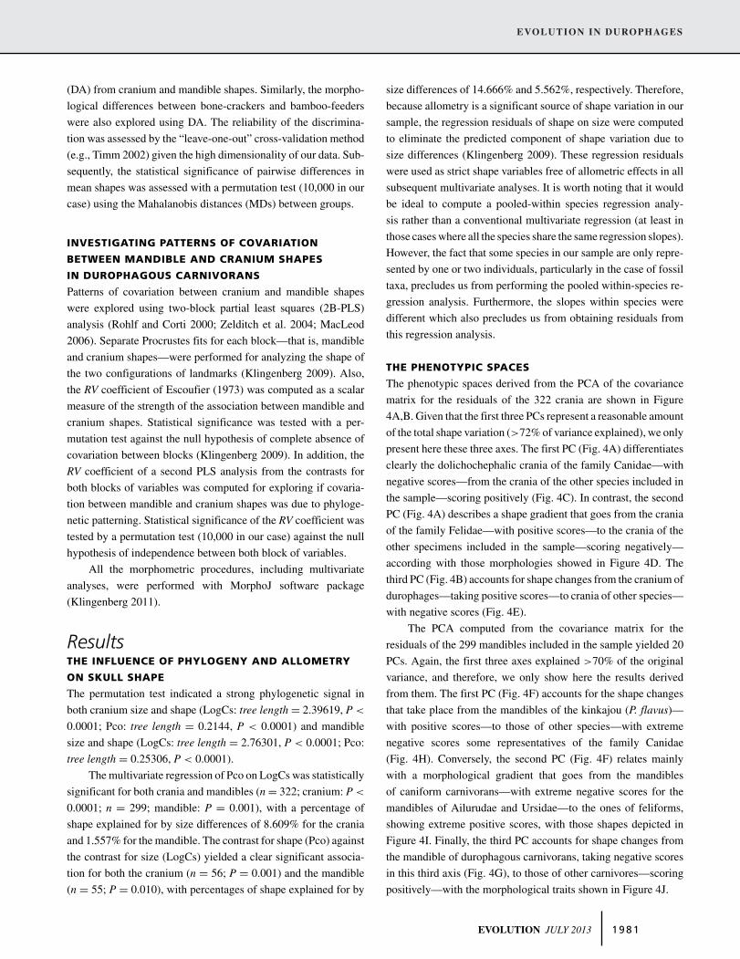
Material and MethodsDATA COLLECTION AND GEOMETRIC
MORPHOMETRICS
We collected 299 mandibles and 322 crania of adult individu-
als belonging to 57 species—45 living and 12 extinct (†)—of
all living terrestrial families of the order Carnivora plus the ex-
tinct family Percrocutidae (see Table 1). We collected data from
only adult individuals—as indicated by closed basilar synchon-
droses and complete tooth eruption. Mandible and cranial shapes
were recovered with a set of landmarks representing functional
key features in the carnivoran skull (Fig. 2). Landmarks were
digitized into two-dimensional Cartesian coordinates (x, y) on
high-resolution digital images using TPSdig V. 211 (Rohlf 2008).
Digital images were collected using a tripod and following a stan-
dardized protocol for avoiding lens distortion and parallax. Later,
interlandmark distances were modeled by means of an outline to
obtain clearer shape transformation models in subsequent multi-
variate analysis. All the specimens were aligned using Procrustes
superimposition procedure and projected onto the tangent space
(Dryden and Mardia 1998).
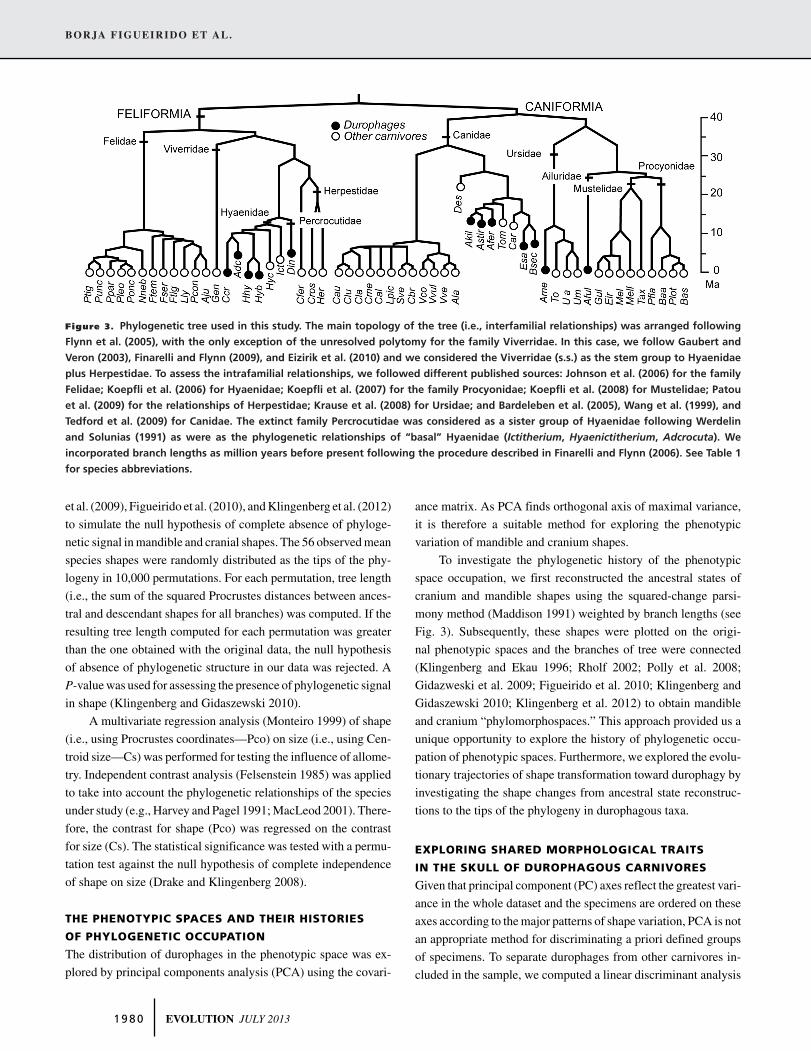
TESTING THE INFLUENCE OF PHYLOGENY
AND ALLOMETRY IN SKULL SHAPE
To test the presence of phylogenetic signal in our data and to assess
for phylogenetic patterning in multivariate analyses, a phyloge-
netic consensus tree was assembled with Mesquite (Maddison and
Maddison 2011) using published sources (see Fig. 3).
We used a permutation approach developed by Laurin
(2004), extended for multivariate analysis by Klingenberg and
Gidaszewski (2010), and applied to shape data by Gidaszeswski
EVOLUTION JULY 2013 1 9 7 9
BORJA FIGUEIRIDO ET AL.
Figure 3. Phylogenetic tree used in this study. The main topology of the tree (i.e., interfamilial relationships) was arranged following
Flynn et al. (2005), with the only exception of the unresolved polytomy for the family Viverridae. In this case, we follow Gaubert and
Veron (2003), Finarelli and Flynn (2009), and Eizirik et al. (2010) and we considered the Viverridae (s.s.) as the stem group to Hyaenidae
plus Herpestidae. To assess the intrafamilial relationships, we followed different published sources: Johnson et al. (2006) for the family
Felidae; Koepfli et al. (2006) for Hyaenidae; Koepfli et al. (2007) for the family Procyonidae; Koepfli et al. (2008) for Mustelidae; Patou
et al. (2009) for the relationships of Herpestidae; Krause et al. (2008) for Ursidae; and Bardeleben et al. (2005), Wang et al. (1999), and
Tedford et al. (2009) for Canidae. The extinct family Percrocutidae was considered as a sister group of Hyaenidae following Werdelin
and Solunias (1991) as were as the phylogenetic relationships of “basal” Hyaenidae (Ictitherium, Hyaenictitherium, Adcrocuta). We
incorporated branch lengths as million years before present following the procedure described in Finarelli and Flynn (2006). See Table 1
for species abbreviations.
et al. (2009), Figueirido et al. (2010), and Klingenberg et al. (2012)
to simulate the null hypothesis of complete absence of phyloge-
netic signal in mandible and cranial shapes. The 56 observed mean
species shapes were randomly distributed as the tips of the phy-
logeny in 10,000 permutations. For each permutation, tree length
(i.e., the sum of the squared Procrustes distances between ances-
tral and descendant shapes for all branches) was computed. If the
resulting tree length computed for each permutation was greater
than the one obtained with the original data, the null hypothesis
of absence of phylogenetic structure in our data was rejected. A
P-value was used for assessing the presence of phylogenetic signal
in shape (Klingenberg and Gidaszewski 2010).
A multivariate regression analysis (Monteiro 1999) of shape
(i.e., using Procrustes coordinates—Pco) on size (i.e., using Cen-
troid size—Cs) was performed for testing the influence of allome-
try. Independent contrast analysis (Felsenstein 1985) was applied
to take into account the phylogenetic relationships of the species
under study (e.g., Harvey and Pagel 1991; MacLeod 2001). There-
fore, the contrast for shape (Pco) was regressed on the contrast
for size (Cs). The statistical significance was tested with a permu-
tation test against the null hypothesis of complete independence
of shape on size (Drake and Klingenberg 2008).
THE PHENOTYPIC SPACES AND THEIR HISTORIES
OF PHYLOGENETIC OCCUPATION
The distribution of durophages in the phenotypic space was ex-
plored by principal components analysis (PCA) using the covari-
ance matrix. As PCA finds orthogonal axis of maximal variance,
it is therefore a suitable method for exploring the phenotypic
variation of mandible and cranium shapes.
To investigate the phylogenetic history of the phenotypic
space occupation, we first reconstructed the ancestral states of
cranium and mandible shapes using the squared-change parsi-
mony method (Maddison 1991) weighted by branch lengths (see
Fig. 3). Subsequently, these shapes were plotted on the origi-
nal phenotypic spaces and the branches of tree were connected
(Klingenberg and Ekau 1996; Rholf 2002; Polly et al. 2008;
Gidazweski et al. 2009; Figueirido et al. 2010; Klingenberg and
Gidaszewski 2010; Klingenberg et al. 2012) to obtain mandible
and cranium “phylomorphospaces.” This approach provided us a
unique opportunity to explore the history of phylogenetic occu-
pation of phenotypic spaces. Furthermore, we explored the evolu-
tionary trajectories of shape transformation toward durophagy by
investigating the shape changes from ancestral state reconstruc-
tions to the tips of the phylogeny in durophagous taxa.
EXPLORING SHARED MORPHOLOGICAL TRAITS
IN THE SKULL OF DUROPHAGOUS CARNIVORES
Given that principal component (PC) axes reflect the greatest vari-
ance in the whole dataset and the specimens are ordered on these
axes according to the major patterns of shape variation, PCA is not
an appropriate method for discriminating a priori defined groups
of specimens. To separate durophages from other carnivores in-
cluded in the sample, we computed a linear discriminant analysis
1 9 8 0 EVOLUTION JULY 2013
EVOLUTION IN DUROPHAGES
(DA) from cranium and mandible shapes. Similarly, the morpho-
logical differences between bone-crackers and bamboo-feeders
were also explored using DA. The reliability of the discrimina-
tion was assessed by the “leave-one-out” cross-validation method
(e.g., Timm 2002) given the high dimensionality of our data. Sub-
sequently, the statistical significance of pairwise differences in
mean shapes was assessed with a permutation test (10,000 in our
case) using the Mahalanobis distances (MDs) between groups.
INVESTIGATING PATTERNS OF COVARIATION
BETWEEN MANDIBLE AND CRANIUM SHAPES
IN DUROPHAGOUS CARNIVORANS
Patterns of covariation between cranium and mandible shapes
were explored using two-block partial least squares (2B-PLS)
analysis (Rohlf and Corti 2000; Zelditch et al. 2004; MacLeod
2006). Separate Procrustes fits for each block—that is, mandible
and cranium shapes—were performed for analyzing the shape of
the two configurations of landmarks (Klingenberg 2009). Also,
the RV coefficient of Escoufier (1973) was computed as a scalar
measure of the strength of the association between mandible and
cranium shapes. Statistical significance was tested with a per-
mutation test against the null hypothesis of complete absence of
covariation between blocks (Klingenberg 2009). In addition, the
RV coefficient of a second PLS analysis from the contrasts for
both blocks of variables was computed for exploring if covaria-
tion between mandible and cranium shapes was due to phyloge-
netic patterning. Statistical significance of the RV coefficient was
tested by a permutation test (10,000 in our case) against the null
hypothesis of independence between both block of variables.
All the morphometric procedures, including multivariate
analyses, were performed with MorphoJ software package
(Klingenberg 2011).
ResultsTHE INFLUENCE OF PHYLOGENY AND ALLOMETRY
ON SKULL SHAPE
The permutation test indicated a strong phylogenetic signal in
both cranium size and shape (LogCs: tree length = 2.39619, P <
0.0001; Pco: tree length = 0.2144, P < 0.0001) and mandible
size and shape (LogCs: tree length = 2.76301, P < 0.0001; Pco:
tree length = 0.25306, P < 0.0001).
The multivariate regression of Pco on LogCs was statistically
significant for both crania and mandibles (n = 322; cranium: P <
0.0001; n = 299; mandible: P = 0.001), with a percentage of
shape explained for by size differences of 8.609% for the crania
and 1.557% for the mandible. The contrast for shape (Pco) against
the contrast for size (LogCs) yielded a clear significant associa-
tion for both the cranium (n = 56; P = 0.001) and the mandible
(n = 55; P = 0.010), with percentages of shape explained for by
size differences of 14.666% and 5.562%, respectively. Therefore,
because allometry is a significant source of shape variation in our
sample, the regression residuals of shape on size were computed
to eliminate the predicted component of shape variation due to
size differences (Klingenberg 2009). These regression residuals
were used as strict shape variables free of allometric effects in all
subsequent multivariate analyses. It is worth noting that it would
be ideal to compute a pooled-within species regression analy-
sis rather than a conventional multivariate regression (at least in
those cases where all the species share the same regression slopes).
However, the fact that some species in our sample are only repre-
sented by one or two individuals, particularly in the case of fossil
taxa, precludes us from performing the pooled within-species re-
gression analysis. Furthermore, the slopes within species were
different which also precludes us from obtaining residuals from
this regression analysis.
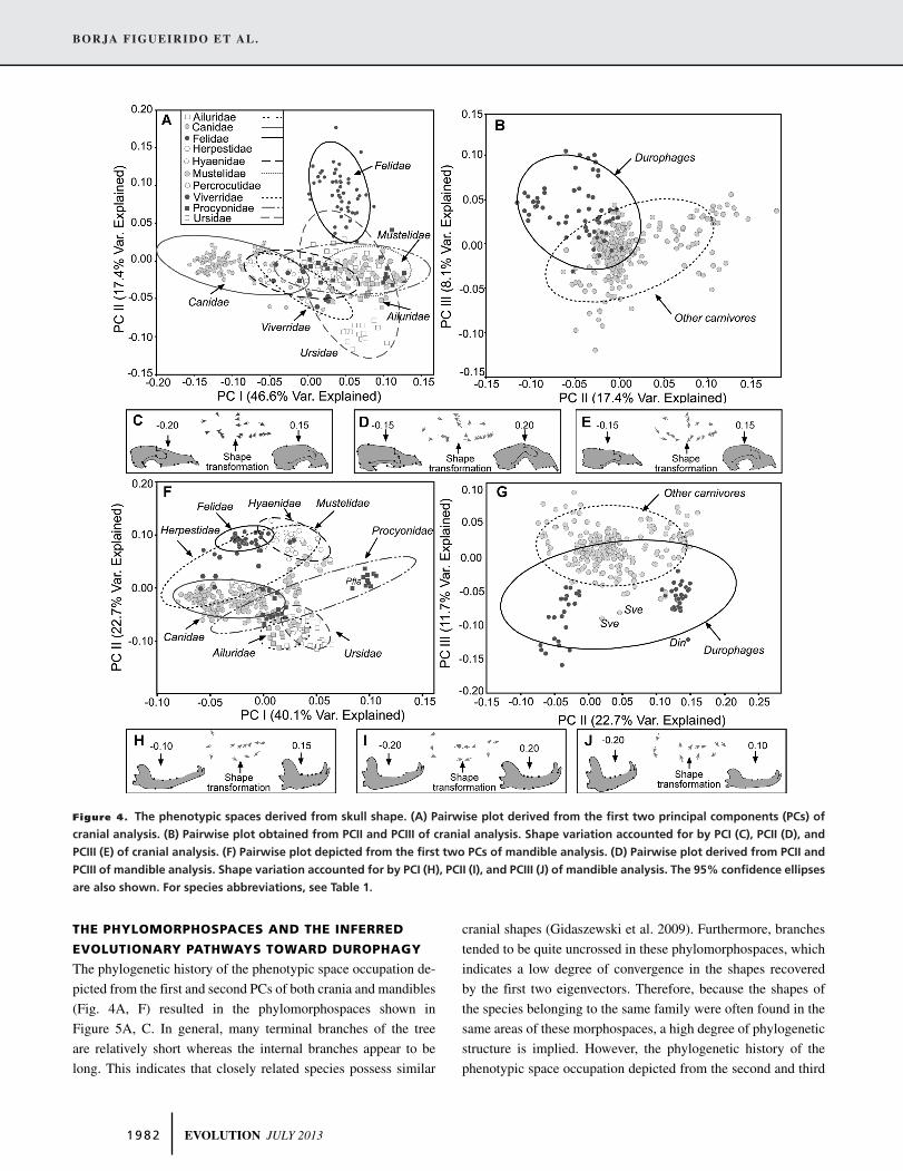
THE PHENOTYPIC SPACES
The phenotypic spaces derived from the PCA of the covariance
matrix for the residuals of the 322 crania are shown in Figure
4A,B. Given that the first three PCs represent a reasonable amount
of the total shape variation (>72% of variance explained), we only
present here these three axes. The first PC (Fig. 4A) differentiates
clearly the dolichochephalic crania of the family Canidae—with
negative scores—from the crania of the other species included in
the sample—scoring positively (Fig. 4C). In contrast, the second
PC (Fig. 4A) describes a shape gradient that goes from the crania
of the family Felidae—with positive scores—to the crania of the
other specimens included in the sample—scoring negatively—
according with those morphologies showed in Figure 4D. The
third PC (Fig. 4B) accounts for shape changes from the cranium of
durophages—taking positive scores—to crania of other species—
with negative scores (Fig. 4E).
The PCA computed from the covariance matrix for the
residuals of the 299 mandibles included in the sample yielded 20
PCs. Again, the first three axes explained >70% of the original
variance, and therefore, we only show here the results derived
from them. The first PC (Fig. 4F) accounts for the shape changes
that take place from the mandibles of the kinkajou (P. flavus)—
with positive scores—to those of other species—with extreme
negative scores some representatives of the family Canidae
(Fig. 4H). Conversely, the second PC (Fig. 4F) relates mainly
with a morphological gradient that goes from the mandibles
of caniform carnivorans—with extreme negative scores for the
mandibles of Ailurudae and Ursidae—to the ones of feliforms,
showing extreme positive scores, with those shapes depicted in
Figure 4I. Finally, the third PC accounts for shape changes from
the mandible of durophagous carnivorans, taking negative scores
in this third axis (Fig. 4G), to those of other carnivores—scoring
positively—with the morphological traits shown in Figure 4J.
EVOLUTION JULY 2013 1 9 8 1
BORJA FIGUEIRIDO ET AL.
Figure 4. The phenotypic spaces derived from skull shape. (A) Pairwise plot derived from the first two principal components (PCs) of
cranial analysis. (B) Pairwise plot obtained from PCII and PCIII of cranial analysis. Shape variation accounted for by PCI (C), PCII (D), and
PCIII (E) of cranial analysis. (F) Pairwise plot depicted from the first two PCs of mandible analysis. (D) Pairwise plot derived from PCII and
PCIII of mandible analysis. Shape variation accounted for by PCI (H), PCII (I), and PCIII (J) of mandible analysis. The 95% confidence ellipses
are also shown. For species abbreviations, see Table 1.
THE PHYLOMORPHOSPACES AND THE INFERRED
EVOLUTIONARY PATHWAYS TOWARD DUROPHAGY
The phylogenetic history of the phenotypic space occupation de-
picted from the first and second PCs of both crania and mandibles
(Fig. 4A, F) resulted in the phylomorphospaces shown in
Figure 5A, C. In general, many terminal branches of the tree
are relatively short whereas the internal branches appear to be
long. This indicates that closely related species possess similar
cranial shapes (Gidaszewski et al. 2009). Furthermore, branches
tended to be quite uncrossed in these phylomorphospaces, which
indicates a low degree of convergence in the shapes recovered
by the first two eigenvectors. Therefore, because the shapes of
the species belonging to the same family were often found in the
same areas of these morphospaces, a high degree of phylogenetic
structure is implied. However, the phylogenetic history of the
phenotypic space occupation depicted from the second and third
1 9 8 2 EVOLUTION JULY 2013
EVOLUTION IN DUROPHAGES
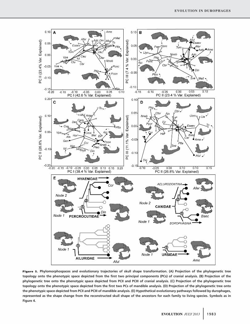
Figure 5. Phylomorphospaces and evolutionary trajectories of skull shape transformation. (A) Projection of the phylogenetic tree
topology onto the phenotypic space depicted from the first two principal components (PCs) of cranial analysis. (B) Projection of the
phylogenetic tree onto the phenotypic space depicted from PCII and PCIII of cranial analysis. (C) Projection of the phylogenetic tree
topology onto the phenotypic space depicted from the first two PCs of mandible analysis. (D) Projection of the phylogenetic tree onto
the phenotypic space depicted from PCII and PCIII of mandible analysis. (E) Hypothetical evolutionary pathways followed by durophages,
represented as the shape change from the reconstructed skull shape of the ancestors for each family to living species. Symbols as in
Figure 4.
EVOLUTION JULY 2013 1 9 8 3
BORJA FIGUEIRIDO ET AL.
PCs (Fig. 4B, G) of both anatomical structures (Fig. 5B, D) indi-
cates the presence of evolutionary convergence. In fact, a visual
inspection of these graphs allows appreciating that the internal
branches of the tree are relatively short compared to the terminal
ones. This suggests that some closely related species showed
clearly different cranial and mandibular shapes (Gidaszewski
et al. 2009). Moreover, in both plots there are many sharp changes
of direction in the evolutionary trajectories, with many crossed
branches leading to a “messy” appearance (Fig. 5B, D).
It is worth noting that the reconstructed evolutionary trajecto-
ries of skull shape transformation are different depending on the
family that durophages belong (Fig. 5E). However, as revealed
by the phenotypic spaces (Fig. 4B, G) and their respective phy-
lomorphospaces (Fig. 5B, D), durophages have attained similar
morphological traits, which suggests their convergent adaptation
to feeding on hard/tough tissues (i.e., bone and bamboo). In con-
trast, other morphological traits have evolved independently as
specific features of bone-crackers and bamboo-feeders (Fig. 5E).
THE MORPHOLOGICAL TRAITS OF THE SKULL
SHARED BY DUROPHAGES
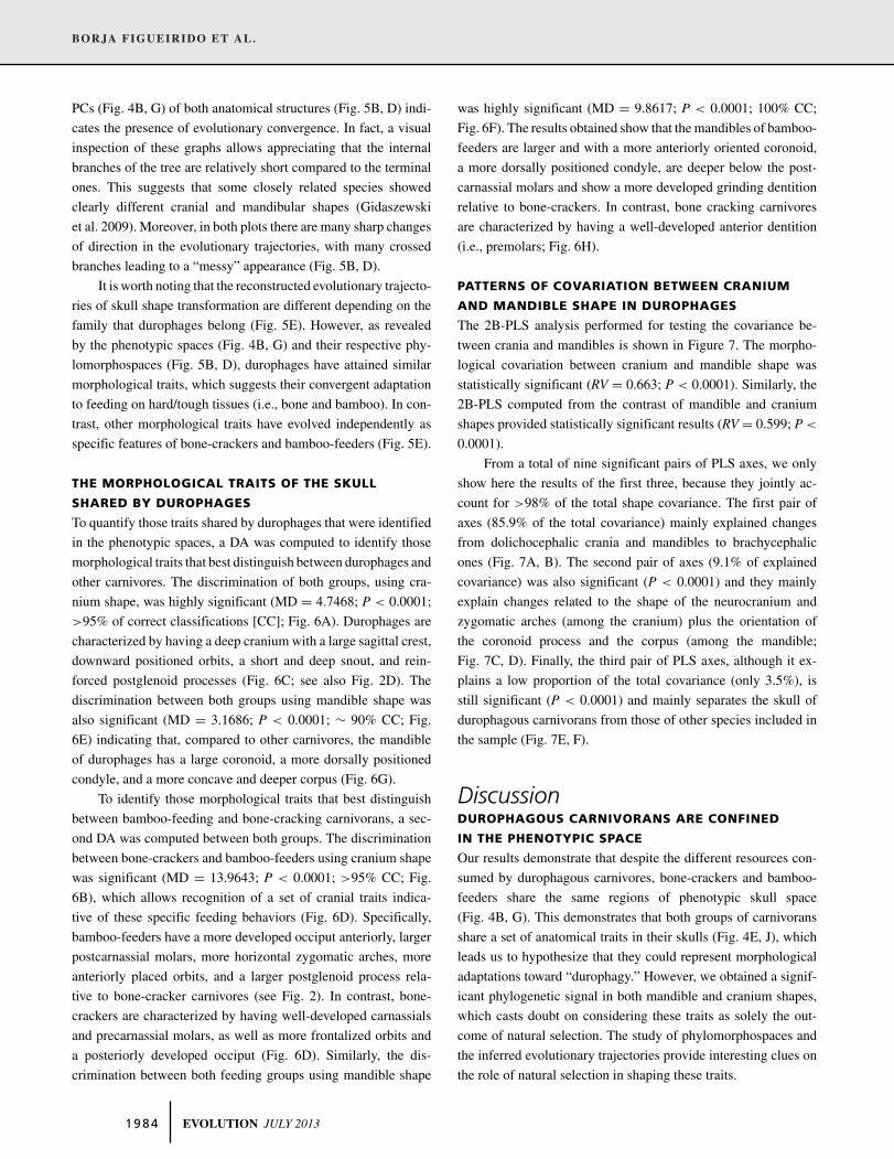
To quantify those traits shared by durophages that were identified
in the phenotypic spaces, a DA was computed to identify those
morphological traits that best distinguish between durophages and
other carnivores. The discrimination of both groups, using cra-
nium shape, was highly significant (MD = 4.7468; P < 0.0001;
>95% of correct classifications [CC]; Fig. 6A). Durophages are
characterized by having a deep cranium with a large sagittal crest,
downward positioned orbits, a short and deep snout, and rein-
forced postglenoid processes (Fig. 6C; see also Fig. 2D). The
discrimination between both groups using mandible shape was
also significant (MD = 3.1686; P < 0.0001; ∼ 90% CC; Fig.
6E) indicating that, compared to other carnivores, the mandible
of durophages has a large coronoid, a more dorsally positioned
condyle, and a more concave and deeper corpus (Fig. 6G).
To identify those morphological traits that best distinguish
between bamboo-feeding and bone-cracking carnivorans, a sec-
ond DA was computed between both groups. The discrimination
between bone-crackers and bamboo-feeders using cranium shape
was significant (MD = 13.9643; P < 0.0001; >95% CC; Fig.
6B), which allows recognition of a set of cranial traits indica-
tive of these specific feeding behaviors (Fig. 6D). Specifically,
bamboo-feeders have a more developed occiput anteriorly, larger
postcarnassial molars, more horizontal zygomatic arches, more
anteriorly placed orbits, and a larger postglenoid process rela-
tive to bone-cracker carnivores (see Fig. 2). In contrast, bone-
crackers are characterized by having well-developed carnassials
and precarnassial molars, as well as more frontalized orbits and
a posteriorly developed occiput (Fig. 6D). Similarly, the dis-
crimination between both feeding groups using mandible shape
was highly significant (MD = 9.8617; P < 0.0001; 100% CC;
Fig. 6F). The results obtained show that the mandibles of bamboo-
feeders are larger and with a more anteriorly oriented coronoid,
a more dorsally positioned condyle, are deeper below the post-
carnassial molars and show a more developed grinding dentition
relative to bone-crackers. In contrast, bone cracking carnivores
are characterized by having a well-developed anterior dentition
(i.e., premolars; Fig. 6H).
PATTERNS OF COVARIATION BETWEEN CRANIUM
AND MANDIBLE SHAPE IN DUROPHAGES
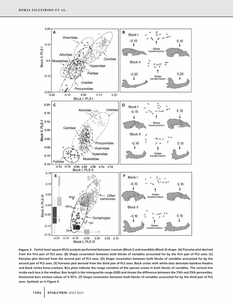
The 2B-PLS analysis performed for testing the covariance be-
tween crania and mandibles is shown in Figure 7. The morpho-
logical covariation between cranium and mandible shape was
statistically significant (RV = 0.663; P < 0.0001). Similarly, the
2B-PLS computed from the contrast of mandible and cranium
shapes provided statistically significant results (RV = 0.599; P <
0.0001).
From a total of nine significant pairs of PLS axes, we only
show here the results of the first three, because they jointly ac-
count for >98% of the total shape covariance. The first pair of
axes (85.9% of the total covariance) mainly explained changes
from dolichocephalic crania and mandibles to brachycephalic
ones (Fig. 7A, B). The second pair of axes (9.1% of explained
covariance) was also significant (P < 0.0001) and they mainly
explain changes related to the shape of the neurocranium and
zygomatic arches (among the cranium) plus the orientation of
the coronoid process and the corpus (among the mandible;
Fig. 7C, D). Finally, the third pair of PLS axes, although it ex-
plains a low proportion of the total covariance (only 3.5%), is
still significant (P < 0.0001) and mainly separates the skull of
durophagous carnivorans from those of other species included in
the sample (Fig. 7E, F).
DiscussionDUROPHAGOUS CARNIVORANS ARE CONFINED
IN THE PHENOTYPIC SPACE
Our results demonstrate that despite the different resources con-
sumed by durophagous carnivores, bone-crackers and bamboo-
feeders share the same regions of phenotypic skull space
(Fig. 4B, G). This demonstrates that both groups of carnivorans
share a set of anatomical traits in their skulls (Fig. 4E, J), which
leads us to hypothesize that they could represent morphological
adaptations toward “durophagy.” However, we obtained a signif-
icant phylogenetic signal in both mandible and cranium shapes,
which casts doubt on considering these traits as solely the out-
come of natural selection. The study of phylomorphospaces and
the inferred evolutionary trajectories provide interesting clues on
the role of natural selection in shaping these traits.
1 9 8 4 EVOLUTION JULY 2013
EVOLUTION IN DUROPHAGES
Figure 6. Discriminant analyses. (A) Histogram showing the distribution of the specimens according to their scores on the discriminant
function adjusted from cranial morphology to separate between durophages and other carnivorans. (B) Histogram showing the distri-
bution of the specimens according to their scores on the discriminant function adjusted from cranial morphology to separate between
bone-crackers and bamboofeeders. (C) Morphological variation accounted for by the discriminant function adjusted from cranial mor-
phology to separate between durophages and other carnivorans. (D) Morphological variation accounted for by the discriminant function
adjusted from cranial morphology to separate between bone-crackers and bamboo-feeders. (E) Histogram showing the distribution of the
specimens according to their scores on the discriminant function adjusted from mandible morphology to separate between durophages
and other carnivorans. (F) Histogram showing the distribution of the specimens according to their scores on the discriminant function
adjusted from mandible morphology to separate between bone-crackers and bamboofeeders. (G) Morphological variation accounted for
by the discriminant function adjusted from mandible morphology to separate between durophages and other carnivorans. (H) Morpho-
logical variation accounted for by the discriminant function adjusted from mandible morphology to separate between bone-crackers and
bamboo-feeders. Dark-gray bars represent the overlapping areas between groups.
THE EVOLUTIONARY ROUTES TO REACH
ADAPTATIONS TOWARD DUROPHAGY
Both the reconstructed phylomorphospaces and the inferred hypo-
thetical evolutionary pathways of skull shape transformation to-
ward durophagy indicate that natural selection is not sufficient for
explaining the shared anatomical traits among bone-crackers and
bamboo-feeders. In fact, although durophages reached similar—
but not identical—skull morphologies (Fig. 5E) and show similar
EVOLUTION JULY 2013 1 9 8 5
BORJA FIGUEIRIDO ET AL.
Figure 7. Partial least square (PLS) analysis performed between cranium (Block I) and mandible (Block II) shape. (A) Pairwise plot derived
from the first pair of PLS axes. (B) Shape covariation between both blocks of variables accounted for by the first pair of PLS axes. (C)
Pairwise plot derived from the second pair of PLS axes. (D) Shape covariation between both blocks of variables accounted for by the
second pair of PLS axes. (E) Pairwise plot derived from the third pair of PLS axes. Black circles with white dots denotate bamboo-feeders
and black circles bone-crackers. Box plots indicate the range variation of the species scores in both blocks of variables. The vertical line
inside each box is the median. Box length is the interquartile range (IQR) and shows the difference between the 75th and 25th percentiles.
Horizontal bars enclose values of 5–95%. (F) Shape covariation between both blocks of variables accounted for by the third pair of PLS
axes. Symbols as in Figure 4.
1 9 8 6 EVOLUTION JULY 2013
EVOLUTION IN DUROPHAGES
deviations from their sister groups, their evolutionary pathways
differed depending on the phylogenetic legacy of each particular
family. In sum, the results of PCA indicate that the traits shared
between both carnivoran groups are strongly influenced by both
phylogenetic inheritance (including here different types of con-
straints; Schwenk and Wagner 2003) and natural selection. How-
ever, further investigation based in Bayesian approaches (e.g.,
Slater et al. 2012) could clarify more finely to which model of
evolution fits the evolutionary patterns described here.
MORPHOLOGICAL ADAPTATIONS OF DUROPHAGOUS
CARNIVORANS
Given that the groups of “durophagous” carnivores and “other
taxa” are both represented by different families belonging to two
different suborders, the results obtained in DA allow us to iden-
tify a shared suite of morphological features among durophages
that are relatively independent of the phylogenetic legacy of each
particular family. Such features could be a priori considered as po-
tential adaptations toward durophagy. However, convergence per
se is not indicative of adaptation (Stayton 2008; Losos 2011). In
fact, evolutionary convergence of traits can occur without the im-
plicit action of natural selection favoring them within a particular
selective environment (see Losos [2011] and references therein).
Therefore, because phenotypes may evolve convergently in simi-
lar environments, even if not directly favored by natural selection,
independent arguments are necessary for testing the adaptative
nature of the traits shared by durophages.
Functional studies of trait evolution could give clues to test
our adaptation hypothesis, as selection operates on the functional
consequences of traits (Arnold 1983). There is compelling evi-
dence from skull biomechanics in mammals (Fig. 2B) that all these
traits enhance the ability to produce the large bite forces required
for feeding on hard/tough foods and for resisting the high com-
pressive loads generated during chewing and biting (e.g., Ewer
1973). For example, a well developed sagittal crest (Fig. 2D)
indicates a large area of attachment for the temporalis muscle
(e.g., Ewer 1973) and temporalis volume correlates well with
bite force (e.g., Christiansen and Adolfsen 2005; Christiansen
and Wroe 2007; Wroe et al. 2005). Similarly, large distances
between the condyle and both the coronoid—that is, insertion
area of the temporalis—and angular—that is, insertion area of
the masseter—processes of the mandible (Fig. 2D) enhance the
input levers of the temporalis and masseter muscles, respectively
(Fig. 2B), which increases mechanical advantage and bite force
(Biknevicius and Van Valeknburgh 1996). Similarly, a shortened
facial skeleton (Fig. 2D) diminishes the output moment arms at
different points of the dentition (Fig. 2B) and also increases bite
force (Turnbull 1976; Biknevicious and Van Valkenburgh 1996;
Christiansen and Wroe 2007). Enlarged postglenoid processes re-
sult in a deep glenoid fossa (Fig. 2D), which precludes dislocating
the lower jaw when large bite forces are exerted at closing angles
(Fig. 2A; Ewer 1973, Werdelin 1989). A deep frontal region of
the skull is the consequence of having large paranasal sinuses
that are involved in dissipating the high compressive loads gen-
erated during chewing (dotted lines in Fig. 2B; Werdelin 1989;
Joeckel 1998; Tanner et al. 2008). Similarly, a deep mandibular
corpus correlates to a large proportion of cortical bone that is
responsible for dissipating compressive forces (Biknevicious and
Ruff 1992). Therefore, because these anatomical features of the
skull enhance the biomechanical performance required to feed
on hard/tough foods, it seems reasonable that these phenotypes
represent convergent adaptations toward durophagy.
THE CRANIUM OF DUROPHAGES REFLECTS THE
“NATURE” OF THE RESOURCES CONSUMED AND
THEIR MANDIBLE THE “TYPE” OF FOOD
Although the DA performed for separating durophagous carni-
vores from other taxa provided significant results using both the
cranium and the mandible, the discrimination using cranial shape
was better than the one with mandible shape (compare the over-
lapping areas—in gray—of the histograms depicted in Fig. 6A,E).
The crania of bone-crackers and bamboo-feeders are more sim-
ilar in shape than their mandible. In fact, this was already noted
from the PCA (compare the areas occupied by durophages in Fig.
4B,G). This result was unexpected, as previous investigations
indicated that lower jaw shape has more evolutionary plasticity
toward particular feeding adaptations than cranial shape (Barone
1986). The reason is that while mandible morphology mainly re-
flects feeding behavior, cranium shape is more constrained by
conflicting functional demands (e.g., feeding, olfaction, vision,
and brain processing; Figueirido and Soiblezon 2010; Figueirido
et al. 2010a,b, 2011a,b). Furthermore, the cranium of carnivores
is made up of a set of bones (i.e., ∼33), but the mandible is only
composed of the dentary bone. As a consequence, the lower jaw
should be a easier structure to modify toward durophagy than the
cranium (Figueirido et al. 2012). However, the results obtained
here point to the opposite direction.
The second round of DA performed to discriminate between
bamboo-feeders and bone-eaters gave clues about this counterin-
tuitive result. Here, we obtained a better discrimination between
both groups of durophages using mandible shape than using cra-
nium shape (compare the histograms depicted in Fig. 6B, F). This
result confirmed previous statements on the higher evolutionary
plasticity of the carnivoran mandible compared to the cranium, as
the latter seems to be more limited to evolve toward the particular
feeding behavior of a durophage. As a result, while there are two
different mandible solutions to be a durophage, depending upon
the resources consumed—that is, bamboo or bone—there is only
one possible solution for the cranium. As a consequence, cranial
shape reflects the nature of the resources consumed—that is, hard
EVOLUTION JULY 2013 1 9 8 7
BORJA FIGUEIRIDO ET AL.
or soft tissues—but mandible morphology more finely reflects the
particular type of food—that is, bone or bamboo. For this reason,
durophagous carnivorans converge more in cranial shape than in
lower jaw shape.
DUROPHAGOUS CARNIVORANS SHARE PATTERNS OF
COVARIATION BETWEEN THE SHAPES OF CRANIA
AND MANDIBLES
The fact that both structures reflect different aspects of feeding be-
havior in durophages opened the possibility to test if the cranium
and the mandible were morphologically integrated. Furthermore,
if cranial shape is more limited to evolve toward durophagy than
mandible shape, we should expect a lower variation in their cra-
nia than in their mandibles. We analyzed patterns of covariation
between cranium and mandible shape using PLS analysis for ex-
ploring quantitatively this hypothesis.
In spite of the fact that the cranium and the mandible of
durophages reflect different aspects of feeding behavior—that is,
nature and type of the resources consumed—our results clearly
demonstrate that cranial and lower jaw shape are highly inte-
grated in carnivores. This result was in part expected because
other authors have identified the same two modules coordinating
shape variability in the cranium—that is, neurocranium and facial
skull; Drake and Klingenberg (2010)—and in the mandible—
that is, ramus and corpus; Meloro et al. (2011). Furthermore,
from a functional perspective this result was also expected, be-
cause the ramus is mainly connected with the neurocranium by
the same main muscles involved in mastication and the corpus
fits with the facial skeleton by the occlusion of upper and lower
teeth.
Durophagous carnivorans also share patterns of covariation
(Fig. 7E,F), most probably as a result of the functional demands
to feed on tough foods. It is obviously inconceivable to have a
“strong” cranium equipped to withstand high bite forces and a
“weak” mandible without these biomechanical adaptations. Fur-
thermore, for those morphological aspects recovered from the
third pair of PLS-axes—which are those traits that best distinguish
the pattern of durophagous covariation—cranial shape shows
much less variation than lower jaw shape (Fig. 7E; box plots),
which confirms that the former has a more limited evolvability.
However, this interpretation is mainly functional and again the
presence of a strong phylogenetic signal in cranium and mandible
shape casts doubts on whether this pattern is exclusively an evi-
dence of adaptation or reflects a constraint.
TERATOLOGICAL CATS AND DOGS PROVIDE
EVIDENCE THAT COVARIATION IN LOWER JAW
AND CRANIAL SHAPE IS EXTERNALLY GOVERNED
The study of some brachycephalic cats (e.g., exotic short-hair
cats) and dogs (e.g., English bulldogs) breeds with a substantially
longer lower jaw relative to the upper jaw (Fox 1963; Schlueter
et al. 2009) constitutes a complementary approach to test if the
pattern of covariation reported between cranial and lower jaw
shape is externally or internally governed. These breeds were
already considered as teratological phenotypes by Charles Darwin
(1875), who identified the English bulldog as a monstrosity fixed
by man’s selection. Experiments on artificial selection provide a
valuable experimental test for assessing whether phenotypes not
observed in nature are the result of selection or constraint (Losos
2011; Olson 2012), as they contribute information about which
phenotypes can and cannot be produced by normal development
(e.g., Alberch 1989; Galis et al. 2006; Olson 2012).
Brachycephalic breeds are known in the veterinary jargon as
“undershot bite.” The “undershot bite” or brachycephalic condi-
tion is considered the result of having a modified achondroplasic
cranium with an extremely shorted snout and palatal region, which
results in a wide face and causes the mandible to overhang beyond
the maxilla (Stockard 1941; Fox 1963). All of these morpholog-
ical traits are the breed standard in brachycephalic dogs (Amer-
ican Kennel Club 2006), which constitutes a documented record
of the selection regime applied by breeders (Young and Bannasch
2006). The genetic cause of the brachycephalic condition—at
least in dogs—is a mutation in the retro-gene coding for fibrob-
last growth factor 4 (FGFR4; Parker et al. 2009), which causes an
abnormal pattern in cartilage formation. In contrast, the opposite
condition, the “overshot bite” or the “the parrot-mouth” seen in
several dogs—for example, in German Shepards (Fox 1963) and
cattle have never been selected in any specific breed. In fact, this
abnormal phenotype, when present, entails a set of injuries caused
by the occlusion of the mandibular teeth in the soft tissues of the
palatal region (Fox 1963). However, even although not being se-
lected by breeders, this deformity seems to appear regularly in
some dog breeds (see Stockard 1941; Fox 1963) and it is cor-
rected, whenever possible, during the first months of individual’s
life.
Both abnormal conditions seen in domesticated animals
indicate that these phenotypes are developmentally possible,
which points to the hypothesis that phenotypes lacking integrated
mandibles and crania are not the result of an internal constraint.
In fact, this was already noted by Fox (1963) referring to Stockard
(1941): “By crossbreeding different types of dogs (dolicocephalic,
mesocephalic, and brachycephalic) a wide spectrum of hybrid
variations have been produced and it was concluded that the length
of the upper jaw is inherited independently of the lower jaw and
vice versa.”
Therefore, both phenotypes which do not have integrated
mandibles and crania are ontogenetically possible. Consequently,
there is no reason to invoke the presence of developmental con-
straints to shape the pattern of covariation between the shapes
of the mandible and the cranium (Fig. 8). Thus, two possible
1 9 8 8 EVOLUTION JULY 2013
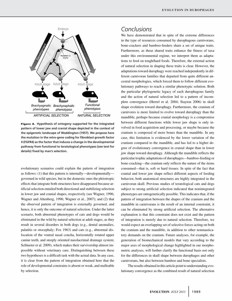
EVOLUTION IN DUROPHAGES
Figure 8. Hypothesis of ontogeny supported for the integrated
pattern of lower jaw and cranial shape depicted in the context of
the epigenetic landscape of Waddington (1957). We propose here
the mutation in the retro-gene coding for fibroblast growth factor
4 (FGFR4) as the factor that induces a change in the developmental
pathway from functional to teratological phenotypes (see text for
details) fixed by man’s selection.
evolutionary scenarios could explain the pattern of integration
as follows: (1) that this pattern is internally—developmentally—
governed in wild species, but in the domestic ones the pleiotropic
effects that integrate both structures have disappeared because ar-
tificial selection entailed both directional and stabilizing selection
in lower jaw and cranial shapes, respectively (see Wagner, 1996;
Wagner and Altenberg, 1996; Wagner et al., 2007); and (2) that
the observed pattern of integration is externally governed, and
hence, it is only the outcome of natural selection. Under the latter
scenario, both abnormal phenotypes of cats and dogs would be
eliminated in the wild by natural selection at adult stages, as they
result in several disorders in both dogs (e.g., dental anomalies,
palatitis or otocephaly; Fox 1963) and cats (e.g., abnormal dis-
location of the ventral nasal concha, horizontally rotated upper
canine teeth, and steeply oriented nasolacrimal drainage system;
Schlueter et al. 2009), which makes their survivorship almost im-
possible without veterinary care. Distinguishing between these
two hypotheses is a difficult task with the actual data. In any case,
it is clear from the pattern of integration obtained here that the
role of developmental constrains is absent or weak, and malleable
by selection.
ConclusionsWe have demonstrated that in spite of the extreme differences
in the type of resources consumed by durophagous carnivorans,
bone-crackers and bamboo-feeders share a set of unique traits.
Furthermore, as these shared traits enhance the fitness of taxa
under this environmental regime, we interpret them as adapta-
tions to feed on tough/hard foods. Therefore, the external action
of natural selection in shaping these traits is clear. However, the
adaptations toward durophagy were reached independently in dif-
ferent carnivoran families that departed from quite different an-
cestral morphologies, which forced them to follow different evo-
lutionary pathways to reach a similar phenotypic solution. Both
the particular phylogenetic legacy of each durophagous family
and the action of natural selection led to a pattern of incom-
plete convergence (Herrel et al. 2004; Stayton 2006) in skull
shape evolution toward durophagy. Furthermore, the cranium of
carnivores is more limited to evolve toward durophagy than the
mandible, perhaps because cranial morphology is a compromise
between different functions while lower jaw shape is only in-
volved in food acquisition and processing, or maybe because the
cranium is composed of more bones than the mandible. In any
case, this limitation is evidenced by the lower variation of the
cranium compared to the mandible, and has led to a higher de-
gree of evolutionary convergence in cranial shape than in lower
jaw shape toward durophagy. Although the mandible reflects the
particular trophic adaptations of durophages—bamboo-feeding or
bone-cracking—the cranium only reflects the nature of the items
consumed—that is, soft or hard tissues. In spite of the fact that
cranial and lower jaw shape reflect different aspects of feeding
behavior, both anatomical structures are highly integrated in the
carnivoran skull. Previous studies of teratological cats and dogs
subject to strong artificial selection indicated that nonintegrated
phenotypes are ontogenetically possible. This indicates that, if the
pattern of integration between the shapes of the cranium and the
mandible in carnivorans is the result of an internal constraint, it
can be eliminated by strong artificial selection. The alternative
explanation is that this constraint does not exist and the pattern
of integration is merely due to natural selection. Therefore, we
would expect an overlapping set of selective forces acting on both
the cranium and the mandible, in addition to other nonmastica-
tory demands on the cranium. Future analyses, for example, the
generation of biomechanical models that vary according to the
major axes of morphological change highlighted in our morpho-
metric analyses, will further clarify the functional basis not only
for the differences in skull shape between durophages and other
carnivorans, but also between bamboo and bone specialists.
The results obtained in this article point to understanding evo-
lutionary convergence as the combined result of natural selection
EVOLUTION JULY 2013 1 9 8 9
BORJA FIGUEIRIDO ET AL.
and constraint. We do not expect evolutionary convergence of
traits in the same environmental regime without the implicit ac-
tion of natural selection (except those cases of chance, exapta-
tions, or correlations on another characters; see Losos [2011] and
references therein), but its action has to be envisaged—in our
view—upon a limited range of phenotypic variants imposed by
different types of constraints—genetic, developmental, or struc-
tural. All of these potential constraints could be grouped under
“phylogenetic constraints.” Although this term has a low oper-
ational value, it is a useful heuristic concept (Schwenk 1994).
Therefore, we refer to phylogenetic constraints as those as-
pects derived from the early definition of body plans determined
early in lineage phylogenies within determined biomaterials and
under highly canalized developmental routes (e.g., Gould and
Lewontin 1979; Seilacher 1979; Cheverud 1982, 1985; Maynard-
Smith 1985; Schluter 1988; Wagner 1988; Gould 1989; Ligon
1993; Thomas and Reif 1993; Schwenk 1995; Wagner 1996),
which strongly biases the production of future phenotypic
variants.
ACKNOWLEDGMENTSWe thank E. Westwig, J. Galkin (AMNH, New York), P. Jenkins, L.Tomsset, and R. Portella (NHM, London) for kindly providing us accessto the specimens under their care. We are also grateful to C. M. Janis,P. Palmqvist, J. A. Perez-Claros, and two anonymous reviewers whosecomments and suggestions greatly improved the rigor of the manuscriptcontents. BF would like to dedicate this paper to M. D. Renzi who gavehim the underlying logic for understanding monsters. This study wassupported by Fulbright Postdoctoral Grant FU2009–0184 (to BF).
LITERATURE CITEDAbouheif, E. 2008. Parallelism as the pattern and process of mesoevolution.
Evol. Dev. 10:3–5.Alberch, P. 1989. The logic of monsters: evidence for internal constraint in
development and evolution. Geobios 12:21–57.Alberch, P., and E. A. Gale. 1985. A developmental analysis of an evolutionary
trend—digital reduction in amphibians. Evolution 39:8–23.American Kennel Club. 2006. The complete dog book. Ballatine Books, New
York, NY.Arendt, J., and D. Reznick. 2008. Convergence and parallelism reconsidered:
what have we learned about the genetics of adaptation? Trends Ecol.Evol. 23:26–32.
Arnold, S. J. 1983. Morphology, performance and fitness. Am. Zool. 23:347–361.
Bardeleben, C., R.L. Moore, and R.K. Wayne. 2005. A molecular phylogenyof the Canidae based on six nuclear loci. Mol. Phylogenet. Evol. 37:815–831.
Barone, R. 1986. Anatomie compare des mammiferes domestiques. Vol. 1.Vigot, Paris.
Blackledge, T. A., and R. G Gillespie. 2004. Convergent evolution of behaviorin an adaptive radiation of Hawaiian web-building spiders. Proc. Natl.Acad. Sci. USA 101:16228–16233.
Bicknevicius, A.R., and B. Van Valkenburgh. 1996. Design for killing: cran-iodental adaptations of predators. Pp. 393–428 in J. L. Gittleman, ed.Carnivore behavior, ecology and evolution. Vol. 2. Cornell Univ. Press,Ithaca, NY.
Biknevicius, A. R., and C. B. Ruff. 1992. The structure of the mandibularcorpus and its relationship to feeding behaviors in extant carnivorans. J.Zool. (Lond.) 228:479–507.
Cheverud, J. M. 1982. Phenotypic, genetic, and environmental morphologicalintegration in the cranium. Evolution 36:499–516.
———. 1985. Quantitative genetics and developmental constraints on evolu-tion by selection. J. Theor. Biol. 110:155–172.
Christiansen P., and J. S. Adolfssen. 2005. Bite forces, canine strength andskull allometry in carnivores (Mammalia, Carnivora). J. Zool. Lond.266:133–151.
Christiansen, P., and S. Wroe. 2007. Bite forces and evolutionaryadaptations to feeding ecology in carnivores. Ecology 88:347–358.
Conway-Morris, S. 2003. Life’s solution: inevitable humans in a lonely uni-verse. Cambridge Univ. Press, Cambridge, U.K.
Darwin, C. 1875. The variation of animals and plants under domestication.Vol. 1. John Murray, London, U.K.
Davis, D. D. 1964. The giant Panda. A morphological study on evolutionarymechanisms. Fieldiana Zool. 3:1–339.
Desutter-Grandcolas, L., F. Legendre, P. Grandcolas, T. Robillard, and J.Murienne. 2005. Convergence and parallelism: is a new life ahead of oldconcepts? Cladistics 21:51–61.
Winter, W., de, and C. E. Oxnard. 2001. Evolutionary radiations and con-vergences in the structural organization of mammalian brains. Nature409:710–714.
Dong, W. 2008. Virtual cranial endocast of the oldest giant panda (Ailuropodamicrota) reveals great similarity to that of its extant relative. Naturwis-senschaften 95:1079–1083.
Drake, A. G., and C. P. Klingenberg. 2008. The pace of morphological change:historical transformation of skull shape in St Bernard dogs. Proc. R. Soc.B 275:71–76.
———. 2010. Large-scale diversification of skull shape in domestic dogs:disparity and modularity. Am. Nat. 175:289–301.
Dryden, I. L., and K. Mardia. 1998. Statistical shape analysis. Wiley, Chich-ester, U.K.
Escoufier, Y. 1973. Le traitement des variables vectorielles. Biometrics29:751–760.
Ewer, R. F. 1973. The carnivores. Cornell Univ. Press, New York, NY.Eizirik, E., W. J. Murphy, K.-P. Koepfli, W. E. Johnson, J. W. Dragoo, R. K.
Wayne, and S. J. O’Brien. 2010. Pattern and timing of diversificationof the mammalian order Carnivora inferred from multiple nuclear genesequences. Mol. Phylogenet. Evol. 56:49–63.
Felsenstein, J. J. 1985. Phylogenies and the comparative method. Am. Nat.125:1–15.
Figueirido, B., and L. H. Soibelzon. 2010. Inferring palaeoecology in extincttremarctine bears (Carnivora, Ursidae) using geometric morphometrics.Lethaia 43:209–222.
Figueirido, B., J. A. Perez-Claros, V. Torregrosa, A. Martın-Serra, and P.Palmqvist. 2010a. Demythologizing Arctodus simus, the “short-faced,”long-legged and predaceous bear that never was. J. Vert. Paleontol.30:262–275.
Figueirido, B., F. J. Serrano-Alarcon, G. J. Slater, and P. Palmqvist. 2010b.Shape at the cross-roads: homoplasy and history in the evolutionof the carnivoran skull towards herbivory. J. Evol. Biol. 23:2579–2594.
Figueirido, B., N. MacLeod, J. Krieger, M. De Renzi, J. A. Perez-Claros,and P. Palmqvist. 2011a. Constraint and adaptation in the evolution ofcarnivoran skull shape. Paleobiology 37:490–518.
Figueirido, B., P. Palmqvist, J. A. Perez-Claros, and W. Dong. 2011b. Cranialshape transformation in the evolution of the giant panda (Ailuropodamelanoleuca). Naturwissenschaften 98:107–116.
1 9 9 0 EVOLUTION JULY 2013
EVOLUTION IN DUROPHAGES
Figueirido, B., F. J. Serrano-Alarcon, and P. Palmqvist. 2012. Geometricmorphometrics shows differences and similarities in skull shape betweenthe red and giant pandas. J. Zool. 286:293–302.
Finarelli, J. A., and J. J. Flynn. 2006. Ancestral state reconstruction of bodysize in the Caniformia (Carnivora, Mammalia): the effects of incorpo-rating data from the fossil record. Syst. Biol. 55:301–313.
Finarelli, J. S., and J. J. Flynn. 2009. Brain-size evolution and sociality inCarnivora. Proc. Natl. Acad. Sci. USA 106:345–9349.
Flynn, J. J., M. Nedbal, J. Dragoo, and L. Honeycutt. 2000. Whence the redpanda? Mol. Phylogenet. Evol. 17:190–199.
Flynn, J. J., J. A. Finarelli, S. Zehr, J. Hsu, and M. Nedbal. 2005. Molecularphylogeny of the Carnivora (Mammalia): assessing the impact of in-creased sampling on resolving enigmatic relationships. Syst. Biol.54:317–337.
Fox, M. L. 1963. Developmental abnormalities of the canine skull. Can. J.Comp. Med. Vet. Sci. 27:219–222.
Futuyma, D. J. 2010. Evolutionary constraint and ecological consequences.Evolution 64:1865–1884.
Galis, F., T. J. M. Van Dooren, H. Feuth, S. Ruinard, A. Witkam, and M. J.Steigenga. 2006. Extreme selection in humans against homeotic trans-formations of cervical vertebrae. Evolution 60:2643–2654.
Gaubert P., and G. Veron. 2003. Exhaustive sample set among Viverridaereveals the sister-group of felids: the Linsangs as a case of extrememorphological convergence within Feliformia. Proc. R. Soc. B Biol.Sci. 270:2523–2530.
Gidaszewski, N. A., M. Baylac, and C. P. Klingenberg. 2009. Evolution of sex-ual dimorphism of wing shape in the Drosophila melanogaster subgroup.BMC Evol. Biol. 9:110.
Gittleman, J. L. 1994. Are the Pandas successful specialists or evolutionaryfailures? Bioscience 44:456–464.
Goswami, A., N. Milne, and S. Wroe. 2011. Biting through constraints:cranial morphology, disparity and convergence across living and fos-sil carnivorous mammals. Proc. Roy. Soc. B Biol. Sci. 278:1831–1839.
Gould, S. J. 1989. A developmental constraint in Cerion, with comments onthe definition and interpretation of constraint in evolution. Evolution43:516–539.
———. 2002. The structure of the evolutionary theory. Harvard Univ. Press,Cambridge, MA.
Gould, S. J., and R. Lewontin. 1979. The spandrels of San Marco and thePanglossian paradigm: a critique of the adaptationist programme. Proc.R. Soc. B 205:581–598.
Grenier, J. L., and R. Greenberg. 2005. A biogeographic pattern in sparrow billmorphology: parallel adaptation to tidal marshes. Evolution 59:1588–1595.
Hall, B. K. 2007. Homoplasy and homology: dichotomy or continuum? J.Hum. Evol. 52:473–479.
Hansen, R. L., M. M. Carr, C. J. Apanavicius, P. Jiang, H. A. Bissell, B. L.Gocinski, F. Maury, M. Himmelreich, S. Beard, J. R. Ouellette, et al.2010. Seasonal shifts in giant panda feeding behavior: relationships tobamboo plant part consumption. Zoo. Biol. 29:470–483.
Harvey, P. H., and M. D. Pagel. 1991. The comparative method in evolution-ary biology. Oxford Univ. Press, Oxford, U.K.
Herrel, A., B. Vanhooydonck, and R. Van Damme. 2004. Omnivory in lacertidlizards: adaptive evolution or constraint? J. Evol. Biol. 17:974–984.
Holliday, J. A., and S. J. Steppan. 2001. Evolution of hypercarnivory: theeffect of specialization on morphological and taxonomic diversity. Pa-leobiology 30:108–128.
Jaekel, M., and D. B. Wake. 2007. Developmental processes underlying theevolution of a derived foot morphology in salamanders. Proc. Natl. Acad.Sci. USA 104:20437–20442.
Joeckel, R. M. 1998. Unique frontal sinuses in fossil and living Hyaenidae(Mammalia, Carnivora): description and interpretation. J. Vert. Paleon-tol. 18:627–639.
Johnson, K. G., G. B. Schaller, and J. Hu. 1988. Comparative behavior ofred and giant pandas in the Wolong Reserve. China J. Mammal. 69:552–564.
Johnson W., E. Eizirik, J. Pecon-Slattery, W. Murphy, A. Antunes, E. Teeling,and S. O’Brian. 2006. The late Miocene radiation of modern Felidae: agenetic assessment. Science 311:73–77.
Klingenberg, C. P. 2009. Morphometric integration and modularity in con-figurations of landmarks: tools for evaluating a-priori hypotheses. Evol.Dev. 11:405–421.
———. 2011. MorphoJ: an integrated software package for geometric mor-phometrics. Mol. Ecol. Resources 11:353–357.
Klingenberg, C. P., and W. Ekau. 1996. A combined morphometric and phylo-genetic analysis of an ecomorphological trend: pelagization in Antarcticfishes (Perciformes: Nototheniidae). Biol. J. Linn. Soc. 59:143–177.
Klingenberg, C. P., and N. A. Gidaszewski. 2010. Testing and quantifyingphylogenetic signals and homoplasy in morphometric data. Syst. Biol.59:245–261.
Klingenberg, C. P., S. Dutke, S. Whelan, and M. Kim. 2012. Developmentalplasticity, morphological variation and evolvability: a multilevel analysisof morphometric integration in the shape of compound leaves. J. Evol.Biol. 25:115–129.
Koepfli K. P., S. M. Jenks, E. Eizirik, T. Zahirpour, B. Van Valkenburgh, andR. K. Wayne. 2006. Molecular systematics of the Hyaenidae: relation-ships of a relictual lineage resolved by a molecular supermatrix. Mol.Phylogenet. Evol. 38:603–620.
Koepfli, K. P., M. E. Gompper, E. Eizirik, C. C. Ho, L. Linden, J. E. Maldon-ado, and R. K. Wayne. 2007. Phylogeny of the Procyonidae (Mammalia:Carnivora): molecules, morphology and the great American interchange.Mol. Phylogenet. Evol. 43:1076–1095.
Koepfli, K. P., K. A. Deere, G. J. Slater, C. Begg, K. Begg, L. Grassman, M.Lucherini, G. Veron, and R. K. Wayne. 2008. Multigene phylogeny of theMustelidae: resolving relationships, tempo and biogeographic history ofa mammalian adaptive radiation. BMC Biol. 6:10.
Krause J., T. Unger, A. Nocon, A. S. Malaspinas, S. O. Kolokotronis, M.Stiller, L. Soibelzon, H. Spriggs, P. H. Dear, A. W. Briggs et al. 2008.Mitochondrial genomes reveal an explosive radiation of extinct andextant bears near the Miocene-Pliocene boundary. BMC Evol. Biol.2008, 8:220.
Kruk, H. 1972. The spotted hyena: a study of predation and social behavior.Univ. of Chicago Press, Chicago. 335 pp.
Langerhans, R. B., J. H. Knouft, and J. B. Losos. 2006. Shared and uniquefeatures of diversification in Greater Antillean Anolis ecomorphs. Evo-lution 60:362–369.
Laurin, M. 2004. The evolution of body size, Cope’s rule and the origin ofamniotes. Syst. Biol. 53:594–622.
Ligon J. D. 1993. The role of phylogenetic history in the evolution of contem-porary avian mating and parental care systems. Curr. Ornithol. 10:1–46.
Losos, J. 2011. Convergence, adaptation and constraint. Evolution 65:1827–1840.
Low, I. M., Z. Y. Che, and B. A. Latella. 2006. Mapping the structure, composi-tion and mechanical properties of bamboo. J. Mater. Res. 21:1969–1976.
MacLeod, N. 2001. The role of phylogeny in quantitative paleobiological dataanalysis. Paleobiology 27:226–240.
———. 2006, Data blocks and partial least squares analysis. PalaeontologicalAssociation Newsletter 63:36–48.
Maddison, W. P. 1991. Squared-change parsimony reconstructions of ancestralstates for continuous-valued characters on a phylogenetic tree. Syst.Zool. 40:304–314.
EVOLUTION JULY 2013 1 9 9 1
BORJA FIGUEIRIDO ET AL.
Maddison, W. P. and D. R. Maddison. 2011. Mesquite: a modular sys-tem for evolutionary analysis. Version 2.75. Available via http://mesquiteproject.org.
Madsen, O., M. Scally, C. J. Douady, D. J. Kao, R. W. DeBry, R. Adkins, H.M. Amrine, M. J. Stanhope, W. W. De Jong, and M. S. Springer. 2001.Parallel adaptive radiations in two major clades of placental mammals.Nature 409:610–614.
Maynard-Smith, J., R. Burian, S. Kauffman, P. Alberch, J. Campbell, B.Goodwin, R. Lande, D. Raup, and L. Wolpert. 1985. Developmentalconstraints and evolution. Q. Rev. Biol. 6:265–287.
Meloro C., P. Raia, F. Carotenuto, and S. Cobb. 2011. Phylogenetic signal,function and integration in the subunits of the carnivoran mandible. Evol.Biol. 38:465–475.
Meyer, A. 1999. Homology and homoplasy: the retention of genetic pro-grammes. Pp. 141–157 in G. Bock, eds. Homology. Wiley and Son,Chichester, U.K.
Monteiro, L. R. 1999. Multivariate regression models and geometric morpho-metrics: the search for causal factors in the analysis of shape. Syst. Biol.48:192–199.
Nevo, E. 2001. Evolution of genome–phenome diversity under environmentalstress. Proc. NY Acad. Sci. 98:623–6240.
Nowak, R. M. 1991. Walker’s mammals of the world. John Hopkins Univ.Press, Baltimore, MD.
Oldfield, C. C., C. R. McHenry, P. D. Clausen, U. Chamoli, W. C. H.Parr, D. Stynder, and S. Wroe. 2012. Finite element analysis of ur-sid cranial mechanics and the prediction of feeding behaviour inthe extinct giant Agriotherium africanum. J. Zool. (Lond.) 286:163–170.
Olson, M. E. 2012. The developmental renaissance in adaptationism. TrendsEcol. Evol. 27:278–287
Palmqvist, P., A. Arribas, and B. Martınez-Navarro. 1999. Ecomorphologicalanalysis of large canids from the lower Pleistocene of southeastern Spain.Lethaia 32:75–88.
Palmqvist, P., B. Martınez-Navarro, J. A. Perez-Claros, V. Torregrosa, B.Figueirido, J. M. Jimenez-Arenas, M. Patrocinio Espigares, S. Ros-Montoya, and M. De Renzi 2011. The giant hyena Pachycrocuta bre-virostris: modelling the bone-cracking behavior of an extinct carnivore.Quaternary Int. 243:61–79.
Parker, H. G., B. M. VonHoldt, P. Quignon, E. H. Margulies, S. Shao, D. S.Mosher, and T. C. Spady. 2009. An expressed Fgf4 retrogene is asso-ciated with breed-defining chondrodyspasia in domestic dogs. Science325:995–998.
Patou M. L., P. A. Mclenachan, C. G. Morley, A. Couloux, C. Cruaud, A. P.Jennings, and G. Veron. 2009. Molecular phylogeny of the Herpestidae(Mammalia, Carnivora) with a special emphasis on the Asian Herpestes.Mol. Phylogenet. Evol. 53:69–80.
———. 2008. Adaptive zones and the pinniped ankle: a 3D quantitativeanalysis of carnivoran tarsal evolution. Pp. 165–194 in E. Sargis andM. Dagosto, eds. Mammalian evolutionary morphology: a tribute toFrederick S. Szalay. Springer, Dordrecht, The Netherlands.
Reid, D. G., J. Hu, and Y. Huang.1991. Ecology of the red panda in the WolongReserve, China. J. Zool. (Lond.) 225:347–364.
Rohlf, F. J. 2002. Geometric morphometrics and phylogeny. Pp. 175–193 in
N. MacLeod and P. L. Forey, eds. Morphology, shape, and phylogeny.Taylor and Francis, London.
———. 2008. TpsDig, ver. 2.11. Digitize landmarks and outlines. Departmentof Ecology and Evolution, State University of New York at Stony Brook[computer program and documentation].
Rohlf, F. J., and Corti, M. 2000. The use of two-block partial least-squares tostudy covariation in shape. Syst. Biol. 49: 740–753.
Sacco, T., and B. Van Valkenburgh. 2004. Ecomorphological indicatorsof feeding behaviour in bears (Carnivora: Ursidae). J. Zool. (Lond.)263:41–54.
Schaller G. B., H. Jinchu, P. Wenshi, and Z. Jing. 1985. The giant pandas ofWolong. Univ. of Chicago Press, Chicago.
Schluter, D. 1988. Estimating the form of natural selection on a quantitativetrait. Evolution 42:849–861.
———. 2000. The ecology of adaptive radiation. Oxford Univ. Press, Oxford,U.K.
———. 2009. Evidence for ecological speciation and its alternative. Science323:737–741.
Schmidt-Nielsen, K. 1984. Scaling: Why is animal size so important? Cam-bridge Univ. Press, Cambridge, NY.
Schwenk, K. 1995. A utilitarian approach to evolutionary constraint. Zoology98:251–262.
Schwenk, K., and G. P. Wagner. 2003. Constraint. Pp. 52–61 in B. K. Hall,and W. M. Olson, eds. Key words and concepts in evolutionary devel-opmental biology. Harvard Univ. Press, Cambridge, MA.
———. 2004. The relativism of constraints on phenotypic evolution. Pp.390–408 in M. Pigliucci, and K. Preston, eds. Phenotypic integration:studying the ecology and evolution of complex phenotypes. OxfordUniv. Press, Oxford.
Slater, G. J., L. J. Harmon, and M. Alfaro. 2012. Integrating fossils withmolecular phylogenies improves inference of trait evolution. Evolution66:3931–3944.
Stayton, C. T. 2006. Testing hypotheses of convergence with multivariate data:morphological and functional convergence among herbivorous lizards.Evolution 60:824–841.
———. 2008. Is convergence surprising? An examination of the frequencyof convergence in simulated datasets. J. Theor. Biol. 252:1–14.
Stefen, C. 1999. Enamel structure of recent and fossil canidae (carnivora,mammalia). J. Vert. Paleontol. 19:576–588.
Stefen, C. 2001. Enamel structure of arctoid carnivora: amphicyonidae, ursi-dae, procyonidae, and mustelidae. J. Mammal. 82:450–462.
Stefen, C., and J. M. Rensberger. 2002. The specialized structure of hyaenidenamel: description and development within the lineage – includingpercrocutids. Zoologische Abhandlungen 52:127–147.
Stockard. C. R. 1941. The genetic and endocrine basis for differences in formand behavior. Wistar Insttute, Philadelphia.
Tanner, J. B., E. R. Dumont, S. T. Sakai, B. L. Lundrigan, and K. E.Holekamp. 2008. Of arcs and vaults: the biomechanics of bone-crackingin spotted hyenas (Crocuta crocuta). Biol. J. Linn. Soc. 95:246–255.
Tedford, R. H., X. Wang, and B. E. Taylor. 2009. Phylogenetic systematics ofthe North American fossil Caninae. Bull. Am. Mus. Nat. Hist. 325:1–218.
Thomas, R. D. K., and W. E. Reif. 1993. The skeleton space: a finite set oforganic designs. Evolution 47:341–360.
Timm, N. H. 2002. Applied multivariate analysis. Springer, New York.Tseng, Z. J. 2009. Cranial function in a late Miocene Dinocrocuta gigantea
(Mammalia: Carnivora) revealed by comparative finite element analysis.Biol. J. Linn. Soc. 96:51–67.
———. 2012. Connecting Hunter–Schreger band microstructure to enamelmicrowear features: new insights from durophagous carnivores. ActaPalaeontol. Pol. 57:473–484.
Tseng Z. J. and W. Binder. 2010. Mandibular biomechanics of Crocuta cro-
cuta, Canis lupus, and the late Miocene Dinocrocuta gigantea (Car-nivora, Mammalia). Zool. J. Linn. Soc. Lond. 158:683–696.
Tseng, Z. J., and X. Wang. 2011. Do convergent ecomorphs evolve throughconvergent morphological pathways? Cranial shape evolution in fossil
1 9 9 2 EVOLUTION JULY 2013
EVOLUTION IN DUROPHAGES
hyaenids and borophagine canids (Carnivora, Mammalia). Paleobiology37:470–489.
Turnbull, W. D. 1976. Mammalian masticatory apparatus. Fieldiana Geol.18:152–356.
Van Valkenburgh, B. 1991. Iterative evolution of hypercarnivory in canids(Mammalia: Carnivora): evolutionary interactions among sympatricpredators. Paleobiology 17:340–362.
———. 1999. Major patterns in the history of carnivorous mammals. Annu.Rev. Earth Planet. Sci. 27:463–493.
———. 2007. Deja vu: the evolution of feeding morphologies in the Car-nivora. Integr. Comp. Biol. 47:147–163.
Van Valkenburgh, B., T. Sacco, and X. Wang. 2003. Pack hunting in Mioceneborophagine dogs: evidence from craniodental morphology and bodysize. Bull. Am. Mus. Nat. Hist. 279:147–162.
Waddington, C. H. 1957. The strategy of the genes: a discussion of someaspects of theoretical biology. George Allen and Unwi LTD, London,UK.
Wang, X., R. H. Tedford, and B. E. Taylor. 1999. Phylogenetic systematics ofthe Borophaginae (Carnivora: Canidae). Bull. Am. Museum Nat. Hist.243:1–391
Wagner, G. P. 1988. The influence of variation and of developmental con-straints on the rate of multivariate phenotypic evolution. J. Evol. Biol.1:45–66.
———. 1996. Homologues, natural kinds and the evolution of modularity.Am. Zool. 36:36–43.
Wagner, G. P., and L. Altenberg. 1996. Complex adaptations and the evolutionof evolvability. Evolution 50:967–976.
Wagner, G. P., M. Pavlicev, and J. M. Cheverud. 2007. The road to modularity.Nat. Rev. Genet. 8:921–931.
Wake, D. 1991. Homoplasy: the result of natural selection, or evidence ofdesign limitations? Am. Nat. 138:543–567.
Wegst, U. G. K. and M. F. Ashby. 2004. The mechanical efficiency of naturalmaterials. Phil. Mag. 21:2167–2186.
Wei, F., Z. Feng, Z. Wang, A. Zhou, and J. Hu. 1999. Use of the nutrients inbamboo by the red panda (Ailurus fulgens). J. Zool. (Lond.) 248:535–541.
Werdelin, L. 1989. Constraint and adaptation in the bone-crackingcanid Osteoborus (Mammalia: Canidae). Paleobiology 15:387–401.
Werdelin, L., and N. Solounias. 1991. The Hyaenidae: taxonomy, systematicand evolution. Fossils Strata 30:1–104.
Wroe, S. 2008. High-resolution 3-D computer simulation of feeding behaviourin marsupial and placental lions. J. Zool. 274:332–339.
Wroe, S., and N. Milne. 2007. Convergence and remarkably consistent con-straint in the evolution of carnivore skull shape. Evolution 61:1251–1260.
Wroe, S., C. McHenry, and J. Thomason. 2005. Bite club: comparative biteforce in big biting mammals and the prediction of predatory behaviourin fossil taxa. Proc. R. Soc. B 272:619–625.
Wroe, S., P. Clausen, C. R. McHenry, K. Moreno, and E. Cunningham. 2007.Computer simulation of feeding behaviour in the thylacine and dingo: anovel test for convergence and niche overlap. Proc. R. Soc. B 274:2819–2828.
Young, A., and D. Bannasch. 2006. Morphological variation in the dog. Pp.47–65 in Ostrander E.A, U. Giger, and K. Lindblad, eds. The dog andits genome. Cold Spring Harbor Laboratory Press, Cold Spring Harbor,NY.
Zelditch, M. L., D. L. Swinderski, H. D. Sheets, and W. L. Fink. 2004.Geometric morphometrics for biologist: a primer. Elsevier AcademicPress, New York/London.
Associate Editor: P. David Polly
EVOLUTION JULY 2013 1 9 9 3


