Embed Size (px)
Citation preview

116 The Role of Saliva Cortisol Measurement in Health and Disease, 2012, 116-128
Margareta Kristenson, Peter Garvin and Ulf Lundberg (Eds) All rights reserved - © 2012 Bentham Science Publishers
CHAPTER 6
Sleep and Salivary Cortisol
Anne Helene Garde1,*, Berndt Karlson2, Åse Marie Hansen3, Roger Persson4 and Torbjörn Åkerstedt5
1Senior reseacher at the National Research Centre for the Working Environment, Lersø Parkallé 105, 2100 København Ø, Copenhagen, Denmark; 2Doctor in Occupational and Environmental Medicine,at the Department of Public Health and Clinical Medicine, Umeå University, Sweden; 3Senior reseacher at the National Research Centre for the Working Environment, Copenhagen, Denmark; 4Senior reseacher at the National Research Centre for the Working Environment, Copenhagen, Denmark and 5Professor at the Stress Research Institute, Stockholm University, Stockholm, Sweden
Abstract: The aim of the present chapter was to analyze whether measures of cortisol in saliva were associated with measures of sleep and to explore if divergent results were related to underlying differences in theoretic assumptions and methods. Measures of sleep quality included sleep duration, overall sleep quality, difficulty falling asleep, disturbed sleep, and sleep deprivation. Twenty-three papers were found to fulfil the inclusion criteria. Cortisol measures were grouped into single time points at different times during the day, deviations at different time periods during the day, reactivity and recovery after a standardized laboratory test, area under the curve and response to dexamethasone test. A large proportion of the studies included showed nonsignificant findings, which, in several cases, may be a result of low power. The most consistent results were a positive association between sleep duration and single measures of salivary cortisol at awakening, which was observed in 3 studies. In these studies, sleep duration was also associated with low evening cortisol levels, steep diurnal deviation of cortisol and/or high area under the curve. Together these findings suggest that longer sleep duration is related to a more dynamic cortisol secretion. Two of the 6 studies on disturbed or restless sleep showed relations to flat diurnal deviation and low laboratory stress test reactivity. This to some extent corroborates the findings on sleep duration. However, the many nonsignificant findings as well as the theoretical and methodological differences (e.g., heterogeneity in measures) complicate comparisons. Conflicting results may be at least partially due to differences in methods and underlying assumptions.
Keywords: Salivary cortisol, sleep, sleep quality, sleep duration, sleep deprivation, difficulty falling asleep, single time point measures, deviations measures, area under the curve, dexamethasone.
INTRODUCTION
The stress response can be described as an increase in arousal in response to a real or anticipated perturbation of homeostasis [1]. The Hypothalamus-Pituitary-Adrenal Cortex (HPA) axis is one of the main stress systems with cortisol as a main actor [2, 3]. The underlying anatomy of the stress response is closely interconnected with the anatomy that regulates sleep and wakefulness [4, 5]. Emotional and cognitive arousal may therefore provide inputs that override the normal circadian and homeostatic processes that otherwise govern sleep and wakefulness in normally healthy humans [4, 6]. The interconnectedness also makes sleep a potent factor that may modulate most components of the endocrine system [6]. To summarize, there is a possible bidirectionality between stress and sleep.
Cortisol levels have a circadian peak early in the morning, show a decline throughout the day and are near the limits of detection in the late evening [6]. The secretion of cortisol is inhibited at sleep onset, and during the early part of the sleep period, and cortisol concentrations continue to decrease until a few hours before normal waking time when they start to rise again [6-8].
In experimental studies, induced sleep deprivation lead to higher cortisol concentrations the subsequent
*Address correspondence to Anne Helene Garde: Senior reseacher at the National Research Centre for the Working Environment, Lersø Parkallé 105, 2100 København Ø, Copenhagen, Denmark; Tel: +45 39 16 52 00; Fax: +45 39 16 52 01; E-mail: [email protected]

Sleep and Salivary Cortisol The Role of Saliva Cortisol Measurement in Health and Disease 117
evening [8] and HPA axis hormones such as cortisol-releasing hormone had a negative effect on sleep quality with increased episodes of rapid eye movement sleep and inhibited Slow-Wave Sleep (SWS). In contrast, cortisol has been shown to promote SWS [9].
Although the theoretic and empirical evidence of a close interconnectedness between sleep and HPA axis hormones is strong, there are still several unknowns with regard to understanding the interplay between stress reactions and sleep. As in other areas of stress research, findings have been disparate on these interactions.
AIM
The aim of the present chapter was to analyze whether measures of cortisol in saliva were associated with measures of sleep and to see if possible divergent results were functions of differences in assumptions made and methods used.
METHOD
In a first step, an online search of the NCBI PubMed database (National Library of Medicine, National Institutes of Health, Bethesda, MD, USA-http://www.ncbi.nlm.nih.gov/PubMed) was conducted. The search covered the time period up to October 1, 2009. The search terms were “sleep AND (saliva OR salivary) AND cortisol”. One hundred and eight-eight papers were found after limiting the search to papers written in English and studies on humans. Of these, 69 were selected for further scrutiny based on the titles and abstracts. They were supplemented with hand searches. In this step, studies were only included in this review if the study group comprised healthy adults and the study included specific statistical analyses of the association between sleep and cortisol.
Measures of sleep quality included (1) sleep duration, (2) overall sleep quality, (3) difficulty falling asleep, (4) disturbed sleep, (5) premature awakening, and (6) sleep deprivation. Sleep duration is a well-defined measure of the number of hours a person sleeps. It may be assessed from self-reports, actigraphy, or polysomnography (PSG). Reports on sleep quality such as ease of awakening, sleep efficiency, and sufficient sleep by use of questionnaire, logbook or actigraphy were all considered as indicators of overall sleep quality. Sleep quality may be related to sleep problems and divided into categories related to different parts of the sleep: difficulty falling asleep, disturbed sleep (difficulties maintaining sleep), and premature awakening. Difficulty falling asleep covered ease of sleep (inverted), speed of sleep onset (inverted), sleep latency, but not sleep onset, and time of falling asleep. Disturbed sleep covered restless sleep, nocturnal awakenings, time awake after sleep onset, number of microarousals during the night, and number of wake periods after sleep onset. In studies of sleep deprivation participants are actively kept awake.
In the following analyses findings were considered significant if p-values were <0.05. As most of the studies had small numbers and seemingly low statistical power, we also included marginally significant results (0.05<p<0.10) denoted by arrows in parentheses in Table 1.
RESULTS
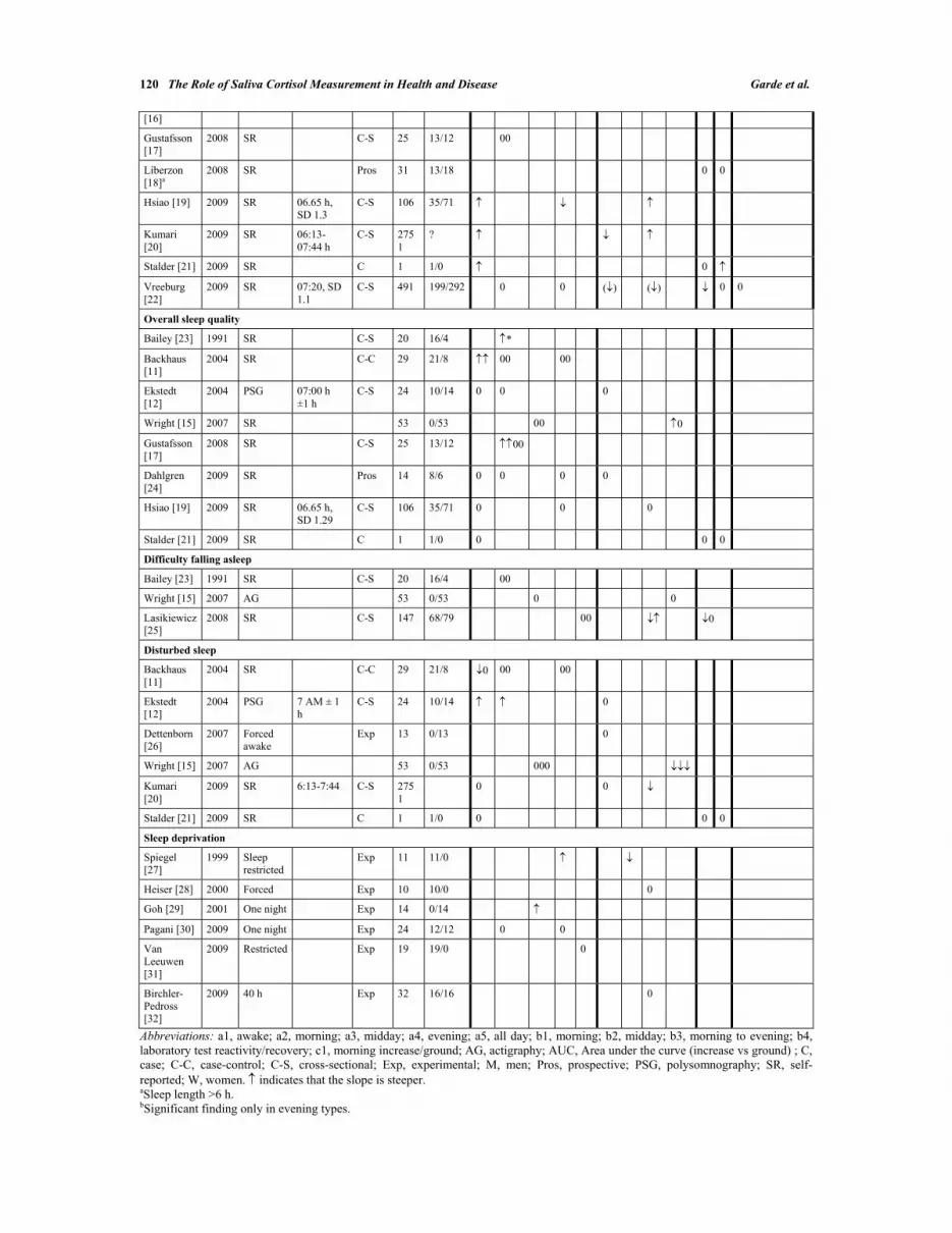
In total 23 papers fulfilled the inclusion criteria. A brief summary of the results (indicated as arrows denoting positive associations, or negative association and zero for a nonsignificant finding) are presented in Table 1. More detailed information on study design, statistical approach, main results, and discussion for each of the 23 papers is presented in Table 2.
Results are presented for each sleep measure. Cortisol measures were grouped as follows. Single time points at: a1, awakening; a2, morning; a3, midday; a4, evening; a5, all day. Deviations during: b1, morning; b2, midday; b3, morning to evening; b4, laboratory test. Area Under the Curve (AUC): c1, morning (increase/ground). Suppression test: d, response to dexamethasone (DST). No studies were found for premature awakening.
Sleep Duration
Thirteen papers were found to test the association between salivary cortisol and sleep duration [10-22]. In the 13 papers there were 37 analyses on relationships between measures of salivary cortisol and sleep

118 The Role of Saliva Cortisol Measurement in Health and Disease Garde et al.
duration. The proportion of significant relationships were 4/16 (25%) for single time points, 6/12 (50%) for deviations, 2/8 (25%) for AUC and 0/1 (0%) for dexamethasone test.
The most consistent results were a positive association between sleep duration and a single measure of salivary cortisol at awakening found in 3 studies [19-21]. In these studies, sleep duration was also associated with low evening cortisol levels [19], steep diurnal deviation of cortisol [19, 20], and with high AUC [21].
In 7 studies the authors failed to find any statistically significant associations between single measures of cortisol and sleep duration [11, 12, 15-17, 22]. The size of these studies was, in general, very small.
The association between sleep duration measures and deviations in cortisol measures was investigated in 7 studies. Morning deviations in cortisol concentrations were found to be positively associated with sleep length in an experimental study of 16 young people (8 morningness and 8 eveningness) using PSG [16]. In 2 ambulatory studies with more than 200 participants [10, 14] and a study of 2761 civil servants using self-reports negative associations to morning deviation in cortisol concentrations [20] were found.
Two studies showed a positive association between self-reported sleep duration and diurnal deviation of cortisol [19, 20]. In 4 other studies, no significant associations were found [11, 12, 15, 22], although tendencies were observed in 1 [22].
Morning AUC was the only AUC investigated in relation to sleep duration [12, 13, 18, 21, 22]. One study, a case study with 50 days of sampling, showed a positive relationship. In contrast, 1 study, which used an insomnia scale and defined sleep duration as “more than six hours sleep”, showed a negative relationship. Two out of 4 studies had only nonsignificant findings.
Overall Sleep Quality
Associations between sleep quality and measures of salivary cortisol were assessed in 8 studies [11, 12, 15, 17, 19, 21, 23, 24]. In the 8 papers there were 28 analyses on relationships between measures of salivary cortisol and overall sleep quality. The proportion of significant relationships was 5/21 (24%) for single time points, 1/5 (20%) for deviations, and 0/2 (0%) for AUC. Sleep quality was measured mainly by use of self-reports, but also PSG [13].
The most consistent pattern, a positive association to a single measure at awakening [11] or in the morning [17, 23], was observed in 3 studies. However, 5 other studies found no associations with a single morning or awakening cortisol measure [12, 15, 19, 21, 24]. In 4 studies, sleep quality was examined in relation to single measures in the afternoon or an evening measure; no associations were found [11, 15, 19, 24]. No significant associations were seen for sleep quality and deviations in cortisol concentrations [12, 19, 24].
One study found a positive relationship between stress reactivity and sleep quality measured as sleep efficiency by actigraphy, but not by self-reports [15]. One study examined associations between sleep quality and morning AUC, and found no significant relationship [21].
Difficulty Falling Asleep
Three studies assessed a total of 10 associations between salivary cortisol and difficulty falling asleep [15, 23, 25]. The proportion of significant relationships was 0/5 (0%) for single time points, 2/3 (67%) for deviations, and 1/2 (50%) for AUC. Difficulty falling asleep was assessed by use of actigraphy and self-reports (ease of sleep (inverted), speed of sleep onset (inverted), sleep latency, and time to fall asleep). The studies all used different types of cortisol measures.
Only 1 of the 3 studies reported significant associations, and the results were mixed [25]. In the same study the association between self-reported difficulty falling asleep in terms of ease of sleep was positively related to slope, whereas speed of sleep onset was negatively related [25]. High self-reported difficulty falling asleep was related to high AUC morning [25]. No other significant associations were observed between self-reported ease of sleep and measures of cortisol [15, 23, 25].

Sleep and Salivary Cortisol The Role of Saliva Cortisol Measurement in Health and Disease 119
Disturbed Sleep/Restless Sleep
Disturbed or restless sleep was examined in 6 studies [11, 12, 15, 20, 21, 26] analyzing a total of 22 relationships. The proportion of significant relationships was 3/13 (23%) for single time points, 4/7 (57%) for deviations, and 0/2 (0%) for AUC. Disturbed sleep was assessed as the number of microarousals during the night using PSG, forced awakening, actigraphy, and self-reports (restless sleep, nocturnal awakenings, time awake after sleep onset, and number of wake periods after sleep onset).
Four studies included associations with a single cortisol measure at awakening or in the morning: 1 found a positive association with the number of microarousals [12], 1 found a negative association with self-reported frequency of nightly awakenings, but no association with self-reported wake time after sleep onset [11], and 2 found no associations [20, 21]. No associations were observed for single measures of cortisol later in the day [11, 15].
One study investigated the relation between disturbed sleep and diurnal deviation and found a negative association [20]. No significant findings were seen in the 3 studies that investigated the relationship between morning deviations of cortisol and disturbed sleep in terms of nightly microarousals[12], forced awakenings [26], and sleep disturbance [20]. One study investigated the effect of disturbed sleep the night before a laboratory stress test, and found negative associations with reactivity [15].
AUC in the morning was tested in relation to disturbed sleep on a day to day basis in a case study with 50 days of sampling; and no significant associations were found [21].
Sleep Deprivation
Six studies investigated a total of 8 associations between sleep deprivation and measures of salivary cortisol with mixed results [27-32]. The proportion of significant relationships was 2/5 (40%) for single time points, 1/3 (33%) for deviations, and 0/0 (0%) for AUC. The studies used either 1 night of sleep deprivation [28-30, 32] or 5-6 nights of only 4 h sleep [27, 31].
In 1 study sleep restriction was associated with increased concentrations of cortisol in the evening and smaller decline in cortisol during the afternoon [27]. In another study it was found that cortisol concentrations were higher in the afternoon after sleep deprivation [29]. In 4 studies using cortisol concentrations in the morning, evening, and during the day following sleep deprivation, no associations were observed [28, 30-32].
Table 1: Summary of main findings of associations between measures salivary cortisol and studied domains sorted by year of publication
References Year Exposure Awakening time
Design n M/W Single time points (or sum/mean of two or more time points)
Deviation Difference/slope for two or more time points
AUC Dexamethasone suppression test
a1 a2 a3 a4 a5 b1 b2 b3 b4 c1 d
Sleep duration
Wüst [10] 2000 SR C-S 509 190/319
Backhaus [11]
2004 SR C-C 29 21/8 () 0 0
Ekstedt [12]
2004 PSG 07:00 h ±1 h
C-S 24 10/14 0 0 0
Federenko [13]
2004 SR 04:00 h Exp 49 0/49 0 0
Schlotz [14]
2004 SR C-S 219 102/117 0
Wright [15] 2007 SR/AG 53 0/53 0 00
Griefahn 2008 PSG Exp 16 16/0 0 b
120 The Role of Saliva Cortisol Measurement in Health and Disease Garde et al.
[16]
Gustafsson [17]
2008 SR C-S 25 13/12 00
Liberzon [18]a
2008 SR Pros 31 13/18 0 0
Hsiao [19] 2009 SR 06.65 h, SD 1.3
C-S 106 35/71
Kumari [20]
2009 SR 06:13-07:44 h
C-S 2751
?
Stalder [21] 2009 SR C 1 1/0 0
Vreeburg [22]
2009 SR 07:20, SD 1.1
C-S 491 199/292 0 0 () () 0 0
Overall sleep quality
Bailey [23] 1991 SR C-S 20 16/4 *
Backhaus [11]
2004 SR C-C 29 21/8 00 00
Ekstedt [12]
2004 PSG 07:00 h ±1 h
C-S 24 10/14 0 0 0
Wright [15] 2007 SR 53 0/53 00 0
Gustafsson [17]
2008 SR C-S 25 13/12 00
Dahlgren [24]
2009 SR Pros 14 8/6 0 0 0 0
Hsiao [19] 2009 SR 06.65 h, SD 1.29
C-S 106 35/71 0 0 0
Stalder [21] 2009 SR C 1 1/0 0 0 0
Difficulty falling asleep
Bailey [23] 1991 SR C-S 20 16/4 00
Wright [15] 2007 AG 53 0/53 0 0
Lasikiewicz [25]
2008 SR C-S 147 68/79 00 0
Disturbed sleep
Backhaus [11]
2004 SR C-C 29 21/8 0 00 00
Ekstedt [12]
2004 PSG 7 AM ± 1 h
C-S 24 10/14 0
Dettenborn [26]
2007 Forced awake
Exp 13 0/13 0
Wright [15] 2007 AG 53 0/53 000
Kumari [20]
2009 SR 6:13-7:44 C-S 2751
0 0
Stalder [21] 2009 SR C 1 1/0 0 0 0
Sleep deprivation
Spiegel [27]
1999 Sleep restricted
Exp 11 11/0
Heiser [28] 2000 Forced Exp 10 10/0 0
Goh [29] 2001 One night Exp 14 0/14
Pagani [30] 2009 One night Exp 24 12/12 0 0
Van Leeuwen [31]
2009 Restricted Exp 19 19/0 0
Birchler-Pedross [32]
2009 40 h Exp 32 16/16 0
Abbreviations: a1, awake; a2, morning; a3, midday; a4, evening; a5, all day; b1, morning; b2, midday; b3, morning to evening; b4, laboratory test reactivity/recovery; c1, morning increase/ground; AG, actigraphy; AUC, Area under the curve (increase vs ground) ; C, case; C-C, case-control; C-S, cross-sectional; Exp, experimental; M, men; Pros, prospective; PSG, polysomnography; SR, self-reported; W, women. indicates that the slope is steeper. aSleep length >6 h. bSignificant finding only in evening types.

Sleep and Salivary Cortisol The Role of Saliva Cortisol Measurement in Health and Disease 121
Table 2: Descriptives of the articles on salivary cortisol and sleep parameters sorted by domain of sleep parameter and year of publication
References Outcome Study design/group characteristics
Sampling Laboratory method and standardization in sampling
Statistical approach for cortisol measurement in relation to sleep
Statistical analysis, cortisol in relation to outcome
Results on cortisol and sleep
Wüst 2000 [10]
Sleep length:
TST
Method: Self-report
Design: C-S
No.: 509
M/W: 190/319
Age: 37.3 (18-71) years
Group: Healthy
Days: 2
Samples per day: 4
Times for sampling: Awakening, +15, 30, and 60 min,
Setting: Ambulatory (at home)
RIA Measurement(s):
a1. Cortisol on awakening
b1. Mean increase from awakening
c1. AUC
Cortisol data: Continuous
Statistics: Pearson correlation and ANOVA with repeated measures
Positive correlation between sleep duration and mean cortisol increase from awakening (b1)
Backhaus 2004 [11]
Sleep length: TST
Sleep quality: PSQI, feeling of recovery
Disturbed sleep: Frequency of nightly awakenings, wake time after sleep onset
Method: Questionnaire (PSQI), feeling of recovery
Design: C-C
No.: 29
M/W: 21/8
Age: 32-62 years
Group: insomniacs (n=14) and healthy controls (n=15)
Days: 7
Samples per day: 3
Times for sampling: Awakening, +15 min and before going to bed
Setting: Ambulatory (at home)
RIA
Not to use food, alcoholic beverages, caffeine, fruit juice, or brush teeth 1 h before sampling
Measurement(s): All by means of same time point over the 3 consecutive days
a1. Cortisol on awakening
a2. Cortisol 15 min after awakening
a4. Cortisol at bedtime
Cortisol data: Continuous
Statistics: Pearson correlation and ANOVAs
Trend for negative correlation between TST and cortisol at awakening (a1)
Positive correlation between sleep quality, and feeling of recovery, and cortisol at awakening (a1)
Negative correlation between frequency of nightly awakenings and cortisol at awakening (a1). No correlation between wake time after sleep onset and awakening cortisol (a1)
No correlation between sleep parameters and cortisol 15 min after awakening (a2) or cortisol at bedtime (a4)
Ekstedt 2004 [12]
Sleep length: TST
Sleep quality: Sleep efficiency
Disturbed sleep: Number of arousals
Method: 2 PSG recordings carried out in the subject’s home (before workday/ day off)
Design: C-S
No.: 24
M/W: 10/14
Age: 30.5 ± 0.5 years
Group: High (n=12) and low (n=12) burnout, recruited from a Swedish IT company
Days: 2
Samples per day: 9
Times for sampling: Awakening, +15, 30, and 60 min, 11:00, 15.00, 19:00, 21:00 h and bedtime
Setting: Ambulatory. Saliva collected at day after the PSG
RIA
No current smokers, non-sedentary lifestyle and moderate alcohol intake
Measurement(s):
a1. Single awakening sample
a2. Awakening cortisol as a mean morning value of 4 samples, at awakening, 15, 30, 60 min post awakening
b1. Deviation morning value (CAR (difference 0-60 min)
Cortisol data: Log transformed
Statistics: Stepwise multiple regression analyses. Pearson correlation coefficient
Confounders:
No association between total sleep time and awakening cortisol or mean cortisol within 60 min after awakening
No association between sleep efficiency and cortisol
More nightly arousals were associated with higher awakening cortisol and mean cortisol within 60 min after awakening
No association between any sleep measure and morning deviation
Federenko 2004 [13]
Sleep duration: TST
Method: Self-report
Design: Exp
No.: 49
M/W: 0/49
Age: nurses: 40.3 years, students: 25 years
Group: Nurses working shifts (n=18) and students with regular sleep cycle (n=31)
Days: 2
Samples per day: 4
Times for sampling: Awakening, +30, 45 and 60 min
Setting: Nurses: collected 1st and 2nd day of 3 different shifts. Students: after early evening nap on 2 days
RIA
Not to smoke, eat and drink just water in the first hour after awakening, not to brush teeth, avoid microinjuries in oral cavity
Measurement(s):
b1. Mean increase from awakening
c1. AUCground
Cortisol data:
Statistics: Person’s correlations
Confounders: Oral contraceptives
No correlation between sleep duration and mean increase from awening or AUCground

122 The Role of Saliva Cortisol Measurement in Health and Disease Garde et al.
Schlotz 2004 [14]
Sleep length:
?
Method: ?
Design: C-S
No.: 219
M/W: 102/117
Age: 48.6 (24-83) years
Group: Healthy
Days: 7 consecutive
Samples per day: 4
Times for sampling: Awakening, +15, 30, and 60 min,
Setting: Ambulatory (at home)
RIA Measurement(s):
a1. Cortisol on awakening
b1. Mean increase from awakening
Cortisol data: Continuous
Statistics: ANOVA with repeated measures
No association between sleep duration and cortisol on awakening (a1)
Positive association between sleep duration and mean cortisol increase from awakening (b1)
Wright 2007 [15]
Sleep duration: TST (actigraphy and self-reports)
Sleep quality: Sleep quality and sleep efficiency
Difficulty falling asleep: Sleep latency
Disturbed sleep: Wake up %, minutes awake, number of wake periods
Method: Actigraph and sleep log (Pittsburgh sleep diary) over 7 days
Design: C-S
No.: 53
M/W: 0/53
Age: 37.3 (± 9.9) years
Group: Healthy
Days: 1
Samples per day: 4
Times for sampling: Base line cortisol before stress test (14:00 h), post test, +30 min and 45 min post test
Setting: Laboratory with stress test
Immunoassay
After stress test the participants were asked to relax and read general interest magazines
Measurement(s):
a3. Single measure at baseline (14:00 h)
b4. Reactivity to test
Cortisol data: Logarithmic (base 10)
Statistics: Pearson´s correlations, univariate analysis and partial correlations adjusting for baseline cortisol
Positive association between sleep efficiency (actigraph) and cortisol reactivity to test
Negative association between all 3 disturbed sleep and cortisol reactivity to test
No association between TST, sleep latency, self-reports of sleep quality and length and cortisol reactivity to test
No association between actigraph measures and baseline cortisol (14:00 h)
Greifahn 2008 [16]
Sleep duration: TST
Method: PSG
Design: Exp
No.: 16
M/W: 16/0
Age: 19-27 years
Group: morningness (n=8), eveningness (n=8)
Days: 6 days (?)
Samples per day: 2
Times for sampling: 7:00 h, +30 min. Only those when wakeup is after 06:50 h
Setting: Laboratory
LIA (IBL)
No smoking, no teeth brushing prior sampling
Measurement(s):
a2. Single measures at 07:00 h
b1. Deviation morning concentration, at 07:00 h and 30 min later
Cortisol data: Continous?
Statistics: ANCOVA with repeated measures correlation
Confounders:
TST had positive association with b1 after night sleep
TST not associated with cortisol at awakening (a2)
Gustafsson 2008 [17]
Sleep duration: Sleep length
Sleep quality: Sufficient sleep, generally difficulties sleeping because of work
Method: Questionnaire
Design: C-S
No.: 25
M/W: 13/12
Age: 24-62 years
Group: White collar workers
Days: 2
Samples per day: 6
Times for sampling: 15-30 min after awakening and every 2 h until 20:00 h
Setting: Ambulatory
RIA
All participants were asked to rise and go to bed at the same times during days of measurement
Measurement(s):
a2. Two measures, approx. 07:00 h and 09:00 h
Cortisol data: ?
Statistics: Linear regression. Repeated measures ANOVA
Confounders:
Association between less sufficient sleep (better sleep quality) and higher morning cortisol. (both measures)
No association between sleep duration or difficulties sleeping because of work and morning cortisol
Liberzon 2008 [18]
Sleep duration:
Method: Not mentioned in methods section
Design: Follow-up
No.: 31
M/W: 13/18
Age: 18-38 years
Group: Students (n=23) and science staff (n=4) and ships crew member (n=4)
Days: 6
Samples per day: 4
Times for sampling: Awakening, +15, 30, 45 min
Setting: Ambulatory
RIA
Not to eat, drink, smoke, brush teeth or rinse mouth until after 45 min sample
Measurement(s):
c1g. AUC with respect to ground
c1i. AUC for increase (awakening response)
Cortisol data:
Statistics:
Confounders: Perceived stress and control (Likert scale)
No other correlation between total sleep time and cortisol measures in total sample
Hsiao 2009 [19]
Sleep duration: Total time slept
Sleep quality: Sleep quality last night
Method: Questionnaire
Design: C-S
No.: 106
M/W: 35/71
Age: 38.5 years (SD 9.7)
Group: 106 healthy subjects (and 126 patients
Days:
Samples per day: 5
Times for sampling:
Awakening, + 45 min, 12:00 h, 17:00 h, 21:00 h
Setting: Ambulatory
RIA
Not to brush teeth, avoid oral blood contamination before sampling, Not to eat 45 min after awakening and 30 min before collecting samples
Measurement(s): single measures at awakening and over a day
a1. Awakening cortisol
a4. Evening cortisol
b3. Deviation (diurnal
Cortisol data: Natural logarithm
Statistics: Two-level individual growth curve model. (multiple regression model for nested, repeated data)
Confounders: Several
Association between longer sleep and steeper slope (b3), higher awakening cortisol (a1), lower cortisol in the evening (a4)
No association between sleep quality and cortisol

Sleep and Salivary Cortisol The Role of Saliva Cortisol Measurement in Health and Disease 123
with major depression - not used in present review)
profile) confounders are adjusted for in two different models
Kumari 2009 [20]
Sleep duration: TST divided into 1-h categories
Disturbed sleep: Sleep disturbance
Method: Logbook and questionnaire
Design: C-S
No.: 2751
M/W: ?
Age: ?
Group: Whitehall
Days:
Samples per day: 5
Times for sampling: Awakening, +30 min, +2.5, 8, 12 h
Setting: Ambulatory
Immunoassay method
Provide 6 samples on a normal weekday
Measurement(s):
a1. Single time point, awake
b1. Deviation morning profile
b3. Deviation morning to evening (profile)
Cortisol data: Log cortisol data
Statistics: Multilevel, interaction term
Confounders: Age, sex, employment grade, awakening time, smoking status, waist circumference
Association between long sleep duration and higher cortisol. at awakening (a1) and steeper diurnal slope (b3). Association between long sleep duration and flatter morning slope (b1)
Association between less disturbed sleep and steeper diurnal slope (b3)
Stalder 2009 [21]
Sleep duration: TST
Sleep quality:
Disturbed sleep: Nocturnal awakenings
Method: Sleep log
Design: Case study
No.: 1
M/W: 1/0
Age: 27 years
Days: 50 with 3 days interval
Samples per day: 4
Times for sampling: Awakening, +15, 30 and 45 min
Setting: Ambulatory
ELISA method
Sat for 15-30 min when sampling, otherwise moved freely in relation to sampling times
Measurement(s):
a1. Awakening concentration
c1. AUCground
c1. AUCincrease
Cortisol data: Log transform
Statistics: Repeated measures ANOVA
Confounders: Alcoholic drinks consumed the evening before measurement day
Positive association between sleep duration and awakening cortisol and AUCground
No association between disturbed sleep and sleep quality and cortisol
No association between any sleep parameters and AUCincrease
Vreeburg 2009 [22]
Sleep duration: Sleep length (more or less than 6 h)
Method: Insomnia rating scale
Design: C-S
No.: 491
M/W: 199/292
Age: 43.0 years Group: volunteers without psychopathology
Excl: Taking antidepressants, pregnant or breastfeeding, on medication with corticosteroids
P rate: 78.3%, with at least one usable cortisol measurement
Days: 1
Samples per day: 7
Times for sampling: Awakening, +30, 45, 60 min, 22:00 h, 23:00 h. Samples taken more than 5 min from protocol time were discarded
Setting: Ambulatory. Day after dexamethasone 0.5 mg directly after sampling time at 23:00 h
Immunoassay method
When sampling no eating, smoking, drinking tea or coffee, or brushing teeth 15 min before
Measurement(s):
a2.
a4.
b1. Deviation morning
b3. Deviation 23:00 h, awakening time divided by numbers of hours in between (diurnal slope)
c1i. AUCincrease morning
c1g. AUCground morning
d. Post dexamethasone (DST)
Cortisol data: AUC, evening cortisol and DST were log transformed
Statistics: Linear mixed models or linear regression analysis
Confounders: Sociodemographic factors, health indicators
Less than 6 h sleep is associated with increase in CAR (AUCincrease)
Tendency for less than 6 h sleep is associated with steeper morning deviation (b1) and a steeper diurnal slope (b3)
No association with morning cortisol (a2), evening cortisol (a4), AUCground morning, or post dexamethasone
Bailey 1991 [23]
Sleep quality: Sleep quality
Difficulty falling asleep: Sleep onset
Method: Sleep log
Design: C-S
No.: 20
M/W: 16/4
Age: 23-39 years
Group: morning types (n=10) and evening types (n=10). Recruited from the general population
Days: 1
Samples per day: 7
Times for sampling: Arising, +20, 40, 60, 80, 100, and 120 min
Setting: Ambulatory
RIA Measurement(s):
a2. Single cortisol levels
Cortisol data: Continuous
Statistics: t-test, Pearson product-moment correlation coefficients, Spearman rho correlation coefficients
Positive association between sleep quality and total cortisol in evening type group, but not in morning type group
No association between sleep onset and total cortisol in evening or morning type group
Dahlgren 2009 [24]
Sleep quality:
Method: Karolinska sleep diary for 4 weeks
Design: C-S
No.: 14
M/W: 8/6
Age: 44 years
Group: Office workers
Days: 28
Samples per day: 3
Times for sampling: Awakening, +15 min, bedtime
Setting: Ambulatory
RIA
No food, no teeth brushing 30 min before saliva sampling
Measurement(s): Single time points:
a1. Awake
a2. Morning
a4. Evening
b1. Deviation morning
Cortisol data: Log data
Statistics: Multiple regression analyses by time. ANOVA of repeated measurements
Confounders: Work day, work load, awakening time, stress at bedtime, sleep quality, stress, sleepiness exhaustion, self related health
No associations between sleep quality and measures of cortisol

124 The Role of Saliva Cortisol Measurement in Health and Disease Garde et al.
Lasikiewicz 2008 [25]
Difficulty falling asleep: Ease of sleep and speed of sleep onset
Method: Questionnaire (Leeds Sleep Evaluation Questionnaire)
Design: C-S
No.: 147
M/W::68/79
Age: :mean age 46.2 years (±7,2)
Group: volunteers
Days: 1 (n=64) or 3 (n=83)
Samples per day: 8
Times for sampling: Awakening, +15, 30, 45 min, +3, 6, 9, 12 h
Setting:
Ambulatory
Immunoassay method
Not to consume food or drink other than water in relation to sample collection. Avoid teeth brushing and vascular leakage
Measurement(s): Mean of same time point on consecutive days
a5.
b4. Deviation evening from 45 min post awakening (slope)
c1. AUC not specified
Cortisol data: Log transformed
Statistics: Pearson’s correlation. Cluster analysis (M)ANOVA
Confounders: Age, gender
Association between higher ease of sleep (less difficulty falling asleep) and low AUC
Association between high ease of sleep (less difficulty falling asleep) and less steep slope (b3)
Association between high speed of sleep onset (less difficulty falling asleep) and more steep slope (b3)
No association between ease of sleep, speed of sleep onset and diurnal mean (a5)
Dettenborn 2007 [26]
Disturbed sleep:
Method: Three experimentally induced awakenings (phone call). The wake up in the morning was optional or set up by alarm clock. No other sleep registration
Design: Exp
No.: 13
M/W: 0/13
Age: 24 years
Days: 3 intervention nights + 3 reference nights
Samples per day: 8 on intervention nights and 2 on recovery nights
Times for sampling: Awakening and +15 min in the morning
Setting: Ambulatory
CLIA Measurement(s):
b1. Repeated measures
Cortisol data: continous
Statistics: ANOVA and ANCOVA
Confounders: Oral contraceptives, thyroid hormone
The morning CAR after disturbed nights was not different from CAR on undisturbed nights
There was a lack of HPA axis activation by forced nightly awakenings
Spiegel 1999 [27]
Sleep deprivation: Sleep restriction
Design: Exp
No.: 11
M/W: 11/0
Age: 18-27 years
Group: Young healthy volunteers
Days: 3
Samples per day: 12-20
Times for sampling: Every 30 min between 15.00 and bedtime
Setting: Laboratory. 3 nights with 8 h in bed, 6 nights with 4 h in bed, and 7 nights with 12 h in bed
RIA a4. Single evening concentration
b2. Deviation between 16:00 h and 21:00 h
Cortisol data: Continuous
Statistics: ANOVA for repeated measures
Higher evening cortisol concentration after sleep restriction
Lower rate of decrease in the afternoon after sleep restriction
Heiser 2000 [28]
Sleep deprivation: 3 days covered, after ordinary sleep, 1 night of total sleep deprivation and 1 night of recovery
Design: Exp
No.: 10
M/W: 10/0
Age: 27.4 ± 2.8 years
Group: healthy volunteers
Days: 3
Samples per day: 3
Times for sampling: 07:00, 13:00, 19:00 h
Setting: Laboratory
RIA
All intake of pineapples, bananas, almonds, nuts, tomatoes, vanilla, or alcohol forbidden, no smokers
Measurement(s):
b3. Diurnal profile with 3 measures per day over 3 days
Cortisol data: ?
Statistics: ANOVA with repeated measures
No effect on salivary cortisol rhythm of sleep deprivation
Goh 2001 [29]
Sleep deprivation: 24 h sleep deprivation or 8 h sleep (control)
Design: Exp
No.: 14
M/W: 14/0
Age: 20-30 years
Group: healthy subjects, military service members
Days: 2
Samples per day: 3-5
Times for sampling: 08:00, 13:30, 18:00, 21:00, 24:00 (day 1), 08:00, 13:30, 18:00 (day 2)
Setting: Laboratory
Immunoassay b3. Deviation, all day Cortisol data: Continuous
Statistics: Two-way ANOVA with repeated measures and interaction terms
Significant interaction between sleep status and time. Cortisol levels at 13:30 h were increased after sleep deprivation
Pagani 2009 [30]
Sleep deprivation: 7 normal nights + 24 h sleep deprivation) or normal living conditions (strict sleep-wake schedule 23:00-07:00 h)
Design: Exp
No.: 24
M/W: 12/12
Age: 27-45 years
Group: healthy subjects
Days: 8?
Samples per day: 2
Times for sampling: 10.30 and 18.00 h
Setting: Laboratory
RIA Measurement(s):
a2.
a4.
Cortisol data: Continuous
Statistics: Mixed model or GLM analysis. Intraclass correlations
Confounders:
No effect of sleep deprivation on cortisol

Sleep and Salivary Cortisol The Role of Saliva Cortisol Measurement in Health and Disease 125
Van Leeuwen 2009 [31]
Sleep deprivation: 2 baseline (8 h sleep), 5 nights of 4 h sleep, and 2 recovery nights of 8 h. Controls (8 h sleep) all nights
Design: Exp
No.: 19
M/W:19/0
Age: 19-29 years
Group: 13 young healthy men + 6 controls
Days:
Samples per day: 10
Times for sampling: Not specified
Setting: Laboratory
Competitive CLIA
Napping during day time was not allowed, meals standardized (calories and time), controlled illumination and room temperature
Measurement(s):
a5. Averaged throughout the day
Cortisol data:
Statistics: t-tests and Wilcoxon signed ranks test for not normally distributed differences
Confounders:
No change in salivary cortisol after sleep restriction
Birchler-Pedross 2009 [32]
Sleep deprivation: 40 h sleep deprivation or nap protocol
Design: Exp
No.: 32
M/W: 16/16
Age: 25.0±3.3 and 65.0±5.5 years
Group: Young and older healthy volunteers
Days: 2
Samples per day: 5-6. Times for sampling: every 30 min collapsed into 08:00, 12:00, 16:00, 20:00, 24:00 h (day 1), 04:00, 08:00, 12:00, 16:00, 20:00, 24:00 h (day 2)
Setting: Laboratory
RIA b3. Deviation, all day Cortisol data: Continuous
Statistics: Repeated measures ANOVA with interaction terms
Significant four-way interaction term (time of day, age, gender, sleep pressure) in model for cortisol, most likely driven by time of day
None of the other variables were significant
Vreeburg 2009 [22]
Sleep duration: Sleep length (more or less than 6 h)
Method: Insomnia rating scale
Design: C-S
No.: 491
M/W: 199/292
Age: 43.0 years
Group: volunteers without psychopatology
Excluded: Taking antidepressants, pregnant or breastfeeding, on medication with corticosteroids
P rate: 78.3%, with at least one usable cortisol measurement
Days: 1
Samples per day: 7
Times for sampling: Awakening, +30, 45, 60 min, 22:00, 23:00 h. Samples taken more than 5 minutes from protocol time were discarded
Setting: Ambulatory. Day after dexamethasone 0.5 mg directly after sampling time at 23:00 h
Immunoassay method
When sampling no eating ,smoking, drinking tea or coffee, or brushing teeth 15 min before
Measurement(s):
a2.
a4.
b1. Deviation morning
b3. Deviation 23:00 h to awakening time divided by number of hours in between (diurnal slope)
c1i. AUCincrease morning
c1g. AUCground morning
d. Post DST
Cortisol data: AUC, evening cortisol and DST were log transformed
Statistics: Linear mixed models or linear regression analysis
Confounders: Sociodemographic factors, health indicators
Less than 6 h sleep is associated with increase in CAR (AUCincrease)
Tendency for less than 6 h sleep is associated with steeper morning deviation (b1) and a steeper diurnal slope (b3)
No association with morning cortisol (a2), evening cortisol (a4), AUCground morning, or post dexamethasone
Abbreviations: AUC, Area under the curve; C-C, case-control; C-S, cross-sectional; CAR, cortisol awakening response; CLIA, chemiluminescence-assay; DST, Dexamethasone test; ELISA, Enzyme Linked Immuno-Sorbant Assay; Exp, experimental; GLM, generalized linear model; LIA, luminescence immunoassay; M, Male; PSG, polysomnography; PSQI, Pittsburgh Sleep Quality Index; P rate, Response rate; RIA, radioimmunoassay; TST, total sleep time, W, women.

126 The Role of Saliva Cortisol Measurement in Health and Disease Garde et al.
DISCUSSION
The aim of the present chapter was to analyze whether measures of cortisol in saliva were associated with measures of sleep to see if divergent results were functions of differences in theoretic assumptions made and methods used. Relatively few papers were first identified (n=188), and only 23 papers met the final inclusion criteria.
The most consistent results were a positive association between sleep duration and a single measure of salivary cortisol at awakening, which was observed in 3 studies [19-21]. In these studies, sleep duration was also associated with low evening cortisol levels [19], steep diurnal deviation of cortisol [19, 20], and with high AUC [21]. Together these findings suggest that longer sleep duration is related to a more dynamic cortisol secretion.
However, long sleep duration is also associated with a lower CAR [10, 14, 20]. Since a lower CAR implies a less dynamic response these observations seems to contradict the suggestion that longer sleep duration is related to a more dynamic cortisol secretion. However, a lower CAR needs not to be mutually exclusive with a dynamic cortisol secretion as assessed by the decline in concentrations during the entire day. Indeed, a lower CAR might just reflect the diurnal rhythmicity of cortisol secretion. For example, to the extent that participants, who sleep long wake up later than usual, they are likely to take their first sample at a time when the concentrations of cortisol already have risen due to the normal diurnal rhythm. Hence, there is simply less room to obtain a high CAR as the morning value has been inflated by the underlying diurnal rhythm. However, there might also be several other explanations and this question deserves more attention in future studies.
Two of the 6 studies on disturbed sleep showed that it was associated with less diurnal deviation [20], and lower reactivity to a laboratory stress test [15]. This to some extent corroborates the findings on sleep duration. However, the many nonsignificant findings as well as the theoretic and methodological differences (e.g. heterogeneity in measures) complicate comparisons.
As expected, statistically significant associations were more often reported in studies with a large number of participants or a high sampling frequency. Among the papers included, statistically significant findings were generally seen in studies with more than 100 participants [10, 14, 19, 20], or samples (case study) [21]. In the large studies, it was more common to focus on general sleep patterns, whereas the case study registered day to day variations within the same person. The results suggest that a relationship between sleep and salivary cortisol is observable both within and between subjects, but requires many observations due to high variability in both cortisol and sleep measures. The use of small study samples and non-optimal measurement procedures might lead to studies with low power and failure to detect statistically significant results.
Even if the inclusion criteria drastically reduced the number of papers, the papers included covered several different types of measures and indicators for cortisol and sleep. This heterogeneity in measures and methods appears to generate a mixed pattern of results that are also conflicting at times. For example, in the same study a positive relationship was observed between stress reactivity and sleep quality measured as sleep efficiency by actigraphy, but not by self-reports [15]. As differences between objectively measured and self-reports of sleep have been observed previously, this conflict may be partially related to the selected instrument for measurement of sleep quality [33].
There are several methodological problems in studies of sleep and saliva cortisol. Because saliva sampling requires that the person is awake, it is virtually impossible to sample saliva during sleep and to establish the relationship with different sleep stages or other processes that may occur during sleep. It is often not practically feasible in field studies to obtain direct measurements during the sleep period with, for example, PSG or endocrine measures. Even if it is technologically possible to use such measures, the procedures and commitments necessary for successful implementation have often been considered to be cumbersome and too expensive to be considered as a realistic option. Stress researchers often have to rely on less sophisticated and simpler approaches such as motion logging (e.g., actigraphy) and subjective reports of various aspects of sleep (e.g., bedtime, awakening time, and perceived quality of sleep).

Sleep and Salivary Cortisol The Role of Saliva Cortisol Measurement in Health and Disease 127
Many of the studies were not conducted primarily to evaluate the relationship between sleep and salivary cortisol. Even so, all studies included in the present chapter have used statistical tests to address the overall question about the relationship between sleep and saliva cortisol. Considering that research have been driven by many points of departures, it is not surprising that most of the result do not act in concert.
CONCLUSIONS
In total 23 studies examining sleep quality in relation to salivary cortisol measures were identified. There was a large proportion of nonsignificant findings and many operational definitions of sleep quality and cortisol secretion. Because many of the studies included were small and entailed few measurements, there is reason to believe that the nonsignificant findings partly reflect low statistical power.
The most consistent results were our observation of a positive association between sleep duration and a single measure of salivary cortisol at awakening, which was observed in 3 studies. In these studies, sleep duration was also associated with low evening cortisol levels, steep diurnal deviation of cortisol, and/or with high AUC. Together these findings suggest that longer sleep duration is related to a more dynamic cortisol secretion. Two of the 6 studies on disturbed or restless sleep showed a relationship with flat diurnal deviation and low laboratory stress test reactivity. This to some extent corroborates the findings regarding sleep duration. However, the many nonsignificant findings as well as the theoretic and methodological differences (e.g., heterogeneity in measures) complicate comparisons. Conflicting results may be at least partially due to differences in methods and underlying assumptions.
REFERENCES
[1] Ursin H, Eriksen HR. The cognitive activation theory of stress. Psychoneuroendocrinology 2004; 29: 567-592. [2] Mason JW. A historical view of the stress field part 2. J Human Stress 1975; 1: 22-36 concl. [3] Mason JW. A historical view of the stress field part 1. J Human Stress 1975; 1: 6-12 contd. [4] Saper CB, Scammell TE, Lu J. Hypothalamic regulation of sleep and circadian rhythms. Nature 2005; 437:
1257-1263. [5] Chrousos GP. Stress and disorders of the stress system. Nat Rev Endocrinol 2009; 5: 374-381. [6] Van Cauter E. temp- in sleep chapter. Presented in draft 100621 from Anne Helene. 2005. [7] Weitzman ED, Zimmerman JC, Czeisler CA, Ronda J. Cortisol secretion is inhibited during sleep in normal
man. J Clin Endocrinol Metab 1983; 56: 352-358. [8] Steiger A. Sleep and the hypothalamo-pituitary-adrenocortical system. Sleep Med Rev 2002; 6: 125-138. [9] Steiger A, Antonijevic IA, Bohlhalter S, Frieboes RM, Friess E, Murck H. Effects of hormones on sleep. Horm
Res 1998; 49: 125-130. [10] Wust S, Wolf J, Hellhammer DH, Federenko I, Schommer N, Kirschbaum C. The cortisol awakening response -
normal values and confounds. Noise Health 2000; 2: 79-88. [11] Backhaus J, Junghanns K, Hohagen F. Sleep disturbances are correlated with decreased morning awakening
salivary cortisol. Psychoneuroendocrinology 2004; 29: 1184-1191. [12] Ekstedt M, Akerstedt T, Soderstrom M. Microarousals during sleep are associated with increased levels of lipids,
cortisol, and blood pressure. Psychosom Med 2004; 66: 925-931. [13] Federenko I, Wust S, Hellhammer DH, Dechoux R, Kumsta R, Kirschbaum C. Free cortisol awakening
responses are influenced by awakening time. Psychoneuroendocrinology 2004; 29: 174-184. [14] Schlotz W, Hellhammer J, Schulz P, Stone AA. Perceived work overload and chronic worrying predict
weekend-weekday differences in the cortisol awakening response. Psychosom Med 2004; 66: 207-214. [15] Wright CE, Erblich J, Valdimarsdottir HB, Bovbjerg DH. Poor sleep the night before an experimental stressor
predicts reduced NK cell mobilization and slowed recovery in healthy women. Brain Behav Immun 2007; 21: 358-363.
[16] Griefahn B, Robens S. The cortisol awakening response: a pilot study on the effects of shift work, morningness and sleep duration. Psychoneuroendocrinology 2008; 33: 981-988.
[17] Gustafsson K, Lindfors P, Aronsson G, Lundberg U. Relationships between self-rating of recovery from work and morning salivary cortisol. J Occup Health 2008; 50: 24-30.
128 The Role of Saliva Cortisol Measurement in Health and Disease Garde et al.
[18] Liberzon J, Abelson JL, King A, Liberzon I. Naturalistic stress and cortisol response to awakening: adaptation to seafaring. Psychoneuroendocrinology 2008; 33: 1023-1026.
[19] Hsiao FH, Yang TT, Ho RT, Jow GM, Ng SM, Chan CL, et al. The self-perceived symptom distress and health-related conditions associated with morning to evening diurnal cortisol patterns in outpatients with major depressive disorder. Psychoneuroendocrinology 2009; 35: 503-515.
[20] Kumari M, Badrick E, Ferrie J, Perski A, Marmot M, Chandola T. Self-reported sleep duration and sleep disturbance are independently associated with cortisol secretion in the Whitehall II study. J Clin Endocrinol Metab 2009; 94: 4801-4809.
[21] Stalder T, Evans P, Hucklebridge F, Clow A. Associations between psychosocial state variables and the cortisol awakening response in a single case study. Psychoneuroendocrinology 2010; 35: 209-214.
[22] Vreeburg SA, Kruijtzer BP, van Pelt J, van Dyck R, DeRijk RH, Hoogendijk WJ, et al. Associations between sociodemographic, sampling and health factors and various salivary cortisol indicators in a large sample without psychopathology. Psychoneuroendocrinology 2009; 34: 1109-1120.
[23] Bailey SL, Heitkemper MM. Morningness-eveningness and early-morning salivary cortisol levels. Biol Psychol 1991; 32: 181-192.
[24] Dahlgren A, Kecklund G, Theorell T, Akerstedt T. Day-to-day variation in saliva cortisol-relation with sleep, stress and self-rated health. Biol Psychol 2009; 82: 149-155.
[25] Lasikiewicz N, Hendrickx H, Talbot D, Dye L. Exploration of basal diurnal salivary cortisol profiles in middle-aged adults: associations with sleep quality and metabolic parameters. Psychoneuroendocrinology 2008; 33: 143-151.
[26] Dettenborn L, Rosenloecher F, Kirschbaum C. No effects of repeated forced wakings during three consecutive nights on morning cortisol awakening responses (CAR): a preliminary study. Psychoneuroendocrinology 2007; 32: 915-921.
[27] Spiegel K, Leproult R, Van Cauter E. Impact of sleep debt on metabolic and endocrine function. Lancet 1999; 354: 1435-1439.
[28] Heiser P, Dickhaus B, Schreiber W, Clement HW, Hasse C, Hennig J, et al. White blood cells and cortisol after sleep deprivation and recovery sleep in humans. Eur Arch Psychiatry Clin Neurosci 2000; 250: 16-23.
[29] Goh VH, Tong TY, Lim CL, Low EC, Lee LK. Effects of one night of sleep deprivation on hormone profiles and performance efficiency. Mil Med 2001; 166: 427-431.
[30] Pagani M, Pizzinelli P, Traon AP, Ferreri C, Beltrami S, Bareille MP, et al. Hemodynamic, autonomic and baroreflex changes after one night sleep deprivation in healthy volunteers. Auton Neurosci 2009; 145: 76-80.
[31] van Leeuwen WM, Lehto M, Karisola P, Lindholm H, Luukkonen R, Sallinen M, et al. Sleep restriction increases the risk of developing cardiovascular diseases by augmenting proinflammatory responses through IL-17 and CRP. PLoS One 2009; 4: e4589.
[32] Birchler-Pedross A, Schroder CM, Munch M, Knoblauch V, Blatter K, Schnitzler-Sack C, et al. Subjective well-being is modulated by circadian phase, sleep pressure, age, and gender. J Biol Rhythms 2009; 24: 232-242.
[33] Harvey AG, Tang NK, Browning L. Cognitive approaches to insomnia. Clin Psychol Rev 2005; 25: 593-611.


