Embed Size (px)
Citation preview
968
Molecular and Cellular Pathobiology
CancerResearch
Smad Signaling Is Required to Maintain Epigenetic Silencingduring Breast Cancer ProgressionPanagiotis Papageorgis1,2, Arthur W. Lambert1, Sait Ozturk1, Fangming Gao1,2,3, Hongjie Pan1,2,Upender Manne4, Yuriy O. Alekseyev2,3, Arunthathi Thiagalingam1, Hamid M. Abdolmaleky1,2,Marc Lenburg1,2,3, and Sam Thiagalingam1,2,3
Abstract
Authors' AMedicineGenomics,School ofPathology,
Note: SupResearch
Current adcogeneticsMA 01757.
CorresponMedicine, 76013; Fax:
doi: 10.115
©2010 Am
Cancer R
Breast cancer progression is associated with aberrant DNA methylation and expression of genes that con-trol the epithelial-mesenchymal transition (EMT), a critical step in malignant conversion. Although the genesaffected have been studied, there is little understanding of how aberrant activation of the DNA methylationmachinery itself occurs. Using a breast cancer cell–based model system, we found that cells that underwentEMT exhibited overactive transforming growth factor β (TGFβ) signaling and loss of expression of the CDH1,CGN, CLDN4, and KLK10 genes as a result of hypermethylation of their corresponding promoter regions. Basedon these observations, we hypothesized that activated TGFβ-Smad signaling provides an “epigenetic memory”to maintain silencing of critical genes. In support of this hypothesis, disrupting Smad signaling in mesenchy-mal breast cancer cells resulted in DNA demethylation and reexpression of the genes identified. This epige-netic reversal was accompanied by an acquisition of epithelial morphology and a suppression of invasiveproperties. Notably, disrupting TGFβ signaling decreased the DNA binding activity of DNA methyltransferaseDNMT1, suggesting that failure to maintain methylation of newly synthesized DNA was the likely cause of DNAdemethylation. Together, our findings reveal a hyperactive TGFβ-TGFβR-Smad2 signaling axis needed to main-tain epigenetic silencing of critical EMT genes and breast cancer progression. Cancer Res; 70(3); 968–78. ©2010 AACR.
Introduction
Epigenetic regulation of gene expression is a fundamentalfeature affecting normal physiologic processes as well as dis-eases such as cancer. Aberrant global DNA hypomethylationas well as hypermethylation of specific regulatory regions ofgenes are considered as hallmarks of cancer progression (1).Silencing of tumor suppressor genes by promoter DNA hy-permethylation has been associated with the expansion ofpremalignant cells and acquisition of an invasive phenotypeleading to metastatic dissemination (2). Except for the recentimplication of the Ras pathway as a potential mediator ofepigenetic gene silencing (3), the upstream signaling cas-cades that control the activity of the DNA methylation ma-chinery remain largely elusive.
ffiliations: Departments of 1Medicine (Genetics and MolecularPrograms and Cancer Research Center), 2Genetics andand 3Pathology and Laboratory Medicine, Boston UniversityMedicine, Boston, Massachusetts and 4Department ofUniversity of Alabama at Birmingham, Birmingham, Alabama
plementary data for this article are available at CancerOnline (http://cancerres.aacrjournals.org/).
dress for A. Thiagalingam: Pharmacogenomics and Pharma-, IPSEN, Biomeasure Incorporated, 27 Maple Street, Milford,
ding Author: Sam Thiagalingam, Boston University School of2 East Concord Street, Boston, MA 02118. Phone: 617-638-617-638-4275; E-mail: [email protected].
8/0008-5472.CAN-09-1872
erican Association for Cancer Research.
es; 70(3) February 1, 2010
Epithelial-mesenchymal transition (EMT) is a critical pro-cess required for the initiation of the metastatic spread oftumor cells to distal tissues (4), and its manifestation hasbeen proposed to involve specific DNA hypermethylationpatterns (5). EMT is initiated by a process whereby epi-thelial cells lose adhesion and cell-cell contacts whileundergoing dramatic remodeling of their cytoskeleton.Concurrently, the expression of epithelial marker genes issuppressed, whereas the expression of mesenchymal com-ponents becomes increased (6). This process is regulatedby factors, such as transforming growth factor β (TGFβ),secreted in the tumor microenvironment (5, 7–9). Althoughthis pleiotropic cytokine mediates transcriptional regulationof downstream genes via the formation of Smad2/3-Smad4complex (10), it also induces the expression of the inhibitorySmad7, a negative feedback regulator of the pathway (11). In-terestingly, studies using mutant TGFβRI constructs that aredefective in binding Smads, but retained signaling via mitogen-activated protein kinases, revealed that Smads are likely to beinvolved in the EMT process (12, 13). Additionally, it has beensuggested that TGFβ could cooperate with other signalingpathways, such as oncogenic Ras, in promoting EMT (9, 14, 15).TGFβ overexpression is commonly observed in advanced
breast cancers concomitant with a prevalence of nuclearphosphorylated Smad2 (16), suggesting that overactivationof TGFβ signaling might promote metastatic breast cancer.Consistent with this notion, reduction in Smad2/3 signalingor ectopic expression of a Smad-binding–defective TGFβRImutant has been shown to suppress metastasis of breast can-cer cells (17, 18).
Epigenetic Regulation of Breast Cancer Progression
Because of the increasing evidence implicating TGFβ inEMT and tumor invasion and due to the likely role of thetumor microenvironment in the induction of DNA methyl-ation during conditions of prolonged EMT (5), we hypoth-esized that the TGFβ signaling pathway might be directlyinvolved in epigenetic regulation of cellular plasticity. Here,we describe the use of a breast cancer progression modelsystem (19–21) to elucidate the role of signaling mediatorsthat are critical for the regulation of aberrant DNA methyl-ation patterns during EMT. Our studies show, for the firsttime, that disruption of the TGFβ pathway results in DNAdemethylation and reexpression of specific genes accompa-nied with reversal to epithelial morphology and suppressionof the invasive properties of breast cancer cells, suggestinga direct role for this cytokine in the establishment andmaintenance of EMT.
Materials and Methods
Cell culture. MCF10A-(MI), MCF10ATk1.cl2-(MII), andMCF10CA1h-(MIII) breast cancer cell lines were obtainedfrom the Barbara Ann Karmanos Cancer Institute (Detroit,MI) and maintained in DMEM-F/12 medium containing 5%heat-inactivated horse serum, 10 μg/mL insulin, 20 ng/mLepidermal growth factor, 0.1 μg/mL cholera enterotoxin,and 0.5 μg/mL hydrocortisone.Antibodies. The working dilutions and sources of the
antibodies were as indicated: E-cadherin (1:1,000 forWestern blot/1:100 for immunofluorescence), β-catenin(1:1,000), and N-cadherin (1:1,000) from BD Biosciences; vi-mentin (1:1,000), Smad4 (1:1,000), γ-catenin (1:1,000), andfibronectin (1:500) from Santa Cruz Biotechnology; anti-hemagglutinin (1:1,000) from Roche; β-actin (1:15,000) fromSigma; Smad2 (1:1,000) and phosphorylated Smad2(1:1,000) from Cell Signaling; DNMT1 (1:500) and DNMT3B
www.aacrjournals.org
(1:500) from Abcam; and anti–5′-methylcytosine (1:50)from EMD Biosciences.Viral transduction. Stable retroviral and lentiviral trans-
ductions were performed using the pBabe and pLKO.1vectors, respectively, at a multiplicity of infection of 5plaque-forming units/cell. Additional details can be foundin Supplementary Materials and Methods.Western blotting and immunofluorescence microscopy.
Western blotting analysis and immunofluorescence micros-copy were performed as previously described (22).Drug treatments. Cells were grown overnight and then
treated with 5 μmol/L 5′-aza-deoxycytidine (5-Aza), 100nmol/L trichostatin A (TSA), or 1 mmol/L sodium butyrate(Sigma).Immunoprecipitation of methylated DNA, methylation-
specific PCR, quantitative MSP, and bisulfite sequencing.Genomic DNA from cells was isolated using the DNeasy tis-sue kit (Qiagen), according to the manufacturer's protocol.Immunoprecipitation of methylated DNA (MeDIP) was per-formed as previously described (23). EpiTect Bisulfite kit(Qiagen) was used for sodium bisulfite treatment of thegenomic DNA. Bisulfite sequencing of the CDH1 promoterinvolved TA cloning of the template as described inSupplementary Materials and Methods.Wound-healing assays. Stably transduced cells (1 × 10⁶)
were grown overnight in 60-mm dishes to reach confluency,and a wound was introduced using a Q-tip. The cell migra-tion rate in the cell-free area was monitored over indicatedtimes using light microscopy.Chemotaxis and Matrigel invasion assays. Chemotaxis
and Matrigel invasion assays were performed using Trans-wells containing 8.0-μm pore membrane (Corning), as de-scribed in Supplementary Materials and Methods.Gene expression analysis. Total RNA was isolated from
three biological replicates corresponding to each cell type(MIIpB, MIIIpB, and MIIIpBSmad7) using RNeasy Mini kit
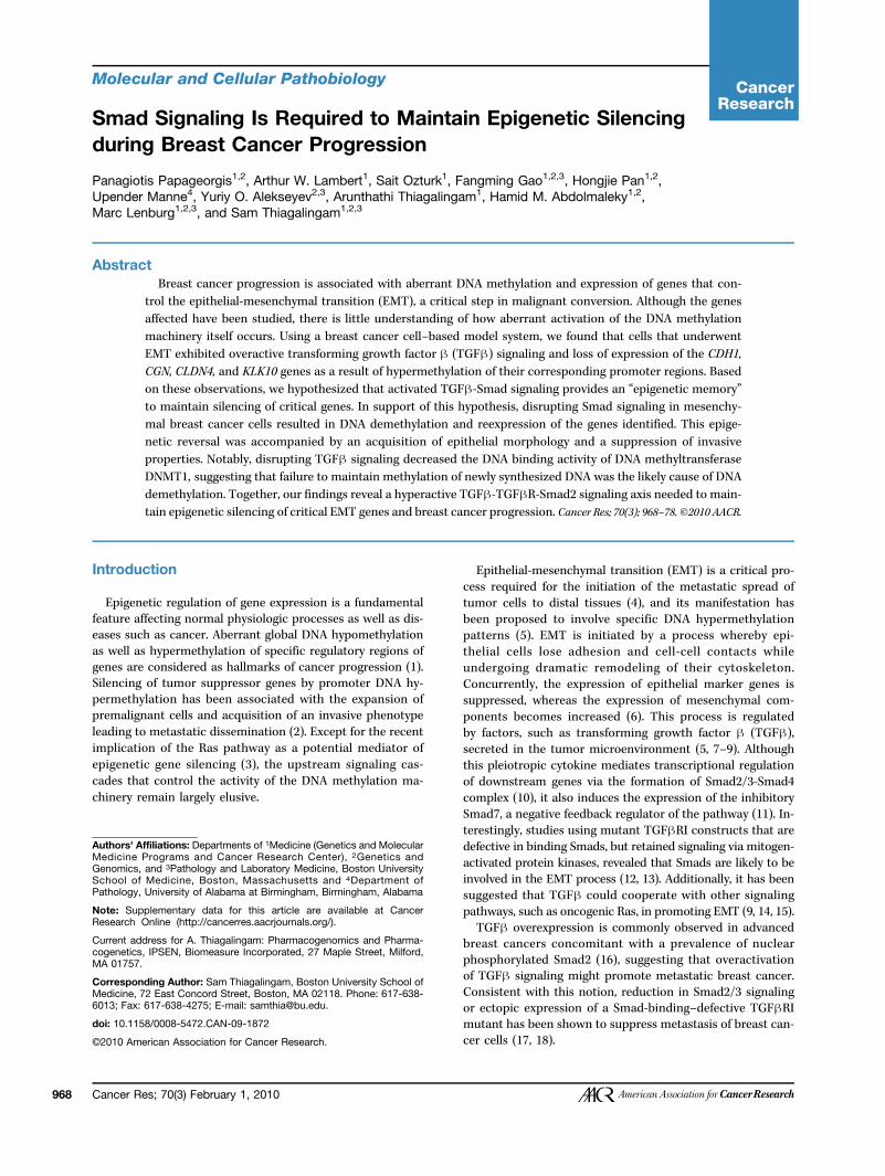
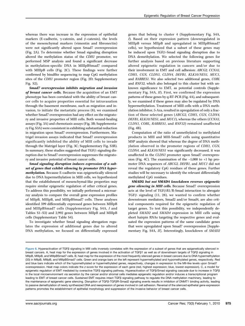
Figure 1. EMT in the MCF10A-based breast cancer progression model correlates with DNA hypermethylation–mediated silencing of E-cadherin expression.A, Western blotting analysis of cell lysates isolated from MI, MII, and MIII cells for detection of epithelial (E-cadherin, β-catenin, and γ-catenin) andmesenchymal (vimentin, fibronectin, and N-cadherin) protein markers. B, MSP analysis of the −160 to +1 bp region of E-cadherin (CDH1) promoter usingbisulfite-treated genomic DNA isolated from MI, MII, and MIII cells. C, MeDIP using either a mouse IgG (mock) or an anti-methylcytosine (Met-Cyt)monoclonal antibody using genomic DNA isolated from MII and MIII cells and PCR analysis of the CDH1 promoter. D, individual and combinations of 5-Aza,TSA, and sodium butyrate (SB) treatments in MIII cells for 72 h and Western blotting for E-cadherin detection.
Cancer Res; 70(3) February 1, 2010 969
Papageorgis et al.
970
(Qiagen), and labeled cRNA fragments were hybridized to hu-man genome U133 Plus 2.0 microarrays (Affymetrix). Geneexpression estimates and the measure of sequence specificityof the hybridization intensities were both determined using
Cancer Res; 70(3) February 1, 2010
standard settings in MAS5 (Affymetrix). Student's t test wasused to assess differential gene expression. Genes with a falsediscovery rate of <0.05 and a >2-fold difference in expres-sion were considered to be differentially expressed. The
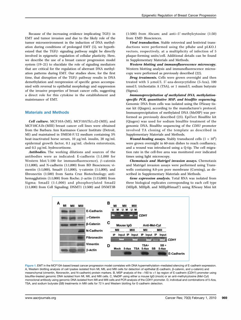
Figure 2. Smad7 overexpressioninduces promoter DNAdemethylation and reexpression ofE-cadherin. A, Western blottinganalysis to detect stableoverexpression of exogenousSmad7 and epithelial andmesenchymal markers in MI, MII,and MIII cells. B, Smad7overexpression induces epithelialmorphology in mesenchymal-likeMIII cells and localization ofE-cadherin at the cell-celljunctions. Representativeexamples of light microscopy (10×)and immunofluorescence images(100×) generated using ananti–E-cadherin primary andFITC-conjugated secondaryantibodies in the MI, MII, and MIIIcells stably transduced witheither pB or pBSmad7 vectors.Blue, cell nuclei were stained with4′,6-diamidino-2-phenylindole.Localization of E-cadherin (green)at the cell-cell junctions isindicated by white arrows. C,semiquantitative MSP analysis ofthe −160 to +1 bp CDH1 promoterregion using bisulfite-treatedgenomic DNA isolated from theMIIIpB and MIIIpBSmad7 cells.D, mapping of the methylated CpGdinucleotides within the −160 to+1 CDH1 promoter region byanalyzing 14 clones each fromthe bisulfite-treated templatesderived from the MIIIpB andMIIIpBSmad7 cells. White andblack squares representunmethylated and methylatedCpGs, respectively. Barchart shows percentages ofclones that exhibited at least onemethylated CpG in relation to thetotal number of clones sequenced.A p-value of less than 0.05(indicated by the * symbol) wasconsidered statistically significantbetween the compared samples.
Cancer Research
Epigenetic Regulation of Breast Cancer Progression
microarray data generated in this study are available fromthe National Center for Biotechnology Information GeneExpression Omnibus (24) under accession code GSE18070.Real-time quantitative reverse transcription-PCR (q-RT-PCR) was performed using SYBR Green Power Master Mix(Applied Biosystems).Chromatin immunoprecipitation assay. Chromatin im-
munoprecipitation (ChIP) assay was performed using theEZ-Magna ChIP G Chromatin Immunoprecipitation kit(Millipore) using chromatin isolated from 1 × 10⁶ cells percondition, according to the manufacturer's protocol.
Results
Characterization of the MCF10A-based breast cancerprogression model.We took advantage of a previously estab-lished cell line model system for breast cancer progression,which consists of a parental spontaneously immortalizedmammary epithelial cell line, MCF10A (MI), and two of itsderivatives: (a) MCF10ATk.cl2 (MII), an H-Ras–transformedMCF10A cell line, and (b) MCF10CA1h (MIII), derived froma xenograft of MII cells in nude mice that progressed to car-cinoma (19–21). These cell lines were previously reported toexhibit distinct tumorigenic properties when reimplanted innude mice; MI is nontumorigenic, MII forms benign hyper-plastic lesions, and MIII forms low-grade, well-differentiatedcarcinomas (20, 21). The advantage of this system is thatthese cell lines were derived from a common genetic back-ground (MCF10A) and accumulated distinct genetic/epige-netic alterations in vivo, enabling them to acquire propertiesassociated with gradual progression from nontumorigenic to
www.aacrjournals.org
carcinogenic state. Interestingly, whereas the MI and MII cellsexhibited a cobble-shaped epithelial morphology, the MIIIcells were spindle shaped with a mesenchymal-like pheno-type representing an apparent EMT during progression fromMII to MIII (Supplementary Fig. S1A).To further investigate EMT using this model system, we
characterized the expression of epithelial and mesenchymalmarkers. There was expression of predominantly epithelialmarkers (E-cadherin, γ-catenin, and β-catenin) in the MIand MII cells and of mesenchymal markers (fibronectin, vi-mentin, and N-cadherin) in the MIII cells with concomitantdownregulation of E-cadherin, β-catenin, and γ-catenin(Fig. 1A). These observations suggested that comparing thefeatures of MII and MIII cells is a logical approach to inves-tigate the molecular events responsible for EMT and theaccompanying epigenetic changes during the progressionfrom in situ to invasive breast carcinoma.The loss of E-cadherin (CDH1) expression, a prominent
biomarker for the epithelial state, due to promoter DNAhypermethylation has been associated with EMT and acqui-sition of invasive properties of breast cancer cells (25, 26).Therefore, we hypothesized that downregulation ofE-cadherin expression in MIII cells is mediated by epigenet-ic silencing. Methylation-specific PCR (MSP) analysis andimmunoprecipitation of genomic DNA from the MII andMIII cells using a monoclonal antibody against methylatedcytosine residues showed that, in contrast to MI and MIIcells, the CDH1 promoter is hypermethylated in MIII cells(Fig. 1B and C), consistent with the observed loss of expres-sion (Fig. 1A). Moreover, whereas treatment with the DNAmethylation inhibitor 5-Aza resulted in a robust increase in
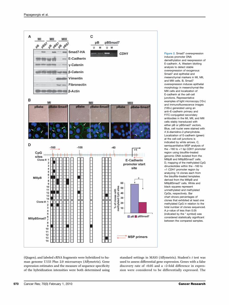
Figure 3. Smad7 overexpressionsuppresses migration and invasionof MIII cells. A, representativelight microscope images ofwound-healing assays for MIIIpBand MIIIpBSmad7 cells to evaluatetheir migration rate into thecell-free area. B, chemotaxisassay. Cells that migrated throughthe 8-μm pore-containingmembrane of the Transwells werestained with propidium iodideand counted. SF, serum-free; FBS,fetal bovine serum. C, Matrigelinvasion assay. Cells that invadedthrough the Matrigel werestained with trypan blue andcounted. All results are presentedas the average of cells counted in10 fields per condition. The resultspresented are the average oftriplicate samples and a p-valueof less than 0.05 (indicated bythe * symbol) was consideredstatistically significant between thecompared samples. Error barsrepresent ± standard error (S.E.)values.
Cancer Res; 70(3) February 1, 2010 971
Papageorgis et al.
972
E-cadherin expression, treatment with the histone deacety-lase inhibitors TSA or sodium butyrate had no effect, indi-cating that E-cadherin silencing occurs predominantly dueto promoter DNA hypermethylation (Fig. 1D).Smad7 overexpression induces an epithelial morphology
in association with CDH1 promoter DNA demethylation.Although a recent report suggested that sustained induc-tion of EMT by the tumor microenvironment inducesDNA methylation of genes, including CDH1 (5), the up-stream signaling events that are critical for the acquisitionand maintenance of these epigenetic changes remainedelusive. Because TGFβ signaling has been associated withthe manifestation of the EMT phenotype (8), we hypothe-sized that it might be directly involved in the regulation ofthe CDH1 promoter DNA methylation.To verify whether all the components of TGFβ pathway
are intact in our model system, we performed luciferasereporter assays using SBE4-luc (27). TGFβ1 treatment sig-nificantly induced luciferase activity, which was inhibited
Cancer Res; 70(3) February 1, 2010
on transient Smad7 overexpression in all three cell lines, in-dicating the requirement for a functional Smad2/3-Smad4complex (Supplementary Fig. S1B). Furthermore, these datasupported the suitability of our in vitromodel system to inter-rogate the role of TGFβ signaling in epigenetic gene silencing.We disrupted the TGFβ signaling pathway by stably over-
expressing Smad7 in MI, MII, and MIII cells to assess theeffects on the DNA methylation status and expression ofE-cadherin (Fig. 2A). As expected, Smad7 overexpression ab-rogated TGFβ/Smad signaling events as evident from theinhibition of TGFβ-mediated Smad2 phosphorylation(Ser465/467; Supplementary Fig. S1C). Furthermore, Smad7overexpression caused a profound effect on the morphologyof MIII cells elicited by the acquisition of a predominantlycobble-shaped epithelial phenotype as opposed to the spindle-shaped precursor cells. These morphologic changes wereaccompanied with upregulation of E-cadherin at the ad-herens junctions, consistent with a role in enhancing theadhesive properties (Fig. 2B). It should be noted that
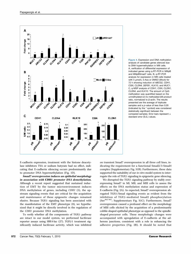
Figure 4. Expression and DNA methylationanalysis of candidate genes silenced dueto DNA hypermethylation in MIII cells.A, verification of differential expression ofindicated genes using q-RT-PCR in MIIIpBand MIIIpBSmad7 cells. B, q-RT-PCRanalysis for expression in MIII cells treatedwith 5 μmol/L 5-Aza or DMSO (Mock) for72 h showing induction of ABCG2, CDH1,CGN, CLDN4, DEFB1, KLK10, and MUC1.C, q-MSP analysis of CDH1, CGN, CLDN1,CLDN4, and KLK10. The amount of CpGmethylation was quantified based on theunmethylated (U ) to methylated (M) productratio, normalized to β-actin. The resultspresented are the average of triplicatesamples and a p-value of less than 0.05(indicated by the * symbol) was consideredstatistically significant between thecompared samples. Error bars represent ±standard error (S.E.) values.
Cancer Research
Epigenetic Regulation of Breast Cancer Progression
www.aacrjournals.org
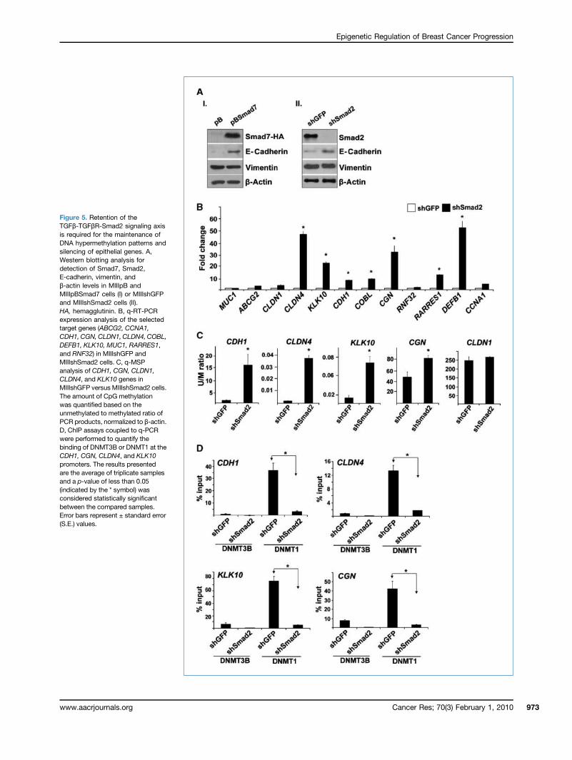
Figure 5. Retention of theTGFβ-TGFβR-Smad2 signaling axisis required for the maintenance ofDNA hypermethylation patterns andsilencing of epithelial genes. A,Western blotting analysis fordetection of Smad7, Smad2,E-cadherin, vimentin, andβ-actin levels in MIIIpB andMIIIpBSmad7 cells (I) or MIIIshGFPand MIIIshSmad2 cells (II).HA, hemagglutinin. B, q-RT-PCRexpression analysis of the selectedtarget genes (ABCG2, CCNA1,CDH1, CGN, CLDN1, CLDN4, COBL,DEFB1, KLK10, MUC1, RARRES1,and RNF32) in MIIIshGFP andMIIIshSmad2 cells. C, q-MSPanalysis of CDH1, CGN, CLDN1,CLDN4, and KLK10 genes inMIIIshGFP versus MIIIshSmad2 cells.The amount of CpG methylationwas quantified based on theunmethylated to methylated ratio ofPCR products, normalized to β-actin.D, ChIP assays coupled to q-PCRwere performed to quantify thebinding of DNMT3B or DNMT1 at theCDH1, CGN, CLDN4, and KLK10promoters. The results presentedare the average of triplicate samplesand a p-value of less than 0.05(indicated by the * symbol) wasconsidered statistically significantbetween the compared samples.Error bars represent ± standard error(S.E.) values.
Cancer Res; 70(3) February 1, 2010 973
Papageorgis et al.
Cancer Res; 70(3) February 1, 2010 Cancer Research974
Epigenetic Regulation of Breast Cancer Progression
whereas there was increase in the expression of epithelialmarkers (E-cadherin, γ-catenin, and β-catenin), the levelsof the mesenchymal markers (vimentin and fibronectin)were not significantly altered upon Smad7 overexpression(Fig. 2A). To determine whether Smad signaling disruptionaltered the methylation status of the CDH1 promoter, weperformed MSP analysis and found a significant decreasein methylation-specific DNA in MIIIpBSmad7 comparedwith MIIIpB cells (Fig. 2C). These findings were furtherconfirmed by bisulfite sequencing to map CpG methylationsites of the CDH1 promoter region (Fig. 2D; SupplementaryFig. S2).Smad7 overexpression inhibits migration and invasion
of breast cancer cells. Because the acquisition of an EMTphenotype has been correlated with the ability of breast can-cer cells to acquire properties essential for intravasationthrough the basement membrane, such as migration and in-vasion, to initiate the metastatic process (8), we examinedwhether Smad7 overexpression had any effect on the migrato-ry and invasive properties of MIII cells. Both wound-healingassays (Fig. 3A) and chemotaxis assays (Fig. 3B; Supplementa-ry Fig. S3A) were consistent in exhibiting substantial reductionin migration upon Smad7 overexpression. Furthermore, Ma-trigel invasion assays indicated that Smad7 overexpressionsignificantly inhibited the ability of MIII cells to invadethrough the Matrigel layer (Fig. 3C; Supplementary Fig. S3B).In summary, these studies suggested that TGFβ signaling dis-ruption due to Smad7 overexpression suppresses the migrato-ry and invasive potential of breast cancer cells.Smad signaling disruption induces expression of a sub-
set of genes that exhibit silencing by promoter DNA hyper-methylation. Because E-cadherin was epigenetically silenceddue to DNA hypermethylation in MIII cells, we hypothesizedthat the establishment of mesenchymal-like properties mayrequire similar epigenetic regulation of other critical genes.To address this possibility, we initially performed a microar-ray analysis to compare the overall gene expression profilesof MIIpB, MIIIpB, and MIIIpBSmad7 cells. These analysesidentified 599 differentially expressed genes between MIIIpBand MIIIpBSmad7 cells (Supplementary Fig. S4A, I andTables S1–S3) and 2,992 genes between MIIpB and MIIIpBcells (Supplementary Table S4).To investigate whether Smad signaling abrogation regu-
lates the expression of additional genes due to alteredDNA methylation, we focused on differentially expressed
www.aacrjournals.org
genes that belong to cluster 4 (Supplementary Fig. S4A,I). Based on their expression pattern (downregulated inMIIIpB versus MIIpB and upregulated in MIIIpBSmad7cells), we hypothesized that a subset of these genes maybe induced upon TGFβ-Smad signaling disruption due toDNA demethylation. We selected the following genes forfurther analysis based on previous literature supportingaltered epigenetic regulation in cancers and/or due totheir involvement in EMT and cell adhesion: ABCG2, CCNA1,CDH1, CGN, CLDN1, CLDN4, DEFB1, KLK10/NES1, MUC1,and RARRES1. We also selected two additional genes, COBLand RNF32, which also belonged to this cluster but with un-known significance to EMT, as potential controls (Supple-mentary Fig. S4A, II). First, we confirmed the expressionpatterns of these genes by q-RT-PCR (Fig. 4A), and subsequent-ly, we examined if these genes may also be regulated by DNAhypermethylation. Treatment of MIII cells with a DNA meth-ylation inhibitor, 5-Aza, resulted in upregulation of only a frac-tion of these selected genes (ABCG2, CDH1, CGN, CLDN4,DEFB1, KLK10/NES1, and MUC1), whereas the others (CCNA1,CLDN1, COBL, RARRES1, and RNF32) remained unaffected(Fig. 4B).Computation of the ratio of unmethylated to methylated
products in MIII and MIII-Smad7 cells using quantitativeMSP analysis showed that whereas the degree of DNA meth-ylation observed in the promoter regions of CDH1, CGN,CLDN4, and KLK10/NES1 was significantly decreased, it wasunaffected in the CLDN1 promoter upon Smad7 overexpres-sion (Fig. 4C). The examination of the −1,000 to +1 bp pro-moter DNA sequences of ABCG2, DEFB1, and MUC1 did notreveal the regulatory CpG residues of these genes. Furtherstudies will be necessary to identify the relevant differentiallymethylated CpG residues.SMAD2 but not SMAD4 knockdown reverses epigenetic
gene silencing in MIII cells. Because Smad7 overexpressionacts at the level of TGFβR1/R-Smad interaction to abrogateTGFβ signaling (11, 28), we wanted to confirm whetherdownstream mediators, Smad2 and/or Smad4, are also crit-ical components required for the epigenetic regulation oftarget genes. To test this possibility, we independently de-pleted SMAD2 and SMAD4 expression in MIII cells usingshort hairpin RNAs targeting the respective genes and eval-uated the expression patterns of the same candidate genesthat were upregulated upon Smad7 overexpression [Supple-mentary Fig. S4A, II]. Interestingly, knockdown of SMAD2
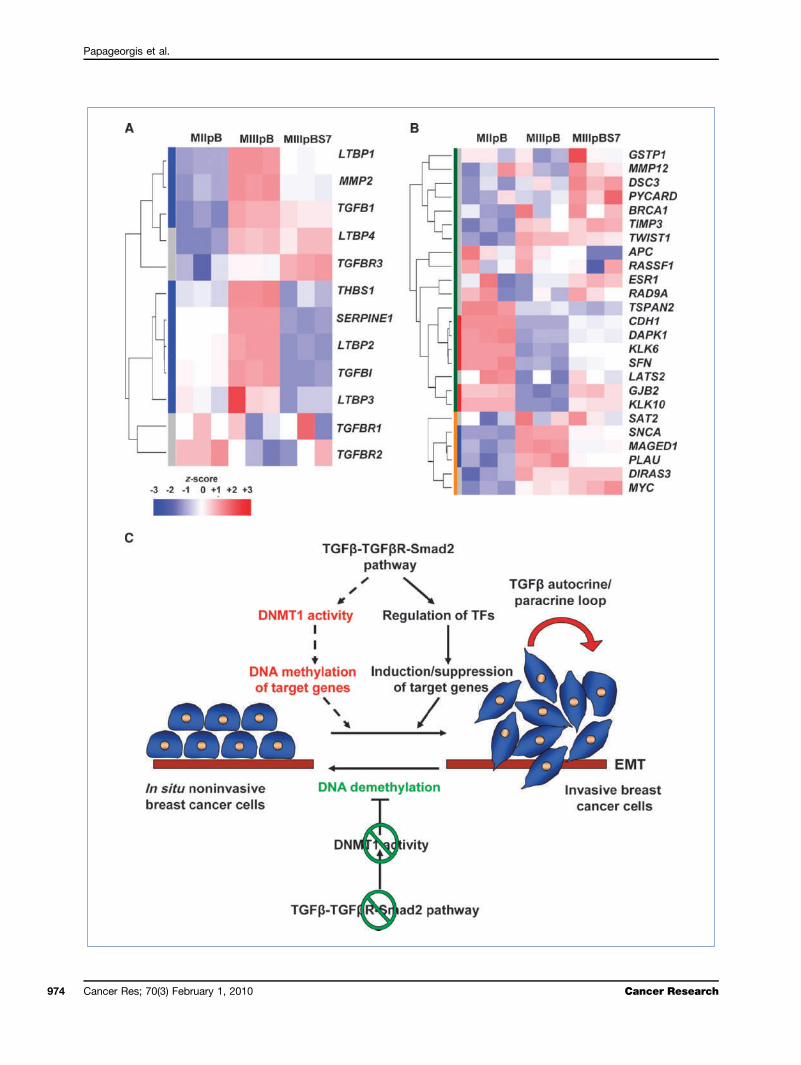
Figure 6. Hyperactivation of TGFβ signaling in MIII cells inversely correlates with the expression of a subset of genes that are epigenetically silenced inbreast cancers. A, heat map for the expression of genes involved in the activation of TGFβ1 as well as of downstream targets of TGFβ signaling inMIIpB, MIIIpB, and MIIIpBSmad7 cells. B, heat map for the expression of the most frequently silenced genes in breast cancers due to DNA hypermethylation(33) in MIIpB, MIIIpB, and MIIIpBSmad7 cells. Green and orange bars on the left represent hypermethylated and hypomethylated genes, respectively. Redand blue bars indicate which of the hypomethylated or hypermethylated genes, respectively, changes in expression to the MII-like levels upon Smad7overexpression. Heat map colors indicate the z-score for the expression of each gene (red, highest expression; blue, lowest expression). C, a model forepigenetic regulation of EMT mediated by overactive TGFβ signaling pathway. Hyperactivation of TGFβ/Smad signaling cascade due to increase in TGFβin the local microenvironment via secretion by the cancer and/or stromal cells mediates epigenetic regulation and/or induces a transcriptional programleading to EMT of breast cancer cells. Sustained EMT requires intact TGFβ signaling pathway to regulate the DNA methylation machinery, leading tothe maintenance of epigenetic gene silencing. Disruption of TGFβ-TGFβR-Smad2 signaling events results in inhibition of DNMT1 binding activity, leadingto passive demethylation of newly synthesized DNA and reexpression of genes involved in cell adhesion. Reversal of the silenced epithelial gene expressionpatterns promotes the establishment of epithelial morphology and suppression of the invasive behavior of breast cancer cells.
Cancer Res; 70(3) February 1, 2010 975
Papageorgis et al.
976
(Fig. 5A, II), but not SMAD4 (Supplementary Fig. S5A), ledto an increase in the expression of CDH1, CGN, CLDN4, andKLK10/NES1 (Fig. 5B; Supplementary Fig. S5B) concomitantwith a decrease in the DNA methylation of the respectiveregulatory regions (Fig. 5C). The specificity of this effectupon Smad2 depletion was further substantiated fromthe observation that SMAD2, but not SMAD4, knockdownresulted in the cells reverting to a more pronounced epithe-lial morphology (Supplementary Fig. S5C) phenocopyingthat of the MIIIpBSmad7 cells (Fig. 2B). These findingssuggest that intact TGFβ-TGFβR-Smad2 signaling axis isrequired for the maintenance of epigenetic gene silencing inour model system and that this phenomenon seems to beSmad4 independent.To determine if the changes in the promoter methylation
status are due to a passive or active demethylation process,we performed ChIP assays to measure the binding of DNMT1and DNMT3B to the promoter of the target genes. We foundthat the maintenance methyltransferase DNMT1 was thepredominant methyltransferase bound to the promoters ofCDH1, CGN, CLDN4, and KLK10 in the MIIIshGFP cells. Inter-estingly, TGFβ signaling disruption caused a significantreduction in the amount of DNMT1 bound to these pro-moters (Fig. 5D) without affecting the corresponding proteinlevels (Supplementary Fig. S6), suggesting that the TGFβ-TGFβR-Smad2 signaling axis regulates DNA methylationmaintenance during EMT, perhaps by modulating DNMT1binding activity.MIII cells exhibit hyperactive TGFβ signaling pathway
and resemble basal B breast cancers. Because our studiessupported that intact TGFβ signaling is required for EMTand DNA methylation maintenance during breast cancerprogression, we compared the gene expression profiles be-tween the invasive mesenchymal-like MIII cells and the non-invasive epithelial MII cells. We found that there wererelatively high expression levels of the downstream targetsof TGFβ signaling, such as MMP2, SERPINE1, and TGFβI,in MIII cells. Moreover, we found that the expression ofTGFβ1 and the TGFβ-activating proteins LTBP1, LTBP2,LTBP3, LTBP4, and THBS1 (29) was also dramatically in-creased in MIII compared with MII cells (Fig. 6A; Supplemen-tary Fig. S7A). Consistent with these observations, ELISAassays confirmed that MIII cells secrete TGFβ1 when cul-tured in serum-free medium (Supplementary Fig. S7B).To further assess the relevance of this phenomenon to
EMT, we compared the differential gene expression patternsin the MIII cells with and without TGFβ-Smad signaling dis-ruption to a previously published microarray data set from51 breast cancer cell lines (30). Interestingly, the genes thatare highly expressed in MIII cells relative to MII cells and re-verted to MII-like levels upon TGFβ-Smad signaling disrup-tion (cluster 1; Supplementary Fig. S4A, I) are found to besimilar to the expression pattern observed in the majorityof the basal B subtype breast cancer cell lines (SupplementaryFig. S8A). Furthermore, the genes with the converse expressionpattern (cluster 4; Supplementary Fig. S4A, I) tend to be alsoexpressed at lower levels in the same basal B-cell lines (Sup-plementary Fig. S8A). Overall, these results suggest that MIII
Cancer Res; 70(3) February 1, 2010
cells exhibit a similar expression pattern as the basal B sub-type cell lines, a subtype associated with acquisition of EMT(31, 32). Additionally, the expression of some TGFβ pathwaycomponents (predominantly LTBP2, MMP2, SERPINE1, TGFBI,and TGFβ1) was also higher in basal B compared with othersubtypes, lending further support to the notion that TGFβpathway overactivation is likely to be an important featureof basal B tumors (Supplementary Fig. S8B). Moreover, we alsofound that a subset of genes, including CDH1, DAPK1, DSC3,GJB2, GSTP1, KLK6, KLK10, LATS2, PYCARD, and SFN, whichwere upregulated upon disruption of TGFβ pathway in MIIIcells, have been consistently reported (33) as targets for silenc-ing due to DNA hypermethylation in breast cancers (Fig. 6B).
Discussion
To delineate the upstream signaling mechanisms respon-sible for the maintenance of aberrant promoter DNA meth-ylation patterns during breast cancer progression, we used apreviously described breast cancer cell line model system.We found that the mesenchymal-like MIII cells, comparedwith its precursor H-Ras–transformed epithelial MII cells(21), harbor hyperactive TGFβ signaling and exhibit anEMT phenotype. Moreover, highly invasive properties of theMIII cells suggesting a prometastatic role were substantiatedby differential expression of several genes in MIII comparedwith the MII cells sharing a similar expression pattern with asubset of genes previously identified as mediators of breastcancer metastasis to the lung (Supplementary Fig. S9; ref. 34).Overall, these results indicate that the MCF10A-based breastcancer cell line model system is an attractive and highly rel-evant model to study the molecular mechanisms responsiblefor epigenetic regulation of EMT during transition fromin situ to invasive breast carcinoma.By using gene expression profiling and by examining the
epigenetic regulation of differentially expressed genes in thisbreast cancer model system, we found that there was DNAhypermethylation–mediated silencing of genes involved incell adhesion and tight junction formation, including CDH1,CGN, and CLDN4, as well as the epithelial protease KLK10/NES1 in basal B-like breast cancer cells that have undergoneEMT. These observations are also consistent with a recentreport showing that suppression of CDH1 expression duringsustained EMT is mediated by the establishment of promoterDNA hypermethylation (5).Furthermore, our studies show that overactive TGFβ sig-
naling events, mediated by an autocrine feedback loop thatmaintains high TGFβ1 levels in the microenvironment, areresponsible for sustaining the altered epigenome and the in-vasive properties of breast cancer cells. Moreover, our studiesprovide direct evidence for the involvement of intact hyper-active TGFβ-TGFβR-Smad2 signaling axis in orchestrating aspecific DNA methylation pattern that favors EMT and theinvasive behavior of breast cancer cells. Several observationssupport this conclusion. First, disruption of TGFβ signalingby either Smad7 overexpression or SMAD2, but not SMAD4,knockdown in the MIII cells reversed the EMT phenotype
Cancer Research
Epigenetic Regulation of Breast Cancer Progression
and caused reestablishment of the epithelial morphology.Second, the observed mesenchymal to epithelial transitionwas accompanied by the upregulation of transcripts for theCDH1 gene, encoding a key cell-cell adhesion molecule andnegative regulator of WNT signaling cascade (35), the tightjunction genes CLDN4 and CGN, as well as the proteaseKLK10/NES1. CDH1 levels have been directly correlated withepithelial phenotype and metastatic properties of cancercells (36), whereas the KLK10/NES1 protease was shown tobe specifically expressed in epithelial cells and suppressbreast tumor growth in vivo (37). Finally, significant de-creases in promoter DNA methylation of the critical targetgenes upon TGFβ-TGFβR-Smad2 signaling disruption stronglysupport a direct involvement of this axis in modulating thefunctionality of the DNA methylation machinery to maintainthe epigenetically silenced state.Despite the identification of putative DNA demethylase
enzymes and evidence for the involvement of a DNA repairpathway in this process (38), the existence of active DNA de-methylation mechanisms in mammals has been elusive (39).Our data favor the alternate mechanism which proposes thatsuppression of the binding of maintenance DNA methyl-transferase, DNMT1, to the target DNA sequences resultsin passive DNA demethylation (40). We found that bindingof DNMT1 to CDH1, CLDN4, CGN, and KLK10 promoters wassignificantly suppressed upon SMAD2 knockdown (Fig. 5D),whereas DNMT1 and DNMT3B protein levels remained un-affected (Supplementary Fig. S6). Therefore, we propose thatreduced DNMT1 binding activity on disruption of TGFβ-Smad signaling could result in loss of DNA methylation main-tenance and passive demethylation of newly synthesized DNA(Fig. 6C). The passive demethylation in the absence of intactSmad2, but not Smad4, suggests that Smad2 may play a role inloading DNMT1 onto specific gene promoters to modulateDNA methylation when TGFβ signaling becomes overactive.Alternatively, Smad2 may interact with other factors to tran-scriptionally regulate target genes or control DNMT1 activityvia post-translational modifications. Finally, it is also likelythat DNMT1 binding is regulated by remodeling of localized
www.aacrjournals.org
chromatin in response to TGFβ signaling–mediated effectsduring breast cancer progression.In summary, our data suggest that increased TGFβ levels
in the breast tumor microenvironment promote hyperactiveSmad signaling to enable the acquisition of EMT-like prop-erties. Furthermore, we propose that overactive TGFβ cas-cades play a major role in the “epigenetic memory” andmaintenance of epithelial gene–specific silencing duringEMT mediated by unique DNA methylation patterns (Fig.6C). To our knowledge, this is the first report to provideconclusive evidence that the reversal of the DNA hyper-methylation status of gene promoters occurs as a result ofa signaling pathway perturbation, in this case the TGFβ/Smad cascade. By extension, our study provides a frame-work for uncovering genes that are coordinately regulatedby epigenetic mechanisms in response to specific signalingevents commonly deregulated during cancer progression.Finally, our findings provide additional credence to theidea that inhibition of TGFβ-TGFβR-Smad2 signaling axismay be a useful therapeutic strategy to target breast cancerprogression.
Disclosure of Potential Conflicts of Interest
No potential conflicts of interest were disclosed.
Acknowledgments
We thank Drs. Didier Trono, Bert Vogelstein, Robert Weinberg, and JeffWrana for generously providing reagents.
Grant Support
Susan G. Komen for the Cure Investigator-Initiated Research AwardKG081435 and NIH grant CA101773 (S. Thiagalingam).
The costs of publication of this article were defrayed in part by the paymentof page charges. This article must therefore be hereby marked advertisement inaccordance with 18 U.S.C. Section 1734 solely to indicate this fact.
Received 5/22/09; revised 11/19/09; accepted 11/22/09; publishedOnlineFirst 1/19/10.
References
1. Herman JG, Baylin SB. Gene silencing in cancer in association withpromoter hypermethylation. N Engl J Med 2003;349:2042–54.2. Baylin SB, Ohm JE. Epigenetic gene silencing in cancer—a mecha-
nism for early oncogenic pathway addiction? Nat Rev Cancer 2006;6:107–16.
3. Gazin C, Wajapeyee N, Gobeil S, Virbasius CM, Green MR. An elab-orate pathway required for Ras-mediated epigenetic silencing. Na-ture 2007;449:1073–7.
4. Gupta GP, Massague J. Cancer metastasis: building a framework.Cell 2006;127:679–95.
5. Dumont N, Wilson MB, Crawford YG, Reynolds PA, Sigaroudinia M,Tlsty TD. Sustained induction of epithelial to mesenchymal transitionactivates DNA methylation of genes silenced in basal-like breastcancers. Proc Natl Acad Sci U S A 2008;105:14867–72.
6. Yang J, Weinberg RA. Epithelial-mesenchymal transition: at thecrossroads of development and tumor metastasis. Dev Cell 2008;14:818–29.
7. Deckers M, van Dinther M, Buijs J, et al. The tumor suppressorSmad4 is required for transforming growth factor β-induced epithe-
lial to mesenchymal transition and bone metastasis of breast cancercells. Cancer Res 2006;66:2202–9.
8. Thiery JP. Epithelial-mesenchymal transitions in tumour progression.Nat Rev Cancer 2002;2:442–54.
9. Oft M, Heider KH, Beug H. TGFβ signaling is necessary for carcino-ma cell invasiveness and metastasis. Curr Biol 1998;8:1243–52.
10. Massague J, Seoane J, Wotton D. Smad transcription factors. GenesDev 2005;19:2783–810.
11. Hayashi H, Abdollah S, Qiu Y, et al. The MAD-related protein Smad7associates with the TGFβ receptor and functions as an antagonist ofTGFβ signaling. Cell 1997;89:1165–73.
12. Yu L, Hebert MC, Zhang YE. TGF-β receptor-activated p38 MAPkinase mediates Smad-independent TGF-β responses. EMBO J2002;21:3749–59.
13. Itoh S, Thorikay M, Kowanetz M, et al. Elucidation of Smad require-ment in transforming growth factor-β type I receptor-induced re-sponses. J Biol Chem 2003;278:3751–61.
14. Oft M, Peli J, Rudaz C, Schwarz H, Beug H, Reichmann E.TGF-β1 and Ha-Ras collaborate in modulating the phenotypic
Cancer Res; 70(3) February 1, 2010 977
Papageorgis et al.
978
plasticity and invasiveness of epithelial tumor cells. Genes Dev1996;10:2462–77.
15. Oft M, Akhurst RJ, Balmain A. Metastasis is driven by sequentialelevation of H-ras and Smad2 levels. Nat Cell Biol 2002;4:487–94.
16. Kang Y, He W, Tulley S, et al. Breast cancer bone metastasis me-diated by the Smad tumor suppressor pathway. Proc Natl Acad SciU S A 2005;102:13909–14.
17. Tian F, Byfield SD, Parks WT, et al. Smad-binding defective mutantof transforming growth factor β type I receptor enhances tumorigen-esis but suppresses metastasis of breast cancer cell lines. CancerRes 2004;64:4523–30.
18. Tian F, DaCosta Byfield S, Parks WT, et al. Reduction in Smad2/3signaling enhances tumorigenesis but suppresses metastasis ofbreast cancer cell lines. Cancer Res 2003;63:8284–92.
19. Santner SJ, Dawson PJ, Tait L, et al. Malignant MCF10CA1 cell linesderived from premalignant human breast epithelial MCF10AT cells.Breast Cancer Res Treat 2001;65:101–10.
20. Strickland LB, Dawson PJ, Santner SJ, Miller FR. Progression of pre-malignant MCF10AT generates heterogeneous malignant variantswith characteristic histologic types and immunohistochemicalmarkers. Breast Cancer Res Treat 2000;64:235–40.
21. Tang B, Vu M, Booker T, et al. TGF-β switches from tumor sup-pressor to prometastatic factor in a model of breast cancer pro-gression. J Clin Invest 2003;112:1116–24.
22. Gao F, Ponte JF, Papageorgis P, et al. hBub1 deficiency triggers anovel p53 mediated early apoptotic checkpoint pathway in mitoticspindle damaged cells. Cancer Biol Ther 2009;8:627–35.
23. WeberM, Davies JJ, Wittig D, et al. Chromosome-wide and promoter-specific analyses identify sites of differential DNA methylation in nor-mal and transformed human cells. Nat Genet 2005;37:853–62.
24. NCBI Gene Expression Omnibus (http://www.ncbi.nlm.nih.gov/geo/).25. Yoshiura K, Kanai Y, Ochiai A, Shimoyama Y, Sugimura T, Hirohashi
S. Silencing of the E-cadherin invasion-suppressor gene by CpGmethylation in human carcinomas. Proc Natl Acad Sci U S A 1995;92:7416–9.
26. Lombaerts M, van Wezel T, Philippo K, et al. E-cadherin transcrip-tional downregulation by promoter methylation but not mutation isrelated to epithelial-to-mesenchymal transition in breast cancer celllines. Br J Cancer 2006;94:661–71.
Cancer Res; 70(3) February 1, 2010
27. Zhou S, Buckhaults P, Zawel L, et al. Targeted deletion ofSmad4 shows it is required for transforming growth factor β and activinsignaling in colorectal cancer cells. Proc Natl Acad Sci U S A 1998;95:2412–6.
28. Abdollah S, Macias-Silva M, Tsukazaki T, Hayashi H, Attisano L,Wrana JL. TβRI phosphorylation of Smad2 on Ser465 and Ser467is required for Smad2-Smad4 complex formation and signaling.J Biol Chem 1997;272:27678–85.
29. Annes JP, Munger JS, Rifkin DB. Making sense of latent TGFβ acti-vation. J Cell Sci 2003;116:217–24.
30. Neve RM, Chin K, Fridlyand J, et al. A collection of breast cancer celllines for the study of functionally distinct cancer subtypes. CancerCell 2006;10:515–27.
31. Blick T, Widodo E, Hugo H, et al. Epithelial mesenchymal transitiontraits in human breast cancer cell lines. Clin Exp Metastasis 2008;25:629–42.
32. Sarrio D, Rodriguez-Pinilla SM, Hardisson D, Cano A, Moreno-BuenoG, Palacios J. Epithelial-mesenchymal transition in breast cancer re-lates to the basal-like phenotype. Cancer Res 2008;68:989–97.
33. Agrawal A, Murphy RF, Agrawal DK. DNA methylation in breast andcolorectal cancers. Mod Pathol 2007;20:711–21.
34. Minn AJ, Gupta GP, Siegel PM, et al. Genes that mediate breast can-cer metastasis to lung. Nature 2005;436:518–24.
35. Jeanes A, Gottardi CJ, Yap AS. Cadherins and cancer: how doescadherin dysfunction promote tumor progression? Oncogene 2008;27:6920–9.
36. Onder TT, Gupta PB, Mani SA, Yang J, Lander ES, Weinberg RA.Loss of E-cadherin promotes metastasis via multiple downstreamtranscriptional pathways. Cancer Res 2008;68:3645–54.
37. Goyal J, Smith KM, Cowan JM, Wazer DE, Lee SW, Band V. The rolefor NES1 serine protease as a novel tumor suppressor. Cancer Res1998;58:4782–6.
38. Gehring M, Reik W, Henikoff S. DNA demethylation by DNA repair.Trends Genet 2009;25:82–90.
39. Ooi SK, Bestor TH. The colorful history of active DNA demethylation.Cell 2008;133:1145–8.
40. Robert MF, Morin S, Beaulieu N, et al. DNMT1 is required to maintainCpG methylation and aberrant gene silencing in human cancer cells.Nat Genet 2003;33:61–5.
Cancer Research


