Embed Size (px)
Citation preview

This article appeared in a journal published by Elsevier. The attachedcopy is furnished to the author for internal non-commercial researchand education use, including for instruction at the authors institution
and sharing with colleagues.
Other uses, including reproduction and distribution, or selling orlicensing copies, or posting to personal, institutional or third party
websites are prohibited.
In most cases authors are permitted to post their version of thearticle (e.g. in Word or Tex form) to their personal website orinstitutional repository. Authors requiring further information
regarding Elsevier’s archiving and manuscript policies areencouraged to visit:
http://www.elsevier.com/authorsrights

Author's personal copy
Original article
Small azobenzene derivatives active against bacteria and fungi
Stefano Piotto a, Simona Concilio b,*, Lucia Sessa a, Amalia Porta a,Elena Concetta Calabrese a, Anna Zanfardino c, Mario Varcamonti c, Pio Iannelli a
aDepartment of Pharmacy, University of Salerno, via Giovanni Paolo II, 132, 84084 Fisciano (Salerno), ItalybDepartment of Industrial Engineering, University of Salerno, via Giovanni Paolo II, 132, 84084 Fisciano (Salerno), ItalycDepartment of Structural and Functional Biology, University of Napoli Federico II, Via Cinthia, Complesso Monte S. Angelo, 80126 Naples, Italy
a r t i c l e i n f o
Article history:Received 26 March 2013Received in revised form11 June 2013Accepted 18 July 2013Available online 11 August 2013
Keywords:AzobenzenesADMETAntimicrobial
a b s t r a c t
ATP synthase and protein kinase (PKs) are prime targets for drug discovery in a variety of diseases. It iswell known that numerous stilbenes are capable to interact and inhibit ATP synthase and PKs. This workfocuses on a series of azobenzene based molecules having high structural similarity with antimicrobialstilbenes. An investigation was carried out analyzing the potential toxicity of a large set of molecules bymeans of computational analysis. A small selection of potential low toxic molecules have been thereforesynthesized, characterized and finally microbiologically tested.
The synthesized compounds show potent bactericidal activity against Gramþ and a fungus, and arecapable of inhibiting biofilm formation. Finally, the compounds demonstrated a thermal stability thatmakes them potential candidates for incorporation in polymer matrix for application as biomedicaldevices and food packaging.
� 2013 Elsevier Masson SAS. All rights reserved.
1. Introduction
The emergence of bacterial strains resistant to the most com-mon classes of antibiotics is prompting a dramatic quest for thedevelopment of new antimicrobial drugs.
Infections caused by resistant microorganisms often fail torespond to conventional treatment, resulting in prolonged illnessand greater risk of death. According to WHO [1], about 440,000new cases of multidrug-resistant tuberculosis (MDR-TB) emergeannually, causing at least 150,000 deaths.
A high percentage of hospital-acquired infections are caused byhighly resistant bacteria, such as methicillin-resistant Staphylo-coccus aureus (MRSA).
Some stilbenes, like resveratrol, are well known natural anti-biotics. Resveratrol is a phytoalexin produced in response to en-vironmental stresses, such as wounding or pathogen attack.Resveratrol (RES) was known to possess an antimicrobial and apronounced antifungal effect [2]. Its mechanism of action isprobably the inhibition of bacterial ATP synthase and PKs [3,4].
However, a recent work of Weber [5] demonstrated that resver-atrol is not effective against Candida albicans and other Candidaspecies. Moreover, Paulo and coworkers [6] pointed out that theactivity of resveratrol against Gram-positive and Gram-negativebacteria is lower than previously reported. Unfortunately, stil-benes derivatives display only moderate antimicrobial effects[7,8], and they are usually toxic compounds, a major drawbackwhen developing new drugs.
The goal of this work was to design and synthesize moleculeswith similar or higher activity compared to antimicrobial stilbenes,but with reduced toxicity. Therefore we considered the azobenzenes,a class of molecules having high structural similarity with stilbenes.Though azobenzenes have been widely studied as dyers or photo-responsive materials [9e12], very little is reported about their po-tential antimicrobial activity. Preliminary results from moleculardocking (data to be published) indicated that these compounds caninhibit ATP synthase binding at the interface between a and g sub-units of ATP synthase. Therefore, a computational investigation wascarried out to study the potential toxicity of a large set of molecules.The five molecules with lowest in silico toxicity values were syn-thesized, chemically and thermally characterized to establish thepossibility of their use in polymer matrices, and they finally under-went to microbiological tests to evaluate the antimicrobial activityand the capability to destroy biofilms.
* Corresponding author. Tel.: þ39 320 7979002; fax: þ39 089 964057.E-mail address: [email protected] (S. Concilio).
Contents lists available at ScienceDirect
European Journal of Medicinal Chemistry
journal homepage: http: / /www.elsevier .com/locate/ejmech
0223-5234/$ e see front matter � 2013 Elsevier Masson SAS. All rights reserved.http://dx.doi.org/10.1016/j.ejmech.2013.07.030
European Journal of Medicinal Chemistry 68 (2013) 178e184

Author's personal copy
2. Results and discussion
2.1. In silico screening
We employed the in silico toxicity tool ADMET from Accelrys, topreliminarily test a series of 6156 azo compounds as listed in Table 4,Section 4.1. For each molecule we calculated the aqueous solubility[13], the bloodebrain penetration after oral administration [14], thecytochrome P450 2D6 inhibition [15], and the potential liver toxicity[16]. Consistent with the literature [17e19], the vast majority of theset yielded unacceptable toxicity; only 22 molecules out of 6156presented values of interest. The five models were calculated for the6156 molecules and the results were plotted in Fig. 1.
In Fig. 1, the distribution of properties is evident using a multi-dimensional representation. The subset that promised less toxicityeffect is indicated within a large red box. The 5 best candidateslisted in Table 1 are all inside the red box. In particular, the twosmall cyan spheres (corresponding to A2 and A4 in Table 1) insidethe red circle, exhibit the best overall indices. Unlike the remainingazobenzenes, that show extremely high probability of hepatotox-icity, the molecules of Table 1 have probability of being nontoxic(For interpretation of the references to color in this figure legend,the reader is referred to the web version of this article.).
2.2. Thermal and optical properties
The proton resonance data for A1eA5 are in agreement with theexpected values, as reported in Section 4.2. Thermodynamicproperties of azo compounds A1eA5 are given in Table 2. Accordingto X-ray diffraction analysis A1eA5, as obtained from the synthesis,are crystalline materials, with melting temperatures ranging be-tween 100 and 175 �C. The thermogravimetric analysis (TGA) showsthat compounds A1, A2, A3 and A5 show initial decompositiontemperatures (5% weight loss) around 200 �C. This stability in-dicates the possibility to incorporate the azo compounds in a largenumber of commercial polymers matrices that undergo high tem-perature melt compounding processing, in order to obtainbiomedical devices and food packaging films. An exception is
represented by A4 that starts to degrade at about 84 �C. Accordingto polarized optical microscopy, each compound shows a brightyellow or red needle crystalline habitus. From DSC analysis westudied the thermal behavior of A1eA5.
A1, A2 and A4 show a sharp melting peak in the first heating runand a crystallization peak in the cooling run. When heated in thesecond run, they show the same melting peak as in the first run. Inparticular, A4 shows also a solid to solid transition in the first heatingrun at 110.3 �C (DHkek ¼ 109.8 J/g) and, after cooling, it shows asecond crystallization during the second heating run at 105.7 �C(DHc2 ¼ 30.1 J/g), as confirmed by polarized optical microscopyobservation and X-ray analysis. A3 has a different behavior: in thefirst heating run it shows a solid to solid transition, confirmed by theobservation at the polarized optical microscope (Tkek ¼ 151.6 �C,DHkek¼ 7.5 J/g); this is followed by themelting of this new solid format 173.3 �C (see Table 2). In the subsequent cooling run the moleculecrystallizes at 135.9 �C in a different crystalline form, which melts at182.3 �C in the second heating run. This behavior was confirmed byX-Ray diffraction patterns. A5 shows only the first melting peak andit is not able to crystallize from the melt; this is probably due to itsasymmetric structure, which comprises a short methyl group and alonger allyl group on the same ring.
Fig. 1. 5D plot of ADMET calculation. The solubility AlogP98, the inhibition of cytochrome D6 and the hepatotoxicity are represented on the orthogonal axes. The PSA 2D is colormapped onto the spheres and the probability to cross the bloodebrain barrier is indicated with the size of the spheres. The square box represents the optimal subset of the 6156molecules of Table 1. The best candidates are indicated within the small round box.
Table 1ADMET parameters of the five synthesized compounds.
Molecule IDa Solubilityb Hepatotoxicity CYP2D6c PSA_2Dd BBBe
A1 1144 �4.212 0.596 0.387 64.277 0.268A2 496 �4.127 0.589 0.395 64.277 0.289A3 820 �4.843 0.655 0.643 64.277 0.409A4 1972 �5.001 0.655 0.712 64.277 0.439A5 2062 �4.683 0.668 0.792 64.277 0.278
a Molecule identification number.b Solubility is the AlogP98.c Cytochrome P450 2D6 inhibition prediction.d Fast polar surface area.e Bloodebrain barrier penetration coefficient. The values of hepatotoxicity and
CYP2D6 fall in the range {0,1}. Values close to 0 correspond to low probability to causedose-dependent toxicity against liver and cytochromes respectively. Values of BBB arethe base 10 logarithm of the ratio (brain concentration)/(blood concentration).
S. Piotto et al. / European Journal of Medicinal Chemistry 68 (2013) 178e184 179

Author's personal copy
The UVevis spectra of A1eA5, in acetonitrile solution, arequalitatively independent on the number and length of flexiblealiphatic segments (methyl, ethyl, allyl) attached to the azobenzenecore, and only depend on the active azo-containing unit, which isthe same for all compounds. The UV absorption spectra of A1eA5 intrans configuration, in the 240e650 nm region, showed two bandscentered around 240 and 360 nm, related respectively to thep / p* and n / p* electronic transitions of the azobenzenechromophore [9]. UVevis data are given in Table 2.
2.3. Antimicrobial activity
The MICs (minimum inhibitory concentrations) of the A1eA5compounds for bacterial strains and C. albicans SC5314, determinedby the microbroth dilution are shown in Table 3.
Table 3 reports the MIC0 values along with the data on resver-atrol published by Weber [5] and Paulo [6]. Compared to the
reference molecule, resveratrol, all azo compounds show a higherantibacterial and antifungal activity. In particular, compounds A4and A5, exhibit MIC0 values 4e6 times smaller than those ofresveratrol.
Fig. 2 shows the effect of azo compounds (A3) on hyphae for-mation of C. albicans. In the absence of our compounds, an extensivehyphae formation was observed, whereas when azo compoundswere present, hyphae formation was severely hampered in aconcentration-dependent manner. Here, we observed a total inhibi-tion of germination above 10 mg/mL of A3. The same analysis madeon A4 and A5 is reported as Supplementary data of this article.
To determine the inhibition of biofilms formationwe performeda semi-quantitative colorimetric assay that does not differentiatebetween live and dead cells, using different concentrations of azocompounds (8, 10, 15, 20, 25, 30, 50 and 60 mg/mL), followed bystaining with CV (crystal violet). As shown in Fig. 3, Candida treatedwith different concentrations of azo compounds displayed a severe
Table 2Optical and thermal characterization.
Molecule Optical characterization Thermal characterization
l*max (nm) 3max (L mol�1 cm�1) Tm# (�C) DHxm (J/g) Tc# (�C) DHxc (J/g) Td# (�C)
A1 357 29,500 156.3 139 100.3 89.6 200.3A2 358 30,300 151.0 112 103.6 98.3 204.9A3 358 28,700 173.3 97.9 135.9 88.7 198.3A4 359 30,500 174.3 110 112.6 13.7 84.9A5 359 30,100 105.3 82.5 e e 203.4
lmax ¼ wavelength at the principal absorption maximum, *instrument error �0.10 nm.3max ¼ molar extinction coefficient at absorption maximum.Tm ¼ melting temperature, from DSC analysis, 10 �C/min, nitrogen flow; Tc ¼ crystallization temperature, from DSC cooling run; Td ¼ temperature of 5% of weight loss in theTGA trace, 10 �C/min, nitrogen flow, #instrument error �0.5 �C.DHm/DHc ¼ melting/crystallization enthalpy, evaluated by integration of the peak, experimental error �5%.
Table 3Antimicrobial and antifungal activity.
MIC0 (mg/mL) after 24 h
Molecule S. aureus L. monocytogenes S. typhimurium P. aeruginosa C. albicans
Resveratrol 100a e >400a >400a >128b
A1 30 50 >60 >60 30A2 20 25 >60 >60 20A3 20 25 >60 >60 20A4 20 25 >60 >60 17A5 25 >60 >60 >60 15
MIC0: minimum inhibitory concentration required to inhibit the growth of 100% of organisms. The values are the geometric mean of at least three determinations.a Data from Ref. [6].b Data from Ref. [5].
Fig. 2. Inhibition of hyphae formation in Candida albicans at different concentrations of A3.
S. Piotto et al. / European Journal of Medicinal Chemistry 68 (2013) 178e184180

Author's personal copy
defect in biofilm formation compared to the control. These resultswere confirmed by the effects of 20 mg/mL of azo compounds onCandida biofilms degradation using an XTT assay, which charac-terizes living biomass (Fig. 4).
S. aureus forms biofilms that can be detected by staining theadhered cells with CV. We analyzed the ability of different con-centrations of azo compounds (8, 10, 15, 20, 25, 30, 50 and 60 mg/mL) to inhibit biofilm formation. As shown in the graph of Fig. 5,while A4 prevents biofilms formation when used at 30 mg/mL, theother azo compounds had the same effect already at 25 mg/mL. Toverify that the inhibitory effect on biofilm formation was not aresult of growth inhibition, different concentrations of azo com-pounds (20, 25, 30 and 50 mg/mL) were added to a preformedS. aureus biofilm. Results in Fig. 6 confirmed that 30 mg/mL of A1, A2,A3 and A5 were able to degrade more than 60% of preformedbiofilm. As expected, 30 mg/mL of A4 degraded only the 40% ofS. aureus biofilm. Complete biofilm degradation required 50 mg/mLof azo compounds.
3. Conclusions
Wehave designed a series of azobenzene compounds and studiedtheir potential toxicity by means of a computational analysis. Wehave synthesized the molecules with the lowest in silico toxicity andstudied their effects on microorganisms’ cultures, representative ofGramþ and Gram� bacteria and fungi. The antimicrobial activity andthe capability to destroy biofilms are promising and it indicates thatthese molecules may have interesting and therapeutically significantapplications. In particular all azo compounds show higher antibac-terial and antifungal activity than resveratrol. The preparation ofpolymers embedding azo-compounds is currently under investiga-tion, since the activity of these compounds and their thermal stabilitysuggest possible applications for food packaging and biomedicaldevices. Future work will include the synthesis of a larger combi-natorial library of these compounds, and the test of their inhibitoryeffects on mammalian ATP synthase and other ATP-dependent en-zymes to assess their specificity.
Fig. 3. Inhibition of C. albicans biofilm formation. Equal numbers of Candida cellsincubated in 96-well plates for 24 h at 37 �C. Adherent cells were stained with CV.
Fig. 4. XTT assay. Metabolic activity of Candida biofilms treated with 20 mg/mL of azocompounds, compared to control (biofilm treated with vehicle only). Each value is themean � S.D. of 3 independent experiments.
Fig. 5. Inhibition of S. aureus biofilm formation. Staphylococcus (0.2 � 107cells/mL) wasincubated in 96-well plates for 24 h at 37 �C. Biofilm was stained with CV and eachassay was performed 3 times.
Fig. 6. Staphylococcus biofilm degradation. Preformed biofilms were treated for 8 hwith different concentrations of azo compounds. The histogram shows the levels oftreated biofilm (biofilm treated with vehicle only) as determined by CV staining. Eachvalue is the mean � S.D. of 3 independent experiments.
S. Piotto et al. / European Journal of Medicinal Chemistry 68 (2013) 178e184 181
Author's personal copy
4. Experimental part
4.1. In silico screening
We have selected 18 substituents, chosen mainly for the easy ofsynthesis, to be linked to different positions on the subgroups AZO_Aand AZO_B. The subgroup AZO_A bears three positions (R1, R2, andR3) where the 18 substituents can be covalently linked. The possiblecombinations are 183 ¼ 5832. The subgroup AZO_B bears only twopositions (R4 and R5) and the same set of substituents. The possiblecombinations are therefore 182 ¼ 324. The number of molecules thathave been prepared for toxicity screening is, therefore, 6156. The6156 considered azobenzenes are listed in Table 4.
We used the module ADMET of Discovery Studio 2.5 [20]. Theaqueous solubility was estimated using the Cheng and Merz pre-dictive model [13]. The model was generated using a dataset con-taining 775 compounds (molecular weight between 50 and 800). Atthe end of the calculation, we chosemolecules with solubility levelsbetween �5 and �3.5. The BBB (bloodebrain barrier) model [14]predicts bloodebrain penetration after oral administration; thismodel contains a quantitative linear regression model for the pre-diction of bloodebrain penetration. The regression is based on 2Dpolar surface area (PSA_2D) and the solubility index AlogP98. Thetraining set lies entirely within the 99% confidence ellipse. BBBvalues are the base 10 logarithm of the ratio (brain concentration)/(blood concentration). Therefore, we restricted the synthesis to thecompounds with the lowest ratio. The cytochrome P450 2D6modelpredicts CYP2D6 enzyme inhibition using 2D chemical structure asinput [15]. The CYP2D6 score is the sumof the predicted values fromall individual trees that comprise the ensemble recursive parti-tioning model, divided by the total number of trees in that model.Azobenzene based compounds are generally toxic, therefore wehave restricted our attention to themolecules having smaller values.The computational model for predicting potential liver toxicity(HEP_TOX) of a compound was reported by Cheng and Dixon [16];the model was developed from available literature data of 382
compounds known to exhibit liver toxicity (i.e., positive dose-dependent hepatocellular, cholestatic, neoplastic, etc.), or triggerdose-related elevated aminotransferase levels in more than 10% ofhuman population. The model generates hepatotoxicity values inthe range {0,1}, and, almost all compounds exhibit the risk oftoxicity. We limited the synthesis to the compounds having thelowest HEP_TOX values.
4.2. Chemistry
All reagents and solvents were purchased from SigmaeAldrichand Carlo Erba and they were used without further purification.
Table 4Azobenzene compounds.
Base name Scaffold R1, R2, R3, R4, R5 Number of molecules
AZO_A Br 5832CH2CH]CH2
CH2CH2CH3
CH2CH3
CH2OHCH2SH
AZO_B CH3 324ClFHIN(CH2CH3)2N(CH3)2NH2
OCH3
OHSCH3
SH
A total of 6156 different azobenzene analogs have been considered for ADMET parameter prediction. Column 3 shows the substituents taken into account. For the scaffoldAZO_A, three positions, R1, R2, and R3, are free to change; for the scaffold AZO_B there are only two positions, R4 and R5, free to change.
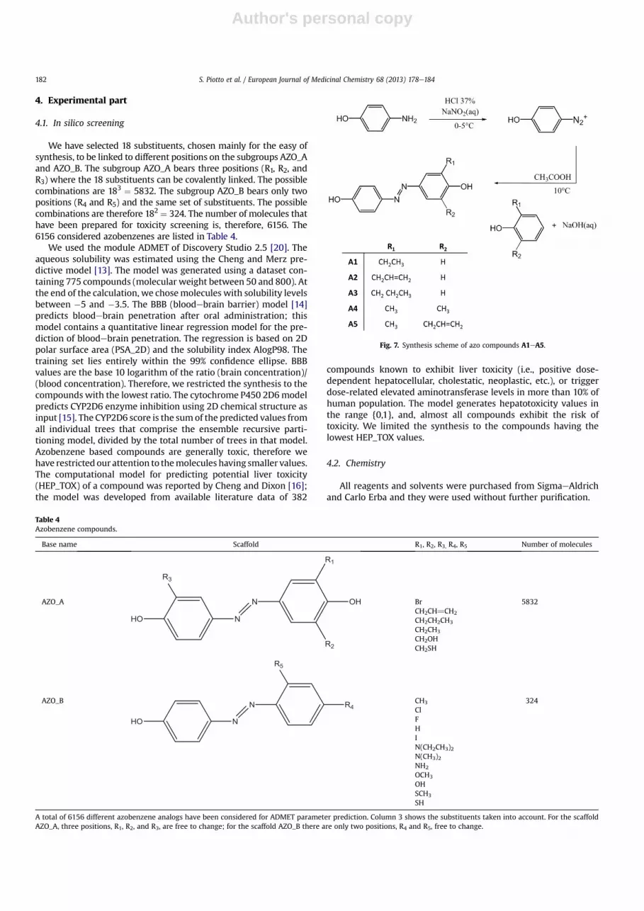
Fig. 7. Synthesis scheme of azo compounds A1eA5.
S. Piotto et al. / European Journal of Medicinal Chemistry 68 (2013) 178e184182

Author's personal copy
Azo compounds A1eA5 were synthesized according to theclassic scheme of diazocopulation reactions, as illustrated in Fig. 7.
The procedure was the following: 2.00 g of 4-aminophenol(0.0183 mol) was suspended in a solution containing 16 mL ofwater and 4mL of HCl 37% (w/w). The solutionwas cooled at 0e5 �Cin a watereice bath. A solution of 1.39 g of sodium nitrite(0.0202 mol) dissolved in 4 mL of water was added drop-wise,obtaining a suspension of the diazonium salt (solution A). Sepa-rately, a solution containing 1.68 g of NaOH (0.0420mol) in 16mL ofwater with 0.0183 mol of the proper phenol (depending on A1eA5)was prepared (solution B). Solution A was added drop-wise to so-lution B, under stirring at 12 �C. The system was left reacting for20min. Then the final solutionwas slowly added to 40mL of an acidsolution (50 mL of water and 4 mL of acetic acid), and then stirredfor 30 min at 15 �C. A dark red precipitate of the azo compoundformed. The crude precipitatewas filtered and dried under vacuum.Yields ranged between 30 and 40%.
4.2.1. 40-Hydroxy-(4-hydroxy-3-ethyl)-azobenzene (A1)The synthesis of compound A1 has been already reported in
Ref. [21]. 4-Aminophenol and 2-ethylphenol were used as startingreagents. 1H NMR (acetone-d6): d (ppm) ¼ 7.77 (d, 2H); 7.61 (s, 1H);7.58 (m, 1H); 6.95 (d, 2H); 6.93 (m, 1H); 2.64 (m, 2H); 1.20 (t, 3H).
4.2.2. 40-Hydroxy-(4-hydroxy-3-allyl)-azobenzene (A2)4-Aminophenol and 2-allylphenol were used as starting re-
agents. The crude product was extracted and crystallized fromboiling n-octane (500 mL) and dried. Final crystallization fromwater/ethanol (10:1) gave pure A2 as red-orange crystalline ma-terial. 1H NMR (acetone-d6): d (ppm) ¼ 7.78 (m, 2H); 7.71 (s, 1H);7.64 (d, 1H); 6.99 (m, 3H); 6.07 (m, 1H); 5.07 (m, 2H); 3.47 (m, 2H).
4.2.3. 40-Hydroxy-(4-hydroxy-3-propyl)-azobenzene (A3)4-Aminophenol and 2-propylphenol were used as starting re-
agents. The crude product was extracted and crystallized fromboiling n-octane (100 mL) and dried. Final crystallization fromboiling water/ethanol (3:1) gave pure A3 as gold yellow crystallinematerial. 1H NMR (DMSO-d6): d (ppm) ¼ 7.72 (d, 2H), 7.58 (d, 1H),7.54 (d, 1H), 6.92 (d, 2H), 2.57 (m, 2H), 1.61 (m, 2H), 0.94 (t, 3H).
4.2.4. 40-Hydroxy-(4-hydroxy-3,5-dimethyl)-azobenzene (A4)4-Aminophenol and 2,6-dimethylphenol were used as starting
reagents. The crude product was extracted and crystallized fromboiling n-octane (100 mL) and dried. Final crystallization fromboiling water/ethanol (3:1) gave pure A4 as orange crystallinematerial. 1H NMR (DMSO-d6): d (ppm) ¼ 7.71 (d, 2H), 7.48 (s, 2H),6.91 (d, 2H), 2.26 (s, 6H).
4.2.5. 40-Hydroxy-(4-hydroxy-3-methyl-5-allyl)-azobenzene (A5)4-Aminophenol and 2-allyl-6-methyphenol were used as
starting reagents. The crude product was extracted and crystallizedfrom boiling n-octane (100mL) and dried. After crystallization fromboiling water/ethanol (3:1), final crystallization from boiling watergave pure A5 as red crystalline material. 1H NMR (DMSO-d6):d (ppm)¼ d 7.72 (d, 2H), 7.49 (d, 2H), 6.91 (d, 2H), 6.00 (m, 1H), 5.10(t, J ¼ 12.0 Hz, 2H), 2.28 (s, 3H).
4.3. Thermal characterization
Thermalmeasurements were performed by a DSC-7 Perkin Elmercalorimeter under nitrogen flow at 10 �C/min rate. Polarized opticalmicroscopy was performed by a Jenapol microscope fitted with aLinkam THMS 600 hot stage. X-ray diffraction patterns on powdersamples of the polymers were recorded on a flat film camera Ni-filtered Cu-Ka radiation. The Fujifilm MS 2025 imaging plate and a
Fuji Bioimaging analyzer System, model BAS-1800, were used fordigitizing thediffractionpatterns.1HNMRspectrawere recordedwitha Bruker DRX/400 Spectrometer. Chemical shifts are reported relativeto the residual solvent peak (acetone-d6: H ¼ 2.05 ppm; dime-thylsulfoxide-d6:H¼2.50ppm).UVabsorption spectraof thesampleswere recorded at 25 �C in acetonitrile solution, on a PerkineElmerLambda 19 spectrophotometer. The spectral region 650e240 nmwasinvestigated by using cell path length of 1 cm. Azobenzene chromo-phore concentration of about 3.0 � 10�5 mol L�1 was used.
4.4. Bacterial strains andminimum inhibitory concentrations (MICs)
The in vitro minimum inhibitory concentrations (MICs) of eachcompound were determined against C. albicans SC5314 [22] by themicro-broth dilutionmethod in 96-wellmicrotest plates according tothe guidelines suggested by the Clinical and Laboratory StandardsInstitute (CLSI) [23], using three separate plates each containing thesame batch of azo compounds. Microtiter plates containing 100 mL oftwo-fold serial dilutions of azo compounds in RPMI 1640 medium(with L-glutamine, without glucose and NaHCO3, buffered to pH 7.0with 0.165M4-morpholinepropanesulfonic acid [MOPS] buffer)wereinoculated with 100 mL of cells containing 2.5 � 103 yeast/mL andincubated at 35 �C for 24 h. The resulting MICs were visually read asthe lowest concentration of compound causing an absence of growth(optically clear) in comparison to the drug-free growth control.
For S. aureusA170 (kindly provided byProf. R. Capparelli from theUniversity of Naples, Italy), Listeria monocytogenes [24], Salmonellatyphimurium [25], Pseudomonas aeruginosaATCC-27853,MIC valuesof each compound were determined by the serial broth micro-dilution method as reported by Patton [26]. Therefore, flat-bottompolystyrene microtiter plates containing 100 mL of two-fold serialdilutions (six replicates per dilution) of azo compounds were inoc-ulated with 100 mL of w5 � 105 CFU/mL of each bacteria grown inMuellereHinton broth 2 (cation-adjusted MHB; SigmaeAldrich,Milan, Italy). Control wells contained broth only (negative control)or bacteria and broth (positive control).
Plates were incubated at 37 �C with shaking at 160 rpm for 24 h.Datawere analyzed according to Patton et al. [26]. Briefly, the opticaldensity was determined just before the incubation (T0) and againafter 24 h incubation (T24) at 600 nm. The OD for each replicate at T0was subtracted fromtheOD foreach replicateatT24. The adjustedODof each control well was then assigned a value of 100% growth. Thepercent inhibition of growth was thus determined using the for-mula: percent inhibition ¼ 1 � (OD test well/OD of correspondingcontrol well)� 100. TheMIC is reported as the lowest concentrationof azo compounds which results in 100% inhibition of growth.
4.5. Candida morphological analysis
Hyphal growth of Candida treated cells was induced using RPMI1640 medium, supplemented with 2.5% fetal calf serum, 20 mMHEPES, 2 mM L-glutamine, and 16 mM sodium hydrogen carbonate(pH 7.0, Gibco-BRL). Stationary yeast cells were inoculated into afresh pre-warmed medium at a density of 6� 106 cells/mL in a flat-bottom 96 well microtiter plates. Different concentrations of azocompounds (ranging from 1 to 50 mg/mL) were added to each well.After incubation at 37 �C for 24 h, each microtiter plate wasexamined using an inverted microscope to monitor phenotypicmodification and hyphae formation.
4.6. Biofilms analysis
C. albicans cells were grown for 24 h at 28 �C in YPD broth (20 g ofpeptone, 10 g of yeast extract, 20 g of glucose per liter). These werewashed twice with sterile PBS (10 mM phosphate buffer, 2.7 mM
S. Piotto et al. / European Journal of Medicinal Chemistry 68 (2013) 178e184 183

Author's personal copy
potassium chloride, 137 mM sodium chloride, pH 7.4, Sigma), and re-suspended in RPMI 1640 supplemented with MOPS at 106 cells/mL.The cell suspension (200 mL) was seeded in pre-sterilized, poly-styrene flat-bottom 96-well plates and incubated for 24 h at 37 �C.After biofilm formation, 20 mg/mL of azo compounds were added toeach well and incubated 8 h at 37 �C. Afterward, the medium wasaspirated, and non-adherent cells were removed bywashing biofilms3 times with 200 mL of sterile PBS. Biofilms were quantified by the2,3-bis(2-methoxy-4-nitro-5-sulfophenyl)-2H-tetrazolium-5-carboxanilide (XTT) reduction assay [27]. Briefly, XTT (SigmaeAldrich, Milan, Italy) was dissolved in PBS at a final concentration of1 mg/mL. The solutionwas filter-sterilized using a 0.22 mm-pore-sizefilter and stored at �70 �C until required. Menadione (SigmaeAldrich, Milan, Italy) solution (0.4 mM) was also prepared andfiltered. Before each assay, XTT solution was thawed and mixed withmenadione solution at a volume ratio of 20O1. TheXTTemenadionesolution (250 mL) was then added to each well. The microtiter plateswere then incubated in the dark for 1 h at 37 �C. Following incuba-tion, 250 mL of the XTT-menadione solution was recovered andcentrifuged (to eliminate interference of cells with colorimetricreadings); 100 mL of the solution was transferred to new wells, andthe color change resulting from XTT reduction was measured at490 nm with a microtiter plate reader (LAB system multiscan EX).Each assay was performed 3 times.
A similar protocol has been used for the inhibition of Candidabiofilm by the azo compounds [28]. Briefly, following the adhesionphase of 100 mL of 2 � 106 cells/mL of C. albicans on flat-bottompolystyrene microtiter plates, wells were washed twice with150 mL of PBS to remove loosely adherent cells. The biofilms wereallowed to develop up to 24 h at 37 �C in the presence of differentconcentrations of azo compounds (ranging from 8 to 60 mg/mL). Forthe photographs, non-adherent cells were removed by washing,and adherent cells were stained with crystal violet (CV) 0.3% so-lution. Images were captured with a Canon EOS 450d.
In order to assess the S. aureus biofilm formation, bacteria weregrown overnight in MHB medium. Cultures were then diluted toapproximately 107 CFU/mL in fresh MHB medium, and 200 mL wasused to inoculate flat-bottom 96-well polystyrene microtiter platescontaining different concentrations of azo compounds (from 8 to60 mg/mL). After incubation for 24 h at 37 �C without shaking, theplate wells were washed twice with phosphate-buffered saline (pH7.2) to remove non-adherent bacteria and dried in an invertedposition. Finally, the adherent cells were stained with CV 0.3%.
Images were acquired using a Canon EOS 450d and processedwith Microsoft Office 2010.
Similarly, S. aureus (200 mL of 104 to 105 CFU/mL) was inoculatedin microtiter plate and incubated for 18e24 h at 35 �C to allow bio-film formation. Afterward, serial diluted solutions of azo compounds(20, 25, 30 and50 mg/mL)were added on eachwell and incubated 8hat 37 �C. For crystal violet staining, wells were rinsed with water toremove loosely adherent cells and then stained for 1minwith 200mLof Gram’s crystal violet.Wells were rinsedwithwater and dried. Theamount of biofilm mass was obtained by destaining the wells with200 mL of 33% acetic acid and then measuring the absorbance of theCV solution in a microplate spectrophotometer set at 595 nm.
Appendix A. Supplementary data
Supplementarydataassociatedwith thisarticlecanbe found in theonline version, at http://dx.doi.org/10.1016/j.ejmech.2013.07.030.
References
[1] WHO. Available from: http://www.who.int/mediacentre/factsheets/fs194/en/.
[2] H.J. Jung, Y. Seu, D. Lee, Candicidal action of resveratrol isolated from grapeson human pathogenic yeast C. albicans, Journal of Microbiology and Biotech-nology 17 (2007) 1324.
[3] P.K. Dadi, M. Ahmad, Z. Ahmad, Inhibition of ATPase activity of Escherichia coliATP synthase by polyphenols, International Journal of Biological Macromole-cules 45 (2009) 72e79.
[4] N. Chinnam, P.K. Dadi, S.A. Sabri, M. Ahmad, M.A. Kabir, Z. Ahmad, Dietarybioflavonoids inhibit Escherichia coli ATP synthase in a differential manner,International Journal of Biological Macromolecules 46 (2010) 478e486.
[5] K. Weber, B. Schulz, M. Ruhnke, Resveratrol and its antifungal activity againstCandida species, Mycoses 54 (2011) 30e33.
[6] L. Paulo, S. Ferreira, E. Gallardo, J.A. Queiroz, F. Domingues, Antimicrobialactivity and effects of resveratrol on human pathogenic bacteria, WorldJournal of Microbiology and Biotechnology 26 (2010) 1533e1538.
[7] Z. Ahmad, M. Ahmad, F. Okafor, J. Jones, A. Abunameh, R.P. Cheniya, I.O. Kady,Effect of structural modulation of polyphenolic compounds on the inhibitionof Escherichia coli ATP synthase, International Journal of Biological Macro-molecules 50 (2012) 476e486.
[8] S. Albert, R. Horbach, H.B. Deising, B. Siewert, R. Csuk, Synthesis and antimi-crobial activity of (E) stilbene derivatives, Bioorganic & Medicinal Chemistry19 (2011) 5155e5166.
[9] D. Acierno, E. Amendola, V. Bugatti, S. Concilio, L. Giorgini, P. Iannelli,S.P. Piotto, Synthesis and characterization of segmented liquid crystallinepolymers with the azo group in the main chain, Macromolecules 37 (2004)6418e6423.
[10] S. Concilio, V. Bugatti, P. Iannelli, S. Piotto, Synthesis and characterization ofnew photoluminescent oxadiazole/carbazole-containing polymers, Interna-tional Journal of Polymer Science 2010 (2010) 1e6.
[11] D. Attianese, M. Petrosino, P. Vacca, S. Concilio, P. Iannelli, A. Rubino, S. Bellone,Switching device based on a thin film of an azo-containing polymer for appli-cation in memory cells, Electron Device Letters, IEEE 29 (2008) 44e46.
[12] L. Angiolini, L. Giorgini, F. Mauriello, P. Rochon, Optically active methacryliccopolymers with side-chain azoaromatic and 9-phenylcarbazole moieties,Reactive and Functional Polymers 72 (2012) 1e10.
[13] A. Cheng, K.M. Merz, Prediction of aqueous solubility of a diverse set ofcompounds using quantitative structureeproperty relationships, Journal ofMedicinal Chemistry 46 (2003) 3572e3580.
[14] W.J. Egan, G. Lauri, Prediction of intestinal permeability, Advanced Drug De-livery Reviews 54 (2002) 273e289.
[15] R.G. Susnow, S.L. Dixon, Use of robust classification techniques for the pre-diction of human cytochrome P450 2D6 inhibition, Journal of Chemical In-formation and Computer Sciences 43 (2003) 1308e1315.
[16] A. Cheng, S.L. Dixon, In silico models for the prediction of dose-dependenthuman hepatotoxicity, Journal of Computer-Aided Materials Design 17(2003) 811e823.
[17] M.A. Brown, S.C. De Vito, Predicting azo dye toxicity, Critical Reviews inEnvironmental Science and Technology 23 (1993) 249e324.
[18] H. Mori, Y. Mori, S. Sugie, N. Yoshimi, M. Takahashi, H. Ni-i, H. Yamazaki,K. Toyoshi, G.M. Williams, Genotoxicity of a variety of azobenzene and ami-noazobenzene compounds in the hepatocyte/DNA repair test and the Sal-monella/mutagenicity test, Cancer Research 46 (1986) 1654e1658.
[19] H. Takahashi, T. Ishioka, Y. Koiso, M. Sodeoka, Y. Hashimoto, Anti-androgenicactivity of substituted azo-and azoxy-benzene derivatives, Biological andPharmaceutical Bulletin 23 (2000) 1387e1390.
[20] Accelrys, Discovery Studio 2.5. Available from: http://accelrys.com/products/discovery-studio/.
[21] S. Piotto, S. Concilio, L. Sessa, P. Iannelli, A. Porta, E.C. Calabrese, M.R. Galdiand,L. Incarnato, Novel antimicrobial polymer films active against bacteria andfungi, Polymer Composites (2013), http://dx.doi.org/10.1002/pc.22410.
[22] E.C. Calabrese, S. Castellano, M. Santoriello, C. Sgherri, M.F. Quartacci,L. Calucci, A.G. Warrilow, D.C. Lamb, S.L. Kelly, C. Milite, I. Granata,G. Sbardella, G. Stefancich, B. Maresca, A. Porta, Antifungal activity of azolecompounds CPA18 and CPA109 against azole-susceptible and -resistantstrains of Candida albicans, Journal of Antimicrobial Chemotherapy (2013).
[23] CLSI, Reference Method for Broth Dilution Antifungal Susceptibility Testing ofYeasts; Approved Standard e Third Edition, Clinical and Laboratory StandardsInstitute CLSI, Wayne, PA, 2008.
[24] S. Sayem, E. Manzo, L. Ciavatta, A. Tramice, A. Cordone, A. Zanfardino, M. DeFelice, M. Varcamonti, Anti-biofilm activity of an exopolysaccharide from asponge-associated strain of Bacillus licheniformis, Microbial Cell Factories 10(2011) 74.
[25] A. Porta, Z. Török, I. Horvath, S. Franceschelli, L. Vígh, B. Maresca, Geneticmodification of the Salmonella membrane physical state alters the pattern ofheat shock response, Journal of Bacteriology 192 (2010) 1988e1998.
[26] T. Patton, J. Barrett, J. Brennan, N. Moran, Use of a spectrophotometricbioassay for determination of microbial sensitivity to manuka honey, Journalof Microbiological Methods 64 (2006) 84e95.
[27] W. da Silva, J. Seneviratne, N. Parahitiyawa, E. Rosa, L. Samaranayake, A. DelBel Cury, Improvement of XTT assay performance for studies involvingCandida albicans biofilms, Brazilian Dental Journal 19 (2008) 364e369.
[28] Y. Jin, H. Yip, Y. Samaranayake, J. Yau, L. Samaranayake, Biofilm-formingability of Candida albicans is unlikely to contribute to high levels of oral yeastcarriage in cases of human immunodeficiency virus infection, Journal ofClinical Microbiology 41 (2003) 2961e2967.
S. Piotto et al. / European Journal of Medicinal Chemistry 68 (2013) 178e184184


