Embed Size (px)
Citation preview
LETTER TO THE EDITOR
Soluble factors derived from tumor mammary cell lines inducea stromal mammary adipose reversion in human and miceadipose cells. Possible role of TGF-b1 and TNF-a
Javier Guerrero Æ Nicolas Tobar Æ Monica Caceres ÆLorena Espinoza Æ Paula Escobar Æ Javier Dotor ÆPatricio C. Smith Æ Jorge Martınez
Published online: 1 August 2009
� Springer Science+Business Media, LLC. 2009
Abstract In carcinomas such as those of breast, pancreas,
stomach, and colon, cancer cells support the expansion of
molecular and cellular stroma in a phenomenon termed
desmoplasia, which is characterized by a strong fibrotic
response. In the case of breast tissue, in which stroma is
mainly a fatty tissue, this response presumably occurs at
the expense of the adipose cells, the most abundant stromal
phenotype, generating a tumoral fibrous structure rich in
fibroblast-like cells. In this study, we aimed to determine
the cellular mechanisms by which factors present in the
media conditioned by MDA-MB-231 and MCF-7 human
breast cancer cell lines induce a reversion of adipose cells
to a fibroblastic phenotype. We demonstrated that soluble
factors generated by these cell lines stimulated the rever-
sion of mammary adipose phenotype evaluated as intra-
cellular lipid content and expression of C/EBPa and
PPARc. We also demonstrated that exogenous TGF-b1 and
TNF-a exerts a similar function. The participation of both
growth factors, components of media conditioned by
tumoral mammary cells, on the expression and nuclear
translocation of C/EBPa and PPARc was tested in 3T3-L1
cells by interfering with the inhibitory effects of media
with agents that block the TGF-b1 and TNF-a activity.
These results allow us to postulate that TGF-b1 and TNF-apresent in this media are in part responsible for this phe-
notypic reversion.
Keywords Desmoplasia � Adipogenic transcription
factors � TGF-b1 � TNF-a
Introduction
It is currently accepted that tumoral progression not only
depends on the intrinsic malignancy of tumor cells but also
on the surrounding microenvironment composed of stromal
cells and the extracellular matrix (ECM) [1]. Moreover, it
has been described that tumor cells are able to modify the
adjacent stroma creating a functional structure that favors
tumor progression [2].
Breast tumors belong to a group of neoplastic lesions
which, under the influence of tumoral cell products, orig-
inate a fibrous structure responsible for the dense and hard
consistency of the tumoral mass. This trait also constitutes
a factor that increases the relative risk of tumor recurrence
[3]. Either in the functional origin or in the cellular com-
position of this fibrotic structure, myofibroblasts are the
predominant stromal phenotype which secretes abundant
amount of collagen and other extracellular matrix (ECM)
proteins through an active phenomenon known as ‘‘Des-
moplastic Response’’ [4]. Some of the changes in the
composition of ECM, derived from the desmoplastic
response, induce changes in breast density that has been
identified as a predisposing factor for breast cancer that
J. Guerrero � N. Tobar � M. Caceres � J. Martınez (&)
Laboratory of Cellular Biology, INTA, University of Chile,
Casilla 138, Santiago 11, Chile
e-mail: [email protected]
L. Espinoza � P. Escobar
East Metropolitan Health Service, Dr. Luis Tisne, Santiago,
Chile
J. Dotor
Digna Biotech, Madrid, Spain
P. C. Smith
Laboratory of Periodontal Physiology, Faculty of Medicine,
Pontifical Catholic University of Chile, Santiago, Chile
123
Breast Cancer Res Treat (2010) 119:497–508
DOI 10.1007/s10549-009-0491-1

confers a higher risk compared with women with fatty
breast [5].
The desmoplastic reaction has been attributed to fibro-
blast-like cells which may derive from adipose tissue in
mammary tumors [6]. Fibroblast accumulation can occur
due to the activity of specific growth factors produced by
tumoral cells. Using 3T3-L1 cells and human adipose
fibroblasts, it has been demonstrated that TNF-a and IL-11
derived from human tumor mammary cell lines inhibit
adipocyte differentiation, a previous step to expansion of
fibroblastic compartment without modifying fibroblast
proliferation [6].
On the other hand, it has been also proposed that
Platelet-derived Growth Factor (PDGF) is a paracrine
factor produced by mammary cells whose primary target
are stromal cells and which is considered the initiator of
tumor desmoplasia [4]. Finally, it has been suggested that
TGF-b1 may be one of the main factors involved in des-
moplastic response. In breast cancer TGF-b1 is highly
expressed, preferentially at the advancing edges of primary
tumors and in lymph node metastasis, suggesting that it
plays a role in interacting with neighboring stromal cells
[7].
Epithelial control of mammary adipose cells is observed
under physiologic conditions as well. During pregnancy
and lactation, reproductive hormones induce the expansion
and terminal differentiation of the mammary epithelium
into secretory, milk-producing, lobular alveoli in a process
that also includes the dedifferentiation of adipocytes into
tiny preadipocytes [8].
CCAAT/enhancer binding proteins (C/EBPs) and per-
oxisome proliferator-activated receptor-gamma (PPARs)
are two families of transcription factors that play a critical
role in either the onset of adipocyte differentiation or the
maintenance of the fully differentiated adipocyte pheno-
type by transactivating adipocyte specific genes [9].
Inhibiting PPARc activity, either in 3T3-L1 adipocytes or
in the adipose tissue of mice was shown to lead to dedif-
ferentiation or adipocyte death [10]. It has also been
demonstrated that C/EBPa is a well-characterized factor
that mediates the expression of genes characteristic of the
terminally differentiated state and also acts as inhibitor of
mitotic growth in most cell lines tested [11].
In the present work, we attempt to unravel some of the
cellular mechanisms of mammary desmoplastic response
by evaluating whether cell-secreted soluble factors pro-
duced by human mammary cell lines (MCF-7 and MDA-
MB-231) are able to induce reversion of mature mammary
adipocytes of human and mouse origin to the fibroblastic
phenotype, in addition to the well-known inhibitory role on
adipose differentiation. To accomplish this, we used human
mammary-derived adipocyte and fully differentiated mur-
ine 3T3-L1 cells to identify the main factors secreted by
the cell lines responsible for the reversion process and the
impact of these factors on the expression and abundance of
PPARc and C/EBPa. Our results, obtained from cell culture
studies and immunohistochemical analysis of human tumor
samples, demonstrated that TGF-b1 and TNF-a of epithe-
lial origin are in part responsible for the adipocyte rever-
sion and that PPARc and C/EBPa expression seem to be a
molecular target of these tumoral factors.
Materials and methods
Antibodies and reagents
Antibodies anti-PPARc and C/EBPa were obtained from
Santa Cruz Biotechnology, (Santa Cruz, CA). Anti-b-actin
was supplied by Sigma (St. Louis, MO), anti-TGF-b was
from Chemicon International, Inc. (Temecula, CA), anti-
human TNF-a (neutralizing of TNF-a) was obtained from
R&D System, Inc. (Minneapolis, MN). The P144 peptide
(TSLDASIIWAMMQN, encompassing aminoacids 730–
743) interfering with TGF-b1 binding to its cellular
receptors [12] was produced by Digna Biotech (Madrid,
Spain). Recombinant TNF-a and TGF-b were purchased
from R&D systems and Calbiochem (La Jolla, CA),
respectively. Insulin, Dexamethasone, isobutylmethylxan-
thine (IBMX), Indomethacin, and Type I collagenase were
purchased from Sigma Chemical Co. Dulbecco’s Modified
Eagle’s Medium (DMEM), fetal bovine serum (FBS), and
bovine serum (BS) were purchase from GibcoTM (Grand
Island, NY).
Patients and normal tissue acquisition
Eight patients undergoing surgical mammary reduction at
the East Metropolitan Health Service (Santiago, Chile)
were recruited for the study. The experimental protocols
were previously approved by institution ethics committee
and all patients gave their informed consent for the pro-
cedure. All subjects were healthy and had no evidence of
diabetes according to routine laboratory tests. Mammary
adipose tissue biopsies were obtained at the time of surgery
and immediately transported to the laboratory in sterile
DMEM for processing.
Adipocyte isolation and three-dimensional
collagen gel culture system
Samples of mammary adipose tissue were minced finely
and digested in a collagenase solution (0.25 mg/ml Type I
collagenase, 3% bovine serum albumin in DMEM) with
shaking at 37�C for 1 h. The resultant digested material
498 Breast Cancer Res Treat (2010) 119:497–508
123

was then centrifuged at 1,500g for 5 min and the super-
natant was washed three times with 3% BSA in DMEM.
Under these experimental conditions, a floating layer that
contains only very low density cells (that we identify as
unilocular mature adipocytes) was generated. This cellular
fraction was extensively washed three times with HBSS.
The remaining more dense phenotypes present in the breast
sample, sediment in the cellular pellet.
A collagen gel solution was prepared as previously
described [13]. Briefly, 8 volumes of type I rat tail collagen
was mixed with 1 volume of tenfold concentrated DMEM
and 1 volume of reconstruction buffer (2.2 g of NaHCO3
and 4.77 g of HEPES in 100 ml of 0.05 N NaOH at 4�C) to
obtain a homogeneous solution. The collagen solution was
mixed with the purified mature adipocytes at room tem-
perature and 1 ml of collagen gel solution containing
nearly 105 cells was placed in a 35 mm culture dish. The
culture dishes were immediately warmed to 37�C to allow
the gel to form and were covered with 2 ml of DMEM
overnight. Afterward, cultures were treated with increasing
concentrations of conditioned media (CM) from mammary
tumoral cells lines MCF-7 and MDA-MB-231 for 10 days.
Collagen was obtained from rat tail tendon as previously
described [14].
Mammary cell lines and preparation
of conditioned media
Mammary tumoral cells lines MCF-7 and MDA-MB-231
cells were purchased from ATCC (Manassas, VA) and
were grown in a phenol red-free DMEM/F12 enriched with
10% FCS. To prepare conditioned media, approximately
105 cells/cm2 of MCF-7 and MDA-MB-231 were cultured
in standard conditions and further incubated for 48 h in
serum-free DMEM. After this, media were collected and
centrifuged for 5 min at 2,000 rpm to clarify.
Oil red O staining
The intracellular lipid content was evaluated with the
lipophilic dye Oil Red O [15]. To do so, media was
removed and cells in culture were fixed and dehydrated for
5 min with 2 ml of 100% isopropanol. Samples were then
stained for 1 h with 2 ml of a saturated solution of Oil Red
O in 60% (v/v) isopropanol and then washed twice with
distilled water for 15 min.
ELISA assay
To determine TGF-b and TNF-a concentrations in mam-
mary tumoral cell conditioned media, TGF-b (559119) BD
Bioscience (San Diego, CA) and TNF-a (T916008) US
Biological (Swampscott, MA) ELISA kits were used. The
samples (in quadruplicate) were processed strictly follow-
ing the supplier’s instructions. At the end of the procedure,
absorbance was immediately determined at a wavelength
of 450 nm for TGF-b and 405 nm for TNF-a, with cor-
rections at 570 and 650 nm, respectively. TGF-b and TNF-
a concentrations were calculated using standard curves
prepared for each individual determination.
Immunohistochemistry
Immunostaining for TGF-b, TNF-a, PPARc, and C/EBPawas performed on 5 lm sections of formalin fixed, paraf-
fin-embedded human mammary tissue biopsies. Tissue
sections were deparaffinized in xylene and hydrated in a
graded sequence of ethanol solutions. For antigen retrieval,
sections were pretreated in a microwave in citrate buffer
(pH 6.0). After cooling, nonspecific binding was blocked
with diluted serum (4% normal goat serum) followed by
incubation with antibodies against TGF-b (1:300), TNF-a(1:150), PPARc (1:50), and C/EBPa (1:2,000) at room
temperature in a humidified chamber. Negative controls
were analyzed on adjacent sections incubated without pri-
mary antibody. After incubation with the primary antibody,
sections were washed with PBS and subsequently treated
using the corresponding biotinylated secondary antibody
from Ultravision ONE kit, Thermo Fisher Scientific (TL-
015-HDJ, Fremont, CA) according to the manufacturer’s
protocol. Peroxidase activity was visualized using the 3,30-diaminobenzidine and sections were counterstained with
haematoxylin. Assessment of staining intensity and distri-
bution was made using the semiquantitative histologic score
(HSCORE) system as described by Budwit-Novotny et al.
[16]. This score was calculated in four control and nine tumor
samples using the following equation: HSCORE = RPi
(i ? 1), where i = intensity of staining with a value of 1, 2,
or 3, (weak, moderate or strong, respectively) and Pi the
percentage of stained mammary adipose cells for each
intensity, varying from 0 to 100%. This HSCORE has been
previously reported [17, 18].
Adipocyte 3T3-L1 differentiation
3T3-L1 cells were purchased from ATCC (Manassas,
VA) and cultured in DMEM media supplemented with
10% BS, 100 lg/ml streptomycin and 100 units/ml of
penicillin in a humidified atmosphere with 5% CO2 at
37�C. Two days after reaching confluence, the induction
of adipocyte differentiation was started by culturing
for 2 days with adipogenic medium containing 1 lM
Dexamethasone, 0.5 mM IBMX, and 400 nM insulin
Breast Cancer Res Treat (2010) 119:497–508 499
123

solution in DMEM 10% FBS. For the following 8 days,
cells were incubated with DMEM 10% FBS containing
400 nM insulin alone. During this period, media was
replaced every 2 days.
Western blots
Mammary mature adipocyte cells or differentiated adipo-
cyte 3T3-L1 cells were lysed with a lysis buffer (50 mM
Hepes, pH 7.4, 150 mM NaCl, 2 mM MgCl2, 2 mM
EGTA, 1% Triton X-100, 10% glycerol, 2 mM PMSF,
2 lg/ml pepstatin, 2 lg/ml leupeptin and 1 mM sodium
orthovanadate) at 4�C. Equal amounts of proteins from
different treatments were resolved by SDS–PAGE and
analyzed by immunoblotting with antibodies anti-PPARc,
C/EBPa using the ECL chemiluminescence detection kit
(Amersham, Arlington Heights, FL).
Immunofluorescence
3T3-L1 cells were plated on cover slips in 24-well plates
and differentiated as described above. After the treat-
ment with mammary cell CMs and inhibitors (neutral-
izing Anti-TNF-a and P144), cells were washed in PBS
with 1 mM Ca?2, fixed with 4% paraformaldehyde for
20 min and permeabilized with 0.25% Triton X-100 for
10 min at RT. Samples were blocked with 10% goat
serum in PBS for 1 h and then co-incubated at 4�C
overnight with the primary antibodies in blocking solu-
tion: monoclonal anti-PPARc (1:50) and polyclonal anti-
C/EBPa (1:100). The cover slips were washed in PBS
and incubated with secondary antibodies (all from
Molecular Probes) diluted 1:100 in blocking solution:
Alexa 488-anti mouse, Alexa 546-anti rabbit at room
temperature for 2 h. Cover slips were washed in PBS
and water. Finally, samples were mounted in fluores-
cence mounting media (Dako).
Adipocyte size image analysis
Sections of 5 lm of paraffin-embedded human mammary
tissue obtained from adjacent regions of both normal and
tumoral samples were stained with haematoxylin and eosin
to evaluate adipocyte size as described by Chen et al. [19],
with some modifications. Briefly, images of the histology
sections were obtained in a jpg format at 109 magnifica-
tion and were converted into a binary format (8-bit) with
Adobe PhotoShop 5.0 (Adobe Systems, San Jose, CA) and
Image J (National Institutes of Health, http://rsbweb.nih.
gov/ij/). The binary images were compared with the
original images to ensure an accurate conversion. For the
determination of the total number and cross-sectional areas
of adipocytes, the commands Image/Adjust/Threshold and
then Analyze/Analyze Particules were used. Adipocyte
cross-sectional areas were expressed as pixel number, and
the results saved as an Excel document for analysis.
Results
Media conditioned by human mammary tumoral
cells revert to mammary adipose phenotype
To test whether soluble factors produced by human tumoral
mammary cells affect the lipid content of mammary adi-
pose tissue, we seeded recently dispersed human mammary
fat cells into a semisolid collagen solution and cultured
them in the presence or absence of media conditioned
(MC) by the weakly invasive MCF-7 and the strongly
invasive MDA-MB-231 mammary cells.
After 10 days, cells were stained with Red Oil O and the
proportion of fibroblast-like cells was scored. Figure 1A
shows a representative image of human adipose mammary
cells exposed to 50% MC by MDA-MB-231. As displayed
in this image, a high proportion of mammary fatty cells lost
their lipid content and acquired a fibroblast shape. Fig-
ure 1B shows the quantitative analysis of these experi-
ments using a range of concentrations of MC derived from
MDA-MB-231 and MCF-7 cells. This data indicated that
MC derived from MDA-MB-231 cells was more effective
in reverting the adipose phenotype to spindle-shaped cells
(Fig. 1B).
It has been proposed that TGF-b1 and TNF-a, two sol-
uble factors produced in a high proportion by tumoral cells,
also play a role in the control of cellular lipid content
[6, 20]. To test whether these factors induce lipid loss in
our system, we assessed their effectiveness in the reversion
of adipose phenotype and found that both factors are
capable of generating a conversion of adipose cells into
fibroblasts (Fig. 1C).
TGF-b1 and TNF-a are differentially expressed
in human mammary cell lines
Next, we analyzed the presence of TGF-b1 and TNF-a in
MC by MCF-7 and MDA-MB-231 cells using an ELISA
assay. Figure 2 shows that while both cell lines produced a
fairly similar amount of TNF-a, the more invasive MDA-
MB-231 cells produced five times more TGF-b1 than the
less invasive MCF-7 line.
500 Breast Cancer Res Treat (2010) 119:497–508
123

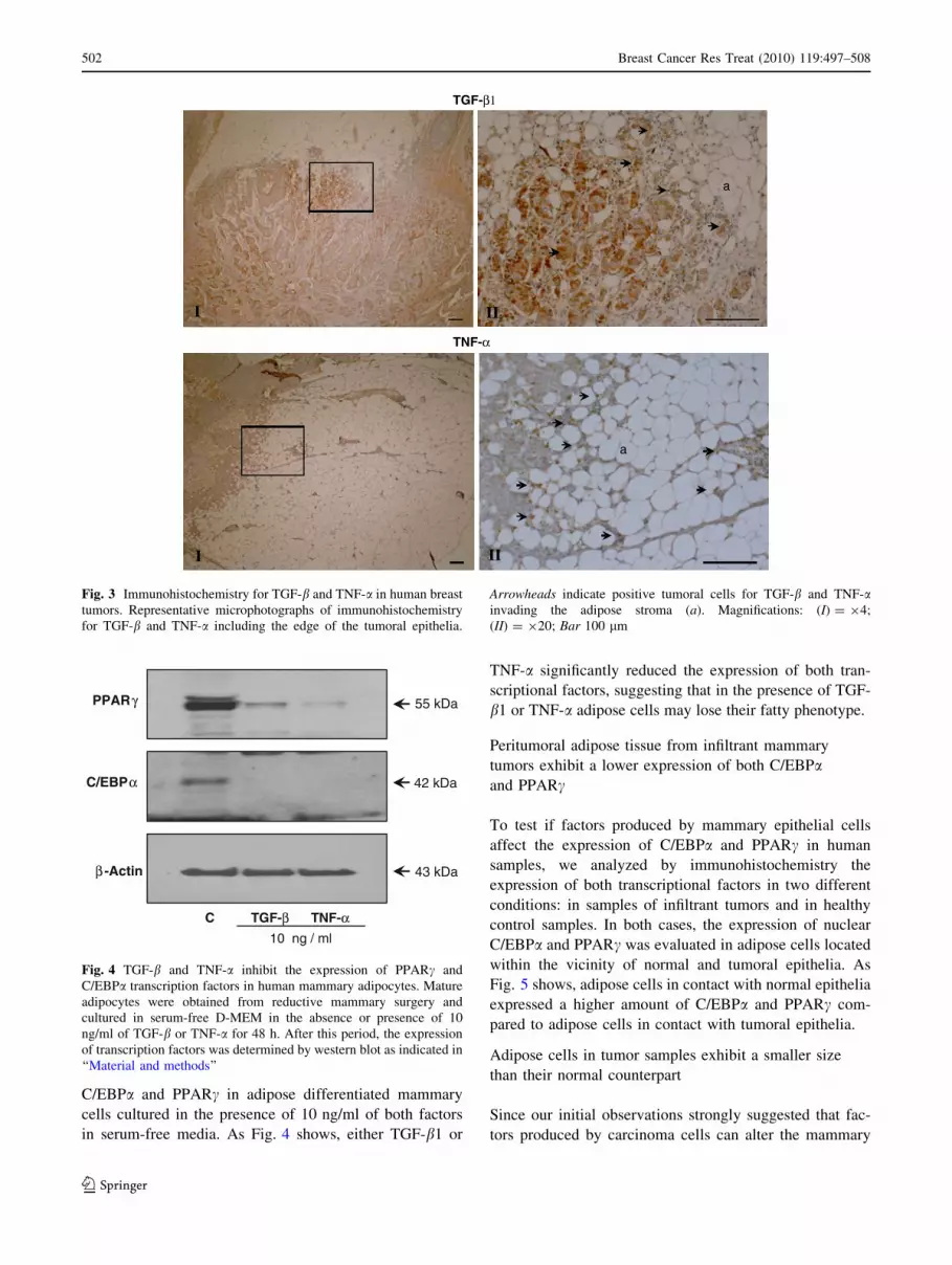
TGF-b1 and TNF-a are expressed in human
mammary tumors
To evaluate the cellular origin and distribution of TGF-b1
and TNF-a in mammary tumor samples, we analyzed by
immunohistotochemistry the presence of these factors in
samples of human ductal infiltrant mammary tumors. As
Fig. 3 shows, both factors were expressed only by tumoral
cells located at the invasive front without a relevant
expression in the stromal adipose cells.
TGF-b1 and TNF-a inhibit the expression
of transcriptional factors involved in the maintenance
of adipose phenotype
The maintenance of the adipose phenotype largely depends
on the expression of specific transcription factors such as C/
EBPa and PPARc, which are involved in the maintenance of
adipose homeostasis [21]. To assess whether the expression
of these transcriptional factors are sensitive to TGF-b1 and
TNF-a, we analyzed by western blotting, the expression of
0
10
20
30
40
50
%MC MCF-7 %MC MDA-231Control 25 50 25 50
* *
**
Control TGF-β TNF-α
% F
ibro
bla
sts-
like
cells
0
10
20
30
40
50
% F
ibro
bla
sts-
like
cells
a
a
a
I
bb
b
b
II
(A)
(B) (C)
Fig. 1 Adipocyte reversion in three-dimensional collagen gel culture.
A 105 human mature mammary adipocytes were cultured in 1.5 ml
semisolid collagen gel for 10 days in the absence (I) or presence (II)of 50% medium conditioned by MDA-MB-231 cells. Cells were
stained with oil red O that identified lipid content in mature spherical
adipocytes (a) and elongated cells with a fibroblast phenotype (b). BQuantification of fibroblast-like cells in cultures treated with
increasing proportions of media conditioned by mammary tumoral
cells MCF-7 and MDA-MB-231. C Quantification of fibroblast-like
cells in collagen matrix cultures treated for 10 days with 10 ng/ml of
TGF-b and TNF-a. Bars represent mean ± SE from five independent
experiments. A Kruskal–Wallis one-way ANOVA followed by a
Dunn’s post hoc analysis was used to determine significant differ-
ences from control. * P \ 0.05; ** P \ 0.001
0
50
100
150
200
250
300
350
400
450
MCF-7 MDA-MB-231
(pg
*ml-1
CM
/106
cells
/48h
)
0
5
10
15
20
25
30
35
40
45
50
(pg
*ml-1
CM
/106
cells
/48h
)
MCF-7 MDA-MB-231
TGF–β1 TNF–α
Fig. 2 Quantification of TGF-b and TNF-a in mammary cancer cell
conditioned media. Samples of media conditioned by mammary
tumoral cell lines MCF-7 and MD-MDA-231 were collected and
immediately quantified by an ELISA immunoassay for TGF-b and
TNF-a as described under ‘‘Materials and methods’’. Data represent
the mean ± SE
Breast Cancer Res Treat (2010) 119:497–508 501
123
C/EBPa and PPARc in adipose differentiated mammary
cells cultured in the presence of 10 ng/ml of both factors
in serum-free media. As Fig. 4 shows, either TGF-b1 or
TNF-a significantly reduced the expression of both tran-
scriptional factors, suggesting that in the presence of TGF-
b1 or TNF-a adipose cells may lose their fatty phenotype.
Peritumoral adipose tissue from infiltrant mammary
tumors exhibit a lower expression of both C/EBPaand PPARc
To test if factors produced by mammary epithelial cells
affect the expression of C/EBPa and PPARc in human
samples, we analyzed by immunohistochemistry the
expression of both transcriptional factors in two different
conditions: in samples of infiltrant tumors and in healthy
control samples. In both cases, the expression of nuclear
C/EBPa and PPARc was evaluated in adipose cells located
within the vicinity of normal and tumoral epithelia. As
Fig. 5 shows, adipose cells in contact with normal epithelia
expressed a higher amount of C/EBPa and PPARc com-
pared to adipose cells in contact with tumoral epithelia.
Adipose cells in tumor samples exhibit a smaller size
than their normal counterpart
Since our initial observations strongly suggested that fac-
tors produced by carcinoma cells can alter the mammary
TGF-β1
a
III
TNF-α
I II
a
Fig. 3 Immunohistochemistry for TGF-b and TNF-a in human breast
tumors. Representative microphotographs of immunohistochemistry
for TGF-b and TNF-a including the edge of the tumoral epithelia.
Arrowheads indicate positive tumoral cells for TGF-b and TNF-ainvading the adipose stroma (a). Magnifications: (I) = 94;
(II) = 920; Bar 100 lm
C TGF-β TNF-α
β -Actin
C/EBPα
PPARγ
43 kDa
42 kDa
55 kDa
10 ng / ml
Fig. 4 TGF-b and TNF-a inhibit the expression of PPARc and
C/EBPa transcription factors in human mammary adipocytes. Mature
adipocytes were obtained from reductive mammary surgery and
cultured in serum-free D-MEM in the absence or presence of 10
ng/ml of TGF-b or TNF-a for 48 h. After this period, the expression
of transcription factors was determined by western blot as indicated in
‘‘Material and methods’’
502 Breast Cancer Res Treat (2010) 119:497–508
123

phenotype, we reasoned that as a final outcome of this
phenomenon, adipose cells within the vicinity of tumors
should contain fewer lipids than their normal counterparts.
Figure 6b represents the cumulative frequency of cell
surface in cross-sectional areas in both types of tissue
samples. Curves show that values representing the lower
surface area were more frequent in tumoral samples. For
example, sixty percent of adipose cells derived from nor-
mal tissue display a size with a higher limit of 150 UR. On
the contrary, the same proportion of cells from the tumoral
sample shows a size below 45 UR. Moreover, on average,
tumoral cells also display a smaller size than their coun-
terparts in the normal sample (Fig. 6c).
Media conditioned by mammary cell lines reverse
the expression of C/EBPa and PPARc in differentiated
3T3-L1 cells
Human adipose cells isolated from mammary tissues fre-
quently suffer lysis when exposed to MC from mammary
C/EBP
aa
t
PPAR
aa
I
I II
II
(A)
(B)
0
1
2
3
4
Normal Tumoral
HS
core
0
1
2
3
4
Normal Tumoral
HS
core
PPAR C/EBP
* *
γ
α
γ α
Fig. 5 Immunohistochemistry for PPARc and C/EBPa in normal and
tumoral mammary samples. A Representative microphotographs of
immunohistochemistry for nuclear PPARc and C/EBPa in adipose
stromal tissue adjacent to normal (I) and tumoral (II) mammary
tissues. Positive and negative immunoreactive cells are indicated by
black or white arrowheads, respectively. Magnification 940; Bar
100 lm. B H-SCORE for staining of PPARc and C/EBPa in normal
and tumoral adipocytes from mammary samples. Assessment of
staining intensity and distribution was made using the semiquantita-
tive histological score calculated as described in ‘‘Material and
methods’’. (a) adipose cells; (t) tumor. A Mann–Whitney test analysis
was used to determine significant differences. * P \ 0.05
Breast Cancer Res Treat (2010) 119:497–508 503
123

cancer cell lines while cultured in cell suspension. To avoid
this consequence, we assessed the potential of MCs MCF-7
and MDA-MB-231 cells to reverse the adipose phenotype
in 3T3-L1 murine adipose cells, a standardized cellular
system frequently used to study the adipose differentiation
process.
Figure 7a shows that after 10-days culturing with an
adipogenic media, 3T3-L1 cells strongly expressed either
PPARc or C/EBPa at the protein level. Further treatment
for 5 days with increasing concentrations of MC from
MCF-7 and MDA-MB-231 cells decreased the expression
of these transcriptional factors in a dose-dependent manner.
To test if this reduction of expression of transcriptional
factors was associated with their functional nuclear loca-
tion, we utilized a similar set of adipose cells to perform an
immunofluorescence experiment. Figure 7b shows that MC
from human mammary cell lines strongly reduces the
nuclear expression of PPARc C/EBPa. In both types of
experiments, MC from MDA-MB-231 cells exert a clearly
more potent effect on the expression of both transcriptional
factors.
Inhibitors of TGF-b1 and TNF-a block the effects
of CM by breast tumoral cell lines
To identify whether the presence of TGF-b1 and TNF-a in
MC from breast tumoral cells is responsible for the
diminished expression of adipose transcriptional factors,
we inhibited the action of both factors using a blocking
antibody against TNF-a and a betaglycan-derived soluble
peptide that inhibits the binding of TGF-b1 to its receptor
[12]. In a similar manner to previous experiments, we
assayed the inhibitory capacity of these two agents by
measuring the expression of PPARc and C/EBPa with
western blotting and immunofluorescence. Figure 8a shows
that the addition of both inhibitors to cell culture contained
MC by MDA-MB-231 cells, effectively counteracted the
inhibitory effect of conditioned media enhancing the
expression of PPARc C/EBPa. On the other hand, Fig. 8b
shows by immunofluorescence that the expression and
nuclear location of PPARc and C/EBPa, initially inhibited
by the breast cell line CM, partially recovered their adipose
characteristic pattern. These results strongly suggest that
TGF-b1 and TNF-a are, in fact, two components of the CM
that are able to modify the adipose phenotype in 3T3-L1
cells.
Discussion
Invasion into neighboring tissue is a crucial event for
cancer progression and a prerequisite to metastasis [22].
During epithelial carcinogenesis, an active molecular
cross-talk is established among cancer and stromal cells.
As a result, functional changes produced in stroma may
contribute to cancer invasion [23]. In human breast cancer,
0
10
20
30
40
50
60
70
80
90
100
5 45 85 125
165
205
245
285
325
365
405
445
485
525
565
Cell Size (UR)
% o
f T
ota
l Cel
ls
Normal
Tumoral
Nomal Tumoral(x102)
0
3000
6000
9000
12000
15000
Cel
l Siz
e (U
R)
I II
(A)
(B) (C)
Fig. 6 Determination of
adipocyte cells size in
histological sections of normal
and tumoral mammary tissue.
a Hematoxylin/eosin stained
sections of a normal (I) and
infiltrating tumor mammary
tissue (II) showing the area
located in the vicinity of
epithelia. Magnification: 920,
bar 50 lm. b Cumulative
frequency of mammary adipose
cells surface area in normal and
tumoral tissue. Cell surface area
was expressed in relative units
according to ‘‘Material and
methods’’. Dotted line shows
that a 60% of adipose cells from
tumoral sample have a size
below 45 UR. In normal tissue
this proportion of cells reaches a
size around 150 UR. c Mean
surface area of adipocytes
derived from normal and
tumoral tissue evaluated
as in (b)
504 Breast Cancer Res Treat (2010) 119:497–508
123

the main stromal change observed during cancer progres-
sion is a fibrotic response that has been identified as a
predictor of a poor prognosis [3].
In the present work, we proposed that part of this fibrotic
response is attributed to a dedifferentiation phenomenon.
Under the effect of soluble factors produced by carcinoma
cells, mammary adipocytes, the main component of the
breast stroma, revert their phenotype to one in which
fibroblast-like cells predominate. Our results show that
mature mammary human adipocytes cultured in the pres-
ence of a media conditioned by tumoral mammary cell
lines (MCF-7 and MDA-MB-231) reverted to a fibroblast-
like phenotype in which fat accumulation is markedly
diminished. This finding is expected to represent a dedif-
ferentiation phenomenon because in these experimental
conditions (culture in semisolid conditions) cell prolifera-
tion does not occur in normal cells. Our data also dem-
onstrated that TNF-a and TGF-b1 produced by mammary
0,0
0,2
0,4
0,6
0,8
1,0
C 10 25 50 10 25 50
Rel
ativ
e In
ten
sity
0,0
0,2
0,4
0,6
0,8
1,0
C 10 25 50 10 25 50
Rel
ativ
e In
ten
sity
%CM MCF- 7 %CM MDA -231
PPARγ
C/EBPα
Control
PPAR C/EBP Merge
CM MDA -231
CM MCF -7
%CM MCF - 7 %CM MDA -231
PPARγ
C/EBPα
* *
*
*
β-actin
γ α
(A)
(C)
(B)
C 10 25 50 10 25 50
Fig. 7 Media conditioned by tumoral mammary cells inhibit the
expression of PPARc and C/EBPa in differentiated 3T3-L1 adipo-
cytes. a PPARc and C/EBPa expression was evaluated by western-
blot in 3T3-L1 cells cultured during 10 days in adipogenic medium
followed by the exposure for 5 days to increasing concentrations of
conditioned media derived from mammary tumoral cells lines MCF-7
and MDA-MB-231. Representative blots obtained from three
different experiments. b Quantitative analysis of PPARc and C/EBPaexpression normalized against b-actin as a loading control. Data are
presented as fold change over control. Bars represents mean ± SE.
* P \ 0.05. c Shows an immunofluorescence for nuclear PPARc(green) and C/EBPa (red) in cells cultured in the presence of CM
derived from mammary cells. (Color figure online)
Breast Cancer Res Treat (2010) 119:497–508 505
123

cell lines are two candidates potentially responsible for this
phenotypic change. Both factors were produced by these
cell lines and were expressed in the human breast tumoral
samples evaluated in this study.
To evaluate whether the intrinsic invasive capacity of
mammary cell lines was correlated with their potential to
revert to the adipose phenotype, we compared this activity
in MCF-7 cells, a weakly invasive cell line, and in MDA-
MB-231, a highly invasive cell line. From our results, two
important conclusions arose. First, media conditioned by
MDA-MB-231 cells behave more actively than media
from MCF-7 cells in terms of reversion of the adipose
phenotype. Second, while the TNF-a content in media
conditioned by both cell lines was similar, the concen-
tration of TGF-b1 in the invasive cell lines is five times
higher than that of MCF-7. This data allow us to suggest
that some of the differential effects in the conversion to a
fibroblast phenotype found with MDA-MB-231 condi-
tioned medium are mainly due to the autocrine production
of TGF-b1.
(A)
CM MDA - 231
N-TNF-α
P144
PPARγ
+
C/EBPα
β−actin
+
+
++
(B)
Control
CM MDA - 231+
P144
CM MDA - 231+
N- TNF - α
CM MDA - 231
PPARγ C/EBPα Merge
Fig. 8 TGF-b and TNF-apresent in media conditioned by
MDA-MB-231 cells are
responsible for the inhibition of
expression of PPARc and C/
EBPa in differentiated 3T3-L1
adipocytes. a Western-blot of
PPARc and C/EBPa from 3T3-
L1 adipose cells prepared as in
Fig. 7 and exposed for 5 days to
a 50% medium conditioned by
MDA-MB-231 cells in the
presence of either 0.6 lg/ml of
blocking antibodies against
TNF-a or a 100 lg/ml of P-144.
b Immunofluorescence for both
transcription factors in cells
treated in the same conditions as
in (a). PPARc (green) and
C/EBPa (red). Magnification
940. (Color figure online)
506 Breast Cancer Res Treat (2010) 119:497–508
123

TGF-b1 is a well-described cancer cell-derived growth
factor, abundantly expressed in tumoral cells, that directly
stimulates the transdifferentiation of stromal fibroblasts
into myofibroblasts and strongly stimulates desmoplasia in
a model of human pancreatic carcinoma [24, 25]. The
participation of TGF-b1 in fat accumulation has been
previously demonstrated in an in vivo model in which the
overexpression of TGF-b1 induces a strong fibrosis in
some tissues and also a severe reduction in body fat [26].
Our results show that TGF-b1 not only induces a reduced
fat content in mammary adipose cells, but also a marked
decreased expression of PPARc and C/EBPa, two of the
most important adipogenic transcriptional factors. In fact, it
has been proposed that, within the concept of microenvi-
ronmental niche, the balance between TGF-b1 and PPARcdefines the fate of the adipose or myofibroblasts differen-
tiation pathway [27]. Accordingly, in samples of human
mammary tumors, stromal cells located within the vicinity
of the epithelial edge that express a higher amount of TGF-
b1 and TNF-a (Fig. 2) also display a lesser proportion of
stain for nuclear PPARc and C/EBPa (Fig. 5). Our results
also showed that around the tumoral cells, which represent
the main source of antiadipogenic growth factors, also a
reduction in the size of the neighbor adipose cells is
observed (Fig. 6). A reduction in fat mass is associated
with a decrease in the size of adipocytes [28]. We suggest
that diminution of size of adipose cells in tumoral samples
is the consequence of an external (tumoral) stimulus that
impairs the expression of PPARc and C/EBPa which, in
turn, diminishes the adipose accretion in these cells.
The confirmation that TGF-b1 and TNF-a, present in the
media conditioned by mammary cells, are responsible for
the adipose phenotypical reversion comes from experi-
ments in which the activity of both growth factors present
in MC from MDA-MB-231 cells was interfered by a
blocking antibody (TNF-a) and a peptide that inhibits the
binding of TGF-b1 to its receptor. Under these experi-
mental conditions the expression and the nuclear location
of PPARc and C/EBPa were restored to the initial condi-
tions in the absence of conditioned media.
From our data, one may extrapolate that mammary epi-
thelial cells are able to promote an adipose dedifferentiation
process that generates a stiffer desmoplastic stroma which is
richer in fibroblast-like cells. As a result, these cells are able
to provoke important changes in ECM composition, integ-
rity, and topology [29]. These conditions generate a new
extracellular microenvironment responsible for a new me-
chanosignalling pattern among stromal and epithelial cells
that is very likely to induce a new transcriptional pattern in
favor of tumoral progression. Previous data from our lab-
oratory shows that the same conditions that inhibit the
expression of PPARc and C/EBPa transcription factors in
mouse adipose cells also activates RhoA, a small GTPase
responsible for the actin cytoskeleton assembly and which
is functionally expressed in the fibroblast phenotype (data
not shown).
Taken together, the results presented in this paper pro-
pose that adipose reversion, an important feature in the
mechanism of mammary desmoplasia response, is medi-
ated by TGF-b1 and TNF-a, which are factors produced by
tumoral cells. These findings will increase our under-
standing of the cellular mechanisms involved in the epi-
thelial-stroma interactions which are key events in the
determination of tumoral cells to invade, and represent a
probable new target for future therapies.
Acknowledgments This work was supported by the grant (1080196
to JM) from the Fondo Nacional de Ciencia y Tecnologıa (FONDE-
CYT) of Chile.
References
1. Bissell MJ, Radisky D (2001) Putting tumours in context. Nat
Rev Cancer 1:1–11
2. Elenbaas B, Weinberg RA (2001) Heterotypic signaling between
epithelial tumour cells and fibroblasts in carcinoma formation.
Exp Cell Res 264:169–184
3. Hasebe T, Tsuda H, Hirohashi S, Shimosato Y, Tsubono Y,
Yamamoto H, Mukai K (1998) Fibrotic focus in infiltrating ductal
carcinoma of the breast: a significant histopathological prognostic
parameter for predicting the long-term survival of the patients.
Breast Cancer Res Treat 49:195–208
4. Shao ZM, Nguyen M, Barsky SH (2000) Human breast carci-
noma desmoplasia is PDGF initiated. Oncogene 19:4337–4345
5. Vacek PM, Geller BM (2004) A prospective study of breast
cancer risk using routine mammographic breast density mea-
surements. Cancer Epidemiol Biomark Prev 13(5):715–722
6. Meng L, Zhou J, Sasano H, Suzuki T, Zeitoun KM, Bulun SE
(2001) Tumor necrosis factor alpha and interleukin 11 secreted
by malignant breast epithelial cells inhibit adipocyte differenti-
ation by selectively down-regulating CAAT/enhancer binding
protein alpha and peroxisome proliferator-activated receptor
gamma: mechanism of desmoplastic reaction. Cancer Res 61:
2250–2255
7. Dalal BI, Keown PA, Greenberg AH (1993) Immunocytochem-
ical localization of secreted transforming growth factor-beta 1 to
the advancing edges of primary tumors and to lymph node
metastases of human mammary carcinoma. Am J Pathol 143:
381–389
8. Wiseman BS, Werb Z (2002) Stromal effects on mammary gland
development and breast cancer. Science 296:1046–1049
9. Gregoire FM, Smas CM, Sul HS (1998) Understanding adipocyte
differentiation. Physiol Rev 78:783–809
10. Tamori Y, Masugi J, Nishino N, Kasuga M (2002) Role of per-
oxisome proliferator-activated receptor-c in maintenance of the
characteristics of mature 3T3-L1 adipocytes. Diabetes 51:2045–
2055
11. Mikkel MB, Porse B (2006) C/EBPa: a tumor suppressor in
multiple tissues? Biochim Biophys Acta 1766:88–103
12. Ezquerro IJ, Lasarte JJ, Dotor J, Castilla-Cortazar I, Bustos M,
Penuelas I, Blanco G, Rodrıguez C, Lechuga Mdel C, Greenwel
P, Rojkind M, Prieto J, Borras-Cuesta F (2003) A synthetic
peptide from transforming growth factor beta type III receptor
inhibits liver fibrogenesis in rats with carbon tetrachloride liver
Breast Cancer Res Treat (2010) 119:497–508 507
123

injury. Cytokine 22(1–2):12–20. Erratum in: Cytokine 2006, 21:
33(2):119
13. Manabe Y, Toda S, Miyazaki K, Sugihara H (2003) Mature
adipocytes, but not preadipocytes, promote the growth of breast
carcinoma cells in collagen gel matrix culture through cancer-
stromal cell interactions. J Pathol 201:221–228
14. Birkedal-Hansen H (1987) Catabolism and turnover of collagens:
collagenases. Methods Enzymol 144:140–171
15. Kang X, Xie Y, Kniss DA (2005) Adipose tissue model using
three-dimensional cultivation of preadipocytes seeded onto
fibrous polymer scaffolds. Tissue Eng 11(3–4):458–468
16. Budwit-Novotny DA, McCarty KS, Cox EB, Soper JT, Mutch
DG, Creasman WT, Flowers JL, McCarty KS Jr (1986) Immu-
nohistochemical analyses of estrogen receptor in endometrial
adenocarcinoma using a monoclonal antibody. Cancer Res
46(10): 5419–5425
17. Lessey BA, Castelbaum AJ, Wolf L, Greene W, Paulson M,
Meyer WR, Fritz MA (2000) Use of integrins to date the endo-
metrium. Fertil Steril 73:779–787
18. Gregory CW, Wilson EM, Apparao KB, Lininger RA, Meyer
WR, Kowalik A, Fritz MA, Lessey BA (2002) Steroid receptor
coactivator expression throughout the menstrual cycle in normal
and abnormal endometrium. J Clin Endocrinol Metab 87(6):
2960–2966
19. Chen HC, Farese RV Jr (2002) Determination of adipocyte size
by computer image analysis. J Lipid Res 43(6):986–989
20. Choy L, Derynck R (2003) Transforming growth factor-beta
inhibits adipocyte differentiation by Smad3 interacting with
CCAAT/enhancer-binding protein (C/EBP) and repressing C/
EBP transactivation function. J Biol Chem 278:9609–9619
21. Fajas L, Fruchart JC, Auwerx J (1998) Transcriptional control of
adipogenesis. Curr Opin Cell Biol 10(2):165–173
22. Mareel M, Leroy A (2003) Clinical, cellular and molecular
aspects of cancer invasion. Physiol Rev 83:337–376
23. Liotta LA, Kohn EC (2001) The microenvironment of the
tumour-host interface. Nature 411:375–379
24. Kalluri R, Zaisberg M (2006) Fibroblast in cancer. Nat Rev
Cancer 6:392–401
25. Lohr M, Schmidt C, Ringel J, Kluth M, Muller P, Nizze H,
Jesnowski R (2001) Transforming growth factor-b1 induces
desmoplasia in an experimental model of human pancreatic car-
cinoma. Cancer Res 61:550–555
26. Clouthier DE, Comerford SA, Hammer RE (1997) Hepatic
fibrosis, glomerulosclerosis, and a lipodystrophy-like syndrome
in PEPCK-TGFb1 transgenic mice. J Clin Invest 100:2697–2713
27. Hong KM, Belperio JA, Keane MP, Burdick MD, Strieter RM
(2007) Differentiation of human circulating fibrocytes as medi-
ated by transforming growth factor-b and peroxisome prolifera-
tor-activated receptor-c. J Biol Chem 282:22910–22920
28. Kamei Y, Ohizumi H, Fujitani Y, Nemoto T, Tanaka T, Takah-
ashi N, Kawada T, Miyoshi M, Ezaki O, Kakizuka A (2003)
PPARc coactivator 1b/ERR ligand 1 is an ERR protein ligand,
whose expression induces a high-energy expenditure and antag-
onizes obesity. Proc Natl Acad Sci USA 100:12378–12383
29. Butcher DT, Alliston T, Weaver VM (2009) A tense situation:
forcing tumour progression. Nat Rev Cancer 9:108–122
508 Breast Cancer Res Treat (2010) 119:497–508
123