Embed Size (px)
Citation preview

Sox11 transcription factor modulates peripheral nerveregeneration in adult mice
Michael P. Jankowskia, Sabrina L. McIlwratha, Xiaotang Jingb, Pamela K. Cornuetb, KathleenM. Salernob, H. Richard Koerbera, and Kathryn M. Albersa,b,*
a Department of Neurobiology, University of Pittsburgh School of Medicine, 200 Lothrop Street, Pittsburgh,PA 15261, USA
b Department of Medicine, University of Pittsburgh School of Medicine, 200 Lothrop Street, Pittsburgh, PA15261, USA
AbstractThe ability of adult peripheral sensory neurons to undergo functional and anatomical recoveryfollowing nerve injury is due in part to successful activation of transcriptional regulatory pathways.Previous in vitro evidence had suggested that the transcription factor Sox11, a HMG-domaincontaining protein that is highly expressed in developing sensory neurons, is an important componentof this regenerative transcriptional control program. To further test the role of Sox11 in an in vivosystem, we developed a new approach to specifically target small interfering RNAs (siRNAs)conjugated to the membrane permeable molecule Penetratin to injured sensory afferents. Injectionof Sox11 siRNAs into the mouse saphenous nerve caused a transient knockdown of Sox11 mRNAthat transiently inhibited in vivo regeneration. Electron microscopic level analysis of Sox11 RNAi-injected nerves showed that regeneration of myelinated and unmyelinated axons was inhibited.Nearly all neurons in ganglia of crushed nerves that were Sox11 immunopositive showed colabelingfor the stress and injury-associated activating transcription factor 3 (ATF3). In addition, treatmentwith Sox11 siRNAs in vitro and in vivo caused a transcriptional and translational level reduction inATF3 expression. These anatomical and expression data support an intrinsic role for Sox11 in eventsthat underlie successful regeneration following peripheral nerve injury.
KeywordsSensory neuron; RNA interference; Axotomy; Cutaneous nerve; Injury; Sry; Penetratin
1. IntroductionThe SRY-box containing gene 11 (Sox11) transcription factor is a member of the group C highmobility group transcription factor family, which also includes Sox4 and Sox12 (Azuma et al.,1999; Dy et al., 2008; Hargrave et al., 1997; Wright et al., 1993). Group C proteins are expressedthroughout the developing nervous system and comprise one of seven sequence homologydefined subgroups in the Sox family (Bergsland et al., 2006; Wegner and Stolt, 2005). Soxgene expression is regulated both spatially and temporally during development (Gubbay et al.,1990; Wilson and Koopman, 2002; Wright et al., 1993) and all appear to have critical roles inembryonic growth (Wegner and Stolt, 2005). Sox factors may activate or repress transcriptionof target genes and in many cases overlap in expression. For example, in developing mice both
*Corresponding author. Department of Medicine Biomedical Science Tower, Rm E1454 Pittsburgh, PA 15261, USA. Fax: +1 412 3835466. E-mail address: [email protected] (K.M. Albers).
NIH Public AccessAuthor ManuscriptBrain Res. Author manuscript; available in PMC 2009 August 23.
Published in final edited form as:Brain Res. 2009 February 23; 1256: 43–54. doi:10.1016/j.brainres.2008.12.032.
NIH
-PA Author Manuscript
NIH
-PA Author Manuscript
NIH
-PA Author Manuscript

Sox11 and Sox4 are required for expression of the pan-neuronal gene Tuj1 (Bergsland et al.,2006). This overlap, coupled with embryonic or perinatal lethality in gene deletion models,has made detailed study of the functional roles of Sox factors challenging (Cheung et al.,2000; Sock et al., 2004).
In addition to their role in development, some Sox proteins have been found to modulate adultinjury responses as well. For example, increased Sox18 expression in epithelial cells correlateswith capillary sprouting after wounding (Darby et al., 2001). Similarly, Sox15 knockout micedisplay disrupted muscle regeneration (Meeson et al., 2007) and increased expression of Sox5, 6 or 9 is important for healing of bone fractures (Uusitalo et al., 2001). Whether Sox11 hasa similar role in adult tissues has not been directly tested. Sox11 is expressed at high levels indeveloping sensory neurons and is hypothesized to regulate neuronal maturation (Hargrave etal., 1997). Its expression is significantly reduced during late phases of gestation and normallyremains at low levels in adult neurons. A robust induction occurs however, in adult dorsal rootsensory neurons following axotomy (Jankowski et al., 2006; Tanabe et al., 2003), suggestinga regulatory role in nerve regeneration. In support of this possibility, cultured adult DRGneurons treated with Sox11 siRNAs exhibit a significant decrease in regeneration as indicatedby reduced neurite length and branching index (Jankowski et al., 2006).
Elements that regulate regeneration in the peripheral nervous system (PNS) following nerveinjury are of significant interest because, in contrast to the central nervous system, axonregeneration in the PNS can occur quite successfully (Cajal, 1928; Silver and Miller, 2004).Transcription factors such as c-Jun, a component of the AP-1 transcription factor complex, andactivated transcription factor 3 (ATF3), a member of the ATF/cAMP-responsive elementbinding protein (CREB) family, may underlie part of this dichotomy in regenerative ability.Both genes are normally expressed at low levels in adult DRG neurons and rise significantlyfollowing peripheral axotomy (Lindwall et al., 2004; Tsujino et al., 2000; Raivich et al.,2004) or after dissociation and culture (Seijffers et al., 2006). For ATF3, the increase inexpression is hypothesized to facilitate expression of survival and axon growth related genes(Lindwall and Kanje, 2005; Seijffers et al., 2006). Indeed, constitutive expression of a Thy-1.2ATF3 transgene in neurons of transgenic mice enhanced PNS regeneration (Seijffers et al.,2007). Because Sox11 is similarly upregulated following axotomy, we tested its role in vivousing a newly developed RNAi nerve injection delivery system. Results indicate that Sox11has an important role in axon growth that may involve interaction with ATF3.
2. Results2.1. Sox11 expression in DRG neurons is increased in response to peripheral but not centralnerve injury
DRG neurons that are axotomized or undergo a crush injury express high levels of Sox11mRNA for up to 2 wks following injury (Jankowski et al., 2006; and this report, Fig. 1). Toassess the degree of association between Sox11 expression and regeneration, we compared itsexpression in DRG following peripheral or central nerve transection. Because injured centralaxon projections do not successfully regenerate, the prediction was that the rise in Sox11expression would be substantially less in rhizotomized DRGs.
The effect of a central nerve cut on Sox11 expression was assayed using RNA collected fromthe L5 DRG three days after a rhizotomy of the L5 dorsal root was performed. Rhizotomyincreased Sox11 mRNA by 51% using semiquantitative real time PCR (RT-PCR) (Fig. 1A).In comparison, peripheral nerve cut of the sciatic nerve produced a 1004% increase in Sox11mRNA in L4/L5 DRG at three days (Fig. 1A). Axotomy of the smaller saphenous nerve causeda 184% increase in the corresponding L2/L3 ganglia (Fig. 1B). Thus, Sox11 mRNA level is
Jankowski et al. Page 2
Brain Res. Author manuscript; available in PMC 2009 August 23.
NIH
-PA Author Manuscript
NIH
-PA Author Manuscript
NIH
-PA Author Manuscript

significantly increased following peripheral nerve injury and only slightly increased inresponse to central nerve injury.
Sox11 antibody labeling of DRG from transected nerves showed Sox11 protein was alsoincreased following injury. Sciatic nerve axotomy significantly enhanced nuclear andcytoplasmic Sox11 immunoreactivity (Fig. 1C) compared to uninjured, contralateral DRGswhere only a low level of staining was evident in a few neurons (Fig. 1D).
2.2. Targeted in vivo knockdown of Sox11 mRNA in DRG neuronsSimilar to the in vivo elevation of Sox11 following axotomy, cultured primary DRG neuronsalso show robust expression of Sox11 mRNA as neurons begin extending processes in the dish(Fig. 2A and Jankowski et al., 2006). This increase in Sox11 mRNA can be detected by 3 hpost plating. To determine if Sox11 expression was important for neurite growth we previouslyused Penetratin-linked Sox11 siRNAs (PenSOX) to achieve a high efficiency of transfectionin order to reduce Sox11 expression in cultured DRG neurons (Davidson et al., 2004;Jankowskiet al., 2006). The Sox11 specificity and effectiveness of these siRNAs in Neuro2a and DRGneuron cell cultures was previously demonstrated in these studies (Jankowski et al., 2006).Penetratin-mediated delivery of Sox11 siRNAs produced transfection efficiencies ofapproximately 90% and resulted in a substantial inhibition of neurite growth. In the presentstudy, RNAi-mediated reduction in Sox11 mRNA lasted at least 24 h and returned to controllevels (relative to PenCON siRNAs) by 4 d (Fig. 2A). On the protein level, westernimmunoblots of DRG neurons treated with Sox11 siRNAs also showed a reduced level ofSox11 (Fig. 2B). Knockdown on the protein level also occurred in treated Neuro2a cell cultures(not shown).
To test if siRNA-mediated reduction in Sox11 affects axon regrowth of DRG neurons invivo, the feasibility of delivering Penetratin-linked siRNAs to nerve fibers by direct nerveinjection was assessed. We used the mouse cutaneous saphenous nerve for these injectionsbecause it is anatomically and functionally well-characterized, a simple surgical approach canbe used that causes minimal damage to surrounding tissue and a crush injury elicits a detectablechange in Sox11 expression (Fig. 1B). Penetratin-linked, CY3 labeled non-targeting control(PenCON) siRNAs or Sox11 targeting (PenSOX) siRNAs were injected into the nerve andsiRNA distribution examined using imaging and RNA expression analysis. Injected siRNAswere taken up into nerve fibers (Fig. 2C) and retrogradely transported back to the associatedcell bodies within the L2/L3 DRGs (Fig. 2D). Retrograde transport of siRNAs occurred overtwo days, although knockdown of Sox11 mRNA was not significantly changed (relative toPenCON injected nerves) until 3 d post-injection where a 38% reduction in Sox11 mRNA wasmeasured (Fig. 2F). It should be noted that injection alone of PenCON siRNAs, siRNA bufferor saline into the saphenous nerve increased Sox11 81%–160%, indicating that even therelatively small injury caused by nerve injection is sufficient to induce Sox11 gene expression.
One possible effect of injecting siRNAs in the nerve is that they would be taken up by othercell types (fibroblasts, Schwann cells) in the nerve fiber that express Sox11. This couldpotentially change their function and affect regeneration. RT-PCR of RNA isolated fromipsilateral and contralateral crushed saphenous nerves (n=3/group) showed however, thatalthough positive for GAPDH, they did not express Sox11 mRNA (not shown).
2.3. Transient reduction in Sox11 inhibits in vivo nerve regenerationBased on the in vitro and in vivo data supporting a role for Sox11 in neurite growth, weexamined whether RNAi-treatment altered the rate of saphenous nerve regeneration. Thesaphenous nerve in mouse is comprised of 80% unmyelinated small to medium diameter fibersand 20% myelinated axons (Baron et al., 1988; Stucky et al., 1999). These axons (sensory and
Jankowski et al. Page 3
Brain Res. Author manuscript; available in PMC 2009 August 23.
NIH
-PA Author Manuscript
NIH
-PA Author Manuscript
NIH
-PA Author Manuscript

sympathetic) innervate hairy skin from the mouse knee to the dorsum of the foot extending tothe toes (Kitao et al., 2002; Koltzenburg, 2000). Because it takes 2–4 wks for functionalregeneration of the saphenous (McIlwrath et al., 2005), the number of unmyelinated andmyelinated axons that regenerated after injury at 3, 7 and 14 days post crush was determinedfor each experimental group. A nerve crush injury was used for this analysis, rather than acomplete axotomy, since crush alone induces a significant rise in Sox11 mRNA, causesdegeneration of all nerve fibers and provides consistency in regeneration rate.
The elevation in Sox11 expression at 3 d and 7 d following saphenous nerve crush was reducedby injection of PenSOX siRNAs, although statistical change was only at 3 d (Fig. 3A). By day14, Sox11 mRNA level was returned to control levels. To test whether the RNAi treatmentrelated to an activation of the interferon pathway (Alvarez et al., 2006), RT-PCR analysis wasused to measure the relative level of the interferon-inducible myxovirus resistance 2 (mx2)transcript (Asano et al., 2003). DRGs of mice that received nerve crush only, PenCON injectionand crush or PenSOX injection and crush had the same level of expression following siRNAtreatment (Fig. 3B). In addition to the panel of controls previously reported in Jankowski et al.(2006), these data support the specificity of the siRNAs used under these conditions.
To determine if RNAi reduction in Sox11 mRNA was sufficient to modulate nerveregeneration, the total number of unmyelinated and myelinated axons in nerves of mice (n=3/group) that received PenCON or PenSOX siRNA injection prior to and at the time of nervecrush was counted at the electron microscopic level. We first analyzed crushed nerves to beable to relate the degree of morphological damage to the time following injury. Analysis ofsaphenous nerves at the crush site showed significant damage to all fibers at 3 d followinginjury (Fig. 4B) compared to uninjured nerves (Fig. 4A). At 1 mm distal to the crush site anoccasional large myelinated fiber could be found, but most fibers had not regenerated (notshown). By 14 d following nerve crush (Fig. 4C), most myelinated and unmyelinated fibershad organizational features and morphologies similar to those seen in uninjured nerves, whichis consistent with prior reports.
Given the absence of regenerating fibers at the 3 d time-point, we focused analysis onregenerated fibers visible at 7 d and 14 d post injury. Electron micrographs of areas 1 mm to2 mm proximal to the injection site and 2 mm, 4 mm and 6 mm distal to the crush site wereused to determine the percentage of fiber regeneration (Figs. 4D–F). At the 7 d timepoint, nodifference in regenerated fibers was found between PenCON and PenSOX siRNA injectedmice 2 mm distal to the crush site (data not shown). At 4 mm distal however, crushed nervestreated with Sox11 siRNA (Fig. 4E) had fewer regenerating myelinated fibers (40%) that werealso smaller relative to crush only (54.2%) and crush plus PenCON (61.2%; Fig. 4D).Regeneration of unmyelinated fibers was also affected by Sox11 knockdown. At 4 mm distalto the crush site, unmyelinated fiber number was reduced in PenSOX injected mice (23.6%PenSOX, 55.7% PenCON and 52.5% crush-only), as was their organization into Remakbundles (Figs. 4D–F). At 14 d after nerve crush, when Sox11 mRNA level had returned tobaseline levels (see Fig. 3), no difference in the percentage of regenerated myelinated axonsbetween crush-only, crush plus PenCON (Fig. 4G) and crush plus PenSOX (Fig. 4H) groupswere measured 6 mm distal to the crush site. However, the percent of unmyelinated axons thathad regenerated by 14 d, relative to crush only, was still significantly reduced. Whereas crush-only nerves had 66% of unmyelinated fibers regenerated and crush/PenCON treated nerveshad 47.7%, only 36.8% of unmyelinated fibers were regenerated in PenSOX/crush treated mice(Fig. 4I).
2.4. In vivo knockdown of Sox11 reduces expression of ATF3The inhibition in the rate of nerve regeneration by Sox11 siRNAs suggested that Sox11 has animportant role in regulating injury-associated transcriptional changes. To further examine
Jankowski et al. Page 4
Brain Res. Author manuscript; available in PMC 2009 August 23.
NIH
-PA Author Manuscript
NIH
-PA Author Manuscript
NIH
-PA Author Manuscript
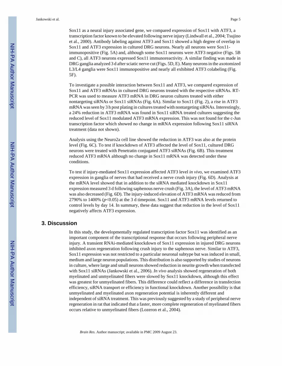
Sox11 as a neural injury associated gene, we compared expression of Sox11 with ATF3, atranscription factor known to be elevated following nerve injury (Lindwall et al., 2004; Tsujinoet al., 2000). Antibody labeling against ATF3 and Sox11 showed a high degree of overlap inSox11 and ATF3 expression in cultured DRG neurons. Nearly all neurons were Sox11-immunopositive (Fig. 5A) and, although some Sox11 neurons were ATF3 negative (Figs. 5Band C), all ATF3 neurons expressed Sox11 immunoreactivity. A similar finding was made inDRG ganglia analyzed 3 d after sciatic nerve cut (Figs. 5D, E). Many neurons in the axotomizedL3/L4 ganglia were Sox11 immunopositive and nearly all exhibited ATF3 colabeling (Fig.5F).
To investigate a possible interaction between Sox11 and ATF3, we compared expression ofSox11 and ATF3 mRNAs in cultured DRG neurons treated with the respective siRNAs. RT-PCR was used to measure ATF3 mRNA in DRG neuron cultures treated with eithernontargeting siRNAs or Sox11 siRNAs (Fig. 6A). Similar to Sox11 (Fig. 2), a rise in ATF3mRNA was seen by 3 h post plating in cultures treated with nontargeting siRNAs. Interestingly,a 24% reduction in ATF3 mRNA was found in Sox11 siRNA treated cultures suggesting thereduced level of Sox11 modulated ATF3 mRNA expression. This was not found for the c-Juntranscription factor which showed no change in mRNA expression following Sox11 siRNAtreatment (data not shown).
Analysis using the Neuro2a cell line showed the reduction in ATF3 was also at the proteinlevel (Fig. 6C). To test if knockdown of ATF3 affected the level of Sox11, cultured DRGneurons were treated with Penetratin conjugated ATF3 siRNAs (Fig. 6B). This treatmentreduced ATF3 mRNA although no change in Sox11 mRNA was detected under theseconditions.
To test if injury-mediated Sox11 expression affected ATF3 level in vivo, we examined ATF3expression in ganglia of nerves that had received a nerve crush injury (Fig. 6D). Analysis atthe mRNA level showed that in addition to the siRNA mediated knockdown in Sox11expression measured 3 d following saphenous nerve crush (Fig. 3A), the level of ATF3 mRNAwas also decreased (Fig. 6D). The injury-induced elevation of ATF3 mRNA was reduced from2790% to 1400% (p<0.05) at the 3 d timepoint. Sox11 and ATF3 mRNA levels returned tocontrol levels by day 14. In summary, these data suggest that reduction in the level of Sox11negatively affects ATF3 expression.
3. DiscussionIn this study, the developmentally regulated transcription factor Sox11 was identified as animportant component of the transcriptional response that occurs following peripheral nerveinjury. A transient RNAi-mediated knockdown of Sox11 expression in injured DRG neuronsinhibited axon regeneration following crush injury to the saphenous nerve. Similar to ATF3,Sox11 expression was not restricted to a particular neuronal subtype but was induced in small,medium and large neuron populations. This distribution is also supported by studies of neuronsin culture, where large and small neurons showed reduction in neurite growth when transfectedwith Sox11 siRNAs (Jankowski et al., 2006). In vivo analysis showed regeneration of bothmyelinated and unmyelinated fibers were slowed by Sox11 knockdown, although this effectwas greatest for unmyelinated fibers. This difference could reflect a difference in transfectionefficiency, siRNA transport or efficiency in functional knockdown. Another possibility is thatunmyelinated and myelinated axon regeneration potential is inherently different andindependent of siRNA treatment. This was previously suggested by a study of peripheral nerveregeneration in rat that indicated that a faster, more complete regeneration of myelinated fibersoccurs relative to unmyelinated fibers (Lozeron et al., 2004).
Jankowski et al. Page 5
Brain Res. Author manuscript; available in PMC 2009 August 23.
NIH
-PA Author Manuscript
NIH
-PA Author Manuscript
NIH
-PA Author Manuscript

To examine the role of Sox11 on regeneration in vivo, we used a novel delivery system in whichsiRNAs conjugated to the lipid permeable peptide Penetratin are injected into a peripheralnerve. The advantage to this system is that it allowed specific targeting of the knockdown effectto injured afferents. A disadvantage is that the injection itself also evoked a small butmeasurable injury response. Conjugation of siRNAs to Penetratin was initially shown toenhance transfection efficiency of primary cortical neurons and cultured DRG neurons withoutinducing cell death and without inhibiting siRNA activity (Davidson et al., 2004; Jankowskiet al., 2006). Here we show nerve injection of Penetratin-linked siRNAs to the saphenouscutaneous nerve, a small nerve that innervates the peripheral field of the lower leg and foot.Visualization using CY3 conjugated siRNAs showed efficient uptake by axons and retrogradetransport to only a percentage of neuronal somas in the L2/L3 DRG that presumably innervatethis restricted target field.
Prior studies using the Neuro2a neuroblastoma cell line suggested Sox11 expression levelmodulated genes involved in cell survival and regeneration (Jankowski et al., 2006). Whethera similar effect on survival genes operates in vivo is unclear although apoptotic neurons werenot detected 3 d following saphenous nerve injury, as measured by immunolabeling foractivated caspase 3 in L2/L3 DRGs of PenCON or PenSOX injected and axotomized mice(data not shown). Thus, at least in this short timeframe, the effects of Sox11 siRNA knockdowndid not produce the same effect in vivo as in vivo. This difference may reflect a more prolongedtime for initiation of cell death as previously reported for injured neurons in vivo (Jiang et al.,2005; Kuo et al., 2005; Thippeswamy et al., 2001) and the more supportive environment ofthe intact in vivo ganglia.
Expression of Sox11 in DRG appears to be highly sensitive to nerve injury being inducedfollowing nerve cut, nerve crush and to a lesser degree, nerve injection. Following crush or cutinjury, the steady rise in Sox11 expression coincided with the time in which injured nervesexhibit Wallerian degeneration and subsequent regeneration (Baba et al., 1982; Kury et al.,2001; Mi et al., 2005; Sebille, 1982; Thomas, 1982). For the saphenous nerve, functionalregeneration occurs approximately 4 wks after nerve transection (McIlwrath et al., 2005) atwhich time Sox11 (and ATF3) mRNAs are returned to baseline level. This time course ofexpression is consistent with a role for Sox11 in functional nerve regeneration that may requireinteraction with other injury-induced transcriptional regulators such as ATF3. ATF3 has beenassociated with neurite sprouting (Pearson et al., 2003), is induced in cells upon injury(Campbell et al., 2005; Isacsson et al., 2005; Lindwall and Kanje, 2005; Tsujino et al., 2000)and improves nerve regeneration when constitutively expressed in DRG neurons (Seijffers etal., 2007). Our data suggests an interaction between ATF3 and Sox11 since ATF3 wasdecreased with Sox11 knockdown. Interestingly, the reverse was not found, i.e., knockdownof ATF3 using ATF3 siRNAs reduced mRNA levels of ATF3, but did not affect Sox11 level.This suggests but does not prove that Sox11 may influence regulation of ATF3 gene expression.Chromatin binding and expression assays to assess interaction of Sox11 with ATF3 generegulatory regions will be necessary to test this possibility.
Another finding from this study is that, similar to ATF3 and c-Jun (Kenney and Kocsis,1997; Shortland et al., 2006), L5 rhizotomy does not induce a significant increase in Sox11 inthe L5 DRG. Indeed, the modest increase in Sox11 mRNA following rhizotomy could be dueto the associated surgical procedure. Given the importance of Sox11 for neurite growth andperipheral nerve regeneration and its potential regulatory influence on ATF3, it is possible thatthe minimal induction of ATF3 expression in DRGs following central injury is due, in part, tothe low level production of Sox11. In this regard, Sox11, as a regulator of gene expression andtissue differentiation, may underlie some of the transcriptional discrepancies that exist in DRGneurons following central and peripheral injuries. A more detailed analysis of Sox11 function
Jankowski et al. Page 6
Brain Res. Author manuscript; available in PMC 2009 August 23.
NIH
-PA Author Manuscript
NIH
-PA Author Manuscript
NIH
-PA Author Manuscript

and its relationship to other transcriptional regulators active following rhizotomy and axotomywill test this possibility and further reveal the regulatory role of Sox11 in nerve regeneration.
4. Experimental procedures4.1. Neuro2a cell culture and siRNA treatment
The mouse neuroblastoma cell line Neuro2a (ATCC clone number CCL-131, Manassas, VA)(Olmsted et al., 1970) was maintained as described in Jankowski et al. (2006). Cells were platedinto 6-well plates at a concentration of 50,000 cells/well, grown to 50% confluence (18–24 h)and then treated with siRNAs. Two hours prior to siRNA transfection fresh medium was addedto cultures and transfection was carried out of 10 nM Sox11 siRNAs (sense strand 5′GGU CCAAGA UCG AGC GCA GUU-3′), ATF-3 siRNAs (5′GUG GUG ACC UAC UGC AUUGUU-3′) or 10 nM non-targeting siRNAs, all purchased from Dharmacon (Lafayette, CO).The non-targeting siRNA sense sequence was 5′-Th-UAG CGA CUA AAC ACA UCA A-dT-dT-3′ and the antisense sequence is 5′-DY547-UUG AUG UGU UUA GUC GCUA-dT-dT-3′.TRANSIT-TKO transfection reagent (Mirus Corporation) (4 μl) and serum-free MEM (100μl) were incubated at room temperature (RT) for 5–20 min and then the appropriate volumeof 1 μm siRNAs was added to the TKO/MEM mixture to obtain a final concentration of 10nM siRNA per well. Solutions were mixed, incubated at RT for 5–20 min and then addeddropwise to the cultures. Cells were allowed to incubate for 24 h prior to protein isolation.
4.2. Primary neuron cultureMale C57/Bl6 (Jackson Laboratories, Bar Harbor, ME) mice approximately 2–3 months of agewere deeply anesthetized with 2.5% avertin and intracardially perfused with 35 mL of Hank’sbalanced salt solution (HBSS; Gibco). DRGs were isolated and grown as described in Malinet al. (2007).
4.3. Penetratin-1/siRNA linkage and transfectionsiRNAs were conjugated to Penetratin-1 (Q-Biogene) peptide as previously described(Davidson et al., 2004; Jankowski et al., 2006). Penetratin-1 is a patented 16-amino acid peptide(NH2-RQIKIWFQNRRMKWKK-COOH) corresponding to the third helix of thehomeodomain of Antennapedia protein that is able to translocate across biological membranesand convey oligonucleotides to the cytoplasm of many cell types (Derossi et al., 1998). siRNAswere synthesized with a 5′ thiol modification on the sense strand to allow Penetratinconjugation. The non-targeting siRNA duplex also contained a 5′ CY3 molecule conjugatedto the antisense strand. Equimolar concentrations of activated Penetratin-1, reconstituted insterile water, were added to each of the siRNAs dissolved in 1× siRNA buffer (Dharmacon),incubated 15 min at 65 °C and then 1 h at 37 °C. Stock siRNAs were brought to 400 nM NaClto enhance solubility of Penetratin linked siRNAs (Pen-siRNAs) and kept at −80 °C until used.Pen-siRNAs were heated to 65 °C for 15 min prior to DRG neuron transfections. 500 μL ofmedia with NGF (50 ng/ml) was removed from DRG cultures and Penetratin-1 linked control(non-targeting; PenCON) and/or Penetratin-1 linked Sox11 (targeting; PenSox) or Penetratinlinked ATF3 (targeting; PenATF3) siRNAs were added. Media containing Pen-siRNAs wasgently mixed and added back to cultures as a 2× solution to give a final concentration of 80nM siRNAs per well. Cells were then incubated at 37 °C/5% CO2 for appropriate times.
4.4. RNA isolation and analysisRNA isolation from cells and tissues was performed using Qiagen RNeasy mini kits using thesupplied protocol. 1 μg of total RNA was treated with DNase I (Invitrogen), annealed to randomprimers, reverse transcribed using Superscript II reverse transcriptase (Invitrogen) and storedat −80 °C until used in SYRB green labeled real time PCR (RT-PCR) reactions. For RT-PCR,
Jankowski et al. Page 7
Brain Res. Author manuscript; available in PMC 2009 August 23.
NIH
-PA Author Manuscript
NIH
-PA Author Manuscript
NIH
-PA Author Manuscript

20 ng of cDNA were added to SYBR Green MasterMix (Applied Biosystems), run in triplicateon an Applied Biosystems Imager and values normalized to GAPDH or neuronal specificenolase (NSE). Both of these internal control genes were unchanged following axotomy or cellculture, respectively (not shown). Changes in expression are reported as a ΔΔCt value that iscalculated by subtracting the gene expression by the GAPDH or NSE control for each sample.Fold change is described as 2ΔΔCt (Applied Biosystems) and 2-fold change equals 100%change.
4.5. Western blotPrimary DRG neurons from 8 C57/Bl6 mice were isolated, pooled, plated into separate wellsand then transfected with either control or Sox11 targeting siRNAs at 1 h and 25 h after plating.At 48 h post plating, neurons from similarly treated wells were pooled and homogenized inlysis buffer containing 20 mM, HEPES pH 7.9, 10 mM KCl2, 5 mM MgCl2, 5 mM EDTA pH8, 10% glycerol, 2 mM DTT, 1% Triton X-100, 300 mM NaCl, 50 mM NaF, 1 mM Na3VO4and protease inhibitors (1 μg/ml pepstatin, 1 μg/ml leupeptin, 1 μg/ml aprotinin; SigmaBiochemicals). Samples were sonicated, incubated on a rotator at 4 °C for 20 min, centrifuged,boiled 10 min in a denaturing buffer containing β-mercaptoethanol and SDS, separated on a10% polyacrylamide SDS-PAGE gel and transferred to nitro-cellulose membrane (BioRad)that was blocked and then incubated with primary antibodies overnight at 4 °C (Sox11, 1:1000,sc-20096, Santa Cruz; ATF3, 1:1000; actin, 1:5000). Antibody binding was visualized usinghorseradish peroxidase-conjugated goat anti-rabbit or donkey anti-goat secondary antibodies(1:5000) and chemiluminescent detection (Pierce Biochemical). Molecular weight of Sox11was estimated by gel interpolation.
Transfected Neuro2a cells were dissolved in lysis buffer containing 1% sodium dodecyl sulfate(SDS), 10 mM Tris–HCl (pH 7.4) and protease inhibitors (1 μg/ml pepstatin, 1 μg/ml leupeptin,1 μg/ml aprotinin, 1 mM sodium orthovanadate and 100 μg/ml phenylmethylsulfonyl fluoride).Samples (25 μg) were centrifuged, boiled 10 min in denaturing buffer containing β-mercaptoethanol and SDS, separated on a 10% polyacrylamide SDS-PAGE gel and transferredto a nitrocellulose membrane (BioRad) that was blocked and then incubated with primaryantibodies overnight at 4 °C (Sox11, 1:100, sc; ATF3, 1:1000; actin, 1:10,000). Antibodybinding was visualized as described above using chemiluminescent detection.
4.6. Nerve injury and siRNA injectionFor saphenous nerve injuries and analysis of regeneration, male Swiss Webster mice were used.DRG neurons cultured from Swiss Webster mice show a similar level of Sox11 induction asfound for the Blk6/C3 H strain suggesting parallel mechanisms of Sox11 actions. Mice 4–6wks of age (Hilltop Lab Animals, Scottdale, PA) were anesthetized by intramuscular injectionof a mixture of ketamine and xylazine (90 mg/kg and 10 mg/kg, respectively). A small incisionmade in the mid-thigh region exposed the saphenous nerve that was transected using fineirredectomy scissors. Wounds were closed using 7.0 silk sutures. Nerve crush was performedusing the same procedure except the exposed nerve was crushed using number 5 forceps (FineScience Tools, Forester City, CA) held together for 4–5 s. For L5 rhizotomy, an incision wasmade just proximal to the iliac crest, a laminectomy was performed to expose the L5 dorsalroot, which was loosened from the spinal cord and transected. Sciatic nerve axotomy was doneon mice anesthetized with isofluorane. The nerve was tightly ligated with 6.0 silk sutures,transected distal to the suture and the muscles sutured and wound closed with microclips(Roboz, Gaithersburg, MD). In animals that received siRNA injection, 0.1–0.2 μL of 90 μMPenetratin-1 linked control (PenCON) or Sox11 targeting (Pen-SOX) siRNAs were pressureinjected into the saphenous nerve using a glass microelectrode connected to a pico-spritzer.This amount was chosen based on dose response/efficiency of knockdown analysis previouslydone in cultured neurons (Jankowski et al., 2006). Injections were done the day before and at
Jankowski et al. Page 8
Brain Res. Author manuscript; available in PMC 2009 August 23.
NIH
-PA Author Manuscript
NIH
-PA Author Manuscript
NIH
-PA Author Manuscript

the time of nerve crush. Nerve cuts/crushes were made 1–2 mm distal to the injection sitewithout cleaning the surrounding area or damage to vasculature. Nerves were collected 2–28d after nerve injury for immuno-cytochemical, electron microscopic and RT-PCR analysis.Animals were cared for and used in accordance with guidelines of the U.S. Public HealthService Policy on Humane Care and Use of Laboratory Animals, the NIH Guide for the Careand Use of Laboratory Animals and following institutional AAALAC approved practices.
4.7. ImmunocytochemistryGanglia were removed from animals perfused with 4% paraformaldehyde (PF), postfixed in4% PF for 1 h, washed 3×s in phosphate buffered saline (PBS) and embedded in 10% gelatinin 0.1 M phosphate buffer (PB). Cultured DRG neurons were washed in 0.1 M PBS and fixed5 min in 4% PF. DRG sections were cut at 30 or 50 μm and collected in 0.1 M PBS. Cells and/or sections were washed in PBS and incubated overnight in goat Sox11 antibody (1:50; SantaCruz) or rabbit ATF3 antibody (1:500, Santa Cruz) diluted in PBS containing 0.25% tritonX-100. Binding was visualized using CY2, CY3 or CY5 conjugated secondary antibodies(1:1000; Jackson Labs). Sections were washed in PBS, mounted on Superfrost slides (VWR)and images captured using a Leica fluorescent microscope.
4.8. Electron microscopyMyelinated and unmyelinated axons in the saphenous nerve were counted at the electronmicroscopic level using photographic montages taken across the entire diameter of thesaphenous nerve. Nerves were collected from deeply anesthesized animals intracardiallyperfused with cold saline following a 5 min fixation in situ with 2% gluteraldehyde/4%paraformaldehyde. Nerves were post fixed in 2% gluteraldehyde/4% PF for 2 h, cut into 1 mmblocks, post-fixed in 2% osmium tetroxide in 0.1 M PB for 1 h and dehydrated in an increasingseries of ethanols and propylene oxide. Blocks were embedded in EMBed 812 (ElectronMicroscopy Sciences), cut on an ultramicrotome at 80–90 nm and collected on formvar/carboncoated copper slot grids. Grids were counter stained in uranyl acetate and lead citrate andanalyzed on a Morgagni electron microscope. Images were taken at 4400× of the entire nerveusing AMT digital imaging software and compiled into montages using Adobe Photoshop.Axon numbers were counted across montaged photographs taken of sections at 7 d and 14 dand then averaged across animals (n = 3 per condition). Axons of comparable morphology touninjured nerves and those not meeting the criteria for electron-dense degeneration accordingto Peters et al. (1991) and Mugnaini and Friedrich (1981) were used to establish the criteriafor degenerating axons. These criteria do not meet those established for filamentous, flocculentor watery degeneration (Mugnaini and Friedrich, 1981). Degenerating axons were determinedto be electron dense with swollen mitochondria (when present) and associated with surroundingglial cells. The number of non-degenerating axons at −1 mm to −2 mm, 2 mm, 4 mm and 6mm distal to the site of the nerve crush at various times after injury were recorded and presentedas an average difference in the percent of regenerating fibers between PenCON and PenSOXinjected mice normalized to the number of axons present proximal (−1 to −2 mm) to theinjection site. Data were segregated into myelinated and unmyelinated groups. Statisticalsignificance was set at p<0.05 for all evaluations.
AcknowledgementsThese studies were supported by grants from the NINDS (NS33730, K.M.A.; NS23725, H.R.K and T32NS007433,M.P.J.).
ReferencesAlvarez VA, Ridenour DA, Sabatini BL. Retraction of synapses and dendritic spines induced by off-
target effects of RNA interference. J Neurosci 2006;26:7820–7825. [PubMed: 16870727]
Jankowski et al. Page 9
Brain Res. Author manuscript; available in PMC 2009 August 23.
NIH
-PA Author Manuscript
NIH
-PA Author Manuscript
NIH
-PA Author Manuscript

Asano A, Jin HK, Watanabe T. Mouse Mx2 gene: organization, mRNA expression and the role of theinterferon-response promoter in its regulation. Gene 2003;306:105–113. [PubMed: 12657472]
Azuma T, Ao S, Saito Y, Yano K, Seki N, Wakao H, Masuho Y, Muramatsu M. Human SOX11, anupregulated gene during the neural differentiation, has a long 3′ untranslated region. DNA Res1999;6:357–360. [PubMed: 10574465]
Baba M, Fowler CJ, Jacobs JM, Gilliatt RW. Changes in peripheral nerve fibres distal to a constriction.J Neurol Sci 1982;54:197–208. [PubMed: 7097298]
Baron R, Janig W, Kollmann W. Sympathetic and afferent somata projecting in hindlimb nerves and theanatomical organization of the lumbar sympathetic nervous system of the rat. J Comp Neurol1988;275:460–468. [PubMed: 3225349]
Bergsland M, Werme M, Malewicz M, Perlmann T, Muhr J. The establishment of neuronal properties iscontrolled by Sox4 and Sox11. Genes Dev 2006;20:3475–3486. [PubMed: 17182872]
Cajal, R. Degeneration and Regeneration of the Nervous System. Oxford Univ. Press; London: 1928.Campbell G, Hutchins K, Winterbottom J, Grenningloh G, Lieberman AR, Anderson PN. Upregulation
of activating transcription factor 3 (ATF3) by intrinsic CNS neurons regenerating axons into peripheralnerve grafts. Exp Neurol 2005;192:340–347. [PubMed: 15755551]
Cheung M, Abu-Elmagd M, Clevers H, Scotting PJ. Roles of Sox4 in central nervous systemdevelopment. Brain Res Mol Brain Res 2000;79:180–191. [PubMed: 10925158]
Darby IA, Bisucci T, Raghoenath S, Olsson J, Muscat GE, Koopman P. Sox18 is transiently expressedduring angiogenesis in granulation tissue of skin wounds with an identical expression pattern to Flk-1mRNA. Lab Invest 2001;81:937–943. [PubMed: 11454981]
Davidson TJ, Harel S, Arboleda VA, Prunell GF, Shelanski ML, Greene LA, Troy CM. Highly efficientsmall interfering RNA delivery to primary mammalian neurons induces MicroRNA-like effectsbefore mRNA degradation. J Neurosci 2004;24:10040–10046. [PubMed: 15537872]
Derossi D, Chassaing G, Prochiantz A. Trojan peptides: the penetratin system for intracellular delivery.Trends Cell Biol 1998;8:84–87. [PubMed: 9695814]
Dy P, Penzo-Mendez A, Wang H, Pedraza CE, Macklin WB, Lefebvre V. The three SoxC proteins–Sox4,Sox11 and Sox12–exhibit overlapping expression patterns and molecular properties. Nucleic AcidsRes 2008;36:3101–3117. [PubMed: 18403418]
Gubbay J, Collignon J, Koopman P, Capel B, Economou A, Munsterberg A, Vivian N, Goodfellow P,Lovell-Badge R. A gene mapping to the sex-determining region of the mouse Y chromosome is amember of a novel family of embryonically expressed genes. Nature 1990;346:245–250. [PubMed:2374589]
Hargrave M, Wright E, Kun J, Emery J, Cooper L, Koopman P. Expression of the Sox11 gene in mouseembryos suggests roles in neuronal maturation and epithelio-mesenchymal induction. Dev Dyn1997;210:79–86. [PubMed: 9337129]
Isacsson A, Kanje M, Dahlin LB. Induction of activating transcription factor 3 (ATF3) by peripheralnerve compression. Scand J Plast Reconstr Surg Hand Surg 2005;39:65–72. [PubMed: 16019731]
Jankowski MP, Cornuet PK, McIlwrath S, Koerber HR, Albers KM. SRY-box containing gene 11(Sox11) transcription factor is required for neuron survival and neurite growth. Neuroscience2006;143:501–514. [PubMed: 17055661]
Jiang Y, Zhang JS, Jakobsen J. Differential effect of p75 neurotrophin receptor on expression of pro-apoptotic proteins c-jun, p38 and caspase-3 in dorsal root ganglion cells after axotomy inexperimental diabetes. Neuroscience 2005;132:1083–1092. [PubMed: 15857712]
Kenney AM, Kocsis JD. Temporal variability of jun family transcription factor levels in peripherally orcentrally transected adult rat dorsal root ganglia. Brain Res Mol Brain Res 1997;52:53–61. [PubMed:9450677]
Kitao Y, Robertson B, Kudo M, Grant G. Proliferation patterns of dorsal root ganglion neurons ofcutaneous, muscle and visceral nerves in the rat. J Neurocytol 2002;31:765–776. [PubMed:14501213]
Koltzenburg M. Neural mechanisms of cutaneous nociceptive pain. Clin J Pain 2000;16:S131–S138.[PubMed: 11014457]
Jankowski et al. Page 10
Brain Res. Author manuscript; available in PMC 2009 August 23.
NIH
-PA Author Manuscript
NIH
-PA Author Manuscript
NIH
-PA Author Manuscript

Kuo LT, Simpson A, Schanzer A, Tse J, An SF, Scaravilli F, Groves MJ. Effects of systemicallyadministered NT-3 on sensory neuron loss and nestin expression following axotomy. J Comp Neurol2005;482:320–332. [PubMed: 15669078]
Kury P, Stoll G, Muller HW. Molecular mechanisms of cellular interactions in peripheral nerveregeneration. Curr Opin Neurol 2001;14:635–639. [PubMed: 11562576]
Lindwall C, Kanje M. Retrograde axonal transport of JNK signaling molecules influence injury inducednuclear changes in p-c-Jun and ATF3 in adult rat sensory neurons. Mol Cell Neurosci 2005;29:269–282. [PubMed: 15911351]
Lindwall C, Dahlin L, Lundborg G, Kanje M. Inhibition of c-Jun phosphorylation reduces axonaloutgrowth of adult rat nodose ganglia and dorsal root ganglia sensory neurons. Mol Cell Neurosci2004;27:267–279. [PubMed: 15519242]
Lozeron P, Krarup C, Schmalbruch H. Regeneration of unmyelinated and myelinated sensory nerve fibresstudied by a retrograde tracer method. J Neurosci Methods 2004;138:225–232. [PubMed: 15325131]
Malin SA, Davis BM, Molliver DC. Production of dissociated sensory neuron cultures and considerationsfor their use in studying neuronal function and plasticity. Natl Protoc Dir 2007;2:152–160.
McIlwrath S, Lawson JJ, Anderson CE, Koerber HR. Sensitization of cutaneous nociceptors after nervecut and regeneration in mouse. Soc Neurosci Abst. 2005Online.
Meeson AP, Shi X, Alexander MS, Williams RS, Allen RE, Jiang N, Adham IM, Goetsch SC, HammerRE, Garry DJ. Sox15 and Fhl3 transcriptionally coactivate Foxk1 and regulate myogenic progenitorcells. EMBO J 2007;26:1902–1912. [PubMed: 17363903]
Mi W, Beirowski B, Gillingwater TH, Adalbert R, Wagner D, Grumme D, Osaka H, Conforti L, ArnholdS, Addicks K, Wada K, Ribchester RR, Coleman MP. The slow Wallerian degeneration gene, WldS,inhibits axonal spheroid pathology in gracile axonal dystrophy mice. Brain 2005;128:405–416.[PubMed: 15644421]
Mugnaini, E.; Friedrich, VL, Jr. Electron microscopy: identification and study of normal and degeneratingneural elements by electron microscopy. In: Heimer, L.; Robards, MJ., editors. NeuroanatomicalTract-Tracing Methods. Plenum Press; New York: 1981. p. 377-406.
Olmsted JB, Carlson K, Klebe R, Ruddle F, Rosenbaum J. Isolation of microtubule protein from culturedmouse neuroblastoma cells. Proc Natl Acad Sci U S A 1970;65:129–136. [PubMed: 5263744]
Pearson AG, Gray CW, Pearson JF, Greenwood JM, During MJ, Dragunow M. ATF3 enhances c-Jun-mediated neurite sprouting. Brain Res Mol Brain Res 2003;120:38–45. [PubMed: 14667575]
Peters, A.; Palay, SL.; Webster, Hd. Neurons and Their Supporting Cells. Oxford; New York: 1991. TheFine Structure of the Nervous System.
Raivich G, Bohatschek M, Da Costa C, Iwata O, Galiano M, Hristova M, Nateri AS, Makwana M, Riera-Sans L, Wolfer DP, Lipp HP, Aguzzi A, Wagner EF, Behrens A. The AP-1 transcription factor c-Jun is required for efficient axonal regeneration. Neuron 2004;43:57–67. [PubMed: 15233917]
Sebille A. [Peripheral neuropathies. Current data concerning nerve regeneration (author’s transl)] NouvPresse Med 1982;11:1206–1215.
Seijffers R, Allchorne AJ, Woolf CJ. The transcription factor ATF-3 promotes neurite outgrowth. MolCell Neurosci 2006;32:143–154. [PubMed: 16713293]
Seijffers R, Mills CD, Woolf CJ. ATF3 increases the intrinsic growth state of DRG neurons to enhanceperipheral nerve regeneration. J Neurosci 2007;27:7911–7920. [PubMed: 17652582]
Shortland PJ, Baytug B, Krzyzanowska A, McMahon SB, Priestley JV, Averill S. ATF3 expression inL4 dorsal root ganglion neurons after L5 spinal nerve transection. Eur J Neurosci 2006;23:365–373.[PubMed: 16420444]
Silver J, Miller JH. Regeneration beyond the glial scar. Nat Rev Neurosci 2004;5:146–156. [PubMed:14735117]
Sock E, Rettig SD, Enderich J, Bosl MR, Tamm ER, Wegner M. Gene targeting reveals a widespreadrole for the high-mobility-group transcription factor Sox11 in tissue remodeling. Mol Cell Biol2004;24:6635–6644. [PubMed: 15254231]
Stucky CL, Koltzenburg M, Schneider M, Engle MG, Albers KM, Davis BM. Overexpression of nervegrowth factor in skin selectively affects the survival and functional properties of nociceptors. JNeurosci 1999;19:8509–8516. [PubMed: 10493751]
Jankowski et al. Page 11
Brain Res. Author manuscript; available in PMC 2009 August 23.
NIH
-PA Author Manuscript
NIH
-PA Author Manuscript
NIH
-PA Author Manuscript

Tanabe K, Bonilla I, Winkles JA, Strittmatter SM. Fibroblast growth factor-inducible-14 is induced inaxotomized neurons and promotes neurite outgrowth. J Neurosci 2003;23:9675–9686. [PubMed:14573547]
Thippeswamy T, Jain RK, Mumtaz N, Morris R. Inhibition of neuronal nitric oxide synthase results inneurodegenerative changes in the axotomised dorsal root ganglion neurons: evidence for aneuroprotective role of nitric oxide in vivo. Neurosci Res 2001;40:37–44. [PubMed: 11311403]
Thomas PK. Selective vulnerability of the centrifugal and centripetal axons of primary sensory neurons.Muscle Nerve 1982;5:S117–121. [PubMed: 6221191]
Tsujino H, Kondo E, Fukuoka T, Dai Y, Tokunaga A, Miki K, Yonenobu K, Ochi T, Noguchi K.Activating transcription factor 3 (ATF3) induction by axotomy in sensory and motoneurons: A novelneuronal marker of nerve injury. Mol Cell Neurosci 2000;15:170–182. [PubMed: 10673325]
Uusitalo H, Salminen H, Vuorio E. Activation of chondrogenesis in response to injury in normal andtransgenic mice with cartilage collagen mutations. Osteoarthr Cartil 2001;9(Suppl A):S174–S179.[PubMed: 11680682]
Wegner M, Stolt CC. From stem cells to neurons and glia: a Soxist’s view of neural development. TrendsNeurosci 2005;28:583–588. [PubMed: 16139372]
Wilson M, Koopman P. Matching SOX: partner proteins and co-factors of the SOX family oftranscriptional regulators. Curr Opin Genet Dev 2002;12:441–446. [PubMed: 12100890]
Wright EM, Snopek B, Koopman P. Seven new members of the Sox gene family expressed during mousedevelopment. Nucleic Acids Res 1993;21:744. [PubMed: 8441686]
Jankowski et al. Page 12
Brain Res. Author manuscript; available in PMC 2009 August 23.
NIH
-PA Author Manuscript
NIH
-PA Author Manuscript
NIH
-PA Author Manuscript

Fig. 1.Sox11 is expressed in regenerating peripheral neurons. (A) RNA was collected from L5 DRGsfollowing L5 dorsal root transection and from L4 DRGs following sciatic nerve transection(n =4/group). Sciatic nerve transection induced a 1004% increase in Sox11 mRNA in the L4/L5 DRG 3 d after cut compared to the 51% increase in L5 DRG following L5 rhizotomy. (B)Axotomy of the saphenous nerve caused a 184% increase in Sox11 mRNA in L2/L3 ganglia3 d after nerve cut. Sox11 remains elevated for at least 7 d with return to baseline by 4 wks.(C) Sox11 immunoreactivity is detected in neuronal nuclei in ipsilateral L4 DRG at 7 d afteraxotomy of the sciatic nerve. Sox11 immunoreactivity is low in contralateral ganglia (D). *pvalue<0.001 (A) and <0.05 (B); values are relative to contralateral DRGs. Scale bar in C=50μm.
Jankowski et al. Page 13
Brain Res. Author manuscript; available in PMC 2009 August 23.
NIH
-PA Author Manuscript
NIH
-PA Author Manuscript
NIH
-PA Author Manuscript

Fig. 2.Sox11 siRNAs reduce the level of Sox11 mRNA and protein in DRG neurons. (A) PrimaryDRG neurons treated with Penetratin-linked Sox11 siRNAs (PenSOX) have reduced levels ofSox11 mRNA compared to untreated or nontargeting control siRNA (PenCON) treatedcultures. Values are relative to mRNA level at time of dissociation and plating (0 h). N=3–4per timepoint. (B) Cultured DRG neurons treated with Sox11 siRNAs (lane 2) show reducedSox11 protein compared to parallel cultures treated with nontargeting siRNA (lane 3) anduntreated cultures (lane 4). Sox11 protein from Neuro2a cells transfected with CMV-Sox11(lane 1) is used to show migration at 44.7 Kd. Arrowhead indicates Sox11; identity of bandbelow Sox11 is unknown. Actin blot indicates sample loading. (C) Injected PenCON Cy3-tagged siRNAs are visible in nerve fibers at injection site (arrowhead). (D) Whole mount imageof L3 DRG shows several Cy3-positive somas (arrows) after saphenous nerve injection ofPenCON siRNA that are absent in the ipsilateral T13 DRG (E). (F) Fold change in Sox11mRNA after injection of nontargeting control (PenCON) or Sox11 (PenSOX) siRNAs into thesaphenous nerve. *p value<0.05. Scale bar in D=50 μm.
Jankowski et al. Page 14
Brain Res. Author manuscript; available in PMC 2009 August 23.
NIH
-PA Author Manuscript
NIH
-PA Author Manuscript
NIH
-PA Author Manuscript

Fig. 3.siRNA injection in crushed saphenous nerve reduces Sox11 mRNA in DRG. (A) At 3 d, nervecrush alone caused a 230% increase in Sox11 in L2/L3 DRGs whereas nerve crush plusPenCON caused a 260% increase. Values are relative to the contralateral DRG where no changein baseline Sox11 mRNA level was measured. PenSOX injection plus nerve crush reducedSox11 mRNA to 140%. Seven days after crush, a 252% increase in Sox11 was measured whilePenCON injected mice had a 225% increase in Sox11. PenSOX siRNA treatment produced a160% increase in Sox11 that was not statistically different from PenCON values. After 14 days,all experimental conditions had similar levels of Sox11 expression (Sox11: 72% vs 51% vs45% increase). N=4 per experimental group. (B) Interferon inducible gene mx2 showed nochange in expression under all conditions. *p value<0.05.
Jankowski et al. Page 15
Brain Res. Author manuscript; available in PMC 2009 August 23.
NIH
-PA Author Manuscript
NIH
-PA Author Manuscript
NIH
-PA Author Manuscript

Fig. 4.Electron photomicrographs of representative regions of the saphenous nerve under normalconditions and after siRNA treatment and nerve crush. (A) Uninjured saphenous nerve showingmyelineated (arrow) and unmyelinated (arrowheads) fibers. (B) Degeneration of myelinatedand unmyelinated fibers occurs 3 d following nerve crush. Extensive myelin unraveling(dashed arrow) and disruption of Remak bundle structure (arrowhead) are evident. (C) Nervecrush site at 7 d post injury. Morphology of myelinated (arrow) and unmyelinated (arrowheads)fibers is almost restored to normal. (D) Regenerating myelinated (arrow) and unmyelinated(arrowheads) axons in PenCON injected nerves 4 mm distal to the crush site at 7 d post injury.(E) PenSOX injected nerves had fewer regenerating myelinated (arrow) and unmyelinatedaxons (arrowhead). (F) Plot of myelinated, unmyelinated and total axons regenerated at 4 mmdistal at 7 d post crush. PenSOX injected nerves had fewer myelinated and unmyelinatedregenerated axons compared to nerves injected with PenCON where the percent of myelinatedand unmyelinated axons were greater (p values<0.02 and <0.001, respectively). (G, H) At 14d after injury, 6 mm distal to the nerve crush, the percent of regenerating myelinated fibers(arrows) in PenCON injected nerves (G) was similar to PenSOX injected nerves (H). InPenSOX nerves (H) unmyelinated fiber (arrowheads) regrowth remained inhibited and a fewdegenerating fibers (dashed arrow) were visible. (I) Plot shows the percentage of regeneratedmyelinated fibers in crush only, PenCON and PenSOX injected mice is the same at 14 d postinjury. Unmyelinated fibers were fewer in PenSOX injected nerves but only when comparedto crush only (*p value<0.01 relative to crush only). Total fiber analysis showed PenSOXinjected nerves with 40.1% regeneration compared to 67.3% in crush only and 49.3% inPenCON samples (*p value<0.01 relative to crush only). Asterisks in E and H indicateinteraxonal space. All scale bars=2 μm.
Jankowski et al. Page 16
Brain Res. Author manuscript; available in PMC 2009 August 23.
NIH
-PA Author Manuscript
NIH
-PA Author Manuscript
NIH
-PA Author Manuscript

Fig. 5.Sox11 expression colocalizes with and modulates ATF3 expression following nerve injury.(A) Cultured DRG neurons show nuclear localization of Sox11 24 h after plating thatcolocalizes with ATF3 immunolabeling (B). Dashed arrow in C illustrates one of manyoverlapping cells. Neurons that were Sox11 positive and ATF3 negative are rare (solid arrows).Following sciatic nerve injury Sox11 expression (D) is found in many neurons of the L3/L4DRG, many of which overlap (F) with ATF3 immunopositive neurons (E). In F, broken arrowindicates overlap, solid arrows indicate neurons positive for Sox11 alone, arrowhead indicatesrare neuron that was only ATF3 positive and asterisk indicates neuron that does not expresseither Sox11 or ATF3.
Jankowski et al. Page 17
Brain Res. Author manuscript; available in PMC 2009 August 23.
NIH
-PA Author Manuscript
NIH
-PA Author Manuscript
NIH
-PA Author Manuscript

Fig. 6.Knockdown of Sox11 modulates ATF3 expression. (A) Cultured DRG neurons transfectedwith PenSOX targeting siRNA have decreased levels of ATF3 mRNA at 24 h post treatment.(B) Cultured DRG neurons treated with PenCON or PenATF3 siRNAs showed reduction inATF3 mRNA at 24 h after transfection but no change in Sox11 mRNA. Values are relative tomRNA level at time of dissociation and plating. (C) Transfection of Sox11 siRNAs decreasedSox11 and ATF3 protein in Neuro2a cells. (D) At 3 d after nerve crush ATF3 mRNA in L2/L3 DRGs from untreated and PenCON injected nerves is increased. PenSOX injection plusnerve crush inhibited the rise in ATF3 mRNA. At 7 d and 14 d after injury no difference inATF3 mRNA level was measured. Values are relative to contralateral side DRG samples.N=3 for all experimental groups. All *p values<0.05. Scale bar in D=40 μm.
Jankowski et al. Page 18
Brain Res. Author manuscript; available in PMC 2009 August 23.
NIH
-PA Author Manuscript
NIH
-PA Author Manuscript
NIH
-PA Author Manuscript


