Embed Size (px)
Citation preview
Hindawi Publishing CorporationComputational and Mathematical Methods in MedicineVolume 2013 Article ID 108910 9 pageshttpdxdoiorg1011552013108910
Research ArticleSpace Constrained Homology Modelling The Paradigm of theRNA-Dependent RNA Polymerase of Dengue (Type II) Virus
Dimitrios Vlachakis Dimitrios Georgios Kontopoulos and Sophia Kossida
Bioinformatics amp Medical Informatics Team Biomedical Research Foundation Academy of Athens Soranou Efessiou 411527 Athens Greece
Correspondence should be addressed to Sophia Kossida skossidabioacademygr
Received 3 July 2013 Accepted 8 July 2013
Academic Editor Carla Sofia Carvalho
Copyright copy 2013 Dimitrios Vlachakis et al This is an open access article distributed under the Creative Commons AttributionLicense which permits unrestricted use distribution and reproduction in any medium provided the original work is properlycited
Protein structure is more conserved than sequence in nature In this direction we developed a novel methodology that significantlyimproves conventional homology modelling when sequence identity is low by taking into consideration 3D structural features ofthe template such as size and shape Herein our new homology modelling approach was applied to the homology modelling ofthe RNA-dependent RNA polymerase (RdRp) of dengue (type II) virus The RdRp of dengue was chosen due to the low sequencesimilarity shared between the dengue virus polymerase and the available templates while purposely avoiding to use the actual X-raystructure that is available for the dengue RdRp The novel approach takes advantage of 3D space corresponding to protein shapeand size by creating a 3D scaffold of the template structureThe dengue polymerase model built by the novel approach exhibited allfeatures of RNA-dependent RNA polymerases and was almost identical to the X-ray structure of the dengue RdRp as opposed tothe model built by conventional homology modelling Therefore we propose that the space-aided homology modelling approachcan be of a more general use to homology modelling of enzymes sharing low sequence similarity with the template structures
1 Introduction
3D structural information provides invaluable insights intothe organization mode of action folding and utterly func-tion of a given protein The 3D structure of proteins isusually experimentally determined using X-ray crystallogra-phy NMR or microscopy [1] However protein expressionpurification and crystallization are quite tedious experimentswith uncertain outcomes and success rates Thus the nextbest thing to experimentally determine the structure of aprotein is done via state of the art computational techniquesand mainly homology modelling [2]
Homology modelling is the current leading techniquefor in silico predicting the three-dimensional structures ofproteins However the quality of the predicted structuresis only limited to the homology shared between the queryprotein and the chosen template structure [3]
Conventional homology modelling methods are com-prised of the following steps [4ndash6] first an initial partialgeometry specification where an initial partial geometry
for each target sequence is copied from regions of one ormore template chains secondly the insertions and deletionstask where residues that still have no assigned backbonecoordinates are modelled Those residues may be in loops(insertions in the model with respect to the template) theymay be outgaps (residues in a model sequence which arealigned before the C-terminus or after the N-terminus of itstemplate) or may be deletions (regions where the templatehas an insertion with respect to the model) For this studyoutgaps have not been included in the homology modellingprocess Third step is the loop selection and side chainpacking where a collection of independentmodels is createdLast step is the final model selection and refinement onewhere the final models are scored and ranked after they havebeen stereochemically checked for persisting errors
In the present work a use case was sought that would beimpossible to model using the previously described conven-tional homology techniques in an effort to put to the test andapply our novel proposed homology modelling technique Inthis direction an example from the highly mutagenic field of
2 Computational and Mathematical Methods in Medicine
RNA viruses was selected In particular we chose to modelthe three-dimensional structure of the RNA-dependent RNApolymerase (RdRp) of Dengue (type II) virus by using thecrystal structures of other polymerases of the Flaviviridaefamily as templates and by applying our novel homologymodelling approach The novel approach takes advantageof the 3D space corresponding to ligands substrates andthe size and shape of the template structures (by creatinga mould made of dummy atoms) in an effort to restrainthe folding of the target protein In this way our proposedmodelling technique manages to overcome fundamentallimitations in the homology modelling methodologies thatoriginate from the low sequence identity shared betweenthe query (Dengue polymerase) and the RdRps used astemplates
The viral family Flaviviridae comprises the genera Fla-vivirus Pestivirus and Hepacivirus and includes numerousimportant human and animal pathogens [10] The smallenveloped virions of the differentmembers of the Flaviviridaefamily contain a single-stranded positive-senseRNAgenomeof about 95ndash125 kb The genome consists of a single longopen reading frame (ORF) which is flanked by untranslatedregions (UTRs) at the 51015840 and 31015840 ends Recent studies onsubgenomic pestivirus and flavivirus RNA replicons haverevealed that the nonstructural (NS) proteins which areencoded by the C-terminal part of the polyprotein playa crucial role in viral RNA replication [11] Accordinglythese proteins are assumed to form replication complexesin conjunction with genomic RNA and possibly with othercellular factors
TheNS5 proteins of hepatitis C virus (HCV) bovine viraldiarrhoea virus (BVDV) and Dengue flavivirus type II havebeen attributed a RNA-dependent RNA polymerase (RdRp)function and thus constitute very good targets for the drugdesign approach [12] Sequence alignments of viral RNA-dependent polymerases (reverse transcriptases and RdRps)have identified several conserved sequence motifs that areimportant for biological functions [8] So far the crystalstructures of RdRps from various RNA viruses have beendetermined including the RdRp from reovirus [13] cali-civirus [14] poliovirus [15] Φ6 [16] hepatitis virus (HCV)[7] and bovine viral diarrhea virus (BVDV) [8] All struc-tures follow the generic shape of a right hand with ldquofingersrdquoldquopalmrdquo and ldquothumbrdquo domains Those structures shed lighton key aspects of the biology of RdRps and confirmedthe hypothesis that RdRps share a common architectureand mechanism for polymerase catalysis [17] In particularcomparison of the crystal structures of the RdRps of HCVand BVDV which belong to the Flaviviridae family revealedthat the ldquofingersrdquo and ldquopalmrdquo domains are structurally similarforming a conserved ldquocorerdquo common to other polymeraseswhereas the ldquothumbrdquo domain is more variable [18] Dengueis the most important mosquito-borne viral disease affectinghumans with a distribution comparable to that of malariaApproximately 25 billion people are living in areas at riskfor epidemic transmission [19] The usual outcome of thistype of disease is the Dengue haemorrhagic fever (DHF) andDengue shock syndrome (DSS) resulting in blood circulationinterruption
Overall it was found that the model derived fromconventional homology modelling was poor in quality andstructurally incapable for the in silico processing of thessRNA fragment since it bared numerous clashes betweenthe oligonucleotide and the proteinrsquos backbone atoms Onthe contrary the model derived from our enhanced homol-ogy modelling methodology that utilises a series of spatialconstrains successfully incorporated all RdRp conservedmotifs and was almost identical to the existing Dengue(type II) X-ray crystal structure [20] Therefore we pro-pose that the space-aided 3D modelling approach is ben-eficial to the homology modelling of enzymes sharing lowsequence similarity with the template structures Finallythe stand-alone application ldquospace-mouldrdquo was developedin an effort to facilitate and automate the incorporationof the 3D spatial constrains that are required by our pro-posed modelling technique The software is available as aGNU licensed scientific freeware package at httpwwwbio-academygrbioinformaticsspaceindexhtml
2 Methods
All computations and simulationswere carried out on an IntelP4-based Microsoft Windows XP workstation mainly usingMOE 200503 Package [21] unless otherwise stated
21 Sequence Analysis The amino acid sequence of denguepolymerase was obtained from the GenBank database (acces-sion no NC 001474 entry name dengue virus type 2 com-plete genome) [22] Secondary structure predictions wereperformed using the NPS (Network Protein Sequence Analy-sis) web server [23] The Gapped-BLAST [24] through NCBIwas used to identify homologous structures by searching theprotein structure database RCSB [25 26]The search detectedthe crystal structures of HCV [27] and BVDV polymerases[8] These structures were subsequently used as templates forthe homology modelling of the dengue polymerase
22 3D Modelling The 3D modelling of the Dengue RdRpwas performed using the MOE package through the builtin homology modelling module The RCSB entries 1NB7and 1S48 corresponding to the crystal structures of theHCV [7] and BVDV [8] RdRps respectively were used astemplates for this purpose Full coordinates from the templatestructures were transferred to the target protein for regionswith sequence identity whereas backbone coordinates wereutilised for regions with sequence similarity For domainscorresponding to deletions or insertions in the sequencealignment a Boltzmann-weighted randomized modellingprocedure [28] was employed This procedure was combinedwith geometric scoring criteria for the proper handling ofinsertions and deletions as reported in Fechteler et al [29]The produced models were evaluated by a residue packingquality function which depends on the number of buriednonpolar side chain groups and on hydrogen bonding [21]
Due to low sequence identity an enhanced approachto conventional homology modelling process was appliedThe novel approach involves the exploitation of common
Computational and Mathematical Methods in Medicine 3
3D space on the template structures For this purpose theconformational space corresponding to the ssRNA UTP andrNTP tunnel regions was first calculated and subsequentlyfilled by alpha spheres [21]The set of alpha spheres cloud wasthen used as a set of user-defined restraint to the homologymodelling process
23 Model Refinement The initial models were further opti-mized by energy minimization using the conjugate gradientmethod as implemented within MOE and the CHARMM22forcefield [30]The energyminimizationwas performed untilthe gradient was less than 10minus5 kJ(moL A) with a distance-dependent dielectric constant of 4 to approximate solventeffects
The quality of the final models was assessed using thePROCHECK suite of programs [31]
24 Molecular Electrostatic Potential (MEP) Electrostaticpotential surfaces were calculated by solving nonlinearPoisson-Boltzmann equation using finite difference method[21] as implemented in the Pymol Software [32]The potentialwas calculated on grid points per side (65 65 65) and theldquogrid fill by soluterdquo parameter was set to 80 The dielectricconstants of the solvent and the solute were set to 800 and20 respectively An ionic exclusion radius of 20 A a solventradius of 14 A and a solvent ionic strength of 0145M wereapplied AMBER99 [33] charges and atomic radii were usedfor this calculation
3 Results and Discussion
31 Sequence Alignment Towards the modelling of the 3Dstructure of the RNA-dependent RNA polymerase (RdRp) ofDengue virus (type II) the known crystal structures of theRdRps of hepatitis C virus (HCV) [27] and bovine viral diar-rhoea virus (BVDV) [8] which also belong to the Flaviviridaefamily were spatially aligned (Figure 1(a)) As described inChoi et al [8] the regions comprising the ldquofingersrdquo and ldquopalmrdquodomains share a high structural similarity between the twopolymerases of this family as well as with RdRps from otherfamilies whereas the region corresponding to the ldquothumbrdquodomain is structurally distant following a different spatialarrangement relatively to ldquofingersrdquo and ldquopalmrdquo domains Thesequence alignment resulting from a least-square minimiza-tion of structurally equivalent 119862120572 atoms between the twocrystal structures of the Flaviviridae family is presented inFigure 1(b)
The Dengue RdRp sequence was included to the abovealignment guided by threading results through the programPHYRE [34] and the eight motifs I to VIII known to beconserved in all RdR polymerases [35] As deduced by thesequence alignment (Figure 1(b)) the Dengue RdRp shareda relatively low overall sequence similarity with the twoknown RdRps 34 sequence similarity (18 identity) withthe HCV and a 31 sequence similarity (18 identity) withthe BVDV RdRp respectively As expected the sequencesimilarity originated mainly from the eight conserved motifsand key residues therein (Figure 1(b))
32 HomologyModelling of the Dengue Virus RNA-DependentRNA Polymerase The modelling of the Dengue RdRpstructure was based on the sequence alignment shown in(Figure 1(b))TheRCSB entries 1NB7 and 1S48 correspondingto the crystal structures of HCV and BVDV RdRps respec-tively were used as templatesTheDengue polymerase regioncomprising the conserved polymerase domains of palm andfingers was modelled based on the HCV structure whereasthe structurally variable ldquothumbrdquo domainwasmodelled basedon the BVDV structure Although the later exhibited overalla higher sequence similarity with the Dengue polymerasethe first structure was preferred for the modelling of thestructurally conserved polymerase region due to the presenceof ssRNA in the HCV polymerase structure which can beused to model the polymerase substratemdashinteracting siteswith higher accuracy
The model produced by the conventional homologymodelling procedure showed only few secondary structureelements and was largely unstructured (Figure 2) In order tofurther evaluate the modelling procedure applied the abilityof the polymerase model to accommodate the substratewas investigated The coordinates of the ssRNA from theHCV RdRp template were transferred to the model for thispurpose The ssRNA fragment had numerous clashes withbackbone atoms of a protein loop (Figure 2) indicating thatthe conventional method failed to predict the binding sitecorrectly This failure was mainly due to the low sequencesimilarity shared between the Dengue polymerase and thetemplate structures used in the modelling procedure
33 3D Space-Aided Homology Modelling In order to over-come the deficiency of the conventional homology mod-elling a novel approach based on additional informationfrom the template structures has been developed Theapproach takes advantage of the space occupied by ligands orsubstrates in the template structures to restrain the folding ofthe target protein In the case of the Dengue RdRp the modelwas enfolded up the 3D conformational space correspondingto the channel occupied by the ssRNA theMn++ ions and therNTP tunnel in the template structures For this purpose theabovementioned 3D space was first filled with alpha-spheres(see Methods) in both templates (Figure 3) The sum of thesphere-filled cavities was subsequently used as a scaffold torestrain the folding of the model (Figure 3)
The quality of the produced model as assessed byPROCHECK [31] was similar to the quality expected for crys-tal structures determined at 29 A Namely the Ramachan-dran plot quality assessment showed that 941ndash100 of theconformational 120593120595 angles of the model were located inallowed regions of the Ramachandran space and the valuesof several geometrical parameters were comparable to typicalvalues obtained from crystal structures determined at 29 A(Table 1)
34 Description of the 3D Space-Aided Model As expectedfrom the sequence alignment (Figure 1(b)) the Dengue poly-merase model produced by the novel approach exhibited thestructural features of RdRps [36] Namely the three distinct
4 Computational and Mathematical Methods in Medicine
Fingers
Palm
Thumb
(a)1205732 1205781 1205722 1205723 1205724
10 20 30 40 5036
152112
1NB7
HCVBVDVDEN
1
1NB7
HCVBVDVDEN
TT 1205725 1205726 1205733 1205734
60 70 80 90 100 11094
215180
1NB7
HCVBVDVDEN
1NB7
HCVBVDVDEN
1205735 1205727 1205782 1205783 1205728
120 130 140 150 160 170 180
II III
151278244
1205736 1205729 12057210 12057211 1205737 1205738TT
190 200 210 220 230 240
IV
216341308
1NB7
HCVBVDVDEN
12057212 1205739 12057310TTTT 250 260 270 280
281404378
VIV
1NB7HCVBVDVDEN
290 300 310 320 330 340 35012057311
12057314
TTTTT12057213
VIIIVII
324454448
1J48
HCVBVDVDEN
12057217 12057218 12057219 TT
360 370 380 390 400 410 420392504513
1J48
HCVBVDVDEN
1J4812057315 12057220 1205789 12057316 12057221 12057222 12057223
430 440 450 460 470 480 490462557574
HCVBVDVDEN
1J48
12057224 12057317
500 510 520526624642
I
120573120573120573120573120573120573 120578120578120578120578120578120578 12057212057212057212057233333
111010010101001101100001000000000 200 3030000 44404440000040040000 5000000000000000000111111111111
(b)
Figure 1 (a) Stereoview of the superimposed crystal structures of the HCV [7] (ribbons in green) and BVDV [8] (ribbons in blue) RNA-dependent RNA polymerases used as templates in the modelling procedure (b) Sequence alignment of the HCV BVDV and Denguepolymerases derived as described in the text Open and red shed boxes correspond to similarities and identities respectively Secondarystructure elements corresponding to the template structures are given on the top of the alignment and colored in green for the HCV and inblue for the BVDVRdRp crystal structuresThe secondary structure prediction for theDengue polymerase and the actual secondary structureelements of the 3Dmodel are shown below the alignment and are colored in black and red respectivelyThe RdRp conservedmotifs are boxedand labelled from I to VIII The figure was produced using the ESPrint utility [9]
Computational and Mathematical Methods in Medicine 5
Table 1 Parameters reflecting the quality of the Dengue polymerase model calculated by PROCHECK [25] Parameter values representobserved values for the Dengue polymerase model produced by the cavity space-aided approach compared to typical values obtained refinedstructures at 29 A
Stereochemical parameters Number ofdata points
Parametervalue Typical value Band width Number of band
widths from mean
Quality comparedto structures at
29 AA main chain parameters
Percentage residues in A B L 490 710 687 100 0 InsideOmega angle st dev 545 90 60 30 10 InsideBad contacts100 residues 0 00 179 100 minus18 BETTERZeta angle st dev 508 33 31 16 01 InsideH-bond energy st dev 290 07 10 02 minus17 BETTEROverall G-factor 547 minus10 minus08 03 minus09 Inside
B side chain parametersChi-1 gauche minus st dev 86 115 263 65 minus23 BETTERChi-1 trans st dev 168 113 257 53 minus27 BETTERChi-1 gauche plus st dev 203 121 243 49 minus25 BETTERChi-1 pooled st dev 457 129 251 48 minus25 BETTERChi-2 trans st dev 109 135 253 50 minus23 BETTER
Figure 2 3D model of the Dengue RNA-dependent RNA poly-merase produced by conventional homology modellingThis modelexhibits only few secondary structure elements Backbone clashesbetween the model and an ssRNA fragment (depicted in greensticks) copied from the HCV template structure indicate that theconventional homology modelling approach failed to model thessRNAchannel correctlyThis figure demonstrates that conventionalhomology modelling of the Dengue polymerase was unsuccessful
domains of RdRps ldquothumbrdquo ldquopalmrdquo and ldquofingersrdquo regionsas well as the eight motifs (IndashVIII) were structurally con-served in the Dengue polymerase model when structurallycompared to the X-ray crystal protein structure of the samespecies (Figure 4)
In order to evaluate the substrate binding site the modelwas subjected to energy minimization in the presence ofthe RdRp substrates The coordinates of the ssRNA orUTPMn++ were transferred to the model from the HCVRdRp template structure (entries 1NB7 and1NB6 resp) for
Figure 3 The novel (3D space-aided) approach to homologymodelling Left the template structures of the HCV (in green)and BVDV (in blue) are shown in ribbon representation and their3D space corresponding to the substrate channel is filled withalpha-spheres (see methods) Middle the sum of alpha-spheresfrom the templates used to restrict the modelling of the Denguepolymerase Right the dengue polymerase model produced by thespace-aided approach The new model is enfolded up the sphere-filled cavity space of the templates which guarantees that the spacecorresponding to substrates will not be occupied by parts of theprotein
this purposeThemodel could accommodate either substrateupon energy minimization in contrast to the model obtainedby conventional homology modelling Invariant residues ofvarious motifs in the vicinity of either substrate in theHCV template structure were conserved structurally in theDengue polymerase model (Figure 5 and Table 3) Takentogether these observations illustrated the viability of thenovel approach to conventional homology modeling
To evaluate further the novel approach the modelsobtained by both conventional homology modelling and the3D space-aided homologymodellingmethodwere comparedwith the X-ray resolved structure of the Dengue RdRpby calculating the root mean square deviations (RMSd)
6 Computational and Mathematical Methods in Medicine
Table 2 Summary of sequence and structural similarities between the homology models of dengue polymerase (novel and conventionalapproaches) and the RdRp known structures (RCSB codes in parenthesis) used as templates
TemplateSequence comparison RMSd (A)
Number of equivalentamino acids Identity () Similarity () Number of
atoms Novel Conventional
HCV RdRp (1NB7) 351 18 34 139 (119862120572) 119 357BVDV RdRp(1S48) 172 18 31 65 (119862120572) 104 304
Table 3 Equivalent invariant residues between the HCV template and the Dengue virus polymerase model in the vicinity of the ssRNAfragment or the UTP substrate
Substrate ssRNA UTPInteracting invariant RdRpresidues
HCV RdRpstructure
Dengue virusModel Motif HCV RdRp
structureDengue virus
Model Motif
D220 D312 IV D318 D441 VID318 D441 VI D319 D442 VID319 D442 VI D220 D312 IVN291 N388 V K155 K247 IIT287 T384 VR158 R250 IIE143 E327 IS282 S379 VK141 K235 IR168 R260 IIR394 R515 Conserved
Figure 4TheDengue polymerasemodel produced by the 3D space-aided approach (colour turquoise) superposed to the Dengue type IIX-ray crystal structure (coloured red PDB entry 2BMF) All majorFlaviviridae polymerase motifs have been structurally conserved inthe Dengue polymerase model
between equivalent atoms Large RMSd values are indicativeof systems of poor quality The 119862120572 RMSd of the modelproduced by the novel approach from the equivalent domainsof the template structures was less than 12 A (Table 2)The low RMSd value indicated that this model remainsconformationally close to the template structures even after
minimization reflecting its good quality On the other handthe 119862120572 RMSd of the conventionally built model from equiva-lent domains of the X-ray establishedDengue RdRp structurewas much higher (304 A) (Table 2) These observationsillustrated further the efficiency of the novel approach overconventional homology modelling
35 Molecular Surface Analysis In order to analyze themolecular surface of the produced Dengue polymerasemodel the electrostatic potential surface of the 3D space-aided model was calculated (see Section 2) For direct com-parison the electrostatic potential surfaces were also calcu-lated for the X-ray-established structures of HCV BVDVand Dengue RdRp that were used in this study The X-ray structure of Dengue and the space-aided model sharedcommon features such as a negatively charged rNTP tunneland a widely positively charged surface in the vicinity of therNTP channel entrance (Figure 6)
36 Comparison of the Modelled RdRp with the X-Ray CrystalStructure The 3D crystal structure of Dengue has beendetermined by X-ray crystallography Therefore a directcomparison can be performed between the homology modeland the crystal structure of the Dengue RdRp The ultimateaim is to judge whether the space-aided homology modellingapproach did make a significant difference and improve-ment to the conventionally built model using unrestrained
Computational and Mathematical Methods in Medicine 7
D312D441
N388
T287
S379
R168
K235
R515
E327
R250
D442
D442
D441D312
K247
(a)
D220
D319
D318 N291
T287
S282
K141
R168R394
E143
R158
D319D318
D220
K155
(b)
Figure 5 Closeup of the ssRNA (upper) and theUTPMn++ (lower) sites in (a) the dengue polymerasemodel produced by the 3D space-aidedapproach and (b) the Dengue X-ray polymerase structure for comparison (PDB entry 2BMF) Coordinates of the substrates were copiedto the model from the template structure (see text) The substrates are depicted in sticks and dotted spheres whereas spheres in magentarepresent the catalytic Mn++ atoms Invariant residues (labeled) of various motifs in the vicinity of either substrate are structurally conservedin this Dengue polymerase model demonstrating the efficiency of the novel approach as opposed to the conventional homology modeling
HCV BVDV HCV BVDV Dengue Model
90∘90∘90∘90∘
Figure 6 Surface comparison of the crystal structures of the HCVBVDV and Dengue RdRp (PDB entry 2BMF) versus the 3D modelof the Dengue polymerase that was produced by the 3D space-aidedhomology modelling approach The surfaces are colored accordingto their electrostatic potential
and unbiased homology modelling The RMSd between thetwo models is 46 A The RMSd between the conventionallybuilt and the space-aided RdRp models is 63 A and 14 Arespectively The 3D structure of the space-aided model isvery similar to that of the crystal structure (Figures 4 and 6)On the other hand it was found that conventional homologymodel produced a not-so viable 3D model There were manybumps and regions of low similarity to the crystal structure
(Figure 2) Most importantly the Ramachandran plot of thespace-aided model was the better one since there were noresidues in the disallowed areas of the plot (data not shown)A structural superposition of the crystal structure and the twomodels is summarized in Figure 4
4 Conclusions
In the current study a novel approach to conventionalhomologymodelling has been developedThe approach takesadvantage of the 3D conformational space corresponding tothe templatersquos shape and size characteristics as well as theexistence of ligands and substrates which are used to restrainthe folding of the target (query) protein With the exampleof the successful modelling of the 3D structure of the RNA-dependent RNA polymerase of Dengue (type II) virus whichshared low sequence identity with the chosen templatesthe new approach illustrated an efficient way to model 3Dstructures of enzymes sharing low sequence identity with themodelling templates
References
[1] A T Brunger ldquoX-ray crystallography and NMR reveal com-plementary views of structure and dynamicsrdquoNature StructuralBiology vol 4 supplement pp 862ndash865 1997
8 Computational and Mathematical Methods in Medicine
[2] H Wieman K Toslashndel E Anderssen and F Drabloslashs ldquoHomo-logy-based modelling of targets for rational drug designrdquoMini-Reviews inMedicinal Chemistry vol 4 no 7 pp 793ndash804 2004
[3] H Venselaar R P Joosten B Vroling et al ldquoHomology mod-elling and spectroscopy a never-ending love storyrdquo EuropeanBiophysics Journal vol 39 no 4 pp 551ndash563 2010
[4] A Kubarenko M Frank and A N R Weber ldquoStructure-function relationships of Toll-like receptor domains throughhomology modelling and molecular dynamicsrdquo BiochemicalSociety Transactions vol 35 no 6 pp 1515ndash1518 2007
[5] MWiltgen andG P Tilz ldquoHomologymodelling a review aboutthe method on hand of the diabetic antigen GAD 65 structurepredictionrdquoWiener Medizinische Wochenschrift vol 159 no 5-6 pp 112ndash125 2009
[6] G Folkers F Alber I Amrhein et al ldquoIntegrated homologymodelling and X-ray study of herpes simplex virus I thymidinekinase a case studyrdquo Journal of Receptor and Signal TransductionResearch vol 17 no 1ndash3 pp 475ndash494 1997
[7] H Ago T Adachi A Yoshida et al ldquoCrystal structure ofthe RNA-dependent RNA polymerase of hepatitis C virusrdquoStructure vol 7 no 11 pp 1417ndash1426 1999
[8] K H Choi J M Groarke D C Young et al ldquoThe structureof the RNA-dependent RNA polymerase from bovine viraldiarrhea virus establishes the role of GTP in de novo initiationrdquoProceedings of the National Academy of Sciences of the UnitedStates of America vol 101 no 13 pp 4425ndash4430 2004
[9] P Gouet E Courcelle D I Stuart and F Metoz ldquoESPriptanalysis of multiple sequence alignments in PostScriptrdquo Bioin-formatics vol 15 no 4 pp 305ndash308 1999
[10] C H Calisher and E A Gould ldquoTaxonomy of the virus familyFlaviviridaerdquo Advances in Virus Research vol 59 pp 1ndash19 2003
[11] S E Behrens C W Grassmann H J Thiel G Meyers andN Tautz ldquoCharacterization of an autonomous subgenomicpestivirus RNA repliconrdquo Journal of Virology vol 72 no 3 pp2364ndash2372 1998
[12] S E Behrens L Tomei and R de Francesco ldquoIdentification andproperties of the RNA-dependent RNA polymerase of hepatitisC virusrdquoThe EMBO Journal vol 15 no 1 pp 12ndash22 1996
[13] Y Tao D L Farsetta M L Nibert and S C Harrison ldquoRNAsynthesis in a cagemdashstructural studies of reovirus polymerase1205823rdquo Cell vol 111 no 5 pp 733ndash745 2002
[14] K K Ng M M Cherney A L Vazquez et al ldquoCrystalstructures of active and inactive conformations of a caliciviralRNA-dependent RNA polymeraserdquo The Journal of BiologicalChemistry vol 277 no 2 pp 1381ndash1387 2002
[15] J L Hansen A M Long and S C Schultz ldquoStructure of theRNA-dependent RNA polymerase of poliovirusrdquo Structure vol5 no 8 pp 1109ndash1122 1997
[16] S J Butcher J M Grimes E V Makeyev D H Bamford andD I Stuart ldquoA mechanism for initiating RNA-dependent RNApolymerizationrdquo Nature vol 410 no 6825 pp 235ndash240 2001
[17] G Kamer and P Argos ldquoPrimary structural comparison ofRNA-dependent polymerases from plant animal and bacterialvirusesrdquo Nucleic Acids Research vol 12 no 18 pp 7269ndash72821984
[18] K H Choi A Gallei P Becher and M G Rossmann ldquoThestructure of bovine viral diarrhea virus RNA-dependent RNApolymerase and its amino-terminal domainrdquo Structure vol 14no 7 pp 1107ndash1113 2006
[19] D J Gubler and G G Clark ldquoDenguedengue hemorrhagicfever the emergence of a global health problemrdquo EmergingInfectious Diseases vol 1 no 2 pp 55ndash57 1995
[20] T L Yap T Xu Y L Chen et al ldquoCrystal structure of the denguevirus RNA-dependent RNA polymerase catalytic domain at185-angstrom resolutionrdquo Journal of Virology vol 81 no 9 pp4753ndash4765 2007
[21] MOE (The Molecular Operating Environment) Version 200506 software available from Chemical Computing Group Inc1010 Sherbrooke Street West Suite 910 Montreal Canada H3A2R7 httpwwwchemcompcom
[22] D A Benson I Karsch-Mizrachi D J Lipman J Ostell andD L Wheeler ldquoGenBankrdquo Nucleic Acids Research vol 35supplement 1 pp D21ndashD25 2007
[23] C Combet C Blanchet C Geourjon and G Deleage ldquoNPSnetwork protein sequence analysisrdquo Trends in BiochemicalSciences vol 25 no 3 pp 147ndash150 2000
[24] S F Altschul T L Madden A A Schaffer et al ldquoGappedBLAST and PSI-BLAST a new generation of protein databasesearch programsrdquo Nucleic Acids Research vol 25 no 17 pp3389ndash3402 1997
[25] S T SherryMHWardM Kholodov et al ldquoDbSNP theNCBIdatabase of genetic variationrdquo Nucleic Acids Research vol 29no 1 pp 308ndash311 2001
[26] H Berman K Henrick H Nakamura and J L Markley ldquoTheworldwide Protein Data Bank (wwPDB) ensuring a singleuniform archive of PDB datardquo Nucleic Acids Research vol 35supplement 1 pp D301ndashD303 2007
[27] D OrsquoFarrell R Trowbridge D Rowlands and J Jager ldquoSub-strate complexes of hepatitis C virus RNA polymerase (HC-J4) structural evidence for nucleotide import and de-novoinitiationrdquo Journal ofMolecular Biology vol 326 no 4 pp 1025ndash1035 2003
[28] M Levitt ldquoAccurate modeling of protein conformation byautomatic segmentmatchingrdquo Journal ofMolecular Biology vol226 no 2 pp 507ndash533 1992
[29] T Fechteler U Dengler and D Schomburg ldquoPrediction ofprotein three-dimensional structures in insertion and deletionregions a procedure for searching data bases of representativeprotein fragments using geometric scoring criteriardquo Journal ofMolecular Biology vol 253 no 1 pp 114ndash131 1995
[30] A DMacKerell Jr N Banavali and N Foloppe ldquoDevelopmentand current status of the CHARMM force field for nucleicacidsrdquo Biopolymers vol 56 no 4 pp 257ndash265 2000
[31] R A Laskowski J A C Rullmann M W MacArthur RKaptein and J M Thornton ldquoAQUA and PROCHECK-NMRprograms for checking the quality of protein structures solvedby NMRrdquo Journal of Biomolecular NMR vol 8 no 4 pp 477ndash486 1996
[32] W L DeLano ldquoThe PyMOL Molecular Graphics SystemrdquoDeLano Scientific San Carlos CA USA 2002 httpwwwpy-molorg
[33] S JWeiner P AKollmanDACase et al ldquoAnew force field formolecularmechanical simulation of nucleic acids and proteinsrdquoJournal of the American Chemical Society vol 106 no 3 pp765ndash784 1984
[34] L A Kelley R M MacCallum and M J E Sternberg ldquoEnhan-ced genome annotation using structural profiles in the program3D-PSSMrdquo Journal ofMolecular Biology vol 299 no 2 pp 499ndash520 2000
Computational and Mathematical Methods in Medicine 9
[35] F Ferron C Bussetta H Dutartre and B Canard ldquoThe mo-deled structure of the RNA dependent RNA polymerase ofGBV-C virus suggests a role for motif E in Flaviviridae RNApolymerasesrdquo BMC Bioinformatics vol 6 article 255 2005
[36] D G Vassylyev and I Artsimovitch ldquoTracking RNA poly-merase one step at a timerdquo Cell vol 123 no 6 pp 977ndash9792005
Submit your manuscripts athttpwwwhindawicom
Hindawi Publishing Corporationhttpwwwhindawicom Volume 2013
ObesityJournal of
Hindawi Publishing Corporation httpwwwhindawicom Volume 2013Hindawi Publishing Corporation httpwwwhindawicom Volume 2013
The Scientific World Journal
Hindawi Publishing Corporationhttpwwwhindawicom Volume 2013
MediatorsinflaMMation
of
ISRN Anesthesiology
Hindawi Publishing Corporationhttpwwwhindawicom Volume 2013
Evidence-Based Complementary and Alternative Medicine
Volume 2013Hindawi Publishing Corporationhttpwwwhindawicom
OphthalmologyJournal of
Hindawi Publishing Corporationhttpwwwhindawicom Volume 2013
Hindawi Publishing Corporationhttpwwwhindawicom Volume 2013
Computational and Mathematical Methods in Medicine
ISRN Allergy
Hindawi Publishing Corporationhttpwwwhindawicom Volume 2013
BioMed Research International
Hindawi Publishing Corporationhttpwwwhindawicom Volume 2013
International Journal of
EndocrinologyHindawi Publishing Corporationhttpwwwhindawicom
Volume 2013
ISRN Addiction
Hindawi Publishing Corporationhttpwwwhindawicom Volume 2013
Hindawi Publishing Corporationhttpwwwhindawicom
OncologyJournal of
Volume 2013
ISRN AIDS
Hindawi Publishing Corporationhttpwwwhindawicom Volume 2013
Hindawi Publishing Corporationhttpwwwhindawicom Volume 2013
Oxidative Medicine and Cellular Longevity
Diabetes ResearchJournal of
Hindawi Publishing Corporationhttpwwwhindawicom Volume 2013
Clinical ampDevelopmentalImmunology
Hindawi Publishing Corporationhttpwwwhindawicom
Volume 2013
Hindawi Publishing Corporationhttpwwwhindawicom Volume 2013
Gastroenterology Research and Practice
Hindawi Publishing Corporationhttpwwwhindawicom Volume 2013
ISRN Biomarkers
PPARRe sea rch
Hindawi Publishing Corporationhttpwwwhindawicom Volume 2013
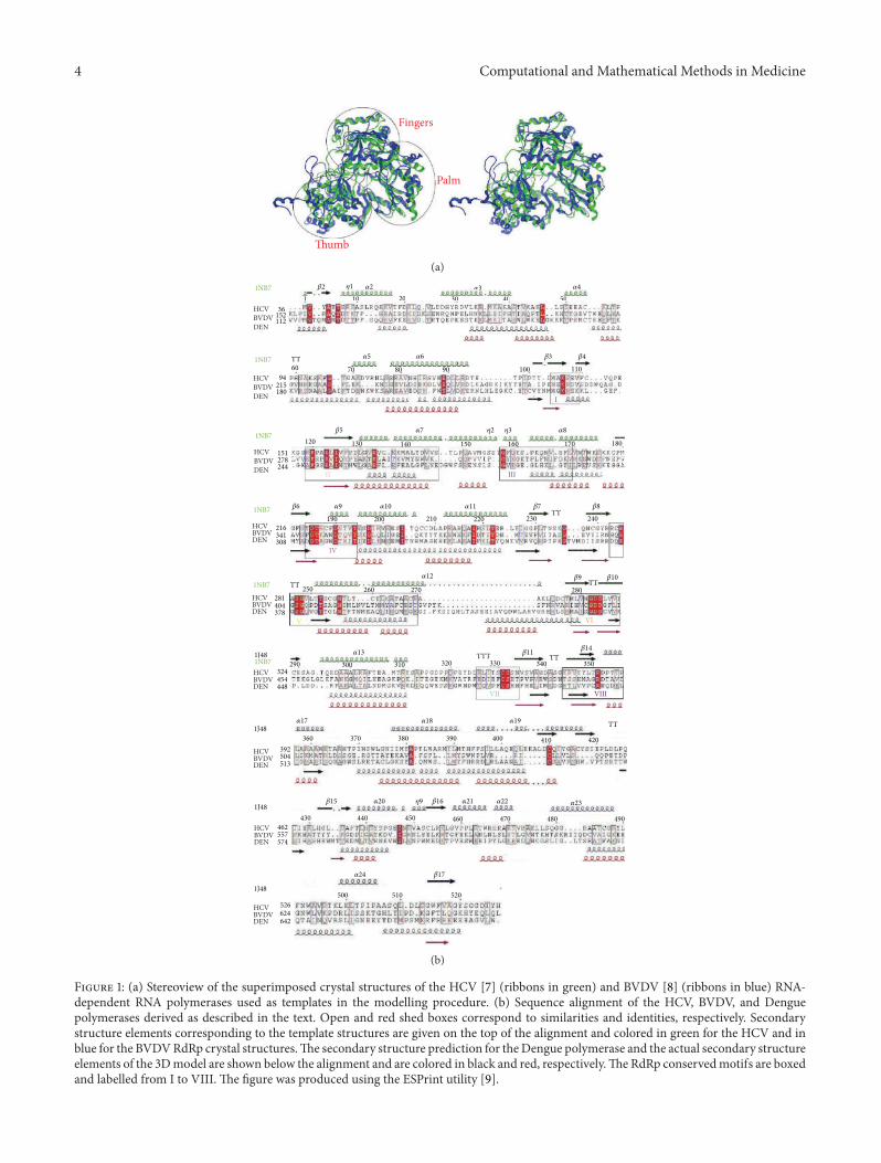
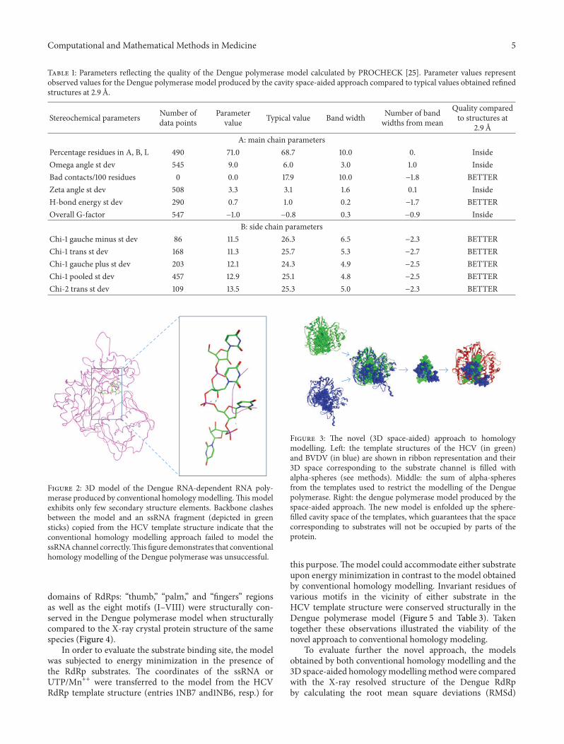
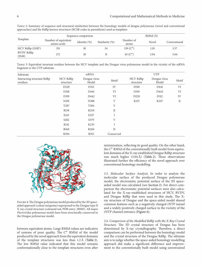
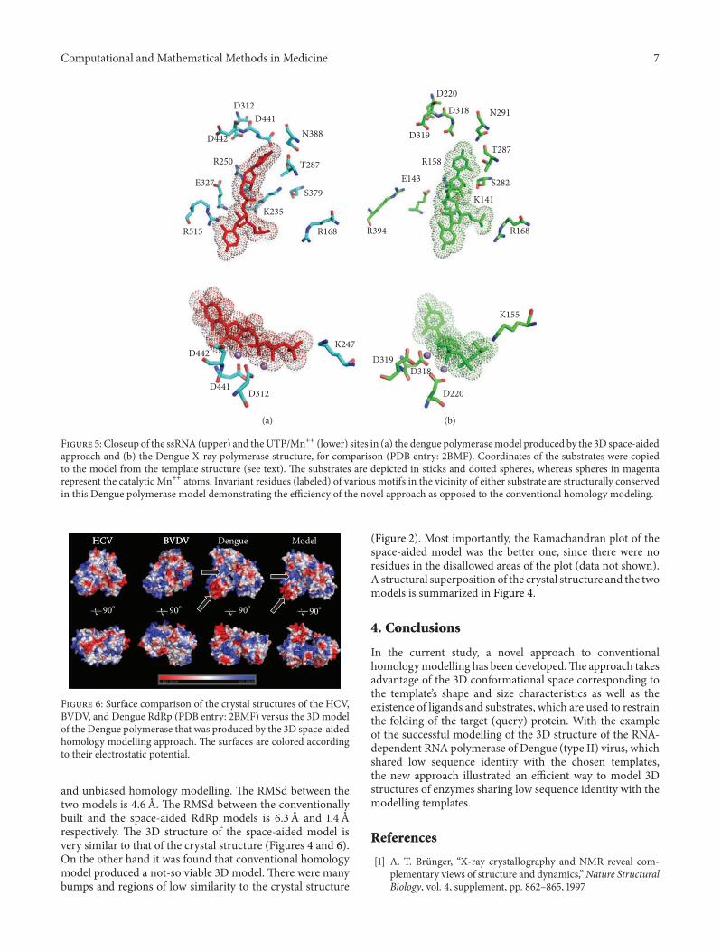
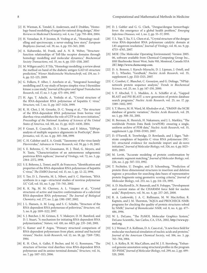


![Protein structure prediction using homology modeling [ Edit ]](https://img.pdfslide.net/doc/110x75/6333b05ba6138719eb0aba9d/protein-structure-prediction-using-homology-modeling-edit-.jpg)


