Embed Size (px)
Citation preview

Brain (1996), 119, 1737-1749
Spasticity and muscle contracture following strokeN. J. O'Dwyer,1 L. Ada1 and P. D. Neilson2
1 School of Physiotherapy, Faculty of Health Sciences,The University of Sydney; 2Cerebral Palsy Research Unit,Institute of Neurological Sciences, The Prince HenryHospital and School of Electrical Engineering,University of New South Wales, Sydney, Australia
Correspondence to: N. J. O'Dwyer, School ofPhysiotherapy, Faculty of Health Sciences, The Universityof Sydney, PO Box 170, Lidcombe, Sydney, NSW 2141,Australia
SummaryIt has become increasingly recognized that the majorfunctional deficits following brain damage are largely dueto 'negative' features such as weakness and loss ofdexterity rather than spasticity. A variety of studies suggestthat spasticity is a distinct problem and separate fromthe loss of dexterity, but that it may be implicated in theformation of muscle contracture and even in the recovery ofstrength. In order to address these issues, we examined therelationship between spasticity, contracture, strength anddexterity in the affected upper limb following stroke.Spasticity was measured both as increased tonic stretchreflexes and increased resistance to passive stretch (hyper-tonia). Twenty-four patients were recruited non-selectivelyfrom three rehabilitation units within 13 months of theirstroke. Few patients exhibited increased tonic reflexes buthalf were found to have muscle contracture, the earliest at
2 months following stroke. Hypertonia was associated withcontracture but not with reflex hyperexcitability. Increasedtonic stretch reflexes were observed only in a subgroup ofthose with contracture and where present could usually beelicited only at the end of muscle range. This finding suggeststhat instead of spasticity causing contracture, contracturemay actually potentiate spasticity in some patients. However,the majority of patients with contracture did not haveincreased tonic stretch reflexes. In addition, we found norelationship between spasticity and either weakness or lossof dexterity. Therefore, while hypertonia remains an importantproblem following cerebral lesions, it would appear thatthe amount of attention directed to reflex hyperexcitabilityassociated with spasticity is out of proportion with its effects.Consequently, hypertonia needs to be clearly distinguishedfrom reflex hyperexcitability in patients with spasticity.
Keywords: spasticity; muscle contracture; stroke; reflex hyperexcitability; hypertonia
Abbreviations: IEMG = rectified and low-pass filtered EMG; MAS = Motor Assessment Scale; MCA = middle cerebral artery
IntroductionIt has been traditional to characterize the signs of braindamage as either 'positive', i.e. those features that are notnormally present such as spasticity and abnormal postures,or 'negative', i.e. those features that have been lost such asstrength and dexterity. Over the past 20 years, it has beenincreasingly recognized that the major functional deficitsfollowing brain damage are largely due to the negativefeatures (e.g. Landau, 1974, 1988; Burke, 1988). However,patients often develop secondary complications such asmuscle contracture. These secondary complications may inturn interfere with the recovery of function. The presentstudy examines the relationship between spasticity, musclecontracture, strength and dexterity across a population ofpatients during recovery of function following stroke.
Much effort has been directed at reducing spasticity aspart of the treatment and rehabilitation of brain-damaged
© Oxford University Press 1996
patients. This, has stemmed from the historical view thatspasticity was the major determinant of motor dysfunction.One of the first investigators to question this view wasLandau (1974) and since then a variety of experiments havesupported his position. The historical view would suggestthat inhibition of spasticity should result in an improvementin function. However, when hyperactive reflexes have beensuppressed with drugs, in people with either stroke (McLellan,1977) or cerebral palsy (Nathan, 1969), there has been noparallel increase in movement control. Similarly, when adultsand children with cerebral palsy have learnt to reducespasticity following training, it has not lead to an improvementin voluntary control of movement (Neilson and McCaughey,1982). In addition, Sahrmann and Norton (1977) demonstratedthat impairment of movement following stroke is notprimarily due to reflexes in the spastic antagonist muscles
by guest on February 10, 2016http://brain.oxfordjournals.org/
Dow
nloaded from
1738 N. J. O'Dwyeret al.
but to abnormalities of agonist contraction. Abnormal motorunit firing patterns have also been documented in the musclesof spastic patients (e.g. Rosenfalck and Andreassen, 1980;Young and Wierzbicka, 1985; Farmer et al, 1993).Notwithstanding these findings, the continued interest inmechanisms of and therapeutic interventions for spasticitysuggests that it retains a focus that is out of step with itstheoretical importance.
Some of the confusion about the role of spasticity inmovement dysfunction has probably arisen because theclinical measurement of spasticity involves gauging theresistance of the limbs to passive movement. This proceduredoes not allow different causes of an increase in resistanceto be identified. Historically, such an increase has beenassumed to be due to exaggerated stretch reflexes, but Dietzet al. (1981) provided evidence that altered mechanicalproperties of muscle may contribute to hypertonia in spasticpatients. Perry (1980) was one of the first researchers todocument the clinical observation that spasticity usuallypresented in conjunction with muscle contracture. Halar et al.(1978) demonstrated muscle shortening in the lower limbin stroke patients with clinical contracture and this wasaccompanied by increased passive stiffness of the ankle.Other investigators also have demonstrated increasedpassive ankle stiffness in spastic patients (Gottlieb et al.,1978; Dietz and Berger, 1983), both with (Hufschmidt andMauritz, 1985) and without (Thilmann et al., 1991b) clinicalsigns of contracture. A similar increase in joint stiffness, alsoattributable to passive soft tissue changes, has been observedin the lower limb of spastic cerebral-palsied children andadults with contracture (Tardieu etal., 1982a; O'Dwyerera/.,1994). Indirect evidence for altered mechanical properties ofupper limb muscles in spastic patients has also been presented(Lee et al., 1987; Dietz et al., 1991). However, despite theseclinical and experimental observations, the nature of therelationship between spasticity and contracture remainsunresolved.
Spasticity has not always been seen in a purely negativelight. Spastic hypertonia has been considered to be superiorto a flaccid paresis (Hufschmidt and Mauritz, 1985) andBerger et al. (1984) suggested that the hypertonicity of legextensor muscles enables hemiparetic patients to supporttheir body during locomotion. According to Dietz et al.(1986), the mechanism underlying this ability may lie inthe alterations of active biomechanical properties of musclefibres implied by histochemical changes in spastic muscle(Edstrom, 1970; Dietz et al., 1986). Both Twitchell (1951)and Brunnstrom (1970) have noted that, during recoveryfollowing hemiplegia, muscle stretch reflexes return beforevolitional movement and it has been shown in stroke patientsthat stretch-evoked reflex activity can augment voluntarymuscle activity (Norton and Sahrmann, 1978). Accordingto the 'servo-assistance' hypothesis of Matthews (1972),voluntary muscle activation is normally augmented by reflexafferents and this notion has been supported by recent studiesin humans showing that muscle afferents provide a net
facilitation to the motoneuron pool, reflexly increasing motoroutput at all levels of voluntary drive by approximately one-third (e.g. Gandevia et al., 1990). Such reflex augmentationof voluntary muscle activity could be even greater in thepresence of spasticity, whether due to lowered reflex threshold(Katz and Rymer, 1989) or increased reflex gain (Thilmannet al., 1991a). It is possible, therefore, that spasticity maybe positively related to strength during recovery of functionfollowing brain damage.
Taken together, these studies suggest that spasticity is adistinct and separate problem to the loss of dexterity whichfollows brain damage, but that it may be implicated in theformation of muscle contracture and even in the recovery ofstrength. It should be noted, however, that the findingsoutlined above include studies of congenital as well asacquired brain damage. In the present study we direct ourattention to hemiparesis following stroke. We measuredspasticity, contracture, strength and dexterity in order toexamine the relations between them in 24 patients who werewithin 1 year of their stroke. It has recently been shown thatthe severity of motor impairment and the patterns of motorrecovery are similar for the upper and lower limbs followingstroke (Duncan et al., 1994). The affected upper limb wasstudied here, specifically the elbow flexor muscles, becauseclinical (Ada and Canning, 1990) and experimental (Leeet al, 1987; Dietz et al, 1991; Thilmann et al, 1991a)observations suggest that they are a common site for thedevelopment of both contracture and spasticity. Furthermore,contrary to earlier clinical impressions, the elbow flexormuscles have been found to be relatively more weakenedthan the extensors (Colebatch et al., 1986).
MethodsSubjectsIn earlier studies of stroke-induced hemiparesis, patientswere selected on the basis of clinically detectable spasticity(e.g. Lee et al., 1987; Powers et al, 1988, 1989; Dietzet al., 1991; Thilmann et al., 1991a; Katz et al., 1992;Ibrahim et al., 1993a). In the present study, we wished tostudy the relationships between spasticity and several othervariables in a group which was representative of the strokepopulation undergoing rehabilitation. Therefore, we testedall hemiparetic patients, both in-patients and out-patients,in three metropolitan rehabilitation units during a 1-monthperiod. Subjects were only excluded if they had suchsevere language, perceptual or cognitive deficits that theywere unable to follow the instructions required to participatein the study. Since most of the procedures did not involveactive participation, this excluded very few patients.Spasticity is a secondary adaptation to upper motor neuronlesions (Burke, 1988) that requires time to develop(Brown, 1994) and similar considerations apply to musclecontracture. Therefore, we did not accept patients earlierthan 1 month post-stroke. This process yielded 24 subjects
by guest on February 10, 2016http://brain.oxfordjournals.org/
Dow
nloaded from

Spasticity and contracture following stroke 1739
Table 1 Subject characteristics
Subject
12*34567*89
1011*121314*151617181920212223*24
Age(years)
877479515861586278366571427874547464674040726552
Gender
MFMMFMFFMFMFFFFMMFMMMFMM
Side ofhemiparesis
RLLLRLRRLLRRRRLRRRRLLRLL
Time sincestroke (months)
22.5312.52.5376
126.57.52.5253.57688
1377.52
Site of lesion (on CT scan)
Brainstem(R) MCA territory(R) Cerebral penduncle(R) Frontoparietal area(L) Frontal lobe(R) Frontoparietal area(L) Frontal lobe(L) Basal ganglia(R) Basal ganglia(R) Parietal and basal ganglia(L) Internal capsule(L) Basal ganglia(L) Basal ganglia(L) Internal capsule(R) Internal capsule(L) Basal ganglia haemorrhage(L) Posterior external capsule(L) Internal capsule(L) Frontoparietal area(R) Frontoparietal area(R) MCA territory(L) Pontomedullary area(R) Frontoparietal area(R) MCA territory
MAS item 6(0-6)
316140001211606261010530
Subjects were all found to be right-handed. MCA = middle cerebral artery. *Subjects with hyperexcitable tonic stretch reflexes.
with a wide range of characteristics (see Table 1), all ofwhom had suffered a stroke within the last year. Theirfunctional ability is indicated in Table 1 by scores between0 and 6 on the upper arm category (item 6) of a clinicalscale, the Motor Assessment Scale (MAS) (Carr et ai,1985), and it can be seen that the degree of impairmentin the group ranged from mild to severe. The experimentalprocedures were approved by the relevant institutionalethics committee and all subjects gave informed consentbefore data collection was undertaken.
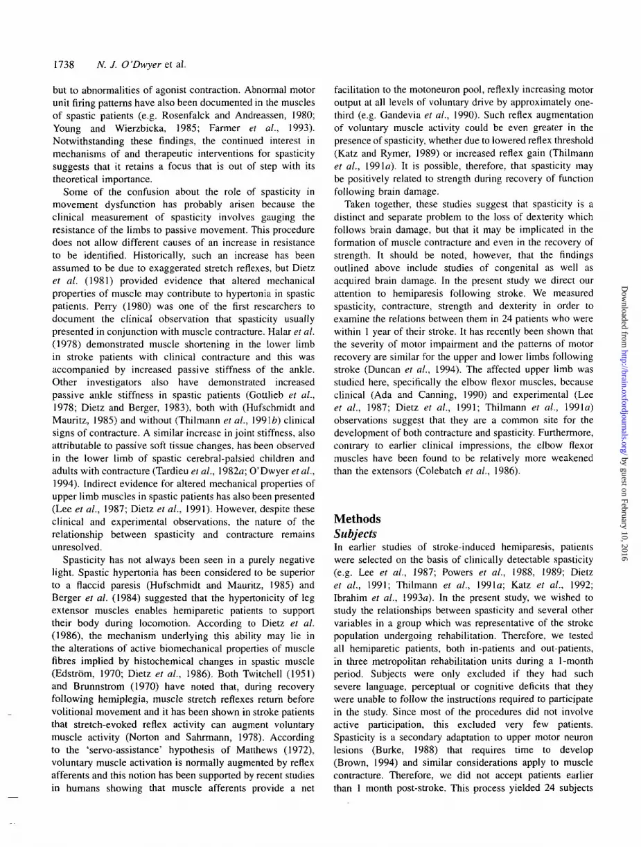
Experimental set-upThe equipment measured elbow joint displacement, torqueand biceps muscle activity (Fig. 1). The subject sat at a tablewith the affected forearm securely supported by a horizontalframe. Rotation of the frame, whether by the experimenteror the subject, produced a change in elbow angle that wasmeasured by a potentiometer aligned directly below theelbow joint. A load cell (capacity 250 N; linearity 97%)attached to the frame measured the resistance of the forearm tomovement. Silver silver-chloride surface electrodes measuredbiceps muscle activity.
After amplification of the EMG activity (X5000) andtorque (X1000), the elbow angle, torque and EMG activitywere sampled by a 16-bit analog to digital converter at1000 Hz and stored on computer. In order to removeany 50 Hz line frequency interference or low-frequencymovement artefact, the EMG was high-pass filtered (digital
8th-order Butterworth) at 80 Hz. Subsequently, the EMGactivity was full-wave rectified and then, along with theelbow angle and torque signals, low-pass filtered (digital 8th-order Butterworth) at 5 Hz to obtain a DC voltage. Thisrectified and low-pass filtered EMG (IEMG) was proportionalto the contraction level of the muscle. The cut-off frequencywas chosen because all frequencies of interest were <5 Hz.
Measurements of spasticity and one measure of dexteritywere carried out using this set-up. The procedures forcollection of EMG activity were standardized betweensubjects to promote reliability. For example, for each subjectthe electrode placement was the same relative to the musclebelly and the impedance was kept as low as possible bythorough abrading and cleaning of the skin. In addition, thesame experimenters always carried out the same part ofthe procedures.
Definition and measurement of spasticityThe most widely accepted definition of spasticity describesit as 'a motor disorder characterized by a velocity-dependentincrease in tonic stretch reflexes ('muscle tone') withexaggerated tendon jerks, resulting from hyperexcitability ofthe stretch reflex, as one component of the upper motorneuron syndrome' (Lance, 1980). The increased stretchreflexes are assumed to cause hypertonia, i.e. increasedresistance to passive movement. Following stroke, however,increased resistance to passive movement may be the resultof altered passive mechanical properties of muscle tissue as
by guest on February 10, 2016http://brain.oxfordjournals.org/
Dow
nloaded from

1740 N. J. O'Dwyeret al.
Fig. 1 Experimental set-up. The arm-frame could be clamped tomeasure isometric strength or could move freely so that either thesubject could track the target (square) on the computer screen orthe experimenter could stretch the biceps. A high-backed chairsupported the subject so that when the arm was resting in thearm-frame, movement was confined to the elbow joint.
well as hyperexcitable reflexes. Therefore, we measured boththe stretch-evoked muscle activity via EMG activity (i.e.reflex hyperexcitability) and the resistance to passive stretchvia the load cell (i.e. hypertonia).
We chose to measure the excitability of the tonic ratherthan the phasic stretch reflex, since it is generally recognizedthat the tonic stretch reflex is of greater physiological andclinical significance (e.g. Lance et al., 1966; Landau, 1974;Neilson, 1993). Reflex hyperexcitability was assessed attwo muscle lengths and velocities since the gain of thetonic stretch reflex has been found to vary with changesin these characteristics of stretch (Neilson and McCaughey,1981; Nash et al., 1989). We stretched the muscles byperforming small amplitude (10° peak-to-peak) quasi-sinusoidal stretching for 30 s at frequencies of 2 and3.5 Hz, producing peak velocities of 60° s"1 and 110° s~",respectively. These frequencies are in the realm of normalmovement but are too fast for consistent voluntary following
(Neilson 1972), particularly for hemiparetic patients. Thestretching was performed at two muscle lengths; the elbow wasflexed at 90° or at 20° from full extension. The latter positionnear the end of the muscle range was designed to gauge theeffect of any muscle contracture that might be present.Therefore, three conditions were tested: 90°±5° at 2 Hz,90°±5° at 3.5 Hz, 20°±5° at 2 Hz.
Subjects relaxed, as confirmed by the absence of EMGactivity, and then the forearm was manually rotated back andforth about the elbow. The rotation was synchronized with ametronome in order to control the frequency. The elbowangle was displayed on the computer monitor so that theamplitude of stretch could be controlled. The consistency ofthe imposed stretch, as measured by spectral analysis, waskept high both for frequency (mean±SD = 2 ±0.1 Hz and3.5±0.1 Hz) and amplitude (13°±1°) (see top traces inFig. 2). Normally, no EMG activity is observed when therelaxed biceps muscles of a neurologically-normal personare stretched in this manner and at these velocities (e.g.Burke, 1983; Ibrahim et al., 1993a) and we have recentlyvalidated this observation in normal elderly subjects (W. Yeo,L. Ada, N. J. O'Dwyer and P. D. Neilson, unpublishedobservations). Therefore, any stretch-induced EMG activityobserved was taken as evidence of reflex hyperexcitability.This procedure makes no assumption regarding whether suchEMG activity is due to lowering of reflex threshold (Katzand Rymer, 1989), increase in reflex gain (Thilmann et al.,1991a) or more complex changes in modulation of reflexthreshold or gain (Neilson and Lance, 1978; Gottlieb andMyklebust, 1993).
The angle and IEMG signals were subjected to crosscorrelational and spectral analysis (McRuer and Krendel,1959; Neilson, 1972) to quantify any tonic stretch reflexespresent. This analysis allows stretch-evoked muscle activityat the stretching frequencies of 2 and 3.5 Hz to bedistinguished from other activity unrelated to the stretch. Themagnitude of these stretch reflexes reflects the degree ofreflex hyperexcitability and was quantified by the gain of thetonic stretch reflex, i.e. by the magnitude of the stretch-evoked IEMG activity divided by the magnitude of stretch.If stretch reflexes of any reasonable magnitude are present,the mean level of IEMG activity would also be expected toincrease during the stretching procedure. Therefore, inaddition to the gain of the reflex, the mean level of IEMGactivity during stretch was measured and compared with thatduring rest. The angle and torque signals were also subjectedto cross correlational and spectral analysis and the resistanceto passive movement was quantified by the gain of thetorque-angle relationship, i.e. by the magnitude of the stretch-evoked torque divided by the magnitude of the stretch.Figure 2 provides examples of angle, torque, EMG and IEMGsignals for subjects with and without tonic stretch reflexes.
Definition and measurement of contractureThe number of sarcomeres in a muscle is not fixed, beingcapable of either increasing or decreasing even in adult
by guest on February 10, 2016http://brain.oxfordjournals.org/
Dow
nloaded from

Spasticity and contracture following stroke 1741
(A)
«03C
degrees
30 T
20 •
10
(B)degrees
30 T
20
10 J
(C)
<
o
degrees
30
20
10
Nm10 T
5
0 -1
-5
-10 J-
^AAAAAA/
microvolts
microvolts30 T
20
10 -
o2
seconds
i o TN m
5
0
-5
-10 J-
AMAAM
microvolts
1 50
0
-50
L _
microvolts30 T
20
* 10 -•
0
(3
1 2seconds
Nm
1]•iWWVW
-10
microvolts
50
•§> - 5 0 Imicrovolts
30 T
20 -•
10 -
2seconds
Fig. 2 Responses to passive stretch of the biceps at 20° at 2 Hz. Traces of elbow angle, torque, biceps EMG and IEMG. (A) Subject 14who showed the largest reflex activity in response to stretch under all conditions. The bursts of muscle activity are coherent with thestretch (coherence 0.96) and have a phase lead (70°) showing their velocity dependence. (B) Subject 11 showing small but similarabnormal stretch reflex responses (coherence 0.99) and phase lead (88°). (C) Subject 6 showing no response to the stretch of biceps(coherence only 0.17). The distortion in the torque signal is due to a small amount of friction in the joint of the arm-frame. It contributed<1% of the power in the torque signal at the stretching frequency and so had negligible impact on the torque-angle gain.
muscle (Tabary et al., 1972; Williams and Goldspink, 1973).Contracture consists of a shortening of muscle length due toa decrease in the number of sarcomeres in series along themyofibrils, accompanied by an increase in the resistance topassive stretch (Tardieu et al., 1982a; Bax and Brown, 1985;O'Dwyer et al., 1989). Muscle fibres are not lost or replacedby connective tissue, as is often assumed (e.g. Lee et al.,1987). Tardieu et al. (1982a) reported that in contracturein cerebral palsy, muscle structure on light and electronmicroscopy was normal apart from reduction of fibre lengthand no excessive connective tissue was observed. The reducedcompliance is probably attributable to remodelling of muscleconnective tissue (O'Dwyer et al., 1989; Goldspink andWilliams, 1990). The range of joint motion, therefore, isreduced both by the shortening of the muscle fibres and bythe loss of muscle compliance (Williams, 1988).
Despite the reduction in muscle compliance, if a contractureis minor in extent it may still be possible to achieve a normalrange of motion by the application of sufficient force. Forexample, Halar et al. (1978) applied a force of 40 lbs (178N) and achieved similar magnitudes of ankle dorsiflexion onthe affected and unaffected sides of hemiplegic patients, even
in the presence of clinical contracture. Consequently, in orderto assess the magnitude of joint motion, it is important notonly to standardize the force applied but also not to exceedthe magnitude of force that is normally sufficient to stretchthe muscles through the joint range. In addition, if a multi-joint muscle is being assessed (as is the case of the bicepsbrachii which crosses both the elbow and shoulder joints), itis important to standardize the position of the joint not beingmeasured. It is not easy to apply these controls in theclinical assessment of muscle contracture, yet without themcomparison cannot be made across subjects or with thenormal population. In the present study, the subjects laysupine with their upper arm resting horizontally on a firmbed, thereby placing the shoulder in neutral. The elbow jointwas extended firmly by the experimenter and held in thisposition for 30 s so as to allow time for relaxation in casemuscle activity was elicited by the manoeuvre. Then the armwas released and maintained in extension solely by theweight of the forearm due to gravity. Selective EMG activitymonitoring confirmed that the elbow flexor muscles wererelaxed in this posture. Normally the forearm will lie flat onthe bed under these conditions. The position of the arm was
by guest on February 10, 2016http://brain.oxfordjournals.org/
Dow
nloaded from

1742 N. J. O'Dwyer et al.
photographed and contracture of the elbow flexor muscleswas quantified by measuring the angle of the forearm relativeto the bed from the photograph. The greater the degree offlexor contracture, the greater the angle. It should be noted,however, that the biceps brachii in this posture is still notfully lengthened across the shoulder joint, so that a forearmflexion measurement of 0° does not entirely rule out a smallcontracture. Therefore, this procedure underestimates the trueextent and frequency of elbow flexor contracture.
Definition and measurement of strengthSince the elbow flexors were the muscles of major interestin this study, strength was measured during a maximalisometric flexor contraction of the elbow, with the arm-framefixed at 90°. Both flexor torque and IEMG were collectedand these two measures were subsequently found to besignificantly correlated (r = 0.56; P<0.01). It is likely that thetorque was influenced to a variable degree by cocontractionof the extensors and since the flexor IEMG provided anunambiguous estimate of the patients' ability to voluntarilyactivate the muscles, we chose to present this as the measureof strength.
The subjects were required to relax for 5 s, pull intoflexion maximally for 5 s and relax again for 5 s. Duringthis procedure the subject was provided with visual feedbackfrom the display, since this has been shown to improve theachievement of maximal output (Jones et al., 1979). Thebest of three attempts was taken to represent the subject'smaximum. Flexor IEMG activity was averaged over the restand contraction periods and the difference between themtaken as maximal voluntary effort.
computer screen. The subject sat at the table with the forearmsupported in the arm-frame and controlled the response cursorvia 10° of elbow flexion and extension (±5°) around a meanposition of 90°, one of the positions at which reflex excitabilitywas assessed. Following familiarization with the task, 1-mintests of a slow and fast target were recorded. The targetsconsisted of random numbers filtered (2nd-order Butterworthlow-pass) at 0.25 Hz for the slow target and at 0.5 Hz forthe fast target.
While performance of this task depended on coordinatedcontrol of the amplitude and timing of elbow flexor andextensor muscle activity, assessment of performance wasbased on the relationship between the target and the subject'sresponse controlled by their elbow angle. A traditionalmeasure of overall tracking performance is the root meansquare value of the error, i.e. the difference between thetarget and the response signal (McRuer and Krendel, 1959).However, this becomes a less satisfactory measure ofperformance as the target moves faster and a significanttime delay is introduced. Therefore, a more detailed cross-correlational and spectral analysis was carried out in orderto assess the similarity of the target and response waveforms.This analysis provides a measure of the overall coherencebetween the target and response, i.e. the proportion of theresponse that is correlated with the target over the frequencybandwidth of the target. The coherence at each frequency isanalogous to the r2 measure in a regression analysis. Forideal tracking, the overall coherence would be one. Figure 3provides examples of both slow and fast tracking from twosubjects with different abilities.
Definition and measurement of dexterityDexterity is adroitness or skill in using the body and it istherefore difficult to assess comprehensively. On the onehand, general measures of everyday tasks which requireco-ordination of limb synergies tend to obscure the roleof individual muscles. On the other hand, measures ofspecific muscles can be criticized for not being relevantto general function. We have, therefore, attempted tomeasure both levels of dexterity.
The overall ability to use the upper arm was measuredusing the MAS which provided a measure of motor functionrelated to everyday tasks. Scores are assigned from 0 to 6,where 0 represents no activity and 6 is the highest scorepossible. Item 6 measures upper limb function and includestasks such as 'raising the arm to shoulder height and holdingfor 10 s'. The scale has been shown to be reliable when usedby a trained tester (Carr et al., 1985), as it was duringthis study.
Specific dexterity of the elbow joint was assessed byrequiring the subject to track the movements of a target ona computer screen using only elbow flexion and extension.The target moved irregularly back and forth across the
Statistical analysisThe measurements of spasticity (reflex hyperexcitability andhypertonia), contracture, strength and dexterity yielded 11variables for statistical analysis. Reflex hyperexcitability wasrepresented by the gain of the tonic stretch reflex andhypertonia by the gain of the torque-angle relationshipduring stretching for the three conditions. Contracture wasrepresented by the angle of elbow flexion. Assessment ofstrength yielded IEMG elbow flexor activity. Generaldexterity measured by the clinical scale yielded one variable,whereas specific dexterity was represented by the overallcoherence in the slow and fast tracking conditions. Thevalues of these variables for all subjects are presented inTable 2.
Most of the data were examined descriptively. StandardANOVAs were used to examine (i) the difference in meanIEMG between rest and stretching, (ii) the difference inresistance to stretch between subjects with reflexes and thosewithout, and (iii) the difference in resistance to stretchbetween subjects with contracture and those without. Finally,the relations between variables, including time since stroke,were analysed by Pearson's product moment correlation.
by guest on February 10, 2016http://brain.oxfordjournals.org/
Dow
nloaded from

Slow Trackingdegrees
Spastlclty and contracture following stroke
Fast Trackingdegrees
Subject
13
1743
Subject20
Coherence0.64
5 seconds 5 seconds
Fig. 3 Ten-second excerpts of tracking responses to the slow and fast targets by two subjects withdiffering levels of performance. The target is the solid line and the subject's response the dashed line.Both subjects track the slow target quite well, although Subject 20 (bottom traces) illustratesovershooting when trying to move the cursor back on target. Subject 13 (top traces) reproduces thefaster target quite well but with a time delay, which is a normal feature, whereas Subject 20 reproducesvery little of the fast target waveform, tending to 'freeze' or move with very little amplitude and aprolonged time delay.
Table 2 Individual variables
Subject
123456789
101112131415161718192021222324
MeanSD
Time since
stroke(months)
2.02.53.01.02.52.53.07.06.0
12.06.57.52.52.05.03.57.06.08.08.0
13.07.07.52.0
5.33.1
Contracture:
elbowflexion(degrees)
632050
14067450
1300052000
220
45.4
Spaslicity
Reflex excitability:gain of ionic stretch reflex
90°@2Hz(uV deg"1)
0.00.00.00.00.00.00.00.00.00.00.00.00.00.50.00.00.00.00.00.00.00.00.00.0
90°@3.5Hz(uV deg-1)
0.00.00.00.00.00.00.00.00.00.00.00.00.01.10.00.00.00.00.00.00.00.00.00.0
20°@2Hz(uV deg-1)
0.000.480.000.000.000.000.370.000.000.000.200.000.001.260.000.000.000.000.000.000.000.000.810.00
Hypertonia:resistance to
90°@2Hz '(Nm deg-1)
0.4460.3360.3450.4280.4110.3160.4320.2230.3440.4220.4120.3690.3500.3800.371 (0.3130.5000.3660.4120.104 (0.2570.317
jassive movement
X)°@3.5HzNm deg-1)
.390
.280
.190
.356
.197
.122
.395
.071
.109
.155
.339
.242
.253
.194).963.244.611.241.501
).9O7.104.047
0.278 0.9610.303
0.351
.124
.2080.083 0.17
20°@2Hz(Nm deg"1)
0.4130.5350.5690.4580.5040.5230.4360.4050.5260.4140.6060.4520.4120.5270.3720.4070.4350.4360.6160.3430.5650.4950.5060.399
0.4730.074
Dexterity
MAS:item 6(0-6)
316140001211606261010530
Slow track:overallcoherence
0.410.000.570.280.280.000.000.000.000.560.000.520.800.00.320.490.530.430.000.640.370.490.460.00
0.300.25
Fast track:overallcoherence
0.320.000.410.080.010.000.000.000.000.310.000.330.670.000.260.170.350.200.000.270.050.300.310.00
0.170.18
Strength:elbow
flexorsIEMG(uV)
8918808270
1242
31149
24255101059
70
478927870
4153
by guest on February 10, 2016http://brain.oxfordjournals.org/
Dow
nloaded from

1744 N. J. O'Dwyeret al.
u.o
- 0 . 5
c
O 0.40)o>c
<i 0.3cr,2
n •?
O M ^ * * ^ . . . . . • • • • • •
0.32 . . - - • "
0.50
0.44. . . 0
•
- e - withcontracture
o withoutcontracture
90° 20°Elbow Position
Fig. 4 Mean torque-angle gains (Nm deg"1) at 2 Hz stretchingat 90° and 20° elbow angle for subjects with and withoutcontracture. Subjects with contracture show a higher resistance topassive stretch than subjects without contracture regardless ofmuscle length. As expected, for all subjects the resistance topassive movement increases at the 20° position near the end ofrange of joint excursion.
ResultsFew subjects had demonstrable tonic stretch reflexes. Fivesubjects exhibited reflexes at 20°±5° at 2 Hz but only oneexhibited reflexes in all three stretching conditions. Wherepresent, the magnitude of this reflex activity was low;typically the bursts of EMG activity during stretching werein the range of ±50 (iV amplitude. However, the change inmean IEMG from rest to stretching was significantly increasedfor those subjects with stretch reflex activity compared withthose without,[F(\,22) = 7.8; P < 0.025], although this wasunlikely to be of functional significance since the increasewas only from 4 to 8 |iV of IEMG.
While very few subjects showed reflex activity, about halfhad a demonstrable contracture. Loss of range of elbow jointextension was observed in 13 of the 24 subjects, the size ofthe flexion contracture ranging from 2° to 22°. Furthermore,contracture was associated with an increased resistance topassive movement. As illustrated in Fig. 4, a significantincrease in torque-angle gain at 2 Hz stretching was observedin subjects who had a contracture compared with thosewithout [F(l,22) = 9.03; P < 0.01]. Not surprisingly, for allsubjects, the resistance to stretch was significantly increasedat 20° near the end of range of joint movement, comparedwith 90° close to the middle of the range [F(l,22) = 33.85;P < 0.001]. However, the size of this increase was notsignificantly different between subjects with and withoutcontracture [F(l,22) = 0.0005; P = 0.98], i.e. in the presenceof contracture, the increased stiffness was present throughoutthe joint range, both at 90° and 20°.
The increased resistance to passive stretch associatedwith contracture was independent of reflex hyper-excitability. Thus, the resistance was still significantlyincreased [F(l,17) = 5.83; P < 0.05] if the subjects withreflex hyperexcitability were excluded from the com-parison. Furthermore, among the subjects with contracture,
the presence of reflexes did not produce a significant increasein the resistance to stretch [F(l,l 1) = 0.04; P = 0.84]. Eventhough the stretch-evoked EMG activity was of a magnitudewhich increased the mean IEMG above that during relaxation,it did not increase the resistance beyond that due to con-tracture.
The correlations between the various measures arepresented in Table 3. There were no significant correlationsbetween the 'positive' and 'negative' features followingstroke but there were significant correlations within thesesubgroups of features. Thus, the negative features, i.e. strengthand the three measures of dexterity (MAS score and slowand fast tracking ability), were all significantly correlated.Only some of the positive features (contracture, reflexhyperexcitability and hypertonia) were correlated and mostof these correlations were attributable to 'outlier' effects dueto the fact that only one subject exhibited tonic stretchreflexes in the 90° stretching conditions. There was, however,one correlation of interest between tonic stretch reflexes at20° and contracture (r = 0.74). This reflects the fact that allfive subjects who had reflex hyperexcitability also hadcontracture. However, four of these subjects exhibited tonicstretch reflex activity only with the biceps in this lengthenedposition and not at 90°. Furthermore, another eight subjectshad contracture but no reflex hyperexcitability. Finally, noneof the variables measured correlated with the time sincethe stroke.
DiscussionOur original expectation that spasticity and contracture wouldbe related was not supported by the findings of this study.Few tonic stretch reflexes were observed in response topassive stretch in this group of hemiparetic patients, eventhough half of them exhibited a contracture. Reflex activitywas present in only seven out of 72 stretching trials and inonly one patient under every stretching condition. This lowoccurrence of reflex hyperexcitability transpired despite thelikely damage to corticofugal pathways in most patients (seeTable 1) and the fact that many patients presented with thecharacteristic 'hemiplegic posture' of a slightly flexed elbowthat is associated clinically with spasticity.
Other studies of hemiparetic stroke patients have reportedreflex responses to relatively slow stretches comparable induration (250 ms and 143 ms) and mean velocity (40°s~'and 70°s~') with those employed in the present study (Powersetai, 1988, 1989; Thilmann et a/., 1991a; KatzetaL, 1992).The only difference that might account for the discrepancyin findings would appear to be the amplitude of stretch,which was 10° in the present study compared with 30° or morein these earlier studies. Nevertheless, smaller stretches (12°),rapidly applied (60 ms, 200°s"'), have been shown to elicitphasic reflexes in hemiparetic patients (Ibrahim et al., 1993a).Perhaps more important than the parameters of stretch arethe subject characteristics. The subjects in earlier studiesusually had clinically manifest, chronic (usually >1 year)
by guest on February 10, 2016http://brain.oxfordjournals.org/
Dow
nloaded from

Spasticity and contracture following stroke 1745
Table 3 Relationship between variables (Pearson product-moment correlations)
Time since Contracture: Positive features
stroke elbow
(months) flexion
(degrees)
Negative features
Hyperreflexia:
gain of ionic stretch reflex
Spasticity: Dexterity
Hypertonia: resistance to passive movement
90°@2Hz 90°@3.5Hz 20°@2Hz 90°@2Hz 90°@3.5Hz 20°@2Hz MAS: Slow track: Fast track:
(HV deg"1) (|iV deg"') (uV deg"1) (Nm deg"1) (Nm deg"1) (Nm deg"1) item 6 overall overall
(0-6) coherence coherence
Strength:
elbow
flexors
IEMG
(UV)
Time since
stroke
Contracture
Tonic
stretch
reflexes
Resistance
to passive
movement
Dexterity
A
A
B
C
D
E
F
G
H
IJ
K
B0.01
C D E F G
-0.22 -0.22 -0.19 -0.10 -0.20
0.35
H0.11
I
-0.11
J
0.25
K
0.09
L
-0.10
0.35
1.00**
0.74*
0.77**
0.77**
0.21
0.15
0.15
0.09
0.00
-0.01
-0.01
-0.03
0.77*
0.07
0.09
0.09
0.15
0.20
0.24
-0.15
-0.20
-0.20
-0.21
0.25
0.00
-0.18
-0.13
-0.24
-0.24
-0.30
-0.11
-0.13
-0.41*
0.65*
-0.06
-0.20
-0.20
-0.21
0.03
-0.05
-0.38
0.75*
0.90*
0.02
0.05
0.05
0.07
0.00
0.06
-0.10
0.46*
0.59*
0.69*
*P < 0.05; **spurious correlations due to the fact thai only one subject had reflex activity at 90° position.
spasticity (Lee et al., 1987; Powers et at., 1988, 1989;Thilmann et al., 1991a; Katz et al., 1992; Ibrahim et al.,1993a), making it highly probable that they would exhibitthe abnormal tonic or phasic stretch reflexes that were reportedin these studies. These successive reports of abnormal reflexactivity may have perpetuated a focus on spasticity in theclinic. In the present study, the subjects were drawn as non-selectively as possible from three standard rehabilitation unitswithin 1 year following their stroke and they are thereforemore representative of stroke patients undergoingrehabilitation than previous studies. We have found only asmall proportion of these hemiparetic patients to havespasticity manifest as exaggerated tonic stretch reflexes.
A possible interpretation of this finding is that reflexhyperexcitability may have been present early followingstroke, preceding our investigation, in some subjects.However, this is an unlikely possibility since spasticityappears to be an adaptation to, rather than a direct result of,cerebral damage (Chapman and Wiesendanger, 1982; Burke,1988) and requires time to develop (Brown, 1994). Forexample, Thilmann et al. (1991a) found that spasticity wasrarely apparent during the first month following stroke butthat stretch reflex gain increased over the second and thirdmonth. Almost half (11) of our subjects were seen within3.5 months after their stroke (Table 2) and only three of thesehad reflex hyperexcitability. Furthermore, this interpretationdepends on the premise that early reflex hyperexcitabilityhad disappeared in our subjects by the time of the study.
We have demonstrated a link between muscle contractureand increased resistance to passive stretch. However, theincreased resistance was not dependent on the presence of
tonic stretch reflexes and patients with both reflexhyperexcitability and contracture were no more stiff thanthose with contracture alone. Antagonist muscle activity,which was not measured in this study, would be importanthere only if hypertonia that was not attributable to bicepsreflex hyperexcitability was instead attributable to tricepsreflex hyperexcitability. This appears unlikely, especiallysince abnormalities of flexor muscles appear more pronouncedthan those of extensor muscles in spastic patients (Dietzet al., 1991; Ibrahim et al., 1993a). Therefore, the increasedpassive resistance appears to be attributable to the presenceof contracture rather than reflex hyperexcitability.
The process of adaptive muscle change following cerebrallesions is a complex one. Contracture obviously affectsthe passive non-contractile properties of muscle, but thecharacteristics of the active muscle length-tension curve arealso altered when fibre length is reduced by loss of sarcomeres(Williams and Goldspink, 1978). Atrophy of type II musclefibres and fibre type transformation have been documentedin spastic patients (Edstrbm, 1970; Dietz et al., 1986) andan increased torque output for a given level of EMG activityhas been reported in a number of studies (e.g. Lee et al.,1987; Dietz et al., 1991; Ibrahim et al., 1993a). Given suchfindings as well as those of the present study, it now seemslikely that adaptive changes in muscle tissue are oftenresponsible for the clinical impression of hypertonia. Clinicalmeasures of spasticity measure hypertonia by gauging theresistance to passive displacement of the limb (e.g. Ash worth,1964) but this method cannot distinguish between theperipheral contribution due to muscle adaptation and theneural contribution due to increased stretch reflexes. As noted
by guest on February 10, 2016http://brain.oxfordjournals.org/
Dow
nloaded from

1746 N. J. O'Dwyeret al.
Reflex hyperexcitabilityincrease in Ionic stretch reflexes withexaggerated tendon jerks (Lance, 1980)
CNS LESION HYPERTONIAincreased resistanceto passive stretch
Altered muscle functionreduced activity due to paresis;gene expression and proteinsynthesis in tissue is linked tomechanical activity (Goldspinkand Williams, 1981, 1990)
Altered mechanical propertiesloss ofsarcomeres (Tabary et al, 1972);remodelling of muscle connective tissue(Williams and Goldspink, 1984);altered periarticular connective tissue(Akesonetal, 1974);altered histochemistry indicative ofmuscle fibre transformation(Edstrom, 1970; Dietzet al., 1986)
Fig. 5 Contributions of reflex and muscle adaptation to hypertonia following cerebral lesions.Resistance to passive movement can be increased via reflex hyperexcitability and/or via alteredmechanical properties. Only altered mechanical properties due to contracture appeared to contribute tohypertonia in the patient group in the present study.
by Katz and Rymer (1989) and illustrated in Fig. 5 here,these are separate contributors to clinical hypertonia followingcerebral lesions. In our present patient group, adaptation ofthe mechanical properties of muscle appeared to be theprimary contributor to hypertonia. Therefore, it is necessaryto recognize a clear distinction between reflex hyper-excitability and hypertonia in patients with spasticity.
It has been commonly assumed that the exaggeratedreflexes of spasticity lead to muscle contracture. However,in the current study, the presence of tonic stretch reflexes inonly five of the 13 patients with muscle contracture andprincipally when elicited with the muscles in a lengthenedposition, suggests instead that muscle contracture maypotentiate the stretch reflex, at least in some patients. If amuscle and its spindles shorten due to contracture, thestretching effects of a given change in joint angle differ froma normal muscle in several ways, all of which may increasethe size of the tonic stretch reflex. First, the muscle is broughtcloser to the end of its range, thereby increasing the effectof length-dependent facilitation (Ashby and Burke, 1971;Neilson and Lance, 1978; Nash et al., 1989); secondly, alarger change in relative muscle length is imposed, therebyproducing a larger amplitude stretch of the spindles; thirdly,as noted by Perry (1980), the forces accompanying themovement are transmitted more completely and morepromptly because the tissues are stiffer as a consequence ofthe contracture. These three theoretical mechanisms indicatehow contracture might potentiate the stretch reflex. In linewith this, Perry (1980) has cited clinical experiences whichsuggest that correction of contracture may reduce spasticity.Nevertheless, the nature of the temporal relation betweenspasticity and muscle contracture would best be resolvedby a longitudinal study of a group of patients beginningimmediately after the stroke.
Our findings suggest that contracture was not caused by
spasticity, and thus raise the question of the actual cause.Contracture of the elbow flexor muscles was identified in13 of the 24 patients in the present study. All the patientswere studied within 1 year of their stroke, but contracturewas documented as early as 2 months. The likely mechanismsof development of contracture in hemiparesis following strokecan be found in studies on regulation of muscle length inexperimental animals (e.g. Tabary et al., 1972; Williams andGoldspink, 1971, 1978, 1984; Tardieu et al., 19826; seeO'Dwyer et al., 1989 for review). Such studies show thatskeletal muscle is highly adaptable, so that its structuralcharacteristics are determined by its conditions of use (aclassic biological example of the relation between structureand function). When a muscle is immobilized in a shortenedposition, a shortening of muscle fibre length occurs due toloss of sarcomeres in series, accompanied by shortening ofmuscle connective tissue and an increase in stiffness ofthe muscle. The extent of these changes is illustrated bythe study of Tabary et al. (1972) who produced a 40%reduction in number of sarcomeres in cat soleus muscle withimmobilization in a shortened position for 4 weeks. In strokepatients, the arm may be effectively immobilized in thepresence of paralysis or weakness and this predisposes thepatients to rest their paretic arm in their lap (Ada andCanning, 1990), particularly if, due to difficulty with walking,they spend much of their time sitting. This posture resultssimply as a consequence of convenience and comfort butit subjects the elbow flexor muscles, among others, toimmobilization in a shortened position, precisely the condi-tions shown to produce muscle contracture in experimentalanimals.
In line with earlier studies cited above, we found norelationship between spasticity and motor function. Thiswas true regardless of whether spasticity was measured ashyperexcitable tonic stretch reflexes or increased resistance
by guest on February 10, 2016http://brain.oxfordjournals.org/
Dow
nloaded from

Spasticity and contracture following stroke 1747
to passive stretch and whether motor function was measuredby a general clinical scale or a more specific tracking task.Similarly, there was no relationship between spasticity andstrength. Since few of these stroke patients exhibitedhyperactive tonic stretch reflexes, especially at the elbowposition (90°) where strength was measured, a role forspasticity in recovery of either motor function or strengthwas unlikely in any event. These findings are consistentwith the view, which is gaining increasing acceptance, thatspasticity and the negative features of weakness and loss ofdexterity following brain damage are separate entities (e.g.Carr and Shepherd, 1987; Katz et al., 1992; Thilmann et al.,1993). Indeed, none of the 'positive' and 'negative' featuresmeasured in this study were found to be correlated.
In the present study reflex excitability was investigatedunder passive conditions only, so we cannot comment on thestretch reflex behaviour of our patient group under activeconditions. Just as in normal subjects, the response to musclestretch in hemiparetic subjects changes between rest andactivity (Ibrahim et al., 1993a). While short-latency reflexesare exaggerated during activity, long-latency reflexes havebeen shown to be reduced in amplitude (e.g. Berger et al.,1984; Cody et al., 1987; Dietz et al, 1991; Ibrahim et al.,1993a). Furthermore, reflexes elicited under active conditionsare more likely to be functionally relevant than those elicitedunder passive conditions. Thus, clinically identifiable stagesof recovery of motor function have recently been shown tobe related to the late EMG response to electrical stimulationand inversely related to the early response (Ibrahim et al.,1993*).
In summary, spasticity does not appear to be related tothe negative features of weakness or loss of dexterityfollowing stroke. Furthermore, it does not seem to be thecause of the common secondary problem of musclecontracture. Although a longitudinal study is desirable toclarify the nature of the relationship between spasticityand contracture, the indications are that contracture maypotentiate the stretch reflex, at least in some patients.Given the common occurrence of contracture in the patientsin this study, the need to maintain muscle length followingstroke seems paramount. Therefore, the amount of attentiondirected to reflex hyperexcitability associated with spasticityof cerebral origin would appear to be out of proportionwith its effects. However, hypertonia following cerebrallesions remains an important problem requiring furtherinvestigation, especially because of its link with contracture.
AcknowledgementsWe are indebted to Cath Dean for the data collection ofcontracture and the clinical scale scores. This work wassupported by the National Health and Medical ResearchCouncil of Australia.
ReferencesAda L, Canning C. Anticipating and avoiding muscle shortening. In:Ada L, Canning C, editors. Key issues in neurological physiotherapy.Oxford: Butterworth-Heinemann, 1990: 219-36.
Akeson WH, Woo SL, Amiel D, Matthews JV. Biomechanical andbiochemical changes in the periarticular connective tissue duringcontracture development in the immobilized rabbit knee. ConnectTissue Res 1974; 2: 315-23.
Ashby P, Burke D. Stretch reflexes in the upper limb of spasticman. J Neurol Neurosurg Psychiatry 1971; 34: 765-71.
Ashworth B. Preliminary trial of Carisprodol in multiple sclerosis.Practitioner 1964; 192: 540-2.
Bax MCO, Brown JK. Contractures and their therapy [editorial].Dev Med Child Neurol 1985; 27: 423^1.
Berger W, Horstmann G, Dietz V. Tension development and muscleactivation in the leg during gait in spastic hemi-paresis: independenceof muscle hypertonia and exaggerated stretch reflexes. J NeurolNeurosurg Psychiatry 1984; 47: 1029-33.
Brown P. Pathophysiology of spasticity [editorial]. [Review]. JNeurol Neurosurg Psychiatry 1994; 57: 773-7.
Brunnstrom S. Movement therapy in hemiplegia. New York: Harper& Row, 1970.
Burke D. Critical examination of the case for or against fusimotorinvolvement in disorders of muscle tone. In: Desmedt JE, editor.Motor control mechanisms in health and disease. Adv Neurol 1983;39: 133-50.
Burke D. Spasticity as an adaptation to pyramidal tract injury.[Review]. In: Waxman SG, editor. Functional recovery inneurological disease. Adv Neurol 1988; 47: 401-23.
Carr JH, Shepherd RB. A motor relearning programme for stroke.2nd ed. London: Heinemann, 1987.
Carr JH, Shepherd RB, Nordholm L, Lynne D. Investigation of anew motor assessment scale for stroke. Phys Ther 1985; 65: 175-80.
Chapman CE, Wiesendanger M. The physiological and anatomicalbasis of spasticity. Physiother Canada 1982; 34: 125-36.
Cody FWJ, Richardson HC, MacDermott N, Ferguson IT. Stretchand vibration reflexes of wrist flexor muscles in spasticity. Brain1987; 110: 433-50.
Colebatch JG, Gandevia SC, Spira PJ. Voluntary muscle strengthin hemiparesis: distribution of weakness at the elbow. J NeurolNeurosurg Psychiatry 1986; 49: 1019-24.
Dietz V, Berger W. Normal and impaired regulation of musclestiffness in gait: a new hypothesis about muscle hypertonia. ExpNeurol 1983; 79: 680-7.
Dietz V, Quintern J, Berger W. Electrophysiological studies of gaitin spasticity and rigidity. Evidence that altered mechanical propertiesof muscle contribute to hypertonia. Brain 1981; 104: 431^9.
Dietz V, Ketelsen U-P, Berger W, Quintern J. Motor unit involvementin spastic paresis. Relationship between leg muscle activation andhistochemistry. J Neurol Sci 1986; 75: 89-103.
Dietz V, Trippel M, Berger W. Reflex activity and muscle toneduring elbow movements in patients with spastic paresis. AnnNeurol 1991; 30: 767-79.
Duncan PW, Goldstein LB, Homer RD, Landsman PB, Samsa GP,Matchar DB. Similar motor recovery of upper and lower extremitiesafter stroke. Stroke 1994; 25: 1181-8.
by guest on February 10, 2016http://brain.oxfordjournals.org/
Dow
nloaded from

1748 N. J. O'Dwyer et al.
Edstrom L. Selective changes in the sizes of red and white musclefibres in upper motor lesions and parkinsonism. J Neurol Sci 1970;11: 537-50.
Farmer SF, Swash M, Ingram DA, Stephens JA. Changes in motorunit synchronization following central nervous lesions in man. JPhysiol (Lond) 1993; 463: 83-105.
Gandevia SC, Macefield G, Burke D, McKenzie DK. Voluntaryactivation of human motor axons in the absence of muscle afferentfeedback. Brain 1990; 113: 1563-81.
Goldspink G, Williams PE. Development and growth of muscle.In: Guba F, Marechal G, Takacs a, editors. Mechanism of muscleadaptation to functional requirements. Advances in physiologicalsciences, Vol. 24. New York: Pergamon Press, 1981: 87-98.
Goldspink G, Williams PE. Muscle fibre and connective tissuechanges associated with use and disuse. In: Ada L, Canning C,editors. Foundations for practice. Topics in neurologicalphysiotherapy. London: Heinemann, 1990: 197-218.
Gottlieb GL, Agarwal GC, Penn R. Sinusoidal oscillation of theankle as a means of evaluating the spastic patient. J NeurolNeurosurg Psychiatry 1978; 41: 32-9.
Gottlieb GL, Myklebust BM. Hyper-reflexia and disorderedvoluntary movement. In: Thilmann AF, Burke DJ, Rymer WZ,editors. Spasticity: mechanisms and management. Berlin: Springer-Verlag, 1993: 155-66.
Halar EM, Stolov WC, Venkatesh B, Brozovich FV, Harley JD.Gastrocnemius muscle belly and tendon length in stroke patientsand able-bodied persons. Arch Phys Med Rehabil 1978; 59: 476-84.
Hufschmidt A, Mauritz K-H. Chronic transformation of muscle inspasticity: a peripheral contribution to increased tone. J NeurolNeurosurg Psychiatry 1985; 48: 676-85.
Ibrahim IK, Berger W, Trippel M, Dietz V. Stretch-inducedelectromyographic activity and torque in spastic elbow muscles.Brain 1993a; 116: 971-89.
Ibrahim IK, el-Abd MAR, Dietz V. Patients with spastic hemiplegiaat different recovery stages: evidence of reciprocal modulation ofearly/late reflex responses. J Neurol Neurosurg Psychiatry 1993b;56: 386-92.
Jones DA, Bigland-Ritchie B, Edwards RH. Excitation frequencyand muscle fatigue: mechanical responses during voluntary andstimulated contractions. Exp Neurol 1979; 64: 401-13.
Katz RT, Rymer WZ. Spastic hypertonia: mechanisms andmanagement. [Review]. Arch Phys Med Rehabil 1989; 70: 144-55.
Katz RT, Rovai GP, Brait C, Rymer WZ. Objective quantificationof spastic hypertonia: correlation with clinical findings. [Review].Arch Phys Med Rehabil 1992; 73: 339-47.
Lance JW. Symposium synopsis. In: Feldman RG, Young RR,Koella WP, editors. Spasticity: disordered motor control. Miami:Symposia Specialists, 1980: 485-94.
Lance JW, De Gail, P, Neilson PD. Tonic and phasic spinal cordmechanisms in man. J Neurol Neurosurg Psychiatry 1966; 29:535-44.
Landau WM. Spasticity: the fable of a neurological demon and theemperor's new therapy [editorial]. Arch Neurol 1974; 31: 217-9.
Landau WM. Parables of palsy pills and PT pedagogy: a spasticdialectic. Neurology 1988; 38: 1496-9.
Lee WA, Boughton A, Rymer WZ. Absence of stretch reflex gainenhancement in voluntarily activated spastic muscle. Exp Neurol1987; 98: 317-35.
Matthews PBC. Mammalian muscle receptors and their centralactions. London: Edward Arnold, 1972.
McLellan DL. Co-contraction and stretch reflexes in spasticityduring treatment with baclofen. J Neurol Neurosurg Psychiatry1977; 40: 30-8.
McRuer DT, Krendel ES. The human operator as a servo element.J Franklin Inst 1959; 267: 381-403, 511-36.
Nash J, Neilson PD, O'Dwyer NJ. Reducing spasticity to controlmuscle contracture of children with cerebral palsy. Dev Med ChildNeurol 1989; 31: 471-80.
Nathan PW. Treatment of spasticity with perineural injections ofphenol. Dev Med Child Neurol 1969; 11: 384.
Neilson PD. Speed of response or bandwidth of voluntary systemcontrolling elbow position in intact man. Med Biol Eng 1972; 10:450-9.
Neilson PD. Tonic stretch reflex in normal subjects and in cerebralpalsy. In: Gandevia SC, Burke D, Anthony M, editors. Science andpractice in clinical neurology. Cambridge: Cambridge UniversityPress, 1993: 169-90.
Neilson PD, Lance JW. Reflex transmission characteristics duringvoluntary activity in normal man and patients with movementdisorders. In: Desmedt JE, editor. Cerebral motor control in man:long loop mechanisms. Progress in clinical neurophysiology, Vol.4. Basel: Karger, 1978: 263-99.
Neilson PD, McCaughey J. Effect of contraction level and magnitudeof stretch on tonic stretch reflex transmission characteristics. JNeurol Neurosurg Psychiatry 1981; 44: 1007-12.
Neilson PD, McCaughey J. Self-regulation of spasm and spasticityin cerebral palsy. J Neurol Neurosurg Psychiatry 1982; 45: 320-30.
Norton BJ, Sahrmann SA. Reflex and voluntary electromyographicactivity in patients with hemiparesis. Phys Ther 1978; 58: 951-5.
O'Dwyer NJ, Neilson PD, Nash J. Mechanisms of muscle growthrelated to muscle contracture in cerebral palsy. [Review]. Dev MedChild Neurol 1989; 31: 543-7.
O'Dwyer NJ, Neilson PD, Nash J. Reduction of spasticity in cerebralpalsy using feedback of the tonic stretch reflex: a controlled study.Dev Med Child Neurol 1994; 36: 770-86.
Perry J. Rehabilitation of spasticity. In: Feldman RG, Young RR,Koella WP, editors. Spasticity: disordered motor control. Miami:Symposia Specialists, 1980: 87-100.
Powers RK, Marder-Meyer J, Rymer WZ. Quantitative relationsbetween hypertonia and stretch reflex threshold in spastichemiparesis. Ann Neurol 1988; 23: 115-24.
Powers RK, Campbell DL, Rymer WZ. Stretch reflex dynamics inspastic elbow flexor muscles. Ann Neurol 1989; 25: 32^42.
Rosenfalck A, Andreassen S. Impaired regulation of force and firing
by guest on February 10, 2016http://brain.oxfordjournals.org/
Dow
nloaded from

Spasticity and contracture following stroke 1749
pattern of single motor units in patients with spasticity. J NeurolNeurosurg Psychiatry 1980; 43: 907-16.
Sahrmann SA, Norton BJ. The relationship of voluntary movementto spasticity in the upper motor neuron syndrome. Ann Neurol1977; 2: 460-5.
Tabary JC, Tabary C, Tardieu C, Tardieu G, Goldspink G.Physiological and structural changes in the cat's soleus muscle dueto immobilization at different lengths by plaster casts. J Physiol(Lond) 1972; 224: 231^4.
Tardieu C, Huet de la Tour E, Bret MD, Tardieu G. Musclehypoextensibility in children with cerebral palsy: I. Clinical andexperimental observations. Arch Phys Med Rehabil 1982a; 63:97-102.
Tardieu C, Tabary JC, Tabary C, Tardieu G. Adaptation of connectivetissue length to immobilization in the lengthened and shortenedpositions in cat soleus muscle. J Physiol (Paris) 1982b; 78: 214-20.
Thilmann AF, Fellows SJ, Garms E. The mechanism of spasticmuscle hypertonus. Brain 1991a; 114: 233-44.
Thilmann AF, Fellows SJ, Ross HF. Biomechanical changes at theankle joint after stroke. J Neurol Neurosurg Psychiatry 1991b; 54:134-9.
Thilmann AF, Burke DJ, Rymer WZ. Preface. In: Thilmann AF,
Burke DJ, Rymer WZ, editors. Spasticity: mechanisms andmanagement. Berlin: Springer-Verlag, 1993: v-vi.
Twitchell TE. The restoration of motor function followinghemiplegia in man. Brain 1951; 74: 443-80.
Williams PE. Effect of intermittent stretch on immobilised muscle.Ann Rheum Dis 1988; 47: 1014-6.
Williams PE, Goldspink G. Longitudinal growth of striated musclefibres. J Cell Sci 1971; 9: 751-67.
Williams PE, Goldspink G. The effect of immobilization on thelongitudinal growth of striated muscle fibres. J Anat 1973; 116:45-55. . :
Williams PE, Goldspink G. Changes in sarcomere length andphysiological properties in immobilized muscle. J Anat 1978; 127:459-68.
Williams PE, Goldspink G. Connective tissue changes inimmobilised muscle. J Anat 1984; 138: 343-50.
Young RR, Wierzbicka M. Behavior of single motor units in normalsubjects and in patients with spastic paresis. In: Delwaide PJ, YoungRR, editors. Clinical neurophysiology in spasticity. Restorativeneurology, Vol. 1. Amsterdam: Elsevier, 1985:
Received December 8, 1995. Revised April 26, 1996.Accepted May 21, 1996
by guest on February 10, 2016http://brain.oxfordjournals.org/
Dow
nloaded from


