Embed Size (px)
Citation preview

Article
The Rockefeller University Press $30.00J. Exp. Med. 2013 Vol. 210 No. 4 683-697www.jem.org/cgi/doi/10.1084/jem.20121798
683
Notch genes encode a highly conserved fam-ily of transmembrane receptors that are involved in various developmental programs and cell fate decisions (Artavanis-Tsakonas et al., 1999). The broad range of processes that require Notch signaling is reflected by the variety of human diseases that result from mutations in compo-nents of the Notch signaling pathway (Koch and Radtke, 2010; Lobry et al., 2011). The mammalian genome encodes for 4 Notch re-ceptors (Notch1–4) that can be activated by binding of ligands that belong to the Serrate-like (Jagged1 and Jagged2) or Delta-like (DLL1, DLL3, and DLL4) families (Kopan and Ilagan, 2009). Although the biological relevance of all
these possible ligand–receptor interactions is still being elucidated, it is clear that they have a crit-ical role in regulating normal developmental pro-cesses (Benedito et al., 2009). Ligand–receptor interactions lead to two sequential proteolytic cleavages of the Notch receptor and these are mediated by a metallo-protease and a multi-protein complex with -secretase activity, respec-tively. After cleavage, the active form of Notch (intracellular Notch [ICN]) migrates to the nu-cleus to activate transcription of downstream target genes, such as Hes1, Nrarp, and Deltex1.
CORRESPONDENCE Tom Taghon: [email protected]
Abbreviations used: CB, cord blood; DN, double negative; DP, double positive; FTOC, fetal thymus organ culture; GSI, -secretase inhibitor; HPC, hematopoietic progenitor cell; ICN, intracellular Notch; icTCR-, intracellular TCR-; TEC, thymic epithelial cell.
I. Van de Walle and E. Waegemans contributed equally to this paper.
Specific Notch receptor–ligand interactions control human TCR-/ development by inducing differential Notch signal strength
Inge Van de Walle,1 Els Waegemans,1 Jelle De Medts,1 Greet De Smet,1 Magda De Smedt,1 Sylvia Snauwaert,1 Bart Vandekerckhove,1 Tessa Kerre,1 Georges Leclercq,1 Jean Plum,1 Thomas Gridley,2 Tao Wang,3 Ute Koch,4 Freddy Radtke,4 and Tom Taghon1
1The Department of Clinical Chemistry, Microbiology and Immunology, Faculty of Medicine and Health Sciences, Ghent University, Ghent University Hospital, 9000 Ghent, Belgium
2Center for Molecular Medicine, Maine Medical Center Research Institute, Scarborough, ME 040743Medical Genetics Research Group and Centre for Molecular Medicine, School of Clinical and Laboratory Sciences, Faculty of Medicine and Human Sciences, The University of Manchester, Manchester M13 9PT, UK
4Swiss Institute for Experimental Cancer Research (ISREC), School of Life Sciences, Ecole Polytechnique Fédérale de Lausanne (EPFL), 1015 Lausanne, Switzerland
In humans, high Notch activation promotes T cell development, whereas lower levels pro-mote -lineage differentiation. How these different Notch signals are generated has remained unclear. We show that differential Notch receptor–ligand interactions mediate this process. Whereas Delta-like 4 supports both TCR- and - development, Jagged1 induces mainly -lineage differentiation. In contrast, Jagged2-mediated Notch activation primarily results in T cell development and represses -lineage differentiation by inhibiting TCR- formation. Consistently, TCR- T cell development is rescued through transduction of a TCR- transgene. Jagged2 induces the strongest Notch signal through interactions with both Notch1 and Notch3, whereas Delta-like 4 primarily binds Notch1. In agreement, Notch3 is a stronger Notch activa-tor and only supports T cell development, whereas Notch1 is a weaker activator supporting both TCR- and - development. Fetal thymus organ cultures in JAG2-deficient thymic lobes or with Notch3-blocking antibodies confirm the importance of Jagged2/Notch3 signaling in human TCR- differentiation. Our findings reveal that differential Notch receptor–ligand interactions mediate human TCR- and - T cell differentiation and provide a mechanistic insight into the high Notch dependency of human T cell development.
© 2013 Van de Walle et al. This article is distributed under the terms of an Attribution–Noncommercial–Share Alike–No Mirror Sites license for the first six months after the publication date (see http://www.rupress.org/terms). After six months it is available under a Creative Commons License (Attribution– Noncommercial–Share Alike 3.0 Unported license, as described at http://creative-commons.org/licenses/by-nc-sa/3.0/).
The
Journ
al o
f Exp
erim
enta
l M
edic
ine

684 Jagged2/Notch3 drives human T cell development | Van de Walle et al.
colonization by bone marrow–derived hematopoietic progeni-tor cells (HPCs). In humans, uncommitted CD34+CD1a thy-mocytes gradually commit to the T cell fate under the influence
Notch signaling is of critical importance during T cell de-velopment (Radtke et al., 2010; Yashiro-Ohtani et al., 2010). In adult life, T lymphocytes are generated in the thymus after
Figure 1. Notch ligands differentially impact TCR- versus TCR- T cell development. (A) Kinetic flow cytometric analysis of CD34+CD4CD1a uncommitted intrathymic progenitors cultured for 11 or 18 d on OP9 cells expressing the different Notch ligands (DLL4, JAG1, or JAG2) as indicated above the dot plots. Numbers in the quadrants indicate the percentage of the corresponding populations. Dot plots shown are representative of three independent experiments. (B–E) Corresponding cell numbers of the kinetic flow cytometric analysis of cultures depicted in A. Graphs show the absolute number of all cells (B), CD4+CD8+ DP cells (C), CD3+TCR-+ (D), and CD3+TCR-+ cells (E). Data shows the mean of three independent experiments and error bars show SEM. (F and G) Relative (F) or absolute (G) frequency of -only, -only, or bipotent - and -containing wells (left graphs) or frequency of TCR- or TCR- T cell–containing wells (right graphs) within single-plated CD34+CD4–CD1a+ T-lineage committed thymocytes on the different OP9 cell lines, as indicated in the graph, that showed T cell reconstitution after 20 d of coculture. Data shows the mean of three independent experiments and error bars show SEM.
JEM Vol. 210, No. 4
Article
685
of the thymic microenvironment. This results in the induction of CD1a expression and the initiation of T cell receptor gene rearrangements at the TCR-, -, and - loci (Dik et al., 2005). While in frame, TCR- and - rearrangements will induce the generation of CD3+ T cells, and a functional TCR- chain will pair with the surrogate pre–TCR- chain to form the pre-TCR complex that drives massive proliferation and differentia-tion toward CD4+CD8+ double-positive (DP) cells, a process called -selection (Taghon et al., 2009). At the DP stage, TCR- rearrangements are initiated to replace the pre-TCR com-plex with a fully functional TCR- chain, and this allows the cells to undergo positive and negative selection, thereby gener-ating immune-competent, non–self-reactive CD4+ and CD8+ single-positive -lineage T cells (Blom and Spits, 2006; Plum et al., 2008).
In the mouse, induction of T cell development in HPCs is critically dependent on the Notch1–DLL4 interaction in the thymus (Radtke et al., 1999; Wilson et al., 2001; Hozumi et al., 2008; Koch et al., 2008). In the absence of either one of these proteins, thymus colonizing HPCs mainly adopt a B cell fate instead of differentiation along the T cell lineage. As a result of this early alteration in lineage choice, it is still unclear if dif-ferential Notch receptor–ligand interactions are involved in intrathymic cell fate decisions within the T-lineage pathway. This is of critical importance because it is well established that differences in Notch signal strength can influence TCR- and - T cell development (De Smedt et al., 2002; García-Peydró et al., 2003; Van de Walle et al., 2009), but it is unclear how this is regulated in vivo. Given our recent finding that dif-ferent Notch ligands induce differences in Notch signal strength when activating the Notch1 receptor, and our observation that different Notch ligands and receptors are expressed in the human postnatal thymus (Van de Walle et al., 2009, 2011), we examined which ligand–receptor interactions are involved in mediating human TCR- and - T cell development. Our findings illustrate that Notch ligands differentially affect both developmental processes through preferential interactions with specific Notch receptors and show that the Jagged2–Notch3 axis drives human T cell development, at the expense of -lineage differentiation.
RESULTSNotch ligands differentially affect TCR- and TCR- T cell developmentIn both mouse and human, Notch signal strength can modulate the / T cell lineage choice (Washburn et al., 1997; Ciofani et al., 2006; Garbe et al., 2006; Taghon et al., 2006; Van de Walle et al., 2009), but the mechanisms through which this is achieved are still unknown. Because we have recently shown that the Notch ligands DLL4, JAG1, and JAG2 are expressed by human thymic epithelial cells (TECs) and that these ligands induce different levels of Notch1 signal strength (Van de Walle et al., 2011), we investigated their impact on human TCR- and TCR- T cell development. Human CD34+CD1aCD4 uncommitted postnatal thymocytes were driven into the CD7+CD1a+ T-lineage pathway upon culture onto OP9-DLL4,
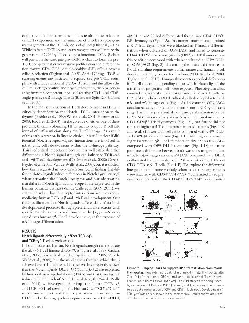
-JAG1, or -JAG2 and differentiated further into CD4+CD8+ DP thymocytes (Fig. 1 A). In contrast, murine uncommitted c-Kit+ fetal thymocytes were blocked in T-lineage differen-tiation when cultured on OP9-JAG1 and failed to generate CD44CD25+ double-negative 3 (DN3) or DP thymocytes in this condition compared with when cocultured on OP9-DLL4 or OP9-JAG2 (Fig. 2), illustrating the critical differences in Notch signaling requirements during mouse and human T cell development (Taghon and Rothenberg, 2008; Archbold, 2009; Taghon et al., 2012). Human thymocytes revealed differences in T cell outcome, depending on to which Notch ligand the intrathymic progenitor cells were exposed. Phenotypic analysis revealed preferential differentiation into TCR- T cells on OP9-JAG1, whereas DLL4 cultured cells developed into both - and -lineage cells (Fig. 1 A). In contrast, OP9-JAG2 cocultured cells differentiated mainly into TCR- T cells (Fig. 1 A). The preferential -lineage differentiation on OP9-JAG1 was seen early at day 6 by an increased number of CD4+CD8+ DP thymocytes (Fig. 1 C) but finally did not result in higher T cell numbers in these cultures (Fig. 1 E) as a result of lower total cell yields compared with OP9-DLL4 and OP9-JAG2 cocultures (Fig. 1 B). Although there was a slight increase in T cell numbers on day 25 in OP9-JAG2 compared with OP9-DLL4 cocultures (Fig. 1 D), the most prominent difference between both was the strong reduction in TCR-–lineage cells on OP9-JAG2 compared with -DLL4, as illustrated by the number of DP thymocytes (Fig. 1 C) and CD3+TCR-+ T cells (Fig. 1 E). To explore the differential lineage outcome more robustly, clonal coculture experiments were initiated with CD34+CD1a+CD4 committed T cell pre-cursors (in contrast to the CD34+CD1aCD4 uncommitted
Figure 2. Jagged1 fails to support DP differentiation from mouse thymocytes. Flow cytometric data of murine c-kit+ fetal thymocytes after 7 or 10 d of coculture on OP9 stromal cells that express different Notch ligands (as indicated above dot plots). Early DN stages are distinguished by expression of CD44 and CD25 (top row) and T cell maturation is moni-tored by the coexpression of CD4 and CD8 (middle row). Development of TCR-+CD3+ cells is shown in the bottom row. Results shown are repre-sentative of three independent experiments.

686 Jagged2/Notch3 drives human T cell development | Van de Walle et al.
the repopulated wells. These data confirmed that DLL4 sup-ports both TCR- and TCR- T cell differentiation, whereas Jagged1 almost exclusively supports T cell development and Jagged2 mainly T cell differentiation. When looking at the clonal outgrowth of all plated wells (Fig. 1 G), no difference in TCR- output was observed in OP9-JAG2 cocultured cells compared with in OP9-DLL4 cocultures, but instead a significant reduction in TCR- output was observed upon
used above), thereby avoiding any potential effects of differ-ences in Notch ligands with respect to inducing T-lineage commitment. In addition, this is the human thymocyte popu-lation from which TCR- and TCR- T cells develop-mentally start to diverge (Van de Walle et al., 2009). Fig. 1 F shows the frequency of -only, -only, or bipotent - and -containing wells (left graph) or frequency of TCR-– or TCR-–containing wells (right graph), but only within
Figure 3. GSI rescues Jagged2-mediated inhibition of -lineage differentiation. (A–C) Flow cytometric analysis of CD34+CD4CD1a+ progenitors cultured during 19 d on OP9 cells expressing the different Notch ligands (DLL4, JAG1, or JAG2), as indicated above the dot plots, in the presence of different concentrations of GSI, as indi-cated on the right of the dot plots. Effects of GSI are shown on TCR-+CD3+ T cells (A), CD4+CD8+ DP thymocytes (B), and TCR-+CD3+ T cells (C). Num-bers in the quadrants indicate the percentage of the corresponding populations and data are representa-tive of three independent experiments. (D–F) Abso-lute number of TCR-+CD3+ T cells (D), CD4+CD8+ DP thymocytes (E), and TCR-+CD3+ T cells (F) of cultures shown in A–C. Triangles below graphs indi-cate an increasing dosage of GSI, corresponding to 0 µM, 0.2 µM, 0.5 µM, and 1 µM GSI. Data shows the mean from three independent experiments (errors bars indicate SEM, * = P < 0.05). (G) Quantitative RT-PCR analysis of human CD34+ thymic precursors after 24 h of culture on equal amounts of DLL4-Fc–, JAG1-Fc–, or JAG2-Fc–coated plates. Units of ex-pression are given relative to GAPDH. Data shows the mean of two sets of independent samples (errors bars indicate SEM, * = P < 0.05).

JEM Vol. 210, No. 4
Article
687
different Notch receptors were expressed by both precursor subsets because different Notch ligand–receptor interactions could potentially explain this difference. Although Notch2 seemed an unlikely candidate based on its low but stable ex-pression in human HSCs and throughout human T cell de-velopment (Van de Walle et al., 2009), both quantitative real- time PCR (Fig. 4 A) and protein expression through FACS analysis (Fig. 4 B) showed that, in addition to Notch2, cord blood (CB) CD34+ HSCs also express Notch1 but not Notch3, whereas thymocytes, already before T cell commitment at the CD34+CD1 stage, express both Notch1 and Notch3, sug-gesting that differential Notch3 expression may account for the differences in Notch ligand responses between human HSCs and intrathymic progenitors. Indeed, binding studies (Fig. 4 C) revealed that Jagged2 can bind to Notch1 and also very efficiently to Notch3, whereas in contrast, DLL4 interacts with Notch1 but virtually not with Notch3. Jagged1 weakly binds to both Notch1 and Notch3. In addition, specific Notch receptor reporter assays (Fig. 4 D) confirmed that DLL4 is the most powerful ligand to activate Notch1-mediated signaling but also revealed that Jagged2 induced the strongest activa-tion when signaling was mediated through Notch3.
Together, these findings reveal that Jagged2 is a very po-tent Notch ligand for human thymocytes as a result of its in-teraction with both Notch1 and Notch3. In contrast, DLL4 only weakly binds and activates Notch3. Because a strong Notch signal favors T cell development, this suggests an important role for Jagged2/Notch3 signaling during TCR- T cell development.
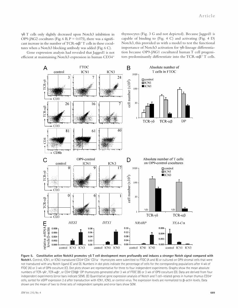
Notch3 activation promotes -lineage differentiation and inhibits TCR- T cell developmentTo functionally assess the influence of differential Notch re-ceptor activation, we transduced uncommitted CD34+CD1a human intrathymic progenitors with the constitutive active forms of Notch1 and Notch3 (ICN1 and ICN3, respectively) and analyzed their impact on human - and -lineage dif-ferentiation. Consistent with previous findings (De Smedt et al., 2002), ICN1 enhanced T cell development and reduced dif-ferentiation into TCR-+ and CD4+CD8+ cells in a fetal thymus organ culture (FTOC) model system, both in terms of frequency and absolute cell number (Fig. 5, A and B). Impor-tantly, ICN3 skewed differentiation toward the -lineage choice in a similar manner but much more profoundly com-pared with ICN1. In addition, we performed coculture ex-periments on OP9 stromal cells that do not overexpress a human Notch ligand, to prevent activation of the endogenous Notch receptors that are present on human thymocytes. This prevents background differentiation of CD3+TCR-+ or CD3+TCR-+ thymocytes as illustrated with control trans-duced cells (Fig. 5, C and D). Under these conditions, ICN1 transduced precursors differentiate into both TCR-+ and TCR-+ T cells, whereas ICN3 transduced precursors only display T cell potential (Fig. 5, C and D). Consistently, gene expression analysis revealed that ICN3 induces a stronger Notch signal compared with ICN1, as revealed by their influence on
OP9-JAG2 coculture (Fig. 1 G), confirming the results from the bulk cocultures (Fig. 1 E).
Overall, these results show that Notch ligands critically influence human TCR- and TCR- T cell development. Although DLL4 supports both - and -lineage differen-tiation, Jagged1 predominantly supports TCR- but not TCR- development. In contrast, Jagged2 mainly supports T cell differentiation at the expense of T cells.
Jagged2 induces the strongest Notch signal in human thymocytesBecause our earlier studies had revealed that high Notch sig-nal strength supports human TCR- T cell development at the expense of TCR- T cell development (De Smedt et al., 2002; Van de Walle et al., 2009), the data in Fig. 1 suggest that Jagged2 induces the strongest Notch signal in human postnatal thymocytes. To investigate this further, we reduced the Notch activation signal with different concentrations of -secretase inhibitor (GSI) in committed human CD34+CD1a+CD4 thymocytes, as we have also shown that a decrease in Notch signaling activation enhances TCR- T cell development (Taghon et al., 2009; Van de Walle et al., 2009). Consistent with the idea that Jagged2 induces the strongest Notch signal, TCR- (Fig. 3 A) and CD4+CD8+ (Fig. 3 B) differentiation could be restored in OP9-JAG2 cocultures at the expense of TCR- T cell development (Fig. 3 C). Addition of low GSI concentrations also increased the frequencies of TCR-+ and CD4+CD8+ cells in Jagged1 and DLL4 cultured cells, but to a lesser extent. In agreement, although the number of TCR- (Fig. 3 D) and CD4+CD8+ (Fig. 3 E) thymocytes consistently and significantly increased upon addition of higher concentration of GSI (up to 1 µM) in OP9-JAG2 cocultures, such an increase in -lineage cells was only observed with lower GSI concentration in OP9-DLL4 cocultures. In addi-tion, the number of TCR- T cells did not significantly decrease with the addition of 1 µM GSI in OP9-JAG2 cocul-tures, in contrast to OP9-DLL4 and OP9-JAG1 cocultures in which higher and significant reductions in T cells were ob-served (Fig. 3 F). Also, gene expression analysis showed that Jagged2 is a stronger inducer of Notch signaling in human CD34+ thymocytes after 24-h exposure to equal amounts of coated Fc-tagged Notch ligands. The Notch target genes HES1, DTX1, NRARP, and NOTCH3 were all up-regulated to a higher level in CD34+ thymocytes exposed to Jagged2 in comparison with DLL4 and much stronger compared with Jagged1 (Fig. 3 G). Thus, these results reveal that for human postnatal thymocytes, Jagged2 is the most potent ligand to in-duce Notch activation, thereby promoting TCR- T cell de-velopment and blocking -lineage differentiation.
Notch ligands display preferential receptor bindingThe idea that Jagged2 induces the strongest Notch signal in human postnatal thymocytes seemed to be in conflict with our recent findings with human HPCs in which Delta-like 4 induces the strongest signal through activation of Notch1 (Van de Walle et al., 2011). Therefore, we investigated whether

688 Jagged2/Notch3 drives human T cell development | Van de Walle et al.
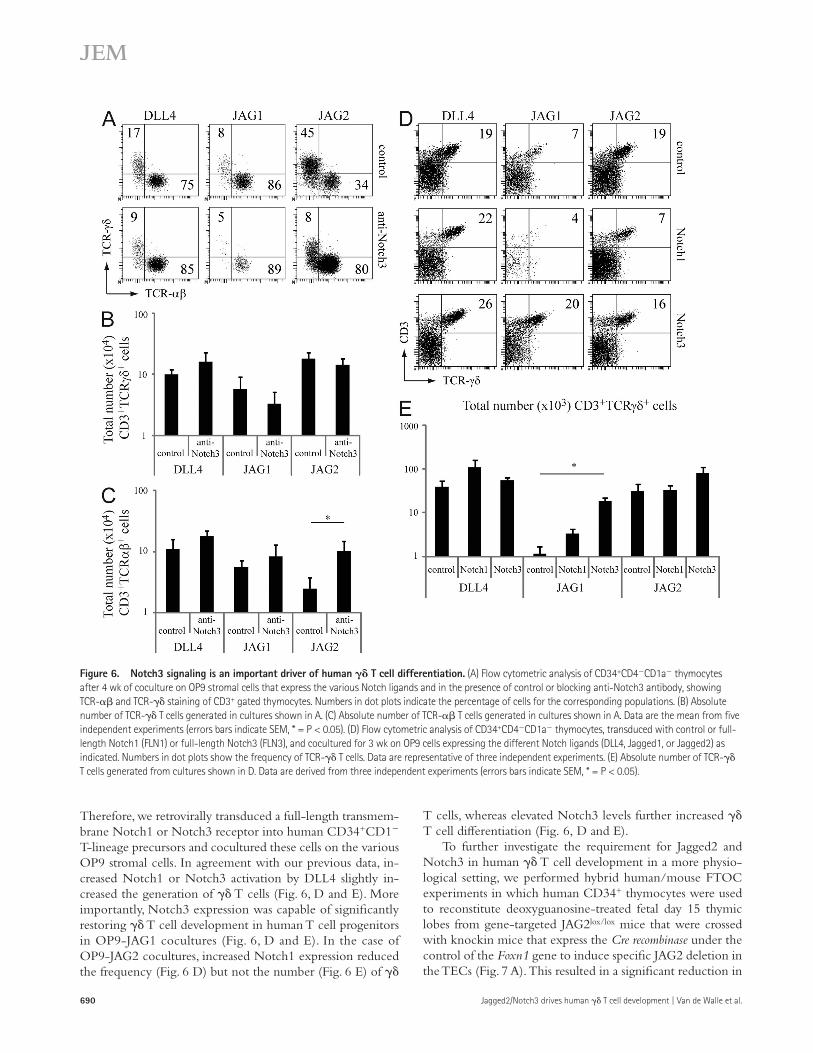
Notch3 activation is critical for promoting T cell developmentTo further show the functional importance of Notch3 activation in mediating human TCR- and - T cell development in the OP9 coculture system, we inhibited Notch3 activation using spe-cific blocking antibodies (Li et al., 2008). Inhibition of Notch3 activation did not significantly alter T cell development in OP9-DLL4 and OP9-JAG1 cocultures but did significantly decrease the frequency of TCR-+ T cells in OP9-JAG2 cocultures while increasing the frequency of TCR-+ cells within the CD3+ fraction of thymocytes (Fig. 6 A). Although the number of
both positively (HES1, DTX1, and NRARP) and negatively (TCR-C) regulated genes (Fig. 5 E). This stronger induction of HES1 and DTX1 expression by ICN3 was confirmed in independent experiments with myc-tagged ICN-fusion proteins in which even lower amounts of ICN3 protein, compared with ICN1 as revealed through FACS-mediated myc staining, still induced much stronger expression of these Notch target genes (unpublished data). Thus, these findings illustrate that Notch3 activation results in a stronger downstream Notch signal com-pared with Notch1 activation and that Notch3 is a strong in-hibitor of T cell development.
Figure 4. Notch3 is expressed in uncommitted human thymocytes and is efficiently bound and activated by Jagged2. (A) Quantitative RT-PCR analysis of Notch1 and Notch3 expression in freshly isolated human CB CD34+ cells and in different human thymocyte subsets. Units of expression are given relative to -actin. Data shows the mean of two to eight sets of independent samples, and error bars indicate SEM. (B) Flow cytometric analysis of Notch1 and Notch3 receptor expression on freshly isolated CB or thymocytes. Density plots shown are representative of three independent experiments. Isotype staining was performed on total human thymocytes suspension. (C) Staining of control-Fc, Notch1-Fc, and Notch3-Fc fusion proteins to K562 cells expressing the different Notch ligands as indicated. Histograms shown are representative of three independent experiments. (D) Luciferase reporter assay of U2OS Tet-on flp-in-Notch1 and U2OS Tet-on flp-in-Notch3 cells cotransfected with CBF-luciferase reporter plasmid pGL2-Gal4-luciferase and the normal-izing plasmid pRL-TK expressing Renilla luciferase. After plasmid transfection, cells were cocultured with K562 expressing DLL4, JAG1, JAG2, or control for 24 h, and thereafter luciferase activity was measured. Bar graphs show the mean of three independent experiments (error bars indicate SEM, * = P < 0.05).
JEM Vol. 210, No. 4
Article
689
thymocytes (Fig. 3 G and not depicted). Because Jagged1 is capable of binding to (Fig. 4 C) and activating (Fig. 4 D) Notch3, this provided us with a model to test the functional importance of Notch3 activation for -lineage differentia-tion because OP9-JAG1 cocultured human T cell progeni-tors predominantly differentiate into the TCR-+ T cells.
T cells only slightly decreased upon Notch3 inhibition in OP9-JAG2 cocultures (Fig. 6 B, P = 0.075), there was a signifi-cant increase in the number of TCR-+ T cells in these cocul-tures when a Notch3 blocking antibody was added (Fig. 6 C).
Gene expression analysis had revealed that Jagged1 is not efficient at maintaining Notch3 expression in human CD34+
Figure 5. Constitutive active Notch3 promotes T cell development more profoundly and induces a stronger Notch signal compared with Notch1. Control, ICN1, or ICN3 transduced CD34+CD4CD1a thymocytes were submitted to FTOC (A and B) or cultured on OP9 stromal cells that were not transduced with any Notch ligand (C and D). Numbers in dot plots indicate the percentage of cells for the corresponding populations after 4 wk of FTOC (A) or 3 wk of OP9 coculture (C). Dot plots shown are representative for three to four independent experiments. Graphs show the mean absolute numbers of TCR-+, TCR-+, or CD4+CD8+ DP thymocytes generated after 3 wk of FTOC (B) or 3 wk of OP9 coculture (D). Data are derived from four independent experiments (error bars indicate SEM). (E) Quantitative gene expression analysis of Notch and T cell–related genes in human thymus CD34+ cells, sorted for eGFP expression 2 d after transduction with ICN1, ICN3, or control virus. The expression levels are normalized to -actin levels. Data shown are the mean of two to three sets of independent samples and error bars show SEM.
690 Jagged2/Notch3 drives human T cell development | Van de Walle et al.
T cells, whereas elevated Notch3 levels further increased T cell differentiation (Fig. 6, D and E).
To further investigate the requirement for Jagged2 and Notch3 in human T cell development in a more physio-logical setting, we performed hybrid human/mouse FTOC experiments in which human CD34+ thymocytes were used to reconstitute deoxyguanosine-treated fetal day 15 thymic lobes from gene-targeted JAG2lox/lox mice that were crossed with knockin mice that express the Cre recombinase under the control of the Foxn1 gene to induce specific JAG2 deletion in the TECs (Fig. 7 A). This resulted in a significant reduction in
Therefore, we retrovirally transduced a full-length transmem-brane Notch1 or Notch3 receptor into human CD34+CD1 T-lineage precursors and cocultured these cells on the various OP9 stromal cells. In agreement with our previous data, in-creased Notch1 or Notch3 activation by DLL4 slightly in-creased the generation of T cells (Fig. 6, D and E). More importantly, Notch3 expression was capable of significantly restoring T cell development in human T cell progenitors in OP9-JAG1 cocultures (Fig. 6, D and E). In the case of OP9-JAG2 cocultures, increased Notch1 expression reduced the frequency (Fig. 6 D) but not the number (Fig. 6 E) of
Figure 6. Notch3 signaling is an important driver of human T cell differentiation. (A) Flow cytometric analysis of CD34+CD4CD1a thymocytes after 4 wk of coculture on OP9 stromal cells that express the various Notch ligands and in the presence of control or blocking anti-Notch3 antibody, showing TCR- and TCR- staining of CD3+ gated thymocytes. Numbers in dot plots indicate the percentage of cells for the corresponding populations. (B) Absolute number of TCR- T cells generated in cultures shown in A. (C) Absolute number of TCR- T cells generated in cultures shown in A. Data are the mean from five independent experiments (errors bars indicate SEM, * = P < 0.05). (D) Flow cytometric analysis of CD34+CD4CD1a thymocytes, transduced with control or full-length Notch1 (FLN1) or full-length Notch3 (FLN3), and cocultured for 3 wk on OP9 cells expressing the different Notch ligands (DLL4, Jagged1, or Jagged2) as indicated. Numbers in dot plots show the frequency of TCR- T cells. Data are representative of three independent experiments. (E) Absolute number of TCR- T cells generated from cultures shown in D. Data are derived from three independent experiments (errors bars indicate SEM, * = P < 0.05).

JEM Vol. 210, No. 4
Article
691
of V1, V2, V3, and V9 expression revealed no differences in the usage of these TCR V gene segments between T cells generated in the presence or absence of Jagged2 (Fig. 7 E). Furthermore, the addition of blocking Notch3 antibodies to FTOC cultures, using deoxyguanosine-treated fetal lobes from
the number (Fig. 7 B) and frequency (Fig. 7, C and D) of human T cells that developed in JAG2-deficient lobes com-pared with the control. Such a reduction in human T cell development was not observed when JAG1-deficient fetal lobes were used (unpublished data). Flow cytometric analysis
Figure 7. Jagged2/Notch3 signaling is critical for human -lineage differentiation. (A) Flow cytometric analysis of Jagged2 expression in TECs from Jag2lox/loxFoxn1-Cre/ (black histogram), Jag2lox/wtFoxn1-Cre+/ (white histogram), and Jag2lox/loxFoxn1-Cre+/ (gray histogram) fetal thymic lobes. Data are representative of three independent experiments. (B) Absolute number of human T cells in FTOCs with Jag2lox/loxFoxn1-Cre/ (black bar) and Jag2lox/lox Foxn1-Cre+/ (gray bar) fetal thymic lobes. Data shows the mean of seven independent experiments and error bars indicate the SEM (* = P < 0.05). (C) Mean frequency of human T cells in FTOCs with Jag2lox/loxFoxn1-Cre/ (black bar) and Jag2lox/loxFoxn1-Cre+/ (gray bar) fetal thymic lobes. Data show the mean of seven independent experiments and error bars indicate the SEM (* = P < 0.05). (D) Flow cytometric analysis of human T cell develop-ment in FTOCs with Jag2lox/loxFoxn1-Cre/ and Jag2lox/loxFoxn1-Cre+/ fetal thymic lobes. Dot plots show CD3 versus TCR- expres-sion and histograms shown TCR- expres-sion in CD3+TCR-+ (white histogram) versus CD3+TCR- (black histogram) cells, gated from human CD45+ cells. Frequen-cies in dot plots show the frequency of -lineage (CD3+TCR-TCR-+) T cells. (E) Histograms show V1, V2, V3, and V9 staining from TCR- gated cells shown in D. Numbers indicate the frequency of positive cells for the corresponding antigen. Data are representative of three independent experiments. (F) Absolute number of human T cells in FTOCs with control (black bar) and blocking Notch3 antibodies (gray bar). Data shows the mean of three independent experiments and error bars indicate the SEM. (G) Mean frequency of human T cells in FTOCs with control (black bar) and blocking Notch3 antibodies (gray bar). (H) Flow cyto-metric analysis of human T cell development in FTOCs with control or blocking Notch3 antibodies. Dot plots show CD3 versus TCR- expression and histograms shown TCR- expression in CD3+TCR-+ (white histogram) versus CD3+TCR- (black his-togram) cells, gated from human CD45+ cells. Frequencies in dot plots show the frequency of -lineage (CD3+TCR-TCR-+) T cells. Data are representative for three indepen-dent experiments.

692 Jagged2/Notch3 drives human T cell development | Van de Walle et al.
wild-type mice, also resulted in a decrease in human dif-ferentiation (Fig. 7, F–H). Overall, these findings clearly illustrate that Jagged2-mediated Notch3 activation is critical for human T cell development.
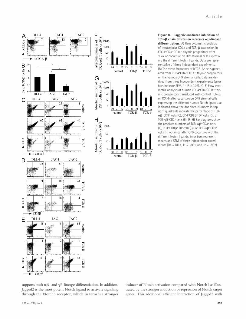
TCR transgenes overcome ligand-induced TCR choicesThe observed differences in TCR- and TCR- T cell de-velopment by the different Notch ligand–receptor interactions could be the result of preferential outgrowth of cells with a particular TCR in those culture conditions rather than a de-velopmental choice on its own. To gain additional insights, we analyzed intracellular TCR- (icTCR-) chain expression in developing human T-lineage progenitors exposed to the dif-ferent Notch ligands, as this is a key early event that distin-guishes most of the developing -lineage progenitors from those differentiating along the T cell pathway. Although virtually all cocultured cells belonged to the T cell lineage as shown by intracellular expression of CD3 (Fig. 8 A), Jagged1-exposed thymocytes contained the highest frequency of icTCR-+ cells—slightly higher compared with DLL4 but sig-nificantly higher compared with thymocytes cocultured on OP9-JAG2, which displayed a low frequency of icTCR-+ cells (Fig. 8, A and B). To explore the functional importance of this reduced TCR- chain expression upon OP9-JAG2 coculture, human CD34+CD4CD1a postnatal thymocytes were transduced with a retrovirus encoding a functional TCR- or TCR- chain before coculture of the cells on the different OP9 stromal cells. Strikingly, Notch ligands had vir-tually no differential impact on the outgrowth of thymocytes with a functional, rearranged TCR. Progenitors transduced with a TCR- chain developed into TCR-+ CD3+ T cells and CD4+CD8+ thymocytes on all three Notch ligands (Fig. 8, C and D), even on Jagged2, and with similar efficien-cies compared with DLL4 with respect to the absolute num-ber of -lineage cells generated (Fig. 8, F and G). In contrast, OP9 cocultures initiated with TCR- transduced thymo-cytes yielded TCR-+ CD3+ T cells on all three Notch ligands (Fig. 8, E and H) and reduced TCR- T cell dif-ferentiation (Fig. 8 F). Even when cocultured on Jagged1, precursors can develop into TCR-+ T cells when a functional TCR- is provided (Fig. 8 E), although with reduced effi-ciency (Fig. 8 H).
Thus, the introduction of a functional TCR transgene into thymic precursors overcomes the ligand-induced lineage outgrowth that is observed with unmanipulated thymocytes. These results therefore indicate that Notch ligand–receptor interactions do not affect the outgrowth of T cell progenitors that express a specific TCR, but that instead they influence the developmental pathway of early T cell precursors into either TCR-– or TCR-–expressing T cells.
DISCUSSIONDuring intrathymic T cell development, differentiating T cell progenitors migrate to different regions within the thymus that provide stage-specific environmental queues that drive their further differentiation (Petrie and Zúñiga-Pflücker, 2007).
One of the most important signaling cascades during early T cell development comprises the Notch signaling pathway and it is well established that the versus T cell lineage choice is modulated by differential Notch signal strength in both mouse and human (Taghon and Rothenberg, 2008; Ciofani and Zúñiga-Pflücker, 2010; Kreslavsky et al., 2010; Taghon et al., 2012). However, it has remained unclear which receptor–ligand interactions are involved in these processes and, moreover, the Notch dependency for TCR- and TCR- T cell development was shown to be different be-tween both species (Washburn et al., 1997; De Smedt et al., 2002; García-Peydró et al., 2003; Ciofani et al., 2006; Garbe et al., 2006; Taghon et al., 2006; Van de Walle et al., 2009). Here, we demonstrate that differential Notch receptor–ligand interactions control human TCR- and TCR- T cell de-velopment by inducing different Notch signal strengths and show that the Jagged2–Notch3 interaction is critical for human T cell development. Importantly, our data provide a mech-anistic insight into the high Notch dependency for develop-ing human T cells.
In the mouse, it is well established that Notch1-DLL4 signaling is essential for inducing T cell differentiation in the immigrating T cell precursors (Wilson et al., 2001; Hozumi et al., 2008; Koch et al., 2008) and we believe that this inter-action is also involved in inducing early T cell development in human. From our previous work, it is clear that Notch1 activation is responsible for inducing human T cell devel-opment (Van de Walle et al., 2009, 2011), and although it remains to be clarified whether Delta-like 4 or Jagged2 is responsible for activating this receptor, the notion that a strong Notch signal is critical at this early stage of human T cell develop-ment (Van de Walle et al., 2009) makes Delta-like 4 the most likely ligand (Van de Walle et al., 2011). More importantly, Notch1 activation at this early stage of T cell development induces high Notch3 expression, as also illustrated previously (Van de Walle et al., 2009, 2011), making this newly expressed receptor accessible for ligand-mediated Notch activation within the thymus.
In both mouse and human, Notch signal strength modu-lates TCR- and TCR- T cell development (Washburn et al., 1997; De Smedt et al., 2002; García-Peydró et al., 2003; Ciofani et al., 2006; Garbe et al., 2006; Taghon et al., 2006; Van de Walle et al., 2009), a process which, in vivo, occurs in the cortex (Petrie and Zúñiga-Pflücker, 2007). We have re-cently shown that JAG2 is expressed by the majority of human cortical TECs, in addition to DLL4 which is less abundantly expressed in this region (Van de Walle et al., 2011), indicat-ing that both ligands can mediate the development of both T cell subsets. Consistent with the notion that human T cells preferentially develop in the presence of high Notch activation (De Smedt et al., 2002; García-Peydró et al., 2003; Van de Walle et al., 2009), we show here that Jagged2 pref-erentially induces human T cell differentiation by inducing the strongest Notch signal in immature thymocytes through interactions with both Notch1 and Notch3, in contrast to Delta-like 4 which predominantly interacts with Notch1 and
JEM Vol. 210, No. 4
Article
693
inducer of Notch activation compared with Notch1 as illus-trated by the stronger induction or repression of Notch target genes. This additional efficient interaction of Jagged2 with
supports both - and -lineage differentiation. In addition, Jagged2 is the most potent Notch ligand to activate signaling through the Notch3 receptor, which in term is a stronger
Figure 8. Jagged2-mediated inhibition of TCR- chain expression represses -lineage differentiation. (A) Flow cytometric analysis of intracellular CD3 and TCR- expression in CD34+CD4CD1a thymic progenitors after 3 wk of coculture on OP9 stromal cells express-ing the different Notch ligands. Data are repre-sentative of three independent experiments. (B) The mean frequency of icTCR-+ cells gener-ated from CD34+CD4CD1a thymic progenitors on the various OP9 stromal cells. Data are de-rived from three independent experiments (error bars indicate SEM, * = P < 0.05). (C–E) Flow cyto-metric analysis of human CD34+CD4–CD1a– thy-mic progenitors transduced with control, TCR-, or TCR- after coculture on OP9 stromal cells expressing the different human Notch ligands, as indicated above the dot plots. Numbers in top right quadrants indicate the percentage of TCR-+CD3+ cells (C), CD4+CD8+ DP cells (D), or TCR-+CD3+ cells (E). (F–H) Bar diagrams show the absolute numbers of TCR-+CD3+ cells (F), CD4+CD8+ DP cells (G), or TCR-+CD3+ cells (H) obtained after OP9 coculture with the different Notch ligands. Error bars represent means and SEM of three independent experi-ments (D4 = DLL4, J1 = JAG1, and J2 = JAG2).

694 Jagged2/Notch3 drives human T cell development | Van de Walle et al.
because these Notch target genes need to be further up-regulated upon T-lineage commitment to reach their peak at the DN3 stage (Taghon et al., 2006). The inability of Jagged1 to induce DTX1, NRARP, and NOTCH3 was also evident in human CB-derived HSCs and might explain why Jagged1 was not sufficient to induce T-lineage specification in human HPCs (Van de Walle et al., 2011). Further studies will need to clarify which Notch target genes are essential for initiating the T-lineage differentiation program.
Importantly, the early up-regulation of Notch3 during human T cell development in comparison with the mouse might explain the difference in Notch dependency of TCR- and TCR- T cell differentiation that is observed between both species (Washburn et al., 1997; Ciofani et al., 2006; Garbe et al., 2006; Taghon et al., 2006; Van de Walle et al., 2009). In humans, both types of T cells start to diverge at the CD34+CD1+ stage when Notch3 is already highly expressed (Van de Walle et al., 2009), allowing Notch3 to influence this lineage choice through activation of Jagged2 that is abun-dantly expressed by human TECs (Van de Walle et al., 2011). In contrast, Notch3 expression in the mouse peaks at the DN3 stage (Taghon et al., 2006; Shi et al., 2011), after the onset of versus T lineage bifurcation (Ciofani et al., 2006). Although there is clear detectable Notch3 expression at the DN2 stage when and T cells start to diverge (Shi et al., 2011), it is unclear if these levels are sufficient to mediate these developmental processes, especially because recent data revealed no obvious defect in mouse T cell development in the absence of Notch3 (Shi et al., 2011; Suliman et al., 2011). In addition, it is unclear how much Jagged2 protein is expressed by the TECs of the mouse, and in situ RNA expression analy-sis suggests that DLL4 is more abundantly expressed in the mouse cortex compared with Jagged2 (Heinzel et al., 2007), raising the possibility that Notch3 might not be sufficiently acti-vated to mediate this early developmental T cell choice. Nev-ertheless, the observation that Jagged2-deficient mice display a reduction in fetal T cell development is consistent with our human findings (Jiang et al., 1998).
The block in TCR- T cell differentiation was not com-plete upon Notch3 inhibition or in the absence of Jagged2, which is consistent with the observation that Notch1 activa-tion can also induce T cell development. However, from the FTOC experiments it is obvious that at least part of human T cells require Jagged2-mediated Notch3 activation for their development. Given our observation that the generation of a TCR- chain is impaired in Jagged2 cocultured thymo-cytes and because introduction of a functional TCR- chain fully restores T cell development under these conditions, it is clear that the failure of TCR- chain production is caus-ing the inhibition of TCR- differentiation that is induced upon Jagged2/Notch3 signaling, thereby favoring the develop-ment of T cells. Given that Notch3 continues to be expressed in DP thymocytes that are the immediate precursors of T cells, we speculate that Notch3 might also be important for allelic exclusion and prevention of further TCR- chain rearrangements when the rearrangement machinery is again
Notch3 explains why the Notch signal strength hierarchy for the different human Notch ligands is different for thymocytes compared with for HPCs that only express Notch1 and Notch2 (Van de Walle et al., 2009), conditions in which DLL4 induces the strongest Notch ligand (Van de Walle et al., 2011). This indicates that the induction of Notch signal strength in signal-receiving cells is highly dependent on its own reper-toire of Notch receptors and on the combination of ligands expressed by the signal-sending cell. Because thymocytes mi-grate through the cortex during their development, our re-sults propose that competition between Notch ligands for engagement of Notch1 and Notch3 will determine the T cell lineage outcome. Despite the abundance of Jagged2 in the human cortex, we anticipate that the strong interaction be-tween DLL4 and Notch1, which results in a strong Notch1 activation, prevents a predominant induction of human T cell development.
Recently, we have shown that Jagged1 does not induce a sufficient strong Notch signal in extrathymic human and mouse HPCs to induce T cell development in contrast to Jagged2, DLL1, and DLL4 (Van de Walle et al., 2011). Here, we demon-strate that the weak Notch signal that is induced by Jagged1 is capable of generating DP and TCR-+CD3+ thymo-cytes from uncommitted postnatal thymocytes from human—in agreement with previous observations (Dontje et al., 2006; Van de Walle et al., 2009)—but not from mouse. Consis-tently, we show that Jagged1 does not support T cell de-velopment in human, whereas this ligand is sufficient for murine TCR- T cell differentiation, illustrating the oppos-ing Notch signaling requirements between mouse and human (Taghon and Rothenberg, 2008; Taghon et al., 2012). This difference corresponds with a differential Notch activation sta-tus during intrathymic T cell development between mouse and human as illustrated by the expression levels of Notch target genes in both species. In humans, it is clear that the strongest Notch signals are delivered in uncommitted CD34+CD1 thymocytes as they express the highest levels of DTX1 and NRARP, genes which are highly sensitive to changes in Notch activation (Van de Walle et al., 2009). In contrast, Notch target genes peak at the DN3 stage of mouse T cell develop-ment when T cell commitment is completed (Taghon et al., 2006; Tydell et al., 2007; Yashiro-Ohtani et al., 2009), a clear difference compared with human (Van de Walle et al., 2009). Also NOTCH3, whose expression is induced upon Notch1 signaling, is expressed earlier during human compared with during mouse T cell development. Because our results show that Jagged1 is incapable of activating these target genes (DTX1, NRARP, and NOTCH3), human postnatal thymo-cytes can further differentiate along the -lineage, as up-regulation of these genes is apparently no longer needed for further differentiation along that pathway. Because human thymocytes continue to express NOTCH3 after the induc-tion of T-lineage commitment, it is unlikely that Jagged1 is involved in vivo in these early stages of human -lineage differentiation. Mouse thymocytes, however, fail to generate DP -lineage cells in OP9-JAG1 cocultures, presumably

JEM Vol. 210, No. 4
Article
695
construct encoding full length Notch1 was provided by W. Pear (University of Pennsylvania, Philadelphia, PA). Viruses encoding functional TCR trans-genes were LZRS-IRES-TCR-EGFP (TCR; Taghon et al., 2009) and LZRS-IRES-TCR-EGFP (TCR, provided by D. Vermijlen, ULB, Gos-selies, Belgium; Vermijlen et al., 2010). Retroviral transduction of thymo-cytes has been described (Taghon et al., 2009).
OP9 cocultures. Purified progenitors were seeded onto plates (24-well or 96-well) confluent with OP9-DLL4, OP9-JAG1, or OP9-JAG2 cells and these cells have been previously well described to express similar levels of each Notch ligand (Van de Walle et al., 2011; Klinakis et al., 2011). OP9 cocultures were all performed in -MEM media (Invitrogen) supple-mented with 20% heat-inactivated FCS (Hyclone) plus 100 U/ml penicil-lin, 100 µg/ml streptomycin, and 2 mM l-glutamine (all from Invitrogen). CD34+CD4CD1 or CD34+CD4CD1+ cells were cultured in the pres-ence of 5 ng/ml IL-7, 5 ng/ml Flt-3L, and 5 ng/ml SCF. For OP9 cocultures started with retrovirally transduced CD34+CD4CD1 or CD34+CD4CD1+ progenitors, EGFP+ transduced cells were first sorted and subsequently subjected to coculture. For -secretase inhibition (GSI) experiments, dif-ferent concentrations of DAPT (0 µM–0.2 µM–0.5 µM–1 µM) were added to the cocultures and an equal concentration of DMSO (0 µM GSI) was added as control. Notch3 blocking experiments were performed by add-ing 1 or 5 µg/ml of the G3 isotype control or A4 anti-Notch3 antibody (provided by C. Siebel, Genentech, San Francisco, CA; Li et al., 2008). Every 2–3 d, half of the medium was refreshed to maintain antibody con-centrations. Cocultures were harvested by forceful pipetting at the indicated time points.
FTOC. FTOCs with control, ICN1, or ICN3 transduced cells were per-formed as described previously (Taghon et al., 2001). NOD-LtSz-scid/scid (NOD-SCID) mice (originally purchased from The Jackson Laboratory) were obtained from our own specific pathogen-free breeding facility. To perform FTOCs with JAG1- and JAG2-deficient fetal thymic lobes, homozygous Jag1lox/lox mice (Mancini et al., 2005) or Jag2lox/lox mice (Xu et al., 2010) were crossed to Foxn1-Cre mice (gift from N. Manley, University of Georgia, Athens, GA; Gordon et al., 2007) to generate Jag1lox/wtFoxn1-Cre+/ and Jag2lox/wt-
Foxn1-Cre+/ mice, respectively. These animals were subsequently inter-crossed to obtain Jag1lox/loxFoxn1-Cre+/ and Jag1lox/loxFoxn1-Cre/ mice, and Jag2lox/loxFoxn1-Cre+/ and Jag2lox/loxFoxn1-Cre/ mice. Fetuses from these intercrossings were isolated at fetal day 15–15.5 of gestation and fetal thymic lobes were treated for 5 d with 1.35 mM 2-deoxyguanosine to remove all endogenous mouse thymocytes. Subsequently, between 5,000 and 10,000 human progenitor cells were added to each lobe in 2-d hanging drop cul-tures, before further culture as described above for FTOCs. Fetal liver from the same fetus was used to genotype the animals. All mice were treated ac-cording to the guidelines of the Laboratory Animal Ethical commission of the University Hospital of Ghent.
Monoclonal antibodies and flow cytometry. Cell suspensions obtained from cocultures were first blocked with anti–mouse FcRII/III (clone 2.4.G2) and human IgG (Fcblock; Miltenyi Biotec) to avoid nonspecific binding. Cell suspensions obtained after FTOCs were also blocked with anti–mouse FcRII/III mAb and stained with rat anti–mouse monoclonal antibody CD45-cychrome to gate out mouse cells during flow cytometry. Subsequently, cells were stained with combinations of anti–human monoclonal antibodies as in-dicated and as described previously (Taghon et al., 2009; Van de Walle et al., 2009). OP9 cocultures were always gated on human CD45+ cells. Cells were examined for the expression of cell surface markers on an LSRII (BD) and human viable cells were gated by excluding propidium iodide–positive cells from analysis.
Reverse transcription PCR. For gene expression analysis, CD34+ thymo-cytes were cultured for 24 h in tissue culture plates that were precoated with Fc-tagged Notch ligands as previously described (Van de Walle et al., 2011). In the case of ICN1, ICN3, and MSCV transduced cells, cells were harvested
activated to induce TCR- rearrangements. Interestingly, the generation of a functional TCR- chain has been shown to be dependent on Notch1 signaling in the mouse (Wolfer et al., 2002) and presumably also in humans (De Smedt et al., 2005). Although our gene expression analysis only reveals quantitative and no qualitative differences between Notch1 and Notch3 in their potential to activate or repress Notch target genes, previ-ous work has proposed that Notch3 can inhibit Notch1 function (Beatus et al., 1999), leaving the possibility that TCR- chain rearrangements are differentially affected by Notch1 and Notch3. Although currently impossible because of the lack of specific ICN3 reagents, genome-wide ChIP studies will help to clarify whether Notch1 and Notch3 target the same genes or not.
Overall, our results reveal a critical role for Notch receptor–ligand interactions in the development of T cells bearing either a or TCR and, as such, propose a novel role for Notch signaling in the generation of these TCRs, possibly by affect-ing TCR rearrangements. Although the expression of such a or TCR does not necessarily correspond with their fur-ther differentiation along the DN - or DP -lineages, respectively, we did observe a correlation between TCR- T cell development and the generation of CD4+CD8+ DP thymocytes in our cultures. Such a correlation between the TCR and the corresponding lineage differentiation program is also observed in the majority of - and -lineage T cells in wild-type mice, but it was also clearly demonstrated that the strength of signal through the TCR is critical in establish-ing the - versus -lineage choice (Haks et al., 2005; Hayes et al., 2005), with Notch signal strength presumably playing a modulatory role (Garbe and von Boehmer, 2007). Such ele-gant genetic studies are difficult to achieve in humans but now become feasible using gene-modified human ES cells that can differentiate along the T cell pathway in vitro (Timmermans et al., 2009). These studies will be required to define the precise contributions of TCR and Notch signal strengths in mediat-ing the - versus -lineage choice in human. Neverthe-less, based on the current evidence from in vitro studies using mouse and human ex vivo–isolated progenitors, it is clear that the requirements for Notch signaling during the early stages of T cell development are distinct between both spe-cies (Taghon et al., 2012). The results in this manuscript in-dicate that this may occur at the level of TCR generation.
MATERIALS AND METHODSIsolation of thymocytes and retroviral transduction. Pediatric thymus samples were obtained and used according to the guidelines of the Medical Ethical Commission of Ghent University Hospital (Belgium). CD34+ thy-mocytes were enriched using CD34 magnetic-activated cell-sorting (MACS; Miltenyi Biotec), according to the manufacturer’s instructions. Enriched CD34+ thymocytes were labeled with CD34-APC, CD4-PE, and CD1-FITC to sort (FACSAria II; BD) CD34+CD4CD1a and CD34+CD4CD1a+ fractions. Isolation of other thymocyte subsets has been described previously (Taghon et al., 2009; Van de Walle et al., 2009). Purity of the sorted cells was checked on a FACSCalibur (BD) and was always >98%. cDNA encod-ing constitutively active or full-length Notch3 was subcloned from previ-ously described constructs (Wang et al., 2007) into the multicloning site of the retroviral vector MSCV-EGFP. Generation of the plasmid containing ICN1 has been described previously (De Smedt et al., 2002) and the MSCV

696 Jagged2/Notch3 drives human T cell development | Van de Walle et al.
Ciofani, M., G.C. Knowles, D.L. Wiest, H. von Boehmer, and J.C. Zúñiga-Pflücker. 2006. Stage-specific and differential notch dependency at the alphabeta and gammadelta T lineage bifurcation. Immunity. 25:105–116. http://dx.doi.org/10.1016/j.immuni.2006.05.010
De Smedt, M., K. Reynvoet, T. Kerre, T. Taghon, B. Verhasselt, B. Vandekerckhove, G. Leclercq, and J. Plum. 2002. Active form of Notch imposes T cell fate in human progenitor cells. J. Immunol. 169:3021–3029.
De Smedt, M., I. Hoebeke, K. Reynvoet, G. Leclercq, and J. Plum. 2005. Different thresholds of Notch signaling bias human precursor cells toward B-, NK-, monocytic/dendritic-, or T-cell lineage in thymus microenvironment. Blood. 106:3498–3506. http://dx.doi.org/10.1182/ blood-2005-02-0496
Dik, W.A., K. Pike-Overzet, F. Weerkamp, D. de Ridder, E.F. de Haas, M.R. Baert, P. van der Spek, E.E. Koster, M.J. Reinders, J.J. van Dongen, et al. 2005. New insights on human T cell development by quantitative T cell receptor gene rearrangement studies and gene ex-pression profiling. J. Exp. Med. 201:1715–1723. http://dx.doi.org/10 .1084/jem.20042524
Dontje, W., R. Schotte, T. Cupedo, M. Nagasawa, F. Scheeren, R. Gimeno, H. Spits, and B. Blom. 2006. Delta-like1-induced Notch1 signaling regulates the human plasmacytoid dendritic cell versus T-cell lineage decision through control of GATA-3 and Spi-B. Blood. 107:2446–2452. http://dx.doi.org/10.1182/blood-2005-05-2090
Garbe, A.I., and H. von Boehmer. 2007. TCR and Notch synergize in al-phabeta versus gammadelta lineage choice. Trends Immunol. 28:124–131. http://dx.doi.org/10.1016/j.it.2007.01.004
Garbe, A.I., A. Krueger, F. Gounari, J.C. Zúñiga-Pflücker, and H. von Boehmer. 2006. Differential synergy of Notch and T cell receptor sig-naling determines versus lineage fate. J. Exp. Med. 203:1579–1590. http://dx.doi.org/10.1084/jem.20060474
García-Peydró, M., V.G. de Yébenes, and M.L. Toribio. 2003. Sustained Notch1 signaling instructs the earliest human intrathymic precursors to adopt a gammadelta T-cell fate in fetal thymus organ culture. Blood. 102:2444–2451. http://dx.doi.org/10.1182/blood-2002-10-3261
Gordon, J., S. Xiao, B. Hughes III, D.M. Su, S.P. Navarre, B.G. Condie, and N.R. Manley. 2007. Specific expression of lacZ and cre recombinase in fetal thymic epithelial cells by multiplex gene targeting at the Foxn1 locus. BMC Dev. Biol. 7:69. http://dx.doi.org/10.1186/1471-213X-7-69
Haks, M.C., J.M. Lefebvre, J.P. Lauritsen, M. Carleton, M. Rhodes, T. Miyazaki, D.J. Kappes, and D.L. Wiest. 2005. Attenuation of gam-madeltaTCR signaling efficiently diverts thymocytes to the alphabeta lineage. Immunity. 22:595–606. http://dx.doi.org/10.1016/j.immuni .2005.04.003
Hayes, S.M., L. Li, and P.E. Love. 2005. TCR signal strength influences alphabeta/gammadelta lineage fate. Immunity. 22:583–593. http://dx.doi .org/10.1016/j.immuni.2005.03.014
Heinzel, K., C. Benz, V.C. Martins, I.D. Haidl, and C.C. Bleul. 2007. Bone marrow-derived hemopoietic precursors commit to the T cell lineage only after arrival in the thymic microenvironment. J. Immunol. 178: 858–868.
Hozumi, K., C. Mailhos, N. Negishi, K.I. Hirano, T. Yahata, K. Ando, S. Zuklys, G.A. Holländer, D.T. Shima, and S. Habu. 2008. Delta-like 4 is indispensable in thymic environment specific for T cell development. J. Exp. Med. 205:2507–2513. http://dx.doi.org/10.1084/jem.20080134
Jiang, R., Y. Lan, H.D. Chapman, C. Shawber, C.R. Norton, D.V. Serreze, G. Weinmaster, and T. Gridley. 1998. Defects in limb, craniofacial, and thymic development in Jagged2 mutant mice. Genes Dev. 12:1046–1057. http://dx.doi.org/10.1101/gad.12.7.1046
Klinakis, A., C. Lobry, O. Abdel-Wahab, P. Oh, H. Haeno, S. Buonamici, I. van De Walle, S. Cathelin, T. Trimarchi, E. Araldi, et al. 2011. A novel tumour-suppressor function for the Notch pathway in myeloid leukae-mia. Nature. 473:230–233. http://dx.doi.org/10.1038/nature09999
Koch, U., and F. Radtke. 2010. Notch signaling in solid tumors. Curr. Top. Dev. Biol. 92:411–455. http://dx.doi.org/10.1016/S0070-2153(10)92013-9
Koch, U., E. Fiorini, R. Benedito, V. Besseyrias, K. Schuster-Gossler, M. Pierres, N.R. Manley, A. Duarte, H.R. Macdonald, and F. Radtke. 2008. Delta-like 4 is the essential, nonredundant ligand for Notch1 dur-ing thymic T cell lineage commitment. J. Exp. Med. 205:2515–2523. http://dx.doi.org/10.1084/jem.20080829
and EGFP+ cells were sorted. Cells were resuspended in RLT buffer and stored at 70°C before RNA isolation. RNA was extracted using RNeasy RNA isolation kit (QIAGEN) and converted into cDNA using Superscript RT II (Invitrogen).
Real-time PCR reactions were performed using the qPCR Core kit for SYBR Green I (Eurogentec) on a 7300 Real-time PCR system (Applied Biosystems). Relative expression levels were calculated for each gene using the Ct method using GAPDH or -actin for normalization.
Luciferase reporter assay and binding studies. Luciferase reporter assays were performed as previously described (Van de Walle et al., 2011). In brief, U2OS Tet-on flp-in cells bearing isogenic transgenes encoding Notch1-Gal4 or Notch3-Gal4 chimeric receptors (gift from J. Aster, Harvard University, Boston, MA; Li et al., 2008) were transfected with Gal4-firefly luciferase and pRL-TK Renilla reporter plasmid. After 1 d, K562 cells expressing Notch ligands were added together with 2 µg/ml tetracycline. 1 d later, luciferase activity was measured in cell lysates using a dual luciferase reporter system (Promega).
Binding studies were performed using K562 cells retrovirally transduced with the different Notch ligands (Delta-like 4, Jagged1, and Jagged2) or the empty vector LZRS-EGFP as a control (Van de Walle et al., 2011). The K562 cells were washed in complete IMDM and aliquoted at 2 × 10⁵ cells in 100 µl of binding buffer (HBSS containing 1 mM CaCl2, 1% BSA, and 0.05% NaN3). The cells were incubated with the Fc-tagged Notch receptors (R&D Systems) or human IgG1 isotype control (Enzo Life Sciences) at 4°C for 40 min. After washing two times with binding buffer, the cells were incubated with a PE-conjugated anti–human Fc (eBioscience).
We thank Chris Siebel (Genentech) for providing the Notch3 blocking antibodies, Warren Pear (University of Pennsylvania) for the full-length Notch1 construct, Nancy Manley (University of Georgia) for FoxN1-Cre mice, Jon Aster (Harvard University) for the U2OS reporter cells, David Vermijlen (Université Libre de Bruxelles) for human TCR antibodies, and Jet Robin and Eelke Vandenberghe for animal care.
This work was supported by the Odysseus program of the Fund for Scientific Research Flanders (FWO) and grants from the FWO, the Flemish Institute for the advancement of Scientific-Technological Research in the Industry (IWT), the Concerted Research Action of Ghent University (GOA) and the Interuniversity Attraction Poles Program (IUAP) from the Belgian Science Policy. I. Van de Walle is supported by grants from the IWT and Special Fund for Scientific Research (BOF) of the Ghent University. E. Waegemans is supported by a grant from Faculty of Medicine and Health Sciences from the Ghent University. S. Snauwaert, T. Kerre, and T. Taghon are supported by the FWO.
The authors have no conflicting financial interests.
Submitted: 8 August 2012Accepted: 20 February 2013
REFERENCESArchbold, J.K. 2009. To be gammadelta or not to be gammadelta? Signaling
pathways in alphabeta versus gammadelta T cell maturation. Sci. Signal. 2:jc2. http://dx.doi.org/10.1126/scisignal.2100jc2
Artavanis-Tsakonas, S., M.D. Rand, and R.J. Lake. 1999. Notch signaling: cell fate control and signal integration in development. Science. 284:770–776. http://dx.doi.org/10.1126/science.284.5415.770
Beatus, P., J. Lundkvist, C. Oberg, and U. Lendahl. 1999. The notch 3 in-tracellular domain represses notch 1-mediated activation through Hairy/Enhancer of split (HES) promoters. Development. 126:3925–3935.
Benedito, R., C. Roca, I. Sörensen, S. Adams, A. Gossler, M. Fruttiger, and R.H. Adams. 2009. The notch ligands Dll4 and Jagged1 have opposing effects on angiogenesis. Cell. 137:1124–1135. http://dx.doi.org/10.1016/ j.cell.2009.03.025
Blom, B., and H. Spits. 2006. Development of human lymphoid cells. Annu. Rev. Immunol. 24:287–320. http://dx.doi.org/10.1146/annurev .immunol.24.021605.090612
Ciofani, M., and J.C. Zúñiga-Pflücker. 2010. Determining versus ß T cell development. Nat. Rev. Immunol. 10:657–663.

JEM Vol. 210, No. 4
Article
697
Kopan, R., and M.X. Ilagan. 2009. The canonical Notch signaling pathway: unfolding the activation mechanism. Cell. 137:216–233. http://dx.doi .org/10.1016/j.cell.2009.03.045
Kreslavsky, T., M. Gleimer, and H. von Boehmer. 2010. Alphabeta versus gam-madelta lineage choice at the first TCR-controlled checkpoint. Curr. Opin. Immunol. 22:185–192. http://dx.doi.org/10.1016/j.coi.2009.12.006
Li, K., Y. Li, W. Wu, W.R. Gordon, D.W. Chang, M. Lu, S. Scoggin, T. Fu, L. Vien, G. Histen, et al. 2008. Modulation of Notch signaling by antibodies specific for the extracellular negative regulatory region of NOTCH3. J. Biol. Chem. 283:8046–8054. http://dx.doi.org/10.1074/jbc.M800170200
Lobry, C., P. Oh, and I. Aifantis. 2011. Oncogenic and tumor suppressor functions of Notch in cancer: it’s NOTCH what you think. J. Exp. Med. 208:1931–1935. http://dx.doi.org/10.1084/jem.20111855
Mancini, S.J., N. Mantei, A. Dumortier, U. Suter, H.R. MacDonald, and F. Radtke. 2005. Jagged1-dependent Notch signaling is dispensable for he-matopoietic stem cell self-renewal and differentiation. Blood. 105:2340–2342. http://dx.doi.org/10.1182/blood-2004-08-3207
Petrie, H.T., and J.C. Zúñiga-Pflücker. 2007. Zoned out: functional map-ping of stromal signaling microenvironments in the thymus. Annu. Rev. Immunol. 25:649–679. http://dx.doi.org/10.1146/annurev.immunol .23.021704.115715
Plum, J., M. De Smedt, G. Leclercq, T. Taghon, T. Kerre, and B. Vandekerckhove. 2008. Human intrathymic development: a selective approach. Semin. Immu-nopathol. 30:411–423. http://dx.doi.org/10.1007/s00281-008-0135-2
Radtke, F., A. Wilson, G. Stark, M. Bauer, J. van Meerwijk, H.R. MacDonald, and M. Aguet. 1999. Deficient T cell fate specification in mice with an induced inactivation of Notch1. Immunity. 10:547–558. http://dx.doi .org/10.1016/S1074-7613(00)80054-0
Radtke, F., N. Fasnacht, and H.R. Macdonald. 2010. Notch signaling in the immune system. Immunity. 32:14–27. http://dx.doi.org/10.1016/ j.immuni.2010.01.004
Shi, J., M. Fallahi, J.L. Luo, and H.T. Petrie. 2011. Nonoverlapping func-tions for Notch1 and Notch3 during murine steady-state thymic lymphopoiesis. Blood. 118:2511–2519. http://dx.doi.org/10.1182/blood- 2011-04-346726
Suliman, S., J. Tan, K. Xu, P.C. Kousis, P.E. Kowalski, G. Chang, S.E. Egan, and C. Guidos. 2011. Notch3 is dispensable for thymocyte -selection and Notch1-induced T cell leukemogenesis. PLoS ONE. 6:e24937. http://dx.doi.org/10.1371/journal.pone.0024937
Taghon, T., and E.V. Rothenberg. 2008. Molecular mechanisms that control mouse and human TCR-alphabeta and TCR-gammadelta T cell devel-opment. Semin. Immunopathol. 30:383–398. http://dx.doi.org/10.1007/ s00281-008-0134-3
Taghon, T., M. De Smedt, F. Stolz, M. Cnockaert, J. Plum, and G. Leclercq. 2001. Enforced expression of GATA-3 severely reduces human thymic cellularity. J. Immunol. 167:4468–4475.
Taghon, T., M.A. Yui, R. Pant, R.A. Diamond, and E.V. Rothenberg. 2006. Developmental and molecular characterization of emerging beta- and gammadelta-selected pre-T cells in the adult mouse thymus. Immunity. 24:53–64. http://dx.doi.org/10.1016/j.immuni.2005.11.012
Taghon, T., I. Van de Walle, G. De Smet, M. De Smedt, G. Leclercq, B. Vandekerckhove, and J. Plum. 2009. Notch signaling is required for proliferation but not for differentiation at a well-defined beta-selection
checkpoint during human T-cell development. Blood. 113:3254–3263. http://dx.doi.org/10.1182/blood-2008-07-168906
Taghon, T., E. Waegemans, and I. Van de Walle. 2012. Notch signal-ing during human T cell development. Curr. Top. Microbiol. Immunol. 360:75–97. http://dx.doi.org/10.1007/82_2012_230
Timmermans, F., I. Velghe, L. Vanwalleghem, M. De Smedt, S. Van Coppernolle, T. Taghon, H.D. Moore, G. Leclercq, A.W. Langerak, T. Kerre, et al. 2009. Generation of T cells from human embryonic stem cell-derived hematopoietic zones. J. Immunol. 182:6879–6888. http://dx.doi.org/10.4049/jimmunol.0803670
Tydell, C.C., E.S. David-Fung, J.E. Moore, L. Rowen, T. Taghon, and E.V. Rothenberg. 2007. Molecular dissection of prethymic progeni-tor entry into the T lymphocyte developmental pathway. J. Immunol. 179:421–438.
Van de Walle, I., G. De Smet, M. De Smedt, B. Vandekerckhove, G. Leclercq, J. Plum, and T. Taghon. 2009. An early decrease in Notch ac-tivation is required for human TCR-alphabeta lineage differentiation at the expense of TCR-gammadelta T cells. Blood. 113:2988–2998. http://dx.doi.org/10.1182/blood-2008-06-164871
Van de Walle, I., G. De Smet, M. Gärtner, M. De Smedt, E. Waegemans, B. Vandekerckhove, G. Leclercq, J. Plum, J.C. Aster, I.D. Bernstein, et al. 2011. Jagged2 acts as a Delta-like Notch ligand during early hema-topoietic cell fate decisions. Blood. 117:4449–4459. http://dx.doi.org/ 10.1182/blood-2010-06-290049
Vermijlen, D., M. Brouwer, C. Donner, C. Liesnard, M. Tackoen, M. Van Rysselberge, N. Twité, M. Goldman, A. Marchant, and F. Willems. 2010. Human cytomegalovirus elicits fetal T cell responses in utero. J. Exp. Med. 207:807–821. http://dx.doi.org/10.1084/jem.20090348
Wang, T., C.M. Holt, C. Xu, C. Ridley, R. P O Jones, M. Baron, and D. Trump. 2007. Notch3 activation modulates cell growth behaviour and cross-talk to Wnt/TCF signalling pathway. Cell. Signal. 19:2458–2467. http://dx.doi.org/10.1016/j.cellsig.2007.07.019
Washburn, T., E. Schweighoffer, T. Gridley, D. Chang, B.J. Fowlkes, D. Cado, and E. Robey. 1997. Notch activity influences the alphabeta ver-sus gammadelta T cell lineage decision. Cell. 88:833–843. http://dx.doi .org/10.1016/S0092-8674(00)81929-7
Wilson, A., H.R. MacDonald, and F. Radtke. 2001. Notch 1-deficient common lymphoid precursors adopt a B cell fate in the thymus. J. Exp. Med. 194:1003–1012. http://dx.doi.org/10.1084/jem.194.7.1003
Wolfer, A., A. Wilson, M. Nemir, H.R. MacDonald, and F. Radtke. 2002. Inactivation of Notch1 impairs VDJbeta rearrangement and allows pre-TCR-independent survival of early alpha beta Lineage Thymocytes. Immunity. 16:869–879. http://dx.doi.org/10.1016/S1074- 7613(02)00330-8
Xu, J., L.T. Krebs, and T. Gridley. 2010. Generation of mice with a con-ditional null allele of the Jagged2 gene. Genesis. 48:390–393. http://dx.doi.org/10.1002/dvg.20626
Yashiro-Ohtani, Y., Y. He, T. Ohtani, M.E. Jones, O. Shestova, L. Xu, T.C. Fang, M.Y. Chiang, A.M. Intlekofer, S.C. Blacklow, et al. 2009. Pre-TCR signaling inactivates Notch1 transcription by antagonizing E2A. Genes Dev. 23:1665–1676. http://dx.doi.org/10.1101/gad.1793709
Yashiro-Ohtani, Y., T. Ohtani, and W.S. Pear. 2010. Notch regulation of early thymocyte development. Semin. Immunol. 22:261–269. http://dx .doi.org/10.1016/j.smim.2010.04.015


