Embed Size (px)
Citation preview
Spectral Radiation Dependent PhotoprotectiveMechanism in the Diatom Pseudo-nitzschia multistriataChristophe Brunet1*, Raghu Chandrasekaran1, Lucia Barra1, Vasco Giovagnetti1, Federico Corato1,
Alexander V. Ruban2
1 Laboratory of Ecology and Evolution of Plankton, Stazione Zoologica Anton Dohrn, Villa Comunale, Napoli, Italy, 2 School of Biological and Chemical Sciences, Queen
Mary University of London, Mile End Road, London, United Kingdom
Abstract
Phytoplankton, such as diatoms, experience great variations of photon flux density (PFD) and light spectrum along themarine water column. Diatoms have developed some rapidly-regulated photoprotective mechanisms, such as thexanthophyll cycle activation (XC) and the non-photochemical chlorophyll fluorescence quenching (NPQ), to protectthemselves from photooxidative damages caused by excess PFD. In this study, we investigate the role of blue fluence rate incombination with red radiation in shaping photoacclimative and protective responses in the coastal diatom Pseudo-nitzschia multistriata. This diatom was acclimated to four spectral light conditions (blue, red, blue-red, blue-red-green), eachof them provided with low and high PFD. Our results reveal that the increase in the XC pool size and the amplitude of NPQis determined by the blue fluence rate experienced by cells, while cells require sensing red radiation to allow thedevelopment of these processes. Variations in the light spectrum and in the blue versus red radiation modulate either thephotoprotective capacity, such as the activation of the diadinoxanthin-diatoxanthin xanthophyll cycle, the diadinoxanthinde-epoxidation rate and the capacity of non-photochemical quenching, or the pigment composition of this diatom. Wepropose that spectral composition of light has a key role on the ability of diatoms to finely balance light harvesting andphotoprotective capacity.
Citation: Brunet C, Chandrasekaran R, Barra L, Giovagnetti V, Corato F, et al. (2014) Spectral Radiation Dependent Photoprotective Mechanism in the DiatomPseudo-nitzschia multistriata. PLoS ONE 9(1): e87015. doi:10.1371/journal.pone.0087015
Editor: Adrianna Ianora, Stazione Zoologica, Italy
Received September 17, 2013; Accepted December 16, 2013; Published January 24, 2014
Copyright: � 2014 Brunet et al. This is an open-access article distributed under the terms of the Creative Commons Attribution License, which permitsunrestricted use, distribution, and reproduction in any medium, provided the original author and source are credited.
Funding: This work was supported by Stazione Zoologica Anton Dohrn. R.C.’s PhD is funded by Stazione Zoologica Anton Dohrn. The funders had no role instudy design, data collection and analysis, decision to publish, or preparation of the manuscript.
Competing Interests: The authors have declared that no competing interests exist.
* E-mail: [email protected]
Introduction
Originating some 2.3222.45 Gyr ago, oxygenic photosynthesis
spread across the Earth, allowing the great diversification of life
and globally altering the community structure and ecological
function of terrestrial and aquatic habitats [1,2]. Phytoplankton,
small floating photosynthetic microorganisms that populate the
aquatic realms, thrive in a light environment naturally variable
over spatial and temporal extremes [3,4]. While penetrating
through the water column, light intensity (photon flux density;
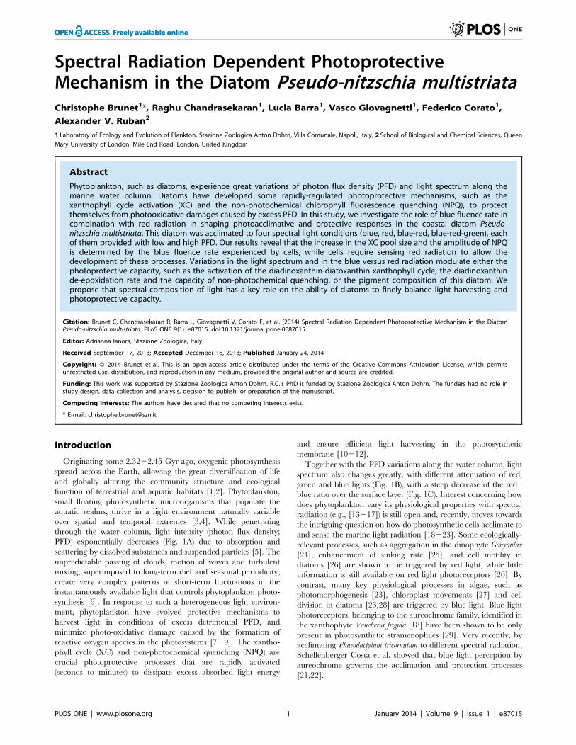
PFD) exponentially decreases (Fig. 1A) due to absorption and
scattering by dissolved substances and suspended particles [5]. The
unpredictable passing of clouds, motion of waves and turbulent
mixing, superimposed to long-term diel and seasonal periodicity,
create very complex patterns of short-term fluctuations in the
instantaneously available light that controls phytoplankton photo-
synthesis [6]. In response to such a heterogeneous light environ-
ment, phytoplankton have evolved protective mechanisms to
harvest light in conditions of excess detrimental PFD, and
minimize photo-oxidative damage caused by the formation of
reactive oxygen species in the photosystems [729]. The xantho-
phyll cycle (XC) and non-photochemical quenching (NPQ) are
crucial photoprotective processes that are rapidly activated
(seconds to minutes) to dissipate excess absorbed light energy
and ensure efficient light harvesting in the photosynthetic
membrane [10212].
Together with the PFD variations along the water column, light
spectrum also changes greatly, with different attenuation of red,
green and blue lights (Fig. 1B), with a steep decrease of the red :
blue ratio over the surface layer (Fig. 1C). Interest concerning how
does phytoplankton vary its physiological properties with spectral
radiation (e.g., [13217]) is still open and, recently, moves towards
the intriguing question on how do photosynthetic cells acclimate to
and sense the marine light radiation [18223]. Some ecologically-
relevant processes, such as aggregation in the dinophyte Gonyaulax
[24], enhancement of sinking rate [25], and cell motility in
diatoms [26] are shown to be triggered by red light, while little
information is still available on red light photoreceptors [20]. By
contrast, many key physiological processes in algae, such as
photomorphogenesis [23], chloroplast movements [27] and cell
division in diatoms [23,28] are triggered by blue light. Blue light
photoreceptors, belonging to the aureochrome family, identified in
the xanthophyte Vaucheria frigida [18] have been shown to be only
present in photosynthetic stramenophiles [29]. Very recently, by
acclimating Phaeodactylum tricornutum to different spectral radiation,
Schellenberger Costa et al. showed that blue light perception by
aureochrome governs the acclimation and protection processes
[21,22].
PLOS ONE | www.plosone.org 1 January 2014 | Volume 9 | Issue 1 | e87015
The general objective of our study is to decipher the role of
spectral radiation on the photophysiological acclimation properties
of coastal diatoms, well-known for their high photosynthetic
flexibility and regulative capacity [7,9,10]. We choose the toxic
Pseudo-nitzschia multistriata species, since the strain has been recently
isolated from the Gulf of Naples (coastal area of the Mediterranean
sea, Stazione Zoologica Anton Dohrn, Italy, strain number
SY717) and its ecological properties are known (e.g., [30]).
We investigate if and how spectral radiation does affect the
photoprotective capacity of this diatom, focussing on the
regulation of pigment content, and on the rapidly activated
protective responses, as the XC and NPQ. Since the different
distribution over water column of the red and blue radiations, and
their essential eco-physiological roles (as introduced before), we
aim to test the hypothesis that photoprotective capacity in diatoms
differs between different mixtures of blue and red radiations,
compared to the same radiations when provided separately. We
also address the question on the biological effect of the red : blue
ratio of the light experienced by cells, as trigger for the
photoprotective response ([22]).
The results suggest that spectral composition of light has a key
role, together with PFD, on the ability of diatoms to finely balance
light harvesting and photoprotective capacity. To our knowledge
this is the first report demonstrating the dependence of the XC
and NPQ to both the blue and red radiation together.
Materials and Methods
Ethics StatementNo specific permits or permissions were required for the field
studies, as the cruise for measuring the vertical light profiles was
carried out in international waters and the isolation of Pseudo-
nitzschia multistriata strain SY717 has been done during the long
term research Mare-Chiara program in the coastal area of the
Gulf of Naples where no specific permits or permissions were
required. This work did not involve endangered or protected
species.
Experimental Strategy and SamplingFour spectral light conditions – blue, red and two mixed light
conditions, namely blue-red-green and blue-red were applied
(Table 1). The two mixed light conditions were characterized by (i)
the same photon flux density (PFD) and relative proportion of red
radiation provided (18–20%), and (ii) two different red : blue
ratios: 0.43 (blue-red-green) and 0.25 (blue-red), determined by the
presence or absence of green light (Table 1). These two values of
red : blue ratio characterize the high light environment, 2 m
(,0.43) and 6 m (,0.25) depths, of the water column during
summer in the Mediterranean Sea (Fig. 1).
For each condition, the daily light dose was kept constant, in
order to be comparable for the provided photon flux density. Two
daily light doses, 6.1 mol m–2 d–1 and 11 mol m–2 d–1 (sinusoidal
light distribution, peaking at 250 and 450 mmol photons m–2 s–1,
respectively; Table 1), have been tested, with a 12:12 hours
light:dark photoperiod. Light intensity was measured inside each
flask by using a laboratory PAR 4 p sensor (QSL 2101,
Biospherical Instruments Inc., San Diego, CA, USA), while
spectral composition (PAR(l)) were measured at light peak by
using a radiometer (Hyper OCR I, Satlantic, Halifax, CA).
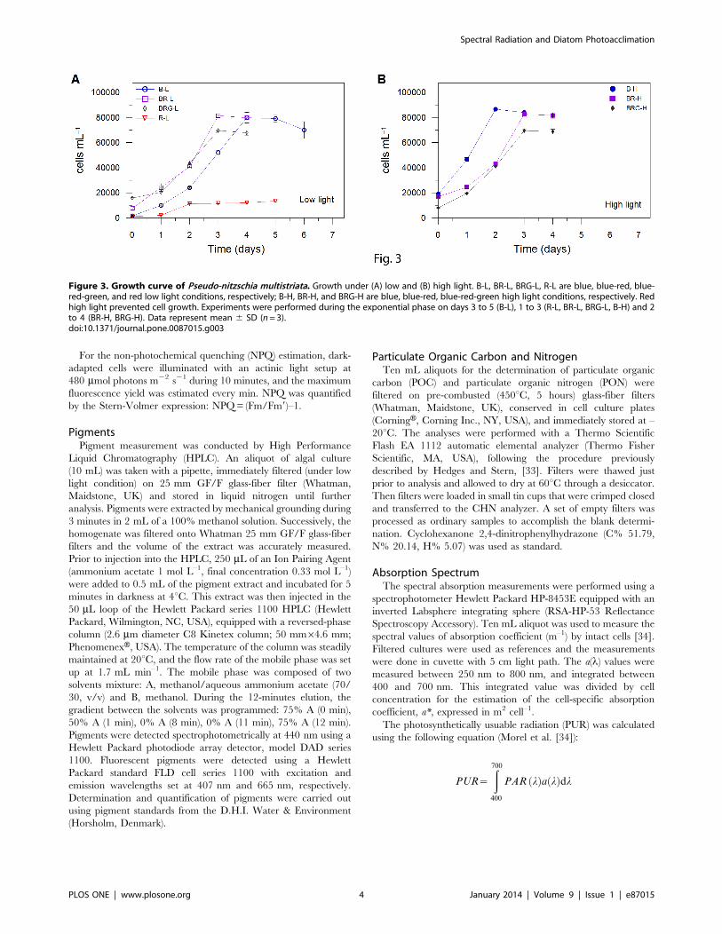
Light was provided by a custom-built illumination system,
which allows to monitor and regulate the light intensity and
quality. The system is composed by blue, green and red light
emitting diodes (peaking at 460, 530 and 626 nm, respectively;
Fig. 2). Experiments were conducted on Pseudo-nitzschia multistriata
strain SY717 isolated in the Gulf of Naples (40u 489 N, 14u 159 E,
Mediterranean Sea). Cells were cultivated at 20uC in 75 cm2
polystyrene canted neck flasks (CorningH flask, Corning Inc., NY,
USA), containing natural sterile seawater amended with f/2
nutrients. All the experiments, lasting three days, were performed
in triplicate during the exponential growth phase (Fig. 3), on
Figure 1. In situ light profile vs depth. (A) PAR (mmol photons m–
2 s–1) distribution along the water column, (B) blue, red and greenradiation distribution along the water column (mW cm–2; blue –422 to496 nm, green –479 to 579 nm and red –589 to 656 nm), and (C)distribution of red : blue ratio along the water column. Data are meanof profiles done on 5 stations in the Mediterranean Sea in June–July2008 (C. Brunet, unpublished data).doi:10.1371/journal.pone.0087015.g001
Spectral Radiation and Diatom Photoacclimation
PLOS ONE | www.plosone.org 2 January 2014 | Volume 9 | Issue 1 | e87015
cultures pre-acclimated to each experimental light condition for
two weeks before the experiments. Under red light, at high PFD
(450 mmol photons m–2 s–1) cells did not grow, preventing any
experimental result.
Samples for pigments, variable fluorescence and electron
transport rate, and elemental composition analysis were taken at
dawn (time 0), midday (time 6 hours) and in the afternoon (time 9
hours), during the first two days of the experiment, and once (time
0) during the third day. Cell counts were performed daily at time
0, while the absorption spectrum were analysed once per
experimental condition (time 6 hours on the second day).
Cell ConcentrationCell concentration was estimated on triplicate sub-samples. An
aliquot of 1 mL was used to fill a Sedgewick Rafter counting cell
chamber, and cell counts were performed using a Zeiss Axioskop
2 Plus microscope.
Photochemical Efficiency and Photosynthetic ParametersPhotochemical efficiency of photosystem (PS) II was estimated
by a Phyto-PAM fluorometer (Heinz Walz, Effeltrich, Germany).
The variable fluorescence analysis was performed on 15-minutes
dark-acclimated samples, to measure the maximum photochem-
ical efficiency (Fv/Fm, [31]). Fm was measured after a saturating
pulse of red light (2400 mmol m–2 s–1, lasting 450 ms), causing a
complete reduction of the PSII acceptor pool.
Electron transport rate (ETR) versus irradiance curves were
determined applying 13 increasing red actinic lights (655 nm) from
1 to 853 mmol photons m22 s21 lasting 1 minute each. The
relative electron transport rate (relETR, expressed in mmol e–1 s21
cell21) was calculated as follows: relETR = (Fv9/Fm9) ? I ? 0.5 ?a*.
where, I is the incident irradiance (expressed in mmol photons
m22 s21), Fv9 and Fm9 are the variable PS II fluorescence yield
and maximal PS II fluorescence yield, respectively, for illuminated
cells (measured at the end of the 1 min lasting actinic light), a* is
the cell-specific absorption coefficient, expressed in m2 cell21 (for
the determination of a* see below). A factor of 0.5 was applied to
correct for the partitioning of photons between PSI and PSII,
assuming that excitation energy is evenly distributed between the
two photosystems.
ETR-I curves were fitted with the equation of Eilers and Peeters
to estimate the photosynthetic parameters [32], relETRmax
(maximal relative rate of linear electron transport), a (maximum
light use efficiency which is the slope of the beginning of the light
curve), and Ek (light intensity for reaching relETRmax).
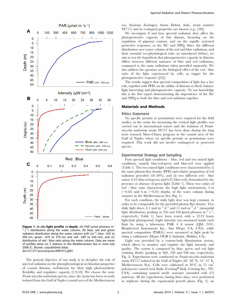
Table 1. Light condition characteristics, and photosynthetic and biochemical properties in Pseudo-nitzschia multistriata cells.
Blue Blue-red Blue-red-green Red
Low High Low High Low High Low
(B-L) (B-H) (BR-L) (BR-H) (BRG-L) (BRG-H) (R-L)
PFD 250 450 250 450 250 450 250
Blue 250 450 200 360 105 189 0
Green 0 0 0 0 100 180 0
Red 0 0 50 90 45 81 250
Red : Blue 0 0 0.25 0.25 0.43 0.43 0
a* 2.51 (0.09) 1.46 (0.07) 2.69 (0.22) 2.89 (0.10) 1.97 (0.63) 1.22 (0.08) 4.44 (0.05)
PUR 2.47 (0.09) 2.4 (0.06) 2.27 (0.23) 4.39 (0.16) 1.49 (0.51) 1.95 (0.12) 1.11 (0.23)
relETRmax 2.72 (0.13) 1.66 (0.11) 3.61 (0.41) 3.45 (0.17) 2.13 (0.57) 1.07 (0.09) 2.63 (0.47)
a 6.7 (1.1) 3.8 (0.27) 7.3 (0.36) 7.1 (0.44) 4.9 (0.96) 3.0 (0.54) 13 (2.2)
Ek 408 (66) 431 (21) 497 (32) 491 (39) 472 (37) 363 (84) 200 (5)
POC 131 (37) 62 (8) 80 (5) 80 (5) 100 (8) 89 (5) 154 (28)
POC/PON 4.9 (0.52) 4.3 (0.63) 4.8 (0.27) 4.7 (0.37) 6.2 (0.76) 4.7 (0.05) 2.9 (0.19)
Chl a/POC 8.2 (4.8) 10.3 (2.9) 13.8 (2.6) 10.7 (1.5) 9.8 (2.2) 10.8 (0.9) 6.2 (2.5)
Blue, green and red fluence rates (mmol photon m22 s21) measured at light peak and red : blue ratio values for the different light conditions. a*610–11, absorptioncoefficient (m2 cell21); PUR610–6, photosynthetically usable radiation (mW cell–1); relETRmax610–6, (maximal relative rate of linear electron transport, nmol e21 s21 cell21),a610–9 (maximum light use efficiency, nmol e21 s21 cell21(mmol photon m22 s21) 21), and Ek (light intensity for reaching relETRmax, mmol photon m22 s21); POC,particulate organic carbon (pg cell21); POC/PON, particulate organic carbon (POC) to particulate organic nitrogen (PON) ratio (pg/pg); Chl a/POC610–3, Chlorophyll a toPOC ratio (pg/pg). Data represent mean and standard deviation. For a* and PUR, n=3; For relETRmax, a and Ek, n=6 (mean of the two days light peak measurements);For POC, PON, POC/PON and Chl a/POC, n= 21.doi:10.1371/journal.pone.0087015.t001
Figure 2. Spectral properties of the LEDs. Blue (4222496 nm),green (4802580 nm) and red light (5902656 nm).doi:10.1371/journal.pone.0087015.g002
Spectral Radiation and Diatom Photoacclimation
PLOS ONE | www.plosone.org 3 January 2014 | Volume 9 | Issue 1 | e87015
For the non-photochemical quenching (NPQ) estimation, dark-
adapted cells were illuminated with an actinic light setup at
480 mmol photons m22 s21 during 10 minutes, and the maximum
fluorescence yield was estimated every min. NPQ was quantified
by the Stern-Volmer expression: NPQ = (Fm/Fm9)–1.
PigmentsPigment measurement was conducted by High Performance
Liquid Chromatography (HPLC). An aliquot of algal culture
(10 mL) was taken with a pipette, immediately filtered (under low
light condition) on 25 mm GF/F glass-fiber filter (Whatman,
Maidstone, UK) and stored in liquid nitrogen until further
analysis. Pigments were extracted by mechanical grounding during
3 minutes in 2 mL of a 100% methanol solution. Successively, the
homogenate was filtered onto Whatman 25 mm GF/F glass-fiber
filters and the volume of the extract was accurately measured.
Prior to injection into the HPLC, 250 mL of an Ion Pairing Agent
(ammonium acetate 1 mol L–1, final concentration 0.33 mol L–1)
were added to 0.5 mL of the pigment extract and incubated for 5
minutes in darkness at 4uC. This extract was then injected in the
50 mL loop of the Hewlett Packard series 1100 HPLC (Hewlett
Packard, Wilmington, NC, USA), equipped with a reversed-phase
column (2.6 mm diameter C8 Kinetex column; 50 mm64.6 mm;
PhenomenexH, USA). The temperature of the column was steadily
maintained at 20uC, and the flow rate of the mobile phase was set
up at 1.7 mL min–1. The mobile phase was composed of two
solvents mixture: A, methanol/aqueous ammonium acetate (70/
30, v/v) and B, methanol. During the 12-minutes elution, the
gradient between the solvents was programmed: 75% A (0 min),
50% A (1 min), 0% A (8 min), 0% A (11 min), 75% A (12 min).
Pigments were detected spectrophotometrically at 440 nm using a
Hewlett Packard photodiode array detector, model DAD series
1100. Fluorescent pigments were detected using a Hewlett
Packard standard FLD cell series 1100 with excitation and
emission wavelengths set at 407 nm and 665 nm, respectively.
Determination and quantification of pigments were carried out
using pigment standards from the D.H.I. Water & Environment
(Horsholm, Denmark).
Particulate Organic Carbon and NitrogenTen mL aliquots for the determination of particulate organic
carbon (POC) and particulate organic nitrogen (PON) were
filtered on pre-combusted (450uC, 5 hours) glass-fiber filters
(Whatman, Maidstone, UK), conserved in cell culture plates
(CorningH, Corning Inc., NY, USA), and immediately stored at –
20uC. The analyses were performed with a Thermo Scientific
Flash EA 1112 automatic elemental analyzer (Thermo Fisher
Scientific, MA, USA), following the procedure previously
described by Hedges and Stern, [33]. Filters were thawed just
prior to analysis and allowed to dry at 60uC through a desiccator.
Then filters were loaded in small tin cups that were crimped closed
and transferred to the CHN analyzer. A set of empty filters was
processed as ordinary samples to accomplish the blank determi-
nation. Cyclohexanone 2,4-dinitrophenylhydrazone (C% 51.79,
N% 20.14, H% 5.07) was used as standard.
Absorption SpectrumThe spectral absorption measurements were performed using a
spectrophotometer Hewlett Packard HP-8453E equipped with an
inverted Labsphere integrating sphere (RSA-HP-53 Reflectance
Spectroscopy Accessory). Ten mL aliquot was used to measure the
spectral values of absorption coefficient (m–1) by intact cells [34].
Filtered cultures were used as references and the measurements
were done in cuvette with 5 cm light path. The a(l) values were
measured between 250 nm to 800 nm, and integrated between
400 and 700 nm. This integrated value was divided by cell
concentration for the estimation of the cell-specific absorption
coefficient, a*, expressed in m2 cell–1.
The photosynthetically usuable radiation (PUR) was calculated
using the following equation (Morel et al. [34]):
PUR~
ð700
400
PAR lð Þa lð Þdl
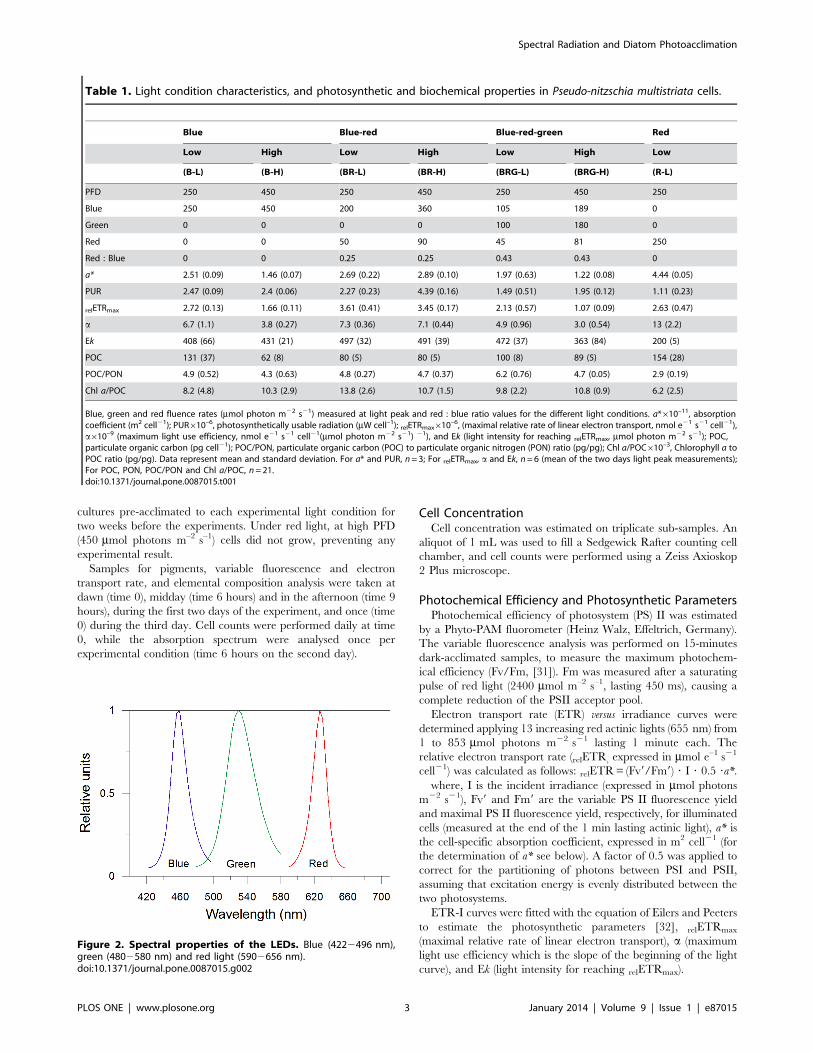
Figure 3. Growth curve of Pseudo-nitzschia multistriata. Growth under (A) low and (B) high light. B-L, BR-L, BRG-L, R-L are blue, blue-red, blue-red-green, and red low light conditions, respectively; B-H, BR-H, and BRG-H are blue, blue-red, blue-red-green high light conditions, respectively. Redhigh light prevented cell growth. Experiments were performed during the exponential phase on days 3 to 5 (B-L), 1 to 3 (R-L, BR-L, BRG-L, B-H) and 2to 4 (BR-H, BRG-H). Data represent mean 6 SD (n=3).doi:10.1371/journal.pone.0087015.g003
Spectral Radiation and Diatom Photoacclimation
PLOS ONE | www.plosone.org 4 January 2014 | Volume 9 | Issue 1 | e87015
Statistical AnalysisStudent’s t-test and Spearman’s rank correlation was performed
using Systat 7 software.
Results and Discussion
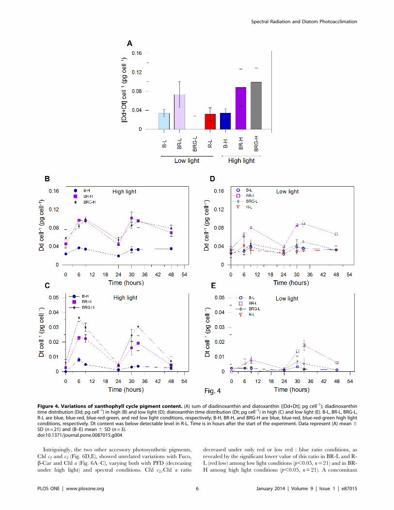
Spectral Radiations and Photoprotective ResponsesThe synthesis of xanthophyll cycle (XC) pigments, diadinox-
anthin (Dd) and diatoxanthin (Dt), is higher under high light than
low light (Fig. 4A, p,0.01, n= 21), with the exception of blue high
light condition (B-H), in which cells did not increase the XC
pigment pool. The low synthesis of both Dd and Dt under B-H
(Fig. 4B,C) is not related to a variation in light absorption, since
the absorption coefficient (a*, Table 1) and photosynthetically
usable radiation (PUR, Table 1) in B-H were similar to the values
found in BRG-H (blue-red-green high; p.0.05, n = 3), in which
Dd and Dt were significantly produced.
Therefore, to explain the absence of Dt and Dd synthesis in B-
H, compared to BR-H (blue-red high) and BRG-H, we propose
that the XC pigment synthesis in diatoms might require sensing of
red light to be triggered, as well as the activation of blue-
photoreceptors by high blue fluence rate to be activated [21]. Red
light might act as a signal for cells to initiate the high light
regulatory pathway, the intensity of the photoprotective response
being thus determined by the blue fluence rate perceived by cells.
This hypothesis fits with the results obtained under low light.
Indeed, red radiation alone prevented Dt synthesis, and blue
radiation alone was not able to enhance the Dt synthesis
(Fig. 4A,D,E). Furthermore, significantly higher XC pigment
content (Dd and Dt; Fig. 4A,D,E) was found in the BR-L (blue-red
low) condition compared to B-L (blue low) and BRG-L (blue-red-
green low; p,0.01, n = 21). The absence of such XC activation
under BRG-L is related to the low blue fluence rate experienced
by cells (105 mmol photons m–2 s–1, Table 1), compared to BR-L
(200 mmol photons m–2 s–1) or to high light conditions
($190 mmol photons m–2 s–1, Table 1). Behind this interpretation,
we know that green light induces much less effect on photo-
regulative processes than blue light in diatoms, which is in
agreement with the lower PUR values measured in BRG (blue-
red-green) compared to B (blue) and BR (blue-red) conditions
(Table 1). This assumption is also supported by the absence of
green-absorbing rhodopsin genes in coastal diatoms [35], as well
as the by higher photosynthetic pigments content measured under
green compared to blue light (data not shown), and by the fact that
the pigments absorbing in blue-green region, Fuco and b-Car,
were similar under BRG-L and BRG-H (see discussion below) and
their distribution perfectly followed the Chl a content.
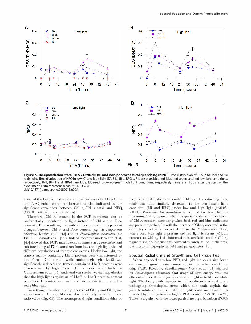
The de-epoxidation state (DES), i.e. the Dd de-epoxidation into
Dt, instead seems to be mainly up regulated by blue fluence rate as
indicated by the higher DES under high than low light (Fig. 5A,B).
This is found even when the synthesis of Dd and Dt is low, i.e.
when red light is absent as in B-H. This feature reveals that Dd de-
epoxidation does not depend on XC activation by the presence of
both red and blue lights together; being enhanced by high blue
fluence rate, as also observed by Schellenberger Costa et al. [21]. It
would mean that the high light dependent-transthylakoidal DpH
build-up [12], which activates the Dd de-epoxidase enzyme for
transforming Dd into Dt, is not under control of the red perception
signal. By contrast, the requirement of red light for enhancing both
the Dd and Dt pigments would indicate that at least one of the
enzymes involved into the XC photoprotective pathway [19] is
under control of the red perception signal. By consequence, the
Dd de-epoxidation rate being up regulated by the XC pigment
content (Fig. 4A–E, 5A,B), is therefore dependent on the
combination of red and blue radiations.
The NPQ capacity was enhanced only in BR-H (Fig. 5C,D),
where the strongest NPQ was measured (0.6660.13 at light peak,
n = 6). Intriguingly, the highest blue fluence rate (B-H) prevented
NPQ increase, suggesting that NPQ development, as XC,
required red light concomitantly with high blue fluence rate.
The higher NPQ in BR-H than in BRG-H (0.2560.03 at light
peak, n = 6; Fig. 5D) is due to the higher blue fluence rate
experienced by cells (360 vs 189 mmol photons m–2 s–1, Table 1).
In BRG-H, NPQ capacity was as low as the values obtained in B-
H (no red radiation) and under low light (p.0.05, n= 21;
Fig. 5C,D), despite the highest XC pool size and DES (Fig. 4A,
5B). Therefore, the NPQ development (Fig. 5C,D) is uncoupled
with both the XC pool size (Fig. 4A) and DES (Fig. 5A,B), as also
reported by Schellenberger Costa et al. [21] on Phaeodactylum
tricornutum grown under different spectral light conditions. This
uncoupling between XC and NPQ in BRG-H can be related to a
weak functional activation of Dt molecules [10,36] and to the
heterogeneous spatial localization of Dt cellular pools [37239].
Furthermore, the BRG-H condition, and the high red : blue ratio
(0.43), might be a potential source of peroxidative damages in cells,
that photoprotective xanthophylls can counter, as already
observed in diatoms [8,39].
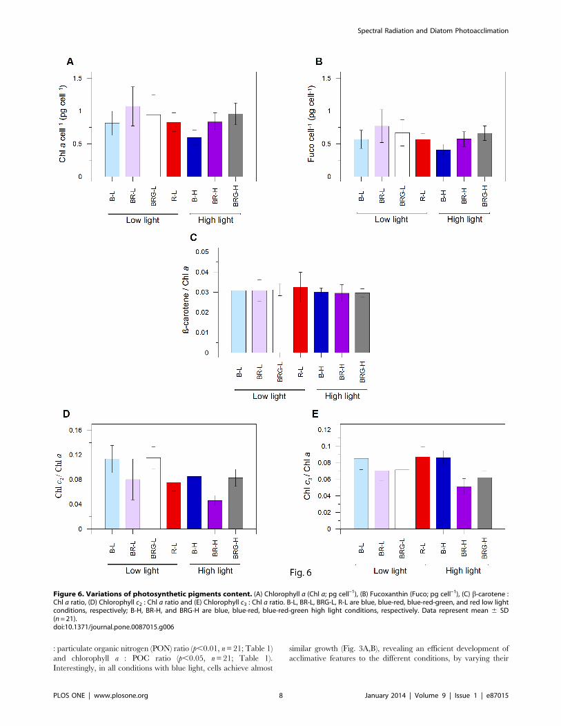
Spectral Radiations and Photosynthetic Pigment ContentAmong the photosynthetic pigments, chlorophyll a (Chl a),
fucoxanthin (Fuco) and b-Carotene (b-Car) followed the same
trend over light conditions (Fig. 6A–C), with a stable ratio between
those pigments (b-Car/Chl a, Fig. 6C; Fuco/Chl a : < 0.70, data
not shown). The contents of these three pigment decreased in B-H
and BR-H compared to B-L and BR-L (Fig. 6A–C) as expected in
a highlight photoacclimation state. The absence of such feature in
BRG-H, where cellular pigment content was similar to BRG-L
(Fig. 6A–C) can be relied to the lower blue fluence rate (189 mmol
photons m–2 s–1 at the light peak) than in B-H and BR-H
($360 mmol photons m–2 s–1, Table 1). This statement reinforces
the strongest role of blue light on photosynthetic regulation in this
diatom compared to green light (see discussion above) and
coincides with the lower PUR values measured in BRG compared
to B and BR conditions (p,0.05, n= 3; Table 1).
The co-variation of Fuco and b-Car with Chl a indicates a
decreasing number of PSII units under high light, reveals an n-
type photoacclimation strategy operated by the coastal diatom P.
multistriata. This strategy allows to co-regulate the number of
antennae and photosystem core complexes to finely tune the
amount of absorbed light energy with the biochemical capacity of
the cell. This is in agreement with the statement of Six et al. [40]
and Lepetit et al. [9], who reported similar photoacclimation
strategy by species growing in the upper mixed layer where light is
variable.
Furthermore, our results reveal that, the high light-induced
pigment variations do not require red radiation to be operated, on
the contrary to XC activation or NPQ. This uncoupling between
pigment variation occurring in the light harvesting complexes and
the photoprotective XC activation, also fits with the absence of a
significant relationship between Dd+Dt and Chl a or Fuco
(p.0.05, n= 147). The reason might be linked to the different
Dd content that the fucoxanthin chlorophyll a/c-binding protein
(FCP) complexes might bind [41,42]. Indeed, these two studies on
two different diatoms revealed that two types of FCPs are present
in diatoms, with different content of Dd, and that high light FCPs
accommodate more Dd compared to low light FCPs.
Spectral Radiation and Diatom Photoacclimation
PLOS ONE | www.plosone.org 5 January 2014 | Volume 9 | Issue 1 | e87015
Intriguingly, the two other accessory photosynthetic pigments,
Chl c2 and c3 (Fig. 6D,E), showed unrelated variations with Fuco,
b-Car and Chl a (Fig. 6A–C), varying both with PFD (decreasing
under high light) and spectral conditions. Chl c2/Chl a ratio
decreased under only red or low red : blue ratio conditions, as
revealed by the significant lower value of this ratio in BR-L and R-
L (red low) among low light conditions (p,0.05, n= 21) and in BR-
H among high light conditions (p,0.05, n= 21). A concomitant
Figure 4. Variations of xanthophyll cycle pigment content. (A) sum of diadinoxanthin and diatoxanthin ([Dd+Dt]; pg cell–1); diadinoxanthintime distribution (Dd; pg cell–1) in high (B) and low light (D); diatoxanthin time distribution (Dt; pg cell–1) in high (C) and low light (E). B-L, BR-L, BRG-L,R-L are blue, blue-red, blue-red-green, and red low light conditions, respectively; B-H, BR-H, and BRG-H are blue, blue-red, blue-red-green high lightconditions, respectively. Dt content was below detectable level in R-L. Time is in hours after the start of the experiment. Data represent (A) mean 6SD (n=21) and (B–E) mean 6 SD (n= 3).doi:10.1371/journal.pone.0087015.g004
Spectral Radiation and Diatom Photoacclimation
PLOS ONE | www.plosone.org 6 January 2014 | Volume 9 | Issue 1 | e87015
effect of the low red : blue ratio on the decrease of Chl c2/Chl a
and NPQ enhancement is observed, as also indicated by the
significant correlation between Chl c2/Chl a ratio and NPQ
(p,0.01, n= 147, data not shown).
Therefore, Chl c2 content in the FCP complexes can be
preferentially modulated by light instead of Chl a and Fuco
content. This result agrees with studies showing independent
changes between Chl c2 and Fuco content (e.g., in Pelagomonas
calceolata, Dimier et al. [43] and in Phaeodactylum tricornutum, see
Fig. 6 in Nymark et al. [44]). Indeed recently Gundermann et al.
[45] showed that FCPs mainly exist as trimers in P. tricornutum and
sub-fractioning of FCP complexes from low and high light, yielded
different populations of trimeric complexes. Under low light, the
trimers mainly containing Lhcf5 proteins were characterised by
low Fuco : Chl c ratio while under high light Lhcf5 was
significantly reduced and trimers containing Lhcf4 proteins were
characterised by high Fuco : Chl c ratio. From both the
Gundermann et al. [45] study and our results, we can hypothesize
that the high light regulation of Lhcf5 vs Lhcf4 proteins content
requires red radiation and high blue fluence rate (i.e., under low
red : blue ratio).
Even though the absorption properties of Chl c3 and Chl c2 are
almost similar, Chl c3/Chl a varied irrespectively to the red : blue
ratio value (Fig. 6E). The monospectral light conditions (blue or
red), presented higher and similar Chl c3/Chl a ratio (Fig. 6E),
while this ratio similarly decreased in the two mixed light
conditions (BR and BRG) under low and high light (p,0.05,
n= 21). Pseudo-nitszchia multistriata is one of the few diatoms
presenting Chl c3 pigment [46]. The spectral radiation modulation
of Chl c3 content, decreasing when both red and blue radiations
are present together, fits with the increase of Chl c3 observed in the
deep, layer below 50 metres depth in the Mediterranean Sea,
where only blue light is present and red light is absent [47]. In
contrast to Chl c2, little information is available on the Chl c3pigment mainly because this pigment is rarely found in diatoms,
but mostly in haptophytes [48] and pelagophytes [43].
Spectral Radiations and Growth and Cell PropertiesWhen provided with low PFD, red light induces a significant
decrease of growth rate compared to the other conditions
(Fig. 3A,B). Recently, Schellenberger Costa et al. [21] showed
on Phaedoactylum tricornutum that usage of light energy was less
efficient when cells were grown under red light as to blue or white
light. The low growth capacity in red condition is related to an
undergoing physiological stress, which also could explain the
growth inhibition under high red light (data not shown), as
revealed by the significantly higher POC content (p,0.05, n= 21;
Table 1) together with the lower particulate organic carbon (POC)
Figure 5. De-epoxidation state (DES=Dt/(Dd+Dt)) and non-photochemical quenching (NPQ). Time distribution of DES in (A) low and (B)high light. Time distribution of NPQ in low (C) and high light (D). B-L, BR-L, BRG-L, R-L are blue, blue-red, blue-red-green, and red low light conditions,respectively; B-H, BR-H, and BRG-H are blue, blue-red, blue-red-green high light conditions, respectively. Time is in hours after the start of theexperiment. Data represent mean 6 SD (n=3).doi:10.1371/journal.pone.0087015.g005
Spectral Radiation and Diatom Photoacclimation
PLOS ONE | www.plosone.org 7 January 2014 | Volume 9 | Issue 1 | e87015
: particulate organic nitrogen (PON) ratio (p,0.01, n= 21; Table 1)
and chlorophyll a : POC ratio (p,0.05, n= 21; Table 1).
Interestingly, in all conditions with blue light, cells achieve almost
similar growth (Fig. 3A,B), revealing an efficient development of
acclimative features to the different conditions, by varying their
Figure 6. Variations of photosynthetic pigments content. (A) Chlorophyll a (Chl a; pg cell–1), (B) Fucoxanthin (Fuco; pg cell–1), (C) b-carotene :Chl a ratio, (D) Chlorophyll c2 : Chl a ratio and (E) Chlorophyll c3 : Chl a ratio. B-L, BR-L, BRG-L, R-L are blue, blue-red, blue-red-green, and red low lightconditions, respectively; B-H, BR-H, and BRG-H are blue, blue-red, blue-red-green high light conditions, respectively. Data represent mean 6 SD(n=21).doi:10.1371/journal.pone.0087015.g006
Spectral Radiation and Diatom Photoacclimation
PLOS ONE | www.plosone.org 8 January 2014 | Volume 9 | Issue 1 | e87015
energy allocation strategies or the arrangement of biochemical
pathways [49].
The fastest cell number increase during the exponential growth
phase recorded under B-H (Fig. 3B) is paralleled by a low POC/
PON ratio (Table 1), in agreement with the findings of Halsey
et al. [49]. The lack of red-dependent activation of the xantho-
phyll cycle and non-photochemical quenching in B-H, limiting the
energetic cost of the photoprotective response [43], allows
therefore a highest energetic investment for growth. In turn, cells
being unable to balance light harvesting and photoprotective
capacity, are not able to cope with high light damages, leading
cells earlier into stationary phase under this condition (B-H)
compared to other high light conditions.
The low red : blue ratio (0.25, BR-H and BR-L) appears to be
peculiar, when compared to the other light conditions, since
growth capacity (Fig. 3), absorption coefficient (a*), as well as
photosynthetic properties and POC and PON content (Table 1)
are similar between low and high PFD. At the exception of R-L,
the highest a* is found in BR (Table 1, p,0.001, n= 3) and is
paralleled by the enhancement of the maximal relative rate of
linear electron transport (relETRmax), light intensity for reaching
the relETRmax (Ek) and the maximum light use efficiency under
low light (a), thus revealing relevant changes of the photosynthetic
properties under this condition (Table 1), irrespective of PFD
experienced. Moreover, as consequence of the highest PUR in
BR-H, which results in an excess energy absorption, cells undergo
an efficient photoprotection, by increasing Dt content (Fig. 4) and
by developing high NPQ (Fig. 5D). The causes of the high a* in
BR-H, similar to BR-L (Table 1), and therefore the highest PUR
in BR-H, are unclear, not being explained by significant variations
in pigment content (Fig. 6A–C). The uncoupling between pigment
variation and a* in BR-H might concern variations in pigment
package effect. The latter can be induced by structural changes in
thylakoid membranes [50,51], occurring in BR-H as revealed by
the variations in Chl c content, and by the changes in LHCs
properties (see discussion above, Gundermann et al. [45]).
Conclusion
Our study leads to consider the spectral composition of light as
an essential trigger for photophysiological acclimation of diatoms.
Our results suggest that the fast photoprotective processes such as
XC and NPQ require red light to be initiated and a high blue
fluence rate to be activated. These results logically fit with the
optical properties of the water column, since red radiation is only
present in the upper layer of the water column, i.e. associated to
high PFD (Fig. 1; [5]). Hypothetically, red radiation sensed by cells
in the surface layer, act as a relevant environmental cue [20] for
signalling high light environment, while blue fluence rate
experienced by cells, narrowly correlated to the depth at which
cells are upwelled, determines the strength of the photoprotective
XC activation, NPQ development and pigment content variations.
Furthermore, the red : blue ratio is also a crucial parameter for
shaping photophysiological properties of the cells, mainly linked to
pigment content related to light-harvesting complex structures.
Acknowledgments
We thank F Margiotta for the POC and PON analysis and F Tramontano
for his help during experiments. We also acknowledge the two reviewers for
helpful criticisms.
Author Contributions
Conceived and designed the experiments: CB FC. Performed the
experiments: RC FC CB LB VG. Analyzed the data: RC FC CB LB
VG AVR. Wrote the paper: CB RC LB VG AVR.
References
1. Falkowski GP, Raven JA (2007) Aquatic photosynthesis. 2nd edition, Princeton
University Press, USA.
2. Rasmussen B, Fletcher IR, Brocks JJ, Kilburn MR (2008) Reassessing the first
appearance of eukaryotes and cyanobacteria. Nature 455: 1101–1104.
3. MacIntyre HL, Kana TM, Geider RJ (2000) The effect of water motion on
short-term rates of photosynthesis by marine phytoplankton. Trends Plant Sci 5:
12–17.
4. Litchman E, Klausmeier CA (2001) Competition of phytoplankton under
fluctuating light. Am Nat 157: 170–187.
5. Kirk JTO (1994) Light and Photosynthesis in Aquatic Ecosystems: Cambridge–
London–New York: Cambridge University Press. 401 p.
6. Falkowski PG (1984) Physiological responses of phytoplankton to natural light
regimes. J Plankton Res 6: 295–307.
7. Lavaud J (2007) Fast regulation of photosynthesis in diatoms: mechanisms,
evolution and ecophysiology. Funct Plant Sci Biotech 1: 267–287.
8. Brunet C, Johnsen G, Lavaud J, Roy S (2011) Pigments and photoacclimation
processes. In Roy S, Llewellyn C, Skarstad Egeland E, Johnsen G editors.
Phytoplankton Pigments, Characterization, Chemotaxonomy and Application in
Oceanography. 445–471.
9. Lepetit B, Goss R, Jakob T, Wilhelm C (2012) Molecular dynamics of the
diatom thylakoid membrane under different light conditions. Photosynth Res
111: 245–257.
10. Ruban AV, Lavaud J, Rousseau B, Guglielmi G, Horton P, et al. (2004) The
super-excess energy dissipation in diatom algae: comparative analysis with
higher plants. Photosynth Res 82: 165–175.
11. Brunet C, Lavaud J (2010) Can the xanthophyll cycle help extract the essence
ofthemicroalgal functional response to a variable light environment? J Plankton
Res 32: 1609–1617.
12. Goss R, Jakob T (2010) Regulation and function of xanthophyll cycle-dependent
photoprotection in algae. Photosynth Res 106: 103–122.
13. Dring MJ (1988) Photocontrol of development in algae. Annu. Rev. Plant
Physiol. Plant Mol Biol 39: 157–174.
14. Roenneberg T, Nakamura H, Hastings JW (1988) Creatine accelerates the
circadian clock in a unicellular alga. Nature 334: 432–434.
15. Falkowski PG, Laroche J (1991) Acclimation to spectral irradiance in algae.
J Phycol 27: 8–14.
16. Lopez-Figueroa F (1992) Diurnal variation in pigment content in Porphyra
laciniara and Chondrus crispus and its relation to the diurnal changes of underwater
light quality and quantity. Mar Ecol 13: 285–305.
17. Mouget JL, Rosa P, Tremblin G (2004) Acclimation of Haslea ostrearia to light of
different spectral qualities confirmation of ‘chromatic adaptation’ in diatoms.
J Photochem Photobiol 75: 1–11.
18. Takahashi F, Yamagata D, Ishikawa M, Fukamatsu Y, Ogura Y, et al. (2007)
AUREOCHROME, a photoreceptor required for photomorphogenesis in
stramenopiles. Proc Natl Acad Sci USA 104: 19625–19630.
19. Coesel S, Obornı́k M, Varela J, Falciatore A, Bowler C (2008) Evolutionary
Origins and Functions of the Carotenoid Biosynthetic Pathway in Marine
Diatoms. PLoS ONE 3: e2896.
20. Depauw FA, Rogato A, d’Alcala MR, Falciatore A (2012) Exploring the
molecular basis of responses to light in marine diatoms. J Exp Bot 63: 1575–
1591.
21. Schellenberger Costa B, Jugandreas A, Jakob T, Weisheit W, Mittag M, et al.
(2013a) Blue light is essential for high light acclimation and photoprotection in
the diatom Phaeodactylum tricornutum. J Exp Bot 64: 483–493.
22. Schellenberger Costa B, Sachse M, Jungandreas A, Bartulos CR, Gruber A, et
al. (2013b) Aureochrome 1a is involved in the photoacclimation of the diatom
Phaeodactylum tricornutum. Plos One 8: e74451.
23. Huysman MJJ, Fortunato AE, Matthijs M, Schellenberger Costa B, Vander-
haeghen R, et al. (2013) AUREOCHROME1a-mediated induction of the
diatom-specific cyclin dsCYC2 controls the onset of cell division in diatoms
(Phaeodactylum tricornutum). Plant Cell 25: 215–228.
24. Roenneberg T, Foster RG (1997) Twilight Times: Light and the Circadian
System. Photochem Photobiol 66: 549–561.
25. Fisher AE, Berges JA, Harrison PJ (1996) Does light quality affect the sinking
rates of marine diatoms? J Phycol 32: 353–360.
26. McLachlan DH, Brownlee C, Taylor AR, Geider RJ, Underwood GJC (2009)
Light-induced motile responses of the estuarine benthic diatoms Navicula
perminuta and Cylindrotheca closterium (Bacillariophyceae). J Phycol 45: 592–599.
27. Shihira-Ishikawa I, Nakamura T, Higashi S-i, Watanabe M (2007) Distinct
responses of chloroplasts to blue and green laser microbeam irradiations in the
centric diatom Pleurosira laevis. Photochem Photobiol 83: 1101–1109.
Spectral Radiation and Diatom Photoacclimation
PLOS ONE | www.plosone.org 9 January 2014 | Volume 9 | Issue 1 | e87015
28. Cao S, Wang J, Chen D (2013) Settlement and cell division of diatom Navicula
can be influenced by light of various qualities and intensities. J Basic Microbiol,doi: 10.1002/jobm.201200315.
29. Ishikawa M, Takahashi F, Nozaki H, Nagasato C, Motomura T, et al. (2009)
Distribution and phylogeny of the blue light receptors aureochromes ineukaryotes. Planta 230: 543–552.
30. D’Alelio D, d’Alcala MR, Dubroca L, Sarno D, Zingone A, et al. (2010) Thetime for sex: A biennial life cycle in a marine planktonic diatom. Limnology and
Oceanography 55: 106–114.
31. Giovagnetti V, Cataldo ML, Conversano F, Brunet C (2012) Growth andphotophysiological responses of two picoplanktonic Minutocellus species, strains
RCC967 and RCC703 (Bacillariophyceae). Eur J Phycol 47: 408–420.32. Eilers PHC, Peeters JCH (1988) A model for the relationship between light
intensity and the rate of photosynthesis in phytoplankton. Ecol Model 42: 199–215.
33. Hedges JI, Stern JH (1984) Carbon and nitrogen determination of carbonate–
containing solids. Limnol Oceanogr 29: 657–663.34. Morel A, Lazzara L, Gostan J (1987) Growth rate and quantum yield time
response for a diatom to changing irradiances (energy and color). LimnolOceanogr 32: 1066–1084.
35. Marchetti A, Schruth DM, Durkin CA, Parker MS, Kodnera RB, et al. (2012)
Comparitive meta transcriptomics identifies molecular bases for the physiolog-ical responses of phytoplankton to varying iron availability. Proc Natl Acad Sci
USA 6: 317–325.36. Lavaud J, Kroth PG (2006) In diatoms, the transthylakoid proton gradient
regulates the photoprotective non-photochemical fluorescence quenchingbeyond its control on the xanthophyll cycle. Plant Cell Physiol 47: 1010–1016.
37. Schumann A, Goss R, Jakob T, Wilhelm C (2007) Investigation of the
quenching efficiency of diatoxanthin in cells of Phaeodactylum tricornutum
(Bacillariophyceae) with different pool sizes of xanthophyll cycle pigments.
Phycologia 46: 113–117.38. Lavaud J, Lepetit B (2013) An explanation for the inter-species variability of the
photoprotective non-photochemical chlorophyll fluorescence quenching in
diatoms. Biochim Biophys Acta 1827: 294–302.39. Lepetit B, Volke D, Gilbert M, Wilhelm C, Goss R (2010) Evidence for the
existence of one antenna-associated, lipid-dissolved, and two protein-boundpools of diadinoxanthin cycle pigments in diatoms. Plant Physiol 154: 1905–
1920.
40. Six C, Finkel Z, Rodriguez F, Marie D, Partensky F, et al. (2008) Contrasting
photoacclimation costs in ecotypes of the marine eukaryote picoplankter
Ostreococcus. Limnol Oceanogr 53: 255–265.
41. Gildenhoff N, Amarie S, Gundermann K, Beer A, Buchel C, et al. (2010)
Oligomerization and pigmentation dependent excitation energy transfer in
fucoxanthin–chlorophyll proteins. Biochim Biophys Acta 1797: 543–549.
42. Nagao R, Yokono M, Akimoto S, Tomo T (2013) High excitation energy
quenching in fucoxanthin chlorophyll a/c-binding protein complexes from the
diatom Chaetoceros gracilis. J Phys Chem B 117: 6888–95.
43. Dimier C, Brunet C, Geider RJ, Raven JA (2009) Growth and photoregulation
dynamics of the picoeukaryote Pelagomonas calceolata in fluctuating light. Limnol
Oceanogr 54: 823–836.
44. Nymark M, Valle KC, Hancke K, Winge P, Andresen K et al. (2013) Molecular
and photosynthetic responses to prolonged darkness and subsequent acclimation
to re-illumination in the diatom Phaeodactylum tricornutum. PLoS ONE 8: e58722.
45. Gundermann K, Schmidt M, Weisheit W, Mittag M, Buchel C (2013)
Identification of several sub-populations in the pool of light harvesting proteins
in the pennate diatom Phaeodactylum tricornutum. Biochim Biophys Acta 1827:
303–310.
46. Zapata M, Rodriguez F, Fraga S, Barra L, Ruggiero MV (2011) Chlorophyll c
pigment patterns in 18 species (51 strains) of the genus Pseudo-nitzschia
(Bacillariophyceae). J Phycol 47: 1274–1280.
47. Brunet C, Casotti R, Vantrepotte V, Conversano F (2007) Vertical variability
and diel dynamics of picophytoplankton in the Strait of Sicily, Mediterranean
Sea, in summer. Mar Ecol Prog Ser 346: 15–26.
48. Zapata M, Jeffrey SW, Wright SW, Rodriguez F, Garrido JL, et al. (2004)
Photosynthetic pigments in 37 species (65 strains) of Haptophyta: implications
for oceanography and chemotaxonomy. Mar Ecol Prog Ser 270: 83–102.
49. Halsey KH, O’Malley RT, Graff JR, Milligan AJ, Behrenfeld MJ (2013) A
common partitioning strategy for photosynthetic products in evolutionarily
distinct phytoplankton species. New Phytol 198: 1030–1038.
50. Fujiki T, Taguchi S (2002) Variability in chlorophyll a specific absorption
coefficient in marine phytoplankton as a function of cell size and irradiance.
J Plankton Res 24: 859–874.
51. Wagner H, Jakob T, Wilhelm C (2006) Balancing the energy flow from captured
light to biomass under fluctuating light conditions. New Phytologist 169: 95–108.
Spectral Radiation and Diatom Photoacclimation
PLOS ONE | www.plosone.org 10 January 2014 | Volume 9 | Issue 1 | e87015


