Embed Size (px)
Citation preview
Sperm ultrastructure in Hemidonax pictus(Hemidonacidae, Bivalvia, Mollusca): comparison withother heterodonts, especially Cardiidae, Donacidaeand Crassatelloidea
JOHN M. HEALY1,3*, PAULA M. MIKKELSEN2† and RÜDIGER BIELER3 FLS
1Biodiversity Program (Malacology), Queensland Museum, PO Box 3300 South Bank, Queensland4101, Australia2Department of Invertebrate Zoology (Malacology), American Museum of Natural History, New York,NY 10024-5192, USA3Department of Zoology (Invertebrates), Field Museum of Natural History, 1400 S. Lake Shore Drive,Chicago, IL 69605-2496, USA
Received 16 March 2007; accepted for publication 20 July 2007
The systematic position and affinities of the marine bivalve genus Hemidonax (Heterodonta, Veneroida) areinvestigated using comparative sperm ultrastructure, with particular focus on the various groups to which thisgenus has been assigned [Donacidae (Tellinoidea), Cardiidae (Cardioidea) and Crassatellidae (Crassatelloidea)].Ultrastructural examination (using transmission electron microscopy) reveals that Hemidonax pictus producessperm of the aquasperm type, with a short, rounded-conical acrosomal vesicle, a short, barrel-shaped nucleus, ashort midpiece (composed of two centrioles and four surrounding mitochondria) and a flagellum containing aconventional 9 + 2 pattern axoneme. The acrosomal vesicle exhibits a wedge-shaped, highly electron-dense, basalring component, and less dense anterior component (including a thin, electron-lucent layer apically, which mayprove to be a useful apomorphy for Hemidonax). A loose, granular deposit of subacrosomal material is locatedwithin a narrow invagination traversing most of the length of the vesicle. Comparison with sperm of otherheterodont bivalves shows no close connection between Hemidonax and the Donacidae (Tellinoidea) or with theCrassatellidae (or other crassatelloideans). Although certain Veneridae (Veneroidea) and Cardiidae (Cardioidea,especially Fragum) show much better conformity in sperm morphology to that observed in Hemidonax, no completematch could be found among studied taxa. We conclude that Hemidonax should be retained in its own, previouslyintroduced family Hemidonacidae, and the latter be placed incertae sedis within the Euheterodonta. © 2008 TheLinnean Society of London, Zoological Journal of the Linnean Society, 2008, 153, 325–347.
ADDITIONAL KEYWORDS: Australian marine fauna – bivalve – gametes – molluscs – reproduction –systematics.
INTRODUCTION
The bivalve genus Hemidonax Mörch, 1871 containsfive extant species, restricted to Australia (four endem-ics), Indonesia and the Philippines, in addition
to one Australian fossil species (Ponder, Colman, Yonge& Colman, 1981). Although often found as beachedvalves, little is known of the habits or ecology ofHemidonax other than that the animals occur inshallow subtidal sandy habitats and are usually col-lected alive only through dredging. The absence ofsiphons and a pallial sinus and the presence of ahatchet-shaped foot indicate that they are shallow andprobably active burrowers (Wilson, 1998). In terms of
*Corresponding author. E-mail: [email protected]†Current address: Paleontological Research Institution, 1259Trumansburg Road, Ithaca, NY 14850, USA.
Zoological Journal of the Linnean Society, 2008, 153, 325–347. With 9 figures
© 2008 The Linnean Society of London, Zoological Journal of the Linnean Society, 2008, 153, 325–347 325
their shell valve profile, Hemidonax species showsome resemblance to the tellinoid family Donacidae,and indeed the genus has often been included in thatfamily (Lamy, 1917; Thiele, 1934; Keen, 1969; Vokes,1980; Abbott & Dance, 1982). Boss (1971) drew atten-tion to the early assignment of Hemidonax to theCardiidae by von Vest (1875) as well as early place-ments within the Crassatellidae (as ‘Crassatellitidae’,Hedley, 1906, 1909, 1923) and (extinct) Tancrediidae(Fischer, 1887). Allan (1959: 319), although aware ofthe often disputed systematic position of Hemidonax,was confident that ‘its anatomical structure placesthis genus in the family Cardiidae, rather than inthose in which it has frequently been placed in thepast.’ She appears to be the first Australian author tohave accepted a cardiid position for the genus.Iredale & McMichael (1962) introduced (withoutdiagnosis) a new family name to accommodate Hemi-donax – the Hemidonacidae (now credited to Scarlato& Starobogatov, 1971)* – that they placed betweenthe Scintillidae and Cardiidae, and distant from theDonacidae and other tellinoidean families. Boss(1971) demonstrated that in terms of their anatomy,Hemidonax spp. lacked key tellinoidean features,most notably the paired, naked siphons and the cru-ciform muscle, and instead showed strong similarityto the Cardiidae. He considered that Hemidonax wasnot distinctive enough to warrant inclusion in its ownfamily, and instead included it in the cardiid sub-family Hemidonacinae (a placement accepted bymany including Kafanov & Popov, 1977; Keen, 1980).Perhaps the most unusual assignment of the Hemi-donacidae was proposed by Scarlato & Starobogatov(1971, 1979), who grouped Hemidonacidae with theDonacidae and the extinct Tancrediidae in the Dona-coidea, and associated with various galeommatoideanfamilies into a suborder Erycina. Ponder et al. (1981)taxonomically reviewed the genus, and providedfurther details of anatomy in support of cardiidaffinities of Hemidonax. However, they also listedseveral anatomical differences and some conchologi-
cal differences between Hemidonax and the Cardi-idae, and therefore maintained the need for aseparate family Hemidonacidae to contain the genus– a view (generally) adopted in subsequent works(e.g. Kafanov & Popov, 1977; Boss, 1982; Vaught,1989; Lamprell & Whitehead, 1992; Wilson, 1998).Schneider (1992) rekindled the debate concerning therelationship of Hemidonax to other Veneroida, pre-ferring to regard the genus as incertae sedis and nota member of the Cardioidea or the tellinoideanDonacidae. Most recently, Schneider & Carter (2001)argued for a closer relationship between Hemidonaxand the tellinoidean family Psammobiidae than withthe Cardiidae, based on their comparative study ofshell microstructure.
Comparative studies of bivalve sperm ultrastruc-ture have shed new light onto higher relationshipswithin the class (Popham, 1979; Healy, 1996a) andthe systematics or phylogeny of several importanttaxa (e.g. Mytiloidea – Hodgson & Bernard, 1986;Kafanov & Drozdov, 1998; Palaeoheterodonta – Healy,1989, 1996a, b; Pteriomorphia – Healy, Keys &Daddow, 2000; Crassatelloidea – Healy, 1995a, b;Galeommatoidea – Jespersen, Lützen & Morton,2002; Veneroidea – Gharagozlou-Van Ginneken &Pochon-Masson, 1971; Healy, 1995b; Healy,Mikkelsen & Bieler, 2006). With this in mind, wehave carried out a sperm ultrastructural study of arepresentative species of Hemidonax [using the typespecies H. pictus (Tryon, 1870)] in order to clarify therelationships of the genus to other Veneroida. In orderbetter to assess the two strongest claims regardingHemidonax affinities (i.e. with the Donacidae or withthe Cardiidae) we also present data for both of thesefamilies, which will help to supplement the availableliterature (Donacidae – Hodgson, Bernard & Van derHorst, 1990; Sousa & Oliveira, 1994; Healy, 1995b;Cardiidae – Popham, 1979; Sousa & Azevedo, 1988;Healy, 1995b, 1996a; Sousa et al., 1998; Keys & Healy,1999, 2000; Drozdov, Frolenko & Ferraguti, 2001).Aside from Pelseneer’s (1911) statement that inHemidonax donaciformis (Schröter, 1786) the sexesare separate, nothing appears to be known concerningthe reproductive biology of Hemidonax. The presentstudy represents the first contribution to knowledgeof gamete morphology in the genus, and it is hopedthat the results will stimulate further studies on thissmall but intriguing group.
MATERIAL AND METHODS
Hemidonax pictus (Tryon, 1870) was dredged from adepth of 20–30 m, approximately 1 km north-westof ‘Yellowpatch’ off the northern coast of MoretonIsland, south-eastern Queensland, Australia(26°57.6′S, 153°24.6′E) on 3 April 2005 and trans-
*[Although Iredale & McMichael (1962) provided bibliographicreferences to both the genus Hemidonax Mörch, 1778 and tothe type species of the genus [H. pictus (Tryon, 1870)], thiscannot be considered a diagnosis of the family as it does notsatisfy the requirements of Article 13.1 of the ICZN (1999)Code. The name Hemidonacidae Iredale & McMichael, 1962 istherefore a nomen nudum (see Bouchet & Rocroi, 2005: 7, fora recent discussion of this aspect of the Code). The first toprovide a formal definition of Hemidonacidae (and thereforemake the name available) were Scarlato & Starobogatov(before 7 July 1971, p. 16) who made no reference to Iredale& McMichael’s checklist, but clearly stated ‘HemidonacidaeScarlato et Starobogatov, fam. nov.’ As their work pre-datesthat of Boss (20 July 1971, Hemidonacinae), Scarlato &Starobogatov (1971) must be credited with authorship ofHemidonacidae, despite later citing (see Scarlato &Starobogatov, 1979) Iredale & McMichael (1962) as theoriginal authors of the family.]
326 J. M. HEALY ET AL.
© 2008 The Linnean Society of London, Zoological Journal of the Linnean Society, 2008, 153, 325–347
ferred to holding tanks at the Moreton Bay ResearchStation (North Stradbroke Island, south-easternQueensland). Donax (Plebidonax) deltoides (Lamarck,1818) was collected from surf beaches near PointLookout, North Stradbroke Island (27°28′S,153°32′E). Vasticardium vertebratum (Jonas, 1844),Fragum unedo (Linnaeus, 1758) and Lunulicardiahemicardium (Linnaeus, 1758) were all collected fromMyora, North Stradbroke Island, Moreton Bay, south-eastern Queensland (27°27′S, 153°26′E, 1986-1990).Eucrassatella cumingii (A. Adams & Angas, 1864)was dredged at a depth of 12 m, Banana Banks,Moreton Bay (27°32′S, 153°20′E, 1992). Cardita muri-cata Sowerby, 1832 was collected intertidally atCockle Bay, Magnetic Island, northern Queensland(19°11′S, 146°49′E, 1991). Papyridea semisulcata(Gray, 1825) was collected from the Florida Keys,USA [station FK-720, 27 April 2004, Looe Key backreef, Monroe County, Florida Keys, 24°32.894′N,81°24.360′W, rubble, sand and seagrass, snorkelling,1.2–2.1 m, FLORIDAYS]. The following processingschedule was followed for most of the material exam-ined. Small (2 mm3) pieces of testicular tissue weretaken from a ripe male and fixed in ice cold (2–4 °C)glutaraldehyde (3.5% in 0.1 M phosphate buffer con-taining 7.5% w/v sucrose) for 3 h. For Papyrideasemisulcata the entire animal was fixed in cold3.5% buffered glutaraldehyde (formula as above) inthe refrigerator for 11 days, then shipped to Chicago(and later sent to Australia for subsequent pro-cessing). Tissue samples were subsequently rinsed inbuffer (three 30-min changes), post-fixed in 1%osmium tetroxide (buffer as above) for 80 min, rinsedagain in three changes of buffer, dehydrated in agraded series of ethanol and embedded in Spurr’sepoxy resin. Samples were maintained at 2–4 °C (onice) for all stages up until 70% ethanol, and thereafterat room temperature (25 °C). Semithin and silver–gold interface ultrathin sections were cut with LeicaUltracut T and LKB IV ultramicrotomes, collected on200-square-mesh copper grids, stained according tothe lead citrate–uranyl acetate–lead citrate ‘sandwichstain’ procedure of Daddow (1986) and examinedusing Jeol 101 or Hitachi 300 transmission electronmicroscopes operating at 75–80 kV or a Philips 300transmission electron microscope at 60 kV. Vouchermaterial of Hemidonax pictus used in the presentstudy has been lodged with the Field Museum ofNatural History (Registration number: FMNH311639) and Queensland Museum (Brisbane, Austra-lia) (QMMO 78087). Voucher material of other speciesof bivalves examined herein have been lodgedwith the Queensland Museum: Vasticardiumvertebratum (QMMO 78083), Fragum unedo (QMMO78084), Lunulicardia hemicardium (QMMO 78085),Donax deltoides (QMMO 78086), Cardita muricata
(QMMO 53310), Eucrassatella cumingii (QMMO53311), or the Field Museum of Natural History(Chicago, USA): Papyridea semisulcata (FMNH311640).
RESULTSHEMIDONAX PICTUS (FIG. 1)
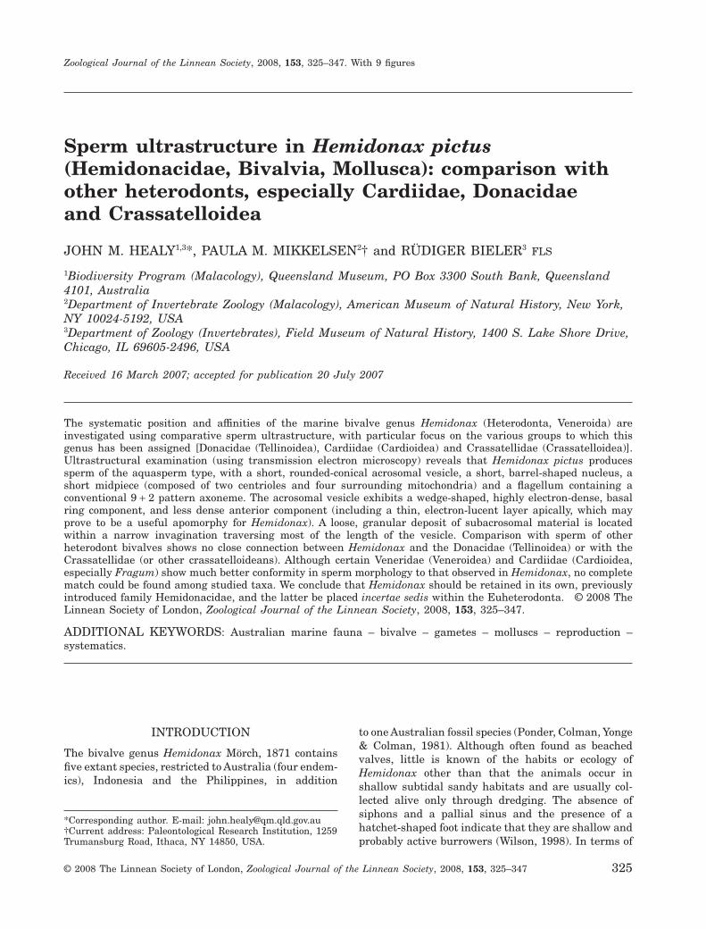
Acrosomal complexThe acrosomal vesicle is conical, measuring0.35 ± 0.02 mm (N = 4) in length and with a maxi-mum diameter of approximately 0.4 ± 0.05 mm (N = 4)(Fig. 1A–D). Contents of the vesicle are differentiatedinto a highly electron-dense, faintly reticulate, basalring sheathed by markedly less dense (and granular)material that also fills the anterior region of thevesicle (Fig. 1A, C, D). Associated with the vesicleapex is a broad, thin, electron-lucent layer, the extentof which is somewhat variable (Fig. 1A, C, arrows). Anarrow basal invagination extends for most of thelength of the acrosomal vesicle and is filled by agranular deposit of subacrosomal material (Fig. 1A–D). The plasma membrane forms the outermostsheath of the acrosomal complex, as it does in otherregions of the spermatozoon (Fig. 1A, F) (this appliesin all the species of bivalves examined herein).
NucleusThe nucleus is short (length 1.8 ± 0.1 mm, N = 4) andbarrel-shaped (tapers apically anteriorly), with amaximum diameter (near base) of 1.0 ± 0.1 mm (N = 4)(Fig. 1B). Nuclear contents are highly electron-dense,with scattered, irregularly shaped, electron-lucentlacunae. Broad, shallow depressions, present basally,contact the anterior surfaces of each midpiece mito-chondrion (Fig. 1B, E). These depressions surround asmaller central recess filled with granular materialloosely associated with the proximal centriole(Fig. 1E).
MidpieceThe midpiece is positioned at the base of the nucleusand consists of four spherical mitochondria sur-rounding a pair of orthogonally arranged centrioles(Fig. 1B, E, F). The entire midpiece region has alength of 0.75 ± 0.05 mm (N = 3) and a maximumdiameter (of mitochondrial cluster) of approximately1.44 ± 0.1 mm (N = 3). The centrioles lie in contactwith each other and exhibit triplet microtubularstructure which is largely obscured by a dense matrix(Fig. 1E–G). The distal centriole is connected by aseries of nine satellite fibres each terminating in aY-shaped fork attached to the plasma membrane(Fig. 1E, G).
FlagellumThe flagellum consists of a 9 + 2 microtubular con-figuration axoneme (nine microtubular doublets
HEMIDONAX SPERM ULTRASTRUCTURE 327
© 2008 The Linnean Society of London, Zoological Journal of the Linnean Society, 2008, 153, 325–347
328 J. M. HEALY ET AL.
© 2008 The Linnean Society of London, Zoological Journal of the Linnean Society, 2008, 153, 325–347
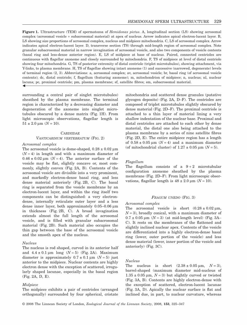
surrounding a central pair of singlet microtubules)sheathed by the plasma membrane. The terminalregion is characterized by a decreasing diameter anddegeneration of the axoneme into singlet micro-tubules obscured by a dense matrix (Fig. 1H). Fromlight microscopic observations, flagellar length is51 ± 2.0 mm (N = 10).
CARDIIDAE
VASTICARDIUM VERTEBRATUM (FIG. 2)
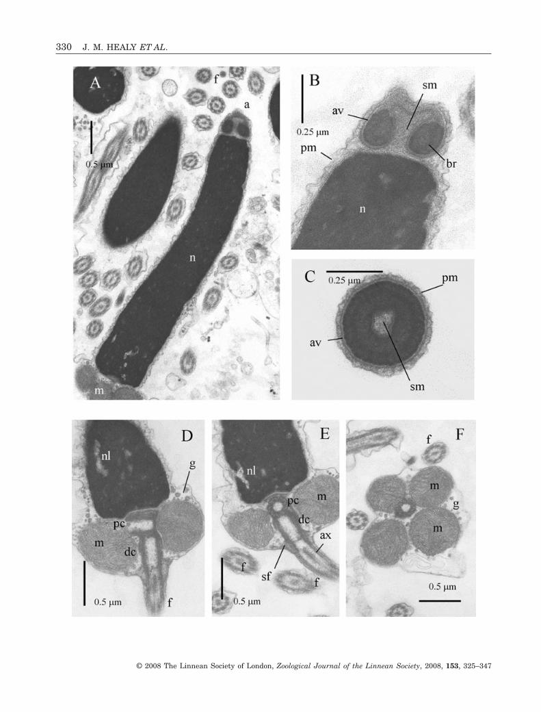
Acrosomal complexThe acrosomal vesicle is dome-shaped, 0.28 ± 0.02 mm(N = 4) in length and with a maximum diameter of0.46 ± 0.02 mm (N = 4). The anterior surface of thevesicle may be flat, slightly concave or, most com-monly, slightly convex (Fig. 2A, B). Contents of theacrosomal vesicle are divisible into a very prominent,and markedly electron-dense basal ring, and lessdense material anteriorly (Fig. 2B, C). The basalring is separated from the vesicle membrane by anelectron-lucent layer, and within the ring itself twocomponents can be distinguished: a very electron-dense, internally reticulate outer layer and a lessdense inner layer, both approximately 0.05–0.06 mmin thickness (Fig. 2B, C). A broad invaginationextends almost the full length of the acrosomalvesicle, and is filled with granular subacrosomalmaterial (Fig. 2B). Such material also occupies thethin gap between the base of the acrosomal vesicleand the smooth apex of the nucleus.
NucleusThe nucleus is rod shaped, curved in its anterior halfand 4.4 ± 0.1 mm long (N = 5) (Fig. 2A). Maximumdiameter is approximately 0.7 ± 0.1 mm (N = 5) justanterior to the midpiece. Nuclear contents are highlyelectron dense with the exception of scattered, irregu-larly shaped lacunae, especially in the basal region(Fig. 2A, D, E).
MidpieceThe midpiece exhibits a pair of centrioles (arrangedorthogonally) surrounded by four spherical, cristate
mitochondria and scattered dense granules (putativeglycogen deposits) (Fig. 2A, D–F). The centrioles arecomposed of triplet microtubules slightly obscured bydense material (Fig. 2D–F). The proximal centriole isattached to a thin layer of material lining a veryshallow indentation of the nuclear base. Proximal anddistal centrioles are attached to each other by densematerial, the distal one also being attached to theplasma membrane by a series of nine satellite fibres(Fig. 2D, E). The entire midpiece region has a lengthof 0.58 ± 0.05 mm (N = 4) and a maximum diameter(of mitochondrial cluster) of 1.27 ± 0.05 mm (N = 5).
FlagellumThe flagellum consists of a 9 + 2 microtubularconfiguration axoneme sheathed by the plasmamembrane (Fig. 2D–F). From light microscopic obser-vations, flagellar length is 48 ± 2.0 mm (N = 10).
FRAGUM UNEDO (FIG. 3)
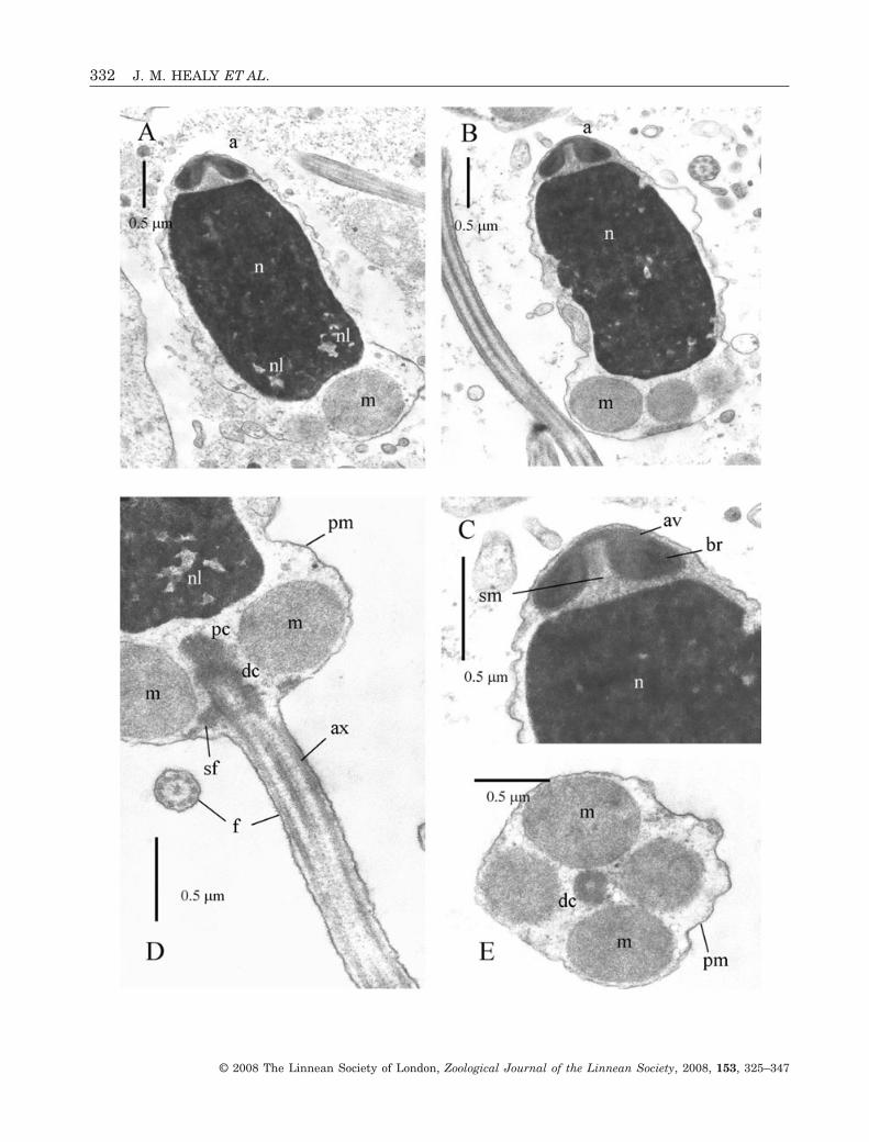
Acrosomal complexThe acrosomal vesicle is short (0.28 ± 0.02 mm,N = 3), broadly conical, with a maximum diameter of0.7 ± 0.05 mm (N = 3) (at mid-length level) (Fig. 3A–C). It rests on the membranes of the flattened andslightly inclined nuclear apex. Contents of the vesicleare differentiated into a highly electron-dense basalring (lower, outer portion of the vesicle) and lessdense material (lower, inner portion of the vesicle andanteriorly) (Fig. 3C).
NucleusThe nucleus is short (2.38 ± 0.05 mm, N = 3),barrel-shaped (maximum diameter mid-nucleus of1.35 ± 0.05 mm, N = 3) but slightly curved or twisted(Fig. 3A, B). Contents are highly electron-dense withthe exception of scattered, electron-lucent lacunae(Fig. 3A, D). Apically the nuclear surface is flat andinclined due, in part, to nuclear curvature, whereas
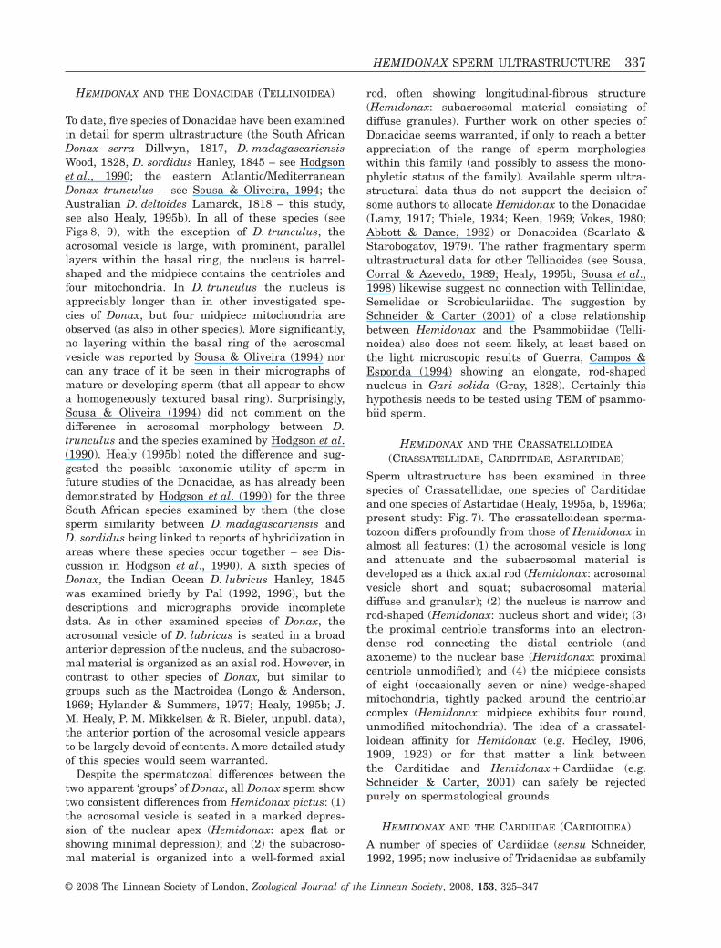
Figure 1. Ultrastructure (TEM) of spermatozoa of Hemidonax pictus. A, longitudinal section (LS) showing acrosomalcomplex (acrosomal vesicle + subacrosomal material) at apex of nucleus. Arrow indicates apical electron-lucent layer. B,LS showing size proportions of acrosomal complex, nucleus and midpiece mitochondria. C, LS of acrosomal complex. Arrowindicates apical electron-lucent layer. D, transverse section (TS) through mid-length region of acrosomal complex. Notegranular subacrosomal material in narrow invagination of acrosomal vesicle, and also two components of vesicle contents(basal ring and less-dense anterior region). E, LS of midpiece at base of nucleus. Paired, connected centrioles arecontinuous with flagellar axoneme and closely surrounded by mitochondria. F, TS of midpiece at level of distal centrioleshowing four mitochondria. G, TS of posterior extremity of distal centriole (triplet microtubules), showing attachment, viaY-links, to plasma membrane. H, TS of flagellae showing intact axoneme (1) and successive (narrowed, degenerate) levelsof terminal region (2, 3). Abbreviations: a, acrosomal complex; av, acrosomal vesicle; br, basal ring (of acrosomal vesiclecontents); dc, distal centriole; f, flagellum (featuring axoneme); m, mitochondrion of midpiece; n, nucleus; nl, nuclearlacuna; pc, proximal centriole; pm, plasma membrane; sf, satellite fibres; sm, subacrosomal material.�
HEMIDONAX SPERM ULTRASTRUCTURE 329
© 2008 The Linnean Society of London, Zoological Journal of the Linnean Society, 2008, 153, 325–347
330 J. M. HEALY ET AL.
© 2008 The Linnean Society of London, Zoological Journal of the Linnean Society, 2008, 153, 325–347
basally, very shallow indentations are associated withthe midpiece mitochondria and proximal centriole(Fig. 3A).
MidpieceThe midpiece contains four spherical mitochondriagrouped around the centriolar pair (proximal anddistal centrioles) (Fig. 3D, E). The centrioles areattached to each other, with the distal one anchoredto the plasma membrane via nine satellite fibres. Theentire midpiece region has a length of 0.75 ± 0.05 mm(N = 3) and a maximum diameter (of mitochondrialcluster) of 1.75 ± 0.08 mm (N = 3).
FlagellumThe flagellum consists of a 9 + 2 microtubular con-figuration axoneme sheathed by the plasma mem-brane. From light microscopic observations, flagellarlength is 48 ± 2.0 mm (N = 10).
LUNULICARDIA HEMICARDIUM (FIG. 4)
Acrosomal complexThe acrosomal vesicle is short (0.24 ± 0.02 mm, N = 2)and dome-shaped (maximum diameter, at base, of0.66 ± 0.01 mm, N = 2) (Fig. 4A, B). It rests on ashallow, circular groove of the nuclear apex (forminga shallow nuclear hump at the nuclear apex)(Fig. 4B). Contents of the vesicle are differentiatedinto a somewhat, dense, basal ring that has an angu-late profile, and is sheathed by a less dense materialthat also fills the anterior half of the vesicle (Fig. 4A,B). The basal invagination of the vesicle is deepin comparison with vesicle length and very broad(maximum diameter at base 0.4 ± 0.02 mm, N = 2) andpartially filled with a granular, but well-defined,deposit of subacrosomal material.
NucleusThe nucleus is short (length 2.15 ± 0.05 mm, N = 3),barrel shaped (maximum diameter, at mid-length, of1.65 ± 0.05 mm, N = 3) and slightly tapered anteriorly(Fig. 4A, B). Contents are highly electron dense withthe exception of electron-lucent lacunae (the lattermore pronounced posteriorly) (Fig. 4A). Apically, the
nucleus projects slightly into the acrosomal vesicleinvagination (Fig. 4A, B). Basally, very shallow inden-tations contact the surfaces of the midpiece mitochon-dria and also form an attachment point for theproximal centriole (Fig. 4A).
MidpieceThe midpiece consists of four or more rarely fivespherical mitochondria and a pair of centrioles(Fig. 4A, C, D). The centrioles (proximal and centri-ole) lie in contact with each other and are arrangedorthogonally. A thin layer of dense material connectsthe proximal centriole to a shallow indentation of thenucleus. Satellite fibres connect the distal centriole tothe plasma membrane (Fig. 4A, C). The entire mid-piece region has a length of 0.7 ± 0.05 mm (N = 3) anda maximum diameter (of mitochondrial cluster) of1.8 ± 0.2 mm (N = 3).
FlagellumThe flagellum consists of a 9 + 2 microtubular con-figuration axoneme, sheathed by the plasmamembrane (Fig. 4C, D). From light microscopic obser-vations, flagellar length is 46 ± 3.0 mm (N = 10).
PAPYRIDEA SEMISULCATA (FIG. 5)
Acrosomal complexThe acrosomal vesicle is dome-shaped, with a flat orslightly domed apex, and measures approximately0.55 ± 0.05 mm (N = 5) in length and 0.7 ± 0.05 mm(N = 5) in maximum diameter (Fig. 5A–D). Contentsof the vesicle are differentiated into an extensive,very electron-dense basal ring, and a less dense (andgranular) anterior component (Fig. 5B, D). A broadbasal invagination extends for approximately half thelength of the acrosomal vesicle and is filled withgranular subacrosomal material (Fig. 5A–C). Dis-cernible within the basal ring are two curved, veryelectron-dense layers and an innermost, electron-lucent layer (Fig. 5B, D).
NucleusThe nucleus is short (length 1.45 ± 0.05 mm, N = 3)and barrel shaped, with a maximum diameter of
Figure 2. Ultrastructure (TEM) of spermatozoa of Vasticardium vertebratum. A, longitudinal section (LS) showingacrosomal complex, curved nucleus, portion of midpiece region. Note also several flagellae cut in transverse section (TS).B, LS of acrosomal complex (acrosomal vesicle + subacrosomal material) and nuclear apex. The degree of anteriordimpling is variable. C, TS at mid-level region of acrosomal complex. D, E, LS at base of nucleus, entire midpiece andproximal portion of flagellum. The proximal centriole shown in LS in D, in TS in E. Putative glycogen granules are alsovisible. F, TS of midpiece showing four mitochondria, distal centriole and putative glycogen granules. Abbreviations: a,acrosomal complex; av, acrosomal vesicle; ax, axoneme; br, basal ring (of acrosomal vesicle contents); dc, distal centriole;f, flagellum; g, putative glycogen granules; m, mitochondrion of midpiece; n, nucleus; nl, nuclear lacuna; pc, proximalcentriole; pm, plasma membrane; sf, satellite fibres; sm, subacrosomal material.�
HEMIDONAX SPERM ULTRASTRUCTURE 331
© 2008 The Linnean Society of London, Zoological Journal of the Linnean Society, 2008, 153, 325–347
332 J. M. HEALY ET AL.
© 2008 The Linnean Society of London, Zoological Journal of the Linnean Society, 2008, 153, 325–347
1.45 ± 0.05 mm (N = 3) (at mid-length) (Fig. 5A).Nuclear contents are highly electron-dense, granu-late, with numerous irregular lacunae (Fig. 5A, E).Apically the nuclear surface is straight or slightlyconcave, while posteriorly shallow indentationscontact the anterior surfaces of each midpiece mito-chondrion (Fig. 5A).
MidpieceThe midpiece consists of four spherical mitochondria(with well-developed cristae) surrounding a pair ofcentrioles (Fig. 5A, F). The entire midpiece region hasa length of 0.7 ± 0.04 mm (N = 3) and a maximumdiameter (of mitochondrial cluster) of 1.7 ± 0.1 mm(N = 3).
FlagellumThe flagellum consists of a 9 + 2 microtubular con-figuration axoneme sheathed by the plasma mem-brane. From light microscopic observations, flagellarlength is 54 ± 2.0 mm (N = 10).
DONAX (PLEBIDONAX) DELTOIDES (FIG. 6)
Acrosomal complexThe acrosomal vesicle measures approximately1.16 ± 0.02 mm (N = 4) in length, has a maximumdiameter of 0.85 ± 0.05 mm (N = 4) (about one-thirdof the distance from the vesicle base) and rests withina broad depression of the nuclear apex (depth0.24 ± 0.03 mm, N = 4) (Fig. 6A–C, E). A narrowinvagination runs almost the full length of the vesicleand is filled with longitudinally fibrous subacrosomalmaterial. Contents of the acrosomal vesicle can bedifferentiated into a very electron-dense, basal ring(curved-cylindrical in longitudinal profile) and homo-geneous, less dense material – the latter enclosing thebasal ring and filling the anterior region of thevesicle. The basal ring shows an internal structure offine, parallel layers (approximately 25–35 in number)(Fig. 6A) that in transverse section (Fig. 6B) arearranged concentrically.
NucleusThe nucleus is short [length 1.4 ± 0.1 mm (N = 4),inclusive of an overlap region with the acrosomal
complex] and squat (maximum diameter 1.5 ±0.04 mm, N = 4), with highly electron-dense contents(Fig. 6A, C–E). The apical surface is broadly indentedto receive the basal portion (0.2 ± 0.05 mm, N = 4) ofthe acrosomal vesicle, while posteriorly, shallowindentations act as contact surfaces for the midpiecemitochondria and the anterior edge of the proximalcentriole. Irregularly shaped electron-lucent lacunaeare present throughout, though largest posteriorly(Fig. 6A, D).
MidpieceThe midpiece exhibits four, roughly spherical, mito-chondria (showing prominent cristae) in addition to apair of orthogonally arranged centrioles (Fig. 6A, E,F). Each centriole is composed of microtubular trip-lets set in a dense matrix (Fig. 6F). The proximalcentriole is loosely connected via a diffuse deposit to ashallow indentation of the nuclear base, and is alsoattached to the distal centriole (Fig. 6A, E). The distalcentriole is anchored to the plasma membrane vianine satellite fibres and is continuous with the dou-blets of the flagellar axoneme (Fig. 6A). The entiremidpiece region has a length of approximately 0.6–0.7 mm and a maximum diameter (of mitochondrialcluster) of 1.6–1.8 mm.
FlagellumThe flagellum consists of a 9 + 2 microtubular con-figuration axoneme, sheathed by the plasma mem-brane (Fig. 6A). From light microscopic observations,flagellar length is 46 ± 3.0 mm (N = 10).
CRASSATELLOIDEA: EUCRASSATELLA CUMINGII
(CRASSATELLIDAE), CARDITA MURICATA (CARDITIDAE)
Acrosomal complexThe acrosomal vesicle is elongate-conical(2.7 ± 0.1 mm E. cumingii, N = 4; length 1.6 ± 0.1 mmC. muricata, N = 4), sharply tapered anteriorlyand almost completely invaginated (Fig. 7A, B). Thevesicle has a maximum diameter (at base) of approxi-mately 0.35 mm. Contents of the acrosomal vesicle aredifferentiated into a dense inner layer enveloped bymarkedly less dense material. (Fig. 7B, C) The sub-acrosomal material is organized as a well-defined
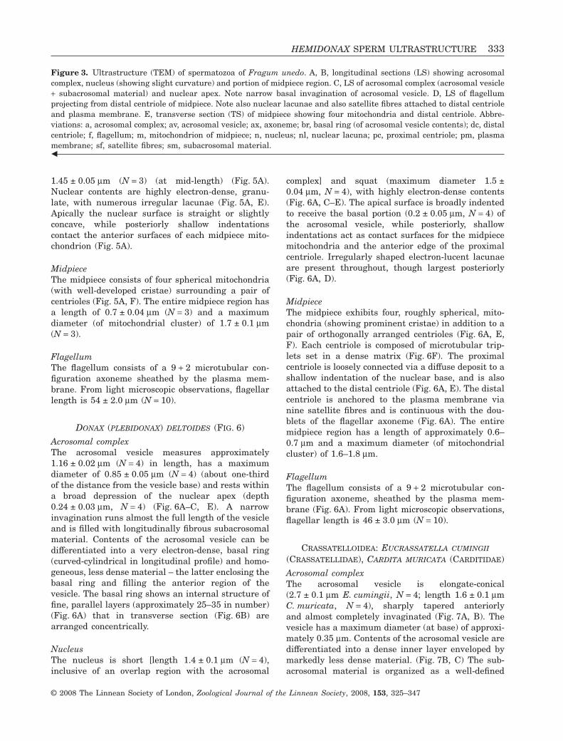
Figure 3. Ultrastructure (TEM) of spermatozoa of Fragum unedo. A, B, longitudinal sections (LS) showing acrosomalcomplex, nucleus (showing slight curvature) and portion of midpiece region. C, LS of acrosomal complex (acrosomal vesicle+ subacrosomal material) and nuclear apex. Note narrow basal invagination of acrosomal vesicle. D, LS of flagellumprojecting from distal centriole of midpiece. Note also nuclear lacunae and also satellite fibres attached to distal centrioleand plasma membrane. E, transverse section (TS) of midpiece showing four mitochondria and distal centriole. Abbre-viations: a, acrosomal complex; av, acrosomal vesicle; ax, axoneme; br, basal ring (of acrosomal vesicle contents); dc, distalcentriole; f, flagellum; m, mitochondrion of midpiece; n, nucleus; nl, nuclear lacuna; pc, proximal centriole; pm, plasmamembrane; sf, satellite fibres; sm, subacrosomal material.�
HEMIDONAX SPERM ULTRASTRUCTURE 333
© 2008 The Linnean Society of London, Zoological Journal of the Linnean Society, 2008, 153, 325–347
334 J. M. HEALY ET AL.
© 2008 The Linnean Society of London, Zoological Journal of the Linnean Society, 2008, 153, 325–347
axial rod that occupies not only the vesicle invagina-tion but also a shallow apical invagination of thenucleus (Fig. 7B).
NucleusThe nucleus is rod-shaped (length 10.0 ± 0.2 mmE. cumingii; 7.4 ± 0.2 mm C. muricata) (N = 5), taper-ing (in each species) from a diameter of 1.0 ± 0.1 mm(N = 6) near the base to 0.4 ± 0.05 mm (N = 6) at theapex (Fig. 7A, B, G, E). Aside from the apical invagi-nation (accommodating part of the subacrosomalmaterial), the nucleus exhibits a short invaginationfor the centriolar rootlet and slight concavities thatcontact the mitochondria (Fig. 7E, G). Irregularlyshaped electron-lucent lacunae are present through-out, though largest and most conspicuous posteriorly(Fig. 7G).
MidpieceThe midpiece exhibits eight (occasionally seven ornine) mitochondria tightly packed around a denserootlet plus distal centriole complex (Fig. 7D–H). Pro-files of the mitochondria are angular in transversesection, with the contacting surfaces of each beingflattened. Although the distal centriole shows nomodifications, the proximal one has been transformedinto a portion of the rootlet attached anteriorly to ashallow indentation at the base of the nucleus andposteriorly to the distal centriole (Fig. 7E–H). Thedistal centriole is anchored to the plasma membranevia a radial array of nine satellite fibres (Fig. 7D), andis continuous with the doublets of the flagellaraxoneme (Fig. 7E). The entire midpiece region(in each species) has a length of approximately0.65 ± 0.05 mm (N = 8) and a maximum diameter of1.7 ± 0.1 mm (N = 8).
FlagellumThe flagellum consists of a 9 + 2 microtubular con-figuration axoneme, sheathed by the plasma mem-brane (Fig. 7F). From light microscopic observations,flagellar length for both species is 47 ± 3.0 mm (N = 10per species).
DISCUSSIONCOMPARISON OF HEMIDONAX SPERM MORPHOLOGY
WITH OTHER HETERODONT BIVALVES
Spermatozoa of Hemidonax pictus are of the simple,aquasperm type characteristic of molluscs showingaquatic fertilization, especially the Bivalvia (Franzén,1955, 1983; Popham, 1979; Hodgson et al., 1990;Healy, 1996a), and also seen in a number of otherinvertebrate groups (e.g. cnidarians, polychaete anne-lids, brachiopods, echiurids, sipunculans – Franzén,1956; Baccetti & Afzelius, 1976; Jamieson & Rouse,1989). Rouse & Jamieson (1987) differentiatedaquasperm into those that fertilize eggs in theambient water (ect-aquasperm) and those that fertil-ize aquatically but within a protected space suchas a worm tube or a molluscan mantle cavity(ent-aquasperm). In the absence of any informationon the fertilization biology of H. pictus, it is impos-sible to characterize the type of aquasperm in thisspecies. However it is worth noting that in shipworms(Teredinidae, Pholadoidea) Popham (1974) found thatspecies fertilizing within the mantle cavity hadsmaller acrosomes than those that fertilized exter-nally (i.e. in the ambient seawater). If such a corre-lation holds among the Bivalvia in general, then thesmall size of the acrosome of H. pictus compared withmost investigated heterodonts (see Figs 8, 9) wouldsuggest the likelihood of ent-aquatic (mantle cavity)fertilization in this species and probably in otherspecies of Hemidonax.
Whereas it is true to state that spermatozoa ofHemidonax pictus do not exhibit any unique or newfeatures, the precise combination of features is dis-tinctive and presumably characteristic of the genusas a whole. Like other heterodonts, H. pictus showsa well-developed basal ring component of the acroso-mal vesicle (see Fig. 8, also for comparative figuresand extensive literature see Healy, 1995b, 1996a).Substantial diversity exists among heterodont taxain the shape of the acrosomal vesicle (and the shapeand internal structure of the basal ring), as well asthe size and length of the nucleus (short or long,straight, curved or helical) and the number ofmidpiece mitochondria (four or five, sometimes
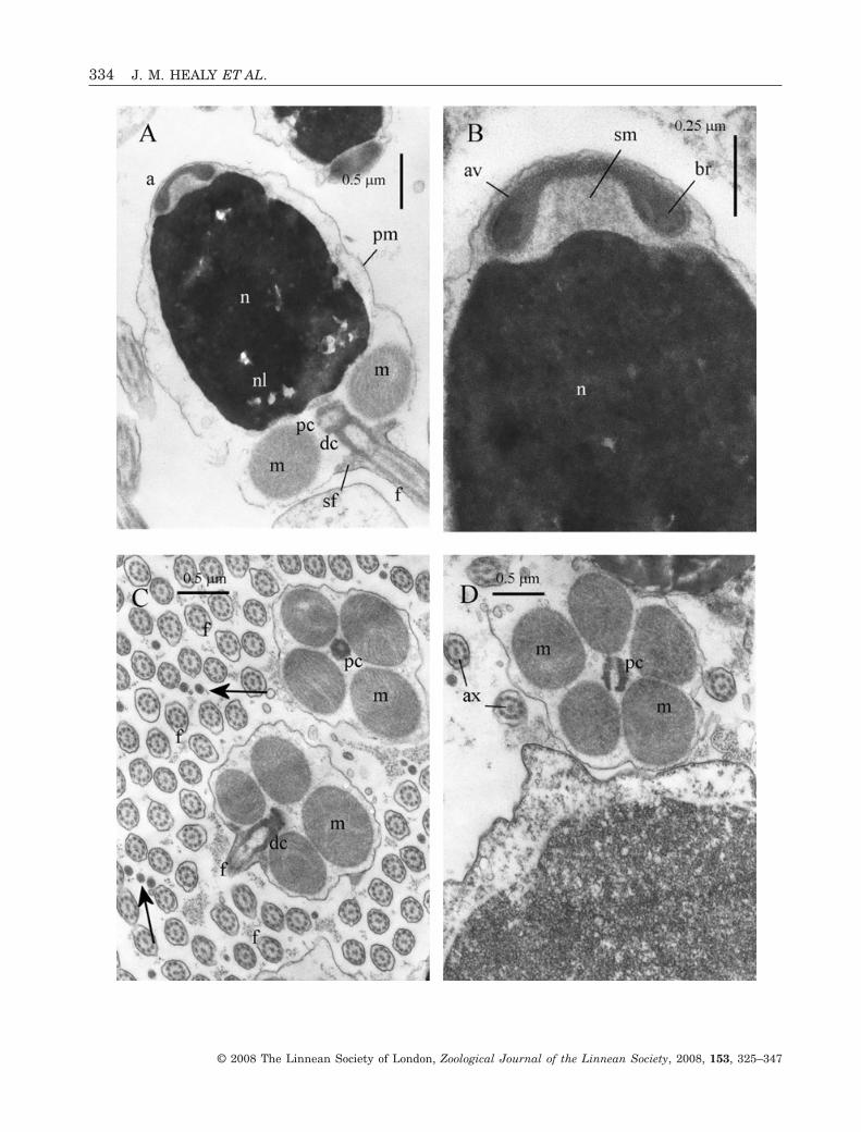
Figure 4. Ultrastructure (TEM) of spermatozoa of Lunulicardia hemicardium. A, longitudinal section (LS) showingacrosomal complex, nucleus (with nuclear lacunae), midpiece region (mitochondria surrounding proximal and distalcentrioles) and proximal portion of flagellum. Note also satellite fibres linking distal centriole to plasma membrane. B,LS of acrosomal complex (acrosomal vesicle + subacrosomal material) and nuclear apex. Note broad but low nuclearprojection. C, transverse section (TS) of midpiece of two spermatozoa and flagella of many others. Note four mitochondria.Arrows indicate terminal region of flagellum. D, TS of midpiece showing five mitochondria variant (proximal centrioleshown in LS) and flagellae. Abbreviations: a, acrosomal complex; av, acrosomal vesicle; ax, axoneme; br, basal ring(of acrosomal vesicle contents); dc, distal centriole; f, flagellum; m, mitochondrion of midpiece; n, nucleus; nl, nuclearlacuna; pc, proximal centriole; pm, plasma membrane; sf, satellite fibres; sm, subacrosomal material.�
HEMIDONAX SPERM ULTRASTRUCTURE 335
© 2008 The Linnean Society of London, Zoological Journal of the Linnean Society, 2008, 153, 325–347
more, but always with a predominating number).As there are a number of differing opinions rela-ting to the placement and affinities of Hemidonaxamong the Heterodonta, the following part of
the discussion will deal with each of these, in-cluding a brief resumé of group features thenprogressing to a comparison with Hemidonaxresults.
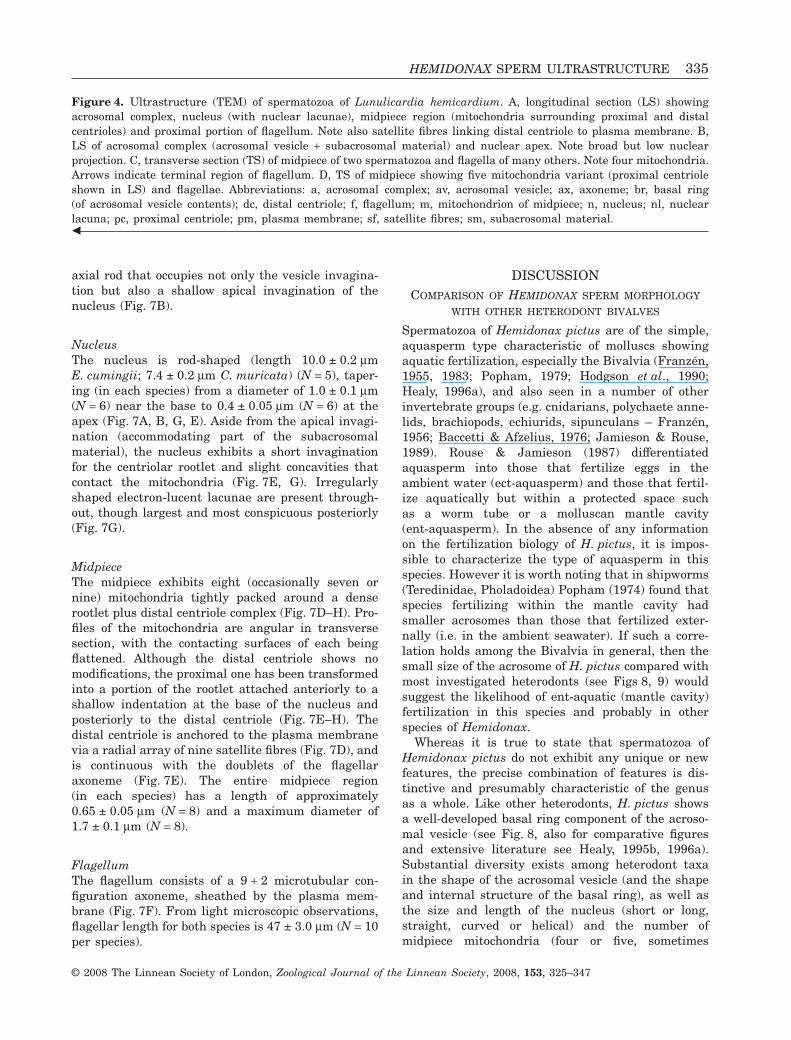
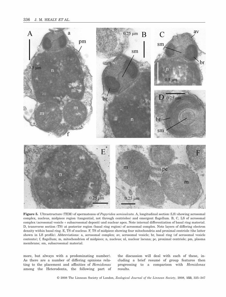
Figure 5. Ultrastructure (TEM) of spermatozoa of Papyridea semisulcata. A, longitudinal section (LS) showing acrosomalcomplex, nucleus, midpiece region (tangential, not through centrioles) and emergent flagellum. B, C, LS of acrosomalcomplex (acrosomal vesicle + subacrosomal deposit) and nuclear apex. Note internal differentiation of basal ring material.D, transverse section (TS) at posterior region (basal ring region) of acrosomal complex. Note layers of differing electrondensity within basal ring. E, TS of nucleus. F, TS of midpiece showing four mitochondria and proximal centriole (the lattershown in LS profile). Abbreviations: a, acrosomal complex; av, acrosomal vesicle; br, basal ring (of acrosomal vesiclecontents); f, flagellum; m, mitochondrion of midpiece; n, nucleus; nl, nuclear lacuna; pc, proximal centriole; pm, plasmamembrane; sm, subacrosomal material.
336 J. M. HEALY ET AL.
© 2008 The Linnean Society of London, Zoological Journal of the Linnean Society, 2008, 153, 325–347
HEMIDONAX AND THE DONACIDAE (TELLINOIDEA)
To date, five species of Donacidae have been examinedin detail for sperm ultrastructure (the South AfricanDonax serra Dillwyn, 1817, D. madagascariensisWood, 1828, D. sordidus Hanley, 1845 – see Hodgsonet al., 1990; the eastern Atlantic/MediterraneanDonax trunculus – see Sousa & Oliveira, 1994; theAustralian D. deltoides Lamarck, 1818 – this study,see also Healy, 1995b). In all of these species (seeFigs 8, 9), with the exception of D. trunculus, theacrosomal vesicle is large, with prominent, parallellayers within the basal ring, the nucleus is barrel-shaped and the midpiece contains the centrioles andfour mitochondria. In D. trunculus the nucleus isappreciably longer than in other investigated spe-cies of Donax, but four midpiece mitochondria areobserved (as also in other species). More significantly,no layering within the basal ring of the acrosomalvesicle was reported by Sousa & Oliveira (1994) norcan any trace of it be seen in their micrographs ofmature or developing sperm (that all appear to showa homogeneously textured basal ring). Surprisingly,Sousa & Oliveira (1994) did not comment on thedifference in acrosomal morphology between D.trunculus and the species examined by Hodgson et al.(1990). Healy (1995b) noted the difference and sug-gested the possible taxonomic utility of sperm infuture studies of the Donacidae, as has already beendemonstrated by Hodgson et al. (1990) for the threeSouth African species examined by them (the closesperm similarity between D. madagascariensis andD. sordidus being linked to reports of hybridization inareas where these species occur together – see Dis-cussion in Hodgson et al., 1990). A sixth species ofDonax, the Indian Ocean D. lubricus Hanley, 1845was examined briefly by Pal (1992, 1996), but thedescriptions and micrographs provide incompletedata. As in other examined species of Donax, theacrosomal vesicle of D. lubricus is seated in a broadanterior depression of the nucleus, and the subacroso-mal material is organized as an axial rod. However, incontrast to other species of Donax, but similar togroups such as the Mactroidea (Longo & Anderson,1969; Hylander & Summers, 1977; Healy, 1995b; J.M. Healy, P. M. Mikkelsen & R. Bieler, unpubl. data),the anterior portion of the acrosomal vesicle appearsto be largely devoid of contents. A more detailed studyof this species would seem warranted.
Despite the spermatozoal differences between thetwo apparent ‘groups’ of Donax, all Donax sperm showtwo consistent differences from Hemidonax pictus: (1)the acrosomal vesicle is seated in a marked depres-sion of the nuclear apex (Hemidonax: apex flat orshowing minimal depression); and (2) the subacroso-mal material is organized into a well-formed axial
rod, often showing longitudinal-fibrous structure(Hemidonax: subacrosomal material consisting ofdiffuse granules). Further work on other species ofDonacidae seems warranted, if only to reach a betterappreciation of the range of sperm morphologieswithin this family (and possibly to assess the mono-phyletic status of the family). Available sperm ultra-structural data thus do not support the decision ofsome authors to allocate Hemidonax to the Donacidae(Lamy, 1917; Thiele, 1934; Keen, 1969; Vokes, 1980;Abbott & Dance, 1982) or Donacoidea (Scarlato &Starobogatov, 1979). The rather fragmentary spermultrastructural data for other Tellinoidea (see Sousa,Corral & Azevedo, 1989; Healy, 1995b; Sousa et al.,1998) likewise suggest no connection with Tellinidae,Semelidae or Scrobiculariidae. The suggestion bySchneider & Carter (2001) of a close relationshipbetween Hemidonax and the Psammobiidae (Telli-noidea) also does not seem likely, at least based onthe light microscopic results of Guerra, Campos &Esponda (1994) showing an elongate, rod-shapednucleus in Gari solida (Gray, 1828). Certainly thishypothesis needs to be tested using TEM of psammo-biid sperm.
HEMIDONAX AND THE CRASSATELLOIDEA
(CRASSATELLIDAE, CARDITIDAE, ASTARTIDAE)
Sperm ultrastructure has been examined in threespecies of Crassatellidae, one species of Carditidaeand one species of Astartidae (Healy, 1995a, b, 1996a;present study: Fig. 7). The crassatelloidean sperma-tozoon differs profoundly from those of Hemidonax inalmost all features: (1) the acrosomal vesicle is longand attenuate and the subacrosomal material isdeveloped as a thick axial rod (Hemidonax: acrosomalvesicle short and squat; subacrosomal materialdiffuse and granular); (2) the nucleus is narrow androd-shaped (Hemidonax: nucleus short and wide); (3)the proximal centriole transforms into an electron-dense rod connecting the distal centriole (andaxoneme) to the nuclear base (Hemidonax: proximalcentriole unmodified); and (4) the midpiece consistsof eight (occasionally seven or nine) wedge-shapedmitochondria, tightly packed around the centriolarcomplex (Hemidonax: midpiece exhibits four round,unmodified mitochondria). The idea of a crassatel-loidean affinity for Hemidonax (e.g. Hedley, 1906,1909, 1923) or for that matter a link betweenthe Carditidae and Hemidonax + Cardiidae (e.g.Schneider & Carter, 2001) can safely be rejectedpurely on spermatological grounds.
HEMIDONAX AND THE CARDIIDAE (CARDIOIDEA)
A number of species of Cardiidae (sensu Schneider,1992, 1995; now inclusive of Tridacnidae as subfamily
HEMIDONAX SPERM ULTRASTRUCTURE 337
© 2008 The Linnean Society of London, Zoological Journal of the Linnean Society, 2008, 153, 325–347
338 J. M. HEALY ET AL.
© 2008 The Linnean Society of London, Zoological Journal of the Linnean Society, 2008, 153, 325–347
Tridacninae) have been examined for sperm ultra-structure (Popham, 1979; Sousa & Azevedo, 1988;Healy, 1995b; Sousa et al., 1998; Keys & Healy, 1999,2000; Drozdov et al., 2001; present study) and eventhough many genera and even some subfamiliesawait investigation, enough is known to provide ameaningful comparison with Hemidonax pictus (seeFigs 8, 9). Sperm morphology in the Cardiidae varieswidely between taxa, to such an extent in fact that nodistinctive, family-defining characters are yet appar-ent. However, it can be stated that in all investigatedspecies: (1) the acrosomal vesicle is never elongate(usually with a rounded or truncated apex); (2) theacrosomal complex is never seated in a depression ofthe nuclear apex; (3) the subacrosomal material isnever formed into a well-defined rod (herein ‘axial rod’or ‘perforatorium’; although this does not preclude thegeneration of such a structure via polymerization ofthe subacrosomal material during the acrosome reac-tion); and (4) the midpiece almost always featuresfour mitochondria (five occurring occasionally asa variant, but not predominating). Comparison ofH. pictus with the range of sperm morphologiesencountered in the Cardiidae to date (see Figs 8, 9)reveals that only Fragum unedo (Fragiinae) showsany degree of similarity to H. pictus, although theacrosomal vesicle is larger and more compressed andthe nuclear profile is curved in longitudinal section(see Fig. 3). The basal invagination of the acrosomalvesicle of H. pictus and F. unedo is narrow within theanterior half of the vesicle, and the wedge-shapedbasal ring profile is comparable between the two taxa.In almost all other investigated species of Cardiidae(including Lunulicardia hemicardium of the Fragii-nae) the anterior region of the invagination is eitheras wide or wider than the basal region. A narrowinvagination also appears present in Cerastodermaspp. (published micrographs are, unfortunately, fewin number and lacking in much detail – see Sousa &Azevedo, 1988; Sousa et al., 1998; Drozdov et al.,2001), but, unlike that in H. pictus or F. unedo, andlike certain species of Tridacninae (Keys & Healy,1999, 2000), the nuclear apex projects deeply into thisinvagination (see Fig. 8). Cerastoderma spp. alsoexhibit marked helical coiling of the nucleus, a
feature long ago recorded by Retzius (1905) and onealso observed in the enigmatic cardioidean (?cardiid)genera Monodacna, Didacna and Adacna (see com-parative light microscopic account by Karpevich,1961). An apparent difference between H. pictusand the Cardiidae (based on available data) is theelectron-lucent layer underlying the curved apexobserved in the former. A similar layer has been seenby us in at least one venerid species (Lioconcha annet-tae Lamprell & Whitehead, 1990) (R. Bieler, P. M.Mikkelsen & J.M. Healy, unpubl. observ.) but we arehesitant to attach undue significance to it, as theapical region of the acrosomal vesicle is the moststructurally unstable component of the acrosomalcomplex. If the electron-lucent layer is not an artefactof fixation, and occurs in other Hemidonax species, itmay prove a useful genus-defining or even family-defining feature.
HEMIDONAX AND THE VENEROIDEA
Several non-cardioid members of the Veneroida havebeen examined for sperm ultrastructure (but still onlya fraction of the known genera) especially among theVeneridae (Pochon-Masson & Gharagozlou, 1970;Gharagozlou-Van Ginneken & Pochon-Masson, 1971;Nicotra & Zappata, 1991; Reunov & Hodgson, 1994;Sousa et al., 1998; Healy et al., 2006). As part of aongoing survey of sperm ultrastructure within theVeneroidea (and especially Veneridae), we have alsoexamined many genera (J. M. Healy, P. M. Mikkelsen& R. Bieler, unpubl. data) and include results for afew previously unstudied examples in this account forcomparison with Hemidonax. Lioconcha annettae andAntigona chemnitzii (Hanley, 1844) show similaracrosomal dimensions to Hemidonax pictus, but inboth, the acrosomal complex rests in an anteriordepression of the nucleus and is angularly tilted, andin neither of these venerids is the acrosomal vesicleinvagination narrow anteriorly (see Figs 8, 9). Inaddition, there are nuclear differences (narrow andcurved in both) and at least in L. annettae five ratherthan four mitochondria. As mentioned previously inthis discussion, L. annettae exhibits an electron-lucent layer at the apex of the acrosomal vesicle,
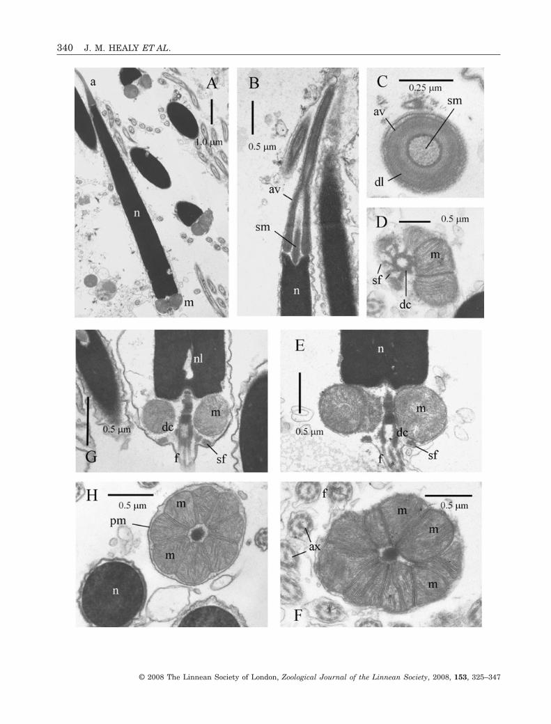
Figure 6. Ultrastructure (TEM) of spermatozoa of Donax (Plebidonax) deltoides. A, longitudinal section (LS) showingacrosomal complex, nucleus, midpiece (with proximal and distal centrioles) and proximal portion of flagellum. Noteprominent depression at nuclear apex accommodating base of acrosomal complex. B, transverse section (TS) throughacrosomal complex, showing parallel internal layers within basal ring, and homogeneous (less electron-dense) innermaterial. C, LS of acrosomal complex. Note longitudinally fibrous nature of subacrosomal material. D, TS of nucleus. E,LS of acrosomal complex, nucleus, midpiece and proximal portion of flagellum. F, TS of midpiece showing fourmitochondria and triplet structure of distal centriole. Abbreviations: a, acrosomal complex; av, acrosomal vesicle; ax,axoneme; br, basal ring (of acrosomal vesicle contents); dc, distal centriole; f, flagellum; m, mitochondrion of midpiece; n,nucleus; nl, nuclear lacuna; pc, proximal centriole; pm, plasma membrane; sf, satellite fibres; sm, subacrosomal material.�
HEMIDONAX SPERM ULTRASTRUCTURE 339
© 2008 The Linnean Society of London, Zoological Journal of the Linnean Society, 2008, 153, 325–347
340 J. M. HEALY ET AL.
© 2008 The Linnean Society of London, Zoological Journal of the Linnean Society, 2008, 153, 325–347
similar to that observed in H. pictus. While this mayprove significant, the lability of this region of theacrosomal complex (i.e. acrosome reaction stages)may be a factor and we reserve our judgement on thenature of this layer. In Dosinia nedigna (Iredale,1930) the nucleus and midpiece are essentially asobserved in H. pictus, but the acrosomal vesicle istilted and exhibits a wide invagination (see Figs 7, 8).In most other investigated venerids, acrosomal,nuclear or midpiece features (usually a combinationof these) do not closely tally with results obtained forH. pictus (see Pochon-Masson & Gharagozlou, 1970;Gharagozlou-Van Ginneken & Pochon-Masson, 1971;Reunov & Hodgson, 1994; Sousa et al., 1998). Only inCallista chione (Linnaeus, 1758) does the acrosomalvesicle approach that of H. pictus in shape, size andnarrowness of the invagination, but even in thisspecies, an axial rod is present within the subacroso-mal material, the nucleus is slightly elongate andstrongly curved, and the midpiece exhibits five mito-chondria (see Nicotra & Zappata, 1991). However,despite various sperm differences between investi-gated Veneridae and Hemidonax, it remains impos-sible, at least at this stage, to rule out unequivocallya relationship between these two taxa, especially inview of the large number of venerid genera awaitingsperm study.
TAXONOMIC AND PHYLOGENETIC CONSIDERATIONS:THE AFFINITIES OF HEMIDONAX
‘After examination and comparison of the shell and ofthe anatomy (both external and internal) of Hemi-donax to both cardiids and donacids, I cannot justifyplacing Hemidonax as a member of the Cardioidea.However, neither can I place Hemidonax within theDonacidae. Instead, I favor placing Hemidonax asincertae sedis within the order Veneroida, until aphylogenetic analysis of the Veneroida is undertaken’(Schneider, 1992: 145). With those words, JaySchneider – a recognized authority on cardioid sys-tematics and phylogeny (see also Schneider, 1995,
1998a, b) – effectively reopened the debate concerningthe affinities of Hemidonax. His rejection of a rela-tionship between Hemidonax and the Donacidae(based on his own anatomical observations) was inaccordance with the views of Boss (1971) and Ponderet al. (1981), and it can be said with confidence thatall available sperm ultrastructural data (Hodgsonet al., 1990; Pal, 1992, 1996; Sousa & Oliveira, 1994;Healy, 1995a; present study) likewise argue stronglyagainst any connection between these two taxa. Thesame conclusion was reached by Schneider & Carter(2001) based on comparative shell microstructure.Schneider’s other conclusion – that Hemidonax isnon-cardioidean – is all the more remarkable when itis considered that he was not persuaded by the argu-ments of either Boss (1971) or Ponder et al. (1981),who strongly supported cardioid affinities for Hemi-donax. Certainly, in terms of its anatomy, Hemidonaxshows a number of features not consistent with place-ment in the Cardiidae as outlined in some detail byPonder et al. (1981; who argued for retention of aseparate family Hemidonacidae). Perhaps signifi-cantly, neither Boss (1971) nor Ponder et al. (1981)identify any affiliations between Hemidonax and anycardioidean genus, lending some degree of credence toSchneider’s (1992) decision to leave Hemidonax asincertae sedis among the Veneroida. It is of interest tonote that Keen (1980), while accepting Boss’s (1971)subfamily Hemidonacinae within the Cardiidae, didnot choose to discuss the relationships of Hemidonax[being evidently influenced by Wilson & Stevenson’s(1977) decision not to include the genus in theirreview of the Western Australian Cardiidae]. Keen,however, grouped the Fraginae and Hemidonacinae inher tabulation of cardiid shell features and also in hertaxonomic keys, and by so doing perhaps was hintingat the possibility of some relationship between thetwo subfamilies. Most recently, Schneider & Carter(2001) have argued, largely on the basis of compara-tive shell microstructure, that Hemidonax showsmuch closer affinity with the tellinoidean familyPsammobiidae than with the Cardiidae (or any other
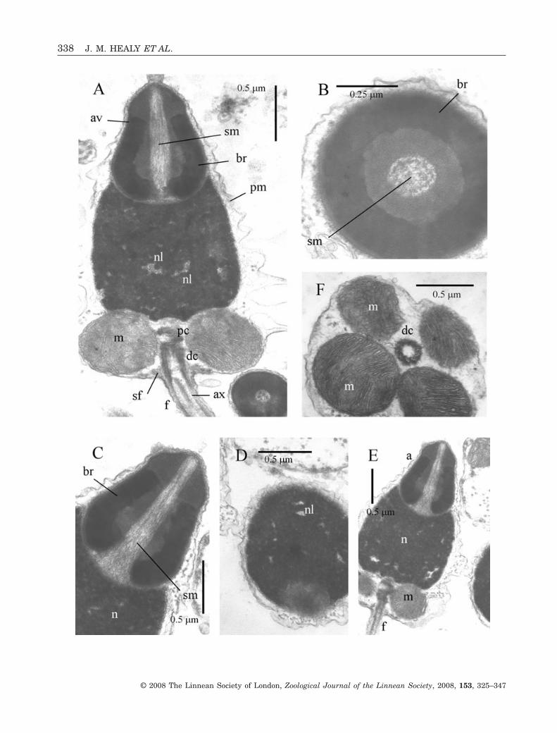
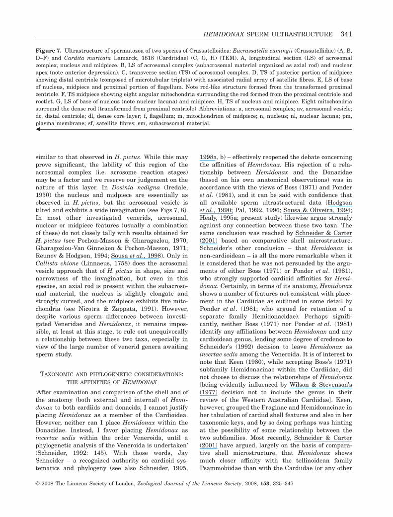
Figure 7. Ultrastructure of spermatozoa of two species of Crassatelloidea: Eucrassatella cumingii (Crassatellidae) (A, B,D–F) and Cardita muricata Lamarck, 1818 (Carditidae) (C, G, H) (TEM). A, longitudinal section (LS) of acrosomalcomplex, nucleus and midpiece. B, LS of acrosomal complex (subacrosomal material organized as axial rod) and nuclearapex (note anterior depression). C, transverse section (TS) of acrosomal complex. D, TS of posterior portion of midpieceshowing distal centriole (composed of microtubular triplets) with associated radial array of satellite fibres. E, LS of baseof nucleus, midpiece and proximal portion of flagellum. Note rod-like structure formed from the transformed proximalcentriole. F, TS midpiece showing eight angular mitochondria surrounding the rod formed from the proximal centriole androotlet. G, LS of base of nucleus (note nuclear lacuna) and midpiece. H, TS of nucleus and midpiece. Eight mitochondriasurround the dense rod (transformed from proximal centriole). Abbreviations: a, acrosomal complex; av, acrosomal vesicle;dc, distal centriole; dl, dense core layer; f, flagellum; m, mitochondrion of midpiece; n, nucleus; nl, nuclear lacuna; pm,plasma membrane; sf, satellite fibres; sm, subacrosomal material.�
HEMIDONAX SPERM ULTRASTRUCTURE 341
© 2008 The Linnean Society of London, Zoological Journal of the Linnean Society, 2008, 153, 325–347
342 J. M. HEALY ET AL.
© 2008 The Linnean Society of London, Zoological Journal of the Linnean Society, 2008, 153, 325–347
group of heterodonts). Whereas it is true that shellmicrostucture has proven a very valuable source ofcharacters for phylogenetic analysis, Schneider &Carter (2001) have not offered any explanation for thekey anatomical differences between Hemidonax andthe Tellinoidea, particularly the absence of the cruci-form muscle (its presence is a synapomorphy of theTellinoidea – see also Boss, 1971, 1982). In this con-nection it is worth noting that Pharus and the Nova-culininae (both originally included in Solecurtidae)were often cited as tellinoidean taxa lacking a cruci-form muscle (Yonge, 1949, 1959; Ponder et al., 1981;Boss, 1982) but are now both placed within the Sole-noidea (e.g. Morton, 1984; von Cosel, 1990; Willan,1998).
CONCLUSIONS
In the present account we have examined the featuresof the mature gonadial spermatozoa of Hemidonaxpictus, and provided comparisons with other hetero-donts, especially the two most favoured affiliates, theDonacidae (Tellinoidea) and the Cardiidae (Cardio-idea). In addition we have drawn attention to impor-tant sperm similarities (some albeit of a rather broadnature) with the Veneridae.
We conclude, based on the available data, that thespermatozoan features of Hemidonax pictus do notshow a close match to those of the investigated 13species of Cardioidea, with Fragum unedo being themost similar. If Keen (1980) did intend to suggest aconnection between the Fragiinae and Hemidonaci-nae, then it would appear that sperm morphologysupplies some supporting evidence for this stance(i.e. a connection with Fragum) and some against (e.g.differences between Hemidonax and Lunulicardia).There are also very interesting sperm similarities(especially acrosomal) to various members of the Ven-eridae, although no species examined to date com-
pletely matches our results for H. pictus. Finally, wecan find no sperm ultrastructural evidence to indicatethat a Hemidonacidae + Cardiidae group either arosefrom or is otherwise related to the Carditidae or othercrassatelloideans (cf. Schneider & Carter, 2001). Sper-matozoa of crassatelloideans are very distinctive(Healy, 1995a, b; present study), and given the antiq-uity of families such as the Astartidae, have long hadtheir own evolutionary pathway.
ACKNOWLEDGEMENTS
We thank the staff of the Centre for Microscopy andMicroanalysis, University of Queensland, for theiradvice and technical assistance throughout thisproject. Our thanks also go to: John Taylor and EmilyGlover (Natural History Museum, London) for sup-plying the live material of Hemidonax pictus; MartinHealy for assistance in collecting live Vasticardiumvertebratum, Lunulicardia hemicardium, Fragumunedo and Plebidonax deltoides; to the late KevinLamprell (Queensland Museum) for live materialof Eucrassatella cumingii; and to Richard Willan(Northern Territory Museum of Arts and Sciences,Darwin) and Gileanne Brodie (James Cook Univer-sity, Queensland) for live material of Cardita muri-cata. We also thank Kevin and Kathy Townsend(Moreton Bay Marine Station) for allowing J.H.access to holding tanks for live material. Papyrideasemisulcata was collected under Florida KeysNational Marine Sanctuary Permit 2002-078 toP.M.M. and R.B. J.H. also wishes to acknowledge thehelp and support of the curatorial and technical staffof the Biodiversity Program of the QueenslandMuseum. We also thank the referees for their con-structive comments on the manuscript. This projectwas supported by NSF-PEET DEB-99781119 to R.B.and P.M.M. and a Grainger Foundation award to R.B.
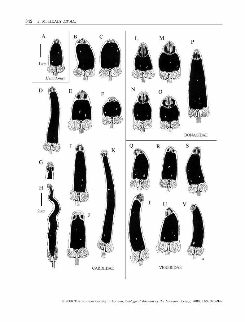
Figure 8. Diagram showing comparative sperm ultrastructure (TEM) (longitudinal sections through head, midpiece andproximal portion of flagellum) of Hemidonax pictus (Tryon, 1870) (A), Cardiidae (B–K), Donacidae (L–P) and selectedVeneridae (Q–V). B–K, Cardiidae. B, Fragum unedo (Linnaeus, 1758); C, Lunulicardia hemicardium (Linnaeus, 1758); D,Vasticardium vertebratum (Jonas, 1844); E, Papyridea semisulcata (Gray, 1825); F, Fulvia tenuicostata (Lamarck, 1819);G, H, Cerastoderma spp.; I, Hippopus hippopus (Linnaeus, 1758); J, Tridacna gigas (Linnaeus, 1758); K, Tridacna maxima(Röding, 1798). L–P, Donacidae. L, Donax deltoides Lamarck, 1818; M, Donax madagascariensis Wood, 1828; N, Donaxsordidus Hanley, 1845; O, Donax serra Röding, 1798; P, Donax trunculus Linnaeus, 1758. Q–V, Veneridae. Q, Venerupisaurea (Gmelin, 1791); R, Venerupis corrugata (Gmelin, 1791); S, Petricola lapicida (Gmelin, 1791); T, Antigona chemnitzii(Hanley, 1844); U, Dosinia nedigna (Iredale, 1930); V, Lioconcha annettae Lamprell & Whitehead, 1990. Scale bar forHemidonax pictus (= 1.0 mm) applies to all other figures except H (= 2.0 mm). Sources of data: A–E, L, S–V (this paper;J. M. Healy, P. M. Mikkelsen & R. Bieler, unpubl. data); F (Popham, 1979); G, H [composite reconstruction based onCerastoderma lamarcki (Reeve, 1844) from Drozdov et al., 2001 and Cerastoderma edule (Linnaeus, 1758) from Sousa &Azevedo, 1988 and Sousa et al., 1998]; I–K (Keys & Healy, 1999, 2000); M–O (Hodgson et al., 1990); P (Sousa & Oliveira,1994); Q, R (Pochon-Masson & Gharagozlou, 1970; Gharagozlou-Van Ginneken & Pochon-Masson, 1971).�
HEMIDONAX SPERM ULTRASTRUCTURE 343
© 2008 The Linnean Society of London, Zoological Journal of the Linnean Society, 2008, 153, 325–347
344 J. M. HEALY ET AL.
© 2008 The Linnean Society of London, Zoological Journal of the Linnean Society, 2008, 153, 325–347
REFERENCES
Abbott RT, Dance SP. 1982. Compendium of seashells. NewYork: E.P. Dutton Inc.
Allan J. 1959. Australian shells, 2nd edn. Melbourne: Geor-gian House.
Baccetti B, Afzelius BA. 1976. Biology of the sperm cell.Monographs in Developmental Biology. Basel: S. Karger.
Boss KJ. 1971. Familial affinities of Hemidonax (Bivalvia).Nautilus 85: 9–12.
Boss KJ. 1982. Mollusca. In: Parker SP, ed. Synopsis andclassification of living organisms, Vol. 2. New York:McGraw-Hill, 945–1166.
Bouchet P, Rocroi J-P. 2005. Classification and nomencla-tor of gastropod families. Malacologia 47: 1–397.
von Cosel R. 1990. An introduction to the razor shells(Bivalvia: Solenacea). In: Morton B, ed. The Bivalvia, Pro-ceedings of a Memorial Symposium in Honour of Sir CharlesMaurice Yonge, Edinburgh, 1986. Hong Kong: Hong KongUniversity Press, 283–311.
Daddow LYM. 1986. An abbreviated method of the doublelead stain technique. Journal of Submicroscopic Cytology18: 221–224.
Drozdov AL, Frolenko LN, Ferraguti M. 2001. Spermato-genesis and gamete structure in a brackishwater cardiidCerastoderma lamarcki (Reeve, 1844). Ruthenica 11: 175–181.
Fischer P. 1887. Manuel de Conchyliologie. Paris: Savy.Franzén Å. 1955. Comparative morphological investigations
into the spermiogenesis among Mollusca. Zoologiska Bidragfran Uppsala 30: 399–456.
Franzén Å. 1956. On spermiogenesis, morphology of thespermatozoon, and the biology of fertilization among inver-tebrates. Zoologiska Bidrag fran Uppsala 31: 355–482.
Franzén Å. 1983. Ultrastructural studies of spermatozoa inthree bivalve species with notes on evolution of elongatedsperm nucleus in primitive spermatozoa. Gamete Research7: 199–214.
Gharagozlou-Van Ginneken ID, Pochon-Masson J. 1971.Étude comparative infrastructurale du spermatozoide chezles palourdes de France. Archives de Zoologie Experimentaleet Génerale 112: 805–817.
Guerra R, Campos B, Esponda P. 1994. Analysis of thespermatozoa of four bivalves with particular reference tothe acrosome and plasma membrane glycoproteins. Journalof Submicroscopic Cytology and Pathology 26: 489–495.
Healy JM. 1989. Spermiogenesis and spermatozoa in the
relict bivalve genus Neotrigonia: relevance to trigonioidrelationships, particularly Unionoidea. Marine Biology 103:75–85.
Healy JM. 1995a. Sperm ultrastructure in the marinebivalve families Carditidae and Crassatellidae and itsbearing on unification of the Crassatelloidea with the Cardi-toidea. Zoologica Scripta 24: 21–28.
Healy JM. 1995b. Comparative sperm ultrastructure in ven-eroid bivalves (Mollusca). In: Jamieson BGM, Ausio J,Justine J-L, eds. Advances in spermatozoal phylogeny andtaxonomy. Memoirs du Muséum National d’HistoireNaturelle 166: 155–166.
Healy JM. 1996a. Molluscan sperm ultrastructure: correla-tion with taxonomic units within the Gastropoda, Cepha-lopoda and Bivalvia. In: Taylor J, ed. Origin andevolutionary radiation of the Mollusca. Oxford: Oxford Uni-versity Press, 99–113.
Healy JM. 1996b. Spermatozoan ultrastructure in the trigo-nioid bivalve Neotrigonia margaritacea Lamarck (Mollusca):comparison with other bivalves, especially Trigonioida andUnionoida. Helgoländer Meeresuntersuchungen 50: 259–264.
Healy JM, Keys J, Daddow LYM. 2000. Comparative spermultrastructure in pteriomorphian bivalves with specialreference to phylogenetic and taxonomic implications. In:Harper EM, Taylor JD, Crame JA, eds. The evolutionarybiology of the Bivalvia. Geological Society, London, SpecialPublications 177: 169–190.
Healy JM, Mikkelsen PM, Bieler R. 2006. Sperm ultra-structure in Glauconome plankta and its relevance to theaffinities of the Glauconomidae Bivalvia: Heterodonta.Invertebrate Reproduction and Development 49: 29–39.
Hedley C. 1906. Studies on Australian Mollusca. Part IX.Proceedings of the Linnean Society of New South Wales(1905), pt 4: 520–546, pls 31–33.
Hedley C. 1909. Mollusca from the Hope Islands, NorthQueensland. Proceedings of the Linnean Society of NewSouth Wales 24: 429–466.
Hedley C. 1923. Studies on Australian Mollusca. Part XIV.Proceedings of the Linnean Society of New South Wales 48:301–316, pls 30–33.
Hodgson AN, Bernard RTF. 1986. Ultrastructure of thesperm and spermatogenesis of three species of Mytilidae(Mollusca, Bivalvia). Gamete Research 15: 123–135.
Hodgson AN, Bernard RTF, Van der Horst G. 1990.Comparative spermatology of three species of Donax(Bivalvia) from South Africa. Journal of Molluscan Studies56: 257–265.
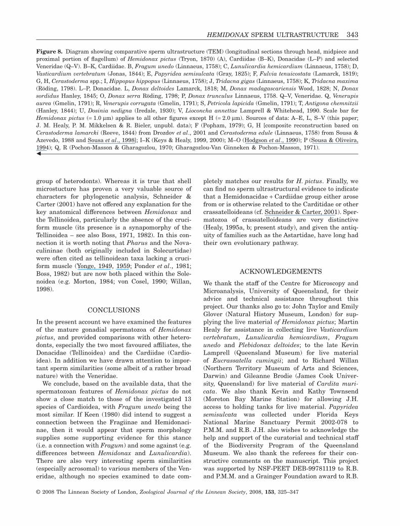
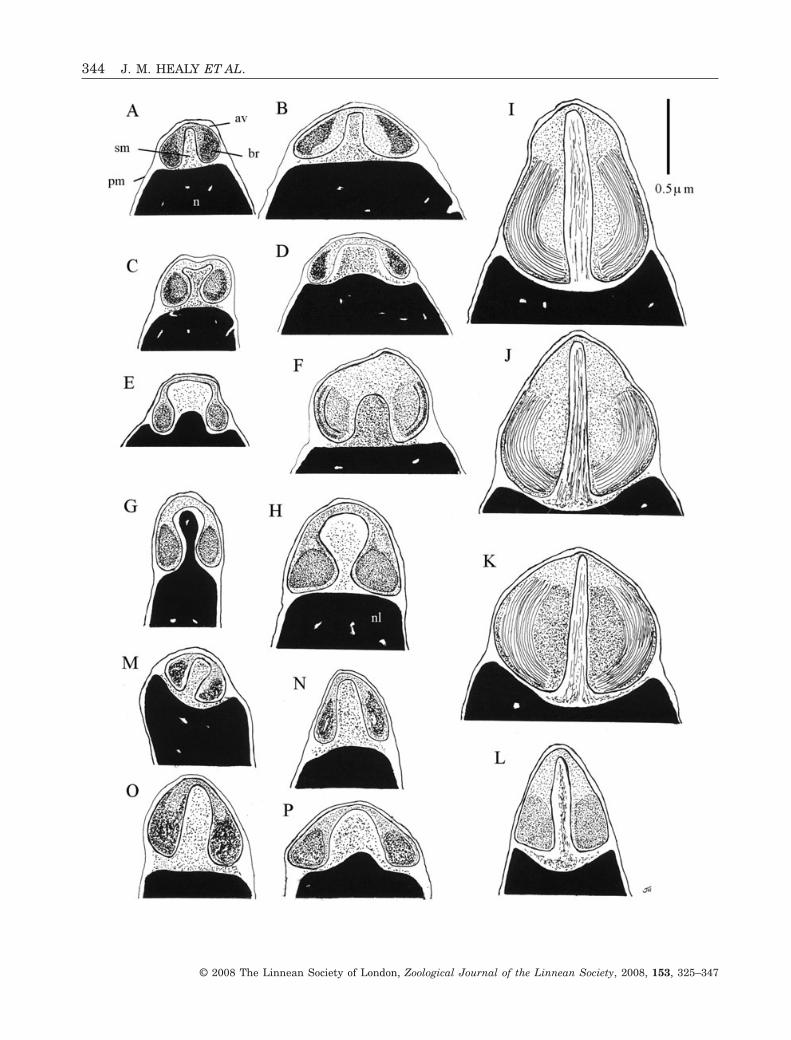
Figure 9. Diagram showing comparative acrosomal complex ultrastructure (TEM) of Hemidonax pictus (A), Cardiidae,Donacidae and selected Veneridae. B–H, Cardiidae: B, Fragum unedo; C, Vasticardium vertebratum; D, Lunulicardiahemicardium; E, Fulvia tenuicostata; F, Papyridea semisulcata; G, Tridacna maxima; H, Hippopus hippopus. I–L,Donacidae: I, Donax deltoides; J, Donax madagascariensis; K, Donax serra; L, Donax trunculus. M–P, Veneridae: M,Lioconcha annettae; N, Dosinia nedigna; O, Antigona chemnitzii; P, Venerupis corrugata. Scale bar (1.0 mm) applies to allfigures. Sources of data: A–D, F, I, M–O (this paper; J. M. Healy, P. M. Mikkelsen & R. Bieler, unpubl. data); E (Popham,1979); G, H (Keys & Healy, 1999, 2000); P (Gharagozlou-Van Ginneken & Pochon-Masson, 1971); J, K (Hodgson et al.,1990); L (Sousa & Oliveira, 1994).�
HEMIDONAX SPERM ULTRASTRUCTURE 345
© 2008 The Linnean Society of London, Zoological Journal of the Linnean Society, 2008, 153, 325–347
Hylander BL, Summers RG. 1977. An ultrastructuralanalysis of the gametes and early fertilization in two bivalvemolluscs, Chama macerophylla and Spisula solidissimawith special reference to gamete binding. Cell and TissueResearch 182: 469–489.
ICZN [International Commission on Zoological Nomen-clature]. 1999. International code of zoological nomencla-ture, 4th edn. London: International Trust for ZoologicalNomenclature.
Iredale T, McMichael DF. 1962. A reference list of marineMollusca of New South Wales. Memoir XI. Sydney: Austra-lian Museum.
Jamieson BGM, Rouse GW. 1989. The spermatozoa of thePolychaeta (Annelida): an ultrastructural view. BiologicalReviews 64: 93–157.
Jespersen Å, Lützen J, Morton B. 2002. Ultrastructure ofdimorphic sperm and seminal receptacle in the hermaphro-dites Barrimysia siphonosomae and Pseudopythina ocheto-stomae (Bivalvia, Galeommatoidea). Zoomorphology 121:159–172.
Kafanov AI, Drozdov AL. 1998. Comparative sperm mor-phology and phylogenetic classification of Recent Mytiloidea(Bivalvia). Malacologia 39: 129–139.
Kafanov AI, Popov SV. 1977. K sisteme kaynozoyskikhKardioidei (Bivalvia) [On the system of the Cenozoic Car-dioidea (Bivalvia)]. Paleontological Journal 1977: 307–314.
Karpevich AF. 1961. Adaptive character of spermatozoaand eggs’ morphology in Bivalvia-molluscs. ZoologicheskiiZhurnal Akademiia Nauk SSSR 40: 340–350 [in Russian,with English summary].
Keen AM. 1969. Donacidae. In: Moore RC, eds. Treatise oninvertebrate paleontology. Part N, Mollusca 6 Bivalvia.Lawrence: The Geological Society of America and TheUniversity of Kansas, N628–N629.
Keen AM. 1980. The pelecypod family Cardiidae: a taxonomicsummary. Tulane Studies in Geology and Paleontology 16:1–40.
Keys JL, Healy JM. 1999. Sperm ultrastructure of the giantclam Tridacna maxima (Tridacnidae: Bivalvia: Mollusca)from the Great Barrier Reef. Marine Biology 135: 41–46.
Keys JL, Healy JM. 2000. Relevance of sperm ultrastructureto the classification of giant clams (Mollusca, Cardioidea,Cardiidae, Tridacninae). In: Harper EM, Taylor JD, CrameJA, eds. The evolutionary biology of the Bivalvia. GeologicalSociety, London, Special Publications 177: 191–205.
Lamprell K, Whitehead T. 1992. Bivalves of Australia, Vol.1. Bathurst: Crawford House Press.
Lamy E. 1917. Révision des Crassatellidae vivants duMuséum d’Histoire Naturelle de Paris. Journal de Con-chologie (Paris) 62: 197–270.
Longo FJ, Anderson E. 1969. Spermiogenesis in the surfclam Spisula solidissima with special reference to the for-mation of the acrosomal vesicle. Journal of UltrastructureResearch 27: 435–443.
Morton B. 1984. The functional morphology of Sinonovaculaconstricta with a discussion on the taxonomic status of theNovaculininae (Bivalvia). Journal of Morphology 202: 299–325.
Nicotra A, Zappata S. 1991. Ultrastructure of the maturesperm and spermiogenesis in Callista chione (Mollusca,Bivalvia). Invertebrate Reproduction and Development 20:213–218.
Pal SG. 1992. Testes of Donax lubrica. In: Giusti F, Manga-nelli G, eds. Abstracts of the 11th International Malacologi-cal Congress. Siena: Unitas Malacologia, 77–80.
Pal SG. 1996. Some observations on the testes of Donaxlubrica (Bivalvia: Donacidae). In: Runham N, Heard WH,Burch JB, eds. Molluscan reproduction.MalacologicalReview 6 (Suppl.): 55–62.
Pelseneer P. 1911. Les lamellibranches de l’expedition duSiboga. Leiden: Partie Anatomique.
Pochon-Masson J, Gharagozlou ID. 1970. Particularitémorphologique de l’acrosome dans le spermatozoide deTapes decussatus L. (Mollusque, Lamellibranche). Annalesdes Sciences Naturelle Zoologie 12: 171–180.
Ponder WF, Colman PH, Yonge CM, Colman MH. 1981.The taxonomic position of Hemidonax Mörch, 1871 with areview of the genus (Bivalvia: Cardiacea). Journal of theMalacological Society of Australia 5: 41–64.
Popham JD. 1974. Comparative morphometrics of theacrosomes of the sperms of ‘externally’ and ‘internally’ fer-tilizing sperms of the shipworms (Teredinidae, Bivalvia,Mollusca). Cell and Tissue Research 150: 291–297.
Popham JD. 1979. Comparative spermatozoon morpho-logy and bivalve phylogeny. Malacological Review 12: 1–20.
Retzius G. 1905. Zur Kenntnis der Spermien der Everte-braten. II. Biologische Untersuchungen 12: 79–102, pls11–18.
Reunov AA, Hodgson AN. 1994. Ultrastructure of the sper-matozoa of five species of South African bivalves (Mollusca),and an examination of early spermatogenesis. Journal ofMorphology 219: 275–283.
Rouse GW, Jamieson BGM. 1987. An ultrastructural studyof the spermatozoa of the polychaetes Eurythoe complanata(Amphinomidae), Clymenella laseroni and Micromaldanelaseroni (Maldanidae) with definition of sperm types inrelation to reproductive biology. Journal of SubmicroscopicCytology 19: 573–584.
Scarlato OA, Starobogatov YI. 1971. Hemidonacidae Scar-lato et Starobogatov, fam. nov. In: Nevesskaja LA, ScarlatoOA, Starobogatov YI, Eberzin AG, eds. Novie predstavleniao sisteme dvustvorchatikh molliuskov [New ideas about thesystem of Bivalvia].Paleontologicheskii Zhurnal 1971: 3–20[in Russian].
Scarlato OA, Starobogatov YI. 1979. Osnovnye ChertyEvolyutsii I sistema Klassa Bivalvia Morfologiya, Sistema-tika I Filogeniya Mollyuskov. Trudy Zoologicheskogo Insti-tuta, Akademiya USSR 80: 5–38. (General evolutionarypatterns and the system of the Class Bivalvia in Morphol-ogy, Systematics and Phylogeny of Mollusks, Transactions ofthe Zoological Institute, Academy of Sciences 80: 5–38.Edited translated version by K. J. Boss, M. K. Jacobson,Special Occasional Publication No. 5, Department ofMollusks, Harvard University, Cambridge Massachusetts,1985.)
346 J. M. HEALY ET AL.
© 2008 The Linnean Society of London, Zoological Journal of the Linnean Society, 2008, 153, 325–347
Schneider JA. 1992. Preliminary cladistic analysis of thebivalve family Cardiidae. American Malacological Bulletin9: 145–155.
Schneider JA. 1995. Phylogeny of the Cardiidae (Mollusca,Bivalvia). Protocardiinae. Laevicardiinae, Lahillinae, Tulon-gogardiinae new subfamily and Pleuriocardiinae new sub-family. Zoologica Scripta 40: 321–373.
Schneider JA. 1998a. Phylogeny of stem-group eucardiids(Bivalvia: Cardiidae) and the significance of the transitionalfossil Perucardia. Malacologia 40: 37–62.
Schneider JA. 1998b. Phylogeny of the Cardiidae (Bivalvia):phylogenetic relationships and morphological evolutionwithin the subfamilies Clinocardiinae. Lymnocardininae,Fraginae and Tridacninae. Malacologia 40: 321–373.
Schneider JA, Carter JG. 2001. Evolution and phylogeneticsignificance of cardioidean shell microstructure (Mollusca,Bivalvia). Journal of Paleontology 75: 607–643.
Sousa M, Azevedo C. 1988. Comparative silver staininganalysis on spermatozoa of various invertebrate species.International Journal of Invertebrate Reproduction 13: 1–8.
Sousa M, Corral L, Azevedo C. 1989. Ultrastructural andcytochemical study of spermatogenesis in Scrobiculariaplana (Mollusca, Bivalvia). Gamete Research 24: 393–401.
Sousa M, Guerra R, Oliveira E, Torres A. 1998. Compara-tive PTA staining of molluscan spermatozoa. Journal ofSubmicroscopic Cytology and Pathology 30: 183–187.
Sousa M, Oliveira E. 1994. Ultrastructural and cytochemi-cal study of spermatogenesis in Donax trunculus (Mollusca,Bivalvia). Journal of Submicroscopic Cytology and Pathol-ogy 26: 305–311.
Thiele J. 1934 (‘1935’). Handbuch der Systematischen Weich-tierkunde. Teil 3 (Scaphopoda; Bivalvia: Cephalopoda; addi-tions and corrections for Teile 1-2; index for Teil 3). Jena:Fischer, 1934, pp. 920–921 (= pp. 1393–1394 of Englishtranslation, 1998, eds R. Bieler and P. M. Mikkelsen,Amerind Publishing Company, India, with SmithsonianInstitution Libraries).
Vaught KC. 1989. A classification of the Living Mollusca.Abbott RT, Boss KJ, eds. Melbourne, FL: American Mala-cologists Inc.
von Vest W. 1875. Über die Genera Adacna, Monodacna undDidacna Eichwald und deren Stellung im System. Jahr-bücher der Deutschen Malakozoologischen Gesellschaft 2:322, 324.
Vokes HE. 1980. Genera of the Bivalvia: a systematic andbibliographic catalogue (Revised and Updated). Ithaca, NY:Paleontological Research Institution.
Willan RC. 1998. Solenoidea. In: Beesley P, Ross G, Wells A,eds. Fauna of Australia, Volume 5, Mollusca: the SouthernSynthesis. Canberra: CSIRO Publishing, 340–342.
Wilson BR. 1998. Cardioidea. In: Beesley P, Ross G, Wells A,eds. Fauna of Australia, Volume 5, Mollusca: the SouthernSynthesis. Canberra: CSIRO Publishing, 328–332.
Wilson BR, Stevenson SE. 1977. Cardiidae of Western Aus-tralia. Western Australian Museum, Special Publication 9.
Yonge CM. 1949. On the structure and adaptations of theTellinacea, deposit-feeding Eulamellibranchia. Philosophi-cal Transactions of the Royal Society, Series B 234: 29–76.
Yonge CM. 1959. On the structure, biology and systematicposition of Pharus legumen (L.). Journal of the MarineBiological Association of the United Kingdom 38: 277–290.
HEMIDONAX SPERM ULTRASTRUCTURE 347
© 2008 The Linnean Society of London, Zoological Journal of the Linnean Society, 2008, 153, 325–347


