Embed Size (px)
Citation preview

Sphingolipids in Human Synovial Fluid - A LipidomicStudyMarta Krystyna Kosinska1, Gerhard Liebisch2, Guenter Lochnit3, Jochen Wilhelm4, Heiko Klein1,
Ulrich Kaesser5, Gabriele Lasczkowski6, Markus Rickert1, Gerd Schmitz2, Juergen Steinmeyer1*
1 Department of Orthopedics, Justus-Liebig-University Giessen, Giessen, Germany, 2 Department of Clinical Chemistry and Laboratory Medicine, University Hospital
Regensburg, Regensburg, Germany, 3 Department of Biochemistry, Justus-Liebig-University Giessen, Giessen, Germany, 4 Medical Clinic II/IV, Justus-Liebig-University
Giessen, Giessen, Germany, 5 Internistisches Praxiszentrum am Krankenhaus Balserische Stiftung, Giessen, Germany, 6 Institute of Forensic Medicine, Justus-Liebig-
University Giessen, Giessen, Germany
Abstract
Articular synovial fluid (SF) is a complex mixture of components that regulate nutrition, communication, shock absorption,and lubrication. Alterations in its composition can be pathogenic. This lipidomic investigation aims to quantify thecomposition of sphingolipids (sphingomyelins, ceramides, and hexosyl- and dihexosylceramides) and minor glyceropho-spholipid species, including (lyso)phosphatidic acid, (lyso)phosphatidylglycerol, and bis(monoacylglycero)phosphatespecies, in the SF of knee joints from unaffected controls and from patients with early (eOA) and late (lOA) stages ofosteoarthritis (OA), and rheumatoid arthritis (RA). SF without cells and cellular debris from 9 postmortem donors (control),18 RA, 17 eOA, and 13 lOA patients were extracted to measure lipid species using electrospray ionization tandem massspectrometry - directly or coupled with hydrophilic interaction liquid chromatography. We provide a novel, detailedoverview of sphingolipid and minor glycerophospholipid species in human SF. A total of 41, 48, and 50 lipid species weresignificantly increased in eOA, lOA, and RA SF, respectively when compared with normal SF. The level of 21 lipid speciesdiffered in eOA SF versus SF from lOA, an observation that can be used to develop biomarkers. Sphingolipids can altersynovial inflammation and the repair responses of damaged joints. Thus, our lipidomic study provides the foundation forstudying the biosynthesis and function of lipid species in health and most prevalent joint diseases.
Citation: Kosinska MK, Liebisch G, Lochnit G, Wilhelm J, Klein H, et al. (2014) Sphingolipids in Human Synovial Fluid - A Lipidomic Study. PLoS ONE 9(3): e91769.doi:10.1371/journal.pone.0091769
Editor: Paul Proost, University of Leuven, Rega Institute, Belgium
Received December 20, 2013; Accepted February 13, 2014; Published March 19, 2014
Copyright: � 2014 Kosinska et al. This is an open-access article distributed under the terms of the Creative Commons Attribution License, which permitsunrestricted use, distribution, and reproduction in any medium, provided the original author and source are credited.
Funding: This work was supported in part by a grant of the DRB foundation and by the ‘‘LipidomicNet’’ project, funded within the Seventh FrameworkProgramme of the European Union (Grant agreement number 202272). The funders had no role in study design, data collection and analysis, decision to publish,or preparation of the manuscript. No additional external funding was received for this study.
Competing Interests: The authors have declared that no competing interests exist.
* E-mail: [email protected]
Introduction
Synovial fluid (SF) can be viewed as an ultrafiltrate of plasma
that contains locally synthesized factors, such as cytokines; growth
factors; and lubricating compounds, such as lubricin, hyaluronic
acid, and phospholipids. The chief functions of SF are shock
absorption; load bearing; lubrication of articular surfaces, such as
those of cartilage, the meniscus, tendons, and ligaments; and
nutrition and communication medium of joints.
Altered composition or concentrations of SF components are
linked to osteoarthritis (OA) and rheumatoid arthritis (RA) [1–3].
A precise profile of the chemical composition of SF during disease-
related alterations will increase our knowledge about the
pathogenesis and possible options to treat these joint diseases.
Sphingolipids (SLs) are a class of lipids that include ceramide
(Cer) species, sphingomyelins (SMs) and more complex glyco-
sphingolipids. A common constituent of all SLs is the sphingoid
base, which is an organic aliphatic amino alcohol sphingosine
(SPH) or a structurally similar compound [4]. The metabolic
pathways of SLs form complex networks of reactions that involve
many enzymes and intermediate metabolites that are needed for
the biosynthesis, degradation, and remodeling of individual SLs
[4–6].
SLs are structural components of plasma membranes and
bioactive molecules that have significant functions in proliferation
and growth as well as differentiation, cellular signal transduction,
and apoptosis in many mammalian cells for instance fibroblast-like
synoviocytes (FLSs) and neural cells [7–11]. The function of SLs
depends on their acyl chain length and degree of saturation.
The balance between levels of individual SLs is critical. For
instance, sphingosine-1-phosphate (S1P) antagonizes Cer-mediat-
ed apoptosis [9]. Exogenous Cer in turn was reported to regulate
proliferation in human FLSs from OA and RA patients [7].
Treatment of FLSs with micromolar concentrations of C6-Cer
inhibits proliferation through Go/G1 arrest, similar to what has
been observed after serum starvation [7].
Cardiolipins (CLs) are tetra-acylated glycerophospholipids that
are part of the inner mitochondrial membranes in eukaryotic cells.
CLs regulate energy production [12,13], whereas free radical
oxidation products of CLs are important mediators of mitochon-
dria-dependent apoptosis [14–16]. Altered levels and composition
of CLs are linked to diseases, such as diabetes [13], heart failure
[13,17], Parkinson disease [18], and a rare cardiomyopathy,
known as Barth syndrome [19].
PLOS ONE | www.plosone.org 1 March 2014 | Volume 9 | Issue 3 | e91769

Bis(monoacylglycero)phosphate (BMP) is an acidic phospholipid
and an isomer of phosphatidylglycerol (PG). BMPs are formed on
the surface of intralysosomal vesicles during the degradation of
PGs and CLs and stimulate the enzymatic hydrolysis of
membrane-bound SLs. The highest levels of negatively charged
BMPs are found in the internal vesicles of lysosomes, and the
concentration of BMPs increases as late endosomes convert into
lysosomes [20]. BMPs are important for endosomal and lysosomal
function, and altered levels of BMP species have been reported in
a group of diseases, known as lysosomal storage disorders, in which
lysosomal dysfunction leads to the accumulation of secondary
metabolites, as observed in Gaucher disease, Fabry disease,
mucopolysaccharidosis, Pompe disease, and drug-induced phos-
pholipidosis [20,21].
Phosphatidic acid (PA) is a biosynthetic precursor of lipids, like
PGs, CLs, and BMPs. There are two lipids that are chemically
similar to PA: lysophosphatidic acid (LPA) and cyclic PA (cPA).
LPA has an identical chemical structure to PA, except that it
contains a single fatty acid (FA). LPA regulates many physiological
and pathophysiological responses and is one of the most active
naturally occurring lipids [22]. cPA act as a lipid mediator with
several biological activities. Notably, cPA was recently reported to
possess some antiinflammatory and chondroprotective activities in
a rabbit model of OA [23].
The normal use of articular joints may damage cells found
within SF which may be thus an additional source of many
components of SF, including intracellular lipids that are not
actively secreted into the SF. Chondrocytes, synovial lining cells,
leukocytes (predominantly T cells), and cells from the meniscus,
tendon, and ligaments are normally found at the borderline of SF
or in SF. In addition, SF is an ultrafiltrate of the plasma so that
lipids might also derive from the blood.
The levels, composition, and functions of SL, CL, LPA, PA,
BMP, PG and lysophosphatidylglycerol (LPG) species in SF are
unknown. Thus, we aimed to identify and quantify SL, CL, LPA,
PA, LPG, PG, and BMP species. In particular, we wanted to
investigate whether these lipid species are related to the health
status of synovial joints using SF from normal donors as well as
from patients with RA, early (eOA) or with late stages (lOA) of
OA.
Materials and Methods
Inclusion and Exclusion CriteriaThe SF originated from 9 deceased donors, 18 patients with RA
and 30 patients with OA (Table 1). The present study was
approved by the Ethical Review Committee of the Faculty of
Medicine (Justus-Liebig-University of Giessen, Germany), and all
patients provided written informed consent to donor samples for
research. The Ethical Review Committee (protocol #62/06)
waived the need for consent to be obtained from relatives of
deceased donors since a judicial order to perform autopsy existed
and, furthermore, to avoid additional emotional drain on relatives.
We used the Outerbridge classification scale (OU) to sub-
categorise the osteoarthritic changes in the joints as early (eOA)
and late (lOA) stages of the disease [24]. For this, the 6 cartilage
surfaces of the patella, trochlea and the medial and lateral sides of
the femoral condyles and the tibia were macroscopically evaluated
and the resulting cumulative OU score was divided by six in order
to obtain an average score valid for the entire joint. Early
osteoarthritic disease stages were defined as those which showed
an average score #2, while late stages of the disease were defined
as those with an OU score .2. Patients with RA were classified as
described in the guidelines of the American College of Rheuma-
tology [25]. SF was removed during autopsies of knee healthy
donors at the Institute of Forensic Medicine, University of Giessen
at the times after death described in Figure 1.
The SF was withdrawn into a syringe either during an
arthroscopy (eOA) or during an otherwise necessary knee puncture
(RA). In contrast, the SF of patients with late stage disease (lOA)
was obtained during a knee prosthetic implantation.
Processing of the Synovial FluidThe SF was processed as described elsewhere [26]. Briefly, SF
was macroscopically evaluated, pressed through a 1.2 micron
filter, and then 10% (v/v) inhibitors were added in order to inhibit
proteases and phospholipases. This was followed by centrifugation
of the SF for 45 min at 16,1006g and RT in order to pellet the
cellular debris [26,27]. Finally, the supernatant was frozen at
286uC for further lipid analysis.
Extraction of Sphingolipids and PhospholipidsThe cell and cellular debris-free supernatant was extracted in
the presence of internal lipid standards (Avanti Polar Lipids,
Alabaster, AL, USA) in order to specifically quantify extracellular
SMs and Cer species [26,28]. In addition, aliquots of the SF
supernatant were extracted with butanol in order to determine
hexosylceramide (HexCer) species, dihexosylceramide (Hex2Cer)
species, sphingoid bases, sphingosylphosphorylcholine (SPC) [29],
S1P, LPAs, CLs, BMPs, PGs, and PAs [30,31]. In brief, SF was
first buffered with 400 ml 30 mM citric acid and 40 mM disodium
hydrogen phosphate. Lipids were then extracted with 1-butanol,
dried, and redissolved in 50 ml ethanol.
Mass Spectrometric Quantification of Lipid SpeciesAs already described the SM and Cer species were determined
using electrospray ionisation mass spectrometry (ESI-MS/MS)
[28,32]. In addition, we used MS/MS after liquid chromato-
graphic separation (LC-MS/MS) to determine HexCer species,
Hex2Cer species, sphingoid bases, SPC [29], S1P, LPAs [30], CLs,
BMPs, PGs and PAs [31]. Lipid species were annotated as
published [33].
Data AnalysisA possible dilution of SF by a joint effusion would result in a
clearly reduced concentration of certain lipids as determined by
ESI-MS/MS. The liver produces urea which is not synthesized or
metabolized in articular joints. Urea diffuses without any
restriction across the synovium and can therefore be used to
evaluate unknown dilution. The dilution effect was corrected by
the method described by Kraus et al [26,34]. For this purpose, the
concentration of urea was first measured in both SF and serum so
that a dilution factor for the SF according to Kraus et al [34] could
be calculated. Urea was determined using a commercially
available kit (BioAssay Systems, Hayward, CA, USA).
Statistical Analysis of the Measured Values ObtainedIn our exploratory study, a non-Gaussian distribution of the
measured values was assumed so that a non-parametric statistical
analysis was carried out. The Kruskal-Wallis test was used to
identify statistically significant differences in the levels of lipid
species between different cohorts. The false discovery rate was
controlled at 5% (Benjamini-Hochberg adjustment).. For the
selected species, the Steel-Dwass multiple comparisons was applied
to determine statistically significant differences in lipid concentra-
tions between individual cohorts (control, eOA, lOA, RA) while
controlling the family-wise error-rate at 5%.
Sphingolipids in Human Synovial Fluid
PLOS ONE | www.plosone.org 2 March 2014 | Volume 9 | Issue 3 | e91769

We used the statistical software ‘‘R’’ version 2.14.0 to perform
the Kruskal-Wallis test, Benjamini-Hochberg adjustment as well as
the Steel-Dwass tests. The data obtained are presented as medians
with interquartile ranges for the box plots. The values quoted in
the text represent the median together with the interquartile range
in brackets.
Results
Sphingomyelin Molecular SpeciesUsing established ESI-MS/MS method the changes in the
concentrations of SM species were examined (28). Of the SL
classes that we examined, SMs were the most abundant in SF. The
concentration of total SMs in control SF was 39 nmol/ml,
multiplying 2.4-fold in eOA (92 nmol/ml; p,0.05), 4.4-fold in
lOA (172 nmol/ml; p#0.001), and 3.2-fold in RA (126 nmol/ml;
p#0.001). Nineteen SM species, based on the length and
saturation of the attached FAs, were identified in human SF as
shown in Figure 2. SM 34:1 was the predominant species among
SMs, accounting for 38% to 44% of total SMs in all cohorts
(Figure 2). In addition, versus control SF, 19 SM species were
elevated by 2.4-fold (2.2–2.8-fold; p,0.05) in eOA, 4.8fold (4.1–
5.4-fold; p#0.001) in lOA, and in RA SF 3.5-fold (2.8–4.2-fold;
p#0.001). Notably, all SM species had risen approximately 2-fold
in SF from eOA to lOA (p#0.05, Figure 2).
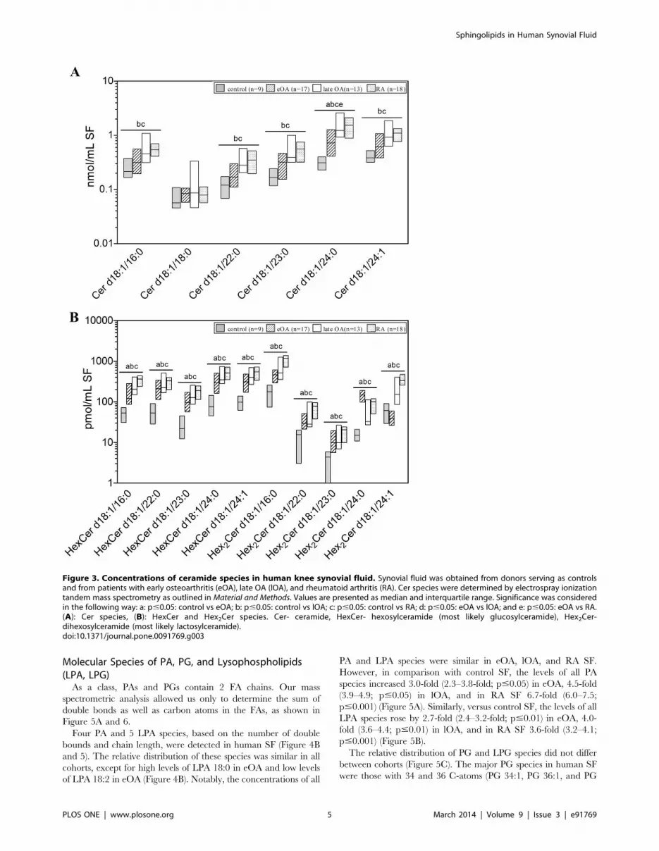
Ceramide Molecular SpeciesIn order to quantify Cer species, ESI-MS/MS analysis was
used. Furthermore, LC-MS/MS method was performed to
quantify HexCer and Hex2Cer species. Within analysed SLs, the
second most prominent group of SLs in human SF was Cer
species. The concentration of total Cer species was 1.4 nmol/ml in
SF from controls, elevated 2-fold in SF from eOA (2.8 nmol/ml;
n.s.), 3.9-fold in lOA SF (5.5 nmol/ml; p#0.01), and 3-fold in SF
from RA (4.2 nmol/ml; p#0.001). Six Cer species were identified;
Cer d18:0/24:0 was the predominant species (Figure 3A). As
expected, the level of most Cer species was higher in lOA and RA
SF, such as Cer d18:1/16:0, Cer d18:1/22:0, Cer d18:1/23:0, and
Cer d18:1/24:1. Notably, most Cer species contained saturated
FAs, constituting 68.9% to 74.9% of total Cer species in all cohorts
(Figure 3A).
Further, the molecular species of HexCer and Hex2Cer were
quantified. Five HexCer species and 5 Hex2Cer species were
detected in human SF (Figure 3B and 4A). The percentage
distribution of these species differed slightly between all cohorts,
except for Hex2Cer d18:1/24:0 which was high in eOA, and
Hex2Cer d18:1/24:1, which was low in eOA (Figure 4A). HexCer
species and Hex2Cer species were low in control SF but higher in
the SF of all other cohorts. In comparison with control SF, the
concentrations of HexCer species increased by 3.9-fold (3.4–4.2-
fold; p#0.001) in eOA, 5.8-fold (4.8–6.1-fold; p#0.01) in lOA,
Table 1. Characteristics of synovial fluid donors.
Postmortem donors. n = 9 Patients with eOA. n = 17 Patients with lOA. n = 13 Patients with RA. n = 18
Age 22 (21–25) 36 (25–49) 70 (67–74) 64 (49–71)
female/male 1/8 6/11 7/6 14/4
BMI 24.8 (20.8–25.2) 24.7 (22.6–26.9) 27.7 (25.8–30.1) 29.5 (24.8–32.8)
CRP nd 0.5 (0.5–1.0) 1.6 (1.13–1.85) 13.2 (5.3–38.8)
No. of cells/ml SF nd nd nd 6100 (2588–12,925)
DAS28 nd nd nd 3.48 (3.32–4.56)
Inclusion criteria: both genders, age 18–85 years inclusive, BMI,40, CRP#3 mg/L, and all CRP levels for RA. Exclusion criteria: joint infection; severe liver or kidneydisease; any surgery within the last 3 months; knee joint surgery within the last 6 months; diabetes mellitus (OA); drug abuse; intraarticular treatment with hyaluronate;or corticosteroid treatment within the last 3 months; HIV infection; tumor/cancer.doi:10.1371/journal.pone.0091769.t001
Figure 1. Postmortem stability of lipids extracted from humansynovial fluid of healthy knee joints used as controls. Lipidswere determined by electrospray ionization tandem mass spectrometry(ESI-MS/MS) or liquid chromatography coupled with tandem massspectrometry (LC-MS/MS) as outlined in Material and Methods. Valuesare displayed as a scatterplot of the concentration of each lipid classand species by postmortem time. (A): Lipid classes, (B): Lipid species.SM-sphingomyelin, Cer-ceramide, HexCer-hexosylceramide (most likelyglucosylceramide), Hex2Cer-dihexosylceramide (most likely lactosylcer-amide), PA-phospatidic acid, LPA-lysophosphatidic acid, PG-phosphati-dylglycerol, LPG-lysophosphatidylglycerol.doi:10.1371/journal.pone.0091769.g001
Sphingolipids in Human Synovial Fluid
PLOS ONE | www.plosone.org 3 March 2014 | Volume 9 | Issue 3 | e91769

and in RA SF 5.8-fold (5.3–6.1-fold; p#0.001) (Figure 3B),
whereas Hex2Cer species rose by 3.4-fold (3.2–5.5-fold; p#0.001)
in eOA, 4.5-fold (4.4–5.2-fold; p#0.05) in lOA, and in RA SF 6.9–
fold (6.9–7.3-fold; p#0.001) (Figure 3B).
Lysosphingolipids and Their PhosphatesThe values for S1P and sphinganine-1-phosphate in SF were
below the limit of detection (6 pmol/ml for undiluted samples).
We identified the following species in RA SF: SPH d18:0
[11.3 pmol/ml (0.0–21.2 pmol/ml)], SPH d18:1 [20.0 pmol/ml
(15.2–24.8 pmol/ml)], SPC d18:0 [1.0 pmol/ml (0.0–1.8 pmol/
ml)], and SPC d18:1 [16.1 pmol/ml (10.8–31.2 pmol/ml)]. Only
2 SL species in OA SF [SPH d18:1, 5.2 pmol/ml (0.0–16.9 pmol/
ml) and SPC d18:1, 10.1 pmol/ml (4.6–19.5 pmol/ml)] were
detected.
Molecular Species of Cardiolipins and BMPsCLs possess a unique dimeric structure, bearing phosphatidyl
moieties that are linked by a bridging glycerol, to which 4 FAs are
attached. Our mass spectrometric analysis only enabled us to
measure the sum of double bounds and carbon atoms in each of
the 2 phosphatidyl moieties. The CL species concentrations were
below the limit of detection, except for CL 36:4/36:4, which was
elevated in RA SF.
Only 2 BMP species (BMP 18:1/18:1 and BMP 18:2/18:1) were
above the limit of detection (Table 2). The highest levels of these
species were observed in RA SF. In comparison with control SF,
the levels of these BMP species rose by 1.6-fold (1.4–1.7-fold; ns) in
all other cohorts.
Figure 2. Concentrations of sphingomyelin species in human knee synovial fluid. Synovial fluid was obtained from donors serving ascontrols and from patients with early osteoarthritis (eOA), late OA (lOA), and rheumatoid arthritis (RA). SM species were determined by electrosprayionization tandem mass spectrometry as outlined in Material and Methods. Species annotation is based on the notion that 2 hydroxyl groups arelinked to a sphingoid base. Values are presented as median and interquartile range. Significance was considered in the following way: a: p#0.05:control vs eOA; b: p#0.05: control vs lOA; c: p#0.05: control vs RA; d: p#0.05: eOA vs lOA; and e: p#0.05: eOA vs RA. SM- sphingomyelin.doi:10.1371/journal.pone.0091769.g002
Sphingolipids in Human Synovial Fluid
PLOS ONE | www.plosone.org 4 March 2014 | Volume 9 | Issue 3 | e91769
Molecular Species of PA, PG, and Lysophospholipids(LPA, LPG)
As a class, PAs and PGs contain 2 FA chains. Our mass
spectrometric analysis allowed us only to determine the sum of
double bonds as well as carbon atoms in the FAs, as shown in
Figure 5A and 6.
Four PA and 5 LPA species, based on the number of double
bounds and chain length, were detected in human SF (Figure 4B
and 5). The relative distribution of these species was similar in all
cohorts, except for high levels of LPA 18:0 in eOA and low levels
of LPA 18:2 in eOA (Figure 4B). Notably, the concentrations of all
PA and LPA species were similar in eOA, lOA, and RA SF.
However, in comparison with control SF, the levels of all PA
species increased 3.0-fold (2.3–3.8-fold; p#0.05) in eOA, 4.5-fold
(3.9–4.9; p#0.05) in lOA, and in RA SF 6.7-fold (6.0–7.5;
p#0.001) (Figure 5A). Similarly, versus control SF, the levels of all
LPA species rose by 2.7-fold (2.4–3.2-fold; p#0.01) in eOA, 4.0-
fold (3.6–4.4; p#0.01) in lOA, and in RA SF 3.6-fold (3.2–4.1;
p#0.001) (Figure 5B).
The relative distribution of PG and LPG species did not differ
between cohorts (Figure 5C). The major PG species in human SF
were those with 34 and 36 C-atoms (PG 34:1, PG 36:1, and PG
Figure 3. Concentrations of ceramide species in human knee synovial fluid. Synovial fluid was obtained from donors serving as controlsand from patients with early osteoarthritis (eOA), late OA (lOA), and rheumatoid arthritis (RA). Cer species were determined by electrospray ionizationtandem mass spectrometry as outlined in Material and Methods. Values are presented as median and interquartile range. Significance was consideredin the following way: a: p#0.05: control vs eOA; b: p#0.05: control vs lOA; c: p#0.05: control vs RA; d: p#0.05: eOA vs lOA; and e: p#0.05: eOA vs RA.(A): Cer species, (B): HexCer and Hex2Cer species. Cer- ceramide, HexCer- hexosylceramide (most likely glucosylceramide), Hex2Cer-dihexosylceramide (most likely lactosylceramide).doi:10.1371/journal.pone.0091769.g003
Sphingolipids in Human Synovial Fluid
PLOS ONE | www.plosone.org 5 March 2014 | Volume 9 | Issue 3 | e91769

36:2 with concentrations between 0.062 and 3.07 pmol/ml;
Figure 6). Five PG and 2 LPG (LPG 16:0 and LPG 18:1) species
were found in human SF at concentrations of 0.1–5.0 pmol/ml
(Figure 6). Notably, in comparison with control SF, the levels of all
PG and LPG species were elevated by 3.5-fold (2.5–5.6-fold;
p#0.01) in eOA, 3.6-fold (2.8–4.6; p#0.05) in lOA, and in RA SF
2.9-fold (1.9–4.5; p#0.01). However, the levels of PG and LPG
species did not differ between eOA and lOA SF.
Post Mortem Stability of Sphingolipids in SFThe 9 postmortem donors died of intoxication (46), multiple
trauma (26), cardiomyopathy (16), pulmonary embolism (16),
and craniocerebral trauma (16). The SF was obtained with a
syringe during the autopsy of 9 adult donors without any known
joint disease within a window of several hours to up to 5 days after
the time-point of death. Since the stability of sphingolipids in SF as
a function of the post-mortem time after time of death is unknown,
the concentrations of several lipid species that exist at high levels in
the SF (SM 34:1, SM 42:2, Cer d18: 1/24: 0) were plotted against
the time between death and autopsy. Figure 6 show that no
relevant changes occur in the level of sphingolipids within the time
frame of sample collection. Furthermore, no discordant values is
obvious in Figure 1 indicating that the cause of death appears to
have no impact on the level of lipid species. However, the numbers
of investigated SF per cause of death is too low to generalize this
observation.
In order to confirm the stability of the lipid species mathemat-
ically, a linear regression of the data was obtained, and the slope of
the line was calculated together with the 95% confidence interval.
None of the slopes differed from 0 (values not shown).
Sphingolipids and Age of DonorsIt is not known whether the level of sphingolipids in SF may be
also dependent on the age of donors. Furthermore, the four
cohorts of our study differ from each other with respect of their
average age. Therefore the concentration of investigated lipid
classes and species was plotted against the age of each donor
within each cohort. Figure 7 as well as Figures S1, S2, S3 shows
that the age of donors had no impact on the concentrations of
lipids within the four investigated cohorts. Also, a linear regression
of the data was calculated together with the slope and the 95%
confidence interval revealing, that none of the slopes differed
significantly from 0 (values not shown).
Adjustment of DataThe adjusted values were obtained from there corresponding
nonadjusted concentrations by multiplying with a dilution factor
[34] and resulting data are shown both for classes (Table 3) and
individual species (Table S1). During OA water SF might be
diluted by water due to inflammation-induced effusion. Therefore,
the concentrations of lipid species were corrected for possible
dilution. The concentrations of urea were determined within
serum and SF in order to calculate a dilution factor for SF; this
procedure was formerly developed to adjust for the dilution
introduced by lavage during some biomarker studies. Compared
with the level of lipids in SF of controls, eOA, lOA, and RA had
elevated concentrations of most lipid species independent of
whether they were adjusted or nonadjusted. However, the
differences between most lipid species in eOA and lOA were
more obvious using the adjusted values (Table S1)
Discussion
Lipids such as SLs, PAs, LPAs, LPGs, and BMPs and other
groups of lipids are known to be bioactive molecules, but their
location and function in SF of articular joints remain to be
discovered. The lack of knowledge on the profile, quantity, and
function of these lipids in human SF underscore the need for
detailed studies in this area. Changes in the composition and
Figure 4. Relative distribution of HexCer and Hex2Cer, PA, LPA,PG and LPG species in in human knee synovial fluid. Synovialfluid was obtained from donors serving as controls and from patientswith early osteoarthritis (eOA), late OA (lOA), and rheumatoid arthritis(RA). The data show the percentage of the lipid species from the totalcorresponding lipid class ( = 100%). Values are presented as median andinterquartile range. (A): HexCer and Hex2Cer species, (B): PA and LPAspecies, (C): PG and LPG species. HexCer- hexosylceramide (most likelyglucosylceramide), Hex2Cer- dihexosylceramide (most likely lactosylcer-amide), PA- phosphatidic acid, LPA- lysophosphatidic acid, PG-phosphatidylglycerol, LPG- lysophosphatidylglycerol.doi:10.1371/journal.pone.0091769.g004
Sphingolipids in Human Synovial Fluid
PLOS ONE | www.plosone.org 6 March 2014 | Volume 9 | Issue 3 | e91769

Figure 5. Concentrations of phosphatidic acid (PA) and lysophosphatidic acid (LPA) species in human knee synovial fluid. Synovialfluid was obtained from donors serving as controls and from patients with early osteoarthritis (eOA), late OA (lOA), and rheumatoid arthritis (RA). PAand LPA species were determined liquid chromatography coupled with tandem mass spectrometry as outlined in Material and Methods. Speciesannotation is based on the notion that only ester bonds are present. Values are presented as median and interquartile range. Significance wasconsidered in the following way: a: p#0.05: control vs eOA; b: p#0.05: control vs lOA; and c: p#0.05: control vs RA. (A): PA species, (B): LPA species.PA- phosphatidic acid, LPA- lysophosphatidic acid.doi:10.1371/journal.pone.0091769.g005
Table 2. Concentrations of bis(monoacylglycero)phosphate (BMP) species.
Concentration [pmol/ml]
BMP-specie control (n = 9) eOA (n = 17) lOA (n = 13) RA (n = 18)
BMP 18:1/18:1 27.8 (17.5–46.8) 23.1 (15.4–37.9) 36.7 (22.9–74.7) 24.2 (19.8–30.9)
BMP 18:2/18:1 15.6 (9.27–20.0) 16.8 (11.5–25.5) 25.4 (15.9–46.3) 18.8 (13.9–23.6)
Concentrations of bis(monoacylglycero)phosphate (BMP) species in knee synovial fluid obtained from donors serving as controls and from patients with earlyosteoarthritis (eOA), late OA (lOA), and rheumatoid arthritis (RA). BMP species were determined by electrospray ionization tandem mass spectrometry as outlined inMaterial and Methods. Values are presented as median and interquartile range in the brackets. BMP-Bis(monoacylglycero)phosphate, eOA-early osteoarthritis, lOA-lateosteoarthritis, RA-rheumatoid arthritis.doi:10.1371/journal.pone.0091769.t002
Sphingolipids in Human Synovial Fluid
PLOS ONE | www.plosone.org 7 March 2014 | Volume 9 | Issue 3 | e91769

concentrations of various lipids have been already linked with
several human disorders [19,26].
We have reported that concentrations of phospholipids species
increase in SF of RA and OA compared with SF obtained from
donors used as controls [26]. The principle finding of our current
study is that a broad spectrum of SL species, their precursors, and
intermediate metabolites was found in human SF. Moreover, the
concentrations of 41 lipids in eOA SF, 48 species in lOA SF, and
50 species in RA SF increase significantly in comparison with
control SF. Notably, the levels of 21 lipid species were altered
between eOA and lOA SF, indicating that the lipid composition of
SF reflects the severity of OA disease. Thus, our findings may be
used to develop biomarkers to discriminate eOA from lOA, and
eOA from healthy joints.
Until recently, there have been no sensitive methods to detect
these species at low concentrations in biological materials. The
lipidomic methods that we used in this study [28–32], allowed us
to identify and quantify many lipid species that have not been
reported in SF. This is why our lipidomic investigation reports for
the first time about the composition of SL and minor glyceropho-
spholipids species in human SF.
SMs and Cer species regulate many processes, including stress
responses, proliferation and differentiation, apoptosis, and senes-
cence [35–37]. Using chondrocytes and explants from rabbit
articular cartilage, exogenous cell-permeable C2-Cer at concen-
trations over 25 mM were noted to induce apoptosis, up regulate
MMP-1, -3, and -13, and ultimately induce articular cartilage
matrix degradation by collagen type II cleavage and proteoglycan
loss from cartilage [38–41]. Further, Cer species are reported to be
mediators that induce proinflammatory cytokine production and
possibly apoptosis in cultured human RA FLS [7,11] as well as
cultured chondrocytes [38]. In vitro administration of synthetic
exogenous C2- or C6-Cer at concentrations below 25 mM did not
affect apoptosis but inhibited the proliferation of FLS in OA and
RA [7]. However, intraarticular injection of 34.5 mg C2-Cer into
the ankle joints of mice increased the population of apoptotic
synovial cells [11].
We observed higher concentrations of several Cer species in SF
from RA and lOA compared with control SF indicating that Cer
species may be involved in the progression of OA and RA.
However, the exact function of extracellular SM and Cer species,
alone and in combination, in synovial joints is unknown. Further
studies are needed to show whether Cer species are synthesized de
novo or are the result of higher sphingomyelinase activity.
CLs are nearly exclusively a constituent of the mitochondrial
inner membrane: extracellular CLs in body fluids have not been
reported. We detected CLs only in RA SF. Thus, we speculate that
the presence of CLs in SF is a hallmark of RA and that CLs can be
used to develop a novel diagnostic tool for RA. However, it is
unknown whether CLs in SF have functions. The source of CLs in
RA SF is not known and further studies are needed to identify
where the CLs come from. One possible source are the cells that
are enriched in RA SF compared with OA and control SF and
which can be damaged by normal joint movement so that
mitochondrial lipids like CLs are released. Although we removed
cells and cellular debris from SF by centrifugation and filtration,
these techniques are unable to eliminate molecular micelles.
LPAs can promote the proliferation of many cell types. In vitro
stimulation with 1 or 5 mM exogenous LPA stimulates the
proliferation of rat primary chondrocytes [42,43]. Moreover, the
expression of six G-protein-coupled LPA receptors increases in RA
FLS. Thus, LPAs appear to function as lipid mediators between
cells displaying growth factor-like activities [44]. We noted
elevated levels of all PA and LPA species in SF of RA and OA
compared with control SF, consistent with previous findings [45].
The higher concentration of LPAs is likely attributed to increased
secretion and activity of enzymes for example phospholipase A2
[46] and may point to a possible repair response. However, further
studies need to evaluate the functions of LPA species as
intercellular lipid mediators that induce cell proliferation and
have growth factor-like activities in damaged articular cartilage.
Figure 6. Concentrations of phosphatidylglycerol (PG) and lysophosphatidylglycerol (LPG) species in human knee synovial fluid.Synovial fluid was obtained from donors serving as controls and from patients with early osteoarthritis (eOA), late OA (lOA), and rheumatoid arthritis(RA). PG and LPG species were determined by liquid chromatography coupled with tandem mass spectrometry as outlined in Material and Methods.Species annotation is based on the notion that only ester bonds are present. Values are presented as median and interquartile range. Significancewas considered in the following way: a: p#0.05: control vs eOA; b: p#0.05: control vs lOA; c: p#0.05: control vs RA; d: p#0.05: eOA vs lOA; e: p#0.05:eOA vs RA; and f: p#0.05: lOA vs RA. PG- phosphatidylglycerol, LPG- lysophosphatidylglycerol.doi:10.1371/journal.pone.0091769.g006
Sphingolipids in Human Synovial Fluid
PLOS ONE | www.plosone.org 8 March 2014 | Volume 9 | Issue 3 | e91769

Recently, cPA was reported to stimulate the production of HA in
human OA articular chondrocytes and to provide some anti-
inflammatory and chondroprotective activities in a rabbit model of
OA [23]. However, our methods applied did not allow us to
quantify cPA in SF.
This study is the first to report about PG species in SF. PGs are
important components of the pulmonary surfactant system [47].
Thus, we hypothesize that PG species have similar functions in
synovial joints. However, our study demonstrates that PG species
exist at low concentrations in human SF. Moreover, the PG
species in human SF are also found in human plasma; albeit,
human plasma appears to have more PG species than SF [20,48].
In, contrast to human plasma, in which only saturated LPG
species (LPG16:0 and LPG 18:0) have been reported [49], human
SF contains a high proportion of monounsaturated LPG 18:1.
Notably, versus control SF, the concentrations of all PG species
were significantly elevated in eOA, lOA, and RA SF. BMP, a
structural isomer of PG, was also detected in SF at low levels, and
2 BMP species were identified. However, the function and source
of extracellular PG, LPG, and BMP species are unknown.
In conclusion, this lipidomic investigation presents for the first
time a comprehensive survey of SLs and minor glyceropho-
spholipids in human SF. Our mass spectrometric analysis of lipids
in SF from patients with eOA, lOA, and RA knee joints indicate
disease and stage-dependent differences. Certain species of SM
and Cer may, at least in part, be involved in the pathogenesis of
OA and RA. The paucity of detailed data on the functions of lipid
species in RA and OA underscore the necessity for further studies.
Our study lays the foundation for addressing specific questions
regarding the biosynthesis and function of lipid species in SF.
Supporting Information
Figure S1 Concentrations of lipids in human synovialfluid as a function of the age of donors used as controls.Synovial fluid was obtained post mortem from donors with healthy
knee joints. Lipids were determined by electrospray ionization
tandem mass spectrometry (ESI-MS/MS) or liquid chromatogra-
phy coupled with tandem mass spectrometry (LC-MS/MS) as
outlined in Material and Methods. Values are displayed as a
scatterplot of the concentration of each lipid class and species by
age of donors. (A): Lipid classes, (B): Lipid species. SM-
sphingomyelin, Cer-ceramide, HexCer-hexosylceramide (most
likely glucosylceramide), Hex2Cer-dihexosylceramide (most likely
lactosylceramide), PA-phospatidic acid, LPA-lysophosphatidic
acid, PG-phosphatidylglycerol, LPG-lysophosphatidylglycerol.
(TIF)
Figure S2 Concentrations of lipids in human synovialfluid as a function of the age of patients with early stageosteoarthritis. Lipids were determined by electrospray ioniza-
tion tandem mass spectrometry (ESI-MS/MS) or liquid chroma-
tography coupled with tandem mass spectrometry (LC-MS/MS)
as outlined in Material and Methods. Values are displayed as a
scatterplot of the concentration of each lipid class and species by
age of donors. (A): Lipid classes, (B): Lipid species. SM-
sphingomyelin, Cer-ceramide, HexCer-hexosylceramide (most
likely glucosylceramide), Hex2Cer-dihexosylceramide (most likely
Figure 7. Concentrations of lipids in human synovial fluid as afunction of the age of patients with late stage osteoarthritis.Lipids were determined by electrospray ionization tandem massspectrometry (ESI-MS/MS) or liquid chromatography coupled withtandem mass spectrometry (LC-MS/MS) as outlined in Material andMethods. Values are displayed as a scatterplot of the concentration ofeach lipid class and species by age of donors. (A): Lipid classes, (B): Lipidspecies. SM-sphingomyelin, Cer-ceramide, HexCer-hexosylceramide(most likely glucosylceramide), Hex2Cer-dihexosylceramide (most likelylactosylceramide), PA-phospatidic acid, LPA-lysophosphatidic acid, PG-phosphatidylglycerol, LPG-lysophosphatidylglycerol.doi:10.1371/journal.pone.0091769.g007
Table 3. Impact of dilution factor on the concentrations oflipid classes.
Lipid class eOA (n = 17) [%] lOA (n = 13) [%] RA (n = 18) [%]
SM 118.2 (110.5–123.8) 157.1 (146.5–173.5) 88.1 (83.4–89.2)
Cer 130.7 (122.6–132.9) 157.7 (154.9–164.1) 81.2 (78.4–87.3)
HexCer 110.1 (110.1–111.6) 153.1 (152.7–158.6) 86.0 (82.8–88.5)
Hex2Cer 103.9 (99.8–105.4) 138.4 (135.5–139.8) 81.2 (77.5–81.9)
PA 103.3 (98.9–106.8) 180.0 (165.7–185.7) 82.8 (79.1–85.6)
LPA 109.9 (108.3–113.3) 176.3 (152.6–190.6) 84.2 (80.1–88.3)
PG 100.0 (98.9–108.5) 123.9 (121.1–126.4) 84.2 (80.8–86.4)
LPG 107.6 (101.9–113.3) 143.2 (133.0–153.4) 82.9 (81.9–83.9)
BMP 97.3 (95.4–99.3) 165.0 (165.0–165.0) 79.6 (77.9–81.2)
Impact of dilution factor on the concentrations of lipid classes as expressed aspercentage of uncorrected values ( = 100%). Lipids were determined byelectrospray ionization tandem mass spectrometry or liquid chromatographycoupled with tandem mass spectrometry, and corrected for possible dilution asoutlined in Material and Methods. Values are median and interquartile range.eOA-early osteoarthritis, lOA-late osteoarthritis, RA-rheumatoid arthritis, SM-sphingomyelin, Cer-ceramide, HexCer-hexosylceramide (most likelyglucosylceramide), Hex2Cer-dihexosylceramide (most likely lactosylceramide),PA-phosphatidic acid, LPA-lysophosphatidic acid, PG-phosphatidylglycerol,LPG-lysophosphatidylglycerol, BMP-bis(monoglycero)phosphate.doi:10.1371/journal.pone.0091769.t003
Sphingolipids in Human Synovial Fluid
PLOS ONE | www.plosone.org 9 March 2014 | Volume 9 | Issue 3 | e91769

lactosylceramide), PA-phospatidic acid, LPA-lysophosphatidic
acid, PG-phosphatidylglycerol, LPG-lysophosphatidylglycerol.
(TIF)
Figure S3 Concentrations of lipids in human synovialfluid as a function of the age of patients with rheumatoidarthritis. Lipids were determined by electrospray ionization
tandem mass spectrometry (ESI-MS/MS) or liquid chromatogra-
phy coupled with tandem mass spectrometry (LC-MS/MS) as
outlined in Material and Methods. Values are displayed as a
scatterplot of the concentration of each lipid class and species by
age of donors. (A): Lipid classes, (B): Lipid species. SM-
sphingomyelin, Cer-ceramide, HexCer-hexosylceramide (most
likely glucosylceramide), Hex2Cer-dihexosylceramide (most likely
lactosylceramide), PA-phospatidic acid, LPA-lysophosphatidic
acid, PG-phosphatidylglycerol, LPG-lysophosphatidylglycerol.
(TIF)
Table S1 Concentrations of lipid species presented inFigure 1–7. Lipids were determined by electrospray ionization
tandem mass spectrometry or liquid chromatography coupled with
tandem mass spectrometry as outlined in Material and Methods. Data
(nmol/ml or pmol/ml) obtained were either uncorrected (normal
font) or corrected with the dilution factor per Kraus et al. (34, bold
font). Values are median and interquartile range. The concentra-
tions of urea were determined within serum and SF to calculate a
dilution factor for SF; this procedure was formerly developed to
adjust for the dilution introduced by lavage during some
biomarker studies. eOA-early osteoarthritis, lOA-late osteoarthri-
tis, RA-rheumatoid arthritis, SM-sphingomyelin, Cer-ceramide,
HexCer-hexosylceramide (most likely glucosylceramide), Hex2-
Cer-dihexosylceramide (most likely lactosylceramide), PA-phospa-
tidic acid, LPA-lysophosphatidic acid, PG-phosphatidylglycerol,
LPG-lysophosphatidylglycerol, SPH-sphingosine, SPC-sphingosyl-
phosphorylcholine, CL-cardiolipin, BMP-bis(monoglycero)pho-
sphate.
(PDF)
Acknowledgments
The authors thank Magdalena Singer, Christiane Hild, and Simone
Duchtel for excellent technical support and Manuela Doller for assistance
with the study organization.
Author Contributions
Conceived and designed the experiments: MKK GS G. Lochnit JS.
Performed the experiments: MKK G. Liebisch G. Lochnit JW HK UK G.
Lasczkowski MR. Analyzed the data: MKK G. Liebisch GS JW JS.
Contributed reagents/materials/analysis tools: G. Liebisch G. Lochnit JW
HK UK G. Lasczkowski MR. Wrote the paper: MKK JS. Involved in
drafting the article or revising it critically for important intellectual content,
and approving the final version to be published: GLI GLO JW HK UK
GL GS MR.
References
1. Bellamy N, Campbell J, Robinson V, Gee T, Bourne R, et al. (2006)
Viscosupplementation for the treatment of osteoarthritis of the knee. Cochrane
Database Syst Rev: CD005321.
2. Ludwig TE, McAllister JR, Lun V, Wiley JP, Schmidt TA (2012) Diminishedcartilage-lubricating ability of human osteoarthritic synovial fluid deficient in
proteoglycan 4: Restoration through proteoglycan 4 supplementation. Arthritis
Rheum 64: 3963–3971.
3. Schmidt TA, Gastelum NS, Nguyen QT, Schumacher BL, Sah RL (2007)Boundary lubrication of articular cartilage: role of synovial fluid constituents.
Arthritis Rheum 56: 882–891.
4. Lahiri S, Futerman AH (2007) The metabolism and function of sphingolipids
and glycosphingolipids. Cell Mol Life Sci 64: 2270–2284.
5. Merrill AH Jr (2011) Sphingolipid and glycosphingolipid metabolic pathways inthe era of sphingolipidomics. Chem Rev 111: 6387–6422.
6. Marchesini N, Hannun YA (2004) Acid and neutral sphingomyelinases: rolesand mechanisms of regulation. Biochem Cell Biol 82: 27–44.
7. Gerritsen ME, Shen CP, Perry CA (1998) Synovial fibroblasts and the
sphingomyelinase pathway: sphingomyelin turnover and ceramide generation
are not signaling mechanisms for the actions of tumor necrosis factor-alpha.Am J Pathol 152: 505–512.
8. Cutler RG, Mattson MP (2001) Sphingomyelin and ceramide as regulators of
development and lifespan. Mech Ageing Dev 122: 895–908.
9. Spiegel S, Milstien S (2003) Sphingosine-1-phosphate: an enigmatic signalling
lipid. Nat Rev Mol Cell Biol 4: 397–407.
10. Luberto C, Kraveka JM, Hannun YA (2002) Ceramide regulation of apoptosisversus differentiation: a walk on a fine line. Lessons from neurobiology.
Neurochem Res 27: 609–617.
11. Ichinose Y, Eguchi K, Migita K, Kawabe Y, Tsukada T, et al. (1998) Apoptosis
induction in synovial fibroblasts by ceramide: in vitro and in vivo effects. J LabClin Med 131: 410–416.
12. Schlame M, Ren M (2009) The role of cardiolipin in the structural organizationof mitochondrial membranes. Biochim Biophys Acta 1788: 2080–2083.
13. Houtkooper RH, Vaz FM (2008) Cardiolipin, the heart of mitochondrial
metabolism. Cell Mol Life Sci 65: 2493–2506.
14. Schug ZT, Gottlieb E (2009) Cardiolipin acts as a mitochondrial signalling
platform to launch apoptosis. Biochim Biophys Acta 1788: 2022–2031.
15. Yin H, Zhu M (2012) Free radical oxidation of cardiolipin: chemicalmechanisms, detection and implication in apoptosis, mitochondrial dysfunction
and human diseases. Free Radic Res 46: 959–974.
16. Ji J, Kline AE, Amoscato A, Samhan-Arias AK, Sparvero LJ, et al. (2012)
Lipidomics identifies cardiolipin oxidation as a mitochondrial target for redoxtherapy of brain injury. Nat Neurosci 15: 1407–1413.
17. Ventura-Clapier R, Garnier A, Veksler V (2004) Energy metabolism in heartfailure. J Physiol 555: 1–13.
18. Ellis CE, Murphy EJ, Mitchell DC, Golovko MY, Scaglia F, et al. (2005)
Mitochondrial lipid abnormality and electron transport chain impairment in
mice lacking alpha-synuclein. Mol Cell Biol 25: 10190–10201.
19. Schlame M, Ren M (2006) Barth syndrome, a human disorder of cardiolipinmetabolism. FEBS Lett 580: 5450–5455.
20. Meikle PJ, Duplock S, Blacklock D, Whitfield PD, Macintosh G, et al. (2008)
Effect of lysosomal storage on bis(monoacylglycero)phosphate. Biochem J 411:
71–78.
21. Hullin-Matsuda F, Luquain-Costaz C, Bouvier J, Delton-Vandenbroucke I
(2009) Bis(monoacylglycero)phosphate, a peculiar phospholipid to control thefate of cholesterol: Implications in pathology. Prostaglandins Leukot Essent Fatty
Acids 81: 313–324.
22. Tokumura A (2002) Physiological and pathophysiological roles of lysopho-
sphatidic acids produced by secretory lysophospholipase D in body fluids.Biochim Biophys Acta 1582: 18–25.
23. Masuda I, Okada K, Momohara S (2013) Cyclic phosphatidic acid (CPA)stimulates the production of hyaluronic acid (HA) in human osteoarthritic
articular chondrocytes, and intraarticular administration of CPA supresses pain,swelling, and cartilage destruction in rabbit experimental osteoarthritis.
Osteoarthritis Cartilage 21: Abstracts:S296–S297.
24. Outerbridge RE (1961) The etiology of chondromalacia patellae. J Bone Joint
Surg Br 43-B: 752–757.
25. Arnett FC, Edworthy SM, Bloch DA, McShane DJ, Fries JF, et al. (1988) The
American Rheumatism Association 1987 revised criteria for the classification ofrheumatoid arthritis. Arthritis Rheum 31: 315–324.
26. Kosinska MK, Liebisch G, Lochnit G, Wilhelm J, Klein H, et al. (2013) Alipidomic study of phospholipid classes and species in human synovial fluid.
Arthritis Rheum 65: 2323–2333.
27. Berckmans RJ, Nieuwland R, Kraan MC, Schaap MC, Pots D, et al. (2005)
Synovial microparticles from arthritic patients modulate chemokine andcytokine release by synoviocytes. Arthritis Res Ther 7: R536–544.
28. Liebisch G, Lieser B, Rathenberg J, Drobnik W, Schmitz G (2004) High-throughput quantification of phosphatidylcholine and sphingomyelin by
electrospray ionization tandem mass spectrometry coupled with isotope
correction algorithm. Biochim Biophys Acta 1686: 108–117.
29. Scherer M, Leuthauser-Jaschinski K, Ecker J, Schmitz G, Liebisch G (2010) A
rapid and quantitative LC-MS/MS method to profile sphingolipids. J Lipid Res51: 2001–2011.
30. Scherer M, Schmitz G, Liebisch G (2009) High-throughput analysis of
sphingosine 1-phosphate, sphinganine 1-phosphate, and lysophosphatidic acid
in plasma samples by liquid chromatography-tandem mass spectrometry. ClinChem 55: 1218–1222.
31. Scherer M, Schmitz G, Liebisch G (2010) Simultaneous quantification ofcardiolipin, bis(monoacylglycero)phosphate and their precursors by hydrophilic
interaction LC-MS/MS including correction of isotopic overlap. Anal Chem 82:8794–8799.
32. Liebisch G, Drobnik W, Reil M, Trumbach B, Arnecke R, et al. (1999)Quantitative measurement of different ceramide species from crude cellular
extracts by electrospray ionization tandem mass spectrometry (ESI-MS/MS).J Lipid Res 40: 1539–1546.
Sphingolipids in Human Synovial Fluid
PLOS ONE | www.plosone.org 10 March 2014 | Volume 9 | Issue 3 | e91769

33. Liebisch G, Vizcaino JA, Kofeler H, Trotzmuller M, Griffiths WJ, et al. (2013)
Shorthand notation for lipid structures derived from mass spectrometry. J LipidRes 54: 1523–1530.
34. Kraus VB, Huebner JL, Fink C, King JB, Brown S, et al. (2002) Urea as a
passive transport marker for arthritis biomarker studies. Arthritis Rheum 46:420–427.
35. Niemela PS, Hyvonen MT, Vattulainen I (2006) Influence of chain length andunsaturation on sphingomyelin bilayers. Biophys J 90: 851–863.
36. Drobnik W, Liebisch G, Audebert FX, Frohlich D, Gluck T, et al. (2003) Plasma
ceramide and lysophosphatidylcholine inversely correlate with mortality in sepsispatients. J Lipid Res 44: 754–761.
37. Mizushima N, Kohsaka H, Miyasaka N (1998) Ceramide, a mediator ofinterleukin 1, tumour necrosis factor alpha, as well as Fas receptor signalling,
induces apoptosis of rheumatoid arthritis synovial cells. Ann Rheum Dis 57:495–499.
38. Sabatini M, Rolland G, Leonce S, Thomas M, Lesur C, et al. (2000) Effects of
ceramide on apoptosis, proteoglycan degradation, and matrix metalloproteinaseexpression in rabbit articular cartilage. Biochem Biophys Res Commun 267:
438–444.39. Sabatini M, Thomas M, Deschamps C, Lesur C, Rolland G, et al. (2001) Effects
of ceramide on aggrecanase activity in rabbit articular cartilage. Biochem
Biophys Res Commun 283: 1105–1110.40. Tetlow LC, Adlam DJ, Woolley DE (2001) Matrix metalloproteinase and
proinflammatory cytokine production by chondrocytes of human osteoarthriticcartilage: associations with degenerative changes. Arthritis Rheum 44: 585–594.
41. Colosimo M, McCarthy N, Jayasinghe R, Morton J, Taylor K, et al. (2000)Diagnosis and management of subdural haematoma complicating bone marrow
transplantation. Bone Marrow Transplant 25: 549–552.
42. Kim MK, Lee HY, Park KS, Shin EH, Jo SH, et al. (2005) Lysophosphatidic
acid stimulates cell proliferation in rat chondrocytes. Biochem Pharmacol 70:
1764–1771.
43. Hurst-Kennedy J, Boyan BD, Schwartz Z (2009) Lysophosphatidic acid
signaling promotes proliferation, differentiation, and cell survival in rat growth
plate chondrocytes. Biochim Biophys Acta 1793: 836–846.
44. Orosa B, Gonzalez A, Mera A, Gomez-Reino JJ, Conde C (2012) Lysopho-
sphatidic acid receptor 1 suppression sensitizes rheumatoid fibroblast-like
synoviocytes to tumor necrosis factor-induced apoptosis. Arthritis Rheum 64:
2460–2470.
45. Song HY, Lee MJ, Kim MY, Kim KH, Lee IH, et al. (2010) Lysophosphatidic
acid mediates migration of human mesenchymal stem cells stimulated by
synovial fluid of patients with rheumatoid arthritis. Biochim Biophys Acta 1801:
23–30.
46. Pruzanski W, Keystone EC, Sternby B, Bombardier C, Snow KM, et al. (1988)
Serum phospholipase A2 correlates with disease activity in rheumatoid arthritis.
J Rheumatol 15: 1351–1355.
47. Agassandian M, Mallampalli RK (2013) Surfactant phospholipid metabolism.
Biochim Biophys Acta 1831: 612–625.
48. Quehenberger O, Armando AM, Brown AH, Milne SB, Myers DS, et al. (2010)
Lipidomics reveals a remarkable diversity of lipids in human plasma. J Lipid Res
51: 3299–3305.
49. Lee JY, Min HK, Moon MH (2011) Simultaneous profiling of lysophospholipids
and phospholipids from human plasma by nanoflow liquid chromatography-
tandem mass spectrometry. Anal Bioanal Chem 400: 2953–2961.
Sphingolipids in Human Synovial Fluid
PLOS ONE | www.plosone.org 11 March 2014 | Volume 9 | Issue 3 | e91769


