Embed Size (px)
Citation preview

Dow
nloadedfrom
http://journals.lww.com
/continuumby
EywjxkqR
lD7PqN
CxD
fpLJJZSLlwgxR
5cduGOuj9zW
DjFsN
AmiDfKrYeG
l60s/bz+JGNN2D
pkfdGyV1SzueC
nPYmG2dO
J/qVkfvSSwTD
s6bHu77W
5acBooFUWQx6JLF1eQ
pd2cetlfpLjlmCNw04qU
uK+0ypuvOIS
on02/06/2021
Downloadedfromhttp://journals.lww.com/continuumbyEywjxkqRlD7PqNCxDfpLJJZSLlwgxR5cduGOuj9zWDjFsNAmiDfKrYeGl60s/bz+JGNN2DpkfdGyV1SzueCnPYmG2dOJ/qVkfvSSwTDs6bHu77W5acBooFUWQx6JLF1eQpd2cetlfpLjlmCNw04qUuK+0ypuvOISon02/06/2021
Spinal Cord Anatomyand LocalizationBy Todd A. Hardy, PhD, MBBS, FRACP
ABSTRACTPURPOSE OF REVIEW:This article focuses on clinically relevant teaching pointsin spinal anatomy and localizing the lesion in myelopathy.
RECENT FINDINGS:The principles underlying spinal cord lesion localization arewell established, but improvements inMRI and the discovery of pathologicantibodies associated with causes of transverse myelitis distinct frommultiple sclerosis, such as aquaporin-4 IgG and myelin oligodendrocyteglycoprotein IgG, have assisted in diagnosis.
SUMMARY: The spinal cord has a highly organized neuroanatomy ofascending and descending tracts that convey sensory, motor, andautonomic information. Using integration of clues from the patient’s historyand neurologic examination, the effective clinician can distinguish spinalcord from peripheral nerve or brain pathology, often determine the leveland parts of the spinal cord affected by a lesion, and focus on a likelydiagnosis. The advent of MRI of the spine has revolutionized investigationof spinal cord disorders, but an important place for strong clinical acumenstill exists in assessing the patient with a myelopathy.
INTRODUCTION
Athorough understanding of the anatomy of the spinal cord is ofvalue to the neurologist because patients can present with an arrayof symptoms and signs referable to the spinal cord, and the capacityto distinguish cord pathology from brain or peripheral nervepathology facilitates early diagnosis and avoids unnecessary
investigations. Early diagnosis is particularly important because the consequencesof delayed diagnosis of spinal cord pathology can be devastating, with thepotential for permanent disability.
The neuroanatomy of the spinal cord is somatotopically and segmentallyorganized, with tracts and pathways that transmit sensory information fromorgans and peripheral receptors to the brain and motor information from thebrain to internal and peripheral effector organs. This means that sensory ormotor deficits in one area of the body can often be deduced to be due to damagein a specific area of the spinal cord.
The localization of lesional pathology is an essential skill for the neurologistand helps with differential diagnosis and targeted investigation. For the mostpart, spinal cord lesions are associated with upper motor neuron signs. Theapproach to a patient with a suspected myelopathy involves ascertaining the
12 FEBRUARY 2021
REVIEW ARTICLE
CONTINUUM AUDIO
INTERVIEW AVAILABLE
ONLINE
C ITE AS :
CONTINUUM (MINNEAP MINN)
2021;27(1, SPINAL CORD DISORDERS):
12–29.
Address correspondence toDr Todd A. Hardy, Department ofNeurology, Concord RepatriationGeneral Hospital, Hospital Road,Concord, NSW 2139, Australia,[email protected].
RELATIONSHIP DISCLOSURE:
Dr Hardy serves as coeditor ofAdvances in ClinicalNeuroscience and Rehabilitationand on an advisory board forMerck & Co, Inc. Dr Hardy hasreceived personal compensationfor speaking engagements fromBiogen;Merck & Co, Inc; NovartisAG; Sanofi Genzyme; and TevaPharmaceutical Industries Ltdand research/grant support fromNovartis AG.
UNLABELED USE OF
PRODUCTS/INVESTIGATIONAL
USE DISCLOSURE:
Dr Hardy reports no disclosure.
© 2021 American Academyof Neurology.
Copyright © American Academy of Neurology. Unauthorized reproduction of this article is prohibited.

motor and sensory level of the lesion. Certain patterns of signs may lead aneurologist to diagnose a spinal cord syndrome, such as a Brown-Séquardsyndrome accompanying a partial transverse myelitis, which may provide a clueto the etiology of the underlying disease process.
The advent of MRI has revolutionized the diagnosis of myelopathies to theextent that specific diagnoses can often be made with MRI alone. Nevertheless,not all myelopathies are associated with MRI lesions, and, occasionally, subtlespinal cord lesions may bemissed or difficult to appreciate onMRI. MRI can alsoidentify unrelated pathologies or red herrings that may divert attention awayfrom the actual diagnosis, which means that the clinical history and examinationmust be used in conjunction with MRI or other adjunctive investigations toestablish a diagnosis.
This article focuses on clinically relevant neuroanatomy of the spinal cord andexplains how neurologic symptoms and signs relate to the anatomy to give cluesthat enable the correct localization of spinal cord pathology.
ANATOMY OF THE SPINAL CORDThe spinal cord is a central nervous system (CNS) structure that arises from theinferior medulla of the brainstem above the C1 vertebra and runs in the spinalcanal to terminate at the L1-L2 level of the lumbar spine (FIGURE 1-11). It iscovered in the meninges, which are contiguous with the meninges of the brain;from the innermost layer to the outermost layer, the meninges are the pia,arachnoid, and duramater. The spinal cord is broadly divided longitudinally intothe cervical, thoracic, and lumbar regions, with the sacral spinal cord region thatrepresents the very lower aspect of the spinal cord referred to as the conusmedullaris. A modified fibrous termination of the pia mater called the filumterminale extends from the conus medullaris caudally to the coccyx, fusing at itslower end with the dura mater, where it anchors the entire spinal cord to thevertebral column.2,3
Enlargements in the cervical region and lumbar regions reflect increasednumbers of neurons in these regions to supply the upper and lower limbs,respectively (FIGURE 1-1). Pairs of sensory and motor nerve roots emerge atdistinct spinal levels throughout the length of the cord.3 Sensory nerve rootsemerge dorsally from the cord and motor nerve roots emerge ventrally beforethey meet to form mixed motor and sensory spinal nerves. The nerve roots arenumbered C1 through C8 when they arise from the cervical cord and T1 throughT12 when they arise from the thoracic cord. The lumbar L1 nerve roots emergejust below the T12 nerve roots. The lowermost nerve roots (lumbar, L2 throughL5; sacral, S1 through S5; and the coccygeal nerve) fan out distally from the lowercord and are referred to as the cauda equina (Latin for horse’s tail). Critically,only the sacral nerve roots emerge from the conus medullaris. The lowermostnerve roots inhabit the CSF-filled subarachnoid space that is formed because thepia-covered spinal cord terminates at L1-L2, but the arachnoid and dura continueto the S2 vertebral level. The subarachnoid space is clinically relevant as the sitefrom which CSF can be collected in a lumbar puncture without risk to the spinalcord, as it is typically performed at the L3-L4 or L4-L5 level.4
Cross-sectional Anatomy of the Spinal CordThe spinal cord is highly somatotopically organized, and its function is totransmit motor, sensory, and autonomic information.2,3 The neurons of the
KEY POINT
● Lumbar puncture istypically performed at theL3-L4 or L4-L5 level.
CONTINUUMJOURNAL.COM 13
Copyright © American Academy of Neurology. Unauthorized reproduction of this article is prohibited.

spinal cord are located centrally in an H-shaped or butterfly-shaped area knownas the central gray matter (FIGURE 1-2). The central gray matter is connectedacross the midline by the gray commissure, within which lies the central canal ofthe cord. The anterior and posterior columns of the gray matter are usuallyreferred to as the ventral and dorsal horns. The ventral horns contain alpha andgamma motor neurons and interneurons. Somatic motor neurons travel in themedial motor columns, where they innervate the axial muscles of the body, or inthe lateral motor columns, where they innervate the upper and lower limbs. Thedorsal horns are where somatic and visceral afferent (sensory) fibers enter thecord. Their cell bodies are in adjacent dorsal root ganglia. The intermediate gray
FIGURE 1-1The gross anatomy of the spinal cord and adjacent spinal nerve roots.Reprinted with permission from Moore KL, et al, Wolters Kluwer/Lippincott Williams & Wilkins.1
© 2014 Lippincott Williams & Wilkins.
SPINAL CORD ANATOMY AND LOCALIZATION
14 FEBRUARY 2021
Copyright © American Academy of Neurology. Unauthorized reproduction of this article is prohibited.

layer between the ventral and dorsal horns is where autonomic preganglioniccells lie, divided into intermediolateral and intermediomedial groups; thepreganglionic sympathetic neurons are located from C8 through L1. Thepreganglionic parasympathetic neurons are located from S2 through S4.
The white matter of the spinal cord surrounds the gray matter and iscomposed of myelinated axons. Principal white matter tracts of clinicalsignificance include the descending motor pathways of the corticospinal(pyramidal) tract, which run laterally and anteriorly (ventrally) in the cordthroughout its length (FIGURE 1-3).5 These convey somatic motor informationarising from the contralateral cerebral cortex, which decussates in the anteriormedulla just before entering the cervical spinal cord. The descending motorpathways of the corticospinal tract synapse with the anterior horns cells thatconvey motor information via the motor nerve roots and then via somatic(peripheral) motor neurons to a target muscle. The descending motor pathwaysin the cord before the anterior horns are called uppermotor neurons, and those ofthe anterior horns and somatic motor nerves are called lower motor neurons.
Additional descending motor pathways referred to as extrapyramidal tractsarise from the brainstem and convey involuntary information important indifferent aspects ofmotor control (FIGURE 1-3).5 Important extrapyramidal tractsinclude the rubrospinal tract, which transmits information to the spinalinterneurons to maintain balance; the reticulospinal tract, which transmitsmotor information from the reticular formation in the brainstem; the tectospinaltract, which assists in coordinating head and eye movements; and thevestibulospinal tract, which transmits balance information to the extremitiesfrom the vestibular system.
Somatosensory information in the form of temperature, pinprick sensation,and touch is carried anterolaterally in the cord via the spinothalamic (also known
FIGURE 1-2Cross-sectional anatomy of the gray and white matter tracts of the spinal cord.Reprinted with permission from Cho TA, Continuum (Minneap Minn).3 © 2015 American
Academy of Neurology.
KEY POINTS
● The butterfly-shapedarea of the spinal cord incross section is known asthe central gray matter.
● The descending motorpathways in the cord beforethe anterior horns are calledupper motor neurons, andthose of the anterior hornsand somatic motor nervesare called lower motorneurons.
CONTINUUMJOURNAL.COM 15
Copyright © American Academy of Neurology. Unauthorized reproduction of this article is prohibited.

as the anterolateral) tracts (FIGURE 1-3).3 This information is referred to thecontralateral thalamus of the brain as the sensory nerve roots entering via the twodorsal horns synapse with second-order neurons. The axons of the second-orderneurons ascend for two to three vertebral segments above where they enter thecord and then decussate across the midline. Proprioception, vibration, and touchare carried in the dorsal columns of the cord to the ipsilateral dorsal columnnuclei of the medulla. The parts of the dorsal columns transmitting informationfrom the lower limbs are referred to as gracile and those from the upper limbs arereferred to as cuneate.
The spinocerebellar tracts (dorsal and ventral) are ipsilateral to the cerebellumand run laterally in the cord, transmitting proprioceptive information frommuscles, tendons, and joints of the lower limbs. Cerebellar information from theupper limbs is conveyed by tracts arising from the external cuneate nucleus ofthe caudal medulla. White matter tracts are also involved in the transmission ofinformation related to autonomic nervous system control of bowel and bladderfunction and reflexes.
The spinal cord also has the capacity to produce a motor response that isindependent of the brain when triggered by external stimuli by way of spinalreflexes.6 The components of themonosynaptic spinal reflex arc include primarysensory afferents arising from peripheral stretch receptors synapsing directly inthe spinal cord on alpha motor neurons that make up the efferent pathway to amuscle and thereforemediate a finalmotor response. The classic example in clinicalpractice is the patellar reflex, which occurs when stretch receptors in the patellartendon are activated by the tendon hammer of a clinician causing reflex contractionof the quadriceps femoris muscle by way of a monosynaptic spinal reflex. Morecomplex polysynaptic reflexes involving an integrated response to sensory inputacrossmultiple spinal segments to produce amotor response also occur as a result ofspinal interneurons between afferent and efferent pathways. A polysynaptic reflexunderlies the startle response to an unexpected touch to the trunk or limbs.
FIGURE 1-3Cross-sectional anatomy of the principal white matter tracts of the spinal cord.Reprinted with permission from Cho TA, Continuum (Minneap Minn).3 © 2015 American
Academy of Neurology.
SPINAL CORD ANATOMY AND LOCALIZATION
16 FEBRUARY 2021
Copyright © American Academy of Neurology. Unauthorized reproduction of this article is prohibited.

Vascular Supply of the Spinal CordThe vascular supply of the spinal cord has clinical importance. The anterior spinalartery runs ventrally along the length of the cord and supplies blood to theanterior two-thirds of the spinal cord (including the corticospinal tracts), withthe remaining dorsal one-third of the spinal cord (including the dorsal columns)supplied by the paired posterior spinal arteries. All three of these arteries emergeas branches of the vertebral arteries at the cervical-cranial junction, but some oftheir blood supply also comes from the thyrocervical trunk via the cervicalarteries. Separate radiculomedullary arteries also arise from the thyrocervicaltrunk to supply the cervical spinal cord sequentially at the different levels atwhich they enter the vertebral canal. More caudal radiculomedullary arteriesemerge directly from the aorta to supply the thoracolumbar cord. Theradiculomedullary artery that anastomotically supplies the anterior spinal arterybetween T9 and T12 in most individuals is the great anterior radiculomedullaryartery, or artery of Adamkiewicz (FIGURE 1-47).8 The artery of Adamkiewicz isresponsible for supplying blood to the anterior cord from its point of entry in thelower thoracic cord down to the conus medullaris.
The venous drainage of the spinal cord is from the dorsal spinal and ventralspinal veins into the internal and external venous plexus adjacent to the dural sacand vertebral bodies. From there, venous blood is transported to the dural venoussinuses.9
IMPORTANCE OF THE CLINICAL HISTORYWhether neurologic symptoms are the result of a myelopathy or brain orperipheral nervous system pathology and the precise location of the lesion in thecord can often be determined by an accurate history and examination combinedwith knowledge of the somatotopic and segmental anatomy of the spinal cord(TABLE 1-1).10 Clues to a spinal cord lesion in the history include bilateral sensory
KEY POINTS
● The monosynaptic spinalreflex is caused byactivation of peripheralstretch receptorstransmitting an impulsealong primary sensoryafferents that synapsedirectly on alpha motorneurons, causing a finalefferent motor response.
● The artery ofAdamkiewicz is the largeradiculomedullary arterythat supplies the anteriorspinal artery between T9and T12 in most individuals.
● The somatotopicorganization of the spinalcord allows determinationof the approximate or, insome cases, precise level ofa spinal cord lesion.
FIGURE 1-4The arterial supply of the spinal cord.Reprinted with permission from Martirosyan NL, et al, J Neurosurg Spine.7 © 2011 Nicholas Theodore, MD.
CONTINUUMJOURNAL.COM 17
Copyright © American Academy of Neurology. Unauthorized reproduction of this article is prohibited.

and/or motor symptoms in the limbs, particularly if concomitant sphincterdisturbance is present. Patients may report stiffness in the legs or sustained(tonic) or rhythmic (clonic) spasm. Other clues include a history of neck or backpain associated with neurologic symptoms when there is extrinsic compressionon the cord, such as due to a cervical or thoracic vertebral metastasis, butintrinsic spinal cord pathology typically does not have associated neck or spinepain. Bladder symptoms, sensory symptoms, and pain that precede thedevelopment of weakness favor an extrinsic (compressive) myelopathy over anintrinsic cord lesion. Particular care should be taken in patients with a history ofknown metastatic cancer or risk factors for cancer (eg, cigarette smoking) thatmay suggest myelopathy due to evolving metastatic spinal cord compression,which is a neurologic emergency.
The Uhthoff phenomenon occurs when patients with a demyelinating disease(such as multiple sclerosis [MS]) develop a recrudescence of symptomsattributable to a CNS demyelinating lesionwith an increase in body temperature,such as when febrile, during exercise, or when ambient temperatures are hot (eg,in the shower). Other features that may be present include a feeling as if a tight
TABLE 1-1 Clues to a Spinal Cord Lesion From the History
General symptoms
◆ Bilateral greater than unilateral sensory and/or motor symptoms
◆ Bladder, bowel, and/or sexual dysfunction
◆ Stiffness in the legs
◆Neck and/or back pain in association with neurologic symptoms, particularly if the patienthad preceding trauma or if pain is exacerbated by neck flexion or extension
◆ Lhermitte or Uhthoff phenomenon
◆ Sensory level across the trunk (often more reliable as a symptom than a sign)
◆ Tight band around the trunk or torso
◆Neurogenic claudication (suggests cauda equina pathology rather than a lower cord lesion)
◆ Sensory ataxia (can also occur with peripheral nervous system disorders)
◆ Dyspnea when lying flat (C3-C5 lesion)
Time course of symptoms
◆ Acute
◇ Trauma
◇ Vascular (infarction, hemorrhage)
◆ Subacute
◇ Demyelination
◇ Space-occupying lesion (eg, epidural abscess)
◆ Chronic or progressive
◇ Primary or secondary progressive multiple sclerosis
◇ Hereditary spastic paraparesis
◇ Motor neuron disease (amyotrophic lateral sclerosis, primary lateral sclerosis)
SPINAL CORD ANATOMY AND LOCALIZATION
18 FEBRUARY 2021
Copyright © American Academy of Neurology. Unauthorized reproduction of this article is prohibited.
band is around the chest or torso. When this is present in patients with MS, it iscommonly referred to as the MS hug.
A careful history is also vital in trying to establish the level of the lesion in thespinal cord. Cervical cord lesions tend to affect the upper limbs but can affect allfour limbs or, more rarely, just the lower limbs. When present, the Lhermittephenomenon (defined as an electrical pain or tingling brought on by neck flexionthat runs from the neck down the back and sometimes into the arms or legs) isa clue to cervical myelopathy most commonly due to a demyelinating lesion, butit may be encountered with other etiologies, including cervical spondyloticmyelopathy. Cervical cord lesions can disrupt the origin of the phrenic nervesand cause diaphragmatic paralysis, hence the medical school mnemonic “C3, 4, 5keeps the diaphragm alive.” Patients with diaphragmatic paralysis typicallyreport dyspnea when lying flat, but severe cervical cord injury can result inrespiratory failure11 and severe autonomic dysfunction (refer to the sectionSpinal Shock Versus Neurogenic Shock). Thoracic lesions typically cause sensoryand/or motor symptoms in the lower limbs.
Painful cramping or weakness in the legs that presents after walking a setdistance and improves with rest (neurogenic claudication) is a feature ofclinically significant lumbar spinal canal stenosis causing intermittentcompression of cauda equina nerve roots; patients may report doing better whenthe spine is in flexion, which widens the lumbar canal (eg, when bent forwardleaning over a shopping cart).10 Sensory ataxia is less specific to spinal cordpathology but is nevertheless a commonmanifestation of cord disease caused bydorsal column dysfunction and can arise from cervical, thoracic, or even lumbarcord or cauda equina lesions.
Ascending numbness with preserved (or increased) reflexes is usuallysecondary to myelopathy (most often sensory myelitis of MS) but is frequentlymistaken for peripheral nervous system involvement. Indeed, if a patientmentions altered sensation or numbness from a particular level on the trunk thatextends downward (often noticed in the shower or when swimming), it is a clueto a spinal sensory level.
Bladder and bowel dysfunction can also relate to lesions in the cervical,thoracic, or lumbar cord; conus medullaris; or cauda equina. Sphincterdisturbance arises because normal voluntary voiding of the bowel and bladderrelies on afferent feedback of a sense of fullness in these organs via the spinalcord to the brain. When this feedback is interrupted, a neurogenic bowel andbladder can develop. Spinal injury can lead to either a flaccid or spastic bladder,with a range of symptoms, including urinary frequency, urgency, incontinence,and urinary retention due to lack of coordination between the detrusor muscle ofthe bladder (which contracts during normal micturition) and the urinarysphincter (which relaxes during normal micturition).12 Similarly, boweldysfunction from spinal cord injury can lead to constipation or fecal urgency andincontinence with a dilated anal sphincter. Male and female sexual dysfunctionoften accompanies neurogenic bowel and bladder symptoms and commonlymanifests as erectile dysfunction, retrograde ejaculation, and anorgasmia inmales and vaginal dryness and anorgasmia in females.12
The demographic profile of the patient and a detailed history can also help togenerate a relevant differential diagnosis list. Older patients are more likely toexperience a spinal cord infarct than younger patients, and MS is 3 times morecommon in females than males.13 A history of trauma raises the possibility of a
KEY POINTS
● The Uhthoff phenomenoncommonly occurs whenpatients with multiplesclerosis experience anexacerbation of symptomswith an increase in bodytemperature.
● Spinal injury can lead toeither a flaccid or spasticbladder, with a range ofsymptoms, including urinaryfrequency, urgency,incontinence, and urinaryretention due to a lack ofcoordination between thedetrusor muscle of thebladder and the urinarysphincter.
● The patient’s previousmedical history and thetemporal onset ofneurologic symptoms can beused to narrow thedifferential diagnosis of aspinal cord lesion.
CONTINUUMJOURNAL.COM 19
Copyright © American Academy of Neurology. Unauthorized reproduction of this article is prohibited.
compressive myelopathy; therefore, a history of trauma to the neck or backpreceding neurologic symptoms should always prompt consideration of spinalcord injury either directly (traumatic myelopathy) or indirectly (eg, spinal cordwatershed infarction secondary to vertebral artery dissection).
The time course of myelopathic symptoms is also informative. Acutesymptoms suggest trauma or a vascular cause, such as infarction or hemorrhage;subacute symptoms may suggest demyelination or an extrinsic compressivecause (eg, epidural abscess); and an insidious onset with chronic progressioncould indicate a progressive form of MS, hereditary spastic paraparesis, orprimary lateral sclerosis.14 Relapsing symptoms could indicate MS or other
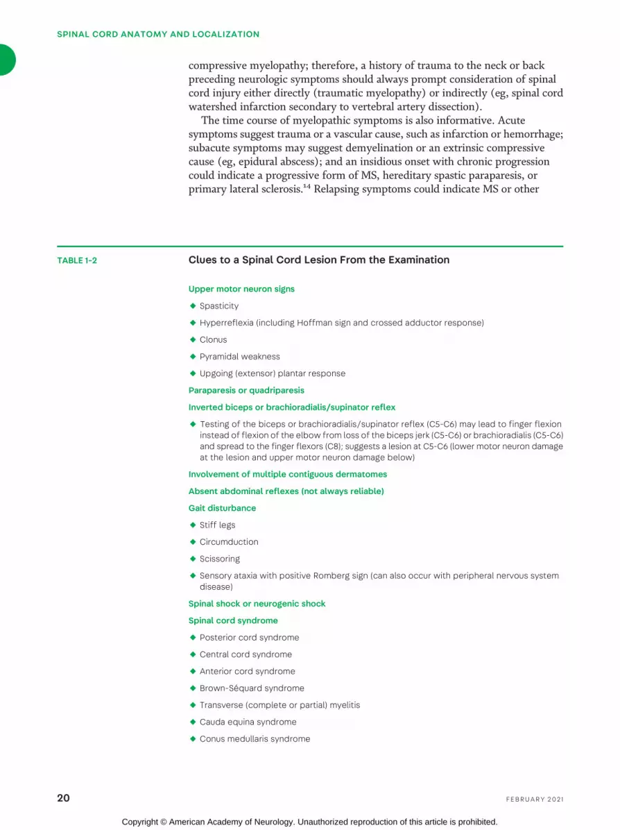
TABLE 1-2 Clues to a Spinal Cord Lesion From the Examination
Upper motor neuron signs
◆ Spasticity
◆ Hyperreflexia (including Hoffman sign and crossed adductor response)
◆ Clonus
◆ Pyramidal weakness
◆ Upgoing (extensor) plantar response
Paraparesis or quadriparesis
Inverted biceps or brachioradialis/supinator reflex
◆ Testing of the biceps or brachioradialis/supinator reflex (C5-C6) may lead to finger flexioninstead of flexion of the elbow from loss of the biceps jerk (C5-C6) or brachioradialis (C5-C6)and spread to the finger flexors (C8); suggests a lesion at C5-C6 (lower motor neuron damageat the lesion and upper motor neuron damage below)
Involvement of multiple contiguous dermatomes
Absent abdominal reflexes (not always reliable)
Gait disturbance
◆ Stiff legs
◆ Circumduction
◆ Scissoring
◆ Sensory ataxia with positive Romberg sign (can also occur with peripheral nervous systemdisease)
Spinal shock or neurogenic shock
Spinal cord syndrome
◆ Posterior cord syndrome
◆ Central cord syndrome
◆ Anterior cord syndrome
◆ Brown-Séquard syndrome
◆ Transverse (complete or partial) myelitis
◆ Cauda equina syndrome
◆ Conus medullaris syndrome
SPINAL CORD ANATOMY AND LOCALIZATION
20 FEBRUARY 2021
Copyright © American Academy of Neurology. Unauthorized reproduction of this article is prohibited.

inflammatory demyelinating disorders, such as aquaporin-4 IgG–seropositiveneuromyelitis optica spectrum disorder (NMOSD) or myelin oligodendrocyteglycoprotein (MOG) IgG–associated demyelination.15
Purely motor symptoms might indicate spinal cord infarction, particularlyif acute in onset, with a common etiology being embolization from theartery of Adamkiewicz to the anterior spinal artery affecting the ventralcorticospinal tracts in the thoracic spine at a point usually between T9 and T12.Unsteadiness when the eyes are closed (eg, in the shower) is highly suggestiveof dorsal column pathology such as from subacute combined degenerationof the spinal cord due to vitamin B12 deficiency or copper deficiency. Fever inthe context of neurologic symptoms referable to the spinal cord shouldinitiate investigations for diskitis, spinal epidural abscess, or vertebralosteomyelitis, particularly if the patient has a history of systemic infection(eg, endocarditis) or is an IV drug user.
CLINICAL EXAMINATION FINDINGSThe neurologic examination is essential for lesion localization in a patient witha suspectedmyelopathy (TABLE 1-2).10 Typically, patients will have upper motorneuron signs, such as spasticity, hyperreflexia, and clonus, or pyramidalweakness characterized by weakness of shoulder abduction, elbow extension,wrist extension, and finger extension and the interossei in the upper limbs.Patients may also have weakness of hip flexion, knee flexion, and ankledorsiflexion in the lower limbs. Uppermotor neuron signs develop consequent toloss of descending inhibitory pathways in the spinal cord, leading tohyperexcitability.16 It is not uncommon to see young patients withhyperreflexia and nonsustained clonus without significant neurologicpathology, so these signs cannot always be relied upon as indicators of CNSdisease; however, the likelihood of upper motor neuron pathology is increasedif hyperreflexia occurs in the company of other upper motor neuron signs,such as an extensor plantar response or pyramidal weakness, or both.Occasionally, patients with spinal cord pathology will have a normal clinicalexamination (eg, asymptomatic spinal cord lesions in MS).
The reflexes most often assessed in the neurologic examination are the bicepsjerks, triceps jerks, and supinator (brachioradialis) reflexes in the upper limbsand knee jerks, ankle jerks, and plantar responses in the lower limbs.17 Reflexesare graded as 0 or absent, 1+ when present but reduced, 2+ when normal,3+ when brisker or more exaggerated than normal (hyperreflexia), and 4+ whenhyperreflexia is accompanied by clonus. Clonus is a rhythmic involuntarymovement most often elicited at the ankle, where a sudden sharp dorsiflexion ofthe ankle by the examiner results in rhythmic plantar flexion of the foot.16 Clonusmay fatigue after a few beats (nonsustained) or occur indefinitely (sustained),with the latter usually indicating a more significant CNS insult.
Other reflexes can also be checked and are often helpful in providing furtherinformation if doubt exists regarding hyperreflexia, including deltoid and pectoralreflexes and finger jerks in the upper limbs and adductor reflexes in the lowerlimbs. A positiveHoffman sign (or reflex) occurswhen the tip of themiddle fingeris flicked downward, causing the thumb of the hand to flex and adduct; this canindicate a cervical myelopathy.18 Spread of reflexes into the fingers may cause thefingers to flex when the biceps jerk or supinator reflex is being elicited in thesetting of hyperactive finger flexors. An inverted brachioradialis/supinator reflex
KEY POINT
● The likelihood of uppermotor neuron pathology isincreased if hyperreflexiaoccurs accompanied byother upper motor neuronsigns, such as an extensorplantar response orpyramidal weakness, orboth.
CONTINUUMJOURNAL.COM 21
Copyright © American Academy of Neurology. Unauthorized reproduction of this article is prohibited.

(C5, C6) occurs when an attempt to elicit the biceps jerk leads to flexion of thefingers rather than elbow flexion because of a lesion at the C5 and/or C6 level.Injury to the C5 and/or C6 nerve root leads to an absent brachioradialis jerk(ie, no elbow flexion or supination), and spinal cord damage below that level leadsto hyperactivity of the finger flexor (C8) jerks and finger flexion from spreadwhen the adjacent brachioradialis tendon is struck.19 An inverted biceps reflex cansimilarly occur from a lesion at this location when loss of the biceps jerk isassociated with spread to the hyperactive triceps, causing elbow extension ratherthan the expected flexion. Cervical spondylosis is the most common etiology ofthese inverted reflexes in the upper limb. The presence of a crossed adductor reflex(contraction of both hip adductors when testing a reflex at the ipsilateral knee)from either or both lower limbs is considered an upper motor neuron sign.
Superficial abdominal reflexes are cutaneous reflexes that can be tested inthe four quadrants of the abdomen by stroking the skin (eg, with an orange stick)in the direction of the umbilicus, which causes reflex contractions of theabdominal wall. The upper abdominal reflexes are supplied by T9 through T11and the lower reflexes by T11 and T12. The superficial abdominal reflexes areabsent when a spinal cord lesion above the segmental level is present, but theirclinical relevance is limited as it can be difficult to elicit abdominal reflexes inpatients who are obese, have undergone abdominal surgery, are pregnant, or areelderly, and they may be absent in 15% of healthy individuals.20 The Beevor sign,which is also nonspecific, refers to upward displacement of the umbilicus whenthe neck is flexed on getting up from a reclining position; it occurs because ofrelative weakness of the lower abdominal muscles compared to the upperabdominalmuscles.21 The Beevor sign can indicate a thoracic cord lesion betweenT10 and T12 but is also seen in fascioscapulohumeral muscular dystrophy.
An extensor plantar response (positive Babinski reflex) is a major clue,particularly if bilateral, as it suggests involvement of the corticospinal tracts inthe spinal cord, whereas an isolated unilateral extensor plantar response could becaused by either ipsilateral cord or contralateral brain pathology. The plantarresponse is elicited by gently scraping along the lateral border of the foot andthen across the ball of the foot to the middle metatarsophalangeal joint; it isconsidered abnormal in adult patients when extension (rather than flexion) ofthe great toe is seen, with or without fanning of the other toes.22 It is worthmentioning that spinal cord pathology can still be present even with a flexorplantar response, so the sign is more helpful in localizing cord pathology whenextensor rather than when flexor.
A dermatome is the area of skin supplied by the sensory nerve root of a singlespinal segment (FIGURE 1-5).23 Knowledge of the dermatomes can help theclinician establish the level of a lesion involving sensation in the nervous system.If a spinal cord lesion is suspected, a dedicated search to try to determine thespinal sensory level of the lesion should be conducted, including over the anteriorand posterior trunk.10 Anatomic landmarks, such as the position of the nipples(which correspond to approximately the T4 dermatomal level) and the umbilicus(which corresponds to the T10 level), can greatly assist the clinician. Light touch,pinprick, and temperature sensation can all be tested over the trunk anteriorly orposteriorly. It is worth remembering that the C4 dermatome abuts the T2dermatome on the chest. A spinal cord lesion affecting the spinothalamic tract(s)may cause anesthesia below the lesion. The most caudal dermatome of normalpain and temperature sensation on the trunk (truncal sensory level) occurs two
SPINAL CORD ANATOMY AND LOCALIZATION
22 FEBRUARY 2021
Copyright © American Academy of Neurology. Unauthorized reproduction of this article is prohibited.

to three vertebral segments belowthe level of the actual lesion inthe spinal cord because thespinothalamic tracts ascend asthey decussate. This anatomicarrangement means that a lesionaffecting the spinothalamic tractof the right hemicord only willcause impaired temperature andpinprick sensation on the lefttrunk and lower limb below thelevel of the cord lesion. A sensorylevel is most reliable if the patienthas described altered sensation atthat level during the history.Involvement of multiplecontiguous dermatomes tends tofavor spinal cord pathology overradiculopathy, particularly if itoccurs in the context of otherupper motor neuron signs. Aspinal motor level can bedetermined by ascertaining whichmuscles are affected by weaknessand relating that to knowledge ofthe myotomes, which are definedas the group ofmuscles innervatedby a single spinal nerve.
Patients with significant spinalcord pathology will often have anabnormal gait. As in other partsof the examination, the locationof the lesion determines the wayin which the gait will beabnormal. Spasticity is usuallypresent and is defined clinicallyas a velocity-dependent increase
in normal resting muscle tone arising from upper motor neuron pathology.16 Theterm paresis is used to denote weakness, whereas plegia is used to denote absenceof any voluntary movement. Patients with a lesion involving the bilateralcorticospinal tracts may be paraplegic (no voluntary movement in either leg) orquadriplegic/tetraplegic (have no movement in any of their four extremities)with severe cervical cord lesions. Patients with spinal cord disease may have aspastic paraparesis (with weakness but some preserved voluntary movement)and an accompanying spastic gait with a tendency for the legs to veer laterallyfrom the midline during each stride (circumduction); they may also exhibitscissoring, in which the feet cross in front of each other during walking.19 Aspastic monoparesis (weakness but some preserved voluntary movement) canoccur when patients have unilateral corticospinal tract pathology, butcontralateral pathology in the brain also should be considered.
KEY POINTS
● The C4 dermatome abutsthe T2 dermatome on thechest.
● A truncal sensory level isdefined as the highestdermatomal area of normalsensation to pinprick andtemperature on the trunk.
● A lesion affecting thespinothalamic tract of theright hemicord only willcause impaired temperatureand pinprick sensation onthe left trunk and lower limbtwo to three vertebral levelsbelow the level of the cordlesion because thespinothalamic tracts ascendas they decussate.
● The term paresis is usedto denote weakness,whereas plegia is used todenote absence of anyvoluntary movement.
FIGURE 1-5The human dermatomes. Evidence-baseddermatome map representing the mostconsistent tactile dermatomal areas for eachspinal dorsal nerve root found inmost individuals,based on the best available evidence. Thedermatomal areas shown are not autonomouszones of cutaneous sensory innervation since,except in the midline where overlap is minimal,adjacent dermatomes overlap to a large andvariable extent. Blank regions indicate areas ofmajor variability and overlap.Modified with permission from Lee MWL, et al, Clin
Anat.23 © 2008 Wiley-Liss, Inc.
CONTINUUMJOURNAL.COM 23
Copyright © American Academy of Neurology. Unauthorized reproduction of this article is prohibited.
Lesions involving afferent pathways in the cord, particularly the dorsalcolumns, give rise to a sensory ataxia; a positive Romberg sign (markedunsteadiness when standing with the feet together and the eyes closed) andpseudoathetosis (involuntary writhing movements more pronounced with eyesclosed from loss of joint position sensation in the hands) may be seen.19
A coexistent stocking-glove peripheral neuropathy causing lower motorneuron signs (hyporeflexia or areflexia, wasting, fasciculations, hypotonia) canmask the upper motor neuron signs of a spinal cord lesion, so it may beappropriate to image the spinal cord in selected patients if a clinical possibility ofa myelopathy exists, despite an absence of upper motor neuron signs.
SPINAL SHOCK VERSUS NEUROGENIC SHOCKSpinal shock occurs when hyperacute or acute injury (particularly trauma) to thespinal cord results in flaccid areflexia below the level of the lesion, and theabsence of upper motor neuron signs can lead to diagnostic confusion. The signsof spinal shock may last from days to weeks. Neurogenic shock occurs in acutetrauma or occasionally in acute nontraumaticmyelopathies above the level of T6,which leads to loss of sympathetic tone below the lesion causing hypotension andunopposed vagal activity leading to bradycardia.24 The hypotension may requireIV fluids, inotropes, and vasopressors or combinations thereof, whereas thebradycardia may require atropine or temporary pacing.
AUTONOMIC DYSREFLEXIAAutonomic dysreflexia occurs when patients have injury above T6, leading to anexaggerated sympathetic nervous system response to sensory stimuli (eg, urinaryretention, urinary tract infection, or constipation) below the level of injury. Areflex response causes a strong sympathetic response, leading to constriction ofsplanchnic blood vessels and arterial hypertension. Carotid baroreceptorstrigger descending inhibitory signals to reduce sympathetic tone via the spinalcord, but a signal cannot reach most sympathetic outflow levels because of thecord lesion at or above T6. A further compensatory parasympathetic vagalresponse tries to reduce blood pressure by lowering the heart rate. In addition tohypertension and bradycardia, the excessive sympathetic tone below the lesionresults in pale cool extremities with piloerection. Above the lesion, theparasympathetic activity results in pupil constriction, flushing, and sweating.Autonomic dysreflexia is managed by treating the offending stimulus.
SPINAL CORD SYNDROMESSpinal cord syndromes are classic examination findings that can suggest the siteor nature of a spinal cord lesion. (TABLE 1-2) (FIGURE 1-625).26
Central Cord SyndromeA central cord syndrome usually occurs as a result of a neck hyperextensioncervical spinal cord injury, leading to damage to medial structures in the cord.A characteristic pattern of distal greater than proximal weakness in the upperlimbs (predominantly the hands from damage to the anterior horn cells at thatlevel) ensues, and the upper limbs are more affected than the lower limbs. Theupper motor neuron pathology can lead to bladder dysfunction and priapism (inmales). Classically, a capelike area of sensory loss to pain and temperature occursover the upper trunk and arms at the level of the lesion as the decussating
SPINAL CORD ANATOMY AND LOCALIZATION
24 FEBRUARY 2021
Copyright © American Academy of Neurology. Unauthorized reproduction of this article is prohibited.

spinothalamic tract fibers areaffected; this is called asuspended sensory level becausethe sensory loss is “suspended”or “hangs” on the trunk withoutinvolving the sacrum or lowerlimbs.27 This is often seen with acentral cord lesion due tosyringomyelia (CASE 1-1). Apatchy loss of pain, temperature,and light touch can be seenbelow the level of the lesion.Relative preservation of sacraland lower limb sensation occursbecause the lower extremityspinothalamic tracts run morelaterally than those of the upperextremities and so are notaffected early. This finding canalso be helpful in distinguishingan intraaxial lesion (within thecord) from an extraaxial lesion(intradural or extradural).Impairment of spinothalamicsensation without involvement ofdorsal column sensation, as seenin this syndrome, is an example ofdissociated sensory loss.
Anterior Cord SyndromeIn an anterior cord syndrome,damage to the ventral cordresults in motor weakness and,
often, loss of pain and temperature sensation below the level of the lesion.Proprioception and vibration are preserved as the dorsal columns run posteriorlyin the cord. Anterior cord syndromes classically occur with anterior spinal arteryinfarction, which is recognized as an iatrogenic complication resulting from crossclamping of the aorta intraoperatively.
Posterior Cord SyndromeA posterior cord syndrome occurs when pathology of one or both dorsal columnsof the spinal cord is present. The prototypic cause is vitamin B12 deficiency,leading to subacute combined degeneration of the cord in which the lateralcolumns are also involved concurrently. Patientsmay presentwith sensory ataxiaand have proprioceptive deficits on examination as well as impaired appreciationof vibration. In the case of vitamin B12 deficiency, peripheral neuropathy,cognitive deficits, and megaloblastic anemia may also be seen. Other causes ofposterior cord syndromes in the absence of a mass lesion or extrinsic compressiononMRI include nitrous oxide abuse (causing vitamin B12 deficiency), neurosyphilis,MS, and copper deficiency. For more information on the effects of vitamin
KEY POINTS
● Spinal shock occurs whenhyperacute or acute injury(particularly trauma) to thespinal cord results in flaccidareflexia below the level ofthe lesion.
● Neurogenic shock occursdue to acute pathologyabove the level of T6, whichleads to loss of sympathetictone below the lesioncausing hypotension andunopposed vagal activityleading to bradycardia.
● Autonomic dysreflexiaoccurs when patients haveinjury above the T6 level,leading to an exaggeratedsympathetic nervous systemresponse to sensory stimulibelow the level of the lesion(eg, bladder filling).
● A central intraaxial spinalcord lesion often causessensory symptoms and signsin the upper limbs and trunkbefore the lower limbs andsacral regions (called asuspended sensory level).This is because the lowerextremity spinothalamictracts run more laterallythan those of the upperextremities and so takelonger to be affected by anexpanding central cordlesion.
FIGURE 1-6Spinal cord syndromes occurring from lesionsabove the conus medullaris. Paraplegia arisesfrom lesions of the thoracic cord affecting motorpathways, and tetraplegia arises from similarlesions of the cervical cord. When transversemyelopathy or transection of the cord occurs,sensation below the level of the lesion is alsoaffected. For simplicity, the other spinal cordsyndromes are depicted as resulting from acervical cord lesion.Reprinted with permission from O’Phelan K, De Jesus I.25
© 2019 Cambridge University Press.
CONTINUUMJOURNAL.COM 25
Copyright © American Academy of Neurology. Unauthorized reproduction of this article is prohibited.

deficiencies and nitrous oxide use, refer to the article “Metabolic and ToxicMyelopathies” by Natalie Elizabeth Parks, MD,28 in this issue of Continuum.
Brown-Séquard SyndromeBrown-Séquard syndrome refers to ipsilateral upper motor neuron motorweakness in one lower limb (and rarely proprioceptive loss) with spinothalamicpain and temperature loss in the contralateral lower limb. It is caused by a lesionthat involves the lateral half of the cord, affecting the corticospinal,spinothalamic, and, sometimes, proprioceptive tracts on that side. Patients willhave a truncal sensory level to pain and temperature on the contralateral side ofthe lesion two or three segments below the level of the lesion. Causes includeMS,unilateral cord compression due to spinal degenerative disease, orhemitransection of the cord due to a knife injury or other trauma.
CASE 1-1 A 50-year-old woman presented with a 13-day history of sensorydisturbance in the upper limbs, pain in the shoulders, andweakness in thehands following a flulike illness. She had no relevant pastmedical history.
Examination revealed mild weakness and wasting of the intrinsicmuscle of the hands, with impaired temperature and pain sensationthroughout the upper limbs and upper trunk in a capelike distribution.Upper limb reflexes were reduced. Lower limb motor and sensoryexamination was normal, and sacral function was intact. Lower limbreflexes were normal. Plantar responses were downgoing.
Routine blood tests and nerve conduction studies were normal. MRI ofthe cervical spine (FIGURE 1-7) showed slight descent of the cerebellartonsils into the foramen magnum, with a cervical cord syrinx extendingfrom C4 to T2. She underwent foramen magnum decompression surgerythat resulted in improvement in her symptoms.
COMMENT The differential diagnosis in this patient included an acquireddemyelinating peripheral neuropathy such as Guillain-Barré syndromebecause of the red herring of the flulike illness, distal sensory and motorsymptoms, and reduced reflexes, at least in the upper limbs, but thewasting of the intrinsic muscles of the hands suggested a more chronicprocess. The downgoing plantar responses did not immediately implicatespinal cord pathology but the key was the capelike distribution of pain andtemperature loss in the upper trunk, which would not be present in chronicinflammatory demyelinating polyradiculoneuropathy (CIDP) and was morein keepingwith a central cord syndrome. Bilateral involvement of the upperlimbs and sparing of the lower limbs with sphincter preservation favoredan intrinsic rather than an extrinsic cord lesion (keeping in mind thatcompressive myelopathies affecting the central cord can sometimespresent with this pattern) and helped to localize the lesion to the cervicalcord. The wasting in the hands and reduced reflexes in the upper limbs arenot upper motor neuron signs but likely occurred in this case as a result ofthe large syrinx chronically impinging on the anterior horn cells.
SPINAL CORD ANATOMY AND LOCALIZATION
26 FEBRUARY 2021
Copyright © American Academy of Neurology. Unauthorized reproduction of this article is prohibited.

Transverse MyelitisThe term transverse myelitis can result in confusion as by definition it shouldinvolve the entire transverse diameter of the spinal cord, yet it is often stratifiedclinically into complete and partial forms. A partial transverse myelitis (typicallyencountered in MS) refers to spinal cord inflammation in which symptomsand signs occur that are attributable to only a portion of the cord axially ratherthan involving the entire transverse diameter. A complete transverse myelitis(typically encountered with aquaporin-4 IgG–seropositive NMOSD) isattributable to spinal cord inflammation involving its entire cross section, usuallyresulting in severe bilateral sensory and motor dysfunction. Transverse myelitisalso can be defined radiologically by the length of the T2-hyperintense lesion onsagittal sequences, which is probably more useful diagnostically than the clinicaldefinition. Involvement may either affect a short segment of the cord (fewer
FIGURE 1-7Imaging of the patient in CASE 1-1, who presented with a central cord syndrome due tocervical syringomyelia. A, Sagittal T2-weighted MRI of the cervical spine shows cerebellarectopia at the level of the foramen magnum (arrowhead ) with a T2-hyperintense lesion ofsimilar signal intensity to CSF in the central cord from C4 to T2 (arrows), consistent with asyrinx. B, Axial T2-weighted MRI confirms that the syrinx involves the central cord. C,Sagittal T2-weighted MRI of the cervical spine after foramen magnum decompressionshows significant improvement in the caliber of the syrinx.
CONTINUUMJOURNAL.COM 27
Copyright © American Academy of Neurology. Unauthorized reproduction of this article is prohibited.
than three vertebral segments in length) on sagittal images, which is typical ofMS, or may be longitudinally extensive (extending three or more vertebralsegments in length), which is characteristic of aquaporin-4 IgG–seropositiveNMOSD.13,15 A variety of causes of transverse myelitis exist. Some have arguedfor a new classification of spinal cord inflammatory disorders that removes theterm transverse from its definition as a large proportion of inflammatorymyelopathies do not involve the entire transverse dimension of the spinal cord.For more information on transverse myelitis, refer to the article “Myelitis andOther Autoimmune Myelopathies” by Sebastian Lopez Chiriboga, MD, andEoin P. Flanagan, MBBCh,29 in this issue of Continuum.
Cauda Equina SyndromeCauda equina syndrome refers to distal greater than proximal weakness andsensory disturbance in the lower limbs accompanied by impairment of sphinctercontrol, including weakness of the anal sphincter, with areflexia of the anklejerks but preserved knee jerks. Patients may report saddle anesthesia. The mostcommon cause is compression of the cauda equina by a structural lesion, such asa prolapsed lumbosacral disk or tumor, but the syndrome can occur due toinfiltration of the cauda equina by neoplasm or involvement by infection orgranulomatous disease. For more information on cauda equina syndrome, referto the article “Disorders of the Cauda Equina” by Samantha LoRusso, MD,30 inthis issue of Continuum.
Conus Medullaris SyndromeThe upper level of the conus medullaris is not well defined. Although thecorresponding spinal cord segments of the conus medullaris are typically from S1through S5, the classic conusmedullaris syndrome usually encompasses lesions ofthe cord as rostral as L2; therefore, the clinical picture is of varying degrees ofmixed upper and lowermotor neuron signs in both lower limbs. The patient mayreport back pain, and the examination may reveal distal greater than proximalweakness, brisk knee jerks, and absent ankle jerks, with saddle and lower limbnumbness; bladder, bowel, and sexual dysfunction is particularly common inthese patients.
CONCLUSIONWhen a patient reports neurologic symptoms that are potentially attributableto the spinal cord or cauda equina, a careful history and examination mayprovide numerous clinical clues that, together with a thorough knowledgeof spinal neuroanatomy, can assist with lesion localization and suggest adifferential diagnosis.
REFERENCES
1 Moore KL, Dalley AF, Agur AMR. Clinicallyoriented anatomy. 7th ed. Wolters Kluwer/LippincottWilliams&WilkinsHealth, 2014: xxviii:1134.
2 Bican O, Minagar A, Pruitt AA. The spinal cord: areview of functional neuroanatomy. Neurol Clin2013;31(1):1-18. doi:10.1016/j.ncl.2012.09.009
3 Cho TA. Spinal cord functional anatomy.Continuum (Minneap Minn) 2015;21(1 Spinal CordDisorders):13-35. doi:10.1212/01.CON.0000461082.25876.4a
KEY POINTS
● A partial transversemyelitis refers to spinal cordinflammation in whichsymptoms and signs occurthat are attributable to onlya portion of the spinal cordin cross section rather thaninvolving the entiretransverse diameter. Acomplete transversemyelitis is attributable tospinal cord inflammationinvolving its entire crosssection.
● Patients with alongitudinally extensivetransverse myelitis shouldbe tested for aquaporin-4IgG and myelinoligodendrocyteglycoprotein IgG.
SPINAL CORD ANATOMY AND LOCALIZATION
28 FEBRUARY 2021
Copyright © American Academy of Neurology. Unauthorized reproduction of this article is prohibited.

4 BezovD, Lipton RB, Ashina S. Post-dural punctureheadache: part I diagnosis, epidemiology,etiology, and pathophysiology. Headache 2010;50(7):1144-1152. doi:10.1111/j.1526-4610.2010.01699.x
5 Lemon RN. Descending pathways in motorcontrol. Annu Rev Neurosci 2008;31:195-218.doi:10.1146/annurev.neuro.31.060407.125547
6 Côté MP, Murray LM, Knikou M. Spinal control oflocomotion: individual neurons, their circuits andfunctions. Front Physiol 2018;9:784. doi:10.3389/fphys.2018.00784
7 Martirosyan NL, Feuerstein JS, Theodore N, et al.Blood supply and vascular reactivity of the spinalcord under normal and pathological conditions.J Neurosurg Spine 2011;15(3):238-251.doi:10.3171/2011.4.SPINE10543
8 Carmichael SW, Gloviczki P. Anatomy of theblood supply to the spinal cord: the artery ofAdamkiewicz revisited. Perspect Vasc SurgEndovasc Ther 1999;12(1):113-122.doi:10.1177/153100359901200118
9 Rabinstein AA. Vascular myelopathies.Continuum (Minneap Minn) 2015;21(1 Spinal CordDisorders):67-83. doi:10.1212/01.CON.0000461085.79241.e0
10 Cho TA, Bhattacharyya S. Approach tomyelopathy. Continuum (Minneap Minn) 2018;24(2, Spinal Cord Disorders):386-406. doi:10.1212/CON.0000000000000583
11 Onders RP, Elmo M, Kaplan C, et al. Long-termexperience with diaphragm pacing for traumaticspinal cord injury: early implantation should beconsidered. Surgery 2018;164(4):705-711.doi:10.1016/j.surg.2018.06.050
12 Benevento BT, Sipski ML. Neurogenic bladder,neurogenic bowel, and sexual dysfunction inpeople with spinal cord injury. Phys Ther 2002;82(6):601-612. doi:10.1093/ptj/82.6.601
13 Brownlee WJ, Hardy TA, Fazekas F, Miller DH.Diagnosis of multiple sclerosis: progress andchallenges. Lancet 2017;389(10076):1336-1346.doi:10.1016/S0140-6736(16)30959-X
14 Mariano R, Flanagan EP, Weinshenker BG, PalaceJ. A practical approach to the diagnosis of spinalcord lesions. Pract Neurol 2018;18(3):187-200.doi:10.1136/practneurol-2017-001845
15 Hardy TA, Reddel SW, Barnett MH, et al. Atypicalinflammatory demyelinating syndromes of theCNS. Lancet Neurol 2016;15(9):967-981.doi:10.1016/S1474-4422(16)30043-6
16 Kheder A, Nair KP. Spasticity: pathophysiology,evaluation and management. Pract Neurol 2012;12(5):289-298. doi:10.1136/practneurol-2011-000155
17 Talley NJ, O'Connor S. Clinical examination: asystematic guide to physical diagnosis. 3rd ed.MacLennan & Petty, 1996.
18 Fogarty A, Lenza E, Gupta G, et al. A systematicreview of the utility of the Hoffmann sign for thediagnosis of degenerative cervical myelopathy.Spine (Phila Pa 1976) 2018;43(23):1664-1669.doi:10.1097/BRS.0000000000002697
19 Morris JGL. The neurology short case. 2nd ed.Edward Arnold Publishers Ltd, 2005.
20 Dick JP. The deep tendon and the abdominalreflexes. J Neurol Neurosurg Psychiatry 2003;74(2):150-153. doi:10.1136/jnnp.74.2.150
21 Pearce JMS. Beevor's sign. Eur Neurol 2005;53(4):208-209. doi:10.1159/000086731
22 Ambesh P, Paliwal VK, Shetty V, Kamholz S. TheBabinski sign: a comprehensive review. J NeurolSci 2017;372:477-481. doi:10.1016/j.jns.2016.10.041
23 Lee MWL, McPhee RW, Stringer MD. Anevidence-based approach to humandermatomes. Clin Anat 2008;21(5):363-373.doi:10.1002/ca.20636
24 Ahuja CS, Wilson JR, Nori S, et al. Traumatic spinalcord injury. Nat Rev Dis Primers 2017;3:17018.doi:10.1038/nrdp.2017.18
25 O'Phelan K, De Jesus I. Management of the spinalcord injury in the neurocritical care unit. In:Torbey MT, ed. Neurocritical care. CambridgeUniversity Press, 2019:290-298.
26 Kunam VK, Velayudhan V, Chaudhry ZA, et al.Incomplete cord syndromes: clinical and imagingreview. Radiographics 2018;38(4):1201-1222.doi:10.1148/rg.2018170178
27 Clarke C, Frackowiak R, Howard R, et al. Thelanguage of neurology: symptoms, signs andbasic investigations. In: Clarke C, Howard R,RossorM, Shorvon S, editors. Neurology: a queensquare textbook. Wiley-Blackwell, 2009:75-108.
28 Parks NE. Metabolic and toxic myelopathies.Continuum (MinneapMinn) 2021:27(1, Spinal CordDisorders):143-162.
29 Lopez Chiriboga S, Flanagan EP. Myelitis andother autoimmune myelopathies. Continuum(MinneapMinn) 2021:27(1, Spinal Cord Disorders):62-92.
30 LoRusso S. Disorders of the cauda equina.Continuum (MinneapMinn) 2021:27(1, Spinal CordDisorders):205-224.
CONTINUUMJOURNAL.COM 29
Copyright © American Academy of Neurology. Unauthorized reproduction of this article is prohibited.


