Embed Size (px)
Citation preview

W.J.J. van den Tweel, A. Harder and R.M. Buitelaar (Eds.), Stability and Stabilization of Enzymes Proceedings of an International Symposiwn held in Maastricht, The Netherlands, 22-25 November 1992 © 1993 Elsevier Science Publishers B.V. All rights reserved.
Stability of engineered antibody fragments
Andreas Pliickthun
81
Max-Planck-Institut fiir Biochemie, Protein Engineering Group, Am Klopferspitz, D-8033 Martinsried, Fed. Rep. Gern1any
Abstract Antibody Fv fragments, consisting of only the variable domains, do bind the
antigen with the same thermodynamic affinity as the whole antibody, but may dissociate into their components VH and VL. Methods of covalently linking the domains also increase the stability against irreversible denaturation, most dramatically by engineering disulfide bonds. New framework positions have been identified in which a majority of antibody fragments can probably be linked, as judged from the structural fit of the disulfides. Surprisingly, almost all antibody Fv fragments contain cavities, albeit in individual positions.
1. INTRODUCTION
Recent progress in preparing and modifying recombinant antibodies has brought about the question of their stability and stabilization. As much smaller antibody fragments are now routinely accessible by bacterial production techniques, an investigation of their stability is both necessary and feasible.
As with all proteins, different types of stability must.be distinguished that may be of relevance for antibodies. The first type is the stability in vivo in the human body, important for therapeutic applications of antibodies. It will be determined both by the stability against thermal denaturation and by the resistance against serum proteases, which may both be related to each other [1]. Other biological factors, such as the rates of plasma clearance, tissue distribution and excretion from the body, very different for different antibody fragments [2, 3], are competing with denaturation as pathways leading to the disappearance of active antibody fragments, and their relative rates will determine whether .
thermodynamic, kinetic and proteolytic stability are actually limiting values for medical applications. As small antibody fragments have become available only very recently (see below), much less is known about their metabolism than for whole antibodies [ 4].
The second type, the stability in vitro, is of interest in applications such as ELISAs or affinity chromatography, where the limiting factor is the stability of

82
the folded antibody against irreversible denaturation. This rate does not have to be directly related to the thermodynamic stability (against reversible unfolding) [5] of the antibody, although of course it frequently will be.
Finally, there is the stability of the antibody in storage, which is again frequently related to thermodynamic stability, the level of contaminating proteases and the susceptibility of the antibody fragment to it, as well as being determined by the composition of the storage solution. Many general conclusions derived from other proteins [6] are also valid for antibodies and are discussed in more detail elsewhere in this volume.
The stability of the antibody against irreversible denaturation and the relation to thermodynamic stability are thus questions central to all aspects of stability, and they will be discussed here. These problems deserve a renewed research effort, since by recent progress in bacterial expression technology [7, 8], it has become feasible to (i) make much smaller antibody fragments which may be advantageous for many applications but may be more limited in stability and (ii) to easily modify these fragments by protein engineering, solving these very stability problems.
2. ANTIBODY STRUCTURE
Antibodies have a domain-like structure (Figure 1), evident both from crystallography and from the internal homology of the domain sequences. The domains are ~-barrel structures (Figure 2) with a conserved disulfide bond linking the two ~-sheets. The variable domain contains an insertion of an additional sheet-turn-sheet element when compared to the constant domains. The thermodynamic stability of the variable domains differs for individual antibodies, and in the case of the phosphorylcholine binding antibody McPC603, as long as the central disulfide bond of either of the variable domains was absent, this stability was found to be insufficient to allow expression of any functional antibody fragment tested (see below) [9].
The framework part of the variable domains tolerates a large variation of sequences of the hypervariable loops (Complementarity Determining Regions, CDR's), not only in amino acid composition, but also in length [10]. Consequently, there will be some range of stability between different antibodies. Those antibody fragments, which only contain the variable domains, may have more pronounced stability differences.

83
F'y fragment
F8b fragment
OOH OOH
Figure 1: Schematic antibody structure with the domains indicated. The Fv and the Fab fragment are labelled.
1
CDR2
4
3b
Figure 2: ~-barrel architecture of antibody domains. On the left, a constant domain is shown and on the right, a variable domain. The disulfide bond linking strand 2 and 6 is indicated. In the variable domain, the complementarity deter1nining regions are labelled.
Bacterial expression technology [11, 12] now allows the production of Fv and Fab fragments of antibodies (Figures 1-3). It was observed, however, that the Fv

84
fragment may dissociate into its components VH and VL [13], and since the interface of the two domains is partially determined by the hypervariable loops CDR3 and also CDR1 (Figure. 2), there will again be some variation in VH-VL dissociation constant.
•S s CH CL I I s S COOH COOH SH
a
COOH COOH
b
NH2 NH2
s~s
~VH VL ~
COOH COOH
c d e
Figure 3: Antibody fragments functionally expressed in bacteria: (a) Fab fragment (a mouse lgA is shown), (b) unlinked Fv fragment, (c) Fv fragment with engineered disulfide bond, (c) single-chain Fv fragment in the orientation VHlinker-VL, (d) single-chain Fv fragment in the orientation VL-linker-VH.
3. DOMAIN DISSOCIATION AND IRREVERSIBLE DENATURATION
Three ways of covalently linking antibody Fv fragments were therefore investigated to prevent this dissociation into VH and VL, (i) by a genetically encoded peptide linker, (ii) by an engineered disulfide bond, and (iii) by chemical cross-linking [13]. It was found that all types of covalent linkage not only lead to functional fragments and did prevent dissociation, but also improved the stability of the fragments against irreversible denaturation at 37°C.
The first method consists of linking VH and VL by a continuous peptide linker [14, 15] to give a single-chain Fv fragment. These fragments are compatible with folding in vivo in E. coli [13]. The linker does not contribute much to stability other than shifting the dissociation equilibrium to the VH-VL associated state, and there are several pieces of evidence for this: The free energy of folding is almost the same for the single-chain Fv fragment VH-linker-VL as for VL-linkerVH [16] (Figure 4), and the dependence of this free energy on linker length is rather small [17]. The most direct evidence, however, comes from recent NMR studies. The chemical shift dispersion of the linker residues is rather small and the T2 relaxation times indicate a high mobility of the linker [18]. The gain of
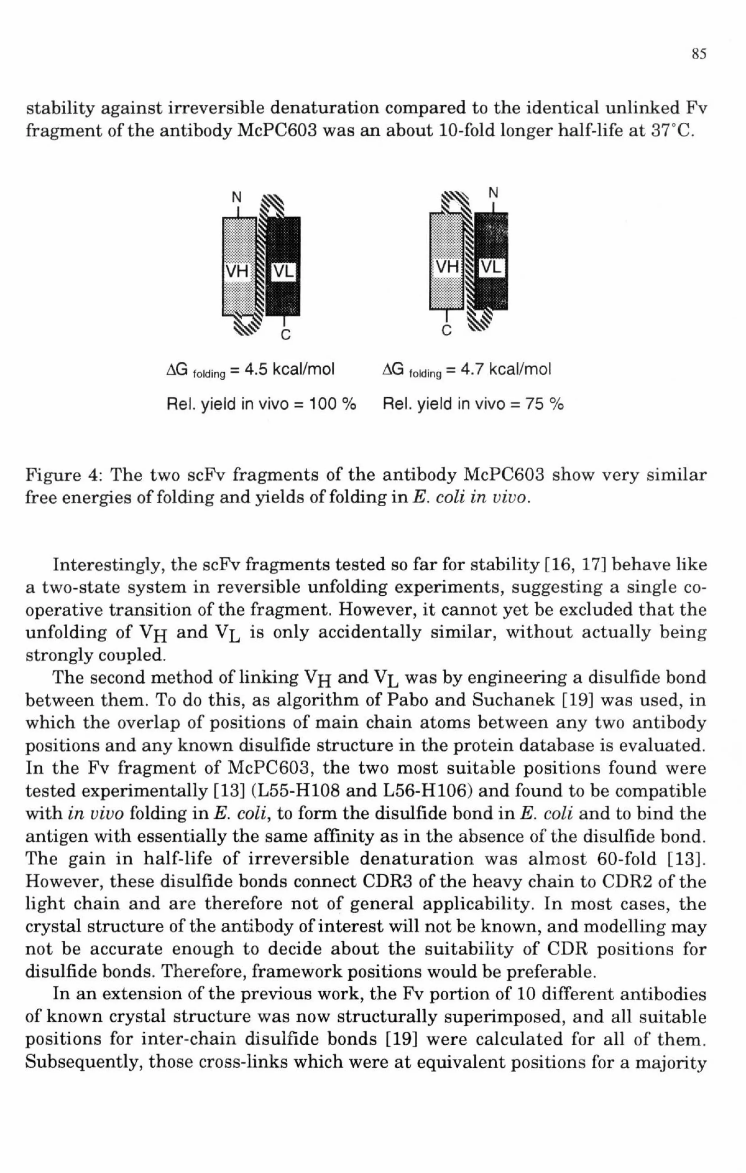
85
stability against irreversible denaturation compared to the identical unlinked Fv fragment of the antibody McPC603 was an about 10-fold longer half-life at 37°C.
N N
VH
c c
~G folding = 4.5 kcal/mol ~G folding = 4. 7 kcal/mol
Rei. yield in vivo = 100 °/o Rei. yield in vivo = 75 o/o
Figure 4: The two scFv fragments of the antibody McPC603 show very similar free energies of folding and yields of folding in E. coli in vivo.
Interestingly, the scFv fragments tested so far for stability [16, 17] behave like a two-state system in reversible unfolding experiments, suggesting a single cooperative transition of the fragment. However, it cannot yet be excluded that the unfolding of VH and VL is only accidentally similar, without actually being strongly coupled.
The second method of linking VH and VL was by engineering a disulfide bond between them. To do this, as algorithm of Pabo and Suchanek [19] was used, in which the overlap of positions of main chain atoms between any two antibody positions and any known disulfide structure in the protein database is evaluated. In the Fv fragment of McPC603, the two most suitable positions found were tested experimentally [13] (L55-H108 and L56-H106) and found to be compatible with in vivo folding in E. coli, to form the disulfide bond in E. coli and to bind the antigen with essentially the same affinity as in the absence of the disulfide bond. The gain in half-life of irreversible denaturation was almost 60-fold [13]. However, these disulfide bonds connect CDR3 of the heavy chain to CDR2 of the light chain and are therefore not of general applicability. In most cases, the crystal structure of the antibody of interest will not be known, and modelling may not be accurate enough to decide about the suitability of CDR positions for disulfide bonds. Therefore, framework positions would be preferable.
In an extension of the previous work, the Fv portion of 10 different antibodies of known crystal structure was now structurally superimposed, and all suitable positions for inter-chain disulfide bonds [19] were calculated for all of them. Subsequently, those cross-links which were at equivalent positions for a majority

86
of fragments were noted. Two types of framework cross-links were found in the majority of fragments, and those should therefore have a reasonable chance of giving a stable fragment for antibodies with unknown 3D structure (Figure 5). However, experimental work on a sufficient number of antibodies is still necessary to validate this idea. The two types of cross-links found are structurally similar and related by the pseudo-two-fold molecular axis relating VH and VL. In both cases, framework region 2 is linked to framework region 4 [10] of the opposite chain.
Preventing dissociation into light and heavy chains is not necessary for Fab fragments, since the constant domains CHl and CL take over the function of holding together the variable domains. The constant domains may restrict the thermal movements of the variable domains and thus also slow down the irreversible denaturation. The differences in irreversible denaturation rates between various types of cross-links between the variable domains also suggests that it is not only a shift in equilibrium to the VH-VL associated state, which guards against irreversible denaturation. Instead, the type of cross-link seems to be essential. Least effective is the "loose" peptide linker of the scFv fragment [18], most effective are the engineered disulfides [13].
Figure 5 (following page): (A) Stereo view of 10 superimposed structures of antibody Fv fragments. These are the same structures as shown individually in Figure 6. The heavy chains are colored in light grey (right), the light chains in dark grey (left). The existing intra-domain disulfide bonds (Figure 2) are shown in black (Ca, C~, Sy) within each domain (left and right). TheCa positions of the newly found disulfide bonds are also shown in black in the center. (B) Stereo view of the same Fv fragments, rotated 180° about they-axis. Now the other positions of the newly found disulfide bonds are visible in the center. (C) Schematic structure (mono view) of the Fv fragment of the antibody McPC603, with the same positions labelled, which were found suitable for linking VH and VL in the framework. (D) Same fragment as in (C) rotated 180° about the y-axis. The similarity of the two possible positions, due to the molecular pseudo-two-fold axis, is apparent. In each case, framework region 2 is connected to framework region 4. (For nomenclature, see ref. [10]).

87
A ...... • ,-,:1. ' . ., '·. • I (/ :v ' ' I ' -· ' ' ... ~ "' ... .... : .•.
8 ~ : .......
•
'
c VH D
VH VL VL

88
3HFM
1FDL
2FB4
2FBJ
~.
·~ ~· ... ~ ' J , ..... "- '·
•
1F19
21GF
3FAB
I ' • '"< . ,. .
v. i ' .:\1\: • \~i i '-... ~ ,.-v··"'
\ •
·- .. , .. ' ~·· .. ' • I
.. ' "· ·'· '!' .. J,. ' /' ·. \ •. . . ' '·'•·:) . ~ ,I ·, .. ~,~·· •• · I ' . . ........ ...._. .
\ :..... ...... .. , ·' -' 2HFL ' ....
I~., ~ .
/· .. /.:\ ·. ' • I '. ' . "' ... ......, ... \ •• •• J ,·· ' ,.... , .... i I "~,,...&,'; i ;' ,. I • I / . . • .. ,.;.._ ... "
2MCP
4FAB
·J • " • .. • '·
Figure 6: Cavities found within 10 structures of Fv fragments, using a probe size 0
of 1.4 A. The VL domains are on the left (black lines), the VH domains are on the right (grey lines). The CDR regions [10] are emphasized by thick lines. Cavities,
0
calculated in full atom models, are drawn as crosses spaced 1 A apart [21] in Ccx tracings of the structures. The PDB access codes is shown below each structure. All are murine antibodies with K light chains, except 3F AB and 2FB4, which are of human origin and have A light chains.

89
4. UNFOLDING OF INDIVIDUAL DOMAINS
Another limitation in stability for antibody fragments may be the partial unfolding of individual domains. The analysis of differential scanning calorimetry profiles [20] suggests that individual regions of the antibody can unfold independently, but it remains to be investigated which domains form individual co-operative folding units.
0
An analysis of antibody structures for cavities accessible to probes of 1.4 A radius was carried out and a surprising number of such regions were found (Figure 6). None of them are conserved between antibodies, however, and it remains to be investigated, whether filling them will be a general method of domain stabilization. Most importantly, the individuality of these cavities would make designing any mutations around them almost impossible in the absence of a high resolution structure of an individual antibody.
Recently, a project was begun to investigate the free energy of folding of mutants of the individual VL domain of the antibody McPC603, for which a 1.97 A structure was obtained from the protein produced in E. coli [22]: While this system is very suitable for combining studies on stabilization with structural information, it will still be necessary to introduce the same mutations into other fragments containing the heavy chain in order to assess the effect on fragments with perhaps larger co-operative folding units than single domains, as well as to test the portability of such changes to other antibodies.
Recent advances in bacterial production of antibody fragments have made it possible to decrease the size of the fragment for antigen binding fragments to just the variable domains. Many side reactions in ELISAs or immunoaffinityapplications may thus be eliminated by reducing the protein surface to its bare essentials, and there is also great interest in the better tumor penetration [2, 3] of such small fragments. Antibody variable domains may, however, differ in their stability. Protein engineering will have to be used on less stable individual antibody fragments, to bring these fragments to their full potential in order to eliminate any limitations imposed by stability, and such experiments have now shown the first promising results.
5. ACKNOWLEDGEMENTS
The author would like to thank Dr. M. Peitsch, Dept. of Biochemistry, University of Lausanne, for the PROMOD software and to Dr. P. Kraulis, Dept. of Mol. Biol., University ofUppsala, for the MOLSCRIPT software.

90
6. REFERENCES
1 G. McLendon and E. Radany, J. Bioi. Chern. 253 (1978) 6335. 2 D. Colcher, R. Bird, M. Roselli, K. D. Hardman, S. Johnson, S. Pope, S. W.
Dodd, M. W. Pantoliano, D. E. Milenic and J. Schlom, J. Natl. Cancer Inst., 82 (1990) 1191.
3 T. Yokota, D. E. Milenic, M. Whitlow and J. Schlom, Cancer Res., 52 (1992) 3402.
4 T. A. Waldmann and W. Strober, Prog. Allergy, 13 (1969) 1. 5 R. Wetzel, Trends Biochem. Sci. 12 (1987) 478. 6 V. V. Mozhaev, I. V. Berezin and K. Martinek, Crit. Rev. Biochemistry 23
(1988) 235. 7 A. Pliickthun, Biotechnology, 9 (1991) 545. 8 A. Pliickthun, Immunolog. Reviews, 130 (1992) in press. 9 R. Glockshuber, T. Schmidt and·A. Pliickthun, Biochemistry, 31 (1992) 1270. 10 E. A. Kabat, T. T. Wu, H. M. Perry, K. S. Gottesman and C. Foeller,
Sequences of proteins of biological interest, National Institute of Health, Bethesda, MD, 1991.
11 A. Skerra and A. Pliickthun, Science, 240 (1988) 1038. 12 M. Better, C. P. Chang, R. R. Robinson and A. H. Horwitz, Science, 240
(1988) 1041. 13 R. Glockshuber, M. Malia, I. Pfitzinger and A. Ph1ckthun, Biochemistry, 29
(1990) 1362. 14 R. E. Bird, K. D. Hardman, J. W. Jacobson, S. Johnson, B. M. Kaufman, S.
Lee, T. Lee, S. H. Pope, G. S. Riordan and M. Whitlow, Science, 242 (1988) 423.
15 J. S. Huston, D. Levinson, M. Mudgett-Hunter, M. Tai, J. Novotny, M. N. Margolies, R. J. Ridge, R. E. Bruccoleri, E. Haber, R. Crea, and H. Oppermann, Proc. Natl. Acad. Sci. USA, 85 (1988) 5879.
16 A. Knappik, C. Krebber and A. Pliickthun, Biotechnology, (1993) in press. 17 M. W. Pantoliano, R. E. Bird, S. Johnson, E. D. Asel, S. W. Dodd, J. F. Wood
and K. D. Hardman, Biochemistry, 30 (1991) 10117. 18 C .. Freund, B. Guth, A. Ross, A. Pliickthun and T. Holak, FEBS Lett., (1993)
• In press. 19 C. 0. Pabo and E. G. Suchanek, Biochemistry, 25 (1986) 5987.
'
20 V. M. Tischenko, V. P. Zavyalov, G. A. Medgyesi, S. A. Potekhin and P. L. Privalov, Eur. J. Biochem., 126 (1982) 517.
21 M. Peitsch, manuscript in preparation 22 B. Steipe, A. Pliickthun and R. Huber, J. Mol. Biol., 225 (1992) 739.


