Embed Size (px)
Citation preview

MOLECULAR AND CELLULAR BIOLOGY, Sept. 2004, p. 7748–7757 Vol. 24, No. 170270-7306/04/$08.00�0 DOI: 10.1128/MCB.24.17.7748–7757.2004Copyright © 2004, American Society for Microbiology. All Rights Reserved.
Stabilization of the E3 Ubiquitin Ligase Nrdp1 by theDeubiquitinating Enzyme USP8
Xiuli Wu, Lily Yen, Lisa Irwin, Colleen Sweeney, and Kermit L. Carraway III*UC Davis Cancer Center, Sacramento, California
Received 15 February 2004/Returned for modification 30 March 2004/Accepted 7 June 2004
Nrdp1 is a RING finger-containing E3 ubiquitin ligase that physically interacts with and regulates steady-state cellular levels of the ErbB3 and ErbB4 receptor tyrosine kinases and has been implicated in the degra-dation of the inhibitor-of-apoptosis protein BRUCE. Here we demonstrate that the Nrdp1 protein undergoesefficient proteasome-dependent degradation and that mutations in its RING finger domain that disruptubiquitin ligase activity enhance stability. These observations suggest that Nrdp1 self-ubiquitination and sta-bility could play an important role in regulating the activity of this protein. Using affinity chromatography, weidentified the deubiquitinating enzyme USP8 (also called Ubpy) as a protein that physically interacts withNrdp1. Nrdp1 and USP8 could be coimmunoprecipitated, and in transfected cells USP8 specifically bound toNrdp1 but not cbl, a RING finger E3 ligase involved in ligand-stimulated epidermal growth factor receptordown-regulation. The USP8 rhodanese and catalytic domains mediated Nrdp1 binding. USP8 markedly en-hanced the stability of Nrdp1, and a point mutant that disrupts USP8 catalytic activity destabilized endogenousNrdp1. Our results indicate that Nrdp1 is a specific target for the USP8 deubiquitinating enzyme and areconsistent with a model where USP8 augments Nrdp1 activity by mediating its stabilization.
Ubiquitination plays central roles in regulating protein sta-bility and activity. The canonical function of ubiquitination is indirecting proteins for proteolytic degradation by proteasomes(23). Ubiquitination is also involved in plasma membrane pro-tein internalization and degradation by lysosomes (2) and inregulating the activities of specific proteins (5, 14, 24). Ubiq-uitin molecules are linked to target proteins through an isopep-tide bond between the carboxyl-terminal glycine of ubiquitinand ε-amino groups of lysine residues of the substrate. Ubiq-uitination requires the activities of three enzymes. E1 enzymesactivate ubiquitin and transfer it to E2 ubiquitin-conjugatingenzymes through thiolester linkages. E3 ubiquitin ligases bringsubstrates to the E2 by binding both the E2 enzyme and spe-cific target proteins. HECT domains and RING finger domainsof E3 ligases mediate their binding to E2-conjugating enzymes(45), while a variety of domains are responsible for substraterecognition.
Ubiquitin modifications take the form of monoubiquitin,where a single ubiquitin moiety is attached to a single targetprotein lysine; multiple monoubiquitination, where several tar-get protein lysines are modified with single ubiquitin moieties;or polyubiquitination, where a ubiquitin moiety attached to atarget protein is iteratively ubiquitinated through one of itsseven lysines (57). The different types of ubiquitin conjugatescontrol different cellular processes. Polyubiquitination throughubiquitin lysine residue K48 is generally thought to directproteasomal delivery and degradation of cytosolic proteins.Polyubiquitination through ubiquitin lysine residue K63 or K29regulates nondegradative processes, while multiple monoubiq-uitination may mediate internalization and trafficking of plas-ma membrane proteins.
Deubiquitinating enzymes can reverse protein ubiquitina-tion and are thought to function in processing ubiquitin pre-cursors, recycling ubiquitin, unclogging proteasomes of ubiqui-tinated proteins, and promoting protein stability. Most knowndeubiquitinating enzymes are cysteine proteases that fall intotwo categories (11, 13, 30, 58). Ubiquitin carboxy-terminalhydrolases (UCHs) comprise a subfamily of closely relatedproteins and contain a catalytic domain of �200 amino acidsthat includes blocks containing conserved cysteine and histi-dine residues. UCHs efficiently remove ubiquitin from pep-tides and adducts but act less efficiently on ubiquitinated pro-teins. These enzymes are thought to suppress the accumulationof nonproductive ubiquitin adducts in the cell and to recycleubiquitin for reuse. Ubiquitin-processing proteases (UBPs)contain a �400-amino-acid-residue catalytic domain that in-cludes blocks of cysteine and histidine residues similar to thoseof UCHs, but the highly divergent intervening sequences maycontribute to substrate specificity. In addition, sequencesunique to the amino or carboxyl side of the catalytic domainsof UBPs are likely to mediate specific protein-protein interac-tions to modulate localization and substrate specificity (34, 35).This class of deubiquitinating enzyme may have evolved tointeract with specific targets to mediate their stabilization. Al-though the human genome encodes at least 63 distinct UBPs,substrate specificities for only a very few mammalian UBPshave been described (8, 31, 33, 52, 53, 55, 58).
Recent studies point to a key role for ubiquitination in thedown-regulation and degradation of a variety of plasma mem-brane proteins (29), including growth factor receptor tyrosinekinases. Upon growth factor binding many receptor tyrosinekinases localize to clathrin-coated pits, become internalized,and are delivered to endosomes. Receptors are sorted in en-dosomes according to whether they are to be recycled to thecell surface or degraded in lysosomes. Ligand binding stimu-lates the multiple monoubiquitination of epidermal growth
* Corresponding author. Mailing address: UC Davis Cancer Center,Research Building III, Room 1400, 4645 2nd Ave., Sacramento, CA95817. Phone: (916) 734-3114. Fax: (916) 734-0190. E-mail: [email protected].
7748
on August 2, 2015 by guest
http://mcb.asm
.org/D
ownloaded from

factor (EGF) receptor and platelet-derived growth factor re-ceptor (20), and it has been demonstrated that monoubiquiti-nation is sufficient to drive EGF receptor internalization anddegradation (20, 38). Moreover, growth factor-stimulated mo-noubiquitination of endosomal sorting accessory proteins mayregulate their function as ubiquitin receptors (17, 21), under-scoring the central role of protein ubiquitination in receptortrafficking and degradation.
Multiple monoubiquitination of EGF receptor is mediated,at least in part, by the RING finger E3 ubiquitin ligase cbl (38).cbl is recruited to the receptor in an activation-dependentmanner by the binding of its tyrosine kinase binding domain tophosphorylated tyrosine 1045 of the EGF receptor (54). Pointmutation of Y1045 (32), or oxidant-induced receptor activa-tion that does not result in Y1045 phosphorylation (48), sup-presses EGF receptor down-regulation. Likewise, cbl mutantsthat are unable to mediate EGF receptor ubiquitination alsopromote receptor stability (32). Hence, escape of receptortyrosine kinases from cbl-mediated down-regulation has beensuggested to promote cellular growth properties associatedwith oncogenesis (44).
We recently identified a novel RING finger E3 ubiquitinligase that regulates steady-state levels of ErbB3 and ErbB4(15, 46), members of the same receptor tyrosine kinase familyas the EGF receptor. Since ErbB3 and ErbB4 are bindingreceptors for the neuregulin subfamily of EGF-like growthfactors, we have named this protein Nrdp1 for neuregulinreceptor degradation pathway protein 1. Our observations sug-gest that cellular levels of Nrdp1 may be critical in regulatingsteady-state levels of receptors (51) and are of particular rel-evance because the aberrant overexpression and activation ofErbB family receptor tyrosine kinases contribute to tumor ma-lignancy (25). More recently it has been reported that Nrdp1,also called FLRF (1), mediates the ubiquitination and degra-dation of BRUCE (47), a large membrane-associated inhibi-tor-of-apoptosis domain-containing protein (22) that inhibitscell death in cultured mammalian cells (10) and in Drosophilamelanogaster eye (56).
In the present study we demonstrate that autoubiquitinationdestabilizes the Nrdp1 protein. We employed an affinity chro-matography approach to identify Nrdp1-interacting proteinsthat might modulate its activity and found that the UBP de-ubiquitinating enzyme USP8 specifically interacts with and sta-bilizes the Nrdp1 protein.
MATERIALS AND METHODS
Antibodies. Mouse (M2; immunoglobulin G1 [IgG1]) and rabbit antibodies toFLAG epitope were purchased from Sigma. Antibody to V5 epitope (IgG2a) waspurchased from Invitrogen, mouse antiactin AC-15 was from Sigma, and rabbitanti-cbl was purchased from Santa Cruz Biotechnologies. Affinity-purified rabbitantibodies to Nrdp1 were described previously (15). Rabbit blotting antibodies toUSP8 were generated using a glutathione S-transferase (GST) fusion of mouseUSP8 amino acid residues 703 to 864. Horseradish peroxidase-conjugated goatanti-mouse IgG and goat anti-rabbit IgG were from Zymed and Chemicon,respectively, and protein A/G agarose was from Oncogene Research Products.
Nrdp1 and USP8 constructs. Construction of FLAG-tagged versions of wild-type human Nrdp1 and the carboxy terminus (clone 32) was described previously(15). The C34S/H36Q mutation of Nrdp1 was also described previously (46). ThecDNA encoding mouse USP8 was obtained by reverse transcriptase PCR withTurbo Pfu (Stratagene) from total C2C12 myotube RNA. The product wassubcloned into the pcDNA3.1� (Invitrogen) expression vector, and sequence wasverified by sequencing both strands. Truncation mutants were generated by PCR
and encompassed the following amino acid residues of the mouse sequence: T1,1 to 92; T2, 1 to 183; T3, 1 to 464; T4, 1 to 612; T5, 1 to 735; T6, 93 to 1080; T7,184 to 1080; T8, 465 to 1080; T9, 613 to 1080; T10, 736 to 1080; T11, 184 to 735;T12, 308 to 735; T13, 465 to 735. The C748A and C748S mutations were madeusing the QuickChange site-directed mutagenesis kit (Stratagene), and muta-tions were verified by sequencing. A plasmid containing the cDNA encodinghuman USP2 was obtained from the American Type Culture Collection, V5epitope tagged at the carboxy terminus, and subcloned into pcDNA3.1� with theuse of PCR.
Cell culture, transfections, and blotting. COS7, 293T, and C2C12 cell lineswere obtained from the American Type Culture Collection. COS7 and 293T cellswere maintained in Dulbecco modified Eagle medium (DMEM) containing 10%fetal calf serum. C2C12 cells were maintained in DMEM containing 15% fetalcalf serum and 0.5% chicken embryo extract. To make myotubes, cells at 60 to70% confluence were switched to DMEM containing 5% horse serum andfurther incubated for 4 days. Transfections were carried out using Fugene 6(Roche) according to the directions of the manufacturer, and cells were allowedto express protein for 48 h following transfection. In some experiments cells weretreated overnight with or without 2 �M MG132 (Calbiochem) prior to lysis. Forexperiments where lysates were blotted with anti-FLAG or anti-V5 antibodies,transfected cells in six-well dishes were lysed in 400 �l of sodium dodecylsulfate-polyacrylamide gel electrophoresis (SDS-PAGE) sample buffer. In someexperiments 293T cells were cotransfected with pBABEpuro plasmid and treatedfor 72 h prior to lysis with 2 �g of puromycin/ml to enrich for the transfectedpopulation. For immunoprecipitation experiments, cells in 100-mm-diameterdishes were lysed in 1 ml of coimmunoprecipitation buffer (12), and clearedlysates were immunoprecipitated with 2 �g of anti-FLAG antibody M2, anti-cbl,or anti-Nrdp1 for 3 h at 4°C with protein A/G Sepharose to capture immunecomplexes. Lysate or immunoprecipitated proteins were resolved by SDS-PAGEand transferred to nitrocellulose; blotted with a 1/1,000 dilution of anti-FLAGM2, anti-V5, anti-Nrdp1, or anti-USP8 or a 1/25,000 dilution of antiactin; anddetected with 1/10,000 dilutions of horseradish peroxidase-conjugated secondaryantibodies.
Isolation of Nrdp1 binding proteins. GST and GST-32 were expressed inDH5� bacteria and purified as described previously (15). Ten 100-mm-diameterdishes of C2C12 myotubes were lysed in a total of 6 ml of binding buffer (20 mMHEPES [pH 7.4]; 150 mM NaCl; 1% Triton X-100; 1 mM EDTA; 0.2 mMNa3VO4; 10 mM sodium pyrophosphate; 1 mM NaF; 5 mM �-glycerophosphate;0.2 mM phenylmethylsulfonyl fluoride; 4 �g each of pepstatin, leupeptin, andaprotinin/ml), and 15 mg of protein was used for batch-wise binding experiments.Fifty micrograms of GST or GST-32 immobilized on glutathione agarose beadswas incubated with binding buffer alone or with cell lysates in a 35-ml totalvolume in a 50-ml conical tube. Incubation was carried out at 4°C with rockingfor 1.5 h. The mix was poured into a 10-ml disposable column (Bio-Rad Labo-ratories), and beads were washed with 50 ml of binding buffer. Beads were elutedwith 200 �l of SDS-PAGE sample buffer, and eluted proteins were resolved bygradient SDS–6 to 10% PAGE. Proteins were stained with Coomassie blue, andbands were excised. The identity of eluted bands was determined by tandem massspectrometry of trypsin-digested proteins by the W. M. Keck Foundation massspectrometry protein identification facility at Yale University.
RESULTS
Nrdp1 protein instability is mediated by autoubiquitination.In characterizing the properties of the Nrdp1 E3 ubiquitinligase, we observed that the protein could not be detected byimmunoblotting when transiently expressed in numerous cul-tured cells of various types (15). For example, Fig. 1B showsthat protein expression was undetectable when COS7 cellswere transiently transfected with FLAG-tagged wild-typeNrdp1. However, Nrdp1 accumulated to very significant levelswhen cells were incubated overnight with 2 �M MG132, aproteasome inhibitor. Similar results were obtained when cellswere treated with lactacystin and proteasome inhibitor 1, twoother proteasome inhibitors, but not with known lysosomeinhibitors (data not shown). These observations indicate thatNrdp1 is very efficiently degraded in cells in a proteasome-dependent manner.
Our previous studies indicate that clone 32 (Fig. 1A), a form
VOL. 24, 2004 USP8 STABILIZATION OF Nrdp1 7749
on August 2, 2015 by guest
http://mcb.asm
.org/D
ownloaded from

of Nrdp1 lacking its RING finger and B-box domains, is in-trinsically stable and that this stability is only marginally in-creased with MG132 (15). However, a form of Nrdp1 consist-ing of the RING finger and the B box alone is intrinsicallyunstable, suggesting that RING finger-mediated ubiquitin li-gase activity may contribute to Nrdp1 instability. To determinewhether Nrdp1 self-ubiquitination could contribute to its in-stability, we examined the expression of a mutant incapable ofautoubiquitination. The C34S/H36Q mutant of Nrdp1 harborstwo point mutations in its RING finger domain (Fig. 1A) thatare predicted to disrupt its binding to E2 ubiquitin-conjugatingenzymes. It has been previously demonstrated that this mutantis incapable of ubiquitinating itself in vitro and incapable ofubiquitinating ErbB3 in vitro and in cells (46). When tran-siently expressed in COS7 cells, the C34S/H36Q mutant wasmarkedly more stable than wild-type Nrdp1, and its stabilitywas further enhanced if proteasome-mediated protein degra-dation was entirely blocked by MG132. These observationsindicate that Nrdp1 is intrinsically unstable in cells and suggestthat autoubiquitination significantly contributes to its instabil-ity. The greater extent of Nrdp1 mutant accumulation in thepresence than in the absence of proteasome inhibitor suggeststhat additional proteasome-dependent mechanisms may alsocontribute to Nrdp1 degradation in these cells.
Identification of USP8 as an Nrdp1 binding protein. Theintrinsic instability of Nrdp1 implies that there may exist cel-lular factors that enhance its stability. To identify proteins thatmight contribute to Nrdp1 activity, we carried out a screen forbinding proteins present in C2C12 myotubes. This cell line waschosen because we have detected Nrdp1 expression by North-ern blotting and by immunoblotting (unpublished observa-
tions). For our screen we employed a GST fusion of the Nrdp1clone 32 because it is much more soluble when expressed inbacteria than is the GST fusion of the full-length protein (un-published observations). GST and GST-32 immobilized onbeads were incubated with lysates from myotubes, and associ-ated proteins were eluted with SDS-PAGE sample buffer andvisualized by SDS-PAGE followed by Coomassie blue staining.
Figure 2 shows that a number of cellular proteins specificallyassociated with GST-32 but not GST, including bands withapparent molecular masses of 270, 185, 120, 80, and 75 kDa.Bands were excised, and the identities of some of the Nrdp1binding proteins were determined by tandem mass spectrom-etry. The identity of the �270-kDa band was determined to beBRUCE, consistent with a previous report that this protein isa binding substrate of Nrdp1 (47). The 120-kDa band, p120,was determined to be mouse USP8 or Ubpy, a member of theUBP subfamily of ubiquitin-specific proteases of unknownfunction. The predicted molecular mass of USP8 is 123 kDa.
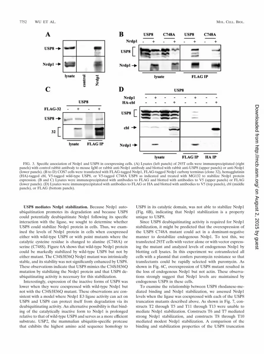
To determine whether USP8 and Nrdp1 form a functionalcomplex, we first determined whether the two proteins couldbe coimmunoprecipitated from lysates of a cell line expressingboth. While many cell lines express very low or undetectablelevels of endogenous Nrdp1, endogenous protein was detectedin 293T human embryonic kidney cells. These cells also expresssignificant levels of endogenous USP8. In the experiment de-picted in Fig. 3A we immunoprecipitated Nrdp1 from lysatesof 293T cells with an affinity-purified rabbit antibody describedpreviously (15) and blotted precipitates with that antibody orwith an antibody to USP8. Endogenous Nrdp1 (�38 kDa)could be detected in these precipitates, along with the 120-kDa
FIG. 1. Mutation of the RING finger domain of Nrdp1 enhances its stability. (A) Domain structure of full-length Nrdp1, C36S/H38Q mutant,and clone 32 used in these studies. (B) COS7 cells were transfected with vector, FLAG-tagged wild-type Nrdp1, or FLAG-tagged C34S/H36Qdouble mutant in the RING finger domain. Cells were then treated overnight without or with 2 �M MG132, as indicated, and lysates wereimmunoblotted with anti-FLAG or antiactin antibodies.
7750 WU ET AL. MOL. CELL. BIOL.
on August 2, 2015 by guest
http://mcb.asm
.org/D
ownloaded from

USP8, indicating that the two proteins exist in a complex incells.
To begin to analyze the functional consequence of Nrdp1-USP8 interaction, we obtained the cDNA encoding the full-length mouse USP8 by reverse transcriptase PCR from C2C12cell RNA and added a V5 epitope tag to its carboxy terminus.When transiently expressed in COS7 cells, V5-tagged USP8yielded the full-length 120-kDa band as well as several smallerfragments (Fig. 3B). This construct could be coimmunoprecipi-tated with FLAG-tagged wild-type Nrdp1 (Fig. 3B) or FLAG-tagged 32 (Fig. 3C) when coexpressed in COS7 cells treatedwith MG132. A catalytically inactive point mutant of USP8(see below) was also capable of interacting with Nrdp1 (Fig.3B). However, USP8 could not be coimmunoprecipitated witheither endogenous or overexpressed cbl (Fig. 3D). Hence,USP8 specifically interacts with the Nrdp1 E3 ubiquitin ligase.
Mapping of Nrdp1 binding sites in USP8. To further char-acterize the interaction of USP8 and Nrdp1, we constructed aseries of 12 mutants with mutations in USP8 that deleted itsvarious domains (Fig. 4A). The USP8 protein consists of twocoiled-coil domains, a rhodanese domain, a putative WW do-main, and a carboxy-terminal catalytic domain responsible fordeubiquitinating enzyme activity. A cysteine residue, C748, hasbeen previously demonstrated to be required for USP8 enzymeactivity (40). Using the coimmunoprecipitation assay, we ana-lyzed the binding of the various V5-tagged USP8 constructs to
full-length FLAG-tagged Nrdp1. We observed that constructsT3 through T11 coimmunoprecipitated with FLAG-Nrdp1 butconstructs T12 and T13 did not (Fig. 4B and C). No coimmu-noprecipitation of USP8 was observed with construct T2; how-ever, we were unable to confirm the presence of this constructin immunoprecipitates (data not shown). Deletion constructs(as shown for T6 and T9 in Fig. 4) did not precipitate withFLAG antibodies unless Nrdp1 was cotransfected. These re-sults map the regions of USP8 responsible for Nrdp1 interac-tion to two domains, the catalytic domain and the rhodanesedomain.
While previous studies have suggested a role for variableregions within the catalytic domain in determining UBP pro-tein target recognition, unique sequences and domains flank-ing the catalytic domains are also proposed to mediate specificprotein-protein interactions (58). Our observations suggestthat the rhodanese domain may play a role in the specificbinding of USP8 to Nrdp1. To determine whether this domainis sufficient to specifically bind Nrdp1, we created a GST fusionof the rhodanese domain (GST-rho; Fig. 5A) and used thisprotein to examine rhodanese binding to Nrdp1 and cbl. Asillustrated in Fig. 5B, the GST-rho specifically bound toFLAG-tagged Nrdp1 and FLAG-tagged clone 32 but was notable to bind to endogenous cbl. These observations suggestthat the rhodanese domain of USP8 is sufficient to mediate itsspecific interaction with Nrdp1.
FIG. 2. Identification of Nrdp1 binding proteins by affinity chromatography. Buffer alone or lysates from C2C12 myotubes were incubated withGST or GST-32 bound to glutathione beads. Beads were washed extensively, and bound proteins were eluted with SDS-PAGE sample buffer.Eluted proteins were resolved by gradient SDS–6 to 10% PAGE and visualized by staining with Coomassie blue. Numbers at left are molecularmasses in kilodaltons.
VOL. 24, 2004 USP8 STABILIZATION OF Nrdp1 7751
on August 2, 2015 by guest
http://mcb.asm
.org/D
ownloaded from
USP8 mediates Nrdp1 stabilization. Because Nrdp1 auto-ubiquitination promotes its degradation and because USP8could potentially deubiquitinate Nrdp1 following its specificinteraction with the ligase, we sought to determine whetherUSP8 could stabilize Nrdp1 protein in cells. Thus, we exam-ined the levels of Nrdp1 protein in cells when coexpressedeither with wild-type USP8 or with point mutants where thecatalytic cysteine residue is changed to alanine (C748A) orserine (C748S). Figure 6A shows that wild-type Nrdp1 proteincould be markedly stabilized by wild-type USP8 but not byeither mutant. The C34S/H36Q Nrdp1 mutant was intrinsicallystable, and its stability was not significantly enhanced by USP8.These observations indicate that USP8 mimics the C34S/H36Qmutation by stabilizing the Nrdp1 protein and that USP8 de-ubiquitinating activity is necessary for this stabilization.
Interestingly, expression of the inactive forms of USP8 waslower when they were coexpressed with wild-type Nrdp1 butnot with the C34S/H36Q mutant. These observations are con-sistent with a model where Nrdp1 E3 ligase activity can act onUSP8 and USP8 can protect itself from degradation via itsdeubiquitinating activity. An alternative possibility is that bind-ing of the catalytically inactive form to Nrdp1 is prolongedrelative to that of wild-type USP8 and serves as a more efficientsubstrate. USP2, the mammalian ubiquitin-specific proteasethat exhibits the highest amino acid sequence homology to
USP8 in its catalytic domain, was not able to stabilize Nrdp1(Fig. 6B), indicating that Nrdp1 stabilization is a propertyunique to USP8.
Since USP8 deubiquitinating activity is required for Nrdp1stabilization, it might be predicted that the overexpression ofthe USP8 C748A mutant could act in a dominant-negativemanner to destabilize endogenous Nrdp1. To test this, wetransfected 293T cells with vector alone or with vector express-ing the mutant and analyzed levels of endogenous Nrdp1 byblotting cell lysates. In this experiment we cotransfected allcells with a plasmid that confers puromycin resistance so thattransfectants could be rapidly selected with puromycin. Asshown in Fig. 6C, overexpression of USP8 mutant resulted inthe loss of endogenous Nrdp1 but not actin. These observa-tions strongly suggest that Nrdp1 levels are maintained byendogenous USP8 in these cells.
To examine the relationship between USP8 rhodanese-me-diated binding and Nrdp1 stabilization, we assessed Nrdp1levels when the ligase was coexpressed with each of the USP8truncation mutants described above. As shown in Fig. 7, con-structs T2 through T5 and T11 through T13 were unable tomediate Nrdp1 stabilization. Constructs T6 and T7 mediatedstrong Nrdp1 stabilization, and constructs T8 through T10mediated modest Nrdp1 stabilization. A comparison of thebinding and stabilization properties of the USP8 truncation
FIG. 3. Specific association of Nrdp1 and USP8 in coexpressing cells. (A) Lysates (left panels) of 293T cells were immunoprecipitated (rightpanels) with control rabbit antibody to mouse IgM or rabbit anti-Nrdp1 antibody and blotted with rabbit anti-USP8 (upper panels) or anti-Nrdp1(lower panels). (B to D) COS7 cells were transfected with FLAG-tagged Nrdp1, FLAG-tagged Nrdp1 carboxy terminus (clone 32), hemagglutinin(HA)-tagged cbl, V5-tagged wild-type USP8, or V5-tagged C748A USP8 as indicated and treated with MG132 to stabilize Nrdp1 proteinexpression. (B and C) Lysates were immunoprecipitated with antibodies to FLAG and blotted with antibodies to V5 (upper panels) or FLAG(lower panels). (D) Lysates were immunoprecipitated with antibodies to FLAG or HA and blotted with antibodies to V5 (top panels), cbl (middlepanels), or FLAG (bottom panels).
7752 WU ET AL. MOL. CELL. BIOL.
on August 2, 2015 by guest
http://mcb.asm
.org/D
ownloaded from

FIG. 4. USP8 binding to Nrdp1 is mediated by the catalytic and rhodanese domains. (A) Domain structure of USP8 mutants. wt, wild type. (Band C) Coimmunoprecipitation of USP8 with Nrdp1. COS7 cells were transfected with wild-type Nrdp1 (FLAG-tagged) and USP8 (V5-tagged)truncation mutants as indicated. Lysates from MG132-treated cells were immunoprecipitated with antibodies to FLAG, and lysates and precip-itates were blotted with antibodies to V5.
VOL. 24, 2004 USP8 STABILIZATION OF Nrdp1 7753
on August 2, 2015 by guest
http://mcb.asm
.org/D
ownloaded from

mutants is depicted in Table 1. These observations indicatethat, while the USP8 catalytic domain itself is moderately ca-pable of mediating Nrdp1 stabilization, full stability requiresNrdp1 association through the rhodanese domain.
DISCUSSION
Previous studies have implicated Nrdp1 as a E3 ubiquitinligase involved in the ligand-independent maintenance ofsteady-state levels of the neuregulin receptor tyrosine kinasesErbB3 and ErbB4 (15, 46) and in the maintenance of cellularlevels of the antiapoptotic protein BRUCE (47). Here wedemonstrate that the Nrdp1 protein is highly labile and that
point mutations in Nrdp1 that eliminate its ubiquitin ligaseactivity also significantly stabilize the protein. This is consistentwith previous observations that autoubiquitination of E3 li-gases, for example, Mdm2 (59), influences their intrinsic sta-bility in cells. Moreover, we demonstrate that the multidomainUBP deubiquitinating enzyme USP8 specifically interacts withand stabilizes Nrdp1. The simplest explanation for these ob-servations is as follows. At low levels of USP8 binding oractivity, constitutive Nrdp1 autoubiquitination efficiently tar-gets the protein for degradation by proteasomes, resulting inlow levels of the Nrdp1 protein. High levels of functional USP8mediate Nrdp1 deubiquitination and stabilization.
FIG. 5. USP8 rhodanese domain is sufficient to bind Nrdp1. (A) Coomassie blue stain of purified GST and GST-rho. Numbers at left aremolecular masses in kilodaltons. (B) USP8 rhodanese domain binds to the carboxy-terminal region of Nrdp1. COS7 cells were transfected withFLAG-tagged wild-type Nrdp1 or clone 32 and treated with MG132 to stabilize Nrdp1 expression, and lysates were incubated with 5 �g of GSTor GST-rho immobilized on Sepharose beads, as indicated. Proteins bound to washed beads were eluted, and lysates and eluates were blotted withanti-FLAG or anti-cbl.
FIG. 6. Stabilization of Nrdp1 by USP8 requires USP8 deubiquitinating activity. (A and B) COS7 cells were transfected with wild-type orC34S/H36Q FLAG-Nrdp1 and cotransfected with the indicated V5-tagged USP constructs. Lysates were immunoblotted with antibodies to FLAG,V5, and actin. (A) USP8 deubiquitinating activity is required for Nrdp1 stabilization. (B) USP2 is incapable of stabilizing Nrdp1. (C) Catalyticallyinactive USP8 destabilizes endogenous Nrdp1. 293T cells were transfected with vector or USP8CA, and lysates were blotted with antibodies toNrdp1, USP8, or actin.
7754 WU ET AL. MOL. CELL. BIOL.
on August 2, 2015 by guest
http://mcb.asm
.org/D
ownloaded from

Interestingly, Nrdp1 may also ubiquitinate USP8 to mediateits destruction, an activity unmasked with the USP8 catalyticcysteine point mutants. Hence, Nrdp1 may be capable of me-diating the destruction of itself and USP8 through its ubiquitinligase activity, and USP8 could be capable of preserving itselfand Nrdp1 through its deubiquitinating activity. When coupledwith the coimmunoprecipitation data and our unpublished ob-servations that Nrdp1 mediates the subcellular relocalizationof USP8, these results underscore the notion that the func-tional form of the two proteins is a complex.
While our model emphasizes the role of USP8 in determin-ing Nrdp1 levels, the mechanism by which the formation oractivity of the Nrdp1/USP8 complex is regulated in cells re-mains an important question. One mode of regulation of thecomplex could be at the level of USP8 expression, throughtranscriptional regulation or through regulation of its half-life
or localization. Alternatively, the binding of USP8 to Nrdp1may be a regulated step that is influenced by posttranslationalevents such as phosphorylation, ubiquitination, or binding toother cellular factors. Finally, it is possible that the catalyticactivity of USP8 is regulated.
The catalytic and rhodanese domains of USP8 mediate itsinteraction with Nrdp1. As catalytic domains in UBPs exhibitmarked divergence that may confer substrate specificity (58),this domain of USP8 probably contributes to Nrdp1 recogni-tion as a target for deubiquitination. The USP8 rhodanesedomain is sufficient to specifically recognize Nrdp1 and may beresponsible for maintaining a stable complex between the twoproteins. Rhodanese domains are ubiquitous structural mod-ules conserved from bacteria to humans whose functions atpresent are unclear. A subset of rhodanese domains possessessulfurtransferase activity, while noncatalytic versions are foundin proteins of various functions including eukaryotic de-ubiquitinating enzymes and mitogen-activated protein (MAP)kinase phosphatases (7). Our observations suggest that non-catalytic rhodanese domains mediate specific protein-proteininteractions. This conclusion is consistent with previous obser-vations indicating that the amino-terminal region of the MAPkinase phosphatase MKP-3 encompassing its rhodanese do-main is responsible for binding to MAP kinases Erk1 and Erk2(39). Fine mapping of the rhodanese binding site on Nrdp1could shed light on sequence motifs selected by rhodanesedomains.
Although the large number and divergence of mammalianUBPs strongly suggest that these enzymes have specific sub-strates and distinct biological activities, substrates for only afew have been described (58). HAUSP (USP7) mediates thedeubiquitination and stabilization of p53 (33), reversing theaction of the Mdm2 E3 ubiquitin ligase and enhancing p53tumor suppressor function. CYLD is a deubiquitinating en-
FIG. 7. Nrdp1 stabilization by USP8 is facilitated by rhodanese domain binding. COS7 cells were transfected with FLAG-tagged wild-typeNrdp1 and cotransfected with the indicated V5-tagged USP8 constructs. Lysates were blotted with antibodies to FLAG, V5, and actin. (A) Effectof truncation constructs CA and T2 through T10 on Nrdp1 stability. (B) Effect of constructs T11 through T13. Numbers at right of each panel aremolecular masses in kilodaltons.
TABLE 1. Summary of Nrdp1 binding and stabilizationby USP8 mutantsa
Construct Nrdp1 binding Nrdp1 stabilization
WT � �CA � �T2 ? �T3 � �T4 � �T5 � �T6 � �T7 � �T8 � �/�T9 � �/�T10 � �/�T11 � �T12 � �T13 � �
a WT, wild type; �, strong binding or stabilization; �, no binding or stabili-zation; ?, could not be determined; �/�, weak stabilization.
VOL. 24, 2004 USP8 STABILIZATION OF Nrdp1 7755
on August 2, 2015 by guest
http://mcb.asm
.org/D
ownloaded from

zyme that is mutated in familial cylindromatosis syndrome,where patients are predisposed to tumors of skin appendages(6). CYLD interacts with the Nemo regulatory component ofthe I� kinase complex and negatively regulates activation ofthe transcription factor NF-�B in response to specific tumornecrosis factor receptors by suppressing the K63 polyubiquiti-nation and activation of TRAF2 and TRAF6 (8, 31, 55).
fam (USP9), the mouse homolog of the D. melanogaster fatfacets gene, colocalizes and interacts with AF-6 (52) and�-catenin (53) at sites of cell-cell contact in cultured epithelialcells. Together with data indicating that AF-6 and �-cateninprotein levels are suppressed by fam loss in developing em-bryos (41), these observations suggest that fam-mediated de-ubiquitination may contribute to the stability of a subset ofproteins at cell adhesion sites. Analogous to its function inDrosophila (26), fam also mediates the deubiquitination ofepsin 1 (9), a protein involved in membrane trafficking. Hence,it appears that a single UBP is capable of mediating the de-ubiquitination of distinct sets of proteins.
The interaction of deubiquitinating enzymes with E3ubiquitin ligases has been reported previously, but the func-tional consequences are unclear. The deubiquitinating enzymeUnpEL was isolated in a screen for proteins that interact withSSA/Ro (16), a putative E3 ubiquitin ligase of unknown func-tion with an overall domain structure similar to that of Nrdp1.Analogous to Nrdp1, SSA/Ro coexpression in cells mediatesthe redistribution of UnpEL; however, this study made noattempt to assess UnpEL impact on SSA/Ro stabilization. As-sociation of the deubiquitinating enzyme BAP1 with the RINGfinger domain of the E3 ubiquitin ligase BRCA1 has beendemonstrated to enhance the tumor suppressor activity of theBRCA1/BARD1 complex in cells (27). Although autopolyu-biquitination enhances the E3 ligase activity of the complex invitro, BAP1 may not function in the deubiquitination of thecomplex (37) but target other substrates.
Previous studies have implicated a role for USP8 in cellulargrowth regulation. USP8 message accumulates upon growthstimulation of serum-starved human fibroblasts, and its levelsdecrease in response to growth arrest induced by cell-cell con-tact. Moreover, antisense oligonucleotides prevented fibro-blasts from entering S phase, suggesting that USP8 expressionis necessary for normal cell cycling (40). Similar to our obser-vations, USP8 has been shown to interact with and stabilize theRING finger E3 ubiquitin ligase GRAIL to mediate T-cellanergy (49). GRAIL is localized to the transferring recyclingendocytic pathway (4), suggesting a function for USP8 in mem-brane protein trafficking. Mouse USP8 has been previouslydemonstrated to interact with the brain-specific ras guaninenucleotide exchange factor cdc25mm and coexpression ofUSP8-mediated cdc25mm deubiquitination and stabilization(19). Interestingly, USP8 has been also been demonstrated tointeract with the Hrs binding protein Hbp/STAM2A (28), amember of the EAST/STAM/hbp family of adapter proteins.Hbp/STAM2A plays roles in receptor endo- and exocytosisand probably also in the regulation of actin cytoskeleton (36).Hence, while the overall function of USP8 is unclear, severallines of evidence point to its involvement in disparate cellularpathways that contribute to cellular growth regulation.
Finally, the overall domain structure of USP8 is similar tothat of a rhodanese domain-containing subfamily of Saccharo-
myces cerevisiae deubiquitinating enzymes that includes yeastUbp4/Doa4, Ubp5, and Ubp7. Of these only Ubp4/Doa4 hasbeen characterized in any detail. Ubp4/Doa4 associates withproteasomes, is required for efficient proteasome activity, andpromotes proteolysis through the removal of ubiquitin fromproteolytic intermediates upon substrate breakdown (42, 43).Hence, Ubp4/Doa4 may play a role in ubiquitin homeostasisby recycling ubiquitin for reuse (50). Evidence has also ac-cumulated that Ubp4/Doa4 plays a necessary role in the de-ubiquitination of plasma membrane proteins prior to theirdegradation in yeast vacuoles (3, 18). Taken together, theseobservations indicate that Ubp4/Doa4 plays a central role incytosolic and membrane protein degradation by mediating thedeubiquitination of target proteins prior to full degradation. Itshould be noted, however, that yeast Ubp5 cannot substitutefor Ubp4/Doa4, suggesting that the rhodanese domain-con-taining deubiquitinating enzymes in yeast harbor distinct func-tions. Therefore, USP8, the only rhodanese domain-containingdeubiquitinating enzyme encoded by the human genome, maybe functionally distinct from Ubp4/Doa4.
ACKNOWLEDGMENTS
This research was supported by NIH grant GM068994 (K.L.C.) andby California Breast Cancer Research Program grant 7KB-0085 (C.S.).
We thank Hamid Band for providing the HA-cbl plasmid, PaolaMarignani for providing pBABEpuro plasmid, and Ronald Wisdomfor critical review of the manuscript.
REFERENCES
1. Abdullah, J. M., X. Li, R. G. Nachtman, and R. Jurecic. 2001. FLRF, a novelevolutionarily conserved RING finger gene, is differentially expressed inmouse fetal and adult hematopoietic stem cells and progenitors. Blood CellsMol. Dis. 27:320–333.
2. Aguilar, R. C., and B. Wendland. 2003. Ubiquitin: not just for proteasomesanymore. Curr. Opin. Cell Biol. 15:184–190.
3. Amerik, A. Y., J. Nowak, S. Swaminathan, and M. Hochstrasser. 2000. TheDoa4 deubiquitinating enzyme is functionally linked to the vacuolar protein-sorting and endocytic pathways. Mol. Biol. Cell 11:3365–3380.
4. Anandasabapathy, N., G. S. Ford, D. Bloom, C. Holness, V. Paragas, C.Seroogy, H. Skrenta, M. Hollenhorst, C. G. Fathman, and L. Soares. 2003.GRAIL: an E3 ubiquitin ligase that inhibits cytokine gene transcription isexpressed in anergic CD4� T cells. Immunity 18:535–547.
5. Bach, I., and H. P. Ostendorff. 2003. Orchestrating nuclear functions: ubiq-uitin sets the rhythm. Trends Biochem. Sci. 28:189–195.
6. Bignell, G. R., W. Warren, S. Seal, M. Takahashi, E. Rapley, R. Barfoot, H.Green, C. Brown, P. J. Biggs, S. R. Lakhani, C. Jones, J. Hansen, E. Blair,B. Hofmann, R. Siebert, G. Turner, D. G. Evans, C. Schrander-Stumpel,F. A. Beemer, A. van Den Ouweland, D. Halley, B. Delpech, M. G. Cleveland,I. Leigh, J. Leisti, and S. Rasmussen. 2000. Identification of the familialcylindromatosis tumour-suppressor gene. Nat. Genet. 25:160–165.
7. Bordo, D., and P. Bork. 2002. The rhodanese/Cdc25 phosphatase superfam-ily. Sequence-structure-function relations. EMBO Rep. 3:741–746.
8. Brummelkamp, T. R., S. M. Nijman, A. M. Dirac, and R. Bernards. 2003.Loss of the cylindromatosis tumour suppressor inhibits apoptosis by activat-ing NF-�B. Nature 424:797–801.
9. Chen, H., S. Polo, P. P. Di Fiore, and P. V. De Camilli. 2003. Rapid Ca2�-dependent decrease of protein ubiquitination at synapses. Proc. Natl. Acad.Sci. USA 100:14908–14913.
10. Chen, Z., M. Naito, S. Hori, T. Mashima, T. Yamori, and T. Tsuruo. 1999.A human IAP-family gene, apollon, expressed in human brain cancer cells.Biochem. Biophys. Res. Commun. 264:847–854.
11. Chung, C. H., and S. H. Baek. 1999. Deubiquitinating enzymes: their diver-sity and emerging roles. Biochem. Biophys. Res. Commun. 266:633–640.
12. Crovello, C. S., C. Lai, L. C. Cantley, and K. L. Carraway III. 1998. Differ-ential signaling by the epidermal growth factor-like growth factors neuregu-lin-1 and neuregulin-2. J. Biol. Chem. 273:26954–26961.
13. D’Andrea, A., and D. Pellman. 1998. Deubiquitinating enzymes: a new classof biological regulators. Crit. Rev. Biochem. Mol. Biol. 33:337–352.
14. Deng, L., C. Wang, E. Spencer, L. Yang, A. Braun, J. You, C. Slaughter, C.Pickart, and Z. J. Chen. 2000. Activation of the I�B kinase complex byTRAF6 requires a dimeric ubiquitin-conjugating enzyme complex and aunique polyubiquitin chain. Cell 103:351–361.
7756 WU ET AL. MOL. CELL. BIOL.
on August 2, 2015 by guest
http://mcb.asm
.org/D
ownloaded from

15. Diamonti, A. J., P. M. Guy, C. Ivanof, K. Wong, C. Sweeney, and K. L.Carraway III. 2002. An RBCC protein implicated in maintenance of steady-state neuregulin receptor levels. Proc. Natl. Acad. Sci. USA 99:2866–2871.
16. Di Donato, F., E. K. Chan, A. D. Askanase, M. Miranda-Carus, and J. P.Buyon. 2001. Interaction between 52 kDa SSA/Ro and deubiquitinatingenzyme UnpEL: a clue to function. Int. J. Biochem. Cell Biol. 33:924–934.
17. Di Fiore, P. P., S. Polo, and K. Hofmann. 2003. When ubiquitin meetsubiquitin receptors: a signalling connection. Nat. Rev. Mol. Cell Biol. 4:491–497.
18. Dupre, S., and R. Haguenauer-Tsapis. 2001. Deubiquitination step in theendocytic pathway of yeast plasma membrane proteins: crucial role of Doa4pubiquitin isopeptidase. Mol. Cell. Biol. 21:4482–4494.
19. Gnesutta, N., M. Ceriani, M. Innocenti, I. Mauri, R. Zippel, E. Sturani, B.Borgonovo, G. Berruti, and E. Martegani. 2001. Cloning and characteriza-tion of mouse UBPy, a deubiquitinating enzyme that interacts with the rasguanine nucleotide exchange factor CDC25(Mm)/Ras-GRF1. J. Biol. Chem.276:39448–39454.
20. Haglund, K., S. Sigismund, S. Polo, I. Szymkiewicz, P. P. Di Fiore, and I.Dikic. 2003. Multiple monoubiquitination of RTKs is sufficient for theirendocytosis and degradation. Nat. Cell Biol. 5:461–466.
21. Haglund, K., P. P. Di Fiore, and I. Dikic. 2003. Distinct monoubiquitinsignals in receptor endocytosis. Trends Biochem. Sci. 28:598–603.
22. Hauser, H. P., M. Bardroff, G. Pyrowolakis, and S. Jentsch. 1998. A giantubiquitin-conjugating enzyme related to IAP apoptosis inhibitors. J. CellBiol. 141:1415–1422.
23. Hershko, A., and A. Ciechanover. 1998. The ubiquitin system. Annu. Rev.Biochem. 67:425–479.
24. Hicke, L. 2001. Protein regulation by monoubiquitin. Nat. Rev. Mol. CellBiol. 2:195–201.
25. Holbro, T., G. Civenni, and N. E. Hynes. 2003. The ErbB receptors and theirrole in cancer progression. Exp. Cell Res. 284:99–110.
26. Huang, Y., R. T. Baker, and J. A. Fischer-Vize. 1995. Control of cell fate bya deubiquitinating enzyme encoded by the fat facets gene. Science 270:1828–1831.
27. Jensen, D. E., M. Proctor, S. T. Marquis, H. P. Gardner, S. I. Ha, L. A.Chodosh, A. M. Ishov, N. Tommerup, H. Vissing, Y. Sekido, J. Minna, A.Borodovsky, D. C. Schultz, K. D. Wilkinson, G. G. Maul, N. Barlev, S. L.Berger, G. C. Prendergast, and F. J. Rauscher III. 1998. BAP1: a novelubiquitin hydrolase which binds to the BRCA1 RING finger and enhancesBRCA1-mediated cell growth suppression. Oncogene 16:1097–1112.
28. Kato, M., K. Miyazawa, and N. Kitamura. 2000. A deubiquitinating enzymeUBPY interacts with the Src homology 3 domain of Hrs-binding protein viaa novel binding motif PX(V/I)(D/N)RXXKP. J. Biol. Chem. 275:37481–37487.
29. Katzmann, D. J., G. Odorizzi, and S. D. Emr. 2002. Receptor downregula-tion and multivesicular-body sorting. Nat. Rev. Mol. Cell Biol. 3:893–905.
30. Kim, J. H., K. C. Park, S. S. Chung, O. Bang, and C. H. Chung. 2003.Deubiquitinating enzymes as cellular regulators. J. Biochem (Tokyo) 134:9–18.
31. Kovalenko, A., C. Chable-Bessia, G. Cantarella, A. Israel, D. Wallach, andG. Courtois. 2003. The tumour suppressor CYLD negatively regulatesNF-�B signalling by deubiquitination. Nature 424:801–805.
32. Levkowitz, G., H. Waterman, S. A. Ettenberg, M. Katz, A. Y. Tsygankov, I.Alroy, S. Lavi, K. Iwai, Y. Reiss, A. Ciechanover, S. Lipkowitz, and Y.Yarden. 1999. Ubiquitin ligase activity and tyrosine phosphorylation underliesuppression of growth factor signaling by c-Cbl/Sli-1. Mol. Cell 4:1029–1040.
33. Li, M., D. Chen, A. Shiloh, J. Luo, A. Y. Nikolaev, J. Qin, and W. Gu. 2002.Deubiquitination of p53 by HAUSP is an important pathway for p53 stabi-lization. Nature 416:648–653.
34. Lin, H., A. Keriel, C. R. Morales, N. Bedard, Q. Zhao, P. Hingamp, S.Lefrancois, L. Combaret, and S. S. Wing. 2000. Divergent N-terminal se-quences target an inducible testis deubiquitinating enzyme to distinct sub-cellular structures. Mol. Cell. Biol. 20:6568–6578.
35. Lin, H., L. Yin, J. Reid, K. D. Wilkinson, and S. S. Wing. 2001. DivergentN-terminal sequences of a deubiquitinating enzyme modulate substrate spec-ificity. J. Biol. Chem. 276:20357–20363.
36. Lohi, O., and V. P. Lehto. 2001. STAM/EAST/Hbp adapter proteins—inte-grators of signalling pathways. FEBS Lett. 508:287–290.
37. Mallery, D. L., C. J. Vandenberg, and K. Hiom. 2002. Activation of the E3
ligase function of the BRCA1/BARD1 complex by polyubiquitin chains.EMBO J. 21:6755–6762.
38. Mosesson, Y., K. Shtiegman, M. Katz, Y. Zwang, G. Vereb, J. Szollosi, andY. Yarden. 2003. Endocytosis of receptor tyrosine kinases is driven by mo-noubiquitylation, not polyubiquitylation. J. Biol. Chem. 278:21323–21326.
39. Muda, M., A. Theodosiou, C. Gillieron, A. Smith, C. Chabert, M. Camps, U.Boschert, N. Rodrigues, K. Davies, A. Ashworth, and S. Arkinstall. 1998. Themitogen-activated protein kinase phosphatase-3 N-terminal noncatalytic re-gion is responsible for tight substrate binding and enzymatic specificity.J. Biol. Chem. 273:9323–9329.
40. Naviglio, S., C. Mattecucci, B. Matoskova, T. Nagase, N. Nomura, P. P. DiFiore, and G. F. Draetta. 1998. UBPY: a growth-regulated human ubiquitinisopeptidase. EMBO J. 17:3241–3250.
41. Pantaleon, M., M. Kanai-Azuma, J. S. Mattick, K. Kaibuchi, P. L. Kaye, andS. A. Wood. 2001. FAM deubiquitylating enzyme is essential for preimplan-tation mouse embryo development. Mech. Dev. 109:151–160.
42. Papa, F. R., and M. Hochstrasser. 1993. The yeast DOA4 gene encodes adeubiquitinating enzyme related to a product of the human tre-2 oncogene.Nature 366:313–319.
43. Papa, F. R., A. Y. Amerik, and M. Hochstrasser. 1999. Interaction of theDoa4 deubiquitinating enzyme with the yeast 26S proteasome. Mol. Biol.Cell 10:741–756.
44. Peschard, P., and M. Park. 2003. Escape from Cbl-mediated downregula-tion: a recurrent theme for oncogenic deregulation of receptor tyrosinekinases. Cancer Cell 3:519–523.
45. Pickart, C. M. 2001. Mechanisms underlying ubiquitination. Annu. Rev.Biochem. 70:503–533.
46. Qiu, X. B., and A. L. Goldberg. 2002. Nrdp1/FLRF is a ubiquitin ligasepromoting ubiquitination and degradation of the epidermal growth factorreceptor family member, ErbB3. Proc. Natl. Acad. Sci. USA 99:14843–14848.
47. Qiu, X. B., S. L. Markant, J. Yuan, and A. L. Goldberg. 2004. Nrdp1-mediated degradation of the gigantic IAP, BRUCE, is a novel pathway fortriggering apoptosis. EMBO J. 23:800–810.
48. Ravid, T., C. Sweeney, P. Gee, K. L. Carraway III, and T. Goldkorn. 2002.Epidermal growth factor receptor activation under oxidative stress fails topromote c-Cbl mediated down-regulation. J. Biol. Chem. 277:31214–31219.
49. Soares, L., C. Seroogy, H. Skrenta, N. Anandasabapathy, P. Lovelace, C. D.Chung, E. Engleman, and C. G. Fathman. 2004. Two isoforms of otubain 1regulate T cell anergy via GRAIL. Nat. Immunol. 5:45–54.
50. Swaminathan, S., A. Y. Amerik, and M. Hochstrasser. 1999. The Doa4deubiquitinating enzyme is required for ubiquitin homeostasis in yeast. Mol.Biol. Cell 10:2583–2594.
51. Sweeney, C., and K. L. Carraway III. 2004. Negative regulation of ErbBreceptor family tyrosine kinases. Br. J. Cancer 90:289–293.
52. Taya, S., T. Yamamoto, K. Kano, Y. Kawano, A. Iwamatsu, T. Tsuchiya, K.Tanaka, M. Kanai-Azuma, S. A. Wood, J. S. Mattick, and K. Kaibuchi. 1998.The Ras target AF-6 is a substrate of the fam deubiquitinating enzyme.J. Cell Biol. 142:1053–1062.
53. Taya, S., T. Yamamoto, M. Kanai-Azuma, S. A. Wood, and K. Kaibuchi.1999. The deubiquitinating enzyme Fam interacts with and stabilizes beta-catenin. Genes Cells 4:757–767.
54. Thien, C. B., and W. Y. Langdon. 2001. Cbl: many adaptations to regulateprotein tyrosine kinases. Nat. Rev. Mol. Cell Biol. 2:294–307.
55. Trompouki, E., E. Hatzivassiliou, T. Tsichritzis, H. Farmer, A. Ashworth,and G. Mosialos. 2003. CYLD is a deubiquitinating enzyme that negativelyregulates NF-�B activation by TNFR family members. Nature 424:793–796.
56. Vernooy, S. Y., V. Chow, J. Su, K. Verbrugghe, J. Yang, S. Cole, M. R. Olson,and B. A. Hay. 2002. Drosophila Bruce can potently suppress Rpr- andGrim-dependent but not Hid-dependent cell death. Curr. Biol. 12:1164–1168.
57. Weissman, A. M. 2001. Themes and variations on ubiquitylation. Nat. Rev.Mol. Cell Biol. 2:169–178.
58. Wing, S. S. 2003. Deubiquitinating enzymes—the importance of driving inreverse along the ubiquitin-proteasome pathway. Int. J. Biochem. Cell Biol.35:590–605.
59. Zhang, Y., and Y. Xiong. 2001. Control of p53 ubiquitination and nuclearexport by MDM2 and ARF. Cell Growth Differ. 12:175–186.
VOL. 24, 2004 USP8 STABILIZATION OF Nrdp1 7757
on August 2, 2015 by guest
http://mcb.asm
.org/D
ownloaded from


