Embed Size (px)
Citation preview

International Journal of
Environmental Research
and Public Health
Case Report
Staged Joint Arthrodesis in the Treatment of Severe SepticAnkle Arthritis Sequelae: A Case Report
Yong-Cheol Hong 1,† , Ki-Jin Jung 1,† , Hee-Jun Chang 1, Eui-Dong Yeo 2, Hong-Seop Lee 3, Sung-Hun Won 4,Jae-Young Ji 5, Dhong-Won Lee 6, Ik-Dong Yoo 7 , Sung-Joon Yoon 1 and Woo-Jong Kim 1,*
�����������������
Citation: Hong, Y.-C.; Jung, K.-J.;
Chang, H.-J.; Yeo, E.-D.; Lee, H.-S.;
Won, S.-H.; Ji, J.-Y.; Lee, D.-W.; Yoo,
I.-D.; Yoon, S.-J.; et al. Staged Joint
Arthrodesis in the Treatment of
Severe Septic Ankle Arthritis
Sequelae: A Case Report. Int. J.
Environ. Res. Public Health 2021, 18,
12473. https://doi.org/10.3390/
ijerph182312473
Academic Editor: Javier Abián-Vicén
Received: 21 October 2021
Accepted: 25 November 2021
Published: 26 November 2021
Publisher’s Note: MDPI stays neutral
with regard to jurisdictional claims in
published maps and institutional affil-
iations.
Copyright: © 2021 by the authors.
Licensee MDPI, Basel, Switzerland.
This article is an open access article
distributed under the terms and
conditions of the Creative Commons
Attribution (CC BY) license (https://
creativecommons.org/licenses/by/
4.0/).
1 Department of Orthopaedic Surgery, Soonchunhyang University Hospital Cheonan, Cheonan 31151, Korea;[email protected] (Y.-C.H.); [email protected] (K.-J.J.); [email protected] (H.-J.C.);[email protected] (S.-J.Y.)
2 Department of Orthopaedic Surgery, Veterans Health Service Medical Center, Seoul 05368, Korea;[email protected]
3 Department of Foot and Ankle Surgery, Nowon Eulji Medical Center, Eulji University, Seoul 01830, Korea;[email protected]
4 Department of Orthopaedic Surgery, Soonchunhyang University Hospital Seoul, Seoul 04401, Korea;[email protected]
5 Department of Anesthesiology and Pain Medicine, Soonchunhyang University Hospital Cheonan,Cheonan 31151, Korea; [email protected]
6 Department of Orthopaedic Surgery, Konkuk University Medical Center, Seoul 05030, Korea;[email protected]
7 Department of Nuclear Medicine, Soonchunhyang University Hospital Cheonan, Cheonan 31151, Korea;[email protected]
* Correspondence: [email protected]; Tel.: +82-41-570-2176† These authors contributed equally to this work and are co-first authors.
Abstract: Septic ankle arthritis is a devastating clinical entity with high risks of morbidity andmortality. Prompt treatment is necessary because delayed or inadequate treatment can lead toirreversible damage that may occur on the articular surface, resulting in cartilage erosion, infectivesynovitis, osteomyelitis, joint deformity, and pain and joint dysfunction. An aggressive surgicalapproach is required when a joint infection causes severe limb-threatening arthritis. A 58-year-old woman visited our clinic with increasing pain in the right ankle, which had been present forthe previous 2 months. She complained of discomfort in daily life due to deformity of the ankle;limping; and severe pain in the ankle even after walking a little. The patient reported a history ofright-ankle injury while exiting a bus in her early 20s. Plain radiographs of the right ankle jointrevealed that the medial malleolus was nearly absent in the right ankle joint on the anteroposteriorview, and severe varus deformity was observed with osteoarthritic changes because of joint spacedestruction. Magnetic resonance imaging revealed diffuse synovial thickening of the destroyedtibiotalar joint with joint effusion. Hybrid 99mTc white blood cell single-photon emission computedtomography/computed tomography showed increased uptake along the soft tissue around the anklejoint; uptake was generally low in the talocrural and subtalar joints. A two-stage operation wasperformed to remove the infected lesions and correct the deformity, thus enabling limb salvage. Thepatient was nearly asymptomatic at the 6-month follow-up, with no discomfort in her daily life andnearly normal ability to carry out full functional activities. She had no complications or recurrentsymptoms at the 1-year follow-up. We have described a rare case of a staged limb salvage procedurein a patient with chronic septic arthritis sequelae. For patients with severe joint deformity because ofseptic ankle sequelae, staged arthrodesis is a reliable method to remove infected lesions, solve softtissue problems, correct deformities, and maintain leg length.
Keywords: septic arthritis; iliac graft; internal fixation; external fixation
Int. J. Environ. Res. Public Health 2021, 18, 12473. https://doi.org/10.3390/ijerph182312473 https://www.mdpi.com/journal/ijerph

Int. J. Environ. Res. Public Health 2021, 18, 12473 2 of 9
1. Introduction
Septic ankle arthritis is generally rare. Among all patients with infectious arthritis,the prevalence of infectious ankle arthritis is limited (3.4–15%) [1–5]. Because this diseaseis a devastating clinical entity with high risks of morbidity and mortality, an accurateunderstanding of the disease, diagnosis, and prompt treatment is essential [6,7].
Infections can be introduced into the joint through hematogenous spread from otherinfection sites, by direct inoculation due to trauma, or by contiguous spread from adjacenttissues [6]. Risk factors in patients susceptible to septic arthritis include nicotine abuse,obesity, age > 80 years, diabetes, alcoholism, trauma, previous joint surgery, rheumatoidarthritis or osteoarthritis, other inflammatory arthropathies, chronic hypoxia, malignantdisease, cutaneous ulcer, hepatic or renal failure, or intravenous or intraarticular injectionhistory [6–9].
Streptococcus aureus is the most common organism causing infectious joints of theankle, and early empirical antibiotic treatment is recommended [7–11]. The diagnosis ofseptic arthritis relies on a modification of the criteria used by Newman [1], which requiresone of four factors to be met: (1) isolation of a pathogenic organism from an affected joint;(2) isolation of a pathogenic organism from another source (e.g., blood) in the context of awarm, red joint that is presumed to exhibit sepsis; (3) typical clinical features and a turbidjoint in the presence of previous antibiotic treatment; or (4) postmortem or pathologicalfeatures that are suggestive of septic arthritis.
Destruction of the joint due to septic arthritis results in functional instability andchanges in lower limb muscle activity patterns during unexpected disturbances, particu-larly in proximal muscles rather than distal muscles [12]. In addition, according to a studyby Casado-Hernández et al. [13], patients with anterior talofibular ligament (ATFL) injuriesshowed a greater presence of calcaneofibular ligament and tibiotalar joint injuries thansubjects with non-injured. These constitute the rationale that functional instability canlead to more severe joint destruction, which accelerates the patient’s limping and pain andintensifies discomfort.
The principles for treatment of septic ankle arthritis and osteomyelitis consist ofinfection control, stabilization, soft tissue coverage, and the management of skeletal defects.An open approach or arthroscopic irrigation and debridement should be performed asearly treatment, along with appropriate empirical antibiotics or antibiotics consistent withculture results. Serial aspiration is required if the surgery cannot be performed [3,4,8,11,14].Inadequate treatment can cause irreversible damage to the cartilage or bone in the joint;infective synovitis; or osteomyelitis, resulting in deformity [11,15,16], sequelae, and painand joint dysfunction. The infection may spread nearby and can cause sepsis [4,8,11].
The mortality caused by a septic joint is approximately 11%, and one-third of affectedpatients experience permanent joint damage [6,17]. However, the diagnosis is not alwaysclear, and imaging tests such as hybrid 99mTc white blood cell (WBC) single-photonemission computed tomography (SPECT)/CT are often performed as part of the diagnostictask [18]. An aggressive surgical approach is required when a joint infection causes severelimb-threatening arthritis. Arthrodesis of the septic arthritic ankle joint combined witheradication of all infected tissue is the final alternative to amputation [7,19–21]. Manyarthrodesis methods are available, but they are difficult for surgeons to perform whilemaintaining contralateral limb length.
Here, we describe a patient who underwent successful treatment of a secondary anklejoint deformity caused by septic ankle arthritis sequelae during staged surgery.
2. Case Presentation
This case report was approved by the Institutional Review Board of SoonchunhyangUniversity Hospital (IRB No. 2021-04-017). The patient provided written informed consentfor the publication of this report and the accompanying images.
A 58-year-old woman visited our clinic with increasing pain in the right ankle, whichhad been present for the previous 2 months. She complained of discomfort in daily life
Int. J. Environ. Res. Public Health 2021, 18, 12473 3 of 9
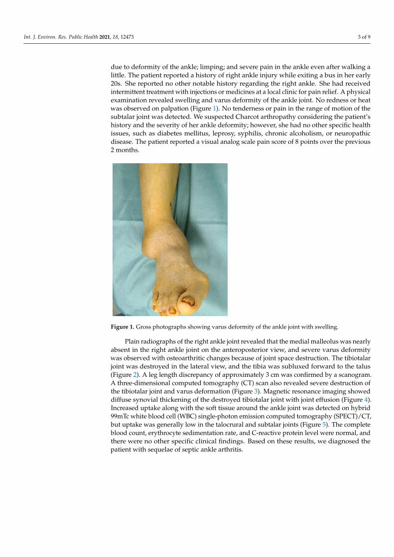
due to deformity of the ankle; limping; and severe pain in the ankle even after walking alittle. The patient reported a history of right ankle injury while exiting a bus in her early20s. She reported no other notable history regarding the right ankle. She had receivedintermittent treatment with injections or medicines at a local clinic for pain relief. A physicalexamination revealed swelling and varus deformity of the ankle joint. No redness or heatwas observed on palpation (Figure 1). No tenderness or pain in the range of motion of thesubtalar joint was detected. We suspected Charcot arthropathy considering the patient’shistory and the severity of her ankle deformity; however, she had no other specific healthissues, such as diabetes mellitus, leprosy, syphilis, chronic alcoholism, or neuropathicdisease. The patient reported a visual analog scale pain score of 8 points over the previous2 months.
Int. J. Environ. Res. Public Health 2021, 18, x FOR PEER REVIEW 3 of 9
A 58-year-old woman visited our clinic with increasing pain in the right ankle, which had been present for the previous 2 months. She complained of discomfort in daily life due to deformity of the ankle; limping; and severe pain in the ankle even after walking a little. The patient reported a history of right ankle injury while exiting a bus in her early 20s. She reported no other notable history regarding the right ankle. She had received intermittent treatment with injections or medicines at a local clinic for pain relief. A phys-ical examination revealed swelling and varus deformity of the ankle joint. No redness or heat was observed on palpation (Figure 1). No tenderness or pain in the range of motion of the subtalar joint was detected. We suspected Charcot arthropathy considering the pa-tient’s history and the severity of her ankle deformity; however, she had no other specific health issues, such as diabetes mellitus, leprosy, syphilis, chronic alcoholism, or neuro-pathic disease. The patient reported a visual analog scale pain score of 8 points over the previous 2 months.
Figure 1. Gross photographs showing varus deformity of the ankle joint with swelling.
Plain radiographs of the right ankle joint revealed that the medial malleolus was nearly absent in the right ankle joint on the anteroposterior view, and severe varus de-formity was observed with osteoarthritic changes because of joint space destruction. The tibiotalar joint was destroyed in the lateral view, and the tibia was subluxed forward to the talus (Figure 2). A leg length discrepancy of approximately 3 cm was confirmed by a scanogram. A three-dimensional computed tomography (CT) scan also revealed severe destruction of the tibiotalar joint and varus deformation (Figure 3). Magnetic resonance imaging showed diffuse synovial thickening of the destroyed tibiotalar joint with joint effusion (Figure 4). Increased uptake along with the soft tissue around the ankle joint was detected on hybrid 99mTc white blood cell (WBC) single-photon emission computed to-mography (SPECT)/CT, but uptake was generally low in the talocrural and subtalar joints (Figure 5). The complete blood count, erythrocyte sedimentation rate, and C-reactive pro-tein level were normal, and there were no other specific clinical findings. Based on these results, we diagnosed the patient with sequelae of septic ankle arthritis.
Figure 1. Gross photographs showing varus deformity of the ankle joint with swelling.
Plain radiographs of the right ankle joint revealed that the medial malleolus was nearlyabsent in the right ankle joint on the anteroposterior view, and severe varus deformitywas observed with osteoarthritic changes because of joint space destruction. The tibiotalarjoint was destroyed in the lateral view, and the tibia was subluxed forward to the talus(Figure 2). A leg length discrepancy of approximately 3 cm was confirmed by a scanogram.A three-dimensional computed tomography (CT) scan also revealed severe destruction ofthe tibiotalar joint and varus deformation (Figure 3). Magnetic resonance imaging showeddiffuse synovial thickening of the destroyed tibiotalar joint with joint effusion (Figure 4).Increased uptake along with the soft tissue around the ankle joint was detected on hybrid99mTc white blood cell (WBC) single-photon emission computed tomography (SPECT)/CT,but uptake was generally low in the talocrural and subtalar joints (Figure 5). The completeblood count, erythrocyte sedimentation rate, and C-reactive protein level were normal, andthere were no other specific clinical findings. Based on these results, we diagnosed thepatient with sequelae of septic ankle arthritis.

Int. J. Environ. Res. Public Health 2021, 18, 12473 4 of 9Int. J. Environ. Res. Public Health 2021, 18, x FOR PEER REVIEW 4 of 9
(a) (b) (c)
Figure 2. Plain anteroposterior (a), lateral (b), and heel alignment (c) radiographs showing an ankle with severe varus deformity, and osteoarthritic changes with destruction of the tibiotalar joint.
(a) (b) (c)
Figure 3. Preoperative coronal CT image showing a severe defect in the distal tibia and nearly undetectable medial malle-olus (a). Sagittal CT scan showing a destroyed tibiotalar joint and anterior tibia migration to the talus (b). The 3D recon-structed CT image shows severe varus deformity of the right ankle joint (c).
(a) (b)
Figure 4. Preoperative T2-weighted coronal (a) and sagittal (b) magnetic resonance images showing a septic ankle joint with diffuse synovial thickening, increased joint effusion with surrounding soft tissue swelling of the distal tibia, and talus osteomyelitis.
(a) (b) (c)
Figure 2. Plain anteroposterior (a), lateral (b), and heel alignment (c) radiographs showing an ankle with severe varusdeformity, and osteoarthritic changes with destruction of the tibiotalar joint.
Int. J. Environ. Res. Public Health 2021, 18, x FOR PEER REVIEW 4 of 9
(a) (b) (c)
Figure 2. Plain anteroposterior (a), lateral (b), and heel alignment (c) radiographs showing an ankle with severe varus deformity, and osteoarthritic changes with destruction of the tibiotalar joint.
(a) (b) (c)
Figure 3. Preoperative coronal CT image showing a severe defect in the distal tibia and nearly undetectable medial malle-olus (a). Sagittal CT scan showing a destroyed tibiotalar joint and anterior tibia migration to the talus (b). The 3D recon-structed CT image shows severe varus deformity of the right ankle joint (c).
(a) (b)
Figure 4. Preoperative T2-weighted coronal (a) and sagittal (b) magnetic resonance images showing a septic ankle joint with diffuse synovial thickening, increased joint effusion with surrounding soft tissue swelling of the distal tibia, and talus osteomyelitis.
(a) (b) (c)
Figure 3. Preoperative coronal CT image showing a severe defect in the distal tibia and nearly undetectable medial malleolus(a). Sagittal CT scan showing a destroyed tibiotalar joint and anterior tibia migration to the talus (b). The 3D reconstructedCT image shows severe varus deformity of the right ankle joint (c).
Int. J. Environ. Res. Public Health 2021, 18, x FOR PEER REVIEW 4 of 9
(a) (b) (c)
Figure 2. Plain anteroposterior (a), lateral (b), and heel alignment (c) radiographs showing an ankle with severe varus deformity, and osteoarthritic changes with destruction of the tibiotalar joint.
(a) (b) (c)
Figure 3. Preoperative coronal CT image showing a severe defect in the distal tibia and nearly undetectable medial malle-olus (a). Sagittal CT scan showing a destroyed tibiotalar joint and anterior tibia migration to the talus (b). The 3D recon-structed CT image shows severe varus deformity of the right ankle joint (c).
(a) (b)
Figure 4. Preoperative T2-weighted coronal (a) and sagittal (b) magnetic resonance images showing a septic ankle joint with diffuse synovial thickening, increased joint effusion with surrounding soft tissue swelling of the distal tibia, and talus osteomyelitis.
(a) (b) (c)
Figure 4. Preoperative T2-weighted coronal (a) and sagittal (b) magnetic resonance images showinga septic ankle joint with diffuse synovial thickening, increased joint effusion with surrounding softtissue swelling of the distal tibia, and talus osteomyelitis.

Int. J. Environ. Res. Public Health 2021, 18, 12473 5 of 9
Int. J. Environ. Res. Public Health 2021, 18, x FOR PEER REVIEW 4 of 9
(a) (b) (c)
Figure 2. Plain anteroposterior (a), lateral (b), and heel alignment (c) radiographs showing an ankle with severe varus deformity, and osteoarthritic changes with destruction of the tibiotalar joint.
(a) (b) (c)
Figure 3. Preoperative coronal CT image showing a severe defect in the distal tibia and nearly undetectable medial malle-olus (a). Sagittal CT scan showing a destroyed tibiotalar joint and anterior tibia migration to the talus (b). The 3D recon-structed CT image shows severe varus deformity of the right ankle joint (c).
(a) (b)
Figure 4. Preoperative T2-weighted coronal (a) and sagittal (b) magnetic resonance images showing a septic ankle joint with diffuse synovial thickening, increased joint effusion with surrounding soft tissue swelling of the distal tibia, and talus osteomyelitis.
(a) (b) (c)
Figure 5. Hybrid 99mTc WBC SPECT/CT images showing increased uptake along the soft tissue around the ankle joint, aswell as generally low uptake in the talocrural and subtalar joints. 99m Tc WBC SPECT (a), hybrid WBC SPECT/CT axialimage (b), and coronal image (c).
A two-stage operation was planned to remove the infected lesions and correct thedeformity, thus enabling limb salvage. The anterior midline approach was used for thearthrotomy and to correct the deformity. First, an open arthrotomy of the ankle jointwas performed with extensive debridement of the infected tissue. The medial malleolarpart of the distal tibia was almost destroyed, and the tibiotalar joint was deformed like aball and socket joint. Because the infection involved bone beyond the articular cartilage,minimal osteotomy of the distal tibia and talus was performed (Figure 6a); an antibiotic-impregnated polymethylmethacrylate spacer was then inserted to control the infectionand maintain leg length for subsequent deformity correction. Antibiotic-impregnatedcement (Simplex; Stryker, Rutherford, NJ, USA; 40 g per pack, erythromycin 0.5 g per pack)was used, and 2 g of vancomycin was mixed with 40 g of cement. The ankle joint wasstabilized with a delta-frame external fixator (Figure 6b,c). No bacteria were detected in aculture of infective tissues removed from the patient, and the acid-fast bacilli stain findingswere negative. The patient was hospitalized and administered intravenous cefazolin for6 weeks as antibiotic therapy. The second step of treatment comprised internal plate fixationtibiotalar arthrodesis. The original midline incision was utilized to access the surgical site,and the vancomycin spacer was removed. Frozen biopsies of surrounding tissues were sentto the Department of Pathology to determine whether the infection had cleared. Fewer than1–2 polymorphonuclear leukocytes per high power field were observed on intraoperativefrozen sections. Bony surfaces of the tibia and talus were prepared by curettage, drilling,and burring; these comprise standard methods for arthrodesis. To maintain leg lengthwhile providing structural support and stability, three cortical bone blocks were harvestedfrom the ipsilateral ileum and then used to construct auto and cancellous chip bone grafts.The iliac crest was reconstructed with bone cement on the donor side ileum because ofdonor site pain [14]. The foot was fixed in a neutral position using an anterior fusion plate(Arthrex Inc., Naples, FL, USA) (Figure 7).
The ankle joint was immobilized for 6 weeks postoperatively in a short-leg cast, withthe ankle and metacarpophalangeal joint in a neutral position. A split-thickness skingraft procedure was performed 3 weeks after arthrodesis because of the need for wounddehiscence at 2 weeks after arthrodesis. The cast was changed to a Walker boot brace at6 weeks postoperatively. Partial weight-bearing was allowed, and the range of foot motionwas allowed. Full weight-bearing began at 8 weeks, and the walking boot brace wasmaintained for 1 additional month. Complete fusion was confirmed on plain radiographs5 months after arthrodesis (Figure 8).

Int. J. Environ. Res. Public Health 2021, 18, 12473 6 of 9
Int. J. Environ. Res. Public Health 2021, 18, x FOR PEER REVIEW 5 of 9
Figure 5. Hybrid 99mTc WBC SPECT/CT images showing increased uptake along the soft tissue around the ankle joint, as well as generally low uptake in the talocrural and subtalar joints. 99m Tc WBC SPECT (a), hybrid WBC SPECT/CT axial image (b), and coronal image (c).
A two-stage operation was planned to remove the infected lesions and correct the deformity, thus enabling limb salvage. The anterior midline approach was used for the arthrotomy and to correct the deformity. First, an open arthrotomy of the ankle joint was performed with extensive debridement of the infected tissue. The medial malleolar part of the distal tibia was almost destroyed, and the tibiotalar joint was deformed like a ball and socket joint. Because the infection involved bone beyond the articular cartilage, min-imal osteotomy of the distal tibia and talus was performed (Figure 6a); an antibiotic-im-pregnated polymethylmethacrylate spacer was then inserted to control the infection and maintain leg length for subsequent deformity correction. Antibiotic-impregnated cement (Simplex; Stryker, Rutherford, NJ, USA; 40 g per pack, erythromycin 0.5 g per pack) was used, and 2 g of vancomycin was mixed with 40 g of cement. The ankle joint was stabilized with a delta-frame external fixator (Figure 6b,c). No bacteria were detected in a culture of infective tissues removed from the patient, and the acid-fast bacilli stain findings were negative. The patient was hospitalized and administered intravenous cefazolin for 6 weeks as antibiotic therapy. The second step of treatment comprised internal plate fixation tibiotalar arthrodesis. The original midline incision was utilized to access the surgical site, and the vancomycin spacer was removed. Frozen biopsies of surrounding tissues were sent to the Department of Pathology to determine whether the infection had cleared. Fewer than 1–2 polymorphonuclear leukocytes per high power field were observed on intraoperative frozen sections. Bony surfaces of the tibia and talus were prepared by cu-rettage, drilling, and burring; these comprise standard methods for arthrodesis. To main-tain leg length while providing structural support and stability, three cortical bone blocks were harvested from the ipsilateral ileum and then used to construct auto and cancellous chip bone grafts. The iliac crest was reconstructed with bone cement on the donor side ileum because of donor site pain [14]. The foot was fixed in a neutral position using an anterior fusion plate (Arthrex Inc., Naples, FL, USA) (Figure 7).
(a) (b) (c)
Figure 6. Intraoperative gross photograph showing open arthrotomy of the ankle joint with extensive debridement of infected tissue (a). Plain anteroposterior (b) and lateral radiographs (c) taken after the first stage showing insertion and fixation of a vancomycin polymethylmethacrylate spacer with an external fixator.
Figure 6. Intraoperative gross photograph showing open arthrotomy of the ankle joint with extensive debridement ofinfected tissue (a). Plain anteroposterior (b) and lateral radiographs (c) taken after the first stage showing insertion andfixation of a vancomycin polymethylmethacrylate spacer with an external fixator.
Int. J. Environ. Res. Public Health 2021, 18, x FOR PEER REVIEW 6 of 9
(a) (b) (c)
Figure 7. Postoperative plain anteroposterior (a), lateral (b), and oblique (c) radiographs showing deformity correction and anterior plating with a bone graft.
The ankle joint was immobilized for 6 weeks postoperatively in a short-leg cast, with the ankle and metacarpophalangeal joint in a neutral position. A split-thickness skin graft procedure was performed 3 weeks after arthrodesis because of the need for wound dehis-cence at 2 weeks after arthrodesis. The cast was changed to a Walker boot brace at 6 weeks postoperatively. Partial weight-bearing was allowed, and the range of foot motion was allowed. Full weight-bearing began at 8 weeks, and the walking boot brace was main-tained for 1 additional month. Complete fusion was confirmed on plain radiographs 5 months after arthrodesis (Figure 8).
(a) (b) (c)
Figure 8. Postoperative plain anteroposterior (a), lateral (b), and heel alignment (c) radiographs at the 5-month follow-up examination showing complete fusion and corrected alignment.
The patient was nearly asymptomatic at the 6-month follow-up, with no discomfort in her daily life and nearly normal ability to carry out full functional activities. The leg length discrepancy was corrected to 1 cm on a follow-up scanogram (Figure 9). She had no complications or recurrent symptoms at the 1-year follow-up. The American Orthope-dic Foot and Ankle Society ankle-hindfoot score improved from 15 points preoperatively to 87 points postoperatively; the visual analog scale score for pain improved from 8 to 1.
Figure 7. Postoperative plain anteroposterior (a), lateral (b), and oblique (c) radiographs showing deformity correction andanterior plating with a bone graft.
Int. J. Environ. Res. Public Health 2021, 18, x FOR PEER REVIEW 6 of 9
(a) (b) (c)
Figure 7. Postoperative plain anteroposterior (a), lateral (b), and oblique (c) radiographs showing deformity correction
and anterior plating with a bone graft.
The ankle joint was immobilized for 6 weeks postoperatively in a short-leg cast, with
the ankle and metacarpophalangeal joint in a neutral position. A split-thickness skin graft
procedure was performed 3 weeks after arthrodesis because of the need for wound dehis-
cence at 2 weeks after arthrodesis. The cast was changed to a Walker boot brace at 6 weeks
postoperatively. Partial weight-bearing was allowed, and the range of foot motion was
allowed. Full weight-bearing began at 8 weeks, and the walking boot brace was main-
tained for 1 additional month. Complete fusion was confirmed on plain radiographs 5
months after arthrodesis (Figure 8).
(a) (b) (c)
Figure 8. Postoperative plain anteroposterior (a), lateral (b), and heel alignment (c) radiographs at the 5-month follow-up
examination showing complete fusion and corrected alignment.
The patient was nearly asymptomatic at the 6-month follow-up, with no discomfort
in her daily life and nearly normal ability to carry out full functional activities. The leg
length discrepancy was corrected to 1 cm on a follow-up scanogram (Figure 9). She had
no complications or recurrent symptoms at the 1-year follow-up. The American Orthope-
dic Foot and Ankle Society ankle-hindfoot score improved from 15 points preoperatively
to 87 points postoperatively; the visual analog scale score for pain improved from 8 to 1.
Figure 8. Postoperative plain anteroposterior (a), lateral (b), and heel alignment (c) radiographs at the 5-month follow-upexamination showing complete fusion and corrected alignment.
The patient was nearly asymptomatic at the 6-month follow-up, with no discomfortin her daily life and nearly normal ability to carry out full functional activities. The leg

Int. J. Environ. Res. Public Health 2021, 18, 12473 7 of 9
length discrepancy was corrected to 1 cm on a follow-up scanogram (Figure 9). She had nocomplications or recurrent symptoms at the 1-year follow-up. The American OrthopedicFoot and Ankle Society ankle-hindfoot score improved from 15 points preoperatively to87 points postoperatively; the visual analog scale score for pain improved from 8 to 1.
Int. J. Environ. Res. Public Health 2021, 18, x FOR PEER REVIEW 7 of 9
(a) (b)
Figure 9. Scanograms showing leg length discrepancy correction from 3 cm before surgery (a) to 1 cm after surgery (b).
3. Discussion Arthrodesis is a reasonable option to prevent amputation of an infected tibiofibular
joint. Various fixation methods and arthrodesis results of septic ankles have been de-scribed in the literature [7,19–21,22–26]. Representative methods include external fixation (Ilizarov, hybrid, or monofixator) [19,23–26], internal fixation [7,21], and a combination of external and internal fixation [20,22]. Thordarson et al. [26] reported good results in four patients who underwent two-stage fusion using an external monofixator. They indicated that aggressive and standardized treatment was required to salvage a septic ankle. Richer et al [19] reported that the use of septic ankle joint arthrodesis with an external AO frame fixator is a useful method to achieve union. The union rate of primary arthrodesis is 62%, although only 39% of patients develop union after revision. Their study showed frequent complications from wound healing problems (22%), non-union (15%), pin-track infections (18%), and revisions (23%), as well as some risk factors for complications. Several studies have reported satisfactory results after arthrodesis of infected ankles with internal fixa-tion. Klouche et al. [7] published the results of “one-stage” arthrodesis with internal fixa-tion consisting of screw fixation, staple fixation, or a combination of both. They reported fusion in 89.5% of patients and an eradicated infection in 85.0% of patients. Simoni et al. [21] reported the results of a “two-stage” surgical treatment for arthrodesis: the first stage was accurate debridement and control of the residual space until the infection normalized, followed by ankle joint arthrodesis. The rate of union in 57 patients was 91.25%. The most frequent complications were weight-bearing ankle foot pain (27%) and ankle foot pain and surgical wound dehiscence (12.25%). Richer et al. [22] reported arthrodesis in 45 pa-tients using internal fixation (compression screw and anterior plate, or compression screw only) and hybrid external fixation; the union rate was 86.6%. They argued that internal osteosynthesis and external fixation are reasonable from a biomechanical perspective. The rationale is that an external fixator protects against torsional rotation, but stability is weak for plantar flexion-dorsiflexion movements in the fusion gap; therefore, internal fixation is needed for neutralization. Persaud et al. [20] described a staged procedure using exter-nal and internal fixation along with infection control and an autologous pillar graft for
Figure 9. Scanograms showing leg length discrepancy correction from 3 cm before surgery (a) to1 cm after surgery (b).
3. Discussion
Arthrodesis is a reasonable option to prevent amputation of an infected tibiofibu-lar joint. Various fixation methods and arthrodesis results of septic ankles have beendescribed in the literature [7,19–26]. Representative methods include external fixation(Ilizarov, hybrid, or monofixator) [19,23–26], internal fixation [7,21], and a combination ofexternal and internal fixation [20,22]. Thordarson et al. [26] reported good results in fourpatients who underwent two-stage fusion using an external monofixator. They indicatedthat aggressive and standardized treatment was required to salvage a septic ankle. Richeret al. [19] reported that the use of septic ankle joint arthrodesis with an external AO framefixator is a useful method to achieve union. The union rate of primary arthrodesis is 62%,although only 39% of patients develop union after revision. Their study showed frequentcomplications from wound healing problems (22%), non-union (15%), pin-track infections(18%), and revisions (23%), as well as some risk factors for complications. Several studieshave reported satisfactory results after arthrodesis of infected ankles with internal fixation.Klouche et al. [7] published the results of “one-stage” arthrodesis with internal fixationconsisting of screw fixation, staple fixation, or a combination of both. They reported fusionin 89.5% of patients and an eradicated infection in 85.0% of patients. Simoni et al. [21]reported the results of a “two-stage” surgical treatment for arthrodesis: the first stage wasaccurate debridement and control of the residual space until the infection normalized,followed by ankle joint arthrodesis. The rate of union in 57 patients was 91.25%. The mostfrequent complications were weight-bearing ankle foot pain (27%) and ankle foot painand surgical wound dehiscence (12.25%). Richer et al. [22] reported arthrodesis in 45 pa-tients using internal fixation (compression screw and anterior plate, or compression screwonly) and hybrid external fixation; the union rate was 86.6%. They argued that internalosteosynthesis and external fixation are reasonable from a biomechanical perspective. The

Int. J. Environ. Res. Public Health 2021, 18, 12473 8 of 9
rationale is that an external fixator protects against torsional rotation, but stability is weakfor plantar flexion-dorsiflexion movements in the fusion gap; therefore, internal fixation isneeded for neutralization. Persaud et al. [20] described a staged procedure using externaland internal fixation along with infection control and an autologous pillar graft for limbsalvage in a patient who had end-stage degenerative joint disease following septic anklearthritis. Similar to the above findings, we encountered loss of limb length because ofseptic arthritis in our patient. We did not perform fibula osteotomy because the presence ofthe fibula would provide more stability during a leg length correction and fusion. Instead,we constructed pillars from two hard tricortical bones in the iliac bone and then insertedand fixed these pillars between the defects. Because the patient did not report pain in thesubtalar joint and no arthritic changes were observed via imaging, we performed tibiotalararthrodesis using anterior plating rather than tibiotalocalcaneal fusion arthrodesis usingintramedullary nails.
4. Conclusions
We have described a rare case of a staged limb salvage procedure in a patient withchronic septic arthritis sequelae. Arthrodesis was the final alternative to avoid amputation,considering the patient’s overall condition. Our findings indicate that an aggressivesurgical approach is required for arthrodesis when joint infection causes severe limb-threatening arthritis. Moreover, for patients with severe joint deformity because of septicankle sequelae, staged arthrodesis is a reliable method to remove infected lesions, solvesoft tissue problems, correct deformities, and maintain leg length.
Author Contributions: Conceptualization, W.-J.K. and K.-J.J.; methodology, Y.-C.H.; software, H.-J.C.; validation, W.-J.K., Y.-C.H.; formal analysis, W.-J.K.; investigation, D.-W.L.; resources, S.-J.Y.; datacuration, Y.-C.H.; writing—original draft preparation, I.-D.Y.; writing—review and editing, H.-S.L.;visualization, S.-H.W.; supervision, S.-H.W.; project administration, Y.-C.H.; funding acquisition,K.-J.J., Y.-C.H., K.-J.J., H.-J.C., E.-D.Y., H.-S.L., S.-H.W., J.-Y.J., D.-W.L., S.-J.Y., W.-J.K. agreed to thepublished version of the manuscript. All authors have read and agreed to the published version ofthe manuscript
Funding: The authors would like to thank the Soonchunhyang University Research Fund for support.(10210019). This research was supported by the Bio and Medical Technology Development Programof the Korea Medical Device Development Fund (KMDF) funded by the Ministry of Health andWelfare (202015X36-03).
Institutional Review Board Statement: The study was conducted according to the guidelines of theDeclaration of Helsinki, and approved by the Institutional Review Board and Human Research EthicsCommittee of Soonchunhyang University Cheonan Hospital (IRB No. 2021-04-017).
Informed Consent Statement: The patient provided written informed consent for the publication ofthis report and the accompanying images.
Conflicts of Interest: The authors declare no conflict of interest.
Abbreviations
CT computed tomographySPECT single-photon emission computed tomographyWBC white blood cell
References1. Newman, J.H. Review of septic arthritis throughout the antibiotic era. Ann. Rheum. Dis. 1976, 35, 198–205. [CrossRef]2. Esterhai, J.L., Jr.; Gelb, I. Adult septic arthritis. Orthop. Clin. N. Am. 1991, 22, 503–514. [CrossRef]3. Stutz, G.; Kuster, M.S.; Kleinstück, F.; Gächter, A. Arthroscopic management of septic arthritis: Stages of infection and results.
Knee Surg. Sports Traumatol. Arthrosc. 2000, 8, 270–274. [CrossRef]4. Vispo Seara, J.L.; Barthel, T.; Schmitz, H.; Eulert, J. Arthroscopic treatment of septic joints: Prognostic factors. Arch. Orthop.
Trauma Surg. 2002, 122, 204–211. [CrossRef] [PubMed]

Int. J. Environ. Res. Public Health 2021, 18, 12473 9 of 9
5. Kang, S.N.; Sanghera, T.; Mangwani, J.; Paterson, J.M.; Ramachandran, M. The management of septic arthritis in children:Systematic review of the English language literature. J. Bone Joint Surg. Br. 2009, 91, 1127–1133. [CrossRef] [PubMed]
6. Mathews, C.J.; Weston, V.C.; Jones, A.; Field, M.; Coakley, G. Bacterial septic arthritis in adults. Lancet 2010, 375, 846–855.[CrossRef]
7. Klouche, S.; El-Masri, F.; Graff, W.; Mamoudy, P. Arthrodesis with internal fixation of the infected ankle. J. Foot Ankle Surg. 2011,50, 25–30. [CrossRef] [PubMed]
8. Movassaghi, K.; Wakefield, C.; Bohl, D.D.; Lee, S.; Lin, J.; Holmes, G.B.J.; Hamid, K.S. Septic Arthritis of the Native Ankle. JBJSRev. 2019, 7, e6. [CrossRef]
9. Hogan, A.; Heppert, V.G.; Suda, A.J. Osteomyelitis. Arch. Orthop. Trauma Surg. 2013, 133, 1183–1196. [CrossRef]10. Holtom, P.D.; Borges, L.; Zalavras, C.G. Hematogenous septic ankle arthritis. Clin. Orthop. Relat. Res. 2008, 466, 1388–1391.
[CrossRef]11. Mankovecky, M.R.; Roukis, T.S. Arthroscopic synovectomy, irrigation, and debridement for treatment of septic ankle arthrosis: A
systematic review and case series. J. Foot Ankle Surg. 2014, 53, 615–619. [CrossRef]12. Kazemi, K.; Arab, A.M.; Abdollahi, I.; López-López, D.; Calvo-Lobo, C. Electromiography comparison of distal and proximal
lower limb muscle activity patterns during external perturbation in subjects with and without functional ankle instability. Hum.Mov. Sci. 2017, 55, 211–220. [CrossRef] [PubMed]
13. Casado-Hernández, I.; Becerro-de-Bengoa-Vallejo, R.; Losa-Iglesias, M.E.; Santiago-Nuño, F.; Mazoteras-Pardo, V.; López-López,D.; Rodríguez-Sanz, D.; Calvo-Lobo, C. Association between anterior talofibular ligament injury and ankle tendon, ligament, andjoint conditions revealed by magnetic resonance imaging. Quant. Imaging Med. Surg. 2021, 11, 84–94. [CrossRef] [PubMed]
14. Lee, J.S.; Park, Y.J.; Wang, L.; Chang, Y.S.; Shetty, G.M.; Nha, K.W. Modified Iliac Crest Reconstruction with Bone Cement forReduction of Donor Site Pain and Morbidity after Open Wedge High Tibial Osteotomy: A Prospective Study. Knee Surg. Relat.Res. 2016, 28, 277–282. [CrossRef]
15. Shadrick, D.; Mendicino, R.W.; Catanzariti, A.R. Ankle joint sepsis with subsequent osteomyelitis in an adult patient withoutidentifiable etiologies: A case report. J. Foot Ankle Surg. 2011, 50, 354–360. [CrossRef] [PubMed]
16. Boffeli, T.J.; Thompson, J.C. Arthroscopic management of the septic ankle joint: Case report of a stage-guided treatment. J. FootAnkle Surg. 2013, 52, 113–117. [CrossRef]
17. Weston, V.; Coakley, G. Guideline for the management of the hot swollen joint in adults with a particular focus on septic arthritis.J. Antimicrob. Chemother. 2006, 58, 492–493. [CrossRef]
18. Love, C.; Palestro, C.J. Nuclear medicine imaging of bone infections. Clin. Radiol. 2016, 71, 632–646. [CrossRef]19. Suda, A.J.; Richter, A.; Abou-Nouar, G.; Jazzazi, M.; Tinelli, M.; Bischel, O.E. Arthrodesis for septic arthritis of the ankle: Risk
factors and complications. Arch. Orthop Trauma Surg 2016, 136, 1343–1348. [CrossRef] [PubMed]20. Persaud, S.J.; Zdenek, C.; Catanzariti, A.R. Staged surgical intervention in the treatment of septic ankle arthritis with autologous
circular pillar fibula augmentation: A case report. Foot Ankle Online J. 2017, 10, 28–33.21. Simoni, G.; Maccauro, G.; Fenga, D.; De Santis, V.; Orani, R.A.; Centofanti, F.; Rosa, M.A. Arthrodesis of the ankle joint in septic
osteoarthritis: Six years long term outcomes in authors’ personal experience. Eur Rev. Med. Pharmacol. Sci. 2019, 23, 139–144.22. Richter, D.; Hahn, M.P.; Laun, R.A.; Ekkernkamp, A.; Muhr, G.; Ostermann, P.A. Arthrodesis of the infected ankle and subtalar
joint: Technique, indications, and results of 45 consecutive cases. J. Trauma 1999, 47, 1072–1078. [CrossRef]23. Johnson, E.E.; Weltmer, J.; Lian, G.J.; Cracchiolo, A., 3rd. Ilizarov ankle arthrodesis. Clin. Orthop. Relat. Res. 1992, 280, 160–169.24. Kollig, E.; Esenwein, S.A.; Muhr, G.; Kutscha-Lissberg, F. Fusion of the septic ankle: Experience with 15 cases using hybrid
external fixation. J. Trauma 2003, 55, 685–691. [CrossRef] [PubMed]25. Cierny, G., 3rd; Cook, W.G.; Mader, J.T. Ankle arthrodesis in the presence of ongoing sepsis. Indications, methods, and results.
Orthop. Clin. N. Am. 1989, 20, 709–721.26. Thordarson, D.B.; Patzakis, M.J.; Holtom, P.; Sherman, R. Salvage of the septic ankle with concomitant tibial osteomyelitis. Foot
Ankle Int. 1997, 18, 151–156. [CrossRef] [PubMed]


![[Knee arthrodesis using a unilateral external fixator for the treatment of infectious sequelae]](https://img.pdfslide.net/doc/110x75/6347e27bf4145ce0ba027068/knee-arthrodesis-using-a-unilateral-external-fixator-for-the-treatment-of-infectious.jpg)


