Embed Size (px)
Citation preview

R
Si
AU
a
ARRAA
KSITAH
1
ehSfpacamssapTcpafm
0h
Immunology Letters 150 (2013) 12– 22
Contents lists available at SciVerse ScienceDirect
Immunology Letters
jou rn al h om epa ge: www.elsev ier .com/ locate / immlet
eview
taphylococcus aureus virulence factors in evasion from innate immune defensesn human and animal diseases
lfonso Zecconi ∗, Federico Scaliniversità degli Studi di Milano, Dip. Scienze Veterinarie e Sanità Pubblica, Via Celoria 10, 20133 Milano, Italy
r t i c l e i n f o
rticle history:eceived 5 November 2012eceived in revised form 9 December 2012ccepted 8 January 2013vailable online 31 January 2013
eywords:taphylococcus aureus
a b s t r a c t
In the last decades, Staphylococcus aureus acquired a dramatic relevance in human and veterinarymedicine for different reasons, one of them represented by the increasing prevalence of antibiotic resis-tant strains. However, antibiotic resistance is not the only weapon in the arsenal of S. aureus. Indeed,these bacteria have plenty of virulence factors, including a vast ability to evade host immune defenses.
The innate immune system represents the first line of defense against invading pathogens. This sys-tem consists of three major effector mechanisms: antimicrobial peptides and enzymes, the complementsystem and phagocytes. In this review, we focused on S. aureus virulence factors involved in the immune
nnate immunityLRdhesinsost defense peptides
evasion in the first phases of infection: TLR recognition avoidance, adhesins affecting immune responseand resistance to host defenses peptides and polypeptides.
Studies of innate immune defenses and their role against S. aureus are important in human and vet-erinary medicine given the problems related to S. aureus antimicrobial resistance. Moreover, due tothe pathogen ability to manipulate the immune response, these data are needed to develop efficacious
ainst
vaccines or molecules ag. Introduction
Staphylococcus aureus has a marked ability to adapt to differentnvironments and represents a primary cause of infection both inumans and in several animal species [1–3]. In the last decades. aureus has acquired a dramatic relevance in human medicineor different reasons, one of them represented by the increasingrevalence of antibiotic resistant strains [4–6]. In United States andmong infective pathogens, S. aureus represents one of the majorauses of death. Indeed, it is involved in over 18,000 deceases pernnum; about 290,000 hospitalizations and almost 12 million ofedical visits and treatments [5–7]. In veterinary medicine the
everity of the diseases are much lower when compared to humanide, but several different pathologies were described in both petsnd food producing animals [8,9]. Particularly in dairy cows, a highrevalence of S. aureus udder infections is observed worldwide.hese infections have an impact on milk yield and quality, and theosts of this infection make S. aureus the most expensive contagious
athogen in dairy cattle, worldwide [10–12]. The importance of S.ureus as a pathogen for human and veterinary medicine increasedurthermore, with the increasing frequency of involvement ofethicillin-resistant S. aureus (MRSA) in severe human disease
∗ Corresponding author. Tel.: +39 0250318073; fax: +39 0250318079.E-mail address: [email protected] (A. Zecconi).
165-2478/$ – see front matter © 2013 Elsevier B.V. All rights reserved.ttp://dx.doi.org/10.1016/j.imlet.2013.01.004
S. aureus.© 2013 Elsevier B.V. All rights reserved.
cases. MRSA has been isolated since many years [13,14]. Initially,they seemed to be restricted to hospital environment (HA-MRSA)[15], but they were soon identified also in non-hospitalized patients(CA-MRSA) [16,17]. Recently, cows and pigs emerged as MRSAreservoirs for human infection (LA-MRSA) [4,18–21]. The problemof MRSA is outside the scope of this paper and this topic was cov-ered by excellent reviews [2,22–27]. However, MRSA problem inhuman and veterinary medicine is one of the best examples for theconcept of “one health” (http://www.onehealthinitiative.com) andsupports the interest in using animal models, other than mice, toincrease our knowledge in S. aureus pathogenesis.
1.1. S. aureus infections in humans
S. aureus has large array of virulence factors and the differentcombinations of these factors, the site of infection and the variabil-ity of host immune response explain the wide range of outcomesrelated to these infections. Indeed, the bacteria may have a negli-gible impact on health, acting as a commensal (on skin or anteriornasal mucosa), or it may cause moderate local infections, but alsosevere and invasive infections [28]. Some authors estimated thatroughly 20% of human beings carries the bacteria in the anterior
nasal mucosa for their entire life and 60% are recurrent carrierswithout any consequences [29]. Indeed, S. aureus can infect boththe upper and the lower respiratory tract, and it is responsible forthe 26% of community-acquired pneumonia in USA, and a serious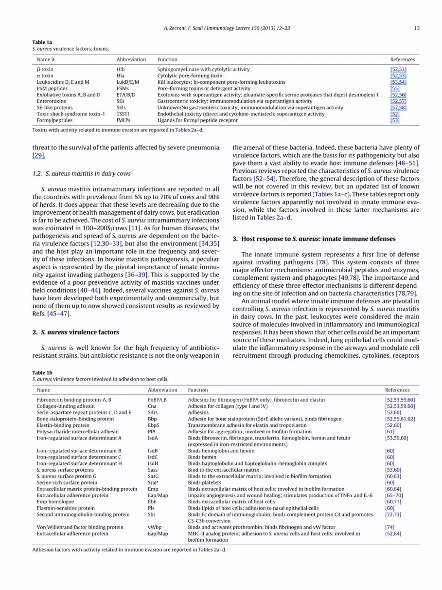
A. Zecconi, F. Scali / Immunology Letters 150 (2013) 12– 22 13
Table 1aS. aureus virulence factors: toxins.
Name it Abbreviation Function References
� toxin Hlb Sphingomyelinase with cytolytic activity [52,53]� toxin Hla Cytolytic pore-forming toxin [52,53]Leukocidins D, E and M LukD/E/M Kill leukocytes; bi-component pore-forming leukotoxins [53,54]PSM peptides PSMs Pore-forming toxins or detergent activity [55]Exfoliative toxins A, B and D ETA/B/D Exotoxins with superantigen activity; gluamate-specific serine proteases that digest desmoglein 1 [52,56]Enterotoxins SEs Gastroenteric toxicity; immunomodulation via superantigen activity [52,57]SE-like proteins SEls Unknown/No gastroenteric toxicity; immunomodulation via superantigen activity [57,58]Toxic shock syndrome toxin-1 TSST1 Endothelial toxicity (direct and cytokine-mediated); superantigen activity [52]
ecept
T
t[
1
toiiwpraianefihnR
2
r
TS
A
Formylpeptides fMLPs Ligands for formyl peptide r
oxins with activity related to immune evasion are reported in Tables 2a–d.
hreat to the survival of the patients affected by severe pneumonia29].
.2. S. aureus mastitis in dairy cows
S. aureus mastitis intramammary infections are reported in allhe countries with prevalence from 5% up to 70% of cows and 90%f herds. It does appear that these levels are decreasing due to themprovement of health management of dairy cows, but eradications far to be achieved. The cost of S. aureus intramammary infections
as estimated in 100–200$/cows [11]. As for human diseases, theathogenesis and spread of S. aureus are dependent on the bacte-ia virulence factors [12,30–33], but also the environment [34,35]nd the host play an important role in the frequency and sever-ty of these infections. In bovine mastitis pathogenesis, a peculiarspect is represented by the pivotal importance of innate immu-ity against invading pathogens [36–39]. This is supported by thevidence of a poor preventive activity of mastitis vaccines undereld conditions [40–44]. Indeed, several vaccines against S. aureusave been developed both experimentally and commercially, butone of them up to now showed consistent results as reviewed byefs. [45–47].
. S. aureus virulence factors
S. aureus is well known for the high frequency of antibiotic-esistant strains, but antibiotic resistance is not the only weapon in
able 1b. aureus virulence factors involved in adhesion to host cells.
Name Abbreviation Function
Fibronectin binding proteins A, B FnBPA,B Adhesins for fibrinoCollagen-binding adhesin Cna Adhesin for collagenSerin-aspartate repeat proteins C, D and E Sdrs Adhesins
Bone sialoprotein-binding protein Bbp Adhesin for bone siaElastin-binding protein EbpS Transmembrane adhPolysaccharide intercellular adhesin PIA Adhesin for aggregaIron-regulated surface determinant A IsdA Binds fibronectin, fib
(expressed in iron-rIron-regulated surface determinant B IsdB Binds hemoglobin aIron-regulated surface determinant C IsdC Binds hemin
Iron-regulated surface determinant H IsdH Binds haptoglobulinS. aureus surface proteins Sass Bind to the extracellS. aureus surface protein G SasG Binds to the extraceSerine-rich surface protein SraP Binds platelets
Extracellular matrix protein-binding protein Emp Binds extracellular mExtracellular adherence protein Eap/Map Impairs angiogenesiEmp homologue Ebh Binds extracellular mPlasmin-sensitive protein Pls Binds lipids of host cSecond immunoglobulin-binding protein Sbi Binds Fc domain of i
C3-C3b conversionVon Willebrand factor binding protein vWbp Binds and activates
Extracellular adherence protein Eap/Map MHC-II analog protebiofilm formation
dhesion factors with activity related to immune evasion are reported in Tables 2a–d.
or [53]
the arsenal of these bacteria. Indeed, these bacteria have plenty ofvirulence factors, which are the basis for its pathogenicity but alsogave them a vast ability to evade host immune defenses [48–51].Previous reviews reported the characteristics of S. aureus virulencefactors [52–54]. Therefore, the general description of these factorswill be not covered in this review, but an updated list of knownvirulence factors is reported (Tables 1a–c). These tables report onlyvirulence factors apparently not involved in innate immune eva-sion, while the factors involved in these latter mechanisms arelisted in Tables 2a–d.
3. Host response to S. aureus: innate immune defenses
The innate immune system represents a first line of defenseagainst invading pathogens [78]. This system consists of threemajor effector mechanisms: antimicrobial peptides and enzymes,complement system and phagocytes [49,78]. The importance andefficiency of these three effector mechanisms is different depend-ing on the site of infection and on bacteria characteristics [78,79].
An animal model where innate immune defenses are pivotal incontrolling S. aureus infection is represented by S. aureus mastitisin dairy cows. In the past, leukocytes were considered the mainsource of molecules involved in inflammatory and immunological
responses. It has been shown that other cells could be an importantsource of these mediators. Indeed, lung epithelial cells could mod-ulate the inflammatory response in the airways and modulate cellrecruitment through producing chemokines, cytokines, receptorsReferences
gen (FnBPA only), fibronectin and elastin [52,53,59,60] (type I and IV) [52,53,59,60]
[52,60]loprotein (SdrE allelic variant), binds fibrinogen [52,59,61,62]esin for elastin and tropoelastin [52,60]
tion; involved in biofilm formation [61]rinogen, transferrin, hemoglobin, hemin and fetuin
estricted environments)[53,59,60]
nd hemin [60][60]
and haptoglobulin–hemoglobin complex [60]ular matrix [53,60]llular matrix; involved in biofilm formation [60,63]
[60]atrix of host cells; involved in biofilm formation [60,64]
s and wound healing; stimulates production of TNF� and IL-6 [65–70]atrix of host cells [60,71]
ells; adhesion to nasal epithelial cells [60]mmunoglobulin; binds complement protein C3 and promotes [72,73]
prothrombin; binds fibrinogen and vW factor [74]in; adhesion to S. aureus cells and host cells; involved in [52,64]

14 A. Zecconi, F. Scali / Immunology Letters 150 (2013) 12– 22
Table 1cS. aureus virulence factors: enzymes and other proteins.
Name Abbreviation Function References
Coagulase Coa Binds and activates prothrombin; promotes conversion of fibrinogen to fibrin [52,74]V8 protease – Serine protease [52,53]Glycerol ester hydrolases lip, geh, beh, Triacylglycerols degradation [53]Fatty acid-modifying enzyme FAME Fatty acids modificationO-acetyltransferase OatA Peptidoglycan O-acetylation [53]PtdIns-phospholipase C Plc Phosphotidylinositol-specific lipase activity [53]Enolase Eno Catalyzes phosphor-glycerate to phosphoenol-pyruvate; binds to laminin [53]Arginine catabolic mobile element (3 types) ACME I/-II/-III Unclear role (aids colonization); seems to contain several enzyme and proteins
(arginine deaminase system, oligopeptide permease, zinc-containing alcoholdehydrogenase, spermine/spermidine acetyltransferase and others)
[75–77]
FPR-like 1 inhibitory protein FLIPr Binds formyl peptide receptor [53]
Enzymes and other proteins with activity related to immune evasion are reported in Tables 2a–d.
Table 2aVirulence factors enabling S. aureus to evade innate immune defenses related to leukocyte migration and phagocytic activity.
Effector mechanism S. aureus evasion factor Abbreviation Function References
Neutrophil migration Staphylococcal superantigen-like 5 SSL5 Specific binding to P-selectin glycoprotein ligand-1blocking PMN rolling
[93,197]
Staphylococcal superantigen-like 11 SSL11 [198]Staphylococcal superantigen-like 1 SSL10 Binds to chemokine receptors [199]Chemotaxis inhibitory protein CHIPS Blocks C5a receptor and formyl peptide receptors [53,142]Staphylococcal superantigen-like 7 SSL7 Binds to the Fc region of IgA and block recognition by
neutrophils[93]
Neutrophils lysis � toxin Hlg Bicomponent leukocidin; hemolysis [52,53]� toxin Hld Cytolytic toxin; binds neutrophils and monocytes [52,53]Panton-Valentine leukocidin PVL Bicomponent leukocidin; pore-forming toxin; kills
leukocytes[52,76]
Leukocidins A and B (alt. names H and G) LukAB/-HG Bi-component pore-forming leukotoxin that kills PMNs [54,200]
Resistance to oxidative burst Staphyloxanthin – Carotenoid (protects against ROS) [53]Catalase and alkylhydroxide reductase CatA, AhpC Inactivate hydrogen peroxide; pivotal for nasal
colonization[53]
Thioredoxin and thioredoxin reductase – Inactivates ROS [53]
Table 2bVirulence factors enabling S. aureus to evade innate immune defenses related to complement.
Effector mechanism S. aureus evasion factor Abbreviation Function References
Complement inactivation Capsular polysaccharides CPSs Alter C3 (CPS5 and 8) or C3b (CPS1) deposition [53,75]Staphylokinase Sak Plasminogen activator (plasminogen-serine
protease plasmin conversion)[53,73]
Staphylococcal complement inhibitor SCIN Inhibits convertase [53,142]Extracellular complement-binding protein Ecb Inhibits convertase [142]Clamp factor A ClfA Platelets adhesion (fibrin-mediated); binds
complement regulator factor I[52,53,59,60,74]
drE
fb
almbt
TV
Staphylococcus aureus surface protein E SExtracellular fibrinogen-binding protein E
nd adhesion molecules [80,81]. Similarly, mammary gland epithe-
ial cells could produce both cytokines and immunomodulatingolecules [36,82–84]. Changes of innate immune response cane observed during lactation. Indeed, recently, Ref. [84] showedhat several soluble factors (lactoferrin, lysozyme, NAGase, and IgG)
able 2cirulence factors enabling S. aureus to evade innate immune defenses related to immuno
Effector mechanism S. aureus evasion factor Abbreviation
Degradation of immunoglobulins Protein A SpA
Staphylokinase Sak
Cloaking of opsonins Serotypes 5,8 PNAGClamp factor A
ClfA
Clamp factor B ClfB
Binds complement regulator factor H [142]Binds fibrinogen; inhibits C3 and C5convertases; binds complement C3
[52,73,142]
have a different pattern during lactation, in absence of intramam-
mary infections.Among the different innate defenses, lysozyme showed a pecu-liar behavior when S. aureus intramammary infection occurred.Indeed, lysozyme activity in milk showed to be decreased in
globulins and opsonization.
Function References
Binds Fc domain of immunoglobulin, vonWillibrand factor and TNFR-1; Bindscomplement protein C3 and promotes C3–C3bconversion
[52,60,137]
Plasminogen activator (plasminogen-serineprotease plasmin conversion)
[53,73]
Platelets adhesion (fibrin-mediated); bindscomplement regulator factor I
[52,53,59,60,74]
Platelets adhesion (fibrin-mediated); bindscytokeratin 10.
[52,59,60,74,134]

A. Zecconi, F. Scali / Immunology Letters 150 (2013) 12– 22 15
Table 2dVirulence factors enabling S. aureus to evade innate immune defenses related to antimicrobial peptides.
Effector mechanism S. aureus evasion factor Abbreviation Function References
Alteration of cell wall components Antimicrobial peptide sensor ApsS/R/X Binds and impairs antimicrobial peptides [182,183]Gra regulatory system GraR/S Impairs phagocytosis and LL-37 [186]Multiple peptide resistance factor F MprF Inserts lysine into teichoic acids [53]Dlt operon DltA/B/C/D Inserts d-alanine into teichoic acids [53]
Lysozyme resistance OatA gene OatA Changes in N-acetylmuramic acid [189]
Cathelicidin resistance Staphylokinase Sak A-defensine binding [192]Aureolysin Aul LL-37 clivage [194]
Mec
SarbMcnc
otmamb
4r
npoaSirr
eataaipd
nocie
apmo
acH
Methicillin resistance
. aureus infected quarters [85]. The decrease may be the result ofn early exhaustion of lysozyme activity and was associated with aeduction in neutrophils respiratory burst activity [86,87], but alsoe related to the different expression of virulence factors [30,44].oreover, recent investigations suggest that lysozyme release from
ells is dose-dependent and at least 100.000 CFU/ml in milk areeeded to trigger its production from mammary gland epithelialells (Zecconi, unpublished data).
Thus, the role of mammary gland epithelial cells as a sourcef innate immunity component cannot be ignored, notably whenhe interactions between bacteria and host are of interest. Defense
olecules released by mammary gland epithelial cells seem to play major role in preventing mastitis, particularly when the level ofilk leucocytes is relatively low (<100.000 cells/ml), as suggested
y Refs. [36,38,88].
. Pathogenesis of S. aureus infections and innate immuneesponse: an evolving fight
The invasion of host tissues and organs triggers innate immu-ity, which is characterized by a quick response against theathogen. The timing of the immune response is strictly dependentn the first indispensable step: the recognition of pathogen-ssociated molecular patterns (PAMPs) [48,78,89]. Additionally,. aureus should adhere to host cell to multiply and, sometimes,nvade host cells [90], while the host replies by the immediateelease of antimicrobial components and of chemoattractants, thusecruiting immune cells for a more efficient response.
However, bacterial pathogens use very efficient strategies tovade host defenses in order to colonize and invade human andnimal tissues [90]. S. aureus has particular characteristics, and inhe initial steps of infections both in human and animals, whendhesion to epithelial cells is required, innate immunity represents
pivotal line of defense [12,48,91,92]. Whereas specific acquiredmmunity seems to play a major role in the further steps in theathogenic process, avoiding the diffusion of the bacteria and theevelopment of bacteremia and sepsis [91,92].
S. aureus virulence factors repertoire has plenty of mecha-isms to evade host innate immunity, including modificationsf structural component and secretion of a large array of spe-ific immune-modulating proteins acting in concert to counteractnnate immune defenses, and to create a microenvironment thatnables even better survival [48,49].
S. aureus has the capability to avoid TLR recognition; theirdhesins are quite efficient in reducing the effectiveness of com-lement system and phagocytes, and they developed resistanceechanism against host defense peptides (HDPs), similarly to the
ne developed against conventional antimicrobials.
Tables 2a–d report a list of the known factors involved in S.ureus immune evasion classified by target: neutrophils and phago-ytosis, complement system, immunoglobulins and opsonins,DPs.
Correlation between methicillin resistance andcathelicin resistance, mechanism to beidentified
[195,196]
It is impossible to cover all the virulence factors and their rolein pathogenesis of S. aureus infection in a single review. There-fore, we focused this review on the S. aureus factors enabling theevasion from immune defenses expressed during the first steps ofthe pathogenic processes (TLR evasion, adhesins and HDPs). Fac-tors involved in avoiding cells’ recruitment and related immuneresponse were covered by other excellent reviews [49,79,93].
4.1. TLR family recognition
S. aureus invasion triggers a complex mechanism through theactivation of specific receptors for PAMPs, eventually startinginnate immunity response [79]. Toll-like receptors (TLRs) were thefirst receptors for PAMPs to be discovered, but in recent years, otherclasses of receptor have been identified namely NOD-like receptors,C-type lectin receptors and RIG-I-like receptors [94].
TLR-2 was one of the most investigated toll-like receptors due toits ability to identify Gram-negative bacteria LPS [95,96] and Gram-positive specific molecular patterns [97,98].
TLR-2 is located on the cell surface forming and heterodimericcomplex with TLR-1 or TLR-6. TLR-2/TLR-1 complex recognizesGram-negative’s triacylated groups, while TLR-2/TLR-6 complexidentifies Gram-positive’s diacylated groups [99]. The lipoteichoicacid and certain lipopeptides of S. aureus trigger TLR-2/TLR-6.TLR-2/TLR-6 complex leads to production and secretion of severalcytokines trough TIRAP and MyD88 pathways. In addition, TLR-2activation seems to be directly involved in the maturation of thephagosome [100,101].
TLR-2 showed to play an important role in the immunity againstS. aureus in different infection models; mutant mice without TLR-2appear more susceptible to cutaneous, intranasal or corneal infec-tion than wild ones; furthermore, these mice seem to be moredisposed to severe infection and eventually death [102–104].
TLR-4 has a central role in the innate immunity againstGram-negative bacteria because of its ability to recognizelipopolysaccharides (LPS) [94,105–107]. Nevertheless, TLR-4 seemsto have a relevant role in S. aureus infections too. In a brain abscessmodel, TLR-4−/− mice have shown more severe lesions, prolongedinfection and higher mortality than the control mice. These resultssuggest the role of TLR-4 for a complete recovery of the diseasedanimal [108]. TLR-4 also seems to be activated by S. aureus leuko-cidins, this activation leads to maturation of dendritic cells in thebone marrow [109].
The other receptors in toll-like family apparently do not seemto have a specific relevance in innate immune response againstS. aureus. However, a recent study highlighted the TLR-9 abil-ity to induce production of IL-12 after S. aureus exposure andthe relevance of TLR-9/TLR-2 crosstalk for the regulation of this
cytokines family. Furthermore, TLR-9 seems to mediate the classI IFN signaling in dendritic cells, role that may be detrimental inMRSA pneumonia due to the allegedly negative effects of IFN� andIFN� in these pneumonias [110].
1 nology
NooiNia
baaa(pbib
irpwa2miwT
tSSp
4
afhc(ptomnIM[pi2at2HopumbroT
6 A. Zecconi, F. Scali / Immu
The NLRs (nucleotide oligomerization domain-like receptor orOD-like) represent a family of receptors involved in the regulationf innate immune response, they are typically located in the cytosolf cells with phagocytic activity. Nevertheless, NLRs can be foundedn various other cell types [111]. NLRs are a large family, over 20LRs were discovered in human cells, a number that probably will
ncrease considering that in mice were founded more than 30 NLRsnd in some invertebrate, like echinoderms, over 200 [111,112].
The role of NLRs still needs to be clarified. However, two mem-ers of the family, nucleotide-binding oligomerization domains 1nd 2 (NOD1 and NOD2), are widely studied, and they seem to have
role in innate immunity against S. aureus. Meso-diaminopimeliccid (m-DAP) triggers NOD1 [113,114] while muramyl dipeptideMDP) activates NOD2 [115,116]. Both m-DAP and MDP are com-onents of peptidoglycan. However, MDP is founded in almost allacterial cell walls whereas, m-DAP appears to be more character-
stic of Gram-negative bacteria. Nevertheless, some Gram-positiveacteria seem to be able to activate NOD1 [117,118].
The exact role of NOD2 in the innate response against S. aureuss still unclear. Indeed, preliminary studies on murine modelseported inconsistent results. In a systemic infection model, NOD2layed a critical role in the innate response. NOD2−/− mutant miceere more susceptible to infection, developed more severe diseases
nd had a larger production of some cytokines (TNF�, IFN� and IL-) than wild mice. Furthermore, NOD2−/− mice showed a higherortality in comparison with TLR-2−/− mice [119]. In a cutaneous
nfection model NOD2−/− mice developed more severe lesions thanild mice; however, NOD2−/− presented an inferior production of
NF� and IL-6 in comparison with wild mice [120].The role of NOD1 in S. aureus infection is even less well-known
han NOD2. Recent studies suggest that NOD1 may be relevant in. aureus killing by neutrophils. Indeed, neutrophils activity against. aureus in NOD1+/+ mice was higher than NOD1−/− ones whenre-stimulated with a Gram-negative bacteria [121].
.2. Evasion from TLRs recognition
The evasion from TLR recognition is a topic which is receivingn increasing interest, even if the studies available are relativelyew. Recently, a novel mechanism for staphylococci to escape theost immune system via interference with recognition by immuneells has been identified [122]. Staphylococcal superantigens likeSSL) proteins play an important role in this mechanism. Theseroteins are in a family of molecules able to interfere with mul-iple components of host immunity, including humoral immunity,psonization, and trafficking of leukocytes [167]. Therefore, theyay be involved in evasion both from innate and adaptive immu-
ity. Indeed, among the 14 members of the SSL family, SSL7 binds togA and C5; SSL5 binds to PSGL-1, chemoattractant receptors, and
MP-9, and SSL10 binds to chemokine receptor and human IgG123]. Among these families, SSL3 is the first-described bacterialrotein that blocks TLR-2 activation through direct extracellular
nteraction with the receptor [124]. Particularly, SSL3 binds to TLR- via its extracellular domain and inhibits tumor necrosis factorlpha (TNF�) production from mouse macrophages in responseo heat-killed S. aureus, peptidoglycan (PGN), or lipopeptide TLR-
ligands. In addition, SSL3 showed to inhibit IL-8 production inEK cells expressing TLR-1/2 and TLR-2/6 dimers through bindingf the extracellular TLR-2 domain. The SSL3-TLR-2 interaction isartially glycan dependent as binding of SSL3 to TLR-2 is reducedpon removal of sialic acid residues. Furthermore, the SSL3 (R308A)utant, lacking glycan-binding properties, shows lower TLR-2 inhi-
ition. An SSL3 mutant, lacking the N-terminal 126 amino acids, stilletains full TLR-2 inhibiting activity. Among the other SSLs tested,nly SSL4, which shares the highest homology with SSL3, blocksLR-2 activation. This unique function of SSL3 adds to the arsenal
Letters 150 (2013) 12– 22
of immune evasive molecules that S. aureus can employ to subvertboth innate and adaptive immunity [123].
The new insights into the SSL-TLR interactions will increase ourunderstanding not only of the bacterial infections but also in all thediseases related to TLR malfunction such as atherosclerosis [125].
4.3. Adhesion
Host-adhesion represents a pivotal phase of pathogenesis of S.aureus, and it developed a wide variety of molecules that aid thisprocess (Tables 1a–c) named adhesins. These molecules mediatethe adhesion to different substrates of the host: extracellular matrixproteins (ECMs), plasma proteins, epithelial cells and endothelialcells [59,60]. Furthermore, some of these adhesins interfere withhost immunity, as described later in this paper. Adhesins can beanchored on the surface of the bacteria, or they can be secreted.Surface adhesins are called microbial surface recognizing adhesivematrix molecules (MSCRAMMs) whereas secretable adhesins arecalled secretable expanded repertoire adhesive molecules (SER-AMs) [53,59]. Both MSCRAMMs and SERAMs are proteins, even ifother molecules, such as polysaccharide intercellular adhesin (PIA)and teichoic acids, can act as adhesins too.
The adhesion is a pre-requisite to have colonization and, eventu-ally, infection of the host. The most important sites of colonizationin human hosts are the nasal mucosa and the skin, and they are avaluable model to understand S. aureus adhesion process [59,126].Colonization of anterior nares can be intermittent or persistent.Persistent subjects seem to have large numbers of carried bacteriaand a single dominant strain while intermittent carriers seem tohave a low amount of bacteria and different strains [127–129]. Ithas been suggested that difference in carrier state could be relatedto different ligands for the adhesins of S. aureus [130]. A similarpattern was observed in bovine intramammary infections [12].
Fibronectin-binding proteins (FnBPs), clumping factors (Clfs),protein A (SpA) and collagen-binding protein (Cna) are the major S.aureus MSCRAMMs. S. aureus synthesizes two fibronectin-bindingproteins: FnBPA and FnBPB. These adhesins bind fibrinoctein andelastin, and FnBPA can bind fibrinogen [52,53,59,60]. FnBPs tiefibronectin on the surface of the bacteria; this structure allows abond between S. aureus and host integrins [131]. FnBPA and FnBPBhave an important role in the colonization of the host. However,their relevance in the infections has been not completely eluci-dated [131,132]. Clumping factors A and B bind fibrinogen of thehost and promote platelets aggregation [52,53,59,60]. Neverthe-less, soluble fibrin seems to be the major protein responsible for theformation of S. aureus-platelets aggregates mediated by Clfs [133].ClfB aids colonization of anterior nares because of its ability to bindcytokeratin 10 [59,60]. Besides, ClfB seems to give a further aid tobacteria–host adhesion by binding cytokeratin 8 [134]. Collagen-binding protein mediates adhesion between S. aureus and host’scollagen; this adhesin provides help in the invasion of diverse tis-sues, and it acts as a virulence factor in different type of infections[52,53,59,60]. Several S. aureus surface proteins can bind the ECMwith a covalent link, however, the ligands of the majority of theseproteins need to be clarified [53,60]. Surface protein G (SasG) seemsto contribute in the colonization of anterior nares [60]; moreover,SasG plays a role in the formation of biofilm when zinc is avail-able for the bacteria [63,135]. Finally, protein A plays mainly a rolein immune evasion than in adhesion and it will be furthermoreconsidered in the next section.
4.4. Adhesins as effector molecules to evade from innate immune
responseMany molecules involved in adhesion are also involved in dif-ferent immune evasion tactics (Tables 2a–d). The role of adhesins

nology
ioomivad
MihiWavts
iorSba
aafbvprp
dcacsaea
dbcaansie
4
aiaamStrt
A. Zecconi, F. Scali / Immu
n evading immune defences is not related to the specific processf adhesion, but their activities interfere with several mechanismsperated by innate immune defences. The role of the differentolecules was summarized in Tables 2a–d, under the specific
mmune mechanism mainly affected by each adhesin. As for otherirulence factors, adhesin expression varies depending on the strainnd on the environment, but some of them are very common andeserve a more detailed description.
Protein A (SpA) is undoubtedly one of the most studiedSCRAMMs, and this protein binds different important molecules
nvolved in the immune response. Indeed, N terminal of SpAas five subdomains biding with Fc region of IgG, thus impair-
ng opsonization and then phagocytosis. SpA also binds to vonillebrand factor and complement protein C3, increasing bacteria
dhesion to platelets. Furthermore, SpA promotes C3–C3b con-ersion [52,60,72]. More recently Spa showed to bind to TNFR1,he receptor for TNF-�, thus reducing TNF-� proinflammatoryignaling [136].
Second immunoglobulin-binding protein (Sbi) has ligands sim-lar to SpA [72,137,138]. This protein can perform its function notnly when secreted but also when it remains anchored to the bacte-ial cell wall due to its ability to bind teichoic acid [72]. Moreover,bi, together with extracellular fibrinogen-binding protein (Efb),inds plasmin in a process that leads to the degradation of C3a, C3bnd C3 [73].
Complement formation is impaired also by other adhesins suchs ClfA, Ecb and SdrE. ClfA binds to complement regulator factor 1,nd this process increases the conversion of C3b to an inactivateorm (iC3b) and impairs phagocytosis of S. aureus [139,140]. Ecbinds C3d and thus inhibits C3 (alternative pathway) and C5 con-ertases (all pathways). This inhibition determines a reduction ofhagocytosis and PMNs migration [141]. SdrE binds to complementegulator factor H, reducing C3 deposition and C5a synthesis, thisrocess impairs also phagocytosis [142].
Extracellular adherence protein (Eap/MaP) is an adhesin withifferent roles such as the formation of bonds between S. aureusells; bonds between bacteria and host’s cells; biofilm formation;ngiogenesis and wound healing inhibition [52,64–67]. Eap showsrystals structure similar to superantigens; however, it do noteems to possess superantigen activities [143]. Furthermore, Eapcts as a MHC-II analog and has several immunomodulatory prop-rties like pro-inflammatory cytokines stimulation (Il-6 and TNF�)nd inhibition of neutrophil migration and adhesion [66,68,69,143].
Adhesins have been targeted as a major antigen for vaccineevelopment both in human and veterinary medicine [43,46,144],ut still the efficacy of S. aureus vaccines based on adhesion isontroversial. A lot of studies were focused on the adhesion mech-nisms and relatively less on investigating the interferences ofdhesins on the innate immune response. This latter aspect can-ot be neglected, and a more holistic approach investigating at theame time the complex adhesion process and the interference withnnate immunity is needed to increase the chances to develop anfficacious vaccine.
.5. Host defense peptides and polypeptides
Once the PAMPs had been recognized and while the bacteriattempt to adhere to host cells, these latter ones react activatingnnate immune defenses and releasing two classes of molecules:ntimicrobial peptides (i.e. cathelicidins) and polypeptides withntimicrobial activity (i.e. lysozyme). The type and amount of theseolecules are dependent from the host, the infected tissues and
. aureus virulence characteristics. Nevertheless, the efficacy ofhis response is pivotal to control the infection and, eventually, toestore the host health both in human and animal S. aureus infec-ions [36,39,145–147].
Letters 150 (2013) 12– 22 17
Host defense peptides (HDPs) are a group of molecules also clas-sically defined as antimicrobial peptides. This latter definition ismisleading, being more related to their history of discovery than tothe potent influence these molecules have on cell immune behav-ior [148]. These molecules are used by multicellular organisms todefend themselves against infective microorganism, and they arewidely present in nature, from plants to humans [81,149–151]. Alarge number of such peptides have been identified, and there isconsistent evidence of their pivotal role in innate immune system[152,153]. The expression of these antimicrobial peptides can beconstitutive or can be induced by infectious stimuli such as bacteriaand bacterial molecules (i.e. LPS, lipoteichoic acid) or inflamma-tory stimuli, such as proinflammatory cytokines [154]. Indeed,HDPs are important effector and modulator molecules of the innateimmune system, being able to enhance phagocytosis, stimulatethe prostaglandin release, neutralize the septic effects of LPS,promote recruitment and accumulation of various immune cellsat inflammatory sites, promote angiogenesis, and induce woundrepair [152–155]. The activity against bacteria is mainly related totheir electrostatic binding to negatively charged bacterial surfacemolecules: LPS in Gram-negative bacteria, lipoteichoic acid andteichoic acid in Gram-positive bacteria. After attracting the pep-tides to the bacterial cell surface, the membrane is made permeable.This involves pore or gap formation by aggregation of the peptidesin the membrane, resulting in bacterial cell death [152–154].
HDPs have a relative low number of aminoacids (12–100), arepositively charged and amphiphilic. HDPs were discovered in theearly 20th century; however, a characterization of these moleculeswas started only recently [150,151]. These peptides can be classi-fied according to their chemicals characteristics: molecules with�-helix structures; molecules with �-sheet structures; moleculeswith extended structures and molecules with loop structures [156].
Among HDPs two large families of antimicrobial peptides havebeen defined: defensins and cathelicidins. Defensins are widelydistributed in mammalian epithelia and phagocytes, while otherpeptides, including cathelicidins, histatins, dermicidin, and anionicpeptides have a more restricted tissue and animal species distribu-tion [146,147,155,157].
Among the different HDPs, cathelicidins represent a family ofheterogeneous antimicrobial peptides. Cathelicidins can be foundin several animals [158–161]; furthermore, in bovines, swineand equines, different cathelicidins in the same species havebeen identified [162–164]. However, in humans, only one gene(CAMP) codifying for cathelicidin was identified, and this geneis similar to mouse CRAMP gene [165,166]. CAMP gene codifiesfor hCAP18, the precursor of active form of human cathelicidin,namely LL-37 [167,168], which is probably the most investigatedhuman cathelicidin. LL-37 seems to adopt a �-helical structurein fluids such as plasma, interstitial liquid and intracellular fluid[169]. Human cathelicidin alters the membrane of sensible bacte-ria through creation of channels and pores [172]. FurthermoreLL-37 has immunomodulatory and chemotactic effects on neu-trophils, T lymphocytes and monocytes [158], and it is involved incutaneous wound healing [148]. Numerous cells like neutrophils,lymphocytes, monocytes, mast cells and natural killer can produceLL-37 [170–172]. Epithelial cells of skin [173], and of respiratory[174,175], gastro-enteric [176] and urogenital tract [177] are alsocapable of secrete this cathelicidin.
4.6. Host defence polypeptides – lysozyme
Host defense polypeptides, usually defined as natural antimi-
crobial enzymes, were discovered since many years [148], but theirrole as immunomodulators has been discovered only more recently[81,148]. Among these molecules, one of the most investigatedand important one is lysozyme (1,4-8-N-acetylmuraminidases)
1 nology
wsbanLgnla
sbGr
cmaa[eitd
4
acmtfsaimt
apaciSvd(acosMg[
usscLptm
8 A. Zecconi, F. Scali / Immu
hich we will use as a focused example of this category of sub-tances. Lysozyme has a wide distribution in nature being presentoth in plants and animals, and exhibits antimicrobial activitiesgainst different microorganisms. The bacterial killing mecha-ism of lysozyme consists in the lytic and non-lytic mechanisms.ysozyme provokes direct cell lysis by hydrolyzing the peptido-lycan layers of bacteria and induction of autolysins [178]. Theon-lytic mechanism is principally based on the properties of
ysozyme to cause membrane perturbation through the binding of specific domain of lysozyme within the bacterial surface [179].
The antibacterial activity of lysozyme depends on the acces-ibility of substrate. Gram-positive bacteria are more susceptibleecause peptidoglycan represents about 90% of the cell wall, whileram-negative bacteria, having <10% peptidoglycan, are more
esistant to the enzyme.The real role of lysozyme in vertebrates is still under dis-
ussion. Generally, the antibacterial activity is considered itsajor role. More recently it has been suggested that lysozyme is
n immunomodulator. Indeed, breakdown products of lysozymection on peptidoglycan demonstrated immunological properties39]. Moreover, recent studies suggest that LL-37 and lysozymexhibit antimicrobial activities synergistically or additively againstnvading microorganisms, in particular S. aureus [180], supportinghe importance of these soluble components of innate immuneefenses.
.7. Host defense peptides and polypeptides resistance
Antimicrobial peptides are conserved in their structure, functionnd mechanisms of action. At high concentration could be bacteri-idal, while at lower concentration, a role in immunomodulation inore plausible [154]. Due to their characteristics, it was assumed
hat resistance to antimicrobial peptides was less probable thanor conventional antimicrobials, but S. aureus infections, once more,howed that this was not the case. Some resistance mechanisms rel-tively common among S. aureus strains were already known, evenf their pathways were elucidated only recently. Moreover, new
echanisms related to the newly discovered HDPs have been iden-ified and more are expected to be discovered in the near future.
Staphylococci can modulate their sensitivity to defensins, and to broad range of antimicrobial peptides, by altering the com-osition and net charge of lipoteichoic acid, wall teichoic acidnd phospholipids [49,181]. A three-component regulatory systemalled the antimicrobial peptide sensor (Aps) has been identifiedn S. epidermidis [182]. The Aps system showed to be conserved intaphylococci, and a similar system is present also among S. aureusirulence factors repertoire [183]. The Aps system includes a histi-ine protein kinase sensory component (ApsS) a response regulatorApsR), and a third protein component (ApsX), which functionsre not yet fully understood [182,183]. Several cationic peptidesan activate Aps system which upregulates expression of the dtlperon and the mrpF gene. Dlt operon expression cause a d-alanineubstitutions of ribitol teichoic acid and lipoteichoic acid [184].prf operon expression adds a l-lysine residue to phosphatidyl-
lycerol exposed on the outer face of the cytoplasmic membrane185].
Also the regulatory system graRS plays a crucial role in reg-lation of dlt operon. Indeed, S. aureus graRS-mutants are moreusceptible to the bactericidal action of LL-37 [186]. The expres-ion of these genes leads S. aureus to reduce his superficial negativeharge in teichoic acids, resulting in a less effective attraction of
L-37 and other cationic HDPs. Modification of teichoic acids andhospholipids with d-alanine and l-lysine, respectively, has addi-ional consequences for the pathogenicity of S. aureus. Indeed, theodifications also interfere with the susceptibility to glycopeptide
Letters 150 (2013) 12– 22
antibiotics such as vancomycin [187,188] and may contribute to thedevelopment of intermediate vancomycin-resistant strains [48].
Similarly, the resistance to lysozyme is related to composi-tional changes of constitutive proteins. Resistance to lysozymeis due to the changes in N-acetylmuramic acid of staphylococ-cal peptidoglycan which is O-acetylated at position C6-OH byan O-acetyltansferase that is an integral membrane protein. Theresponsible gene, oatA, was recently identified, and the oatA dele-tion mutant had an increased sensitivity to lysozyme [189].
Moreover, many S. aureus strains secrete staphylokinase (Sak), aprotein that, in addition to its plasminogen-activating role, showedto be able to inactivate several defensins [190]. The binding ofdefensins by Sak inhibits their bactericidal effects and, in vivo,staphylococcal strains producing Sak were protected against thebactericidal effect of a-defensins. Notably, the site within Sak thatbinds �-defensins is different from its plasminogen-binding site.Sak seems to be also able to form a complex with LL-37 that increaseits fibrinolytic action and improve the invasivity of S. aureus inhuman airways [191]. Therefore, due to its different activities inter-fering with innate immune defenses, Sak is considered to be animportant virulence factor of S. aureus [192].
As described previously, cathelicidin LL-37 is one of the fewhuman bactericidal peptides with potent anti-staphylococcal activ-ity. Until some years ago, LL-37 seemed to show a good activityagainst S. aureus [193]. However, in recent years, different strainsof S. aureus showed to be resistant to human cathelicidin [194].Indeed, aureolysin, a metalloproteinase in S. aureus, was found toinactivate LL-37 in a time- and concentration-dependent manner[194]. Aureolysin A cleaves LL-37 at positions 19–20, 23–24 and31–32, thereby directly degrading human cathelicidin and impair-ing its activity [194]. On the contrary, the V8 proteinase of S. aureusalso cleaves LL-37 but the resulting LL-17–37 fragment retainedthe antibacterial activity against S. aureus [194]. Some of theseresistance mechanisms showed to be related to MRSA resistance.Indeed, when MSSA with MRSA strains were compared, the MRSAstrains showed lower susceptibility to HDPs, indicating that someMRSA strains are resistant not only to various antibiotics, includingmethicillin, but also to some of HDPs [165,174]. More specifically,recent studies showed a correlation between methicillin and LL-37resistance [195,196].
Staphylococcal resistance against HDPs seems to be a multifac-torial process and, while some bacterial factors limiting the activityof HDPs have been identified, many others are still unknown. Thereare increasing research activities in the area of HDPs discovery,as a way to overcome the widespread problem of antimicrobialresistance. However, the S. aureus models show as the evolutionof pathogens could already have affected the sensitivity to thesenatural molecules. Therefore, before applying them as therapeu-tics, we should be very careful in avoiding the mistakes we made inchemotherapeutical approach in human and veterinary medicine.
5. Conclusions
Even if innate immunity is the oldest and most widespreaddefense system in mammals, our knowledge on it is far to be com-pleted. Moreover, the studies on the relationship between thissystem and invading pathogens require a holistic approach whichshould include both the host defense mechanisms and the pathogencapability to avoid them. Undoubtedly, it is hard to believe thatwe will be able to elucidate all the potential relationships, becausethese latter ones will co-evolute naturally, as long as immune sys-
tem will be efficient. However, the still increasing importance ofS. aureus in human and veterinary medicine, the problems relatedto antimicrobial resistance and the need to have efficacious vac-cines or molecules against S. aureus, strongly support the ongoing
nology
raph
R
A. Zecconi, F. Scali / Immu
esearch on the innate immune defenses and their role against S.ureus. This will allow to have new and, hopefully, more powerfulreventive and therapeutical tools to reduce S. aureus impact onealth both in humans and animals.
eferences
[1] Al-Dabbagh M, Dobson S. Infectious hazards from pets and domestic animals.In: Curtis N, Finn A, Pollard AJ, editors. Hot topics in infection and immunityin children Vii. 2011. p. 261–72.
[2] Fluit AC. Livestock-associated Staphylococcus aureus. Clin Microbiol Infect2012;18:735–44.
[3] Hasman H, Moodley A, Guardabassi L, Stegger M, Skov RL, Aarestrup FM. spatype distribution in Staphylococcus aureus originating from pigs, cattle andpoultry. Vet Microbiol 2010;141:326–31.
[4] Vanderhaeghen W, Hermans K, Haesebrouck F, Butaye P. Methicillin-resistantStaphylococcus aureus (MRSA) in food production animals. Epidemiol Infect2010;138:606–25.
[5] Goetghebeur M, Landry PA, Han D, Vicente C. Methicillin-resistant Staphylo-coccus aureus: a public health issue with economic consequences. Can J InfectDis Med Microbiol 2007;18:27–34.
[6] McCaig LF, McDonald LC, Mandal S, Jernigan DB. Staphylococcus aureus-associated skin and soft tissue infections in ambulatory care. Emerg InfectDis 2006;12:1715–23.
[7] Klevens RM, Morrison MA, Nadle J, Petit S, Gershman K, Ray S, et al. Invasivemethicillin-resistant Staphylococcus aureus infections in the United States.JAMA 2007;298:1763–71.
[8] Leonard FC, Markey BK. Meticillin-resistant Staphylococcus aureus in animals:a review. Vet J 2008;175:27–36.
[9] Rich M. Staphylococci in animals: prevalence, identification and antimicro-bial susceptibility, with an emphasis on methicillin-resistant Staphylococcusaureus. Br J Biomed Sci 2005;62:98–105.
[10] Zecconi A. Staphylococcus aureus mastitis: what we need to know to controlthem. Isr J Vet Med 2010;65:93–9.
[11] Zecconi A, Calvinho LF, Fox KL. Staphylococcus aureus intramammary infec-tions. IDF Bull 2006;408:1–42.
[12] Piccinini R, Borromeo V, Zecconi A. Relationship between S. aureus genepattern and dairy herd mastitis prevalence. Vet Microbiol 2010;145:100–5.
[13] Barber M. Methicillin-resistant staphylococci. J Clin Pathol 1961;14:385–93.[14] Boyce JM. Methicillin-resistant Staphylococcus aureus in hospital and long-
tem care facilities – microbiology, epidemiology, and preventive measures.Infect Control Hosp Epidemiol 1992;13:725–37.
[15] Johnson AP, Aucken HM, Cavendish S, Ganner M, Wale MCJ, Warner M,et al. Dominance of EMRSA-15 and-16 among MRSA causing nosocomialbacteraemia in the UK: analysis of isolates from the European Antimi-crobial Resistance Surveillance System (EARSS). J Antimicrob Chemother2001;48:143–4.
[16] Bukharie HA, Abdelhadi MS, Saeed IA, Rubaish AM, Larbi EB. Emergence ofmethicillin-resistant Staphylococcus aureus as a community pathogen. DiagnMicrobiol Infect Dis 2001;40:1–4.
[17] Salgado CD, Farr BM, Calfee DP. Community-acquired methicillin-resistantStaphylococcus aureus: a meta-analysis of prevalence and risk factors. ClinInfect Dis 2003;36:131–9.
[18] Devriese LA, Van Damme LR, Fameree L. Methicillin-(cloxacillin)-resistantStaphylococcus aureus strains isolated from bovine mastitis cases. ZentralblHyg Umweltmed 1972;19:598–605.
[19] Voss A, Loeffen F, Bakker J, Klaassen C, Wulf M. Methicillin-resistant Staphy-lococcus aureus in pig farming. Emerg Infect Dis 2005;11:1965–6.
[20] van Loo I, Huijsdens X, Tiemersma E, de Neeling A, van de Sande-Bruinsma N,Beaujean D, et al. Emergence of methicillin-resistant Staphylococcus aureus ofanimal origin in humans. Emerg Infect Dis 2007;13:1834–9.
[21] Khanna T, Friendship R, Dewey C, Weese JS. Methicillin resistant Staphy-lococcus aureus colonization in pigs and pig farmers. Vet Microbiol2008;128:298–303.
[22] Calfee DP. Methicillin-resistant Staphylococcus aureus and vancomycin-resistant enterococci, and other Gram-positives in healthcare. Curr OpinInvestig Drugs 2012;25:385–94.
[23] Petinaki E, Spiliopoulou I. Methicillin-resistant Staphylococcus aureus amongcompanion and food-chain animals: impact of human contacts. Clin MicrobiolInfect 2012;18:626–34.
[24] Fitzgerald JR. Livestock-associated Staphylococcus aureus: origin, evolutionand public health threat. Trends Microbiol 2012;20:192–8.
[25] Stefani S, Chung DR, Lindsay JA, Friedrich AW, Kearns AM, Westh H,et al. Meticillin-resistant Staphylococcus aureus (MRSA): global epidemiol-ogy and harmonisation of typing methods. Int J Antimicrob Agents 2012;39:273–82.
[26] Gould IM, David MZ, Esposito S, Garau J, Lina G, Mazzei T, et al. New insightsinto meticillin-resistant Staphylococcus aureus (MRSA) pathogenesis, treat-
ment and resistance. Int J Antimicrob Agents 2012;39:96–104.[27] Skov R, Christiansen K, Dancer SJ, Daum RS, Dryden M, Huang YC, et al.Update on the prevention and control of community-acquired meticillin-resistant Staphylococcus aureus (CA-MRSA). Int J Antimicrob Agents 2012;39:193–200.
Letters 150 (2013) 12– 22 19
[28] Lowy FD. Medical progress – Staphylococcus aureus infections. N Engl J Med1998;339:520–32.
[29] Kluytmans J, vanBelkum A, Verbrugh H. Nasal carriage of Staphylococcusaureus: epidemiology, underlying mechanisms, and associated risks. ClinMicrobiol Rev 1997;10:505.
[30] Zecconi A, Cesaris L, Liandris E, Daprà V, Piccinini R. Role of several Staphy-lococcus aureus virulence factors on the inflammatory response in bovinemammary gland. Microb Pathog 2006;40:177–83.
[31] Taverna F, Negri A, Piccinini R, Zecconi A, Nonnis S, Ronchi S, et al. Charac-terization of cell wall associated proteins of a Staphylococcus aureus isolatedfrom bovine mastitis case by a proteomic approach. Vet Microbiol 2007;119:240–7.
[32] Kerro Dego O, van Dijk J, Nederbragt H. Factors involved in the early patho-genesis of bovine Staphylococcus aureus mastitis with emphasis on bacterialadhesion and invasion. A review. Vet Q 2002;24:181–98.
[33] Sutra L, Poutrel B. Virulence factors involved in the pathogenesis of bovineintramammary infections due to Staphylococcus aureus. J Med Microbiol1994;40(2):79–89.
[34] Fox LK, Gershaman M, Hancock DD, Hutton CT. Fomities and reservoirsof S. aureus causing intramammary infections as determined by phagetyping: the effect of milking hygiene practises. Cornell Vet 1990;81:183–93.
[35] Robertson JR, Fox LK, Hancock DD, Gay JM, Besser TE. Sources of intramam-mary infections from Staphylococcus aureus in dairy heifers at first parturition.J Dairy Sci 1998;81:687–93.
[36] Rainard P, Riollet C. Innate immunity of the bovine mammary gland. Vet Res2006;37:369–400.
[37] Rinaldi M, Li RW, Bannerman DD, Daniels KM, Evock-Clover C, Silva MVB,et al. A sentinel function for teat tissues in dairy cows: dominant innateimmune response elements define early response to E. coli mastitis. FunctIntegr Genomics 2010;10:21–38.
[38] Riollet C, Rainard P, Poutrel B. Cells and cytokines in inflammatorysecretions of bovine mammary gland. Adv Exp Med Biol 2000;480:247–58.
[39] Zecconi A, Smith KL. Ruminant mammary gland immunity. Bruxelles: FIL-IDF;2003.
[40] Watson D. Staphylococcal mastitis vaccine. Vaccine 1992;10(5):359.[41] Leitner G, Lubashevsky E, Trainin Z. Staphylococcus aureus vaccine against
mastitis in dairy cows, composition and evaluation of its immunogenicity ina mouse model. Vet Immunol Immunopathol 2003;93:159–67.
[42] Pellegrino M, Giraudo J, Raspanti C, Odierno L, Bogni C. Efficacy of immuniza-tion against bovine mastitis using a Staphylococcus aureus avirulent mutantvaccine. Vaccine 2010;28:4523–8.
[43] Castagliuolo I, Piccinini R, Beggiao E, Palù G, Mengoli C, Ditadi F, et al. Mucosalgenetic immunization against four adhesins protects against Staphylococcusaureus-induced mastitis. Vaccine 2006;24:4393–402.
[44] Scarpa M, Piccinini R, Brun P, Grillo A, Palu G, Mengoli C, et al. Relationshipbetween virulence factor genes in bovine Staphylococcus aureus subclini-cal mastitis isolates and binding to anti-adhesin antibodies. J Dairy Res2010;77:159–67.
[45] Pereira UP, Oliveira DGS, Mesquita LR, Costa GM, Pereira U. Efficacy ofStaphylococcus aureus vaccines for bovine mastitis: a systematic review. VetMicrobiol 2011;148:117–24.
[46] Middleton JR. Staphylococcus aureus antigens and challenges in vaccine devel-opment. Expert Rev Vac 2008;7:805–15.
[47] Middleton JR, Luby CD, Adams DS. Efficacy of vaccination against staphylo-coccal mastitis: a review and new data. Vet Microbiol 2009;134:192–8.
[48] Fedtke I, Gotz F, Peschel A. Bacterial evasion of innate host defenses – theStaphylococcus aureus lesson. Int J Med Microbiol 2004;294:189–94.
[49] Rooijakkers SHM, van Kessel KPM, van Strijp JAG. Staphylococcal innateimmune evasion. Trends Microbiol 2005;13:596–601.
[50] Urban CF, Lourido S, Zychlinsky A. How do microbes evade neutrophil killing.Cell Microbiol 2006;8:1687–96.
[51] Graves SF, Kobayashi SD, DeLeo FR. Community-associated methicillin-resistant Staphylococcus aureus immune evasion and virulence. J Mol Med2010;88:109–14.
[52] Peacock SJ, Moore CE, Justice A, Kantzanou M, Story L, Mackie K, et al. Vir-ulent combinations of adhesin and toxin genes in natural populations ofStaphylococcus aureus. Infect Immun 2002;70:4987–96.
[53] Chavakis T, Preissner KT, Herrmann M. The anti-inflammatory activities ofStaphylococcus aureus. Trends Immunol 2007;28:408–18.
[54] DuMont AL, Nygaard TK, Watkins RL, Smith A, Kozhaya L, Kreiswirth BN, et al.Characterization of a new cytotoxin that contributes to Staphylococcus aureuspathogenesis. Mol Microbiol 2011;79:814–25.
[55] Perret M, Badiou C, Lina G, Burbaud S, Benito Y, Bes M, et al. Cross-talkbetween Staphylococcus aureus leukocidins-intoxicated macrophages andlung epithelial cells triggers chemokine secretion in an inflammasome-dependent manner. Cell Microbiol 2012;14:1019–36.
[56] Kato F, Kadomoto N, Iwamoto Y, Bunai K, Komatsuzawa H, Sugai M. Regulatorymechanism for exfoliative toxin production in Staphylococcus aureus. InfectImmun 2011;79:1660–70.
[57] Baker MD, Acharya KR. Superantigens structure–function relationships. Int JMed Microbiol 2004;293:529–37.
[58] Ortega E, Abriouel H, Lucas R, Galvez A. Multiple roles of Staphylococcusaureus enterotoxins: pathogenicity, superantigenic activity, and correlationto antibiotic resistance. Toxins 2010;2.

2 nology
0 A. Zecconi, F. Scali / Immu[59] Speziale P, Pietrocola G, Rindi S, Provenzano M, Provenza G, Di PotoA, et al. Structural and functional role of Staphylococcus aureus surfacecomponents recognizing adhesive matrix molecules of the host. Future Micro-biol 2009;4.
[60] Clarke SR, Foster SJ. Surface adhesins of Staphylococcus aureus. Adv MicrobPhysiol 2006;51:51.
[61] Arciola CR, Visai L, Testoni F, Arciola S, Campoccia D, Speziale P, et al. Concisesurvey of Staphylococcus aureus virulence factors that promote adhesion anddamage to peri-implant tissues. Int J Artif Organs 2011;34:771–80.
[62] Vazquez V, Liang XW, Horndahl JK, Ganesh VK, Smeds E, Foster TJ,et al. Fibrinogen is a ligand for the Staphylococcus aureus microbial sur-face components recognizing adhesive matrix molecules (MSCRAMM) bonesialoprotein-binding protein (Bbp). J Biol Chem 2011;286:29797–805.
[63] Geoghegan JA, Corrigan RM, Gruszka DT, Speziale P, O’Gara JP, Potts JR, et al.Role of surface protein SasG in biofilm formation by Staphylococcus aureus. JBacteriol 2010;192.
[64] Johnson M, Cockayne A, Morrissey JA. Iron-regulated biofilm formation inStaphylococcus aureus Newman requires ica and the secreted protein Emp.Infect Immun 2008;76:1756–65.
[65] Thompson KM, Abraham N, Jefferson KK. Staphylococcus aureus extracellularadherence protein contributes to biofilm formation in the presence of serum.FEMS Microbiol Lett 2010;305:143–7.
[66] Harraghy N, Hussain M, Haggar A, Chavakis T, Sinha B, Herrmann M, et al. Theadhesive and immunomodulating properties of the multifunctional Staphy-lococcus aureus protein Eap. Microbiology-SGM 2003;149:2701–7.
[67] Athanasopoulos AN, Ecnomopoulou M, Orlova VV, Sobke A, Schneider D,Weber H, et al. The extracellular adherence protein (Eap) of Staphylococcusaureus inhibits wound heating by interfering with host defense and repairmechanisms. Blood 2006;107:2720–7.
[68] Edwards AM, Bowden MG, Brown EL, Laabei M, Massey RC. Staphylococcusaureus extracellular adherence protein triggers TNF alpha release, promotingattachment to endothelial cells via protein A. PLoS ONE 2012;7:8.
[69] Scriba TJ, Sierro S, Brown EL, Phillips RE, Sewell AK, Massey RC. The Staphyloc-cous aureus eap protein activates expression of proinflammatory cytokines.Infect Immun 2008;76:2164–8.
[70] Haggar A, Ehrnfelt C, Holgersson J, Flock JI. The extracellular adherence pro-tein from Staphylococcus aureus inhibits neutrophil binding to endothelialcells. Infect Immun 2004;72:6164–7.
[71] Sakamoto S, Tanaka Y, Tanaka I, Takei T, Yu J, Kuroda M, et al.Electron microscopy and computational studies of Ebh, a giant cell-wall-associated protein from Staphylococcus aureus. Biochem Biophys Res Commun2008;376:261–6.
[72] Smith EJ, Corrigan RM, van der Sluis T, Gruendling A, Speziale P, GeogheganJA, et al. The immune evasion protein Sbi of Staphylococcus aureus occurs bothextracellularly and anchored to the cell envelope by binding lipoteichoic acid.Mol Microbiol 2012;83.
[73] Koch TK, Reuter M, Barthel D, Böhm S, van den Elsen J, Kraiczy P, et al.Staphylococcus aureus proteins Sbi and Efb recruit human plasmin to degradecomplement C3 and C3b. PLoS ONE 2012;7:e47638.
[74] McAdow M, Missiakas DM, Schneewind O. Staphylococcus aureus secretescoagulase and von Willebrand factor binding protein to modify the coag-ulation cascade and establish host infections. J Innate Immun 2012;4:141–8.
[75] Gordon RJ, Lowy FD. Pathogenesis of methicillin-resistant Staphylococcusaureus infection. Clin Infect Dis 2008;46:S350–9.
[76] Thurlow LR, Joshi GS, Richardson AR. Virulence strategies of the dominantUSA300 lineage of community-associated methicillin-resistant Staphylococ-cus aureus (CA-MRSA). FEMS Immunol Med Microbiol 2012;65:5–22.
[77] Urushibara N, Kawaguchiya M, Kobayashi N. Two novel arginine catabolicmobile elements and staphylococcal chromosome cassette mec com-posite islands in community-acquired methicillin-resistant Staphylococcusaureus genotypes ST5-MRSA-V and ST5-MRSA-II. J Antimicrob Chemother2012;67:1828–34.
[78] Medzhitov R, Janeway CJ. Advances in immunology: innate immunity. N EnglJ Med 2000;343:338–44.
[79] Fournier B, Philpott DJ. Recognition of Staphylococcus aureus by the innateimmune system. Clin Microbiol Rev 2005;18:521–40.
[80] López-Boado YS, Rubin BK. Macrolides as immunomodulatory medicationsfor the therapy of chronic lung diseases. Curr Opin Pharmacol 2008;8:286–91.
[81] Ganz T. Antimicrobial polypeptides in host defense of the respiratory tract. JClin Invest 2002;109:693–7.
[82] Fitzgerald DC, Meade KG, McEvoy AN, Lillis L, Murphy EP, Machugh DE, et al.Tumour necrosis factor-alpha (TNF-alpha) increases nuclear factor kappaB(NFkappaB) activity in and interleukin-8 (IL-8) release from bovine mammaryepithelial cells. Vet Immunol Immunopathol 2007;116:59–68.
[83] Didier A, Kessel S. Novel in-vitro co-culture system for studies on leukocyte-mammary gland epithelial cell cross-talk. Milchwissenschaft-Milk Sci Int2004;59:236–9.
[84] Piccinini R, Binda E, Belotti M, Daprà V, Zecconi A. Evaluation of milk compo-nents during whole lactation in healthy quarters. J Dairy Res 2007;74:226–32.
[85] Piccinini R, Bronzo V, Moroni P, Luzzago C, Zecconi A. Study on the relation-
ship between milk immune factors and Staphylococcus aureus intramammaryinfections in dairy cows. J Dairy Res 1999;66:501–10.[86] Niwa Y, Sakane T, Yokoyama M, Shosey JL, Miyachi Y. Reverse relationshipbetween lysosomal-enzyme release and active-oxygen generation in stimu-lated human neutrophils. MolImmunol 1985;22:973–80.
Letters 150 (2013) 12– 22
[87] Paape MJ, Capuco AV, Nickerson SC, Dulin AM. Effects of neutrophils on lac-tating mammary tissue. J Dairy Sci 1984;67(1):95–6.
[88] Alluwaimi AM, Cullor JS. Cytokines gene expression patterns of bovine milkduring middle and late stages of lactation. J Vet Med Ser B-Infect Dis Vet PublHealth 2002;49:105–10.
[89] Kumar H, Kawai T, Akira S. Pathogen recognition by the innate immune sys-tem. Int Rev Immunol 2011;30:16–34.
[90] Garzoni C, Kelley WL. Return of the Trojan horse: intracellular phenotypeswitching and immune evasion by Staphylococcus aureus. EMBO Mol Med2011;3:115–7.
[91] van Belkum A. Staphylococcal colonization and infection: homeostasis versusdisbalance of human (innate) immunity and bacterial virulence. Curr OpinInvestig Drugs 2006;19:339–44.
[92] Broker BM, van Belkum A. Immune proteomics of Staphylococcus aureus. Pro-teomics 2011;11:3221–31.
[93] Foster TJ. Immune evasion by Staphylococci. Nat Rev Microbiol2005;3:948–58.
[94] Kawai T, Akira S. Toll-like receptors and their crosstalk with other innatereceptors in infection and immunity. Immunity 2011;34:637–50.
[95] Yang RB, Mark MR, Gray A, Huang A, Xie MH, Zhang M, et al. Toll-likereceptor-2 mediates lipopolysaccharide-induced cellular signalling. Nature1998;395:284–8.
[96] Kirschning CJ, Wesche H, Ayres TM, Rothe M. Human Toll-like recep-tor 2 confers responsiveness to bacterial lipopolysaccharide. J Exp Med1998;188:2091–7.
[97] Schwandner R, Dziarski R, Wesche H, Rothe M, Kirschning CJ. Peptidoglycan-and lipoteichoic acid-induced cell activation is mediated by Toll-like receptor2. J Biol Chem 1999;274:17406–9.
[98] Yoshimura A, Lien E, Ingalls RR, Tuomanen E, Dziarski R, Golenbock D.Cutting edge: recognition of Gram-positive bacterial cell wall componentsby the innate immune system occurs via Toll-like receptor 2. J Immunol1999;163:1–5.
[99] Kawai T, Akira S. The role of pattern-recognition receptors in innate immu-nity: update on Toll-like receptors. Nat Immunol 2010;11:373–84.
[100] Blander JM, Medzhitov R. Regulation of phagosome maturation by signalsfrom Toll-like receptors. Science 2004;304:1014–8.
[101] Watanabe I, Ichiki M, Shiratsuchi A, Nakanishi Y. TLR2-mediated survival ofStaphylococcus aureus in macrophages: a novel bacterial strategy against hostinnate immunity. J Immunol 2007;178:4917–25.
[102] Gonzalez-Zorn B, Senna JPM, Fiette L, Shorte S, Testard A, ChignardM, et al. Bacterial and host factors implicated in nasal carriage ofmethicillin-resistant Staphylococcus aureus in mice. Infect Immun 2005;73:1847–51.
[103] Sun Y, Hise AG, Kaisow CM, Pearlman E. Staphylococcus aureus-inducedcorneal inflammation is dependent on Toll-like receptor 2 and myeloid dif-ferentiation factor 88. Infect Immun 2006;74:5325–32.
[104] Hoebe K, Georgel P, Rutschmann S, Du X, Mudd S, Crozat K, et al. CD36 is asensor of diacylglycerides. Nature 2005;433:523–7.
[105] Takeuchi O, Hoshino K, Kawai T, Sanjo H, Takada H, Ogawa T, et al.Differential roles of TLR2 and TLR4 in recognition of Gram-negativeand Gram-positive bacterial cell wall components. Immunity 1999;11:443–51.
[106] Hoshino K, Takeuchi O, Kawai T, Sanjo H, Ogawa T, Takeda Y, et al. Cut-ting edge: Toll-like receptor 4 (TLR4)-deficient mice are hyporesponsive tolipopolysaccharide: evidence for TLR4 as the Lps gene product. J Immunol1999;162:3749–52.
[107] Raetz CRH, Whitfield C. Lipopolysaccharide endotoxins. Annu Rev Biochem2002;71:635–700.
[108] Stenzel W, Soltek S, Sanchez-Ruiz M, Akira S, Miletic H, Schluter D, et al. BothTLR2 and TLR4 are required for the effective immune response in Staphy-lococcus aureus-induced experimental murine brain abscess. Am J Pathol2008;172:132–45.
[109] Inden K, Kaneko J, Miyazato A, Yamamoto N, Mouri S, Shibuya Y, et al. Toll-likereceptor 4-dependent activation of myeloid dendritic cells by leukocidin ofStaphylococcus aureus. Microbes Infect 2009;11:245–53.
[110] Parker D, Soong G. Induction of IFN-beta signaling by Staphylcoccus aureusUSA300 is mediated by TLR9. Cytokine 2011;56:92.
[111] Franchi L, Wamer N, Viani K, Nunez G. Function of Nod-like recep-tors in microbial recognition and host defense. Immunol Rev 2009;227:106–28.
[112] Fritz JH, Ferrero RL, Philpott DJ, Girardin SE. Nod-like proteins in immunity,inflammation and disease. Nat Immunol 2006;7:1250–7.
[113] Girardin SE, Boneca IG, Carneiro LAM, Antignac A, Jehanno M, Viala J, et al.Nod1 detects a unique muropeptide from Gram-negative bacterial peptido-glycan. Science 2003;300:1584–7.
[114] Chamaillard M, Hashimoto M, Horie Y, Masumoto J, Qiu S, Saab L, et al. Anessential role for NOD1 in host recognition of bacterial peptidoglycan con-taining diaminopimelic acid. Nat Immunol 2003;4:702–7.
[115] Girardin SE, Boneca IG, Viala J, Chamaillard M, Labigne A, Thomas G, et al.Nod2 is a general sensor of peptidoglycan through muramyl dipeptide (MDP)detection. J Biol Chem 2003;278:8869–72.
[116] Inohara N, Ogura Y, Fontalba A, Gutierrez O, Pons F, Crespo J, et al. Host recog-nition of bacterial muramyl dipeptide mediated through NOD2. J Biol Chem2003;278:5509–12.
[117] Philpott DJ, Girardin SE. The role of Toll-like receptors and Nod proteins inbacterial infection. Mol Immunol 2004;41:1099–108.

nology
[
[
[
[
[
[
[
[
[
[
[
[
[
[
[
[
[
[
[
[
[
[
[
[
[
[
[
A. Zecconi, F. Scali / Immu
118] Hasegawa M, Yamazaki T, Kamada N, Tawaratsumida K, Kim YG, Nunez G,et al. Nucleotide-binding oligomerization domain 1 mediates recognitionof Clostridium difficile and induces neutrophil recruitment and protectionagainst the pathogen. J Immunol 2011;186:4872–80.
119] Deshmukh HS, Hamburger JB, Ahn SH, McCafferty DG, Yang SR, Fowler VG.Critical role of NOD2 in regulating the immune response to Staphylococcusaureus. Infect Immun 2009;77:1376–82.
120] Hruz P, Zinkernagel AS, Jenikova G, Botwin GJ, Hugot JP, Karin M, et al. NOD2contributes to cutaneous defense against Staphylococcus aureus throughalpha-toxin-dependent innate immune activation. Proc Natl Acad Sci USA2009;106:12873–8.
121] Clarke TB, Davis KM, Lysenko ES, Zhou AY, Yu YM, Weiser JN. Recognitionof peptidoglycan from the microbiota by Nod1 enhances systemic innateimmunity. Nat Med 2010;16:228-U137.
122] Yokoyama R, Itoh S, Kamoshida G, Takii T, Fujii S, Tsuji T, et al. Staphylococ-cal superantigen-like protein 3 binds to the Toll-like receptor 2 extracellulardomain and inhibits cytokine production induced by Staphylococcus aureus,cell wall component, or lipopeptides in murine macrophages. Infect Immun2012;80:2816–25.
123] Bardoel BW, van Strijp JAG. Molecular battle between host and bacterium:recognition in innate immunity. J Mol Recognit 2011;24:1077–86.
124] Bardoel BW, Vos R, Bouman T, Aerts PC, Bestebroer J, Huizinga EG, et al. Eva-sion of Toll-like receptor 2 activation by staphylococcal superantigen-likeprotein 3. J Mol Med 2012;90:1109–20.
125] Luft FC. Staphylococcus aureus, Toll-like receptors, superantigens, and theirderivatives. J Mol Med 2012;90:1091–3.
126] Miller LS, Cho JS. Immunity against Staphylococcus aureus cutaneous infec-tions. Nat Rev Immunol 2011;11:505–18.
127] Eriksen NHR, Espersen F, Rosdahl VT, Jensen K. Carriage of Staphylococcusaureus among 104 healthy-persons during a 19-month period. EpidemiolInfect 1995;115:51–60.
128] VandenBergh MFQ, Yzerman EPF, van Belkum A, Boelens HAM, SijmonsM, Verbrugh HA. Follow-up of Staphylococcus aureus nasal carriage after8 years: redefining the persistent carrier state. J Clin Microbiol 1999;37:3133–40.
129] Nouwen JL, Ott A, Kluytmans-Vandenbergh MFQ, Boelens HAM, HofmanA, van Belkum A, et al. Predicting the Staphylococcus aureus nasal carrierstate: derivation and validation of a culture rule. Clin Infect Dis 2004;39:806–11.
130] Corrigan RM, Miajlovic H, Foster TJ. Surface proteins that promote adherenceof Staphylococcus aureus to human desquamated nasal epithelial cells. BMCMicrobiol 2009;9.
131] Hoffmann C, Ohlsen K, Hauck CR. Integrin-mediated uptake of fibronectin-binding bacteria. Eur J Cell Biol 2011;90:891–6.
132] Ythier M, Entenza JM, Bille J, Vandenesch F, Bes M, Moreillon P, et al. Natu-ral variability of in vitro adherence to fibrinogen and fibronectin does notcorrelate with in vivo infectivity of Staphylococcus aureus. Infect Immun2010;78:1711–6.
133] Niemann S, Spehr N, Van Aken H, Morgenstern E, Peters G, Herrmann M,et al. Soluble fibrin is the main mediator of Staphylococcus aureus adhesion toplatelets. Circulation 2004;110:193–200.
134] Haim M, Trost A, Maier CJ, Achatz G, Feichtner S, Hintner H, et al. Cytokeratin8 interacts with clumping factor B: a new possible virulence factor target.Microbiology-SGM 2010;156:3710–21.
135] Corrigan RM, Rigby D, Handley P, Foster TJ. The role of Staphylococcus aureussurface protein SasG in adherence and biofilm formation. Microbiology-SGM2007;153:2435–46.
136] Gomez MI, O’Seaghdha M, Magargee M, Foster TJ, Prince AS. Staphylococcusaureus protein A activates TNFR1 signaling through conserved IgG bindingdomains. J Biol Chem 2006;281:20190–6.
137] Smith EJ, Visai L, Kerrigan SW, Speziale P, Foster TJ. The Sbi protein is a mul-tifunctional immune evasion factor of Staphylococcus aureus. Infect Immun2011;79:3801–9.
138] Kim HK, Thammavongsa V, Schneewind O, Missiakas D. Recurrent infectionsand immune evasion strategies of Staphylococcus aureus. Curr Opin Microbiol2012;15:92–9.
139] Hair PS, Echague CG, Sholl AM, Watkins JA, Geoghegan JA, Foster TJ,et al. Clumping factor A interaction with complement factor I increasesC3b cleavage on the bacterial surface of Staphylococcus aureus anddecreases complement-mediated phagocytosis. Infect Immun 2010;78:1717–27.
140] Hair PS, Ward MD, Semmes OJ, Foster TJ, Cunnion KM. Staphylococcus aureusclumping factor A binds to complement regulator factor I and increases factorI cleavage of C3b. J Infect Dis 2008;198.
141] Jongerius I, Garcia BL, Geisbrecht BV, van Strijp JAG, Rooijakkers SHM. Con-vertase inhibitory properties of staphylococcal extracellular complement-binding protein. J Biol Chem 2010;285:14973–9.
142] Sharp JA, Echague CG, Hair PS, Ward MD, Nyalwidhe JO, Geoghegan JA, et al.Staphylococcus aureus surface protein SdrE Binds complement regulator factorH as an immune evasion tactic. PLoS ONE 2012;7.
143] Haggar A, Flock JI, Norrby-Teglund A. Extracellular adherence protein (Eap)
from Staphylococcus aureus does not function as a superantigen. Clin MicrobiolInfect 2010;16:1155–8.144] DeDent A, Kim HK, Missiakas D, Schneewind O. Exploring Staphylococcusaureus pathways to disease for vaccine development. Semin Immunopathol2012;34:317–33.
Letters 150 (2013) 12– 22 21
[145] Mazzilli M, Zecconi A. Assessment of epithelial cells’ immune and inflamma-tory response to Staphylococcus aureus when exposed to a macrolide. J DairyRes 2010;77:404–10.
[146] Brogden KA, Ackermann M, McCray Jr PB, Tack BF. Antimicrobial pep-tides in animals and their role in host defences. Int J Antimicrob Agents2003;22:465–78.
[147] Ganz T. Antimicrobial polypeptides. J Leukoc Biol 2004;75:34–8.[148] Nakatsuji T, Gallo RL. Antimicrobial peptides: old molecules with new ideas.
J Invest Dermatol 2012;132:887–95.[149] Hoffmann JA, Kafatos FC, Janeway CA, Ezekowitz RAB. Phylogenetic perspec-
tives in innate immunity. Science 1999;284:1313–8.[150] Zasloff M. Antimicrobial peptides in health and disease. N Engl J Med
2002;347:1199–200.[151] Brogden KA. Antimicrobial peptides: pore formers or metabolic inhibitors in
bacteria. Nat Rev Microbiol 2005;3:238–50.[152] Hancock REW, Diamond G. The role of cationic antimicrobial peptides in
innate host defences. Trends Microbiol 2000;8:402–10.[153] Hancock REW, Scott MG. The role of antimicrobial peptides in animal
defenses. Proc Natl Acad Sci USA 2000;97:8856–61.[154] Jenssen H, Hamill P, Hancock REW. Peptide antimicrobial agents. Clin Micro-
biol Rev 2006;19:491.[155] Cederlund A, Gudmundsson GH, Agerberth B. Antimicrobial peptides impor-
tant in innate immunity. FEBS J 2011;278:3942–51.[156] Lai YP, Gallo RL. AMPed up immunity: how antimicrobial peptides have mul-
tiple roles in immune defense. Trends Immunol 2009;30:131–41.[157] Ganz T. Defensins: antimicrobial peptides of vertebrates. C R Biol
2004;327:539–49.[158] Maier VH, Schmitt CNZ, Gudmundsdottir S, Gudmundsson GH. Bacterial
DNA indicated as an important inducer of fish cathelicidins. Mol Immunol2008;45:2352–8.
[159] Zhao H, Gan TX, Liu XD, Jin Y, Lee WH, Shen JH, et al. Identification andcharacterization of novel reptile cathelicidins from elapid snakes. Peptides2008;29:1685–91.
[160] Xiao YJ, Cai YB, Bommineni YR, Fernando SC, Prakash O, Gilliland SE, et al.Identification and functional characterization of three chicken cathelicidinswith potent antimicrobial activity. J Biol Chem 2006;281:2858–67.
[161] Daly KA, Digby MR, Lefevre C, Nicholas KR, Deane EM, Williamson P.Identification, characterization and expression of cathelicidin in the pouchyoung of tammar wallaby (Macropus eugenii). Comp Biochem Physiol B2008;149:524–33.
[162] Scocchi M, Wang SL, Zanetti M. Structural organization of the bovinecathelicidin gene family and identification of a novel member. FEBS Lett1997;417:311–5.
[163] Wu HA, Zhang GL, Ross CR, Blecha F. Cathelicidin gene expression inporcine tissues: roles in ontogeny and tissue specificity. Infect Immun1999;67:439–42.
[164] Bruhn O, Grotzinger J, Cascorbi I, Jung S. Antimicrobial peptides and proteinsof the horse – insights into a well-armed organism. Vet Res 2011;42.
[165] Gallo RL, Kim KJ, Bernfield M, Kozak CA, Zanetti M, Merluzzi L, et al. Identi-fication of CRAMP, a cathelin-related antimicrobial peptide expressed in theembryonic and adult mouse. J Biol Chem 1997;272:13088–93.
[166] Termen S, Tollin M, Olsson B, Svenberg T, Agerberth B, GudmundssonGH. Phylogeny, processing and expression of the rat cathelicidin rCRAMP:a model for innate antimicrobial peptides. Cell Mol Life Sci 2003;60:536–49.
[167] Sorensen OE, Follin P, Johnsen AH, Calafat J, Tjabringa GS, Hiemstra PS,et al. Human cathelicidin, hCAP-18, is processed to the antimicrobial pep-tide LL-37 by extracellular cleavage with proteinase 3. Blood 2001;97:3951–9.
[168] Durr UHN, Sudheendra US, Ramamoorthy A. LL-37, the only human memberof the cathelicidin family of antimicrobial peptides. Biochim Biophys Acta-Biomembranes 2006;1758:1408–25.
[169] Braff MH, Hawkins MA, Di Nardo A, Lopez-Garcia B, Howell MD, Wong C, et al.Structure–function relationships among human cathelicidin peptides: disso-ciation of antimicrobial properties from host immunostimulatory activities.J Immunol 2005;174:4271–8.
[170] Turner J, Cho Y, Dinh NN, Waring AJ, Lehrer RI. Activities of LL-37, a cathelin-associated antimicrobial peptide of human neutrophils. Antimicrob AgentsChemother 1998;42:2206–14.
[171] Agerberth B, Charo J, Werr J, Olsson B, Idali F, Lindbom L, et al. Thehuman antimicrobial and chemotactic peptides LL-37 and alpha-defensinsare expressed by specific lymphocyte and monocyte populations. Blood2000;96:3086–93.
[172] Di Nardo A, Vitiello A, Gallo RL. Cutting edge: mast cell antimicrobial activityis mediated by expression of cathelicidin antimicrobial peptide. J Immunol2003;170:2274–8.
[173] Ong PY, Ohtake T, Brandt C, Strickland I, Boguniewicz M, Ganz T, et al. Endoge-nous antimicrobial peptides and skin infections in atopic dermatitis. N Engl JMed 2002;347:1151–60.
[174] Cole AM, Liao HI, Stuchlik O, Tilan J, Pohl J, Ganz T. Cationic polypeptidesare required for antibacterial activity of human airway fluid. J Immunol
2002;169:6985–91.[175] Lehrer RI, Ganz T. Cathelicidins: a family of endogenous antimicrobial pep-tides. Curr Opin Hematol 2002;9:18–22.
[176] Eckmann L. Defence molecules in intestinal innate immunity against bacterialinfections. Curr Opin Gastroenterol 2005;21:147–51.

2 nology
2 A. Zecconi, F. Scali / Immu[177] Chromek M, Slamova Z, Bergman P, Kovacs L, Podracka L, Ehren I, et al. Theantimicrobial peptide cathelicidin protects the urinary tract against invasivebacterial infection. Nat Med 2006;12:636–41.
[178] Masschalck B, Michiels CW. Antimicrobial properties of lysozyme in relationto foodborne vegetative bacteria. Crit Rev Microbiol 2003;29:191–214.
[179] Masschalck B, Deckers D, Michiels CW. Lytic and nonlytic mechanism of inac-tivation of Gram-positive bacteria by lysozyme under atmospheric and highhydrostatic pressure. J Food Prot 2002;65:1916–23.
[180] Chen XJ, Niyonsaba F, Ushio H, Okuda D, Nagaoka I, Ikeda S, et al. Synergisticeffect of antibacterial agents human beta-defensins, cathelicidin LL-37 andlysozyme against Staphylococcus aureus and Escherichia coli. J Dermatol Sci2005;40:123–32.
[181] Kraus D, Peschel A. Staphylococcus aureus evasion of innate antimicrobialdefense. Future Microbiol 2008;3:437–51.
[182] Li M, Lai YP, Villaruz AE, Cha DJ, Sturdevant DE, Otto M. Gram-positive three-component antimicrobial peptide-sensing system. Proc Natl Acad Sci USA2007;104:9469–74.
[183] Li M, Cha DJ, Lai YP, Villaruz AE, Sturdevant DE, Otto M. The antimicro-bial peptide-sensing system aps of Staphylococcus aureus. Mol Microbiol2007;66:1136–47.
[184] Peschel A, Otto M, Jack RW, Kalbacher H, Jung G, Gotz F. Inactivation of the dltoperon in Staphylococcus aureus confers sensitivity to defensins, protegrins,and other antimicrobial peptides. J Biol Chem 1999;274:8405–10.
[185] Peschel A, Jack RW, Otto M, Collins LV, Staubitz P, Nicholson G, et al. Staphylo-coccus aureus resistance to human defensins and evasion of neutrophil killingvia the novel virulence factor MprF is based on modification of membranelipids with l-lysine. J Exp Med 2001;193:1067–76.
[186] Kraus D, Herbert S, Kristian SA, Khosravi A, Nizet V, Gotz F, et al. The GraRS reg-ulatory system controls Staphylococcus aureus susceptibility to antimicrobialhost defenses. BMC Microbiol 2008;8.
[187] Peschel A, Vuong C, Otto M, Gotz F. The d-alanine residues of Staphylococcusaureus teichoic acids alter the susceptibility to vancomycin and the activityof autolytic enzymes. Antimicrob Agents Chemother 2000;44:2845–7.
[188] Ruzin A, Severin A, Moghazeh SL, Etienne J, Bradford PA, Projan SJ, et al. Inac-tivation of mprF affects vancomycin susceptibility in Staphylococcus aureus.Biochim Biophys Acta-Gen Subjects 2003;1621:117–21.
[189] Bera A, Herbert S, Jakob A, Vollmer W, Gotz F. Why are pathogenic staphylo-cocci so lysozyme resistant? The peptidoglycan O-acetyltransferase OatA is
Letters 150 (2013) 12– 22
the major determinant for lysozyme resistance of Staphylococcus aureus. MolMicrobiol 2005;55:778–87.
[190] Jin T, Bokarewa M, Foster T, Mitchell J, Higgins J, Tarkowski A. Staphylococ-cus aureus resists human defensins by production of staphylokinase, a novelbacterial evasion mechanism. J Immunol 2004;172:1169–76.
[191] Braff MH, Jones AL, Skerrett SJ, Rubens CE. Staphylococcus aureus exploitscathelicidin antimicrobial peptides produced during early pneumonia to pro-mote staphylokinase-dependent fibrinolysis. J Infect Dis 2007;195:1365–72.
[192] Bokarewa MI, Jin T, Tarkowski A. Staphylococcus aureus: staphylokinase. Int JBiochem Cell Biol 2006;38:504–9.
[193] Travis SM, Anderson NN, Forsyth WR, Espiritu C, Conway BD, Greenberg EP,et al. Bactericidal activity of mammalian cathelicidin-derived peptides. InfectImmun 2000;68:2748–55.
[194] Sieprawska-Lupa M, Mydel P, Krawczyk K, Wojcik K, Puklo M, Lupa B,et al. Degradation of human antimicrobial peptide LL-37 by Staphylococ-cus aureus-derived proteinases. Antimicrob Agents Chemother 2004;48:4673–9.
[195] Ouhara K, Komatsuzawa H, Kawai T, Nishi H, Fujiwara T, FujiueY, et al. Increased resistance to cationic antimicrobial peptide LL-37in methicillin-resistant strains of Staphylococcus aureus. J AntimicrobChemother 2008;61:1266–9.
[196] Wang GS, Epand RF, Mishra B, Lushnikova T, Thomas VC, Bayles KW, et al.Decoding the functional roles of cationic side chains of the major antimi-crobial region of human cathelicidin LL-37. Antimicrob Agents Chemother2012;56:845–56.
[197] Bestebroer J, Poppelier MJJG, Ulfman LH, Lenting PJ, Denis CV, van Kessel KPM,et al. Staphylococcal superantigen-like 5 binds PSGL-1 and inhibits P-selectin-mediated neutrophil rolling. Blood 2007;109:2936–43.
[198] Chavakis T, Hussain M, Kanse SM, Peters G, Bretzel RG, Flock JI, et al.Staphylococcus aureus extracellular adherence protein serves as anti-inflammatory factor by inhibiting the recruitment of host leukocytes. NatMed 2002;8:687–93.
[199] Walenkamp AME, Boer IGJ, Bestebroer J, Rozeveld D, Timmer-Bosscha H,
Hemrika W, et al. Staphylococcal superantigen-like 10 inhibits CXCL12-induced human tumor cell migration. Neoplasia 2009;11:333–44.[200] Ventura CL, Malachowa N, Hammer CH, Nardone GA, Robinson MA, KobayashiSD, et al. Identification of a novel Staphylococcus aureus two-componentleukotoxin using cell surface proteomics. PLoS ONE 2010;5:11.