Embed Size (px)
Citation preview
Doictrttobe6(i
FiM
a
Journal of the American College of Cardiology Vol. 54, No. 21, 2009© 2009 by the American College of Cardiology Foundation ISSN 0735-1097/09/$36.00P
Cardiac Imaging and Stent Failure
Stent Gap by 64-DetectorComputed Tomographic AngiographyRelationship to In-Stent Restenosis, Fracture, and Overlap Failure
Harvey S. Hecht, MD, Sotir Polena, MD, Vladimir Jelnin, MD, Marcelo Jimenez, MD,Tandeep Bhatti, DO, Manish Parikh, MD, Georgia Panagopoulos, PHD, Gary Roubin, MD, PHD
New York, New York
Objectives The goal of this study was to define the frequency of stent gaps by 64-detector computed tomographic angiogra-phy (CTA) and their relation to in-stent restenosis (ISR), stent fracture (SF), and overlap failure (OF).
Background SF defined by catheter angiography or intravascular ultrasound has been implicated in ISR.
Methods A total of 292 consecutive patients, with 613 stents, who underwent CTA were evaluated for stent gaps associ-ated with decreased Hounsfield units. Correlations with catheter coronary angiography (CCA) were available in143 patients with 384 stents.
Results Stent gaps were noted in 16.9% by CTA and 1.0% by CCA. ISR by CCA was noted in 46.1% of the stent gaps(p � 0.001) as determined by CCA, and stent gaps by CTA accounted for 27.8% of the total ISR (p � 0.001). Inunivariate analysis, stent diameter �3 mm was the only CCA characteristic significantly associated with stentgaps (p � 0.002), but was not a significant predictor by multivariate analysis. Bifurcation stents, underlying cal-cification, stent type, location, post-dilation, and overlapping stents were not observed to be predisposing fac-tors. Excessive tortuosity and lack of conformability were not associated with stent gaps; however, their fre-quency was insufficient to permit meaningful analysis.
Conclusions Stent gap by CTA: 1) is associated with 28% of ISR, and ISR is found in 46% of stent gaps; 2) is associated with�3-mm stents by univariate (p � 0.002) but not by multivariate analysis; 3) is infrequently noted on catheterangiography; and 4) most likely represents SF in the setting of a single stent, and may represent SF or OF inoverlapping stents. (J Am Coll Cardiol 2009;54:1949–59) © 2009 by the American College of CardiologyFoundation
ublished by Elsevier Inc. doi:10.1016/j.jacc.2009.06.045
oa
M
PtddewCC5rPCcs
rug-eluting stents have revolutionized percutaneous cor-nary intervention by dramatically reducing the incidence ofn-stent restenosis (ISR). Stent fracture (SF), although aommon finding with significant negative clinical implica-ions in the peripheral vasculature (1–4), has never beeneported in multicenter randomized clinical coronary stentrials (5–14). However, recent data (15–32) have suggestedhat SF, as defined by catheter coronary angiography (CCA)r intravascular ultrasound (IVUS) evidence of a gap, maye a significant contributor to ISR, particularly in sirolimus-luting stents (15,16). ISR has been extensively evaluated by4-detector computed tomographic angiography (CTA)33). This study was designed to determine the character-stics and relationship of stent gaps on CTA to ISR, SF, and
rom the Lenox Hill Heart & Vascular Institute, New York, New York. Dr. Hechts on the Speakers’ Bureau of Philips Medical Systems. Dr. Parikh is a consultant for
edtronic, and is on the Speakers’ Bureau for Cordis and Abbott Vascular.
NManuscript received April 15, 2009; revised manuscript received May 29, 2009,
ccepted June 2, 2009.
verlap failure (OF) by CCA, in those selected for invasivengiography.
ethods
opulation. Two hundred ninety-two consecutive pa-ients, with implantation of 613 stents, undergoing 64-etector CTA were evaluated retrospectively. The patientemographics are shown in Table 1. All were referred forvaluation of symptoms. Of the 292 patients, CCA dataere available in 143, allowing comparison of CTA andCA findings for 384 stents.TA protocol. Metoprolol 50 to 100 mg by mouth and/ormg intravenously �4 was administered to reduce the heart
ate to �60 beats/min. The CTA were acquired on thehilips Brilliance-64 scanner (Philips Medical Systems,leveland, Ohio) using the 64 � 0.625-mm detector
onfiguration, 120 kVp, 600 to 1,050 mA, 0.2 pitch, andtandard or sharp filters (Philips CC and CD filters).
onionic contrast (Ioversol 350 mg/ml at 5 to 6 ml/s) was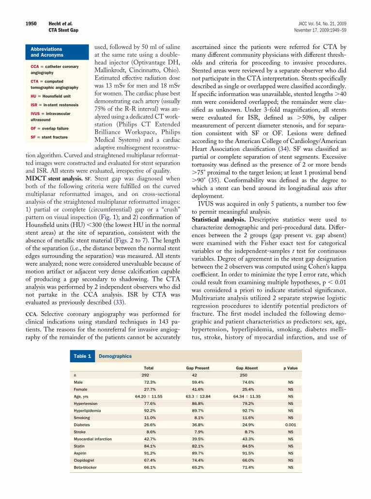
ttaMbma1pHsaoewmoane
C
ctr
amoSndImswmtaHpt��wd
tScewvvbccwMrfght
1950 Hecht et al. JACC Vol. 54, No. 21, 2009CTA Stent Gap November 17, 2009:1949–59
used, followed by 50 ml of salineat the same rate using a double-head injector (Optivantage DH,Mallinkrodt, Cincinnatto, Ohio).Estimated effective radiation dosewas 13 mSv for men and 18 mSvfor women. The cardiac phase bestdemonstrating each artery (usually75% of the R-R interval) was an-alyzed using a dedicated CT work-station (Philips CT ExtendedBrilliance Workspace, PhilipsMedical Systems) and a cardiacadaptive multisegment reconstruc-
ion algorithm. Curved and straightened multiplanar reformat-ed images were constructed and evaluated for stent separationnd ISR. All stents were evaluated, irrespective of quality.
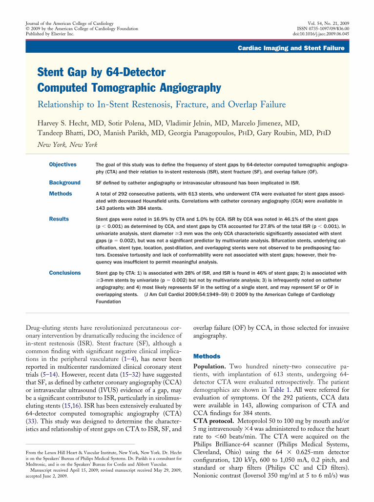
DCT stent analysis. SF. Stent gap was diagnosed whenoth of the following criteria were fulfilled on the curvedultiplanar reformatted images, and on cross-sectional
nalysis of the straightened multiplanar reformatted images:) partial or complete (circumferential) gap or a “crush”attern on visual inspection (Fig. 1); and 2) confirmation ofounsfield units (HU) �300 (the lowest HU in the normal
tent areas) at the site of separation, consistent with thebsence of metallic stent material (Figs. 2 to 7). The lengthf the separation (i.e., the distance between the normal stentdges surrounding the separation) was measured. All stentsere analyzed; none were considered unevaluable because ofotion artifact or adjacent very dense calcification capable
f producing a gap secondary to shadowing. The CTAnalysis was performed by 2 independent observers who didot partake in the CCA analysis. ISR by CTA wasvaluated as previously described (33).
CA. Selective coronary angiography was performed forlinical indications using standard techniques in 143 pa-ients. The reasons for the nonreferral for invasive angiog-aphy of the remainder of the patients cannot be accurately
DemographicsTable 1 Demographics
Total
n 292
Male 72.3%
Female 27.7%
Age, yrs 64.20 � 11.55
Hypertension 77.6%
Hyperlipidemia 92.2%
Smoking 11.0%
Diabetes 26.6%
Stroke 8.6%
Myocardial infarction 42.7%
Statin 84.1%
Aspirin 91.2%
Clopidogrel 67.4%
Abbreviationsand Acronyms
CCA � catheter coronaryangiography
CTA � computedtomographic angiography
HU � Hounsfield unit
ISR � in-stent restenosis
IVUS � intravascularultrasound
OF � overlap failure
SF � stent fracture
scertained since the patients were referred for CTA byany different community physicians with different thresh-
lds and criteria for proceeding to invasive procedures.tented areas were reviewed by a separate observer who didot participate in the CTA interpretation. Stents specificallyescribed as single or overlapped were classified accordingly.f specific information was unavailable, stented lengths �40m were considered overlapped; the remainder were clas-
ified as unknown. Under 3-fold magnification, all stentsere evaluated for ISR, defined as �50%, by calipereasurement of percent diameter stenosis, and for separa-
ion consistent with SF or OF. Lesions were definedccording to the American College of Cardiology/Americaneart Association classification (34). SF was classified as
artial or complete separation of stent segments. Excessiveortuosity was defined as the presence of 2 or more bends
75° proximal to the target lesion; at least 1 proximal bend90° (35). Conformability was defined as the degree tohich a stent can bend around its longitudinal axis aftereployment.IVUS was acquired in only 5 patients, a number too few
o permit meaningful analysis.tatistical analysis. Descriptive statistics were used toharacterize demographic and peri-procedural data. Differ-nces between the 2 groups (gap present vs. gap absent)ere examined with the Fisher exact test for categoricalariables or the independent-samples t test for continuousariables. Degree of agreement in the stent gap designationetween the 2 observers was computed using Cohen’s kappaoefficient. In order to minimize the type I error rate, whichould result from examining multiple hypotheses, p � 0.01as considered a priori to indicate statistical significance.ultivariate analysis utilized 2 separate stepwise logistic
egression procedures to identify potential predictors ofracture. The first model included the following demo-raphic and patient characteristics as predictors: sex, age,ypertension, hyperlipidemia, smoking, diabetes melli-us, stroke, history of myocardial infarction, and use of
Present Gap Absent p Value
42 250
59.4% 74.6% NS
41.6% 25.4% NS
� 12.84 64.34 � 11.35 NS
86.8% 79.2% NS
89.7% 92.7% NS
8.1% 11.6% NS
36.8% 24.9% 0.001
7.9% 8.7% NS
39.5% 43.3% NS
82.1% 84.5% NS
89.7% 91.5% NS
74.4% 66.0% NS
Gap
63.3
Beta-blocker 66.1% 65.2% 71.4% NS
soi
uaS
1951JACC Vol. 54, No. 21, 2009 Hecht et al.November 17, 2009:1949–59 CTA Stent Gap
tatins, aspirin, clopidogrel, and beta-blockers. The sec-nd model included all coronary angiography character-stics as presented in Table 2. A value of p � 0.05 was
Figure 1 Stent Gap Patterns
Normal (A), partial (B), crush (C), and complete (D, center, preceded [left] and fo
Figure 2 A 63-Year-Old Female With Recurrent Atypical Chest
(A) Computed tomographic angiography (CTA) curved multiplanar reconstruction (MPRdensity consistent with neointimal hyperplasia. (B) Catheter angiography demonstrate(arrows), but is not seen on a subsequent frame, D. (E) Cross sections obtained fromthe presence of metallic stent material. Densities in the normal area were �400 HU.
sed to indicate statistical significance in the multivariatenalyses. All statistical analyses were performed withPSS version 16.0.2 (SPSS, Chicago, Illinois). The study
[right] by normal areas).
6 Months After Placement of Overlapping Stents in the LAD
ls clear stent separation (arrow) consistent with fracture as well as luminal hypo-in-stent restenosis (arrow). (C) Stent fracture is evident on a frame without contrasttraightened MPR reveal a low HU of 192 at the separation site, incompatible withHounsfield units; LAD � left anterior descending coronary artery.
llowed
Pain
) reveas mild
the sHU �
wH
R
Tgfnmw
abCp2sCpif
0(cgtitt1dadm0sinlha
1952 Hecht et al. JACC Vol. 54, No. 21, 2009CTA Stent Gap November 17, 2009:1949–59
as approved by the Institutional Review Board of Lenoxill Hospital.
esults
he patient demographics are displayed in Table 1. Stentap was noted in 14.4%; diabetic patients were morerequently found in the SF/OF group (p � 0.001) but wasot a significant predictor in a multivariate analysis. Theean � SD interval between the CCA and CTA studiesas 57.4 � 130 days.There were 384 stents in the 143 patients with both CCA
nd CTA data, Stent gap was noted in 16.9% of the stentsy CTA. There were 4 stents with the crush pattern onTA and 1 with total separation; the remainder had theartial gap pattern. SF was observed in only 1.0% by CCA;had total and 2 had partial separation. There was a highly
ignificant association of stent gap by CTA with ISR onCA (Tables 2 and 3). There were 229 stents in the 159atients who did not proceed to CCA; stent gap was notedn 6.6% (p � 0.001 compared with those with CCA
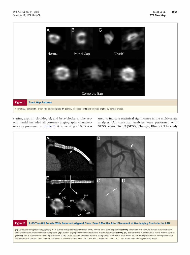
Figure 3 A 58-Year-Old Asymptomatic Male 8 Months After Pla
(A) Two areas of separation are noted on the CTA curved MPR (arrows) as well asreveals significant in-stent restenosis (ISR) (arrow). (C) A single noncontrast fram(D) Cross sections obtained from the straightened MPR reveal partial absence offormly higher HU (right). Fx � fracture; RCA � right coronary artery; other abbrevia
ollow-up). g
ISR on CCA was noted in 46.1% of stent gaps (p �.001), and stent gaps accounted for 27.8% of the total ISRp � 0.001). The HU for the gap area was 196.9 � 81.1ompared with 481.9 � 161.8 for the intact portion. Theap length was 2.3 � 0.9 mm. Stent gap agreement betweenhe 2 observers was very strong (kappa � 0.904). Stentmplantation information was available for 124 patients;here were no differences in the interval between implanta-ion and the CCA in stents with (median 618 days, range,851 days) and without (median 559 days, range 2,369ays) stent gaps. The only CCA characteristic significantlyssociated with a stent gap by univariate analysis was stentiameter �3 mm; stent gaps were present in 20.0% of �3m stents compared with 3.4% of �3-mm stents (p �
.002). By multivariate analysis, stent diameter was not aignificant predictor. Stent type, location, length, underly-ng calcification, post-dilation, and bifurcation stents wereot predisposing factors (Table 2). Excessive tortuosity and
ack of conformability were not associated with stent gaps;owever, their frequency was insufficient to permit meaningfulnalysis. There were no differences in the frequency of stent
nt of Overlapping RCA Stents
al hypodensity consistent with neointimal hyperplasia. (B) Catheter angiographylays complete separation, which is not visible on any subsequent frames.
aterial with HU �300 at both sites (left, middle). The normal area reveals uni-as in Figure 2.
ceme
lumine dispstent mtions
aps noted in single versus overlapped stents (Table 2).
HagipTb
goiaf(
(isfctm
c4sgt
tiottatmp6w
ol
1953JACC Vol. 54, No. 21, 2009 Hecht et al.November 17, 2009:1949–59 CTA Stent Gap
owever, 26.6% of the stents were in the unknown categorynd could not be classified as single or overlapped; 46.6% of theaps were noted in this group. Due to sample-size limitations,t is possible that there was not sufficient power to detectotentially significant predictors in the multivariate analysis.he sensitivity and specificity of the CTA for detection of ISRy CCA were 89.3% and 79.2%, respectively.
Figures 2 to 7 demonstrate the CTA and catheter angio-raphic characteristics of stent gaps. In Figure 2A, there isbvious separation at an overlap site on the CTA. Highlight-ng the difficulty inherent in the limited sampling of catheterngiographic analysis is the clear gap noted in 1 noncontrastrame (Fig. 2C), which is totally unapparent in a second frameFig. 2D). There was only mild ISR (Fig. 2B).
In Figure 3, there are 2 areas of stent separation by CTAFig. 3A), with significant ISR at the more proximal site. Asn Figure 2, a single noncontrast frame revealed the stenteparation on catheter angiography (Fig. 3C); all otherrames revealed a normal-appearing stent. In both cases,ross-sectional analyses (Figs. 2E and 3E) revealed HU athe gap sites that were below the threshold for metallic stent
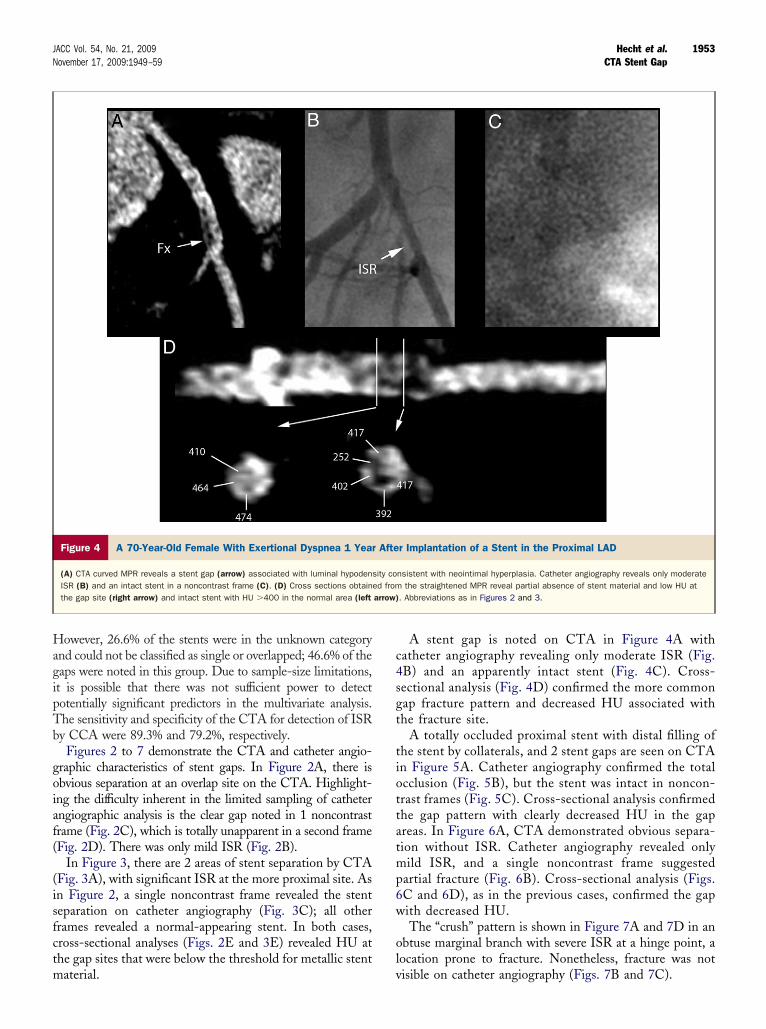
Figure 4 A 70-Year-Old Female With Exertional Dyspnea 1 Year
(A) CTA curved MPR reveals a stent gap (arrow) associated with luminal hypodenISR (B) and an intact stent in a noncontrast frame (C). (D) Cross sections obtainthe gap site (right arrow) and intact stent with HU �400 in the normal area (left
aterial. v
A stent gap is noted on CTA in Figure 4A withatheter angiography revealing only moderate ISR (Fig.B) and an apparently intact stent (Fig. 4C). Cross-ectional analysis (Fig. 4D) confirmed the more commonap fracture pattern and decreased HU associated withhe fracture site.
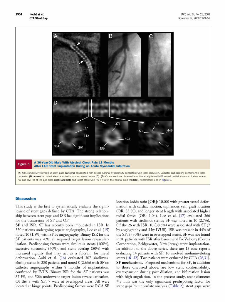
A totally occluded proximal stent with distal filling ofhe stent by collaterals, and 2 stent gaps are seen on CTAn Figure 5A. Catheter angiography confirmed the totalcclusion (Fig. 5B), but the stent was intact in noncon-rast frames (Fig. 5C). Cross-sectional analysis confirmedhe gap pattern with clearly decreased HU in the gapreas. In Figure 6A, CTA demonstrated obvious separa-ion without ISR. Catheter angiography revealed onlyild ISR, and a single noncontrast frame suggested
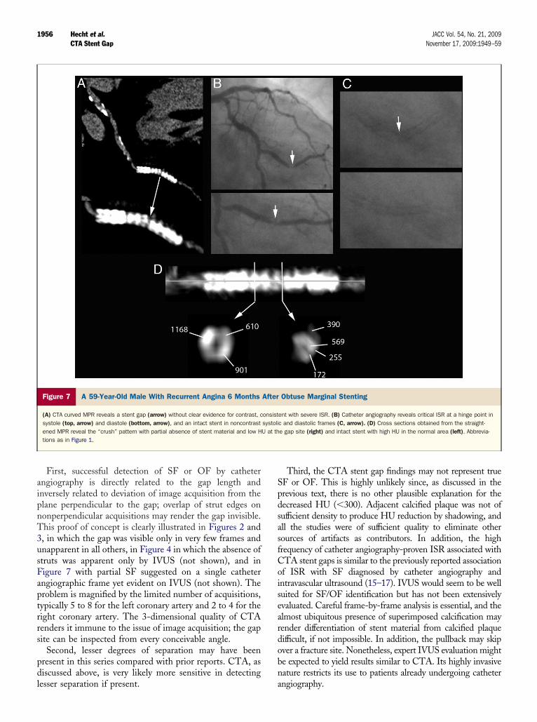
artial fracture (Fig. 6B). Cross-sectional analysis (Figs.C and 6D), as in the previous cases, confirmed the gapith decreased HU.The “crush” pattern is shown in Figure 7A and 7D in an
btuse marginal branch with severe ISR at a hinge point, aocation prone to fracture. Nonetheless, fracture was not
r Implantation of a Stent in the Proximal LAD
nsistent with neointimal hyperplasia. Catheter angiography reveals only moderatethe straightened MPR reveal partial absence of stent material and low HU at
. Abbreviations as in Figures 2 and 3.
Afte
sity coed fromarrow)
isible on catheter angiography (Figs. 7B and 7C).
D
TisfS5nSieidecc3Ol
lm(rpObtiCIesStow�
1954 Hecht et al. JACC Vol. 54, No. 21, 2009CTA Stent Gap November 17, 2009:1949–59
iscussion
his study is the first to systematically evaluate the signif-cance of stent gaps defined by CTA. The strong relation-hip between stent gaps and ISR has significant implicationsor the occurrence of SF and OF.F and ISR. SF has recently been implicated in ISR. In30 patients undergoing repeat angiography, Lee et al. (15)oted 10 (1.8%) with SF by angiography. Binary ISR for theF patients was 70%; all required target lesion revascular-
zation. Predisposing factors were sirolimus stents (100%),xcessive tortuosity (40%), and stent overlap (50%) withncreased rigidity that may act as a fulcrum for metaleformation. Aoki et al. (16) evaluated 307 sirolimus-luting stents in 280 patients and noted 8 (2.6%) with SF onatheter angiography within 8 months of implantation,onfirmed by IVUS. Binary ISR for the SF patients was7.5%, and 50% underwent target lesion revascularization.f the 8 with SF, 7 were at overlapped areas. All were
Figure 5 A 36-Year-Old Male With Atypical Chest Pain 18 MonAfter LAD Stent Implantation During an Acute Myocar
(A) CTA curved MPR reveals 2 stent gaps (arrows) associated with severe luminaocclusion (B, arrow); an intact stent is noted in a noncontrast frame (C). (D) Crosrial and low HU at the gap sites (right and left) and intact stent with HU �400 in
ocated at hinge points. Predisposing factors were RCA SF s
ocation (odds ratio [OR]: 10.00) with greater vessel defor-ation with cardiac motion, saphenous vein graft location
OR: 35.88), and longer stent length with associated higheradial forces (OR: 1.04). Lee et al. (17) evaluated 366atients with sirolimus stents; SF was noted in 10 (2.7%).f the 26 with ISR, 10 (38.5%) were associated with SF (7
y angiography and 3 by IVUS). ISR was present in 44% ofhe SF; 3 (30%) were in overlapped stents. SF was not foundn 30 patients with ISR after bare-metal Bx Velocity (Cordisorporation, Bridgewater, New Jersey) stent implantation.
n addition to the above series, there are 13 case reportsvaluating 14 patients with SF: 10 involved sirolimus-elutingtents (18–32). Two patients were evaluated by CTA (28,31).F mechanisms. Proposed mechanisms for SF, in addition
o those discussed above, are low stent conformability,verexpansion during post-dilation, and bifurcation lesionith high angulation. In the present study, stent diameter3 mm was the only significant predisposing factor for
nfarction
ensity consistent with total occlusion. Catheter angiography confirms the totaltions obtained from the straightened MPR reveal partial absence of stent mate-rmal area (middle). Abbreviations as in Figure 2.
thsdial I
l hypods secthe no
tent gaps by univariate analysis (Table 2); stent gaps were

p�itcrr
mtgliwCcodamaTa
fowsdggiaaso
sbfLis2b
1955JACC Vol. 54, No. 21, 2009 Hecht et al.November 17, 2009:1949–59 CTA Stent Gap
resent in 20.0% of �3-mm stents compared with 3.4% of3-mm stents (p � 0.002). However, it was not a signif-
cant predictor in multivariate analysis. Stent length, loca-ion and type, overlapped stents, post-dilation, underlyingalcification, and bifurcation stents were not significantlyelated. Excessive tortuosity and lack of conformability werearely noted, and their contribution could not be evaluated.
The high incidence of ISR associated with stent gapsost likely results from the absence of drug-elution protec-
ion from neointimal hyperplasia, or a drug-free zone, at theap site. Other possibilities include broken struts causingocal mechanical stimulation of the vessel wall, resulting innflammation and development of intimal hyperplasia, asell as local uncovered unstable plaque.TA diagnosis of SF or OF. The ability of CTA to
onfidently identify stent gaps is dependent not just on dem-nstration of a “gap,” which may be more apparent than real,epending on the window settings. Rather, the gap must bessociated with HUs below the minimum density of stentaterial, which is independent of window center and width,
nd the absence of artifact that may contribute to this finding.he study must be carefully evaluated for shadowing effects of
Figure 6 A 62-Year-Old Male With Dyspnea 9 Months After Ove
(A) CTA curved MPR reveals obvious separation without ISR (arrow). (B) Catheter angfracture (bottom) (arrows). (C and D) Cross-sectional analysis confirmed the gright) stent segments.
djacent dense calcification and motion artifacts. i
In the absence of artifacts, the most likely explanationsor the hypodense gap are fracture- or OF-related absencef strut material. Overinflation, with spreading of strutsithout true fracture, cannot be excluded. Bifurcation
tents, with inflation into a side branch and possible strutamage, were not associated with a higher incidence of stentaps (Table 2). The absence of strut material for a distancereater than the normal average interstrut distance (1 mm)s convincing evidence for an uncovered portion of thertery. The 46% incidence of ISR in stent gaps and the 28%ssociation of stent gaps with ISR support the pathologicignificance of this observation and the likely presence of SFr OF, even though there is no confirmatory gold standard.The most common gap was a hypodense partial gap
uggestive of incomplete SF. The crush pattern, manifestedy a flattening of the stent with a hypodense gap, was lessrequent. Only 1 case of total separation was noted.imitations of catheter angiography and IVUS. The
nfrequency of catheter angiographic identification of stenteparation in the present series (1.0%) is similar to the 1.8% to.7% in previously reported studies (15–17). The discrepancyetween the CTA (16.9%) and catheter angiography frequency
ing LAD Stent Implantation
hy revealed only mild ISR (top) and a single noncontrast frame suggested partialdecreased HU (D, middle), compared with proximal (D, left) and distal (D,
rlapp
iograpap with
n this and previous reports has several possible explanations.
aipnT3usFaptrrs
pdl
SpdsasfCoiseardobn
1956 Hecht et al. JACC Vol. 54, No. 21, 2009CTA Stent Gap November 17, 2009:1949–59
First, successful detection of SF or OF by catheterngiography is directly related to the gap length andnversely related to deviation of image acquisition from thelane perpendicular to the gap; overlap of strut edges ononperpendicular acquisitions may render the gap invisible.his proof of concept is clearly illustrated in Figures 2 and, in which the gap was visible only in very few frames andnapparent in all others, in Figure 4 in which the absence oftruts was apparent only by IVUS (not shown), and inigure 7 with partial SF suggested on a single catheterngiographic frame yet evident on IVUS (not shown). Theroblem is magnified by the limited number of acquisitions,ypically 5 to 8 for the left coronary artery and 2 to 4 for theight coronary artery. The 3-dimensional quality of CTAenders it immune to the issue of image acquisition; the gapite can be inspected from every conceivable angle.
Second, lesser degrees of separation may have beenresent in this series compared with prior reports. CTA, asiscussed above, is very likely more sensitive in detecting
Figure 7 A 59-Year-Old Male With Recurrent Angina 6 Months
(A) CTA curved MPR reveals a stent gap (arrow) without clear evidence for contrast, csystole (top, arrow) and diastole (bottom, arrow), and an intact stent in noncontrastened MPR reveal the “crush” pattern with partial absence of stent material and low Htions as in Figure 1.
esser separation if present. a
Third, the CTA stent gap findings may not represent trueF or OF. This is highly unlikely since, as discussed in therevious text, there is no other plausible explanation for theecreased HU (�300). Adjacent calcified plaque was not ofufficient density to produce HU reduction by shadowing, andll the studies were of sufficient quality to eliminate otherources of artifacts as contributors. In addition, the highrequency of catheter angiography-proven ISR associated withTA stent gaps is similar to the previously reported associationf ISR with SF diagnosed by catheter angiography andntravascular ultrasound (15–17). IVUS would seem to be welluited for SF/OF identification but has not been extensivelyvaluated. Careful frame-by-frame analysis is essential, and thelmost ubiquitous presence of superimposed calcification mayender differentiation of stent material from calcified plaqueifficult, if not impossible. In addition, the pullback may skipver a fracture site. Nonetheless, expert IVUS evaluation mighte expected to yield results similar to CTA. Its highly invasiveature restricts its use to patients already undergoing catheter
Obtuse Marginal Stenting
ent with severe ISR. (B) Catheter angiography reveals critical ISR at a hinge point inc and diastolic frames (C, arrow). (D) Cross sections obtained from the straight-e gap site (right) and intact stent with high HU in the normal area (left). Abbrevia-
After
onsistsystoliU at th
ngiography.
C(ISia
tnlsii2tswofIeSr
hic anght coro
R
I
1957JACC Vol. 54, No. 21, 2009 Hecht et al.November 17, 2009:1949–59 CTA Stent Gap
The lower incidence of stent gap in patients not referred forCA compared with those who underwent invasive evaluation
6.6% vs. 16.9%, p � 0.001) may reflect a lower incidence ofSR-related symptoms requiring further testing.F versus OF. In patients with a gap in a single stent, SF
s the most likely explanation. In prior reports of fracture atn overlap site, stent separation has always been attributed
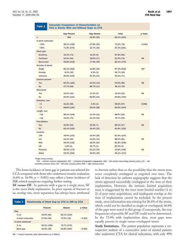
Univariate Comparison of Characteristics onCCA in Stents With and Without Gaps on CTATable 2 Univariate Comparison of CharacterCCA in Stents With and Without Ga
Gap Present G
n 384
In-stent restenosis
�50% 28.1% (108)
�50% 71.9% (276)
Stent type
Sirolimus 19.3% (74)
Paclitaxel 24.0% (92)
Bare-metal 56.8% (218)
Number of stents
Single 65.1% (250)
Overlap 8.3% (32)
Unknown 26.6% (102)
Calcium present
Yes 52.3% (102)
No 47.7% (93)
Bifurcation
Yes 15.5% (30)
No 84.5% (163)
Diameter, mm
�3 31.2% (59)
�3 68.8% (130)
Length, mm
�18 58.1% (104)
�18 41.9% (75)
Post-dilation
Yes 33.9% (64)
No 66.1% (125)
Location
LAD 39.9% (153)
LCx 24.0% (92)
RCA 34.5% (132)
LM 1.6% (6)
Proximal 45.5% (172)
Distal 54.5% (206)
*Single versus overlap.CCA � catheter coronary angiography; CTA � computed tomograp
circumflex coronary artery; LM � left main coronary artery; RCA � rig
elationship of Stent Gap by CTA to ISR by CCATable 3 Relationship of Stent Gap by CTA to ISR by CCA
Present Absent p Value
Gap
% (n) 16.9% (65) 83.1% (319)
In-stent restenosis 27.8% (30) 72.2% (78) �0.001
In-stent restenosis
% (n) 29.3% (108) 70.7% (276)
Stent gap 46.2% (30) 53.8% (350) �0.001
wSR � in-stent restenosis; other abbreviations as in Table 2.
o fracture rather than to the possibility that the stents wereever completely overlapped or migrated over time. The
ack of detection by catheter angiography suggests that thetents appeared successfully overlapped at the time of theirmplantation. However, the intrinsic limited acquisitionssue is exaggerated by the even more limited number (1 to) of post-stent acquisitions, and inadequate overlap at theime of implantation cannot be excluded. In the presenttudy, stent information was missing for 26.6% of the stents,hich could not be classified as single or overlapped; 46.6%f the gaps were noted in this group. Consequently, the truerequencies of possible SF and OF could not be determined.n the 73.4% with implantation data, stent gaps werequally present in single versus overlapped stents.tudy limitations. The patient population represents a ret-ospective analysis of a consecutive series of stented patients
onCTA
sent Total p Value
(65) 83.1% (319)
(30) 72.2% (78) �0.001
(35) 87.3% (241)
(9) 87.8% (65)
(17) 81.5% (75) NS
(39) 82.1% (179)
(32) 87.2% (218) NS*
(2) 93.7% (30)
(31) 69.6% (71)
(13) 53.9% (89) NS
(17) 46.1% (76)
(5) 15.2% (25) NS
(24) 84.8% (139)
(2) 96.6% (57) 0.002
(26) 80.0% (104)
(12) 88.5% (92) NS
(16) 78.7% (59)
(7) 89.1% (57) NS
(23) 81.6% (102)
(29) 81.0% (124)
(11) 88.0% (81) NS
(24) 81.8% (108)
(1) 83.3% (5)
(34) 43.9% (138) NS
(30) 56.1% (176)
iography; LAD � left anterior descending coronary artery; LCx � leftnary artery.
isticsps on
ap Ab
16.9%
27.8%
12.7%
12.2%
18.5%
17.9%
12.8%
6.3%
30.4%
43.3%
56.7%
17.2%
82.8%
3.4%
20.0%
11.5%
21.3%
10.9%
18.4%
19.0%
12.0%
18.2%
16.7%
53.1%
46.9%
ho underwent CTA for clinical indications, with only 49%
uoCsgispHiaceigoCpbIvitpesftivTacd
AHN
R
1
1
1
1
1
1
1
1
1
1
2
2
2
2
2
2
2
2
2
2
3
3
1958 Hecht et al. JACC Vol. 54, No. 21, 2009CTA Stent Gap November 17, 2009:1949–59
ndergoing CCA, rather than a prospective, consecutive seriesf patients who underwent stenting with follow-up CCA andTA. Consequently, there is significant selection bias, and the
tudy very likely overestimates the incidence of SF or OF in theeneral population of stented patients. Reflecting the problemsnherent in tertiary referral centers, in which data regardingtents implanted elsewhere may not be available, is the incom-lete stent implantation data in those who underwent CCA.owever, sufficient numbers of stents were available for mean-
ngful analysis, and this study is the first to address this topic inlarge series of patients. SF and OF cannot be absolutely
onfirmed in the absence of postmortem examination. How-ver, the absence of other plausible explanations, the 46%ncidence of ISR in stent gaps, and the 28% association of stentaps with ISR support the pathologic significance of thisbservation.linical implications. The incidence of stent gaps withossible fracture or OF in a large series of stents evaluatedy CTA and CCA was 16.9%, representing 28% of the totalSR population; ISR was present in 46% of SF/OF. Thisery strong association of stent gaps with ISR suggests that,n patients with drug-eluting stents, it may not be failure ofhe drug-eluting compound to prevent neointimal hyper-lasia that is responsible for all ISR, but rather lack ofxposure of the arterial segment to the compound at a gapite in a substantial number. Greater emphasis on thoseactors that promote SF and on manufacturing techniqueshat prevent SF appear to be in order. In overlapping stentmplantations, IVUS may prove to be a reliable tool forerifying the accuracy of overlap at the time of insertion.he inability of catheter angiography to detect stent gaps
nd the highly invasive nature of IVUS, and its potentialonfounding by calcified plaque, suggest that CTA is theiagnostic procedure of choice.
ddress for correspondence: Dr. Harvey S. Hecht, Lenox Hilleart and Vascular Institute, 130 East 77th Street, New York,ew York 10021. E-mail: [email protected].
EFERENCES
1. Sacks BA, Miller A, Gottlieb M. Fracture of an iliac artery Palmazstent. J Vasc Interv Radiol 1996;7:53–5.
2. Scheinert D, Scheinert S, Sax J, et al. Prevalence and clinical impact ofstent fractures after femoropopliteal stenting. J Am Coll Cardiol2005;45:312–5.
3. Duda SH, Pusich B, Richter G, et al. Sirolimus-eluting stents for thetreatment of obstructive superficial femoral artery disease: six-monthresults. Circulation 2002;106:1505–9.
4. Phipp LH, Scott DJ, Kessel D, Robertson I. Subclavian stents andstent-grafts: cause for concern? J Endovasc Surg 1999;6:223–6.
5. Morice MC, Serruys PW, Sousa JE, et al. A randomized comparisonof a sirolimus-eluting stent with a standard stent for coronary revas-cularization. N Engl J Med 2002;346:1773–80.
6. Moses JW, Leon MB, Popma JJ, et al. Sirolimus-eluting stents versusstandard stents in patients with stenosis in a native coronary artery.N Engl J Med 2003;349:1315–23.
7. Stone GW, Ellis SG, Cox DA, et al. A polymer-based, paclitaxel-
eluting stent in patients with coronary artery disease. N Engl J Med2004;350:221–31.8. Schofer J, Schluter M, Gershlick AH, et al. Sirolimus-eluting stentsfor treatment of patients with long atherosclerotic lesions in smallcoronary arteries: double-blind, randomised controlled trial (E-SIRIUS). Lancet 2003;362:1093–9.
9. Schampaert E, Cohen EA, Schluter M, et al. The Canadian study ofthe sirolimus-eluting stent in the treatment of patients with long denovo lesions in small native coronary arteries (C-SIRIUS). J Am CollCardiol 2004;43:1110–5.
0. Hong MK, Mintz GS, Lee CW, et al. Paclitaxel coating reduces in-stentintimal hyperplasia in human coronary arteries: a serial volumetric intra-vascular ultrasound analysis from the Asian Paclitaxel-Eluting StentClinical Trial (ASPECT). Circulation 2003;107:517–20.
1. Gershlick A, De Scheerder I, Chevalier B, et al. Inhibition ofrestenosis with a paclitaxel-eluting, polymer-free coronary stent: theEuropean evaLUation of pacliTaxel Eluting Stent (ELUTES) trial.Circulation 2004;109:487–93.
2. Grube E, Silber S, Hauptmann KE, et al. TAXUS I: six- andtwelve-month results from a randomized, double-blind trial on aslow-release paclitaxel-eluting stent for de novo coronary lesions.Circulation 2003;107:38–42.
3. Colombo A, Drzewiecki J, Banning A, et al., TAXUS II Study Group.Randomized study to assess the effectiveness of slow- and moderate-release polymer-based paclitaxel-eluting stents for coronary arterylesions. Circulation 2003;108:788–94.
4. Lansky A, Costa RA, Mintz GS, Tsuchiya Y, et al. Non-polymer-basedpaclitaxel-coated coronary stents for the treatment of patients with denovo coronary lesions. Angiographic follow-up of the DELIVER clinicaltrial. Circulation 2004;109:1948–54.
5. Lee MS, Jurewitz D, Aragon J, et al. Stent fracture associated withdrug-eluting stents: clinical characteristics and implications. CatheterCardiovasc Interv 2007;69:387–94.
6. Aoki J, Nakazawa G, Tanabe K, Hoye A, et al. Incidence and clinicalimpact of coronary stent fracture after sirolimus-eluting stent implan-tation. Catheter Cardiovasc Interv 2007;69:380–6.
7. Lee S, Park J, Shin D, Kim Y, et al. Frequency of stent fracture as acause of coronary restenosis after sirolimus-eluting stent implantation.Am J Cardiol 2007;100:627–30.
8. Chowdhury PS, Ramos RG. Images in clinical medicine. Coronary-stent fracture. N Engl J Med 2002;347:581.
9. Brilakis ES, Maniu C, Wahl M, Barsness G. Unstable angina due tostent fracture. J Invasive Cardiol 2004;16:545.
0. Sianos G, Hofma S, Ligthart JM, et al. Stent fracture and restenosis inthe drug-eluting stent era. Catheter Cardiovasc Interv 2004;61:111–6.
1. Halkin A, Carlier S, Leon MB. Late incomplete lesion coveragefollowing Cypher stent deployment for diffuse right coronary arterystenosis. Heart 2004;90:e45.
2. Hamilos MI, Papafaklis MI, Ligthart JM, et al. Stent fracture and restenosisof a paclitaxel-eluting stent. Hellenic J Cardiol 2005;46:439–42.
3. Surmely JF, Kinoshita Y, Dash D, et al. Stent strut fracture-inducedrestenosis in a bifurcation lesion treated with the crush stentingtechnique. Circ J 2006;70:936–8.
4. Min PK, Yoon YW, Moon Kwon H. Delayed strut fracture ofsirolimus-eluting stent: a significant problem or an occasional obser-vation? Int J Cardiol 2006;106:404–6.
5. Park JS, Shin DG, Kim YJ. Fractured DES with a patent coronaryartery: clinical implications. J Invasive Cardiol 2007;19:E43–5.
6. Wilczynska J, Rdzanek A, Kochman J, et al. Sirolimus eluting stentfracture following angioplasty of diffuse in-stent restenosis in the rightcoronary artery. Int J Cardiol 2007;118:126–7.
7. Makaryus AN, Lefkowitz L, Lee ADK. Coronary artery stent fracture.Int J Cardiovasc Imaging 2007;23:305–9.
8. Zaizen H, Tamura A, Miyamoto K, et al. Complete fracture ofsirolimus-eluting stent detected by multislice computed tomography. JInt J Cardiol 2007;118:120–1.
9. Leong DP, Dundon BK, Puri R, Yeend RAS. Very late stent fractureassociated with a sirolimus-eluting stent. Heart Lung Circ 2008;17:426–8.
0. Jin X, Zhang S, Xie H, et al. Strut fracture of DES: An increasingproblem? Int J Cardiol 2007;118:e54–6.
1. Kuboyama O, Takei H, Tokunaga T. Strut fracture of a sirolimus-eluting stent at the ostium of the right coronary artery. Heart
2007;93;1608.
3
3
3
3
1959JACC Vol. 54, No. 21, 2009 Hecht et al.November 17, 2009:1949–59 CTA Stent Gap
2. Shite J, Matsumoto D, Yokoyama M. Sirolimus-eluting stent fracturewith thrombus, visualization by optical coherence tomography. EurHeart J 2006;27:1389.
3. Hecht HS, Zaric M, Jelnin V, et al. Usefulness of 64-detectorcomputed tomographic angiography for diagnosing in-stent restenosisin native coronary arteries. Am J Cardiol 2008;101:820–4.
4. Ryan TJ, Faxon DP, Gunnar RM, et al. Guidelines for percutaneoustransluminal coronary angioplasty: a report of the American College of
on Percutaneous Transluminal Coronary Angioplasty). Circulation1988;78:486–502.
5. Freed FS, Safian RD. Proximal vessel tortuosity and angulatedlesions. In: Safian RD, Freed MS, editors. Manual of InterventionalCardiology. 3rd edition. Royal Oak, MI: Physician’s Press, 2001:237– 43.
Cardiology/American Heart Association Task Force on Assessment ofDiagnostic and Therapeutic Cardiovascular Procedures (Subcommittee
Key Words: stent fracture y stent gap y overlap failure y in-stentrestenosis y computed tomographic angiography.


