Embed Size (px)
Citation preview
Stress-induced differences in primary and secondary resistanceagainst bacterial sepsis corresponds with diverse corticotropinreleasing hormone receptor expression by pulmonary CD11c+
MHC II+ and CD11c− MHC II+ APCs
Xavier F. Gonzales1, Aniket Desmutkh1, Mark Pulse1, Khaisha Johnson2, and Harlan P.Jones1,*1 Department of Molecular Biology and Immunology, University of North Texas Health ScienceCenter in Fort Worth, Fort Worth, Texas 761072 Department of Biology, Fisk University, Chicago, IL
AbstractStress responses have been associated with altered immunity and depending upon the type of stressor,can have diverse effects on disease outcomes. As the first line of defense against potential pathogens,alterations in cellular immune responses along the respiratory tract can have a significant impact onthe manifestation of local and systemic disease. Utilizing a murine model of respiratory pneumonia,the current study investigated the effects of restraint stress on the induction of primary and secondaryimmunity along the respiratory tract, influencing host susceptibility. Female CD-1 mice weresubjected to three hours of restraint stress over a period of four days followed by primary andsecondary Streptococcus pneumoniae infection via intranasal route. Stress exposure led to increasedretention of bacterial carriage in the lungs, enhanced polymorphonuclear cells and a preferentialdecrease in pulmonary CD11c+ MHC II+ cells resulting in delayed lethality during primary infectionbut significant impairment of acquired immune protection after secondary infection. We also provideevidence to support a role for lung-associated corticotrophin releasing hormone regulation throughperipheral CRH and diverse CRH receptor expression by MHC II+ antigen presenting cells (APCs).We conclude that repeated restraint stress has distinct influences on immune cell populations thatappear to be important in the generation of innate and adaptive immune responses along therespiratory tract with the potential to influence local and systemic protection against diseasepathogenesis.
Keywordsrestraint stress; respiratory immunity; antigen presentation; corticotrophin releasing hormone
Correspondence: Harlan P. Jones, 3500 Camp Bowie Boulevard, Department of Molecular Biology and Immunology, Phone: (817)735-2448, Email: [email protected]'s Disclaimer: This is a PDF file of an unedited manuscript that has been accepted for publication. As a service to our customerswe are providing this early version of the manuscript. The manuscript will undergo copyediting, typesetting, and review of the resultingproof before it is published in its final citable form. Please note that during the production process errors may be discovered which couldaffect the content, and all legal disclaimers that apply to the journal pertain.
NIH Public AccessAuthor ManuscriptBrain Behav Immun. Author manuscript; available in PMC 2010 April 5.
Published in final edited form as:Brain Behav Immun. 2008 May ; 22(4): 552–564. doi:10.1016/j.bbi.2007.11.005.
NIH
-PA Author Manuscript
NIH
-PA Author Manuscript
NIH
-PA Author Manuscript
IntroductionExposure to stressful situations has been associated with health status (Fava et al., 2007;McClellan and Cohen, 2007; Schindler, 1985; Yang and Glaser, 2002), lending credence tothe belief that communication between the neuroendocrine and immune systems play a majorrole in disease susceptibility. Evidence to support this notion originate from previous in vitroand in vivo studies demonstrating that neuroendocrine response elements of the central andperipheral nervous systems affect immune function including: cellular proliferation,differentiation, cytolytic function and cytokine secretion (Bryndina and Danilov, 2002;Calcagni and Elenkov, 2006; Dhabhar and McEwen, 1996; Dhabhar et al., 1996; Emeny et al.,2007; Engler et al., 2005b; Leu and Singh, 1993; Saha et al., 2001; Sternberg, 2006; Weiss etal., 1989). In this regard, stress-induced immune modulation has been found to be diverse.Experimentally, studies have documented stress susceptibility to predict immune competencebased upon strain and gender differences (Li et al., 2000; Sieve et al., 2006). For example, CKiank et al. demonstrated disease susceptibility to be greater in male mice, highlightingdifferences in stress responsiveness and inherent hormonal and behavioral dispositions (Kianket al., 2006). In addition, numerous studies have characterized stress-immune relationships tobe dependent upon gender, neuroendocrine response factors, the type and quality of the stressevent, antigenic stimuli and host genetic factors (Butcher and Lord, 2004; Engler et al.,2005b; Gross and Siegel, 1981; Ishihara et al., 1999; Ising and Holsboer, 2006; Matalka,2003; Okuyama et al., 2007). Furthermore, studies have shown that stress effects to beassociated with regional differences in the generation of immune responses (Engler et al.,2005b). Thus, it has been predicted that defining the relationships between neuroendocrine andimmune function during disease to be very complex.
As a primary site of pathogenic exposure, immune regulation along the respiratory tract playsa major protective role against potential local and systemic disease. Importantly, because therespiratory tract is constantly exposed to foreign stimuli, the respiratory immune defensesystem must be able to temper its’ activation between innocuous antigen and potentialpathogens. Stress exposures have been implicated in the pathogenesis of numerous respiratorydisease states: including asthma, bacterial pneumonia, viral infection and tumor development(Boyce et al., 1995; Bryndina and Danilov, 2002; Datti et al., 2002; Engler et al., 2005a;Joachim et al., 2003; Konstantinos and Sheridan, 2001; MacQueen and Bienenstock, 2006;Okuyama et al., 2007). Yet, the mechanisms involved in the activation of neuroendocrineresponses that influence the regulation of respiratory immunity remain uncertain. Our missionin understanding the relationships of stress and respiratory immunity is to define how stressmediates cell-specific responses that predict protective verses immunopathogenesis,influencing disease susceptibility.
Utilizing various in vivo experimental models of stress, researchers have made progress in thisarea. For example, previous studies have documented the role that stress has in mediation ofviral infection and tumor defenses along the respiratory tract by demonstrating the influenceof stress on cell-mediated immune responses and NK cell activity (Engler et al., 2005a;Hunzeker et al., 2004; Konstantinos and Sheridan, 2001; Sheridan et al., 2000; Strange et al.,2000). In particular, Tseng et. al. demonstrated that susceptibility to experimental viralinfluenza infection was associated with glucocorticoid and opiod-induced suppression of NKactivity in response to restraint stress (Tseng et al., 2005). Likewise, studies have shown thatin conditions such as asthma, neuroendocrine activation can promote polarization of cell-mediated immune responses, facilitating a hyperactive immune reaction toward innocuousantigens (Datti et al., 2002; Okuyama et al., 2007; Portela Cde et al., 2001). A study by Joachimet. al. showed that stress-induced exacerbation of asthma was associated with a biasedactivation of Th2 cellular immune responses including: elevations in IL-4 and IL-5 cytokineactivity and eosinophillia (Joachim et al., 2003). These and other published studies reveal that
Gonzales et al. Page 2
Brain Behav Immun. Author manuscript; available in PMC 2010 April 5.
NIH
-PA Author Manuscript
NIH
-PA Author Manuscript
NIH
-PA Author Manuscript
proper orchestration of different cellular components of the immune system in response tovarious infectious or non-pathogenic insults can be influenced by neuroendocrine activation.
As a leading cause of community acquired respiratory pneumonia, Streptococcus pneumoniae(S. pneumoniae) infection poses a serious problem for the health care system. Despitetherapeutic advances, effective vaccines against recurrent infections are unavailable andtherefore resistance against disease hinges greatly on host natural immunity. Protection againstS. pneumoniae infection involves the activation of the innate and adaptive compartments ofthe immune defense system. In that proper immune activation can determine the outcome ofdisease, it is conceivable that stress-associated alterations in immune responsiveness can havea significant impact on host susceptibility. Support for this idea is found in studiesdemonstrating that children, elderly, hospitalized patients and the immunocompromised,experiencing stressful conditions correlated with increased risk of respiratory-related disease(Butcher and Lord, 2004; Chiang et al., 2007; Ginesu and Pirina, 1995; Graham et al., 2006;Hortal et al., 2007; McClellan and Cohen, 2007). Few experimental studies have investigatedthe relationships between stress and S. pneumoniae infection. In on such study, Murray et. al.demonstrated compensatory effects of cellular host immune defenses was associated withsusceptibility to S. pneumoniae infection in a model of CRH expression (Murray et al.,2001). Using an experimental model of stress and pneumococcal respiratory infection in mice,the purpose of the current study was to determine the influence of stress on cellular immunityalong the respiratory tract and how potential alterations would predict disease susceptibility.In this study, we revealed that stress-induced differences in protective responses during primaryand secondary pneumococcal infection are associated with preferences in cellular immuneresponsiveness along the respiratory tract. We also provide evidence to support a role for lung-associated diversity of corticotrophin releasing hormone (CRH) and CRH receptor expressionby antigen presenting cells (APCs), as a potential link in the induction of acquired immuneprotection in during exposure to stressful events.
Materials and methodsMice
Adult (7–8 weeks of age) female CD-1 mice (Harlan-Sprague Dawley, Indianapolis, Indiana)were used in all studies. Mice were maintained under specific pathogen-free conditions on a12:12 light/dark cycle (7:00 PM to 7:00 AM). Mice were housed 5 to a cage under optimaltemperature and humidity controlled conditions and provided proper care as directed by theinstitutional animal care and use committee. All mice were allowed to acclimate to the colonyuntil of age prior to experiment.
Stress paradigmRestraint stress (RS) was induced in mice by placing them in a sterile 50 ml conical tubesupplied with air holes for sufficient ventilation. All mice were stressed daily for 4 daysperformed for 3 hrs exactly between the hours of 1:00 PM and 4:00 PM. No food or water wassupplied to subjects during the stress period, including non-restraint stress (NRS) animals,which remained in their home cage environment.
Bacteria and infectionStreptococcus pneumoniae strain #6301 (ATCC, Manassas, VA) was grown overnight toachieve mid-log phase cultures on Blood Agar plates. Mice were infected with S.pneumoniae (106 cells) in a volume of 40 μl of Todd Hewitt broth (Sigma, St. Louis, MO) byintranasal inoculation. Similar groups of mice were inoculated with Todd Hewitt broth (SigmaAldrich) to serve as vehicle control. All inoculations were performed during the same time inwhich the stress paradigm was performed. Prior to infection and sacrifice, mice were
Gonzales et al. Page 3
Brain Behav Immun. Author manuscript; available in PMC 2010 April 5.
NIH
-PA Author Manuscript
NIH
-PA Author Manuscript
NIH
-PA Author Manuscript
anesthetized by intramuscular injection of ketamine-xylazine. In selected experiments survivalanalysis was performed.
Colony forming assayBacterial loads in lung, spleen and blood were evaluated at different time points after S.pneumoniae infection. After sacrifice, organs were harvested and homogenized in PBS. Ten-fold serial dilutions of organ homogenates were plated in triplicate onto blood agar plates andincubated at 37° C overnight. Colonies on plates were enumerated, and the results wereexpressed as log10 CFU per μl.
Cell IsolationBronchiolar lavage cells were isolated from bronchiolar lavage fluid (BALF). Specifically,lungs were perfused twice via tracheal instillation of 1–2 ml of wash media using 25 gaugeblunted syringe needle. BALF containing cells was aspirated and the volume of fluid retrievedwas recorded. Single cell suspensions from lung tissue were prepared as previously described(Jones et al., 2001). Briefly, lungs were infused with sterile PBS to eliminate contaminatingRBCs. Lung tissue was separated into single lobes finely minced and placed into digestionmedium containing 500 units/ml collagenase type II (Worthington, Lakewood, NJ.) and 50units/ml DNAse (Sigma Aldrich, St. Louis, MO). Lung tissue was digested for 1.5 hr per lung.After digestion, lung digestions were passed through a nylon mesh filter (LabPak, Depew,NY.) into sterile 15 ml conical tubes and washed 2X in wash media (Hyclone, Logan, UT.).Lymphocytes were prepared by ficoll-hypaque (Lympholyte M, Cedarlane, Laboratories, ltd.,Ontario, CA) centrifugation. Spleen cell suspensions were prepared by pressing spleen tissuethrough sterile mesh. Contaminating RBCs were removed using ACK lysis buffer as previouslydescribed (Kruisbeek, 1999). Whole blood samples were prepared from cardiac puncture andplaced into sterile tubes containing heparin-sulfate to impede coagulation.
Flow cytometryThe phenotype of immune cells was determined by characterization of cell surface markersusing flow cytometric methods. Lung single cell suspensions were washed and incubated withan optimal concentration of anti-FcBlock Antibody (clone 2.4G2) (BD PharMingen, SanDiego, CA) to block non-specific binding to FcRs for 30 min. Two-color immunofluorescentstaining was performed by incubating cells with combinations of pre-tittered concentrationsof the following antibodies: anti-PE GR1+, anti-FITC MHC II+, anti-FITC F4/80+, anti-FITCCD11b+, anti-PE CD3+, anti-FITC CD4+ or anti-FITC-CD8+ and anti-PE-Cy7 CD45R.Detection of CD11c+ cells was performed using biotin-conjugate anti-CD11c+ antibodyfollowed by secondary labeling using streptavidin Texas Red conjugate. Antibodies werepurchased from BD PharMingen, San Diego, CA. with the exception of CD45R conjugatedantibody that was purchased from Invitrogen, Corporation (Invitrogen, Corporation, Carlsbad,CA) and biotin-conjugated CD11c+ antibody and streptavidin Texas Red that were purchasedfrom eBioscience (eBioscience, San Diego, CA.). Gating of lymphocytes was identified byforward-scatter/side-scatter profile. Percent positive staining was determined by subtractingautofluoresence from cells stained with appropriate anti-fluorescent labeled iso-typed controls(BD PharMingen). Data was collected on an FC500 flow cytometer (Beckman Coulter, Miami,FL.). Further analysis was performed using EPIC software (Beckman Coulter). Absolute cellnumbers was determined by multiplying the percent positive cells by the total number of cellsisolated from lung tissue.
Cell SortingThe purification of pulmonary APC populations was performed by cell-sorting techniques.Briefly, total pulmonary lymphocytes were labeled with Texas Red-anti CD11c+ (eBioscience)
Gonzales et al. Page 4
Brain Behav Immun. Author manuscript; available in PMC 2010 April 5.
NIH
-PA Author Manuscript
NIH
-PA Author Manuscript
NIH
-PA Author Manuscript
and FITC anti-MHC II+ (BD, PharMingen). After labeling, total lung lymphocytes were sortedusing an InFlux cell sorter (Cytopeia, Seattle, WA). CD11c+ MHC II+ and CD11c− MHCII+ cell populations were collected. Selected cell populations were determined to have a purityof at least 99%; analyzed using Spigot software (Cytopeia).
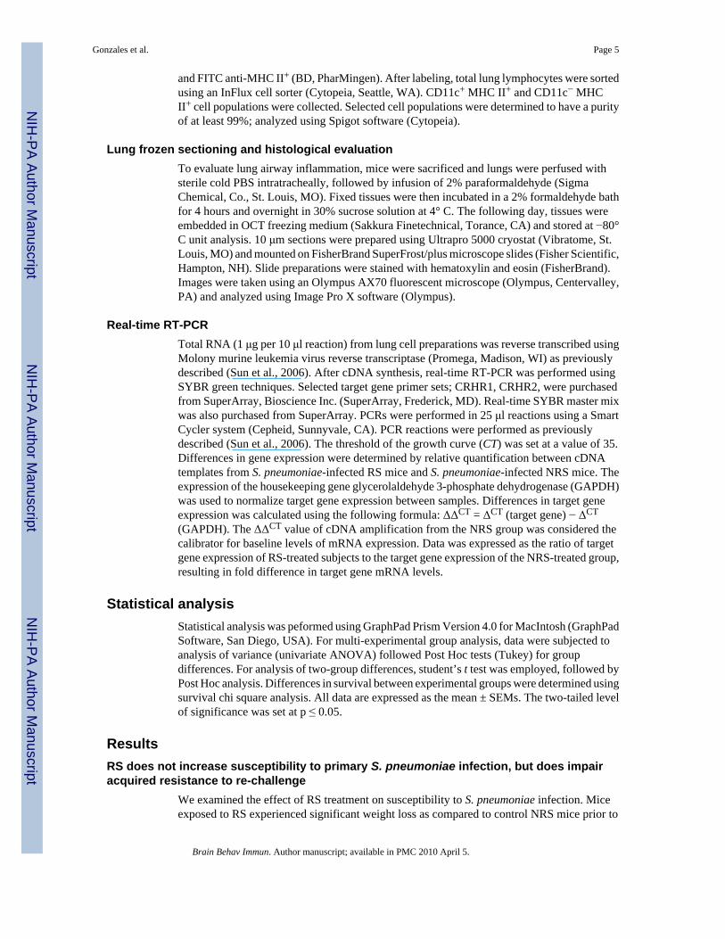
Lung frozen sectioning and histological evaluationTo evaluate lung airway inflammation, mice were sacrificed and lungs were perfused withsterile cold PBS intratracheally, followed by infusion of 2% paraformaldehyde (SigmaChemical, Co., St. Louis, MO). Fixed tissues were then incubated in a 2% formaldehyde bathfor 4 hours and overnight in 30% sucrose solution at 4° C. The following day, tissues wereembedded in OCT freezing medium (Sakkura Finetechnical, Torance, CA) and stored at −80°C unit analysis. 10 μm sections were prepared using Ultrapro 5000 cryostat (Vibratome, St.Louis, MO) and mounted on FisherBrand SuperFrost/plus microscope slides (Fisher Scientific,Hampton, NH). Slide preparations were stained with hematoxylin and eosin (FisherBrand).Images were taken using an Olympus AX70 fluorescent microscope (Olympus, Centervalley,PA) and analyzed using Image Pro X software (Olympus).
Real-time RT-PCRTotal RNA (1 μg per 10 μl reaction) from lung cell preparations was reverse transcribed usingMolony murine leukemia virus reverse transcriptase (Promega, Madison, WI) as previouslydescribed (Sun et al., 2006). After cDNA synthesis, real-time RT-PCR was performed usingSYBR green techniques. Selected target gene primer sets; CRHR1, CRHR2, were purchasedfrom SuperArray, Bioscience Inc. (SuperArray, Frederick, MD). Real-time SYBR master mixwas also purchased from SuperArray. PCRs were performed in 25 μl reactions using a SmartCycler system (Cepheid, Sunnyvale, CA). PCR reactions were performed as previouslydescribed (Sun et al., 2006). The threshold of the growth curve (CT) was set at a value of 35.Differences in gene expression were determined by relative quantification between cDNAtemplates from S. pneumoniae-infected RS mice and S. pneumoniae-infected NRS mice. Theexpression of the housekeeping gene glycerolaldehyde 3-phosphate dehydrogenase (GAPDH)was used to normalize target gene expression between samples. Differences in target geneexpression was calculated using the following formula: ΔΔCT = ΔCT (target gene) − ΔCT
(GAPDH). The ΔΔCT value of cDNA amplification from the NRS group was considered thecalibrator for baseline levels of mRNA expression. Data was expressed as the ratio of targetgene expression of RS-treated subjects to the target gene expression of the NRS-treated group,resulting in fold difference in target gene mRNA levels.
Statistical analysisStatistical analysis was peformed using GraphPad Prism Version 4.0 for MacIntosh (GraphPadSoftware, San Diego, USA). For multi-experimental group analysis, data were subjected toanalysis of variance (univariate ANOVA) followed Post Hoc tests (Tukey) for groupdifferences. For analysis of two-group differences, student’s t test was employed, followed byPost Hoc analysis. Differences in survival between experimental groups were determined usingsurvival chi square analysis. All data are expressed as the mean ± SEMs. The two-tailed levelof significance was set at p ≤ 0.05.
ResultsRS does not increase susceptibility to primary S. pneumoniae infection, but does impairacquired resistance to re-challenge
We examined the effect of RS treatment on susceptibility to S. pneumoniae infection. Miceexposed to RS experienced significant weight loss as compared to control NRS mice prior to
Gonzales et al. Page 5
Brain Behav Immun. Author manuscript; available in PMC 2010 April 5.
NIH
-PA Author Manuscript
NIH
-PA Author Manuscript
NIH
-PA Author Manuscript
infection (t (38) = 4.986, p ≤ 0.0001) and among non-survivors (t (38) = 3.059, p ≤ 0.0041)(Fig. 1). RS exposure resulted in a delayed onset of endotoxemia, resulting in significantly(x2 = 98.67, p ≤ 0.001) fewer deaths within the first 96 hrs of infection (Fig. 2A). However,no significant (x2 = 1.095, p ≤ 0.5178) difference in overall percent survival was found betweenRS and NRS mice exposed to S. pneumoniae. We also determined the effect of RS on naturalresistance among survivors of primary S. pneumoniae infection. RS and NRS survivors werere-challenged 7 days following initial infection. As shown in fig. 2B, a significant (x2 = 4.598,p ≤ 0.0320) decrease in percent survival was found among RS-treated mice.
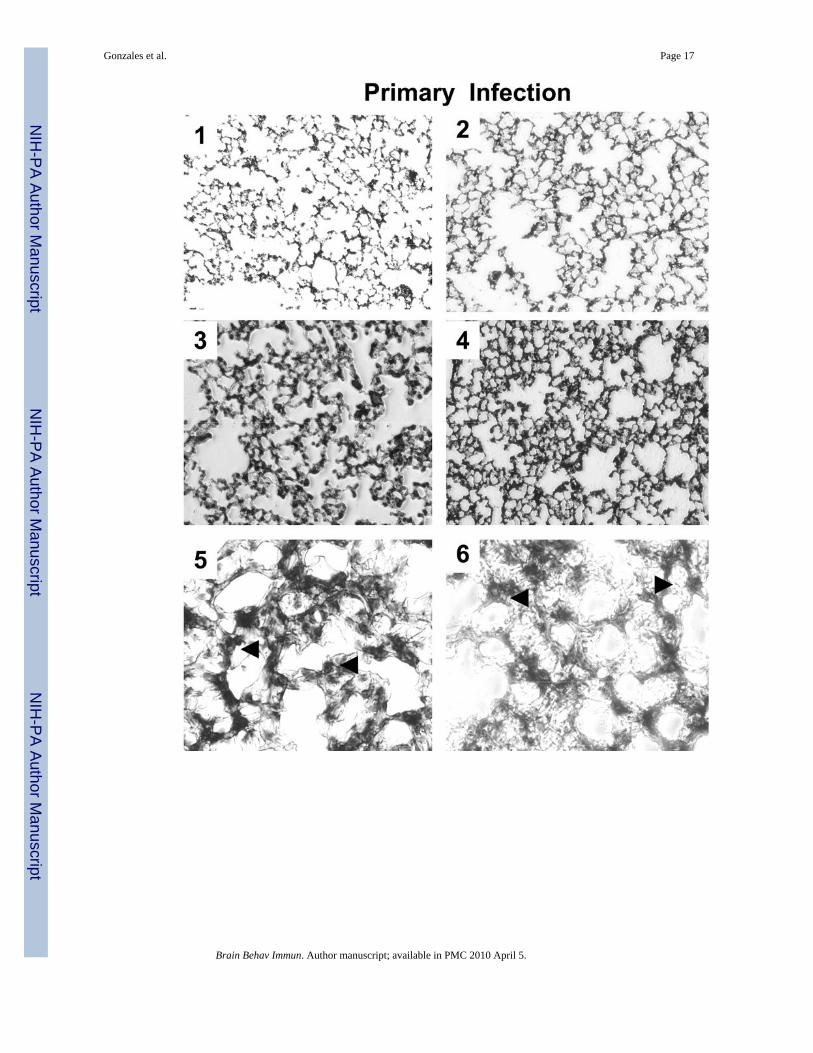
RS survivors from re-challenge demonstrate greater chronic lung injuryWe compared the histopathological changes in the lungs between NRS and RS treated miceexposed to primary infection and of NRS and RS survivors from re-exposure to S.pneumoniae. Despite minimal differences in cellular consolidation within the alveolar spacesbetween NRS and RS survivors from primary infection (Fig. 3., 3 and 4), NRS micedemonstrated a preferential monocytic infiltrate as compared to a polymorphonuclear infiltrateamong RS mice (Fig. 3., 5 and 6). In contrast, RS-treated mice subjected to secondary infection,demonstrated greater gross immunopathological changes as defined by a preferentialpolymorphonuclear infiltrate within the alveolar spaces as compared to NRS challenged mice(Fig. 3., 9 and 10).
RS decreases pulmonary bacterial clearanceWe determined the bacterial loads within the lung, blood and spleen of RS and NRS mice 18hr after S. pneumoniae infection. No differences in the number of organisms were found withinthe spleen and blood between RS and NRS mice 18 hr after infection. In contrast, a significantly(t (12) = 2.577, p ≤ 0.0242) higher number of organisms were apparent in the lungs of RS-treated mice than NRS mice (Fig. 4).
RS influences leukocyte distribution along the respiratory airways in response to S.pneumoniae infection
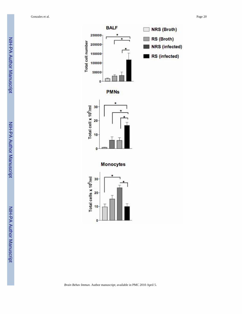
We assessed whether differences in the distribution of immune cells along the respiratorytissues would be observed in response to RS. Differential cell characterization within thebronchiolar lavage fluid (BALF), examination of immune cell numbers in respiratory draininglymphnodes and phenotypic examination of lymphocytes isolated from the lung interstitialtissues was performed. The absolute number of polymorphonuclear cells (PMNs), monocytesand lymphocytes from BALF and cellular phenotypes resident in interstitial lung tissue of NRSand RS infected mice were determined. Total BALF cell numbers were significantly higher(F= 5.047; p≤ 0.0172) in RS stressed mice exposed to S. pneumoniae than in respective controls(Fig. 5A). PMNs were significantly (F= 11.46; p ≤ 0.0029) increased among RS infected miceas compared to respective controls (Fig. 5A). By contrast, monocyte numbers among RS-infected were significantly (F= 8.433; p ≤ 0.0043) diminished as compared to NRS-infectedmice (Fig. 5A). Figure 5B confirms morphologically the distribution of PMNs and monocytesdistributed in BALF of each experimental group.
We also determined the changes in immune cell populations in the draining respiratory nodes(LRN) and total lung tissue of mice exposed to RS. No significant differences in total LRNwere found (NRS-broth; 5.79 × 10⁶ ± 2.3 × 10⁶, RS-broth; 5.5 × 10⁶ ± 1.5 × 10⁶, NRS-infected;1.0 ×10⁷ ± 8.9 ×10⁶, RS-infected; 6.2 ×10⁶ ± 2.1 ×10⁶;(n=4)). Whereas RS led to a moderateincrease in the total number of BALF cells, RS exposure was found to elicit a decrease inlymphoid infiltrate within the interstitial lung tissue (Fig. 6). Despite the observed decrease intotal cells, Gr1+CD11b+ cells; markers typically used to designate neutrophils were the majorcell-type found to significantly increase (F=505.9; p ≤ .0001) in response to S. pneumoniaeinfection (Fig. 6). On the contrary, RS-treatment corresponded with a significant (F=18.58; p
Gonzales et al. Page 6
Brain Behav Immun. Author manuscript; available in PMC 2010 April 5.
NIH
-PA Author Manuscript
NIH
-PA Author Manuscript
NIH
-PA Author Manuscript
≤ 0.0082) decrease in a subset of APCs; CD11c+ MHC II+ (DCs) and B220+ (B Cells) (F=10.44;p ≤ 0.0231) 18 hrs after infection (Fig. 6). No significant changes in F4/80+ MHC II+ APCc(Macrophages) subset or CD3+ CD4+ and CD3+ CD8+ T cells were found (data not shown).
Diverse CRH receptor expression by lung APCs in response to restraint stress and infectionUtilizing real-time RT-PCR methods, we examined total CRH expression in lung tissue as wellas CRHR1 and CRHR2 receptor expression by pulmonary CD11c+ MHC II+ and CD11c−MHC II+ cells of infected mice exposed to RS. CRH mRNA levels were significantly (t = (3)10.71; p ≤ .0086) increased in the lungs of RS-infected mice (Fig. 7A). CRHR1 mRNA levelsby CD11c+ MHC II+ was significantly (F= 48.41; p ≤ 0.0013) increased in the lungs of RS-infected mice as compared to non-stressed mice. However, CRHR2 expression by CD11c+
MHC II+ cells among RS-infected mice was not significantly different as compared to NRS-infected mice (Fig 7B). The converse was shown by lung CD11c− MHC II+ cells, CRHR2expression was preferentially increased (4-fold) and CRHR1 mRNA levels were onlymoderately increased (Fig. 7B).
DiscussionThe studies presented here demonstrate that repeated regiments of acute restraint stress elicitsselective cellular immune responsiveness along the respiratory tract, leading to divergentoutcomes between primary and secondary S. pneumoniae infection. Such findings suggeststhat stress-induced preferences in the induction of pulmonary immune responses are likely toplay a critical role in determining the course of disease. To date, the exact regulatory pathwayswhich stress responses influence cellular immune function, particularly along the respiratorytract are not fully understood. Here, we demonstrate stress-induced preferences in thedistribution of pulmonary cellular populations as a potential mechanism responsible for thecontrasting outcomes of primary and secondary pneumococcal infection. We also implicateperipheral CRH and CRH receptor expression in the lung and pulmonary APCs respectivelyas a potential link between neuroendocrine-immune interactions in the generation of acquiredimmune protection.
Many studies have reported the generation of immune responses to be dependent on a broadrange of physiological, environmental and genetic variables. Likewise, exposure to varioustypes of stress has been characterized for the type of immune response that is affected,depending on its duration (e.g. short periods verses longer exposure times) (Ishihara et al.,1999; Okuyama et al., 2007; Persoons et al., 1997). By applying 4 consecutive days of repeateddurations of stress (3 hrs) and subsequent delivery of S. pneumoniae along the respiratoryairways 18 hrs later, our findings demonstrated that subjecting female mice to this form ofstress did not diminish overall lethality compared to NRS mice. In fact, RS provided temporaryprotection by delaying the onset of death within the first 0–96 hrs that gradually declinedthroughout the course of primary infection which was consistent with the indistinguishabledifferences in lung histopathology found between NRS and RS mice. This finding was incontrast to our prediction considering the significant decrease in weight among mice subjectedto RS prior to infection. Support for such findings has been documented previously. Forexample, C. Kiank et al. demonstrated that female BALB/c mice were more resistant towardbacterial infections given RS exposure despite increased weight loss (Kiank et al., 2006). Thus,based upon these results, we suspected that this form of stress was consistent with previousstudies demonstrating the temporal immuno-enhancing effects of acute RS (Fleshner et al.,1998; Roozen and Magnusson, 1996) at least to the extent that did not result in increasedsusceptibility in female mice.
Lethality from bacterial dissemination and sepsis due to S. pneumoniae infection is oftendependent upon the degree of bacterial outgrowth from the respiratory tract into systemic
Gonzales et al. Page 7
Brain Behav Immun. Author manuscript; available in PMC 2010 April 5.
NIH
-PA Author Manuscript
NIH
-PA Author Manuscript
NIH
-PA Author Manuscript
compartments (e.g. blood). Given that no significant difference in disease outcome was foundbetween mice exposed to RS as compared to NRS mice, suggested that little if any differencesin bacterial colonization along the respiratory tract and subsequently within the peripheraltissues would be observed. Alternatively, one might consider that the observed delay in onsetof death among RS-treated mice could be a result of a preferential inhibition of initial bacterialcolonization within the lungs. On the contrary, the results from this study demonstrated apreferential retention of bacterial growth within the lung and no significant differences inoutgrowth in the blood or spleen 18 hrs after infection. One potential explanation for thesefindings could be that the preference for bacterial colonization in the lung under stressconditions may be preventing dissemination allowing for delayed sepsis, but ultimately withoutproviding a selective advantage of survival. We considered whether the initial bacterial loadmasked resistance among RS mice. The fact that reducing the infection dose to the LD50
demonstrated a similar survival pattern (data not shown), suggest that initial pulmonarycarriage was not influencing the effects of stress. Furthermore, we found that after 36 hr, theextent of bacterial outgrowth in the lungs, spleen and blood were similar between RS and NRSmice and would therefore correspond with little differences in overall survival (data not shown).Based upon these findings, we concluded that restraint stress demonstrated transient protectiveinfluences, presumably by elevating the status of innate immune responses during primaryinfection.
Natural acquired resistance to S. pneumoniae infection is believed to be an important factor inpreventing recurrent disease. In that no differences in host susceptibility and at least temporalprotection upon stress exposure was observed, one might expect that the induction of naturalresistance would reflect a similar pattern. However, in contrast to primary infection, resistanceagainst re-challenge was shown to be significantly altered by demonstrating the following: 1)re-challenging surviving subjects previously exposed to RS led to a significant decrease inoverall survival as compared to NRS survivors and 2) greater histopathological changes werefound in the lungs of RS survivors, indicating chronic lung injury. Based upon these findings,we concluded that although initial exposure did not adversely affect primary resistance tosepsis, RS exposure was substantial enough to diminish acquired adaptive immune responsesleading to decreased survival and chronic pulmonary disease.
The respiratory airways serve as an initial site of defense against local and potentially systemicdisease. Thus, it is likely that stress-induced alterations in local respiratory immune responsescould facilitate host susceptibility, warranting an investigation of the phenotypes governingrespiratory immune defenses against S. pneumoniae infection. The preferential increase inPMNs/neutrophils within BALF prior to and after infection is consistent with its documentedrole in early host defense against bacterial infection (Edwards et al., 1982; Gordon et al.,1986; Hart et al., 1986; Mold et al., 1982) and may explain the observed delayed onset of deathgiven the enhanced BAL infiltrate of PMNs in response to RS. Utilizing this model of stress,we have documented early chemokine activity prior to and 3hr post-infection (unpublisheddata), suggesting that RS maybe enhancing cellular recruitment of innate immune cellpopulations (e.g. neutrophils) that could potentially influence protection. In support Speyer et.al. demonstrated that during sepsis, alterations in chemokine activity resulted in trafficking ofPMNs to the lung (Speyer et al., 2004). Interestingly, pulmonary monocytes (comprising ofmacrophages), which also have an important role in immune surveillance and constituteimportant liaisons and activators of acquired immunity, were not responsive toward RS withinthe BALF. Alveolar macrophages, a major constituent of the monocytic population, are knownto play a major regulatory role as phagocytes in defense against bacterial infections (Marriottet al., 2006). Thus, the findings presented here are seemingly contradictory with respect to thelack of bacterial clearance and delayed lethality. In support of our observations, other studieshave demonstrated that neuroendocrine activation can promote disease pathogenesis along therespiratory tract that is mediated in part by altered neutrophil function (Chang et al., 2004;
Gonzales et al. Page 8
Brain Behav Immun. Author manuscript; available in PMC 2010 April 5.
NIH
-PA Author Manuscript
NIH
-PA Author Manuscript
NIH
-PA Author Manuscript
Meagher et al., 1996; Nishida et al., 1998). Thus, one might consider that although neutrophilicrecruitment under RS conditions may be associated with the observed delayed onset of death,the lack of bacterial clearance may be a result of a dysregulation in their functional responseor other cellular components that are instrumental in neutrophil function (e.g. complementactivation) (Gordon et al., 1986; Liles and Klebanoff, 1995; Noya et al., 1993). Also, the lackof the activation by alveolar macrophages in response to infection may have been a contributingfactor for increased bacterial load within the lungs of RS mice. In support, Flesher et. al. havepreviously demonstrated that nitric oxide activation by peritoneal macrophages required pre-activation via antigenic recognition and its effect was dependent upon time between stress andantigenic exposure (Fleshner et al., 1998). Thus, at least given our current stress regimen priorto antigenic challenge, impairment of bacterialcidal activity along the respiratory tractcontradictory to the delayed lethality may be facilitated by a lack of continuity betweenneutrphillic and macrophages or other innate cell populations.
Subsequently to collecting lavage fluid, we further assessed the phenotypic distribution ofresidual immune cells within the lung parenchyma and draining respiratory nodes. While nosignificant changes in cellular responses were observed in the respiratory draining lymphnodes,we noted a preferential reduction in the total lymphoid infiltrate within the lungs in responseto RS, suggesting the mobilization of immune cells within the alveolar spaces in response toinfection. This would be consistent with the observed BALF-associated increase in cellularinfiltrate demonstrated in RS-infected subjects. The depression of total cells within the lungsinvolved a selective distribution of cellular populations among RS-infected subjects. Namely,while GR1+ CD11b+ cells (typically markers for neutrophils) were increased in the lungs,F4/80+ MHC II+ specific staining of macrophages demonstrated no increase in their responsegiven RS toward S. pneumoniae infection. Interestingly, however, we found a significantdecrease in CD11c+ MHC II+ and B220+ (B cells) cells among RS infected mice. Therefore,while similar trends in neutrophil and macrophage responses were observed along therespiratory tract, the finding that other major cells types were dramatically decreased wasinteresting and provided a potential explanation for the differential effect on primary andacquired immune protection given RS. Specifically, the diminished number of CD11c+ MHCII+ cells along the respiratory tract in response to stress during infection, suggest a potentialmechanism responsible for the observed decrease in host protection. CD11c+ MHC II+ cellscomprise mainly of dendritic cells (DCs) and can elicit protection against S. pneumoniae(Colino and Snapper, 2007). However, more studies are needed to define the mechanismsinfluencing DC functionality under stress conditions. Our results demonstrating a concomitantdecrease in CD11c+ MHC II+ cells with decreased acquired protection provides a potentialexplanation for the observed difference in primary and secondary responses to S.pneumoniae infection. Moreover, decreases in B cell populations would be consistent withaltered priming of adaptive immune protection via impaired humoral and cell-mediatedimmune responses (van Rossum et al., 2005). In support, alterations in adaptive immunity havebeen previously described to influence disease susceptibility under stress conditions (Cao andLawrence, 2002; Mediratta and Sharma, 2002). Future studies are planned to examine therelationships between stress and priming of humoral and T cell-mediated immune responsesto primary and secondary infection.
Stress-associated activation of the CNS results in the neuroendocrine response through thehypothalamic-pituitary axis (HPA) resulting in corticosterone release and the autonomicresponse via stimulation of catecholamines such as norepinephrine. Indeed, the findingspresented here suggest the activation of both stress response pathways in modulation of thecellular immune responses generated during primary and secondary infection. For example,stress exposure has been documented to be a trigger of both the hypothalamic and adrenergicnervous systems with the capacity to amplify local innate and inflammatory responses as a firstline of defense against acute infection (Ader and Cohen, 1993). In particular, numerous studies
Gonzales et al. Page 9
Brain Behav Immun. Author manuscript; available in PMC 2010 April 5.
NIH
-PA Author Manuscript
NIH
-PA Author Manuscript
NIH
-PA Author Manuscript
have highlighted the significance of adrenergic activation as primary mediators during earlystages of disease (Cao et al., 2003), while prolonged stress exposure may be responsible inregulation of adaptive immune responses through HPA-mediate corticosterone activity(Truckenmiller et al., 2006). Therefore, although the contribution of catecholamines and/orcorticosteroids were not demonstrated in the current study, the findings here suggest thatpreferences in their activity will be important in defining the underlying differences in diseaseoutcome.
Once believed to be primarily derived from the HPA, CRH can also be expressed peripherallyat sites of inflammation and believed to impact immune responses (Jain et al., 1991; Murrayet al., 2004). CRH activation has been shown to modulate several disease states including:colitis (Anton et al., 2004), asthma (Silverman et al., 2004; Tantisira et al., 2004) andrheumatoid arthritis (Crofford et al., 1992; Jessop et al., 2001). To date, numerous cellularimmune targets have been documented including: mast cells, T cells, B cells, neutrophils andmacrophages (Agelaki et al., 2002; Baker et al., 2003; Cao et al., 2006; Cao et al., 2005; Iavicoliet al., 1998; Radulovic et al., 1999). The functional activity of CRH is regulated by two majorreceptors, CRHR1 and CRHR2 having diverse affinities for CRH and the CRH homologues,Urocortin (UCN1–3) (Jain et al., 1991; Jessop et al., 2001). A number of studies haveimplicated a role for CRH and its receptors as inflammatory mediators (Moffatt et al., 2006;Papadopoulou et al., 2005; Radulovic et al., 1999). To our best knowledge, we report for thefirst time the expression of CRH receptors by pulmonary CD11c+ MHC II+ cells; a subset ofDCs. Specifically, we provide in vivo evidence that CD11c+ MHC II+ cells and CD11c− MHCII+ cells (presumably macrophages and B cells) exhibit diverse CRH receptor expression inthe lungs of mice exposed to a stress event. Moreover, our finding that CRH was expressed inthe lungs and significantly increased given RS exposure provides the potential for direct ligand-receptor interaction. In that CD11c+ MHC II+ cells revealed a preferential increase in CRHR1expression by DCs whereas CD11c− MCH II+ cells expressed higher levels of CRHR2 isintriguing. Previous studies have described a function role of CRHR1 expression onmacrophages and regulation of anti-inflammatory activity by macrophages in the presence ofCRH1 and CRH agonist (Tsatsanis et al., 2007). Thus, based upon these findings one mayspeculate that differential receptor expression could have influences on APC function andtherefore impact downstream APC-T cell interactions, leading to altered adaptive immuneresponses (e.g. cytokine production by T cells). Although not examined in the current study,we cannot discount the potential influence of CRH regulation of neutrophils that were observedto have opposing responsiveness within the airways as compared to immune cells populationsexamined. Previous studies have demonstrated the role of CRH on neutrophil function (Iavicoliet al., 1998). It would be interesting to determine whether diverse cellular effects correspondwith CRH/CRH receptor regulation. Thus, given these results, we believe that furtherinvestigation into the role of CRH on antigen presenting cell function as well as on other targetcell populations will reveal potential mechanisms responsible for stress-induced alterations inimmune function along the respiratory tract.
In conclusion, our findings restate the complexities of stress responses on immune regulation.The studies presented increase our knowledge of the effects that a given stress event can impactpulmonary immune responses and how such changes influence both innate and acquiredimmunity. Importantly, the studies begin to address how peripheral CRH activation on APCfunction as a mechanism influencing stress-effects on immune modulation.
AcknowledgmentsWe would like to acknowledge Dr. Jerry Simecka, Byung-Jin Kim and Xiangle Sun for guidance in the execution ofstudies and preparation of this manuscript. The project described was supported by Grant Number P20-MD001633from NCMHD and its contents are solely the responsibility of the authors and do not necessarily represent the officialviews of the NCMHD.
Gonzales et al. Page 10
Brain Behav Immun. Author manuscript; available in PMC 2010 April 5.
NIH
-PA Author Manuscript
NIH
-PA Author Manuscript
NIH
-PA Author Manuscript
ReferencesAder R, Cohen N. Psychoneuroimmunology: conditioning and stress. Annu Rev Psychol 1993;44:53–
85. [PubMed: 8434895]Agelaki S, Tsatsanis C, Gravanis A, Margioris AN. Corticotropin-releasing hormone augments
proinflammatory cytokine production from macrophages in vitro and in lipopolysaccharide-inducedendotoxin shock in mice. Infect Immun 2002;70:6068–74. [PubMed: 12379683]
Anton PM, Gay J, Mykoniatis A, Pan A, O’Brien M, Brown D, et al. Corticotropin-releasing hormone(CRH) requirement in Clostridium difficile toxin A-mediated intestinal inflammation. Proc Natl AcadSci U S A 2004;101:8503–8. [PubMed: 15159534]
Baker C, Richards LJ, Dayan CM, Jessop DS. Corticotropin-releasing hormone immunoreactivity inhuman T and B cells and macrophages: colocalization with arginine vasopressin. J Neuroendocrinol2003;15:1070–4. [PubMed: 14622437]
Boyce WT, Chesney M, Alkon A, Tschann JM, Adams S, Chesterman B, et al. Psychobiologic reactivityto stress and childhood respiratory illnesses: results of two prospective studies. Psychosom Med1995;57:411–22. [PubMed: 8552730]
Bryndina IG, Danilov GE. [Substance P as a factor enhancing resistance of the surfactant lung system tochronic immobilization stress]. Ross Fiziol Zh Im I M Sechenova 2002;88:84–9. [PubMed: 11868267]
Butcher SK, Lord JM. Stress responses and innate immunity: aging as a contributory factor. Aging Cell2004;3:151–60. [PubMed: 15268748]
Calcagni E, Elenkov I. Stress system activity, innate and T helper cytokines, and susceptibility to immune-related diseases. Ann N Y Acad Sci 2006;1069:62–76. [PubMed: 16855135]
Cao J, Cetrulo CL, Theoharides TC. Corticotropin-releasing hormone induces vascular endothelialgrowth factor release from human mast cells via the cAMP/protein kinase A/p38 mitogen-activatedprotein kinase pathway. Mol Pharmacol 2006;69:998–1006. [PubMed: 16332989]
Cao J, Papadopoulou N, Kempuraj D, Boucher WS, Sugimoto K, Cetrulo CL, et al. Human mast cellsexpress corticotropin-releasing hormone (CRH) receptors and CRH leads to selective secretion ofvascular endothelial growth factor. J Immunol 2005;174:7665–75. [PubMed: 15944267]
Cao L, Hudson CA, Lawrence DA. Acute cold/restraint stress inhibits host resistance to Listeriamonocytogenes via beta1-adrenergic receptors. Brain Behav Immun 2003;17:121–33. [PubMed:12676574]
Cao L, Lawrence DA. Suppression of host resistance to Listeria monocytogenes by acute cold/restraintstress: lack of direct IL-6 involvement. J Neuroimmunol 2002;133:132–43. [PubMed: 12446016]
Chang LC, Madsen SA, Toelboell T, Weber PS, Burton JL. Effects of glucocorticoids on Fas geneexpression in bovine blood neutrophils. J Endocrinol 2004;183:569–83. [PubMed: 15590983]
Chiang WC, Teoh OH, Chong CY, Goh A, Tang JP, Chay OM. Epidemiology, clinical characteristicsand antimicrobial resistance patterns of community-acquired pneumonia in 1702 hospitalizedchildren in Singapore. Respirology 2007;12:254–61. [PubMed: 17298459]
Colino J, Snapper CM. Dendritic cell-derived exosomes express a Streptococcus pneumoniae capsularpolysaccharide type 14 cross-reactive antigen that induces protective immunoglobulin responsesagainst pneumococcal infection in mice. Infect Immun 2007;75:220–30. [PubMed: 17043104]
Crofford LJ, Sano H, Karalis K, Webster EL, Goldmuntz EA, Chrousos GP, et al. Local secretion ofcorticotropin-releasing hormone in the joints of Lewis rats with inflammatory arthritis. J Clin Invest1992;90:2555–64. [PubMed: 1281840]
Datti F, Datti M, Antunes E, Teixeira NA. Influence of chronic unpredictable stress on the allergicresponses in rats. Physiol Behav 2002;77:79–83. [PubMed: 12213504]
Dhabhar FS, McEwen BS. Stress-induced enhancement of antigen-specific cell-mediated immunity. JImmunol 1996;156:2608–15. [PubMed: 8786326]
Dhabhar FS, Miller AH, McEwen BS, Spencer RL. Stress-induced changes in blood leukocytedistribution. Role of adrenal steroid hormones. J Immunol 1996;157:1638–44. [PubMed: 8759750]
Edwards KM, Gewurz H, Lint TF, Mold C. A role for C-reactive protein in the complement-mediatedstimulation of human neutrophils by type 27 Streptococcus pneumoniae. J Immunol 1982;128:2493–6. [PubMed: 7077077]
Gonzales et al. Page 11
Brain Behav Immun. Author manuscript; available in PMC 2010 April 5.
NIH
-PA Author Manuscript
NIH
-PA Author Manuscript
NIH
-PA Author Manuscript
Emeny RT, Gao D, Lawrence DA. Beta1-adrenergic receptors on immune cells impair innate defensesagainst Listeria. J Immunol 2007;178:4876–84. [PubMed: 17404268]
Engler A, Roy S, Sen CK, Padgett DA, Sheridan JF. Restraint stress alters lung gene expression in anexperimental influenza A viral infection. J Neuroimmunol 2005a;162:103–11. [PubMed: 15833365]
Engler H, Engler A, Bailey MT, Sheridan JF. Tissue-specific alterations in the glucocorticoid sensitivityof immune cells following repeated social defeat in mice. J Neuroimmunol 2005b;163:110–9.[PubMed: 15885313]
Fava GA, Fabbri S, Sirri L, Wise TN. Psychological factors affecting medical condition: a new proposalfor DSM-V. Psychosomatics 2007;48:103–11. [PubMed: 17329602]
Fleshner M, Nguyen KT, Cotter CS, Watkins LR, Maier SF. Acute stressor exposure both suppressesacquired immunity and potentiates innate immunity. Am J Physiol 1998;275:R870–8. [PubMed:9728086]
Ginesu F, Pirina P. Etiology and risk factors of adult pneumonia. J Chemother 1995;7:277–85. [PubMed:8568539]
Gordon DL, Johnson GM, Hostetter MK. Ligand-receptor interactions in the phagocytosis of virulentStreptococcus pneumoniae by polymorphonuclear leukocytes. J Infect Dis 1986;154:619–26.[PubMed: 2943825]
Graham JE, Christian LM, Kiecolt-Glaser JK. Stress, age, and immune function: toward a lifespanapproach. J Behav Med 2006;29:389–400. [PubMed: 16715331]
Gross WB, Siegel PB. Long-term exposure of chickens to three levels of social stress. Avian Dis1981;25:312–25. [PubMed: 7020680]
Hart PH, Spencer LK, Nikoloutsopoulos A, Lopez AF, Vadas MA, McDonald PJ, et al. Role of cellsurface receptors in the regulation of intracellular killing of bacteria by murine peritoneal exudateneutrophils. Infect Immun 1986;52:245–51. [PubMed: 3514456]
Hortal M, Estevan M, Iraola I, De Mucio B. A population-based assessment of the disease burden ofconsolidated pneumonia in hospitalized children under five years of age. Int J Infect Dis2007;11:273–7. [PubMed: 16997592]
Hunzeker J, Padgett DA, Sheridan PA, Dhabhar FS, Sheridan JF. Modulation of natural killer cell activityby restraint stress during an influenza A/PR8 infection in mice. Brain Behav Immun 2004;18:526–35. [PubMed: 15331123]
Iavicoli S, Lopez-Perez E, Buehring GC, Thomas HA, Wei ET, Kishimoto T. Bipolar-shape response ofhuman neutrophils to corticotropin-releasing factor. Eur J Pharmacol 1998;349:301–6. [PubMed:9671111]
Ishihara Y, Matsunaga K, Iijima H, Fujii T, Oguchi Y, Kagawa J. Time-dependent effects of stressorapplication on metastasis of tumor cells in the lung and its regulation by an immunomodulator inmice. Psychoneuroendocrinology 1999;24:713–26. [PubMed: 10451907]
Ising M, Holsboer F. Genetics of stress response and stress-related disorders. Dialogues Clin Neurosci2006;8:433–44. [PubMed: 17290801]
Jain R, Zwickler D, Hollander CS, Brand H, Saperstein A, Hutchinson B, et al. Corticotropin-releasingfactor modulates the immune response to stress in the rat. Endocrinology 1991;128:1329–36.[PubMed: 1999154]
Jessop DS, Harbuz MS, Lightman SL. CRH in chronic inflammatory stress. Peptides 2001;22:803–7.[PubMed: 11337094]
Joachim RA, Quarcoo D, Arck PC, Herz U, Renz H, Klapp BF. Stress enhances airway reactivity andairway inflammation in an animal model of allergic bronchial asthma. Psychosom Med 2003;65:811–5. [PubMed: 14508025]
Jones HP, Hodge LM, Fujihashi K, Kiyono H, McGhee JR, Simecka JW. The pulmonary environmentpromotes Th2 cell responses after nasal-pulmonary immunization with antigen alone, but Th1responses are induced during instances of intense immune stimulation. J Immunol 2001;167:4518–26. [PubMed: 11591779]
Kiank C, Holtfreter B, Starke A, Mundt A, Wilke C, Schutt C. Stress susceptibility predicts the severityof immune depression and the failure to combat bacterial infections in chronically stressed mice.Brain Behav Immun 2006;20:359–68. [PubMed: 16330179]
Gonzales et al. Page 12
Brain Behav Immun. Author manuscript; available in PMC 2010 April 5.
NIH
-PA Author Manuscript
NIH
-PA Author Manuscript
NIH
-PA Author Manuscript
Konstantinos AP, Sheridan JF. Stress and influenza viral infection: modulation of proinflammatorycytokine responses in the lung. Respir Physiol 2001;128:71–7. [PubMed: 11535264]
Kruisbeek, A., editor. Isolation and fractionation of mononuclear cell populations. Vol. 1. 1999.Leu SJ, Singh VK. Suppression of in vitro antibody production by corticotropin-releasing factor
neurohormone. J Neuroimmunol 1993;45:23–9. [PubMed: 8331162]Li KS, Liege S, Moze E, Neveu PJ. Plasma corticosterone and immune reactivity in restrained female
C3H mice. Stress 2000;3:285–98. [PubMed: 11342394]Liles WC, Klebanoff SJ. Regulation of apoptosis in neutrophils--Fas track to death? J Immunol
1995;155:3289–91. [PubMed: 7561020]MacQueen GM, Bienenstock J. Stress and immune regulation. Clin Exp Allergy 2006;36:969–71.
[PubMed: 16911352]Marriott HM, Hellewell PG, Cross SS, Ince PG, Whyte MK, Dockrell DH. Decreased alveolar
macrophage apoptosis is associated with increased pulmonary inflammation in a murine model ofpneumococcal pneumonia. J Immunol 2006;177:6480–8. [PubMed: 17056580]
Matalka KZ. Neuroendocrine and cytokines-induced responses to minutes, hours, and days of mentalstress. Neuro Endocrinol Lett 2003;24:283–92. [PubMed: 14646999]
McClellan CB, Cohen LL. Family functioning in children with chronic illness compared with healthycontrols: a critical review. J Pediatr 2007;150:221–3. 223 e1–2. [PubMed: 17307532]
Meagher LC, Cousin JM, Seckl JR, Haslett C. Opposing effects of glucocorticoids on the rate of apoptosisin neutrophilic and eosinophilic granulocytes. J Immunol 1996;156:4422–8. [PubMed: 8666816]
Mediratta PK, Sharma KK. Differential effects of benzodiazepines on immune responses in non-stressedand stressed animals. Indian J Med Sci 2002;56:9–15. [PubMed: 12508625]
Moffatt JD, Lever R, Page CP. Activation of corticotropin-releasing factor receptor-2 causesbronchorelaxation and inhibits pulmonary inflammation in mice. Faseb J 2006;20:1877–9. [PubMed:16855006]
Mold C, Edwards KM, Gewurz H. Effect of C-reactive protein on the complement-mediated stimulatedof human neutrophils by Streptococcus pneumoniae serotypes 3 and 6. Infect Immun 1982;37:987–92. [PubMed: 7129640]
Murray SE, Lallman HR, Heard AD, Rittenberg MB, Stenzel-Poore MP. A genetic model of stressdisplays decreased lymphocytes and impaired antibody responses without altered susceptibility toStreptococcus pneumoniae. J Immunol 2001;167:691–8. [PubMed: 11441072]
Murray SE, Rosenzweig HL, Johnson M, Huising MO, Sawicki K, Stenzel-Poore MP. Overproductionof corticotropin-releasing hormone blocks germinal center formation: role of corticosterone andimpaired follicular dendritic cell networks. J Neuroimmunol 2004;156:31–41. [PubMed: 15465594]
Nishida K, Ohta Y, Ishiguro I. Contribution of NO synthases to neutrophil infiltration in the gastricmucosal lesions in rats with water immersion restraint stress. FEBS Lett 1998;425:243–8. [PubMed:9559657]
Noya FJ, Baker CJ, Edwards MS. Neutrophil Fc receptor participation in phagocytosis of type III groupB streptococci. Infect Immun 1993;61:1415–20. [PubMed: 8454344]
Okuyama K, Ohwada K, Sakurada S, Sato N, Sora I, Tamura G, et al. The distinctive effects of acute andchronic psychological stress on airway inflammation in a murine model of allergic asthma. AllergolInt 2007;56:29–35. [PubMed: 17259807]
Papadopoulou NG, Oleson L, Kempuraj D, Donelan J, Cetrulo CL, Theoharides TC. Regulation ofcorticotropin-releasing hormone receptor-2 expression in human cord blood-derived cultured mastcells. J Mol Endocrinol 2005;35:R1–8. [PubMed: 16326828]
Persoons JH, Moes NM, Broug-Holub E, Schornagel K, Tilders FJ, Kraal G. Acute and long-term effectsof stressors on pulmonary immune functions. Am J Respir Cell Mol Biol 1997;17:203–8. [PubMed:9271308]
Portela Cde P, Massoco Cde O, de Lima WT, Palermo-Neto J. Stress-induced increment on totalbronchoalveolar cell count in OVA-sensitized rats. Physiol Behav 2001;72:415–20. [PubMed:11274686]
Radulovic M, Dautzenberg FM, Sydow S, Radulovic J, Spiess J. Corticotropin-releasing factor receptor1 in mouse spleen: expression after immune stimulation and identification of receptor-bearing cells.J Immunol 1999;162:3013–21. [PubMed: 10072553]
Gonzales et al. Page 13
Brain Behav Immun. Author manuscript; available in PMC 2010 April 5.
NIH
-PA Author Manuscript
NIH
-PA Author Manuscript
NIH
-PA Author Manuscript
Roozen AW, Magnusson U. Effects of short-term restraint stress on leukocyte counts, lymphocyteproliferation and lysis of erythrocytes in gilts. Zentralbl Veterinarmed B 1996;43:505–11. [PubMed:8921740]
Saha B, Mondal AC, Basu S, Dasgupta PS. Circulating dopamine level, in lung carcinoma patients,inhibits proliferation and cytotoxicity of CD4+ and CD8+ T cells by D1 dopamine receptors: an invitro analysis. Int Immunopharmacol 2001;1:1363–74. [PubMed: 11460316]
Schindler BA. Stress, affective disorders, and immune function. Med Clin North Am 1985;69:585–97.[PubMed: 3892192]
Sheridan JF, Stark JL, Avitsur R, Padgett DA. Social disruption, immunity, and susceptibility to viralinfection. Role of glucocorticoid insensitivity and NGF. Ann N Y Acad Sci 2000;917:894–905.[PubMed: 11270350]
Sieve AN, Steelman AJ, Young CR, Storts R, Welsh TH, Welsh CJ, et al. Sex-dependent effects of chronicrestraint stress during early Theiler’s virus infection on the subsequent demyelinating disease in CBAmice. J Neuroimmunol 2006;177:46–62. [PubMed: 16762424]
Silverman ES, Breault DT, Vallone J, Subramanian S, Yilmaz AD, Mathew S, et al. Corticotropin-releasing hormone deficiency increases allergen-induced airway inflammation in a mouse model ofasthma. J Allergy Clin Immunol 2004;114:747–54. [PubMed: 15480311]
Speyer CL, Gao H, Rancilio NJ, Neff TA, Huffnagle GB, Sarma JV, et al. Novel chemokineresponsiveness and mobilization of neutrophils during sepsis. Am J Pathol 2004;165:2187–96.[PubMed: 15579460]
Sternberg EM. Neural regulation of innate immunity: a coordinated nonspecific host response topathogens. Nat Rev Immunol 2006;6:318–28. [PubMed: 16557263]
Strange KS, Kerr LR, Andrews HN, Emerman JT, Weinberg J. Psychosocial stressors and mammarytumor growth: an animal model. Neurotoxicol Teratol 2000;22:89–102. [PubMed: 10642118]
Sun X, Jones HP, Hodge LM, Simecka JW. Cytokine and chemokine transcription profile duringMycoplasma pulmonis infection in susceptible and resistant strains of mice: macrophageinflammatory protein 1beta (CCL4) and monocyte chemoattractant protein 2 (CCL8) andaccumulation of CCR5+ Th cells. Infect Immun 2006;74:5943–54. [PubMed: 16988274]
Tantisira KG, Lake S, Silverman ES, Palmer LJ, Lazarus R, Silverman EK, et al. Corticosteroidpharmacogenetics: association of sequence variants in CRHR1 with improved lung function inasthmatics treated with inhaled corticosteroids. Hum Mol Genet 2004;13:1353–9. [PubMed:15128701]
Truckenmiller ME, Bonneau RH, Norbury CC. Stress presents a problem for dendritic cells:corticosterone and the fate of MHC class I antigen processing and presentation. Brain Behav Immun2006;20:210–8. [PubMed: 16504465]
Tsatsanis C, Androulidaki A, Dermitzaki E, Gravanis A, Margioris AN. Corticotropin releasing factorreceptor 1 (CRF1) and CRF2 agonists exert an anti-inflammatory effect during the early phase ofinflammation suppressing LPS-induced TNF-alpha release from macrophages via induction ofCOX-2 and PGE2. J Cell Physiol 2007;210:774–83. [PubMed: 17117478]
Tseng RJ, Padgett DA, Dhabhar FS, Engler H, Sheridan JF. Stress-induced modulation of NK activityduring influenza viral infection: role of glucocorticoids and opioids. Brain Behav Immun2005;19:153–64. [PubMed: 15664788]
van Rossum AM, Lysenko ES, Weiser JN. Host and bacterial factors contributing to the clearance ofcolonization by Streptococcus pneumoniae in a murine model. Infect Immun 2005;73:7718–26.[PubMed: 16239576]
Weiss JM, Sundar SK, Becker KJ, Cierpial MA. Behavioral and neural influences on cellular immuneresponses: effects of stress and interleukin-1. J Clin Psychiatry 1989;50(Suppl):43–53. discussion54–5. [PubMed: 2654130]
Yang EV, Glaser R. Stress-induced immunomodulation and the implications for health. IntImmunopharmacol 2002;2:315–24. [PubMed: 11811934]
Gonzales et al. Page 14
Brain Behav Immun. Author manuscript; available in PMC 2010 April 5.
NIH
-PA Author Manuscript
NIH
-PA Author Manuscript
NIH
-PA Author Manuscript
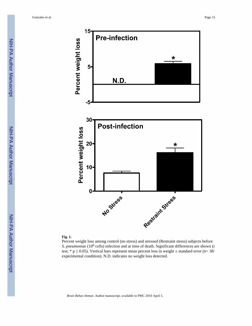
Fig. 1.Percent weight loss among control (no stress) and stressed (Restraint stress) subjects beforeS. pneumoniae (106 cells) infection and at time of death. Significant differences are shown (ttest, * p ≤ 0.05). Vertical bars represent mean percent loss in weight ± standard error (n= 38/experimental condition). N.D. indicates no weight loss detected.
Gonzales et al. Page 15
Brain Behav Immun. Author manuscript; available in PMC 2010 April 5.
NIH
-PA Author Manuscript
NIH
-PA Author Manuscript
NIH
-PA Author Manuscript
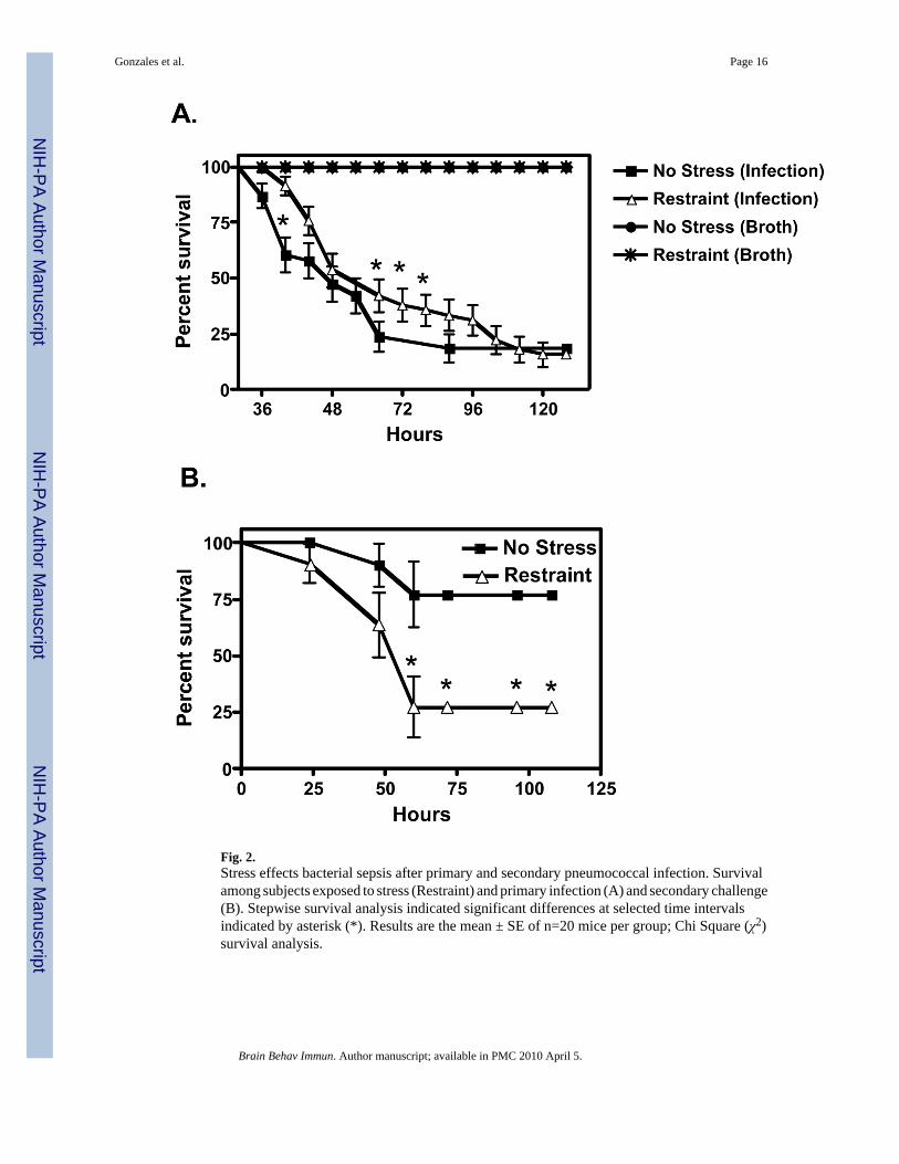
Fig. 2.Stress effects bacterial sepsis after primary and secondary pneumococcal infection. Survivalamong subjects exposed to stress (Restraint) and primary infection (A) and secondary challenge(B). Stepwise survival analysis indicated significant differences at selected time intervalsindicated by asterisk (*). Results are the mean ± SE of n=20 mice per group; Chi Square (χ2)survival analysis.
Gonzales et al. Page 16
Brain Behav Immun. Author manuscript; available in PMC 2010 April 5.
NIH
-PA Author Manuscript
NIH
-PA Author Manuscript
NIH
-PA Author Manuscript
Gonzales et al. Page 17
Brain Behav Immun. Author manuscript; available in PMC 2010 April 5.
NIH
-PA Author Manuscript
NIH
-PA Author Manuscript
NIH
-PA Author Manuscript
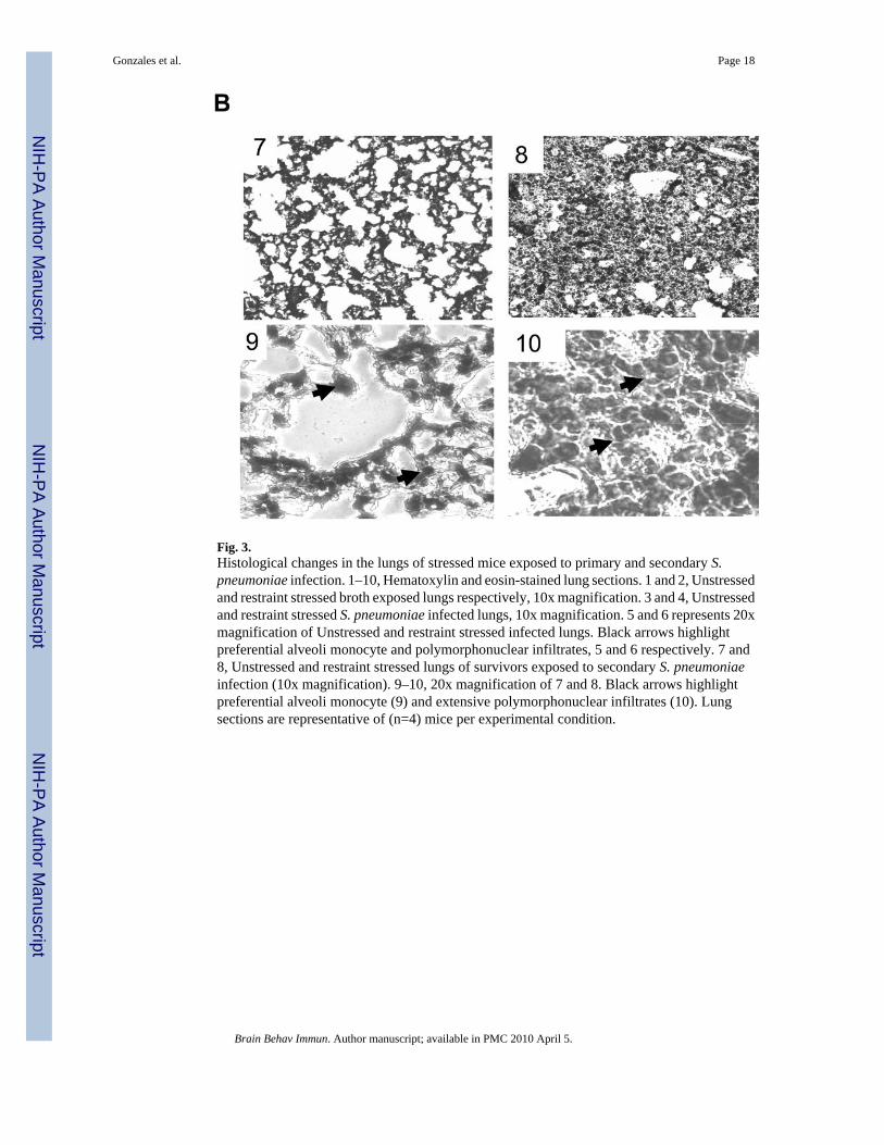
Fig. 3.Histological changes in the lungs of stressed mice exposed to primary and secondary S.pneumoniae infection. 1–10, Hematoxylin and eosin-stained lung sections. 1 and 2, Unstressedand restraint stressed broth exposed lungs respectively, 10x magnification. 3 and 4, Unstressedand restraint stressed S. pneumoniae infected lungs, 10x magnification. 5 and 6 represents 20xmagnification of Unstressed and restraint stressed infected lungs. Black arrows highlightpreferential alveoli monocyte and polymorphonuclear infiltrates, 5 and 6 respectively. 7 and8, Unstressed and restraint stressed lungs of survivors exposed to secondary S. pneumoniaeinfection (10x magnification). 9–10, 20x magnification of 7 and 8. Black arrows highlightpreferential alveoli monocyte (9) and extensive polymorphonuclear infiltrates (10). Lungsections are representative of (n=4) mice per experimental condition.
Gonzales et al. Page 18
Brain Behav Immun. Author manuscript; available in PMC 2010 April 5.
NIH
-PA Author Manuscript
NIH
-PA Author Manuscript
NIH
-PA Author Manuscript
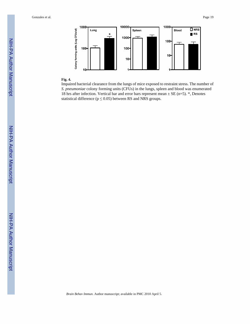
Fig. 4.Impaired bacterial clearance from the lungs of mice exposed to restraint stress. The number ofS. pneumoniae colony forming units (CFUs) in the lungs, spleen and blood was enumerated18 hrs after infection. Vertical bar and error bars represent mean ± SE (n=5). *, Denotesstatistical difference (p ≤ 0.05) between RS and NRS groups.
Gonzales et al. Page 19
Brain Behav Immun. Author manuscript; available in PMC 2010 April 5.
NIH
-PA Author Manuscript
NIH
-PA Author Manuscript
NIH
-PA Author Manuscript
Gonzales et al. Page 20
Brain Behav Immun. Author manuscript; available in PMC 2010 April 5.
NIH
-PA Author Manuscript
NIH
-PA Author Manuscript
NIH
-PA Author Manuscript
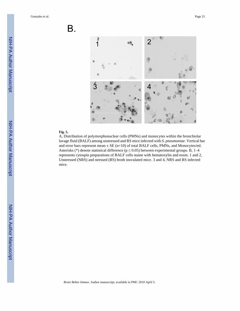
Fig. 5.A, Distribution of polymorphonuclear cells (PMNs) and monocytes within the bronchiolarlavage fluid (BALF) among unstressed and RS mice infected with S. pneumoniae. Vertical barand error bars represent mean ± SE (n=10) of total BALF cells, PMNs, and Monocytes/ml.Asterisks (*) denote statistical difference (p ≤ 0.05) between experimental groups. B, 1–4represents cytospin preparations of BALF cells staine with hematoxylin and eosin. 1 and 2,Unstressed (NRS) and stressed (RS) broth inoculated mice. 3 and 4, NRS and RS infectedmice.
Gonzales et al. Page 21
Brain Behav Immun. Author manuscript; available in PMC 2010 April 5.
NIH
-PA Author Manuscript
NIH
-PA Author Manuscript
NIH
-PA Author Manuscript
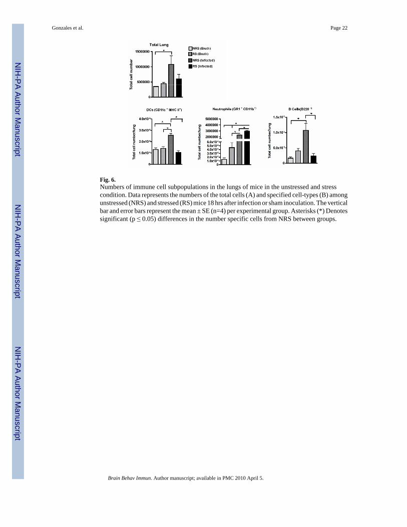
Fig. 6.Numbers of immune cell subpopulations in the lungs of mice in the unstressed and stresscondition. Data represents the numbers of the total cells (A) and specified cell-types (B) amongunstressed (NRS) and stressed (RS) mice 18 hrs after infection or sham inoculation. The verticalbar and error bars represent the mean ± SE (n=4) per experimental group. Asterisks (*) Denotessignificant (p ≤ 0.05) differences in the number specific cells from NRS between groups.
Gonzales et al. Page 22
Brain Behav Immun. Author manuscript; available in PMC 2010 April 5.
NIH
-PA Author Manuscript
NIH
-PA Author Manuscript
NIH
-PA Author Manuscript
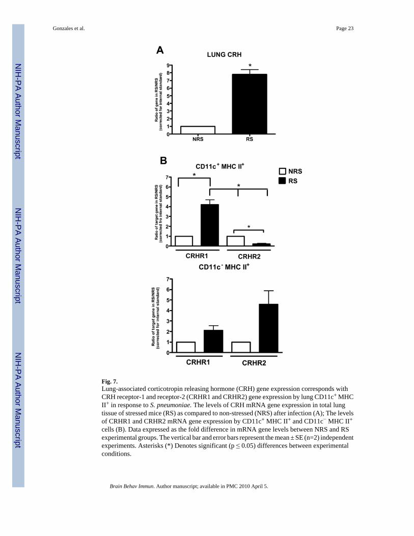
Fig. 7.Lung-associated corticotropin releasing hormone (CRH) gene expression corresponds withCRH receptor-1 and receptor-2 (CRHR1 and CRHR2) gene expression by lung CD11c+ MHCII+ in response to S. pneumoniae. The levels of CRH mRNA gene expression in total lungtissue of stressed mice (RS) as compared to non-stressed (NRS) after infection (A); The levelsof CRHR1 and CRHR2 mRNA gene expression by CD11c+ MHC II+ and CD11c− MHC II+
cells (B). Data expressed as the fold difference in mRNA gene levels between NRS and RSexperimental groups. The vertical bar and error bars represent the mean ± SE (n=2) independentexperiments. Asterisks (*) Denotes significant (p ≤ 0.05) differences between experimentalconditions.
Gonzales et al. Page 23
Brain Behav Immun. Author manuscript; available in PMC 2010 April 5.
NIH
-PA Author Manuscript
NIH
-PA Author Manuscript
NIH
-PA Author Manuscript


