Embed Size (px)
Citation preview
❖ Describe the structure, innervation and function of the muscle spindle.
❖ Describe the components of monosynaptic muscle stretch reflexes, including the
role of alpha (α) and gamma (γ) motorneurons.
❖ Distinguish between a static and dynamic stretch reflex.
❖ Describe the spinal and supra-spinal regulation of the stretch reflex.
❖ Describe the structure and function of the Golgi tendon organ and the inverse
stretch reflex.
❖ Appreciate the clinical importance of the stretch reflexes.
5th Lecture ∣The Physiology Team
Objectives:
نسان إل ما يس لل وأن لسعى
Colour index :● important● Numbers● Extra
Stretch reflex and tendon jerks
Done by:
❖ Team leaders: irammalA ilA ,irassodlA haleludbA
Fatima Balsharaf, Rahaf Alshammari
❖ Team members: Abdurhman Alhayssoni
Abdulaziz Aldurgham
Abdullah Alsergani, Rasheed Albalaa
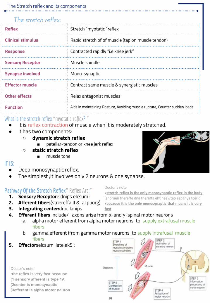
The Stretch reflex and its components
What is the stretch reflex “myotatic reflex ”?● It is reflex contraction of muscle when it is moderately stretched.● it has two components:
○ dynamic stretch reflex■ patellar-tendon or knee jerk reflex
○ static stretch reflex■ muscle tone
IT IS:● Deep monosynaptic reflex.● The simplest ;it involves only 2 neurons & one synapse.
Pathway Of the Stretch Reflex“ Reflex Arc ”:1. Sensory Receptoreldnips elcsum :2. Afferent fibers)stnereffa II & aI puorg(3. Integrating center:droc lanips4. Efferent fibers include/ axons arise from α-and γ-spinal motor neurons
a. alpha motor efferent from alpha motor neurons to supply extrafusal muscle fibers
b. gamma efferent (from gamma motor neurons to supply intrafusal muscle fibers
5. Effectorselcsum latelekS :
Reflex Stretch “myotatic ”reflex
Clinical stimulus Rapid stretch of of muscle (tap on muscle tendon)
Response Contracted rapidly “i.e knee jerk”
Sensory Receptor Muscle spindle
Synapse involved Mono-synaptic
Effector muscle Contract same muscle & synergistic muscles
Other effects Relax antagonist muscles
Function Aids in maintaining Posture, Avoiding muscle rupture, Counter sudden loads
The stretch reflex:
Doctor’s note:•stretch reflex is the only monosynaptic reflex in the body )snoruen tnereffe dna tnereffa eht neewteb espanys tcerid(•because it is the only monosynaptic that means it is very fast
Doctor’s note:
•the reflex is very fast because
1) sensory afferent is type 1A
2) center is monosynaptic
3) efferent is alpha motor neuron
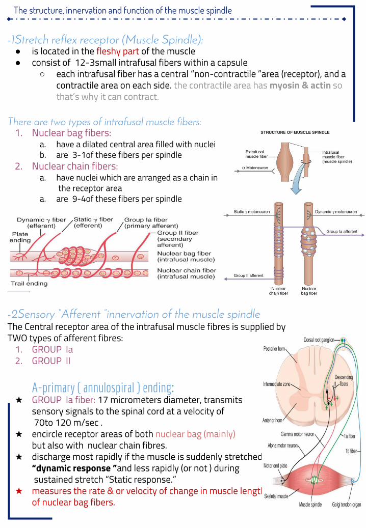
The structure, innervation and function of the muscle spindle
1- Stretch reflex receptor (Muscle Spindle):● is located in the fleshy part of the muscle● consist of 3-12 small intrafusal fibers within a capsule
○ each intrafusal fiber has a central “non-contractile ”area (receptor), and a contractile area on each side. the contractile area has myosin & actin so that’s why it can contract.
There are two types of intrafusal muscle fibers:1. Nuclear bag fibers:
a. have a dilated central area filled with nucleib. are 1-3 of these fibers per spindle
2. Nuclear chain fibers:a. have nuclei which are arranged as a chain in
the receptor areaa. are 4-9 of these fibers per spindle
2- Sensory “Afferent ”innervation of the muscle spindleThe Central receptor area of the intrafusal muscle fibres is supplied byTWO types of afferent fibres:
1. GROUP Ia2. GROUP II
A-primary ( annulospiral ) ending:★ GROUP Ia fiber: 17 micrometers diameter, transmits
sensory signals to the spinal cord at a velocity of70to 120 m/sec .
★ encircle receptor areas of both nuclear bag (mainly)but also with nuclear chain fibres.
★ discharge most rapidly if the muscle is suddenly stretched“dynamic response ”and less rapidly (or not ) duringsustained stretch “Static response ”.
★ measures the rate & or velocity of change in muscle lengthof nuclear bag fibers.
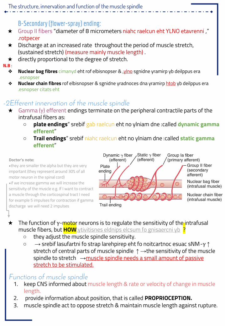
The structure, innervation and function of the muscle spindle
B-Secondary (flower-spray) ending:★ Group II fibers “diameter of 8 micrometers ” , niahc raelcun eht YLNO etavrenni
rotpecer.★ Discharge at an increased rate throughout the period of muscle stretch,
(sustained stretch) (measure mainly muscle length) .★ directly proportional to the degree of stretch.
N.B :❖ Nuclear bag fibres sgnidne yramirp yb deilppus eraylnoeht rof elbisnopser & , cimanyd
esnopser .❖ Nuclear chain fibres yb deilppus erahtob rof elbisnopser & sgnidne yradnoces dna yramirp
esnopser citats eht.
2- Efferent innervation of the muscle spindle★ Gamma (γ) efferent endings terminate on the peripheral contractile parts of the
intrafusal fibers as:○ plate endings eht no ylniam dne :gab raelcun srebif“ called dynamic gamma
efferent”○ Trail endings eht no ylniam dne :niahc raelcun srebif“ called static gamma
efferent”
★ The function of γ-motor neurons is to regulate the sensitivity of the intrafusal muscle fibers, but HOW ?ytivitisnes eldnips elcsum fo gnisaercni yb.○ they adjust the muscle spindle sensitivity.○ ↑γ- srebif lasufartni fo strap larehpirep eht fo noitcartnoc esuac sNM→
stretch of central parts of muscle spindle →↑ the sensitivity of the muscle spindle to stretch →muscle spindle needs a small amount of passive stretch to be stimulated.
Functions of muscle spindle1. keep CNS informed about muscle length & rate or velocity of change in muscle
length.2. provide information about position, that is called PROPRIOCEPTION.3. muscle spindle act to oppose stretch & maintain muscle length against rupture.
Doctor’s note:•they are smaller the alpha but they are very important (they represent around 30% of all motor neuron in the spinal cord)•if we increase gamma we will increase the sensitivity of the muscle e.g. If I want to contract a muscle through the corticospinal tract I need for example 5 impulses for contraction if gamma discharge we will need 2 impulses
The structure, innervation and function of the muscle spindle
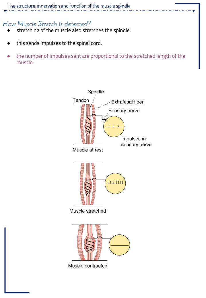
How Muscle Stretch Is detected?● stretching of the muscle also stretches the spindle.
● this sends impulses to the spinal cord.
● the number of impulses sent are proportional to the stretched length of the muscle.
The components of monosynaptic muscle stretch reflexes, including the role ofalpha (α) and gamma (γ) motorneurons
Stretch reflex .cont:
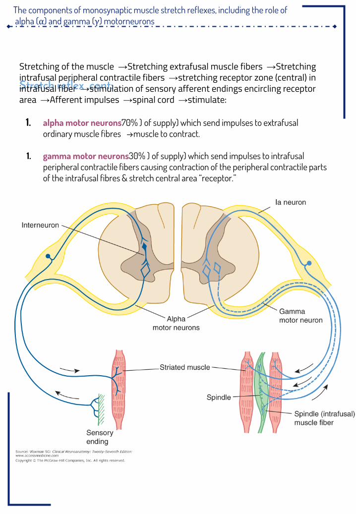
Stretching of the muscle →Stretching extrafusal muscle fibers →Stretching intrafusal peripheral contractile fibers →stretching receptor zone (central) in intrafusal fiber →stimulation of sensory afferent endings encircling receptor area →Afferent impulses →spinal cord →stimulate:
1. alpha motor neurons (70% of supply) which send impulses to extrafusal ordinary muscle fibres →muscle to contract.
1. gamma motor neurons (30% of supply) which send impulses to intrafusal peripheral contractile fibers causing contraction of the peripheral contractile parts of the intrafusal fibres & stretch central area “receptor ”.
Static and Dynamic stretch reflex
Dynamic stretch reflex● sudden “phasic ”rapid stretch of a muscle causes synchronous strong burst of
excitatory discharges from Nuclear bag srebif laripsolunna ni“ Primary ”afferents to the alpha motor neuron .
● this causes the latter to send strong motor excitatory impulses to extrafusal fibers →causing sudden, jerky “brief ”muscle contraction )tnemevom ykreJ(.
● as the muscle shortens →the spindle becomes lax →and ceases to discharge →no more stimulation of alpha motor neuron →no more excitatory impulses from alpha motor neuron to the extrafusal fibers →muscle relaxes.
● This is the basis of Tendon Jerks “Dynamic stretch reflexes ”.Static stretch reflex
● Maintained “Tonic ”stretch of muscle →
● impulses from Nuclear chain srebif tnereffa eldnips hguorht levart srebif ylniam( yarpS rewolF gnola“ secondary ”ending) to alpha motor neuron, stimulating it to
produce muscle contraction.● Causing sustained “continuous ”contraction of the muscle as long as it is stretch .● The static stretch reflex is the basis of muscle tone; sa yllacinilc denifed si hcihw
hcterts elcsum ot ecnatsiser.
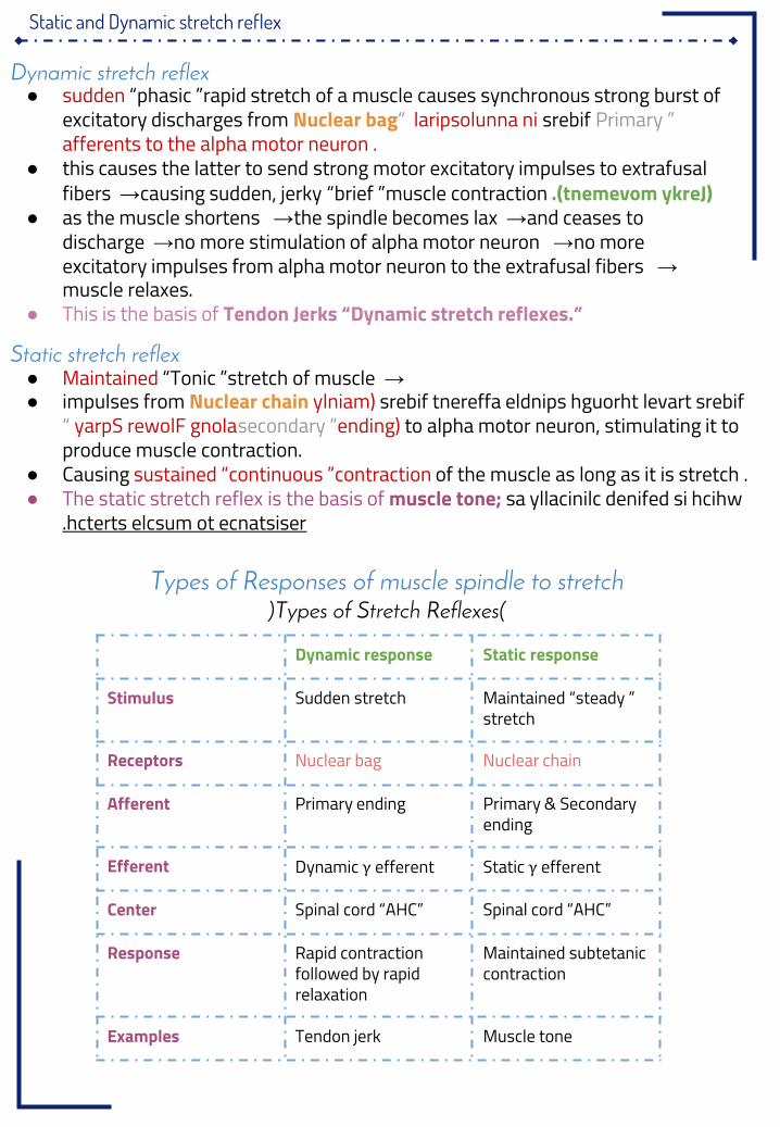
Dynamic response Static response
Stimulus Sudden stretch Maintained “steady ”stretch
Receptors Nuclear bag Nuclear chain
Afferent Primary ending Primary & Secondary ending
Efferent Dynamic γ efferent Static γ efferent
Center Spinal cord “AHC” Spinal cord “AHC”
Response Rapid contraction followed by rapid relaxation
Maintained subtetanic contraction
Examples Tendon jerk Muscle tone
Types of Responses of muscle spindle to stretch(Types of Stretch Reflexes)
Muscle tone & damping mechanism
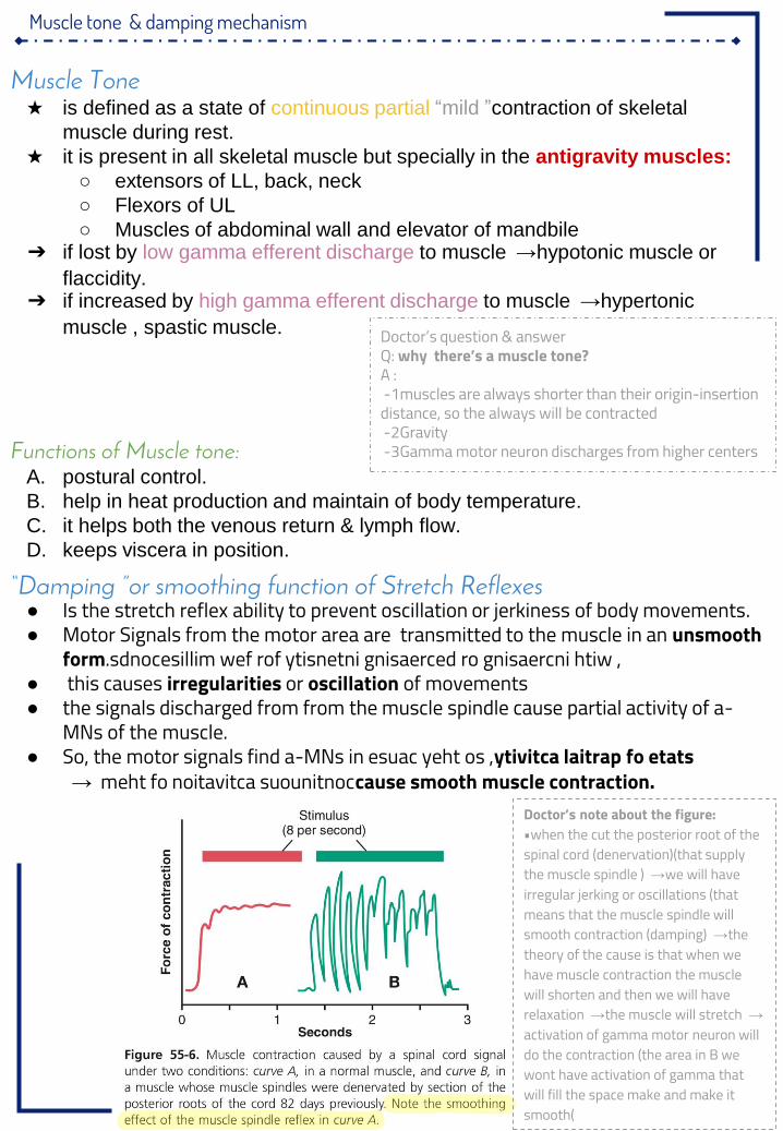
“Damping ”or smoothing function of Stretch Reflexes● Is the stretch reflex ability to prevent oscillation or jerkiness of body movements.● Motor Signals from the motor area are transmitted to the muscle in an unsmooth
form sdnocesillim wef rof ytisnetni gnisaerced ro gnisaercni htiw ,.● this causes irregularities or oscillation of movements● the signals discharged from from the muscle spindle cause partial activity of a-
MNs of the muscle.● So, the motor signals find a-MNs in ytivitca laitrap fo etats esuac yeht os ,
meht fo noitavitca suounitnoc→ cause smooth muscle contraction.
Muscle Tone★ is defined as a state of continuous partial “mild ”contraction of skeletal
muscle during rest.
★ it is present in all skeletal muscle but specially in the antigravity muscles:
○ extensors of LL, back, neck
○ Flexors of UL
○ Muscles of abdominal wall and elevator of mandbile➔ if lost by low gamma efferent discharge to muscle →hypotonic muscle or
flaccidity.➔ if increased by high gamma efferent discharge to muscle →hypertonic
muscle , spastic muscle.
Functions of Muscle tone:A. postural control.
B. help in heat production and maintain of body temperature.
C. it helps both the venous return & lymph flow.
D. keeps viscera in position.
Doctor’s question & answerQ: why there’s a muscle tone?A :
1- muscles are always shorter than their origin-insertion distance, so the always will be contracted
2- Gravity3- Gamma motor neuron discharges from higher centers
Doctor’s note about the figure:•when the cut the posterior root of the spinal cord (denervation)(that supply the muscle spindle ) →we will have irregular jerking or oscillations (that means that the muscle spindle will smooth contraction (damping) →the theory of the cause is that when we have muscle contraction the muscle will shorten and then we will have relaxation →the muscle will stretch →activation of gamma motor neuron will do the contraction (the area in B we wont have activation of gamma that will fill the space make and make it smooth)
Spinal and Supra-spinal regulation of the stretch reflex.
How Are Muscle Spindles Stimulated?1. Passive stretch of the whole muscle:
a. it causes stretch of muscle spindle which lies parallel to muscle fibers2. Activation of the γ-MNs:
a. by supraspinal centers or reflexelyb. it causes contraction of the peripheral part of the intrafusal fibers →
stretch of receptor area3. Co-activation of α-and γ-Motor Neurons “α-γegakniL”:
a. Signals from motor cortex to the alpha motor neuron, mostly transmitted to the gamma motor neurons simultaneously, an effect called Coactivation.
★ what is the significance of this coactivation?○ Regulate the sensitivity of the spindle by keeping its length constant○ oppose sudden changes in muscle length.
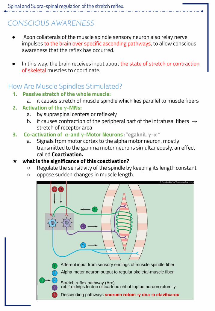
CONSCIOUS AWARENESS
● Axon collaterals of the muscle spindle sensory neuron also relay nerve impulses to the brain over specific ascending pathways, to allow conscious awareness that the reflex has occurred.
● In this way, the brain receives input about the state of stretch or contraction of skeletal muscles to coordinate.
Afferent input from sensory endings of muscle spindle fiber
Alpha motor neuron output to regular skeletal-muscle fiber
Stretch reflex pathway (Arc)γ-rebif eldnips fo dne elitcartnoc eht ot tuptuo noruen rotom
Descending pathways oc-α etavitca-γ dna-snoruen rotom
Spinal and Supra-spinal regulation of the stretch reflex.
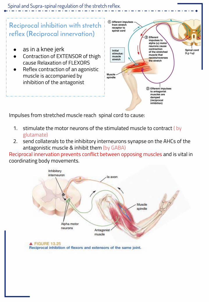
Reciprocal inhibition with stretch reflex (Reciprocal innervation)
● as in a knee jerk● Contraction of EXTENSOR of thigh
cause Relaxation of FLEXORS● Reflex contraction of an agonistic
muscle is accompanied by inhibition of the antagonist
Impulses from stretched muscle reach spinal cord to cause:
1. stimulate the motor neurons of the stimulated muscle to contract ( by glutamate)
2. send collaterals to the inhibitory interneurons synapse on the AHCs of the antagonistic muscle & inhibit them (by GABA)
Reciprocal innervation prevents conflict between opposing muscles and is vital in coordinating body movements.
Spinal and Supra-spinal regulation of the stretch reflex.
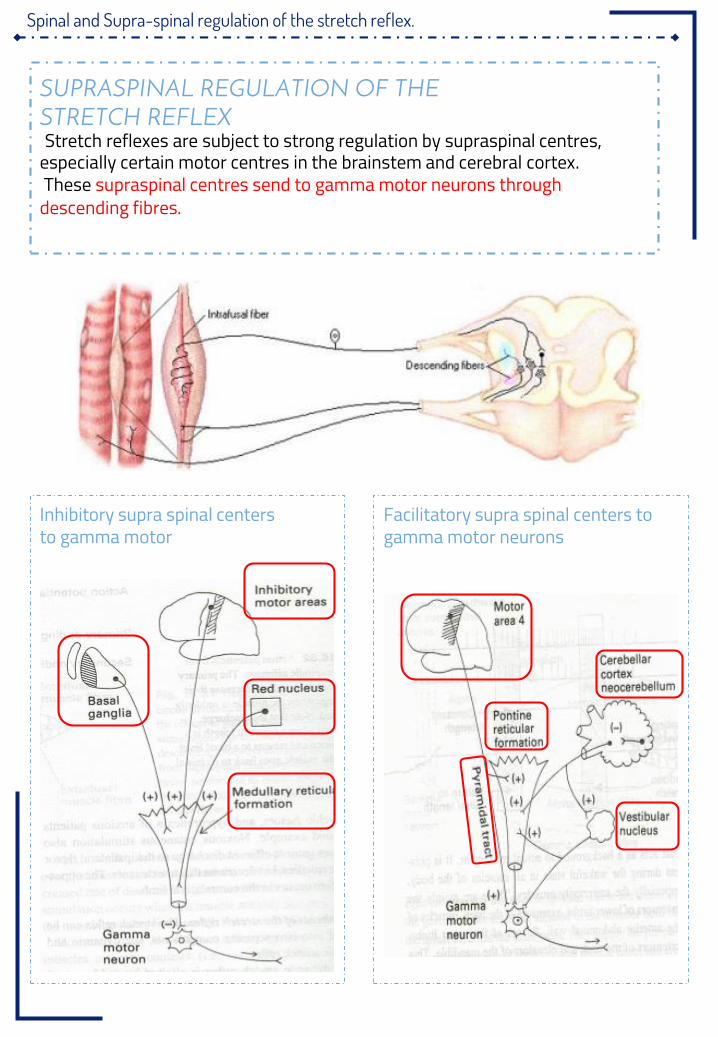
SUPRASPINAL REGULATION OF THESTRETCH REFLEXStretch reflexes are subject to strong regulation by supraspinal centres,
especially certain motor centres in the brainstem and cerebral cortex.These supraspinal centres send to gamma motor neurons through
descending fibres.
Inhibitory supra spinal centersto gamma motor
Facilitatory supra spinal centers to gamma motor neurons
Spinal and Supra-spinal regulation of the stretch reflex.
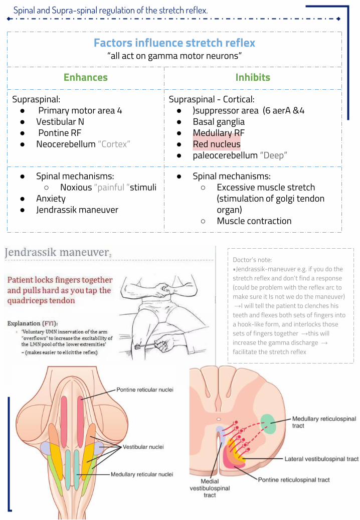
Factors influence stretch reflex“all act on gamma motor neurons”
Enhances InhibitsSupraspinal:● Primary motor area 4● Vestibular N● Pontine RF● Neocerebellum “Cortex”
Supraspinal - Cortical:● (suppressor area 4 aerA &6 )● Basal ganglia● Medullary RF● Red nucleus● paleocerebellum “Deep”
● Spinal mechanisms:○ Noxious “painful ”stimuli
● Anxiety● Jendrassik maneuver
● Spinal mechanisms:○ Excessive muscle stretch
(stimulation of golgi tendon organ)
○ Muscle contraction
Doctor’s note:•Jendrassik-maneuver e.g. if you do the stretch reflex and don’t find a response (could be problem with the reflex arc to make sure it Is not we do the maneuver) →I will tell the patient to clenches his teeth and flexes both sets of fingers into a hook-like form, and interlocks those sets of fingers together →this will increase the gamma discharge →facilitate the stretch reflex
The clinical application & importance of stretch reflexes
Clinical application of stretch reflex: Knee Jerk Reflex➔ contraction of the muscle being stretch “Quadriceps ”.➔ Reciprocal inhibition of the antagonist muscle “Hamstring ”through
reciprocal innervation.
What is the Clinical Significance of Tendon Reflexes?They are carried out clinically to test the integrity of reflex arc.Areflexia or hypo-reflexia (hypo-tonia) indicates that thereflex arc is interrupted at one of its components by:❏ Lesions of lower motor neuron, e.g. poliomyelitis❏ Peripheral nerve lesions e.g. peripheral neuropathy, Diabetic neuropathy❏ Neuromuscular junction disorder e.g. myasthenia gravis❏ Primary muscle disorder e.g. myopathy
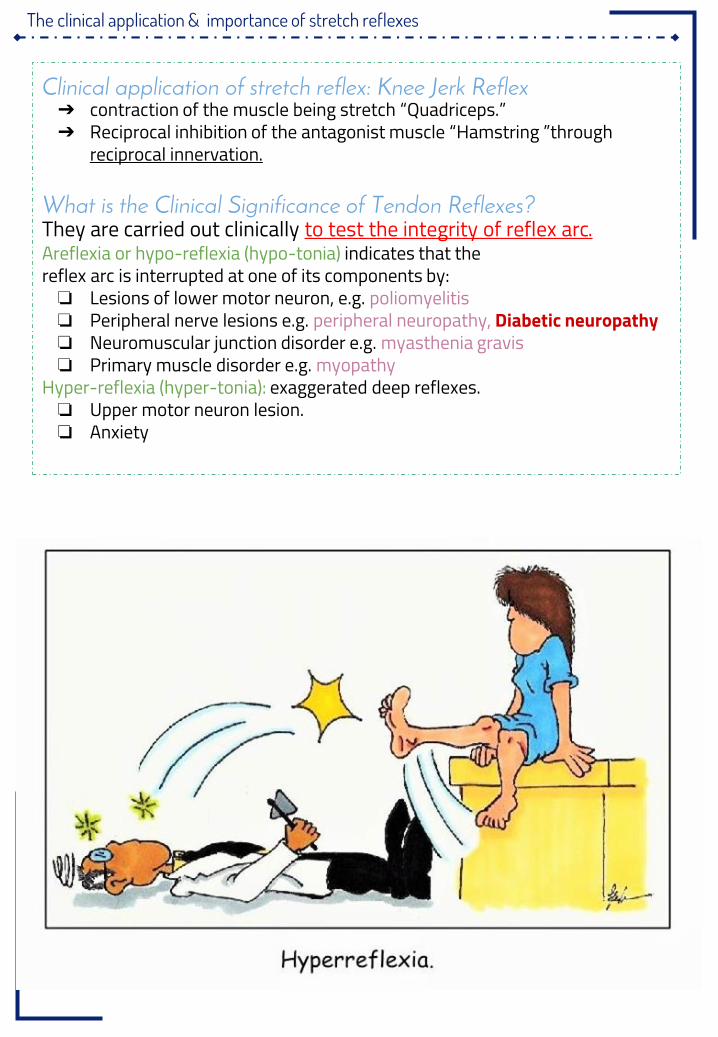
Hyper-reflexia (hyper-tonia): exaggerated deep reflexes.❏ Upper motor neuron lesion.❏ Anxiety
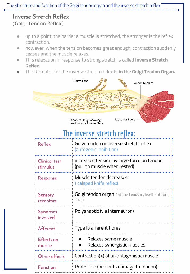
The structure and function of the Golgi tendon organ and the inverse stretch reflex
The inverse stretch reflex:Reflex Golgi tendon or inverse stretch reflex
(autogenic inhibition)Clinical test stimulus
increased tension by large force on tendon (pull on muscle when rested)
Response Muscle tendon decreases( calsped knife reflex)
Sensory receptors
Golgi tendon organ “at the tendon yhself eht ton ,trap”
Synapses involved
Polysnaptic (via interneuron)
Afferent Type Ib afferent fibresEffects on muscle
● Relaxes same muscle● Relaxes synergistic muscles
Other effects Contraction(+) of an antagonistic muscleFunction Protective (prevents damage to tendon)
Inverse Stretch Reflex(Golgi Tendon Reflex)
● up to a point, the harder a muscle is stretched, the stronger is the reflex contraction.
● however, when the tension becomes great enough, contraction suddenly ceases and the muscle relaxes.
● This relaxation in response to strong stretch is called Inverse Stretch Reflex.
● The Receptor for the inverse stretch reflex is in the Golgi Tendon Organ.
The structure and function of the Golgi tendon organ and the inverse stretch reflex
Golgi tendon organs (3-25 :) They are present intendons and encapsulated sensory receptorsthrough which muscle tendon fibers pass.
● About 10 to 15 muscle fibers are usually connected to each golgi tendon organ● they Transmit information about tendon tension or rate of change of tension.● the organ is stimulated when this small bundle of muscle fibers is “tensed ”by severe
contraction.● Thus, the major difference in excitation of the Golgi tendon organ versus the muscle
spindle eldnips eht taht si,htgnel elcsum ni segnahc dna htgnel elcsum stceted nagro nodnet eht saerehw noisnet elcsum stcetedflesti ni noisnet eht yb detcelfer sa.
Receptor Golgi tendon organs
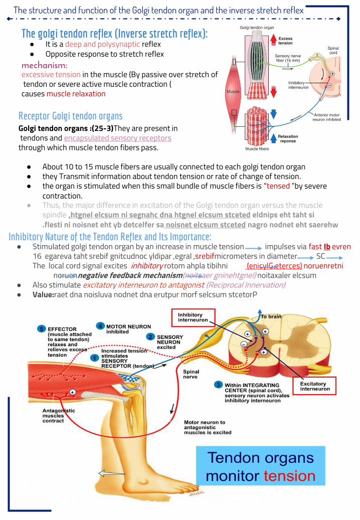
Inhibitory Nature of the Tendon Reflex and Its Importance:● Stimulated golgi tendon organ by an increase in muscle tension impulses via fast Ib evren
srebif egareva taht srebif gnitcudnoc yldipar ,egral ,16 micrometers in diameter SC The local cord signal excites inhibitory noruenretni )enicylG eterces( rotom ahpla tibihni noruennegative feedback mechanism noitaxaler elcsum)noitcaer gninehtgnel(
● Also stimulate excitatory interneuron to antagonist (Reciprocal Innervation)● Value:raet dna noisluva nodnet dna erutpur morf selcsum stcetorP
The golgi tendon reflex (Inverse stretch reflex):● It is a deep and polysynaptic reflex● Opposite response to stretch reflex
mechanism:excessive tension in the muscle (By passive over stretch oftendon or severe active muscle contraction )causes muscle relaxation
The structure and function of the Golgi tendon organ and the inverse stretch reflex
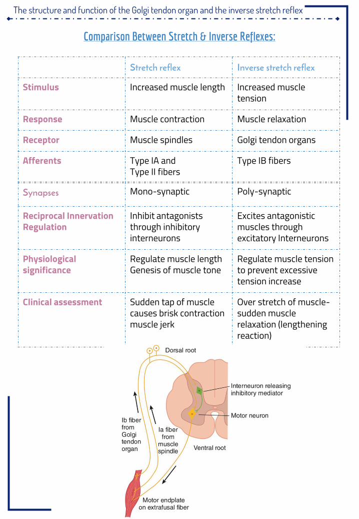
Comparison Between Stretch & Inverse Reflexes:
Stretch reflex Inverse stretch reflex
Stimulus Increased muscle length Increased muscle tension
Response Muscle contraction Muscle relaxationReceptor Muscle spindles Golgi tendon organsAfferents Type IA and
Type II fibersType IB fibers
Synapses Mono-synaptic Poly-synaptic
Reciprocal Innervation Regulation
Inhibit antagonists through inhibitory interneurons
Excites antagonistic muscles through excitatory Interneurons
Physiological significance
Regulate muscle lengthGenesis of muscle tone
Regulate muscle tension to prevent excessive tension increase
Clinical assessment Sudden tap of muscle causes brisk contraction muscle jerk
Over stretch of muscle-sudden muscle relaxation (lengthening reaction)
Questions
1. The number of synapses in
the stretch reflex are:
A. .One (monosynaptic)
B. .Two (disynaptic)
C. .Three (trisynaptic)
D. .B&C
2 . the spindles can send to the
spinal cord in:
A. .Positive signals
B. .Negative signals
C. .No signal
D. .A&B
3 . Which of the following is not a
factor INHIBIT the stretch:
A. .Anxiety
B. .Basal ganglia
C. .excessive stretch reflex of
muscle
D. .medullary RF
4 . The response of golgi tendon
reflex is:
A. .Muscle contraction
B. .no response
C. .muscle relaxation
D. .A&C
5 . Muscle spindle is:
A. .sensory neuron
B. .motor neuron
C. .effector
D. .sensory receptor
6 . motor neuron of stretch reflex:
A. .Alpha
B. .Type II
C. .Gamma
D. .A&C
7 . Which of the following is NOT a
factor enhance the stretch:
A. .Anxiety
B. .Jendrassik-manuver
C. .Excessive stretch of muscle
D. .Noxious painful stimuli
8 . muscle spindles detect:
A. .Length
B. .Tension
C. .nothing
D. .A&B
9 . golgi tendon organ activates
group:
A. .Ib afferent nerves
B. .II afferent nerves
C. .Ia afferent nerves
D. .A&B
10 . When a person steps on a tack
with their left foot, flexor muscles on
the right leg and extensor muscles
on the left leg will be stimulated:
A. .True
B. .False
An
sw
ers
:
1.A
2.D
3.A
4.C
5.D
6.D
7.C
8.A
9.A
10
.B


