Embed Size (px)
Citation preview
R
S
LPa
b
c
d
e
h
•••••
a
ARRAA
KNDDSS
dN
N
h0
Behavioural Brain Research 280 (2015) 160–171
Contents lists available at ScienceDirect
Behavioural Brain Research
jou rn al hom epage: www.elsev ier .com/ locate /bbr
esearch report
triatal dopamine receptor plasticity in neurotensin deficient mice
ucy G. Chastaina,∗, Hongyan Qua, Chase H. Bourkea, P. Michael Iuvoneb,c,aul R. Dobnerd, Charles B. Nemeroffe, Becky Kinkeada
Department of Psychiatry and Behavioral Sciences, Emory University School of Medicine, Atlanta, GA, USADepartment of Ophthalmology, Emory University School of Medicine, Atlanta, GA, USADepartment of Pharmacology, Emory University School of Medicine, Atlanta, GA, USADepartment of Microbiology and Physiological Systems, Program in Neuroscience, University of Massachusetts Medical School, Worcester, MA, USADepartment of Psychiatry and Behavioral Sciences, University of Miami Miller School of Medicine, Miami, FL, USA
i g h l i g h t s
We investigated dopaminergic tone and function in mice lacking neurotensin (NT−/−).NT−/− mice show increased striatal D1, D2 and DAT mRNA compared to wildtype mice.NT−/− mice show increased striatal D2 receptor binding densities.Some of the behavioral effects of D1- and D2-type receptor agonists are altered in NT−/− mice.Results implicate NT deficiency in dopamine-related disorders such as schizophrenia.
r t i c l e i n f o
rticle history:eceived 22 August 2014eceived in revised form 4 November 2014ccepted 7 November 2014vailable online 15 November 2014
eywords:eurotensinopamineopamine receptortriatumchizophrenia
a b s t r a c t
Schizophrenia is thought to be caused, at least in part, by dysfunction in striatal dopamine neuro-transmission. Both clinical studies and animal research have implicated the dopamine neuromodulatorneurotensin (NT) in the pathophysiology of schizophrenia. Utilizing male mice lacking the NT gene(NT−/−), these studies examined the consequences of NT deficiency on dopaminergic tone and func-tion, investigating (1) dopamine concentrations and dopamine receptor and transporter expression andbinding in dopaminergic terminal regions, and (2) the behavioral effects of selective dopamine receptoragonists on locomotion and sensorimotor gating in adult NT−/− mice compared to wildtype (NT+/+) mice.NT−/− mice did not differ from NT+/+ mice in concentrations of dopamine or its metabolite DOPAC in anybrain region examined. However, NT−/− mice showed significantly increased D1 receptor, D2 receptor,and dopamine transporter (DAT) mRNA in the caudate putamen compared to NT+/+ controls. NT−/− micealso showed elevated D2 receptor binding densities in both the caudate putamen and nucleus accumbensshell compared to NT+/+ mice. In addition, some of the behavioral effects of the D1-type receptor agonistSKF-82958 and the D2-type receptor agonist quinpirole on locomotion, startle amplitude, and prepulse
−/−
inhibition were dose-dependently altered in NT mice, showing altered D1-type and D2-type receptorsensitivity to stimulation by agonists in the absence of NT. The results indicate that NT deficiency altersstriatal dopamine receptor expression, binding, and function. This suggests a critical role for the NT sys-tem in the maintenance of striatal DA system homeostasis and implicates NT deficiency in the etiologyof dopamine-associated disorders such as schizophrenia.© 2014 Published by Elsevier B.V.
Abbreviations: CP, caudate putamen; CSF, cerebrospinal fluid; DA, dopamine; DAT, dopihydroxyphenylacetic acid; FCTX, frontal cortex; HPLC, high-pressure liquid chromatogT+/+, wildtype; NTS1, neurotensin receptor 1; NTS2, neurotensin receptor 2; PPI, prepuls∗ Corresponding author. Present address: Endocrine Research Facility, Department of Aew Brunswick, NJ 08901, USA. Tel.: +1 848 932 7462; fax: +1 732 932 7504.
E-mail address: [email protected] (L.G. Chastain).
ttp://dx.doi.org/10.1016/j.bbr.2014.11.014166-4328/© 2014 Published by Elsevier B.V.
amine transporter; DHBA, 3,4-dihydroxybenzylamine hydrobromide; DOPAC, 3,4-raphy; NAcc, nucleus accumbens; NT, neurotensin; NT−/− , neurotensin knockout;e inhibition; RT-PCR, reverse transcription polymerase chain reaction.nimal Sciences, Rutgers, The State University of New Jersey, 67 Poultry Farm Lane,
l Brain
1
hntfsacstnDiitpDeda
tsSsc[ttcaa
bntNaDpstaSpatdiDpnaatD
ordTt(d
L.G. Chastain et al. / Behavioura
. Introduction
Disrupted dopamine (DA) neurotransmission has long beenypothesized to play a role in the pathophysiology of schizophre-ia. The ‘dopamine hypothesis’ of schizophrenia posits dysfunc-ional hyperactivity in the striatal DA system to be responsibleor psychotic symptoms of schizophrenia [1–3]. This hypothe-is is supported by early studies noting that administration ofmphetamine, an indirect DA agonist that increases synapticoncentrations of DA in striatal regions, exacerbated psychoticymptoms in schizophrenic patients [3]. Also in support of thisheory, all clinically effective antipsychotic drugs act as antago-ists at the dopamine D2 receptor, producing decreased striatalAergic activity [1,4]. Finally, many imaging and post mortem stud-
es have shown increases in striatal D2 receptor binding densitiesn the brains of schizophrenic patients, confirming a disruption inhe striatal DA system in schizophrenia [5–7]. These studies sup-ort the dopamine hypothesis of schizophrenia and implicate the2 receptor in particular in schizophrenia neuropathology. How-ver it remains unclear whether striatal DA disruption is a primaryysfunction in schizophrenia or whether it is a compensatory alter-tion due to some other defect [8].
In addition to DA system dysfunction, many other neuro-ransmitters have been implicated in the neuropathology ofchizophrenia, including the tridecapeptide neurotensin (NT).everal clinical studies implicate NT in the neurobiology ofchizophrenia. Decreased concentrations of NT are found in theerebrospinal fluid (CSF) of a subset of schizophrenic subjects9–12], and NT levels normalize in these subjects following effec-ive treatment with antipsychotic drugs [13,14]. These studies ledo the hypothesis that NT may act as an endogenous antipsy-hotic [15]. Preclinical studies utilizing animal models of psychosislso supported the use of NT receptor agonists as potential novelntipsychotic drugs [16].
Laboratory animal studies have shown endogenous NT toe a neuromodulator of nigrostriatal and mesocorticolimbic DAeurotransmission (for review see [17]). Increasing NT neuro-ransmission by administration of NT receptor agonists such asT69L alter the behavioral effects of DA receptor agonists such asmphetamine, an indirect DA receptor agonist, and haloperidol, a2 receptor antagonist [16]. For example, NT69L administrationrevents haloperidol-induced catalepsy in rats [18]. In addition,tudies utilizing inhibition of NT neurotransmission also suggesthe effects of pharmacological manipulation of the DA systemsre in fact dependent on intact NT neurotransmission [19–21].pecifically, NT gene knockout in mice results in blunted effects ofharmacological activation of striatal DA neurons by amphetaminend by the antipsychotic drug haloperidol [22,23]. In addition,he behavioral effects of amphetamine and some antipsychoticrugs on prepulse inhibition (PPI), a measure of sensorimotor gat-
ng which is regulated by the nigrostriatal and mesocorticolimbicA systems, are altered in NT−/− mice [21]. These findings arearalleled by studies utilizing pharmacological antagonism of NTeurotransmission that demonstrate that NT receptor antagonismlso alters the physiological and behavioral effects of DA receptorgonists and antagonists [19,20,23–25]. These studies suggest func-ional alterations in both the mesocorticolimbic and nigrostriatalA systems in the absence of NT neurotransmission.
Although disrupting NT neurotransmission by NT gene knock-ut is known to result in altered physiological and behavioralesponses to activation of the DA systems, it is unknown whether aeficit in NT produces any developmental changes in these circuits.
he experiments detailed in this paper sought to evaluate DAergicone in DA terminal regions in NT−/− mice compared to wildtypeNT+/+) mice. Specifically, these studies measured DA receptor andopamine transporter (DAT) gene expression and binding and theResearch 280 (2015) 160–171 161
concentrations of DA and its metabolite 3,4-dihydroxyphenylaceticacid (DOPAC) in the nucleus accumbens (NAcc), caudate putamen(CP), and frontal cortex (FCTX). In light of the previously observedaltered behavioral response to amphetamine on PPI in NT−/− mice[21], these studies also sought to investigate possible changes in DAreceptor function or sensitivity in NT−/− mice by measuring behav-ioral response to selective DA receptor agonists compared to NT+/+
mice.
2. Materials and methods
2.1. Animals
NT−/− mice were generated as previously described [22]. Onlymale mice were used in these experiments. Mice (60 days of age andolder) from the lab’s NT−/− breeding colony backcrossed againstthe C57BL/6J strain were used for these studies. Mice heterozygousfor the NT gene were bred to generate wildtype (NT+/+) and NT−/−
mice. Animals were housed in an environmentally controlled ani-mal facility with a 12 h light–dark cycle (lights off: 10:00, lightson: 22:00). Food and water were available ad libitum. Mice wereweaned on postnatal day 21 and housed in same sex groups oftwo to six per cage. All behavioral testing and euthanasia proce-dures were completed in the dark phase. All animal protocols wereapproved by the Emory University Institutional Animal Care andUse Committee (IACUC) in compliance with the National Institutesof Health and AAALAC guidelines for use of laboratory animals.
2.2. Genotyping
At weaning, ear punches were obtained from all mice andDNA was extracted from the tissue. The presence or absenceof the NT gene was identified using custom PCR primers (Invi-trogen) to amplify the wildtype NT gene or the disrupted NTgene construct. Primer sequences to detect the wildtype NTgene allele were 5′-CATCCCTCACAGTTCACTCACTTTG-3′ (25 mer,Tm = 74 ◦C) and 5′-CCTGGATTCATTTACCTGAGTAGCA-3′ (25 merTm = 72 ◦C). Primer sequences to detect the NT−/− gene allele were5′-CATCCCTCACAGTTCACTCACTTTG-3′ (25 mer, Tm = 74 ◦C) and 5′-CCCAGTCACGACGTTGTAAAACGAC-3′ (25 mer, Tm = 76 ◦C). The PCRproducts for the wildtype NT gene and for the NT−/− gene allelewere 270 bp and 188 bp, respectively. PCR products were run ongel electrophoresis to identify the genotype for each animal.
2.3. HPLC
Concentrations of DA and its metabolite DOPAC were assayed inNT+/+ (n = 6–8) and NT−/− (n = 8–12) mice by high-pressure liquidchromatography (HPLC). All mice used for these studies first under-went behavioral testing for startle amplitude and PPI as describedbelow. One week after testing, mice were euthanized by decapita-tion and brains were collected and quickly frozen on dry ice. Brainswere later dissected according to a mouse brain atlas [26]. NAcc, CP,and FCTX regions were collected. Samples of mouse brains wereprepared by adding 200 �l of ice-cold 0.1 N perchloric acid con-taining 0.01% sodium metabisulfite and 25 ng/ml internal standard3,4-dihydroxybenzylamine hydrobromide (DHBA) to the tissue.Samples were then homogenized and centrifuged at 15,000 × g for10 min at 4 ◦C. The supernatant was injected at a constant flow rateof 1 mL/min onto an Ultrasphere ODS 250 mm × 4.6 mm column,5 �m (Beckman Coulter, Fullerton, CA) with mobile phase (0.1 mM
EDTA; 0.35 mM sodium octyl sulfate; 50 mM phosphoric acid; 5%acetonitrile, adjusted to pH 2.7 with NaOH). A coulometric detector(ESA Inc., Chelmsford, MA; guard cell set at 600 mV and analyti-cal cell at 300 mV) was used to detect the DA, DOPAC, and DHBA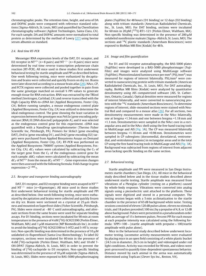
1 l Brain
catcFps
2
DdrbOtwatemHC(iemaqSDg(tttedwe
2
afianonPnatWttur4ObwS
by placing mice in an open field consisting of a white plastic bucket(24.5 cm in diameter, 26.5 cm in height) and videotaped under red
62 L.G. Chastain et al. / Behavioura
hromatographic peaks. The retention time, height, and area of DAnd DOPAC peaks were compared with reference standard solu-ions (Sigma-Aldrich, St. Louis, MO) and quantified by ChemStationhromatography software (Agilent Technologies, Santa Clara, CA).or each sample, DA and DOPAC amounts were normalized to totalrotein as determined by the method of Lowry [27] using bovineerum albumin as standard.
.4. Real time RT-PCR
Messenger RNA expression levels of the DAT, D1 receptor, and2 receptor in NT+/+ (n = 8 pairs) and NT−/− (n = 8 pairs) mice wereetermined by real time reverse transcription polymerase chaineaction (RT-PCR). All mice used in these studies first underwentehavioral testing for startle amplitude and PPI as described below.ne week following testing, mice were euthanized by decapita-
ion and brains were collected and quickly frozen on dry ice. Brainsere later dissected according to a mouse brain atlas [26]. NAcc, CP,
nd FCTX regions were collected and pooled together in pairs fromhe same genotype matched on overall % PPI values to generatenough tissue for RNA extraction. RNA was extracted by the TRIzolethod (Invitrogen, Carlsbad, CA) and reverse transcribed with theigh Capacity RNA-to-cDNA kit (Applied Biosystems, Foster City,A). Before running samples, a mouse endogenous control plateApplied Biosystems, Foster City, CA) was utilized to determine thedeal endogenous control. The gene showing the least variation inxpression between the genotypes was Polr2a (gene encoding poly-erase (RNA) II (DNA directed) polypeptide A), and it was selected
s the endogenous control gene for this experiment. cDNA wasuantified with a NanoDrop spectrophotometer (Thermo Fishercientific Inc, Pittsburgh, PA). Primers for Slc6a3 (gene encodingAT), Drd1a (gene encoding D1), and Drd2 (gene encoding D2) tar-ets were purchased from Applied Biosystems Assays on DemandApplied Biosystems, Foster City, CA). RT-PCR was performed onhe Applied Biosystems 7900HT system (Applied Biosystems, Fos-er City, CA). dCT values were calculated by subtracting the CT ofhe target gene from the CT of the endogenous control gene forach sample. ddCT values were calculated by subtracting the meanCT of NT+/+ from the mean dCT of NT−/−. Gene expression changesere then assessed with the following formula: Fold change in gene
xpression = 2—ddCT.
.5. Receptor and transporter binding autoradiography
DAT, D1 receptor, and D2 receptor binding were assayed in NT+/+
nd NT−/− mice (n = 8/genotype). All mice used in these studiesrst underwent behavioral testing for startle amplitude and PPIs described below. One week following testing, mice were eutha-ized by decapitation and brains were collected and quickly frozenn dry ice. Brains were sectioned on a cryostat at 25 �m thick-ess and mounted on Superfrost slides (Fisher Scientific, Pittsburgh,A). Slides were stored at −80 ◦C until autoradiography, and alter-ate sections from the same brains were used for separate bindingssays. For D1 binding, sections were incubated for 90 min at roomemperature in the presence of 4 nM [3H]-SCH23390 (Perkin Elmer,
altham, MA) and 1 �M mianserin (MP Biomedicals Inc), in ordero avoid the binding of [3H]-SCH23390 to 5-HT2 and 5-HT1c recep-ors. Non-specific binding was determined in the presence of 10 �Mnlabeled cis-flupenthixol (Santa Cruz Biotechnology). To label D2eceptors, sections were incubated for 60 min in the presence of
nM [3H]-raclopride (Perkin Elmer, Waltham, MA) and 10 nM 7-
H-DPAT (Sigma-Aldrich, St. Louis, MO) in order to prevent theinding of [3H]-raclopride to D3 receptors. Non-specific bindingas determined in the presence of 10 �M sulpiride (Sigma-Aldrich,t. Louis, MO). Slides were exposed to BAS-5000 phosphorimaging
Research 280 (2015) 160–171
plates (FujiFilm) for 48 hours (D1 binding) or 12 days (D2 binding)along with tritium standards (American Radiolabeled Chemicals,Inc., St. Louis, MO). For DAT binding, sections were incubatedfor 60 min in 20 pM [125I]-RT1-121 (Perkin Elmer, Waltham, MA).Non-specific binding was determined in the presence of 200 �Munlabeled nomifensine maleate (Sigma–Aldrich, St. Louis, MO). Thesections and 14C plastic standards (Amersham Biosciences) wereexposed to BioMax MR film (Kodak) for 48 h.
2.6. Image and film quantification
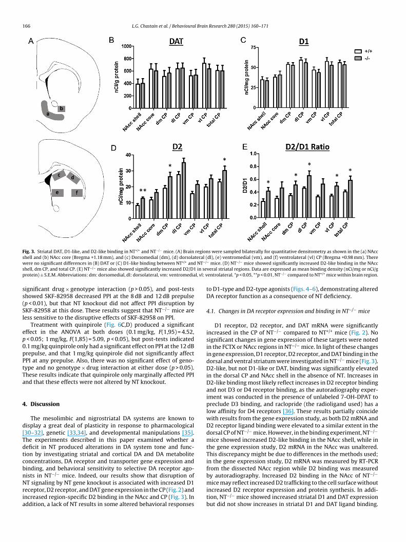
For D1 and D2 receptor autoradiography, the BAS-5000 plates(FujiFilm) were developed in a BAS-5000 phosphorimager (Fuji-Film) and images were analyzed using MultiGauge software(FujiFilm). Photostimulated luminescence per mm2 (PSL/mm2) wasmeasured for regions of interest bilaterally. PSL/mm2 were con-verted to nanocuries/mg protein with tritium standards (AmericanRadiolabeled Chemicals, Inc., St. Louis, MO). For DAT autoradiog-raphy, BioMax MR films (Kodak) were analyzed by quantitativedensitometry using AIS computerized software (AIS, St. Cather-ines, Ontario, Canada). Optical densities were measured for regionsof interest bilaterally, and were converted to nanocuries/mg pro-tein with the 14C standards (Amersham Biosciences). To determineregions of interest, slide-mounted sections were stained with Neu-tral Red and compared to a mouse atlas [26]. For all sections, twodensitometry measurements were made in the NAcc bilaterally,one at bregma +1.34 mm and one between bregma +1.18 mm and+1.1 mm. Densitometries were sampled in the NAcc core in a rect-angular area and in the NAcc shell using the free hand tracing toolsin MultiGauge and AIS (Fig. 3A). The CP was measured bilaterallybetween bregma +1.18 mm and +0.98 mm. Densitometries weresampled in CP subregions (dorsomedial, dorsolateral, ventrome-dial, and ventrolateral regions) in rectangular areas and in the totalCP using the free hand tracing tools in MultiGauge and AIS (Fig. 3A).Background was subtracted from regions of interest from adjacentareas lacking specific binding on the same section.
2.7. Behavioral testing
Startle amplitude and PPI were measured in San Diego Instru-ments startle chambers (San Diego, CA). All mice in the behavioralstudy described below and in the tissue studies described aboveunderwent startle testing. Startle amplitude was measured fromvibrations of a Plexiglas cylinder (resting on a platform) causedby whole-body response. Vibrations were converted into analogsignals using a piezoelectric unit attached to the platform. Thesesignals were digitized and stored in a personal computer. Thetesting session began with a 5 min acclimatization to the startlechamber in the presence of 65 dB background white noise. Testingsessions consisted of eleven 120 dB pulses alone, eleven no stimulustrials, and 18 pulses preceded (100 ms) by a prepulse of 4, 8, or 12 dBabove background. Pulses were presented in a pseudorandom orderwith an average of 15 s between pulses. Percent PPI for each mouseat each prepulse intensity was calculated using the following for-mula: %PPI = 100 − (startle amplitude with prepulse × 100/startleamplitude with pulse alone).
Mice in the behavioral study described below underwent loco-motor testing. Locomotor activity measurements were evaluated
light conditions. Activity was recorded for 90 min, and videos werepost-processed to quantify time-dependent spontaneous behavior.Distance moved by each animal in the arena was automaticallydetermined using TopScan (Clever Sys Inc., Reston, VA).
l Brain
2
8aDos8STAPBttwl1s
2
fSt
pPcoltmtetauwwdtdpuaway(J
3
3
DmiFtp
L.G. Chastain et al. / Behavioura
.8. DA receptor agonist study
The behavioral effects of the D1-type receptor agonist SKF-2958 and the D2-type receptor agonist quinpirole on locomotorctivity and PPI were tested in NT+/+ (n = 8) and NT−/− mice (n = 13).rugs were dissolved in 0.9% saline and injected i.p. at a volumef 1.0 ml/kg body weight. Mice were weighed before each testingession to determine the appropriate dose for each animal. SKF-2958 hydrobromide was obtained through the NIMH Chemicalynthesis and Drug Supply Program (RTI International, Researchriangle Park, NC), and quinpirole was purchased from Sigma-ldrich. For all tests, drugs or saline were injected i.p. 10 min beforePI testing, which was immediately followed by locomotor testing.efore drug testing, NT+/+ and NT−/− mice first underwent baselineesting twice (receiving injections of 0.9% saline i.p. before testing)o obtain baseline PPI and locomotor values for each animal. Oneeek after baseline testing, all animals underwent weekly PPI and
ocomotor testing, receiving each treatment (0.1 mg/kg quinpirole, mg/kg quinpirole, 0.3 mg/kg SKF-82958, 1 mg/kg SKF-82958, oraline) in a counterbalanced, within-subjects design.
.9. Statistical analysis
For the HPLC, RT-PCR, and autoradiography experiments, dif-erences between genotypes for each measure were analyzed by atudent’s t test. For the gene expression studies, Pearson correla-ions were calculated between −dCT values across brain regions.
For the DA receptor agonist study, the behavioral effects of quin-irole and SKF-82958 on locomotor behavior, startle response, andPI were analyzed utilizing ANOVAs. Following ANOVAs, plannedomparisons were tested using Tukey’s HSD post-test. The effectf each drug dose on each behavior was analyzed separately. Forocomotor behavior and PPI for each drug dose, the effects of geno-ype and drug were analyzed using three-way repeated measures
ixed ANOVAs (SAS PROC GLIMMIX specifying a gamma distribu-ion and a log link for the dependent variable and robust standardrrors) with the factors: drug, genotype, and timepoint for locomo-or behavior and drug, genotype, and prepulse for PPI. For startlemplitude, the effects of genotype and drug dose were analyzedsing two-way ANOVAs (factors: genotype and drug). When thereas a significant genotype × drug interaction, Tukey’s post-testsere used for pairwise comparisons of saline controls to each drugose within each genotype to assess drug response within geno-ype. When there was a significant effect of drug but no significantrug × genotype interaction, saline and drug groups were com-ared collapsed across genotypes for each timepoint or prepulsesing Tukey’s post-tests. Baseline saline control values for startlemplitude and PPI values were not statistically different and, thus,ere averaged to generate control values for startle amplitude
nd PPI comparisons. Significance was set at p < 0.05 for all anal-ses. Statistical analyses were performed with GraphPad Prism 3.0GraphPad Software, San Diego, CA), SysStat SigmaPlot 12.3 (Sanose, CA), and SAS (Cary, NC) statistical software.
. Results
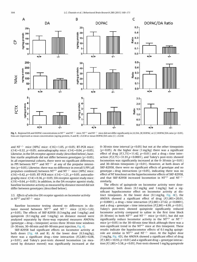
.1. DA and DOPAC concentrations in NT+/+ and NT−/− mice
To compare DAergic tone in NT+/+ and NT−/− mice, DA andOPAC concentrations in the NAcc, CP, and FCTX in NT+/+ and NT−/−
ice were examined by HPLC. There were no significant differences
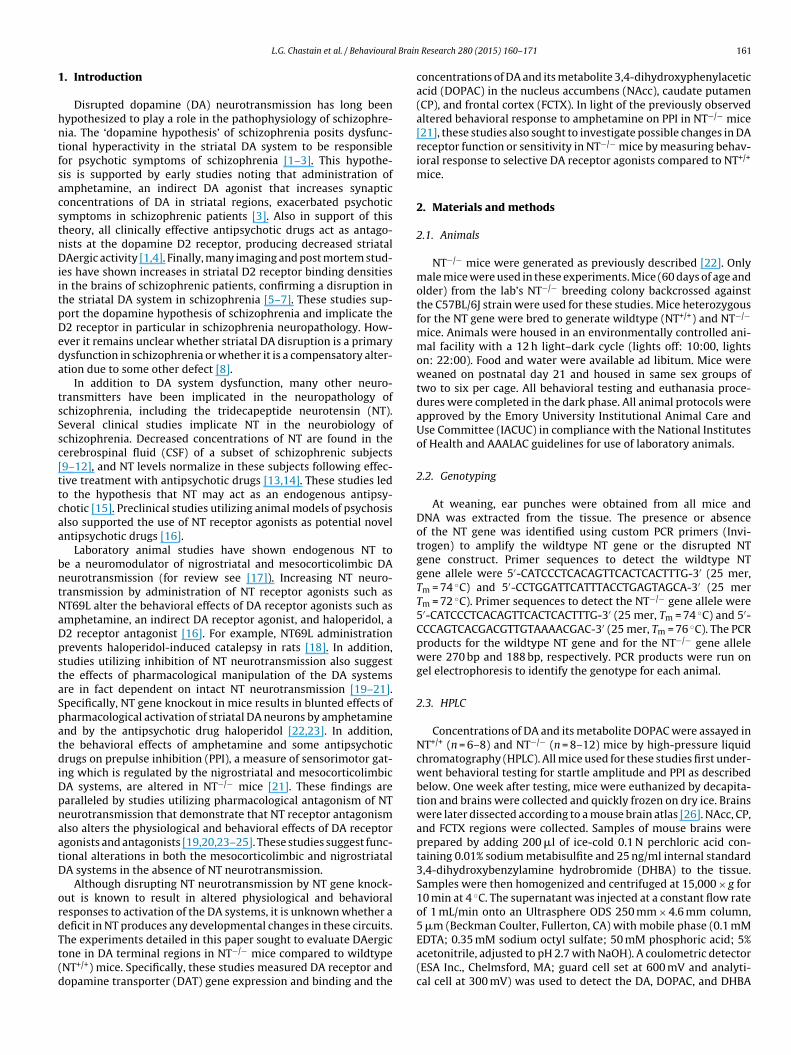
n DA (Fig. 1A; CP: t(18) = 0.30), p > 0.05; NAcc: t(14) = 1.27, p > 0.05;CTX: t(16) = 0.01, p > 0.05) or DOPAC concentrations (Fig. 1B; CP:(18) = 0.05, p > 0.05; NAcc: t(14) = 0.06, p > 0.05; FCTX: t(15) = 0.22,> 0.05) in any of the brain regions examined. To evaluate DA
Research 280 (2015) 160–171 163
turnover in NT+/+ and NT−/− mice, the DOPAC/DA ratio was calcu-lated and compared between genotypes. There was no differencein the DOPAC/DA ratio (Fig. 1C; CP: t(18) = 0.69, p > 0.05; NAcc:t(14) = 1.37, p > 0.05; FCTX: t(15) = 0.81, p > 0.05) between NT+/+ andNT−/− mice in any of the brain regions examined. The NAcc andFCTX are anatomically and functionally connected directly via corti-costriatal projections and indirectly through striatopallidothalamicprojections [28], and experimental disruption of frontocortical DAneurotransmission in rodent models has been shown to producehyperactivity of DAergic function in the striatum, especially theventral striatum [29]. For this reason, DA and DOPAC concentra-tions were compared between NAcc and FCTX regions by ratioanalyses in NT+/+ and NT−/− mice. There was no difference inDA (t(13) = 0.58, p > 0.05) and DOPAC (t(12) = 1.13, p > 0.05) con-centrations in the NAcc compared to the FCTX (NAcc/FCTX ratio),consistent with the idea that there is no major disruption of fron-tocortical DA transmission in NT−/− mice.
3.2. DA receptor and DAT gene expression in NT+/+ and NT−/−
mice
Differences in mRNA levels of the dopamine D1 receptor(Fig. 2A), dopamine D2 receptor (Fig. 2B), and the DAT (Fig. 2C)between NT+/+ and NT−/− mice were examined by real time RT-PCR.NT−/− mice had significantly increased D1 (t(13) = 5.04, p < 0.001),D2 (t(13) = 2.39, p < 0.05), and DAT mRNA levels (t(13) = 2.52,p < 0.05) compared to NT+/+ mice in the CP. NT−/− mice showed atrend for increased DAT mRNA in the FCTX compared to NT+/+ mice,but this difference did not reach significance (t(13) = 1.98, p = 0.07).There were no significant differences in mRNA levels of any of thetargets in the NAcc between genotypes (p > 0.05).
Again, as DA neurotransmission in the FCTX and NAcc arefunctionally related, D1 and D2 mRNA levels (−dCT values) wereanalyzed for regional correlation between the FCTX and NAcc.Receptor expression in the FCTX was not correlated with its expres-sion in the NAcc in either genotype (p > 0.05).
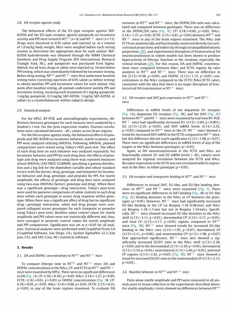
3.3. DA receptor and transporter binding in NT+/+ and NT−/− mice
Differences in striatal DAT, D1-like, and D2-like binding den-sities in NT+/+ and NT−/− mice were examined (Fig. 3). Therewere no significant differences in DAT binding (Fig. 3B) or D1-like(Fig. 3C) binding densities in the NAcc or CP between the geno-types (p > 0.05). However, NT−/− mice had significantly increasedD2-like binding in the CP (at Bregma 1.18–0.98 mm) and NAcc(at Bregma 1.18–1.1 mm but not at Bregma 1.34 mm). Specifi-cally, NT−/− mice showed increased D2-like densities in the NAccshell (t(13) = 3.11, p < 0.01), dorsomedial CP (t(13) = 2.17, p < 0.05),and total CP (t(13) = 2.17, p < 0.05) regions compared to NT+/+
mice (Fig. 3D). NT−/− mice showed trends for increased D2-likebinding in the NAcc core (t(13) = 1.95, p = 0.07), dorsolateral CP(t(13) = 2.11, p = 0.06), and ventromedial CP (t(13) = 1.98, p = 0.07)that approached significance. NT−/− mice also showed a sig-nificantly increased D2/D1 ratio in the NAcc shell (t(13) = 2.28,p < 0.05) and in the dorsomedial (t(13) = 2.39, p < 0.05), dorsolateral(t(13) = 2.34, p < 0.05), ventrolateral (t(13) = 2.40, p < 0.05), and totalCP regions (t(13) = 2.42, p < 0.05) (Fig. 3E). NT−/− mice showed atrend for increased D2/D1 ratio in the ventromedial CP (t(13) = 2.13,p = 0.05).
3.4. Baseline behavior in NT+/+ and NT−/− mice
Pulse alone startle amplitude and PPI were measured in all ani-mals prior to tissue collection in the experiments described above.For startle amplitude, t-tests showed no differences between NT+/+
164 L.G. Chastain et al. / Behavioural Brain Research 280 (2015) 160–171
F −/− mD n DOP
atLlIitptgtbd
3i
tpqa((
btps
ig. 1. Regional DA and DOPAC concentrations in NT+/+ and NT−/− mice. NT+/+ and NTata are expressed as mean concentration (ng/mg protein, A and B) ±S.E.M or mea
nd NT−/− mice (HPLC mice: t(16) = 1.05, p > 0.05; RT-PCR mice:(14) = 0.32, p > 0.05; autoradiography mice: t(14) = 0.04, p > 0.05).ikewise, in the DA receptor agonist study (described below), base-ine startle amplitude did not differ between genotypes (p > 0.05).n all experimental cohorts, there were no significant differencesn PPI between NT+/+ and NT−/− at any of the prepulse intensi-ies (p > 0.05). Likewise, there was no difference in overall % PPI (allrepulses combined) between NT+/+ and NT−/− mice (HPLC mice:(16) = 0.42, p > 0.05; RT-PCR mice: t(14) = 1.21, p > 0.05; autoradio-raphy mice: t(14) = 0.36, p > 0.05; DA receptor agonist study mice:(19) = 0.64, p > 0.05). In addition, in the DA receptor agonist study,aseline locomotor activity as measured by distance moved did notiffer between genotypes (described below).
.5. Effects of selective DA receptor agonists on locomotor activityn NT+/+ and NT−/− mice
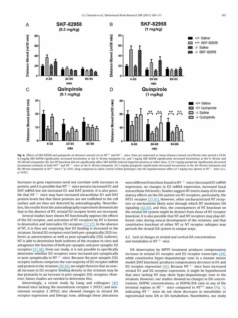
Baseline locomotor testing showed no differences in dis-ance moved between NT+/+ and NT−/− mice (t(16) = 1.02,
> 0.05). The effects of SKF-82958 (0.3 mg/kg and 1 mg/kg) anduinpirole (0.1 mg/kg and 1 mg/kg) on distance moved werenalyzed separately by three-way repeated measures ANOVAsgenotype × drug × timepoint) across three 30 min time windows0–30 min, 30–60 min and 60–90 min post injection, Fig. 4).
SKF-82958 had significant effects on locomotor activity at
oth doses (Fig. 4A and B). At the lower dose (0.3 mg/kg),here was a significant drug × time interaction (F(2,80) = 6.08,< 0.01), and Tukey’s post-tests showed locomotion (as mea-ured by distance moved) was significantly increased at the
ice did not differ significantly in (A) DA, (B) DOPAC, or (C) DOPAC/DA ratio (p > 0.05).AC/DA ratio (C) ±S.E.M.
0–30 min time interval (p < 0.05) but not at the other timepoints(p > 0.05). At the higher dose (1 mg/kg) there was a significanteffect of drug (F(1,75) = 11.42, p < 0.01) and a drug × time inter-action (F(2,75) = 31.91,p < 0.0001), and Tukey’s post-tests showedlocomotion was significantly increased at the 0–30 min (p < 0.01)and 30–60 min timepoints (p < 0.01). However, at both doses ofSKF-82958, there were no significant effects of genotype and nogenotype × drug interactions (p > 0.05), indicating there was noeffect of NT knockout on the hyperlocomotor effects of SKF-82958,and that SKF-82958 increased locomotion in NT+/+ and NT−/−
similarly.The effects of quinpirole on locomotor activity were dose-
dependent; both doses (0.1 mg/kg and 1 mg/kg) had a sig-nificant hypolocomotor effect on locomotor activity at dis-tinct timepoints. At the lower dose (0.1 mg/kg, Fig. 4C), theANOVA showed a significant effect of drug (F(1,80) = 20.01,p < 0.0001), a drug × time interaction (F(2,80) = 27.62, p < 0.0001),and a drug × genotype × time interaction (F(2,80) = 4.99, p < 0.01).Tukey’s post-tests showed quinpirole significantly reducedlocomotor activity compared to saline in the first time block(0–30 min) in both NT+/+ and NT−/− mice (p < 0.01), but did notsignificantly reduce locomotor activity in the NT+/+ or NT−/−
mice (p > 0.05) in the 30–60 min time block although there was anon-significant trend in the NT+/+ mice at this timepoint. Theseresults indicate the hypolocomotor effects of 0.1 mg/kg quinpi-
role are similar in NT+/+ and NT−/− mice. At the higher dose(1 mg/kg, Fig. 4D), the ANOVA showed a significant effect of drug(F(1,80) = 10.93, p < 0.01) and a significant drug × genotype interac-tion (F(1,80) = 5.58, p < 0.05). Post-tests showed 1 mg/kg quinpirole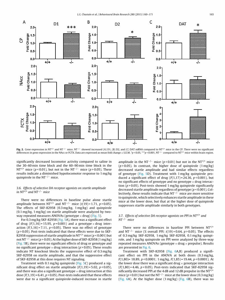
L.G. Chastain et al. / Behavioural Brain Research 280 (2015) 160–171 165
F (B) D2d chang
stNrq
3i
aT(w
oa(8n(niSo
nadw
ig. 2. Gene expression in NT+/+ and NT−/− mice. NT−/− showed increased (A) D1,
ifferences in gene expression in the NAcc or FCTX. Data are expressed as mean fold
ignificantly decreased locomotor activity compared to saline inhe 30–60 min time block and the 60–90 min time block in theT+/+ mice (p < 0.01), but not in the NT−/− mice (p > 0.05). These
esults indicate a diminished hypolocomotor response to 1 mg/kguinpirole in the NT−/− mice.
.6. Effects of selective DA receptor agonists on startle amplituden NT+/+ and NT−/− mice
There were no differences in baseline pulse alone startlemplitude between NT+/+ and NT−/− mice (t(19) = 1.71, p > 0.05).he effects of SKF-82958 (0.3 mg/kg, 1 mg/kg) and quinpirole0.1 mg/kg, 1 mg/kg) on startle amplitude were analyzed by two-ay repeated measures ANOVAs (genotype × drug) (Fig. 5).
For 0.3 mg/kg SKF-82958 (Fig. 5A), there was a significant effectf drug (F(1,16) = 15.95, p = 0.001) and a genotype × drug inter-ction (F(1,16) = 7.11, p < 0.05). There was no effect of genotypep > 0.05). Post-tests indicated that these effects were due to SKF-2958 suppression of startle amplitude in NT+/+ mice (p < 0.001) butot NT−/− mice (p > 0.05). At the higher dose of SKF-82958 (1 mg/kg)Fig. 5B), there were no significant effects of drug or genotype ando significant genotype × drug interaction (p > 0.05). These results
ndicate NT knockout blocks the suppressive effect of 0.3 mg/kgKF-82958 on startle amplitude, and that the suppressive effectf SKF-82958 at this dose requires NT signaling.
Treatment with 0.1 mg/kg quinpirole (Fig. 5C) produced a sig-
ificant drug effect on startle amplitude (F(1,19) = 5.02, p < 0.05),nd there was also a significant genotype × drug interaction at thisose (F(1,19) = 6.41, p < 0.05). Post-tests indicated that these effectsere due to a significant quinpirole-induced increase in startle, and (C) DAT mRNA compared to NT+/+ mice in the CP. There were no significante ± S.E.M. *p < 0.05, ***p < 0.001, NT−/− compared to NT+/+ mice within brain region.
amplitude in the NT−/− mice (p < 0.01) but not in the NT+/+ mice(p > 0.05). In contrast, the higher dose of quinpirole (1 mg/kg)decreased startle amplitude and had similar effects regardlessof genotype (Fig. 5D). Treatment with 1 mg/kg quinpirole pro-duced a significant effect of drug (F(1,17) = 24.36, p < 0.001), butno significant effects of genotype and no genotype × drug interac-tion (p > 0.05). Post-tests showed 1 mg/kg quinpirole significantlydecreased startle amplitude regardless of genotype (p < 0.001). Col-lectively, these results indicate that NT−/− mice are more sensitiveto quinpirole, which selectively enhances startle amplitude in thesemice at the lower dose, but that at the higher dose of quinpirolesuppresses startle amplitude similarly in both genotypes.
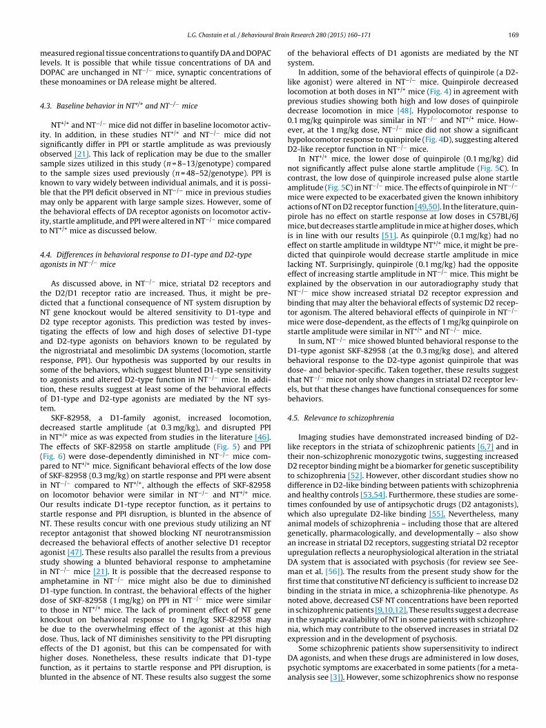
3.7. Effects of selective DA receptor agonists on PPI in NT+/+ andNT−/− mice
There were no differences in baseline PPI between NT+/+
and NT−/− mice (% overall PPI: t(19) = 0.64, p > 0.05). The effectsof 0.3 mg/kg SKF-82958, 1 mg/kg SKF-82958, 0.1 mg/kg quinpi-role, and 1 mg/kg quinpirole on PPI were analyzed by three-wayrepeated measures ANOVAs (genotype × drug × prepulse). Resultsare presented in Fig. 6.
Treatment with SKF-82958 (Fig. 6A,B) produced a signifi-cant effect on PPI in the ANOVA at both doses (0.3 mg/kg,F(1,80)= 18.89, p < 0.0001; 1 mg/kg, F(1,85) = 19.44, p < 0.0001). Atthe lower dose there was a significant drug × genotype interaction
(F(1,80) = 4.46, p < 0.05). Post-tests indicated that SKF-82958 sig-nificantly decreased PPI at the 4 dB and 12 dB prepulse in the NT+/+mice (p < 0.01) but not the NT−/− mice at the lower dose (0.3 mg/kg)(Fig. 6A). At the higher dose (1 mg/kg) (Fig. 6B), there was no
166 L.G. Chastain et al. / Behavioural Brain Research 280 (2015) 160–171
Fig. 3. Striatal DAT, D1-like, and D2-like binding in NT+/+ and NT−/− mice. (A) Brain regions were sampled bilaterally for quantitative densitometry as shown in the (a) NAccshell and (b) NAcc core (Bregma +1.18 mm), and (c) Dorsomedial (dm), (d) dorsolateral (dl), (e) ventromedial (vm), and (f) ventrolateral (vl) CP (Bregma +0.98 mm). Therew NT−/
s in sep l, vl: v
ss(Sl
ep0pPtTa
4
d[TdtcbnNria
ere no significant differences in (B) DAT or (C) D1-like binding between NT+/+ andhell, dm CP, and total CP. (E) NT−/− mice also showed significantly increased D2/D1rotein) ± S.E.M. Abbreviations: dm: dorsomedial, dl: dorsolateral, vm: ventromedia
ignificant drug × genotype interaction (p > 0.05), and post-testshowed SKF-82958 decreased PPI at the 8 dB and 12 dB prepulsep < 0.01), but that NT knockout did not affect PPI disruption byKF-82958 at this dose. These results suggest that NT−/− mice areess sensitive to the disruptive effects of SKF-82958 on PPI.
Treatment with quinpirole (Fig. 6C,D) produced a significantffect in the ANOVA at both doses (0.1 mg/kg, F(1,95) = 4.52,
< 0.05; 1 mg/kg, F(1,85) = 5.09, p < 0.05), but post-tests indicated.1 mg/kg quinpirole only had a significant effect on PPI at the 12 dBrepulse, and that 1 mg/kg quinpirole did not significantly affectPI at any prepulse. Also, there was no significant effect of geno-ype and no genotype × drug interaction at either dose (p > 0.05).hese results indicate that quinpirole only marginally affected PPInd that these effects were not altered by NT knockout.
. Discussion
The mesolimbic and nigrostriatal DA systems are known toisplay a great deal of plasticity in response to pharmacological30–32], genetic [33,34], and developmental manipulations [35].he experiments described in this paper examined whether aeficit in NT produced alterations in DA system tone and func-ion by investigating striatal and cortical DA and DA metaboliteoncentrations, DA receptor and transporter gene expression andinding, and behavioral sensitivity to selective DA receptor ago-ists in NT−/− mice. Indeed, our results show that disruption of
T signaling by NT gene knockout is associated with increased D1eceptor, D2 receptor, and DAT gene expression in the CP (Fig. 2) andncreased region-specific D2 binding in the NAcc and CP (Fig. 3). Inddition, a lack of NT results in some altered behavioral responses
− mice. (D) NT−/− mice showed significantly increased D2-like binding in the NAccveral striatal regions. Data are expressed as mean binding density (nCi/mg or nCi/gentrolateral. *p < 0.05, **p < 0.01, NT−/− compared to NT+/+ mice within brain region.
to D1-type and D2-type agonists (Figs. 4–6), demonstrating alteredDA receptor function as a consequence of NT deficiency.
4.1. Changes in DA receptor expression and binding in NT−/− mice
D1 receptor, D2 receptor, and DAT mRNA were significantlyincreased in the CP of NT−/− compared to NT+/+ mice (Fig. 2). Nosignificant changes in gene expression of these targets were notedin the FCTX or NAcc regions in NT−/− mice. In light of these changesin gene expression, D1 receptor, D2 receptor, and DAT binding in thedorsal and ventral striatum were investigated in NT−/− mice (Fig. 3).D2-like, but not D1-like or DAT, binding was significantly elevatedin the dorsal CP and NAcc shell in the absence of NT. Increases inD2-like binding most likely reflect increases in D2 receptor bindingand not D3 or D4 receptor binding, as the autoradiography exper-iment was conducted in the presence of unlabeled 7-OH-DPAT topreclude D3 binding, and raclopride (the radioligand used) has alow affinity for D4 receptors [36]. These results partially coincidewith results from the gene expression study, as both D2 mRNA andD2 receptor ligand binding were elevated to a similar extent in thedorsal CP of NT−/− mice. However, in the binding experiment, NT−/−
mice showed increased D2-like binding in the NAcc shell, while inthe gene expression study, D2 mRNA in the NAcc was unaltered.This discrepancy might be due to differences in the methods used;in the gene expression study, D2 mRNA was measured by RT-PCRfrom the dissected NAcc region while D2 binding was measuredby autoradiography. Increased D2 binding in the NAcc of NT−/−
mice may reflect increased D2 trafficking to the cell surface withoutincreased D2 receptor expression and protein synthesis. In addi-tion, NT−/− mice showed increased striatal D1 and DAT expressionbut did not show increases in striatal D1 and DAT ligand binding.
L.G. Chastain et al. / Behavioural Brain Research 280 (2015) 160–171 167
Fig. 4. Effects of SKF-82958 and quinpirole on distance moved (m) in NT+/+ and NT−/− mice. Data are expressed as mean distance moved (m)/30 min time period ± S.E.M.0.3 mg/kg SKF-82958 significantly increased locomotion at the 0–30 min timepoint (A), and 1 mg/kg SKF-82958 significantly increased locomotion at the 0–30 min and30–60 min timepoints (B), but NT knockout did not significantly affect SKF-82958-induced hyperlocomotion at either dose. (C) 0.1 mg/kg quinpirole significantly decreasedl 1 mg/6 thin gp
IpDbpslt
otosfNardoraade
srr
ocomotion similarly in both NT+/+ and NT−/− mice at the 0–30 min timepoint. (D)0–90 min timepoint in NT+/+ mice (**p < 0.01, drug compared to saline control wi
> 0.05).
ncreases in gene expression need not correlate with increases inrotein, and it is possible that NT−/− mice possess increased D1 andAT mRNA but not increased D1 and DAT protein. It is also possi-le that NT−/− mice may have increased intracellular D1 and DATrotein levels but that these proteins are not trafficked to the cellurface and are thus not detected by autoradiography. Neverthe-ess, the results from the autoradiography experiment demonstratehat in the absence of NT, striatal D2 receptor levels are increased.
Several studies have shown NT functionally opposes the effectsf the D2 receptor, and activation of NT receptors by NT is knowno desensitize and internalize D2 receptors [17,37]. In the absencef NT, it is thus not surprising that D2 binding is increased in thetriatum. Striatal D2 receptors exist both pre-synaptically (D2S iso-orm) as autoreceptors as well as post-synaptically (D2L isoform).T is able to desensitize both isoforms of the receptor in vitro andntagonize the function of both pre-synaptic and post-synaptic D2eceptors [37,38]. From our study, it is not possible to specificallyetermine whether D2 receptors were increased pre-synapticallyr post-synaptically in NT−/− mice. Because the post-synaptic D2Leceptor isoform comprises the vast majority of D2 receptor mRNAnd protein in the striatum [30,39], we might theorize that an over-ll increase in D2 receptor binding density in the striatum may beue primarily to an increase in post-synaptic D2L receptors. How-ver, future studies are needed to determine this.
Interestingly, a recent study by Liang and colleagues [40]howed mice lacking the neurotensin receptor 1 (NTS1) and neu-otensin receptor 2 (NTS2) also showed changes in striatal DAeceptor expression and DAergic tone, although these alterations
kg quinpirole significantly decreased locomotion at the 30–60 min timepoint andenotype), but the hypolocomotor effect of 1 mg/kg was absent in NT−/− mice (n.s.,
were different from those found in NT−/− mice (decreased D1 mRNAexpression, no changes in D2 mRNA expression, increased basalextracellular DA levels). Studies suggest NT exerts many of its mod-ulatory effects on the DA system via NT receptors, particularly, theNTS1 receptor [37,41]. However, other uncharacterized NT recep-tors or mechanisms likely exist through which NT modulates DAsignaling [42,43], and thus, the consequences of NT knockout onthe striatal DA system might be distinct from those of NT receptorknockout. It is also possible that NT and NT receptors may play dif-ferent roles during neural development of the striatum, and thatconstitutive knockout of either NT or NT receptor subtypes mayperturb the striatal DA system in unique ways.
4.2. Lack of changes in striatal and cortical DA concentrationsand metabolites in NT−/− mice
DA denervation by MPTP treatment produces compensatoryincreases in striatal D1 receptor and D2 receptor transcripts [44],while constitutive hyper-dopaminergic tone in a mutant mousemodel (DAT knockout) produces compensatory decreases in D1 andD2 receptor expression [45]. Because NT−/− mice have increasedstriatal D1 and D2 receptor expression, it might be hypothesizedthat mice lacking NT may show hypo-dopaminergic tone in thestriatum. However, our studies showed no changes in DA concen-
trations, DOPAC concentrations, or DOPAC/DA ratio in any of theterminal regions in NT−/− mice compared to NT+/+ mice (Fig. 1),indicating NT−/− mice do not show decreases in mesolimbic ornigrostriatal tonic DA or DA metabolism. Nonetheless, our study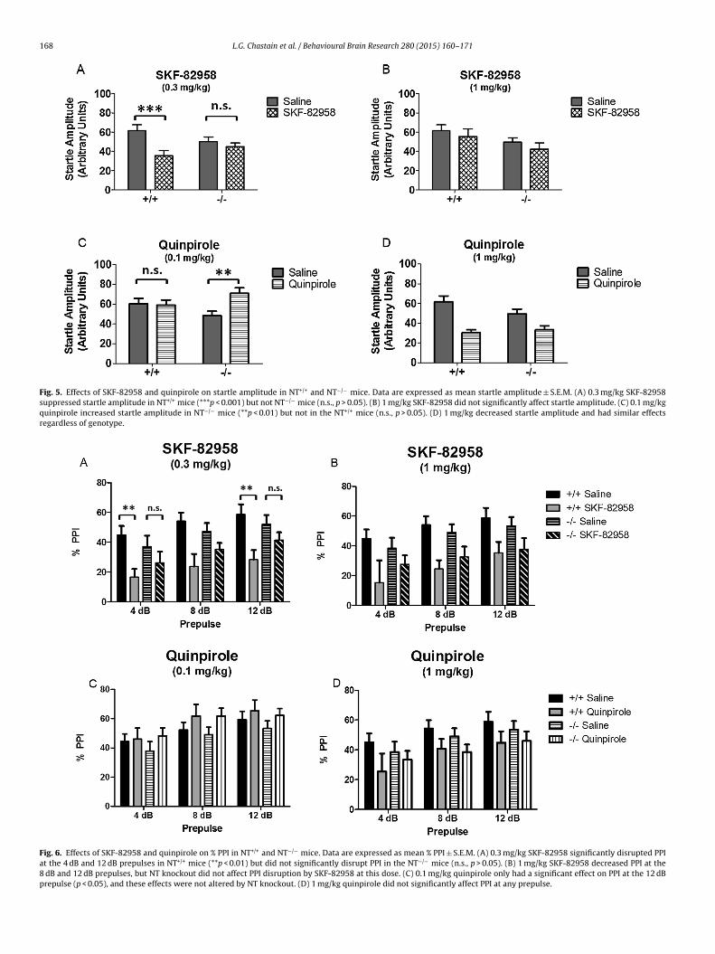
168 L.G. Chastain et al. / Behavioural Brain Research 280 (2015) 160–171
Fig. 5. Effects of SKF-82958 and quinpirole on startle amplitude in NT+/+ and NT−/− mice. Data are expressed as mean startle amplitude ± S.E.M. (A) 0.3 mg/kg SKF-82958suppressed startle amplitude in NT+/+ mice (***p < 0.001) but not NT−/− mice (n.s., p > 0.05). (B) 1 mg/kg SKF-82958 did not significantly affect startle amplitude. (C) 0.1 mg/kgquinpirole increased startle amplitude in NT−/− mice (**p < 0.01) but not in the NT+/+ mice (n.s., p > 0.05). (D) 1 mg/kg decreased startle amplitude and had similar effectsregardless of genotype.
Fig. 6. Effects of SKF-82958 and quinpirole on % PPI in NT+/+ and NT−/− mice. Data are expressed as mean % PPI ± S.E.M. (A) 0.3 mg/kg SKF-82958 significantly disrupted PPIat the 4 dB and 12 dB prepulses in NT+/+ mice (**p < 0.01) but did not significantly disrupt PPI in the NT−/− mice (n.s., p > 0.05). (B) 1 mg/kg SKF-82958 decreased PPI at the8 dB and 12 dB prepulses, but NT knockout did not affect PPI disruption by SKF-82958 at this dose. (C) 0.1 mg/kg quinpirole only had a significant effect on PPI at the 12 dBprepulse (p < 0.05), and these effects were not altered by NT knockout. (D) 1 mg/kg quinpirole did not significantly affect PPI at any prepulse.
l Brain
mlDt
4
isostkbmtit
4a
tdNDtatrsttot
diT(poioOsNrdasiaDdtkbdehfb
L.G. Chastain et al. / Behavioura
easured regional tissue concentrations to quantify DA and DOPACevels. It is possible that while tissue concentrations of DA andOPAC are unchanged in NT−/− mice, synaptic concentrations of
hese monoamines or DA release might be altered.
.3. Baseline behavior in NT+/+ and NT−/− mice
NT+/+ and NT−/− mice did not differ in baseline locomotor activ-ty. In addition, in these studies NT+/+ and NT−/− mice did notignificantly differ in PPI or startle amplitude as was previouslybserved [21]. This lack of replication may be due to the smallerample sizes utilized in this study (n = 8–13/genotype) comparedo the sample sizes used previously (n = 48–52/genotype). PPI isnown to vary widely between individual animals, and it is possi-le that the PPI deficit observed in NT−/− mice in previous studiesay only be apparent with large sample sizes. However, some of
he behavioral effects of DA receptor agonists on locomotor activ-ty, startle amplitude, and PPI were altered in NT−/− mice comparedo NT+/+ mice as discussed below.
.4. Differences in behavioral response to D1-type and D2-typegonists in NT−/− mice
As discussed above, in NT−/− mice, striatal D2 receptors andhe D2/D1 receptor ratio are increased. Thus, it might be pre-icted that a functional consequence of NT system disruption byT gene knockout would be altered sensitivity to D1-type and2 type receptor agonists. This prediction was tested by inves-
igating the effects of low and high doses of selective D1-typend D2-type agonists on behaviors known to be regulated byhe nigrostriatal and mesolimbic DA systems (locomotion, startleesponse, PPI). Our hypothesis was supported by our results inome of the behaviors, which suggest blunted D1-type sensitivityo agonists and altered D2-type function in NT−/− mice. In addi-ion, these results suggest at least some of the behavioral effectsf D1-type and D2-type agonists are mediated by the NT sys-em.
SKF-82958, a D1-family agonist, increased locomotion,ecreased startle amplitude (at 0.3 mg/kg), and disrupted PPI
n NT+/+ mice as was expected from studies in the literature [46].he effects of SKF-82958 on startle amplitude (Fig. 5) and PPIFig. 6) were dose-dependently diminished in NT−/− mice com-ared to NT+/+ mice. Significant behavioral effects of the low dosef SKF-82958 (0.3 mg/kg) on startle response and PPI were absentn NT−/− compared to NT+/+, although the effects of SKF-82958n locomotor behavior were similar in NT−/− and NT+/+ mice.ur results indicate D1-type receptor function, as it pertains to
tartle response and PPI disruption, is blunted in the absence ofT. These results concur with one previous study utilizing an NT
eceptor antagonist that showed blocking NT neurotransmissionecreased the behavioral effects of another selective D1 receptorgonist [47]. These results also parallel the results from a previoustudy showing a blunted behavioral response to amphetaminen NT−/− mice [21]. It is possible that the decreased response tomphetamine in NT−/− mice might also be due to diminished1-type function. In contrast, the behavioral effects of the higherose of SKF-82958 (1 mg/kg) on PPI in NT−/− mice were similaro those in NT+/+ mice. The lack of prominent effect of NT genenockout on behavioral response to 1 mg/kg SKF-82958 maye due to the overwhelming effect of the agonist at this highose. Thus, lack of NT diminishes sensitivity to the PPI disrupting
ffects of the D1 agonist, but this can be compensated for withigher doses. Nonetheless, these results indicate that D1-typeunction, as it pertains to startle response and PPI disruption, islunted in the absence of NT. These results also suggest the some
Research 280 (2015) 160–171 169
of the behavioral effects of D1 agonists are mediated by the NTsystem.
In addition, some of the behavioral effects of quinpirole (a D2-like agonist) were altered in NT−/− mice. Quinpirole decreasedlocomotion at both doses in NT+/+ mice (Fig. 4) in agreement withprevious studies showing both high and low doses of quinpiroledecrease locomotion in mice [48]. Hypolocomotor response to0.1 mg/kg quinpirole was similar in NT−/− and NT+/+ mice. How-ever, at the 1 mg/kg dose, NT−/− mice did not show a significanthypolocomotor response to quinpirole (Fig. 4D), suggesting alteredD2-like receptor function in NT−/− mice.
In NT+/+ mice, the lower dose of quinpirole (0.1 mg/kg) didnot significantly affect pulse alone startle amplitude (Fig. 5C). Incontrast, the low dose of quinpirole increased pulse alone startleamplitude (Fig. 5C) in NT−/− mice. The effects of quinpirole in NT−/−
mice were expected to be exacerbated given the known inhibitoryactions of NT on D2 receptor function [49,50]. In the literature, quin-pirole has no effect on startle response at low doses in C57BL/6Jmice, but decreases startle amplitude in mice at higher doses, whichis in line with our results [51]. As quinpirole (0.1 mg/kg) had noeffect on startle amplitude in wildtype NT+/+ mice, it might be pre-dicted that quinpirole would decrease startle amplitude in micelacking NT. Surprisingly, quinpirole (0.1 mg/kg) had the oppositeeffect of increasing startle amplitude in NT−/− mice. This might beexplained by the observation in our autoradiography study thatNT−/− mice show increased striatal D2 receptor expression andbinding that may alter the behavioral effects of systemic D2 recep-tor agonism. The altered behavioral effects of quinpirole in NT−/−
mice were dose-dependent, as the effects of 1 mg/kg quinpirole onstartle amplitude were similar in NT+/+ and NT−/− mice.
In sum, NT−/− mice showed blunted behavioral response to theD1-type agonist SKF-82958 (at the 0.3 mg/kg dose), and alteredbehavioral response to the D2-type agonist quinpirole that wasdose- and behavior-specific. Taken together, these results suggestthat NT−/− mice not only show changes in striatal D2 receptor lev-els, but that these changes have functional consequences for somebehaviors.
4.5. Relevance to schizophrenia
Imaging studies have demonstrated increased binding of D2-like receptors in the striata of schizophrenic patients [6,7] and intheir non-schizophrenic monozygotic twins, suggesting increasedD2 receptor binding might be a biomarker for genetic susceptibilityto schizophrenia [52]. However, other discordant studies show nodifference in D2-like binding between patients with schizophreniaand healthy controls [53,54]. Furthermore, these studies are some-times confounded by use of antipsychotic drugs (D2 antagonists),which also upregulate D2-like binding [55]. Nevertheless, manyanimal models of schizophrenia – including those that are alteredgenetically, pharmacologically, and developmentally – also showan increase in striatal D2 receptors, suggesting striatal D2 receptorupregulation reflects a neurophysiological alteration in the striatalDA system that is associated with psychosis (for review see See-man et al. [56]). The results from the present study show for thefirst time that constitutive NT deficiency is sufficient to increase D2binding in the striata in mice, a schizophrenia-like phenotype. Asnoted above, decreased CSF NT concentrations have been reportedin schizophrenic patients [9,10,12]. These results suggest a decreasein the synaptic availability of NT in some patients with schizophre-nia, which may contribute to the observed increases in striatal D2expression and in the development of psychosis.
Some schizophrenic patients show supersensitivity to indirectDA agonists, and when these drugs are administered in low doses,psychotic symptoms are exacerbated in some patients (for a meta-analysis see [3]). However, some schizophrenics show no response
1 l Brain
ta[sboNiDntNtci[isimt
4
gidDNw
C
wSoPBSvRm(S$PP(aei
A
tsRRaPy
[
[
[
[
[
[
[
[
[
[
[
[
[
[
[
[
[
[
[
70 L.G. Chastain et al. / Behavioura
o DA agonists, and there is evidence that DA agonists might actu-lly improve negative symptoms in some schizophrenic patients57,58]. Most animal models relevant to schizophrenia also showupersensitivity to direct and indirect DA agonists, as measuredy the effects of these drugs on locomotor activity and sensorim-tor gating, while a few show subsensitivity to DA agonists [59].T−/− mice may fall in the latter category, as they show behav-
oral and physiological subsensitivity to the effects of the indirectA agonist amphetamine [21,23] and to the selective D1-like ago-ist SKF-82958, although they also showed altered responsivity tohe D2-like agonist quinpirole in our study. These findings suggestT−/− mice may have potential as a unique animal model of nega-
ive schizophrenia-like symptoms. In line with this reasoning, somelinical studies have shown the decreased CSF NT levels observedn some schizophrenics are associated with negative symptoms12,14]. Historically, most animal studies have emphasized model-ng positive symptoms of schizophrenia while neglecting negativeymptoms, and negative symptoms are notoriously difficult to treatn schizophrenic patients [60,61]. For this reason, future studies
ight investigate NT−/− mice for physiology and behaviors relevanto the negative symptoms of schizophrenia.
.6. Conclusions
The results of these studies indicate that NT deficiency due to NTene knockout results in DA system plasticity, particularly, changesn DA receptor expression, binding, and function. These resultsemonstrate a regulatory role for NT in the maintenance of striatalA system homeostasis. Finally, these studies implicate a deficit inT neurotransmission in the neurobiology of disorders associatedith DA system abnormalities, specifically in schizophrenia.
onflicts of interest
Chastain, Qu, Iuvone, Dobner, Kinkead: None; Bourke: Currentlyorks for the FDA; Nemeroff: Consulting: Xhale, Takeda, SK Pharma,
hire, Roche, Lilly, Allergan, Mitsubishi Tanabe Pharma Devel-pment America, Taisho Pharmaceutical Inc., Lundbeck, Prismicharmaceuticals; Stockholder: CeNeRx, BioPharma, PharmaNeuro-oost, Revaax Pharma, Xhale, Celgene, Seattle Genetics, Abbvie;cientific Advisory Boards: American Foundation for Suicide Pre-ention (AFSP), CeNeRx BioPharma (2012), National Alliance foresearch on Schizophrenia and Depression (NARSAD), Xhale, Phar-aNeuroBoost (2012), Anxiety Disorders Association of America
ADAA), Skyland Trail; Board of Directors: AFSP, NovaDel (2011),kyland Trail, Gratitude America, ADAA; Income sources of equity of10,000 or more: PharmaNeuroBoost, CeNeRx BioPharma, NovaDelharma, Reevax Pharma, American Psychiatric Publishing, Xhale;atents: Method and devices for transdermal delivery of lithiumUS 6,375,990B1), Method of assessing antidepressant drug ther-py via transport inhibition of monoamine neurotransmitters byx vivo assay (US 7,148,027B2); Honoraria: Various; Royalties: Var-ous; Expert Witness: Various.
cknowledgments
This work was supported by a grant from the National Insti-ute of Mental Health (NIMH grant MH39415). The HPLC work wasupported by grants from the National Eye Institute (NEI grants01EY004864 and P30EY006360). We thank Aaron DeLaRosa fromutgers University, Department of Animal Sciences for help in cre-
ting the brain illustrations in Fig. 3. We also thank Dr. Anthonyawlak from Rutgers University for consultation on statistical anal-ses.[
Research 280 (2015) 160–171
References
[1] van Rossum JM. The significance of dopamine-receptor blockade for themechanism of action of neuroleptic drugs. Arch Int Pharmacodyn Ther1966;160:492–4.
[2] Meltzer HY, Stahl SM. The dopamine hypothesis of schizophrenia: a review.Schizophr Bull 1976;2:19–76.
[3] Lieberman JA, Kane JM, Alvir J. Provocative tests with psychostimulant drugsin schizophrenia. Psychopharmacology (Berl) 1987;91:415–33.
[4] Grace AA, Bunney BS, Moore H, Todd CL. Dopamine-cell depolarization blockas a model for the therapeutic actions of antipsychotic drugs. Trends Neurosci1997;20:31–7.
[5] Seeman P. Dopamine receptors and the dopamine hypothesis of schizophrenia.Synapse 1987;1:133–52.
[6] Seeman P, Kapur S. Schizophrenia: more dopamine, more D2 receptors. ProcNatl Acad Sci USA 2000;97:7673–5.
[7] Wong DF, Pearlson GD, Tune LE, Young LT, Meltzer CC, Dannals RF, et al. Quan-tification of neuroreceptors in the living human brain: IV. Effect of aging andelevations of D2-like receptors in schizophrenia and bipolar illness. J CerebBlood Flow Metab 1997;17:331–42.
[8] Carlsson A, Waters N, Holm-Waters S, Tedroff J, Nilsson M, Carlsson ML. Inter-actions between monoamines, glutamate, and GABA in schizophrenia: newevidence. Ann Rev Pharmacol Toxicol 2001;41:237–60.
[9] Breslin NA, Suddath RL, Bissette G, Nemeroff CB, Lowrimore P, Weinberger DR.CSF concentrations of neurotensin in schizophrenia: an investigation of clinicaland biochemical correlates. Schizophr Res 1994;12:35–41.
10] Lindström LH, Widerlöv E, Bissette G, Nemeroff CB. Reduced CSF neurotensinconcentration in drug-free schizophrenic patients. Schizophr Res 1988;1:55–9.
11] Nemeroff CB, Bissette G, Widerlov E, Beckmann H, Gerner R, Manberg PJ,et al. Neurotensin-like immunoreactivity in cerebrospinal fluid of patients withschizophrenia, depression, anorexia nervosa-bulimia, and premenstrual syn-drome. J Neuropsychiatry Clin Neurosci 1989;1:16–20.
12] Sharma RP, Janicak PG, Bissette G, Nemeroff CB. CSF neurotensin concentrationsand antipsychotic treatment in schizophrenia and schizoaffective disorders.Am J Psychiatry 1997;154:1019–21.
13] Widerlöv E, Lindström LH, Besev G, Manberg PJ, Nemeroff CB, Breese GR,et al. Subnormal CSF levels of neurotensin in a subgroup of schizophrenicpatients: normalization after neuroleptic treatment. Am J Psychiatry 1982;139:1122–6.
14] Garver DL, Bissette G, Yao JK, Nemeroff CB. Relation of CSF neurotensin concen-trations to symptoms and drug response of psychotic patients. Am J Psychiatry1991;148:484–8.
15] Nemeroff CB. Neurotensin: perchance an endogenous neuroleptic. Biol Psychi-atry 1980;15:283–302.
16] Boules M, Li Z, Smith K, Fredrickson P, Richelson E. Diverse roles of neurotensinagonists in the central nervous system. Front Endocrinol (Lausanne) 2013;4:36.
17] Binder EB, Kinkead B, Owens MJ, Nemeroff CB. Neurotensin and dopamineinteractions. Pharmacol Rev 2001;53:453–86.
18] Cusack B, Boules M, Tyler BM, Fauq A, McCormick DJ, Richelson E. Effects of anovel neurotensin peptide analog given extracranially on CNS behaviors medi-ated by apomorphine and haloperidol. Brain Res 2000;856:48–54.
19] Costa FG, Frussa-Filho R, Felicio LF. The neurotensin receptor antagonist,SR48692, attenuates the expression of amphetamine-induced behavioural sen-sitisation in mice. Eur J Pharmacol 2001;428:97–103.
20] Cáceda R, Binder EB, Kinkead B, Nemeroff CB. The role of endogenous neu-rotensin in psychostimulant-induced disruption of prepulse inhibition andlocomotion. Schizophr Res 2012;136:88–95.
21] Kinkead B, Dobner PR, Egnatashvili V, Murray T, Deitemeyer N, Nemeroff CB.Neurotensin-deficient mice have deficits in prepulse inhibition: restoration byclozapine but not haloperidol, olanzapine or quetiapine. J Pharmacol Exp Ther2005;315:256–64.
22] Dobner PR, Fadel J, Deitmeyer N, Carraway RE, Deutch AY. Neurotensin-deficient mice show altered responses to antipsychotic drugs. Proc Natl AcadSci U S A 2001;98:8048–53.
23] Fadel J, Dobner PR, Deutch AY. Amphetamine-elicited striatal Fos expression isattenuated in neurotensin null mutant mice. Neurosci Lett 2006;402:97–101.
24] Binder EB, Kinkead B, Owens MJ, Nemeroff CB. Neurotensin receptor antag-onist SR 142948A alters Fos expression and extrapyramidal side effectprofile of typical and atypical antipsychotic drugs. Neuropsychopharmacology2004;29:2200–7.
25] Fadel J, Dobner PR, Deutch AY. The neurotensin antagonist SR 48692 atten-uates haloperidol-induced striatal Fos expression in the rat. Neurosci Lett2001;303:17–20.
26] Franklin KBJ, Paxinos G. The Mouse Brain in Stereotaxic Coordinates. San Diego:Academic Press; 1997.
27] Lowry OH, Rosebrough NJ, Farr AL, Randall RJ. Protein measurement with theFolin phenol reagent. J Biol Chem 1951;193:265–75.
28] Alexander GE, DeLong MR, Strick PL. Parallel organization of function-ally segregated circuits linking basal ganglia and cortex. Ann Rev Neurosci
1986;9:357–81.29] Deutch AY. Prefrontal cortical dopamine systems and the elaboration of func-tional corticostriatal circuits: implications for schizophrenia and Parkinson’sdisease. J Neural Trans 1993;91:197–221.
l Brain
[
[
[
[
[
[
[
[
[
[
[
[
[
[
[
[
[
[
[
[
[
[
[
[
[
[
[
[
[
[
L.G. Chastain et al. / Behavioura
30] Neve KA, Neve RL, Fidel S, Janowsky A, Higgins GA. Increased abundance ofalternatively spliced forms of D2 dopamine receptor mRNA after denervation.Proc Natl Acad Sci USA 1991;88:2802–6.
31] Shilling PD, Kelsoe JR, Segal DS. Dopamine transporter mRNA is up-regulated inthe substantia nigra and the ventral tegmental area of amphetamine-sensitizedrats. Neurosci Lett 1997;236:131–4.
32] Belin D, Deroche-Gamonet V, Jaber M. Cocaine-induced sensitization is asso-ciated with altered dynamics of transcriptional responses of the dopaminetransporter, tyrosine hydroxylase, and dopamine D2 receptors in C57Bl/6J mice.Psychopharmacology (Berl) 2007;193:567–78.
33] Jones SR, Gainetdinov RR, Jaber M, Giros B, Wightman RM, Caron MG. Profoundneuronal plasticity in response to inactivation of the dopamine transporter.Proc Natl Acad Sci USA 1998;95:4029–34.
34] Fauchey V, Jaber M, Bloch B, Le Moine C. Dopamine control of striatal geneexpression during development: relevance to knockout mice for the dopaminetransporter. Eur J Neurosci 2000;12:3415–25.
35] Gatzke-Kopp LM. The canary in the coalmine: the sensitivity of mesolimbicdopamine to environmental adversity during development. Neurosci BiobehavRev 2011;35:794–803.
36] Lahti RA, Evans DL, Stratman NC, Figur LM. Dopamine D4 versus D2 receptorselectivity of dopamine receptor antagonists: possible therapeutic implica-tions. Eur J Pharmacol 1993;236:483–6.
37] Thibault D, Albert PR, Pineyro G, Trudeau LE. Neurotensin triggers dopamineD2 receptor desensitization through a protein kinase C and beta-arrestin1-dependent mechanism. J Biol Chem 2011;286:9174–84.
38] Fuxe K, O’ Connor WT, Antonelli T, Osborne PG, Tanganelli S, Agnati LF, et al.Evidence for a substrate of neuronal plasticity based on pre- and postsynapticneurotensin-dopamine receptor interactions in the neostriatum. Proc Natl AcadSci U S A 1992;89:5591–5.
39] Mack KJ, Todd RD, O’Malley KL. The mouse dopamine D2A receptor gene:sequence homology with the rat and human genes and expression of alter-native transcripts. J Neurochem 1991;57:795–801.
40] Liang Y, Boules M, Li Z, Williams K, Miura T, Oliveros A, et al. Hyperactivityof the dopaminergic system in NTS1 and NTS2 null mice. Neuropharmacology2010;58:1199–205.
41] Jomphe C, Lemelin PL, Okano H, Kobayashi K, Trudeau LE. Bidirectional regu-lation of dopamine D2 and neurotensin NTS1 receptors in dopamine neurons.Eur J Neurosci 2006;24:2789–800.
42] Trudeau LE. Neurotensin regulates intracellular calcium in ventral tegmentalarea astrocytes: evidence for the involvement of multiple receptors. Neuro-science 2000;97:293–302.
43] Mandell AJ, Owens MJ, Selz KA, Morgan WN, Shlesinger MF, Nemeroff CB. Modematches in hydrophobic free energy eigenfunctions predict peptide–proteininteractions. Biopolymers 1998;46:89–101.
44] Smith TS, Trimmer PA, Khan SM, Tinklepaugh DL, Bennett Jr JP. Mitochondrialtoxins in models of neurodegenerative diseases. II: Elevated zif268 transcrip-tion and independent temporal regulation of striatal D1 and D2 receptormRNAs and D1 and D2 receptor-binding sites in C57BL/6 mice during MPTPtreatment. Brain Res 1997;765:189–97.
[
[
Research 280 (2015) 160–171 171
45] Fauchey V, Jaber M, Caron MG, Bloch B, Le Moine C. Differential regulationof the dopamine D1, D2 and D3 receptor gene expression and changes in thephenotype of the striatal neurons in mice lacking the dopamine transporter.Eur J Neurosci 2000;12:19–26.
46] Frau R, Pillolla G, Bini V, Tambaro S, Devoto P, Bortolato M. Inhibition of 5alpha-reductase attenuates behavioral effects of D(1)-, but not D(2)-like receptoragonists in C57BL/6 mice. Psychoneuroendocrinology 2012;38:542–51.
47] Poncelet M, Souilhac J, Gueudet C, Terranova JP, Gully D, Le Fur G, et al. Effectsof SR 48692, a selective non-peptide neurotensin receptor antagonist, on twodopamine-dependent behavioural responses in mice and rats. Psychopharma-cology (Berl) 1994;116:237–41.
48] Halberda JP, Middaugh LD, Gard BE, Jackson BP. DAD1- and DAD2-like agonisteffects on motor activity of C57 mice: differences compared to rats. Synapse1997;26:81–92.
49] Shi WS, Bunney BS. Neurotensin attenuates dopamine D2 agonist quinpirole-induced inhibition of midbrain dopamine neurons. Neuropharmacology1990;29:1095–7.
50] Shi WX, Bunney BS. Neurotensin modulates autoreceptor mediated dopamineeffects on midbrain dopamine cell activity. Brain Res 1991;543:315–21.
51] Ralph-Williams RJ, Lehmann-Masten V, Geyer MA. Dopamine D1 rather thanD2 receptor agonists disrupt prepulse inhibition of startle in mice. Neuropsy-chopharmacology 2003;28:108–18.
52] Hirvonen J, van Erp TG, Huttunen J, Aalto S, Nagren K, Huttunen M, et al.Increased caudate dopamine D2 receptor availability as a genetic marker forschizophrenia. Arch Gen Psychiatry 2005;62:371–8.
53] Nordstrom AL, Farde L, Eriksson L, Halldin C. No elevated D2 dopamine recep-tors in neuroleptic-naive schizophrenic patients revealed by positron emissiontomography and [11C]N-methylspiperone. Psychiatry Res 1995;61:67–83.
54] Farde L, Wiesel FA, Stone-Elander S, Halldin C, Nordstrom AL, Hall H,et al. D2 dopamine receptors in neuroleptic-naive schizophrenic patients. Apositron emission tomography study with [11C]raclopride. Arch Gen Psychiatry1990;47:213–9.
55] Burt DR, Creese I, Snyder SH. Antischizophrenic drugs: chronic treatment ele-vates dopamine receptor binding in brain. Science 1977;196:326–8.
56] Seeman P, Schwarz J, Chen JF, Szechtman H, Perreault M, McKnight GS,et al. Psychosis pathways converge via D2high dopamine receptors. Synapse2006;60:319–46.
57] Lindenmayer JP, Nasrallah H, Pucci M, James S, Citrome L. A systematic review ofpsychostimulant treatment of negative symptoms of schizophrenia: challengesand therapeutic opportunities. Schizophr Res 2013;147:241–52.
58] Sanfilipo M, Wolkin A, Angrist B, van Kammen DP, Duncan E, Wieland S, et al.Amphetamine and negative symptoms of schizophrenia. Psychopharmacology(Berl) 1996;123:211–4.
59] Seeman P. All roads to schizophrenia lead to dopamine supersensitivity and
elevated dopamine D2(high) receptors. CNS Neurosci Ther 2011;17:118–32.60] Foussias G, Remington G. Negative symptoms in schizophrenia: avolition andOccam’s razor. Schizophr Bull 2010;36:359–69.
61] Hanson E, Healey K, Wolf D, Kohler C. Assessment of pharmacotherapy fornegative symptoms of schizophrenia. Curr Psychiatry Rep 2010;12:563–71.


